Article

CIRBP Enhances the Function of Yak Cumulus Cells by Activating AMPK/mTOR-Mediated Mitophagy

1
College of Veterinary Medicine, Gansu Agricultural University, Lanzhou 730000, China
2
Gansu Livestock Embryo Engineering Technology Innovation Center, Lanzhou 730000, China
*
Authors to whom correspondence should be addressed.
Biomolecules 2025, 15(6), 759; https://doi.org/10.3390/biom15060759
Submission received: 2 April 2025 / Revised: 12 May 2025 / Accepted: 17 May 2025 / Published: 24 May 2025
(This article belongs to the Section Cellular Biochemistry)

Abstract

:
Cold-inducible RNA-binding protein (CIRBP) has been reported to be involved in various cellular functions by regulating programmed cell death (PCD). However, the specific mechanism and function of CIRBP in regulating mitochondrial autophagy are still unclear. In this study, we found that CIRBP induced mitophagy through the AMPK/mTOR pathway to improve the function of yak cumulus cells (YCCs). We observed that low temperatures (32 °C) activated autophagy, increased E2 and P4 secretion, and up-regulated CIRBP expression. CIRBP overexpression activated mitophagy in YCCs, promoted cumulus diffusion, enhanced E2 and P4 synthesis and secretion, and inhibited apoptosis. CIRBP overexpression significantly attenuated the dysfunction of YCCs induced by the inhibition of mitophagy, whereas the activation of mitophagy exerted the same effect as CIRBP overexpression. DOX HCL is an AMPK/mTOR pathway inhibitor. CIRBP overexpression can successfully alleviate the inhibition of mitophagy caused by DOX HCL inhibiting the AMPK/mTOR pathway and can significantly enhance the mitophagy induced by AMPK/mTOR pathway activation in YCCs. Furthermore, we found that the increased expression of CIRBP protein alleviated the apoptosis caused by AKT pathway activation. In summary, CIRBP promoted mitophagy by activating AMPK/mTOR pathway, thereby promoting the synthesis and secretion of steroid hormones and cumulus diffusion in YCCs and enhancing YCCs survival through activating autophagy and AKT signaling pathway, and then improve the function of YCCs. Our research provided new perspectives on CIRBP’s regulation of cell death and highlighted its potential role in female reproductive systems.

Graphical Abstract

1. Introduction

As a protein that reacts to various environmental stressors, Cold-inducible RNA-binding protein (CIRBP) boosts cellular resistance to conditions like hypothermia, lack of oxygen, and UV light. It was first discovered as a transcript that is activated by DNA damage [1]. CIRBP is widely expressed in multiple organs and various cells of many animals [2]. Research indicates that the CIRBP levels increase in the brains and hearts of rats subjected to chronic intermittent hypothermia [3]. At hypothermia, by binding to the CIRBP promoter, Sp1 induces CIRBP protein displacement and stimulates its expression [4]. In addition, during testicular injury, heat stress down-regulates CIRBP expression by transforming growth factor β [5]. This indicates that different cell types, cell microenvironment and cell localization all affect CIRBP transcription. As an RNA chaperone that promotes RNA translation, after the transcription of target genes, CIRBP regulates the level of target genes by binding to their mRNA. Subsequently, CIRBP is involved in the regulation of a variety of signaling pathways associated with cell proliferation and reproductive development [6,7,8]. CIRBP plays an important role in male mammalian reproductive physiology by binding to infertility-related mRNA in the testis and maintaining its gene stability or regulating pathways related to spermatogenesis. For example, down-regulation of CIRBP activates key pathways that affect spermatogenesis, such as SAPK/JNK MAPK and p44/p42 [9]. Dryk1b/Mirk is a key enzyme involved in the regulation of cell growth arrest, and CIRBP promotes spermatogonia differentiation by interacting with it [10]. CIRBP plays a role in oocyte and embryo cryopreservation during female reproduction. Studies have shown that CIRBP maintains the integrity and protects the function of vitrified mature oocytes in mice [11]. CIRBP is involved in regulating the process of embryo attachment, involving adhesion molecules such as β-catenin [12]. In summary, existing data suggest the potential role of CIRBP in mammalian reproductive development; however, most current research on the regulation of reproductive physiology by CIRBP is related to male animals, and its specific mechanism in female animal reproduction is unknown. Notably, a study found that CIRBP promotes ferritin deposition by activating ferritin autophagy [7]. It shows the regulatory role of CIRBP in programmed cell death (PCD).
PCD is a process in which the irreversible degradation of cell function eventually leads to the loss of cell integrity, including apoptosis, autophagy, pyroptosis and ferroptosis [13]. Apoptosis is a highly autonomous and PCD process of normal cells in the body under external stimulation. This involves complex signal cascade amplification, which eventually leads to the orderly termination of cells [14]. Existing evidence suggests that CIRBP alleviates multiorgan injury by modulating apoptosis. CIRBP reportedly alleviates acute kidney injury by inhibiting mitophagy pathway [15]. CIRBP up-regulation significantly reduces oxidative damage-induced cardiomyocyte injury and apoptosis in mice [16]. CIRBP protects rat neuronal cells from amyloid β toxic damage by anti-apoptotic via Akt and ERK pathways [17]. In contrast, some researchers have found that CIRBP released in shock can directly activate lung endothelial cells (ECs) pyroptosis, leading to lung injury [18]. High CIRBP levels increase oxidative stress and apoptosis in rat hepatocytes, which aggravates hepatic ischemia-reperfusion injury [19]. These data indicate that the regulation of apoptosis by CIRBP is dynamic, which may be related to the cell type and the conditions that induce its expression. Autophagy is another mechanism of PCD that occurs in eukaryotes under various physiological and pathological conditions. Its primary function is to decompose unwanted components into energy and essential materials [20]. Studies have shown that CIRBP activates ferritin autophagy to promote ferroptosis [7]. Another study showed that CIRBP mediates TLR4 signaling to induce mitophagy and regulate macrophage death after trauma [21]. Although these data indicate that CIRBP can participate in the regulation of a variety of cellular physiological and pathological processes by regulating programmed cell death. However, the molecular mechanism of its regulation of programmed cell death is still unclear, and research on yak cell death has not been reported.
A recent study identified CIRBP and the autophagy-related factors LC3B and P62 as direct or indirect regulators of cholesterol efflux and phagocytosis [22]. Steroid hormones are derivatives of cholesterol [23]. This seems to suggest a potential regulatory relationship between CIRBP and autophagy on steroid hormone synthesis.
Autophagy has been shown to regulate steroid synthesis and secretion. For example, activation of autophagy in porcine Leydig cells can alleviate the decrease in testosterone secretion caused by diethyl hexyl phthalate (DEHP) exposure through m6A epigenetic regulation [24]. Further, inhibition of autophagy leads to a decrease in the expression of the estradiol-synthesis-related factor CYP19A1, thereby reducing estradiol secretion [25]. Granulosa cells (GCs) provide nutrition and microenvironment for oocyte development, and the expansion degree of cumulus cells plays an important role in oocyte maturation. Previous studies have shown that HIF-1α can promote the expression of diffusion factors in yak cumulus cells by activating autophagy, affecting yak oocyte maturation and early embryonic development [26]. However, the underlying mechanism of PCD involved in cumulus diffusion and steroid hormone synthesis and secretion regulation remains to be determined.
Yaks are typical plateau mammals that inhabit cold hypoxic environments, and their low reproduction rate restricts the development of the yak industry [27]. Yak cumulus cells provide the developmental microenvironment for oocytes, and normal oocyte function is necessary for oocyte development. In summary, the purpose of this study was to investigate the effects of CIRBP regulation of mitophagy on the function and apoptosis of yak cumulus cells, and to study the mechanism of CIRBP regulation of mitophagy and apoptosis. The results provide new evidence for exploring the mechanism of CIRBP regulating PCD and provide basic data for CIRBP participating in the regulation of mammalian reproductive physiology.

2. Materials and Methods

2.1. Culture of Primary Yak Cumulus Cells (YCCs)

Ovarian samples were collected from a slaughtering company in Xining City, Qinghai Province, in line with the national animal welfare protocols. Collection of follicular stage ovaries from adult yaks was carried out. The ovaries were washed with normal saline containing penicillium streptomycin at 37 °C until there was no obvious blood and odor. Cumulus–oocyte complex (COC) in follicular fluid was collected and cultured in maturation medium. Upon the emergence of the first polar body, a marker of COC maturation, the CCs and oocytes were digested with hyaluronidase. After separation, the oocytes were discarded, and the CCs cells were collected and made into a suspension, which was cultured at 5% CO2 and 37 °C.

2.2. Low-Temperature Treatment and Transfection of YCCs

When the well-grown YCCs reached an appropriate degree of fusion, the cells were treatment to a low temperature (32 °C), and the samples were collected after 6 and 12 h of low-temperature culture. pGCMV/MCS/Neo-CIRBP is abbreviated as PEX-CIRBP as the CIRBP overexpression group, and pGCMV/MCS/Neo (PEX3) is abbreviated as PEX-NC as the negative control group (the vectors were all constructed by Shanghai Jima Co., Shanghai, China); at the same time, a blank control group, ‘control’, was set up to clarify the differences between the treatment group, the control group and the negative control group. According to the instructions of the transfection reagent, Lipo8000TM Transfection Reagent (Lipo8000, Beyotime Biotechnology, C0533, Shanghai, China), the plasmid DNA (μg) and Lipo8000 (μL) should be mixed in a ratio of 1:1.6 to prepare the transfection complex. PEX-CIRBP or PEX-NC should be transfected into YCCs cells under conditions of 37 °C and 5% O2 for 24 h or 48 h. In this experiment, the transfection efficiency at 24 h was approximately 60%, while at 48 h, it was approximately 80%.

2.3. Drug Treatment of YCCs

The drugs were diluted according to the manufacturer’s instructions for 3-Methyladenine (3-MA), Rapamycin (RAPA), Acadesine (AICAR), Doxorubicin (DOX) HCL, LY294002, and 740 Y-P. After 24 h of transfection, the drug treatment group (3MA treatment for 12 h) was added, and samples were collected after 24 h of culture. Drugs 3-MA, RAPA, AICAR, DOX HCL, LY294002 and 740 Y-P were purchased from China MCE. YCCs were treated with 3MA to 7.5 mM/mL as an autophagy inhibition group. YCCs were treated with RAPA to 15 μM/mL as an autophagy activation group. YCCs were treated with 740 Y-P diluted to 20 μM/mL as a PI3K/AKT activation group. YCCs were treated with LY294002 to 30 μM/mL as a PI3K/AKT inhibition group. YCCs were treated with AICAR diluted to 500 μM/mL as an AMPK/mTOR activation group or mitophagy activation group. YCCs were treated with DOX HCL to 200 μM/mL as an AMPK/mTOR inhibition group or mitochondrial inhibition group.

2.4. Immunofluorescence Staining

YCCs with good morphology were cultured on coverslips. When the degree of cell fusion reached 70%, paraformaldehyde fixation, Triton X-100 solution permeabilization, blocking in 2% BSA solution, primary antibody incubation overnight, and DAPI staining of nuclei after secondary antibody staining were performed. Final protein localization was carried out using laser confocal microscopy. The antibodies and dilution ratios used in this experiment are as follows: CIRBP (Affinity, DF2643, Liyang, China, 1:200), COX2 (Affinity, AF7003, Liyang, China, 1:200), PTX3 (Affinity, DF3132, Liyang, China, 1:200), TSG6 (Affinity, AF5492, Liyang, China, 1:200), HAS2 (Affinity, BF0034, Liyang, China, 1:200).

2.5. RT-PCR

Total RNA was extracted and quantified using TransZol reagent (TransGen, Beijing, China), Evo M-MLV reagent and SYBR Green Premix Pro Taq HS qPCR reagent (Accurate Biotechnology, Changsha, China). The specific primer sequences were as follows (Table 1):

2.6. Detection of E2 and P4 Secretion

According to the instructions of the Bovine Estradiol ELISA kit (Shanghai Fenxi Biology, FXs01960, Shanghai, China) and the Bovine PROG ELISA kit (Shanghai Meilian Biology, E—0002921, Shanghai, China), the E2 and P4 secretion levels in the cell supernatants of the treatment group, PEX-CIRBP, 3MA, PEX + 3MA and RAPA; the negative control group, PEX-NC; and the blank control group were detected.

2.7. Western Blotting

Total cellular proteins were extracted according to the instructions of RIPA Tissue Lysate (Transgenic, Beijing, China). The protein was separated by SDS-PAGE, transferred to PVDF and blocked with 5% skim milk, and then specifically bound to primary antibody CIRBP (Affinity, DF2643, Liyang, China, 1:1000), LC3B (Proteintech, 14600-1-AP, Wuhan, China, 1:1500), ATG5 (Bioss, bs-4005R, Beijing, China, 1:800), Beclin-1 (Proteintech, 11306-1-AP, 1:1500), P62 (Proteintech, 18420-1-AP, Wuhan, China 1:1500), STAR (Affinity, Liyang, China, DF6192, 1:2000), CYP1A1 (Affinity, AF5312, Liyang, China, 1:1000), CYP11A1 (Affinity, DF4697, Liyang, China, 1:1000), CYP17A1 (Affinity, AF5210, Liyang, China, 1:1000), CYP19A1 (Affinity, AF5229, Liyang, China, 1:1000), COX2 (Affinity, AF7003, Liyang, China, 1:1000), PTX3 (Affinity, Liyang, China, DF3132, 1:1000), TSG6 (Affinity, AF5492, Liyang, China, 1:1000), HAS2 (Affinity, BF0034, Liyang, China, 1:1000), β-actin (Affinity, AF7018, Liyang, China, 1:8000), GAPDH (Proteintech, 10494-1-AP, Wuhan, China 1:20,000). After washing, the secondary antibody was incubated and a chemiluminescent solution (ECL Luminescent Solution, Abcam, Cambridge, UK) was added dropwise. Imaging and semi-quantitative protein expression were performed using GE AI600 imaging system and Image J 1.51j8 software.

2.8. Flow Cytometry

The cells were treated with drugs or plasmid transfection experiments; the cells were digested by trypsin, washed twice with buffer, and the cell precipitates were collected and resuspended, added to the antibody group and isotype control group centrifuge tubes for co-incubation in the dark at room temperature for 15 min, and then centrifuged and resuspended. Blank control, isotype control and cells stained with target antibody were detected separately on the flow-cytometry-measuring equipment (BriCyte E6 Flow Cytometer, Shenzhen mindary global, China). The blank control and isotype control parameters were set and sample data were collected according to the parameters to collect 2 × 10⁴ valid cells. The same group of samples was tested 3 times to ensure the accuracy of the data.

2.9. Detection of Ad-mCherry-GFP-LC3B

YCCs were labeled with Ad-mCherry-GFP-LC3B for 24 h. After treatment of the cells with reference to Method (Section 2.2), the cells were fixed with paraformaldehyde, and the nucleus was stained with DAPI and blocked with an anti-fluorescence quencher. Autophagy flux was assessed by imaging using laser confocal fluorescence microscopy.

2.10. Statistical Analysis

Relative expression was calculated by the 2−ΔΔCt algorithm, and the statistical significance of the difference was analyzed by one-way analysis of variance (ANOVA and average paired comparison. GraphPad Prism 9.5 was used to draw the data graph. The specific data repetition number (n value) and the significance of the difference were marked in the graph. The results were expressed as mean ± standard error (mean ± SE), *: p < 0.05, **: p < 0.01, NS: no difference.

3. Results

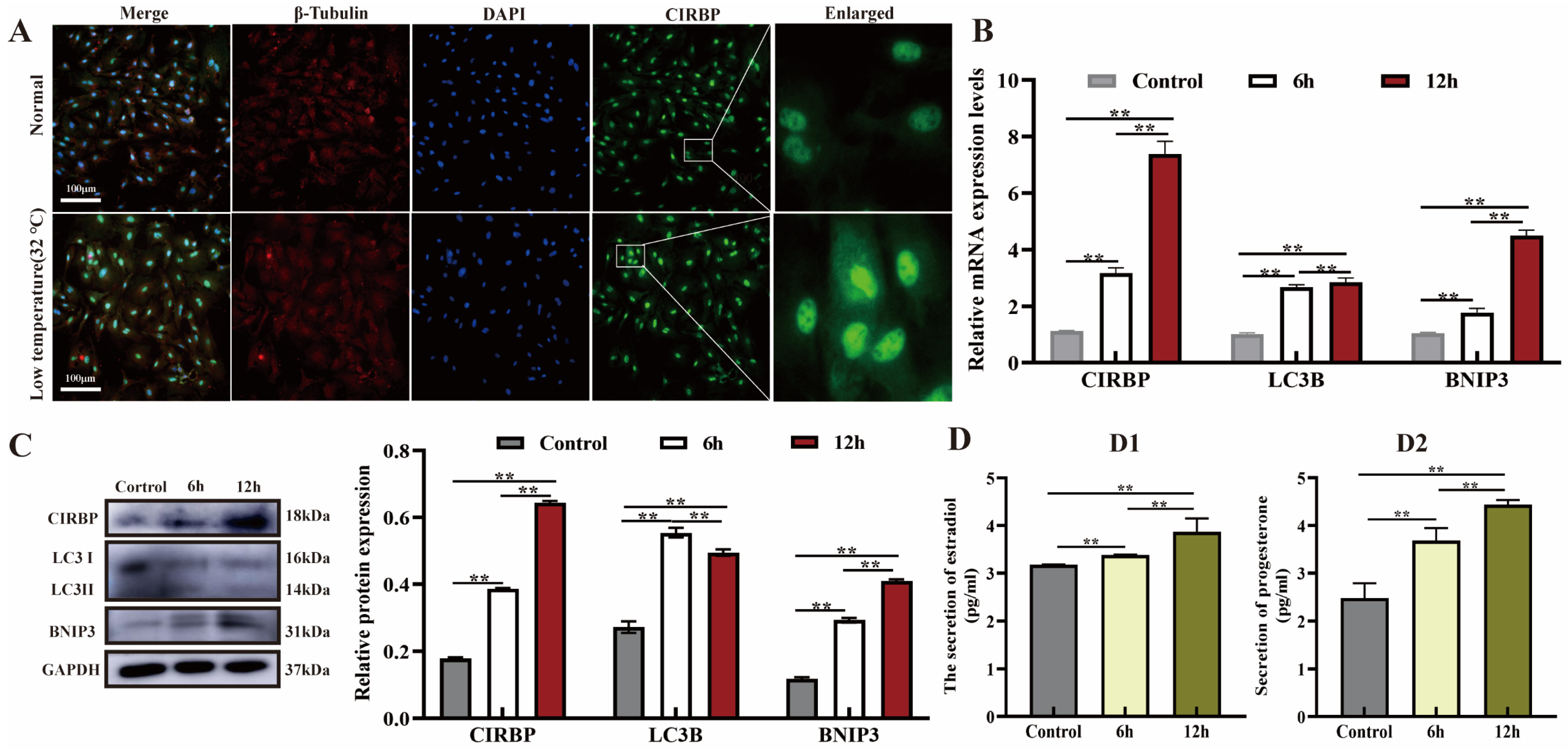
3.1. Effect of Low Temperature on CIRBP Expression, Mitophagy and E2 and P4 Secretion in Cells

CIRBP is up-regulated in response to low temperature and its protein position undergoes migratory changes to play an important role [28]. We observed the subcellular localization of CIRBP after low-temperature treatment of YCCs for 6 h and 12 h and evaluated mitophagy, E2 and P4 secretion, and CIRBP expression. The results showed that low-temperature treatment for 12 h significantly up-regulated the expression of CIRBP in YCCs and resulted in the migration of CIRBP from the nucleus to the cytoplasm (Figure 1A,B). In addition, the expression levels of BNIP3 and LC3B proteins in YCCs were significantly up-regulated by low temperature, and E2 and P4 secretion increased (Figure 1C,D). These results suggest that low temperature induces the expression of CIRBP in YCCs, activates mitophagy, and increases steroid hormone secretion.

3.2. Overexpression of CIRBP Activates Mitophagy in YCCs

Mitophagy controls the quantity and quality of intracellular mitochondria and is essential for maintaining cell survival. We previously found that CIRBP regulates autophagy. In order to illustrate the relationship between CIRBP and mitophagy induced by low temperature, we established a CIRBP overexpression model to evaluate the regulatory effect of CIRBP on YCC mitophagy. The data showed that CIRBP overexpression significantly up-regulated the ratio of LC3 I/II, induced the expression of BNIP3, Beclin-1 and ATG5 proteins, and inhibited P62 (Figure 2A–D). To further clarify the effect of CIRBP on mitophagy in YCCs, we used flow cytometry to detect the mitochondrial membrane potential and used AD-Ad-mCherry-GFP-LC3B double-label staining to detect the autophagic fluxes in YCCs after CIRBP overexpression. The results showed that the mitochondrial membrane potential of the CIRBP overexpression group was depolarized, and more red (autolysosomes) and yellow (autophagosomes) spots were observed (Figure 2E–H). In addition, JC-1 staining showed that the change in mitochondrial membrane potential after up-regulation of CIRBP was similar to that after Carbonyl cyanide 3-chlorophenylhydrazone (CCCP, a type of mitochondrial electron transport chain inhibitor, as a positive control to induce the decrease in mitochondrial membrane potential)-positive treatment, and green fluorescence was enhanced (Figure 2I). This suggested that the mitochondrial membrane potential was impaired. These results indicated that overexpression of CIRBP caused impaired depolarization of the mitochondrial membrane potential in YCCs, which, in turn, activated mitophagy.

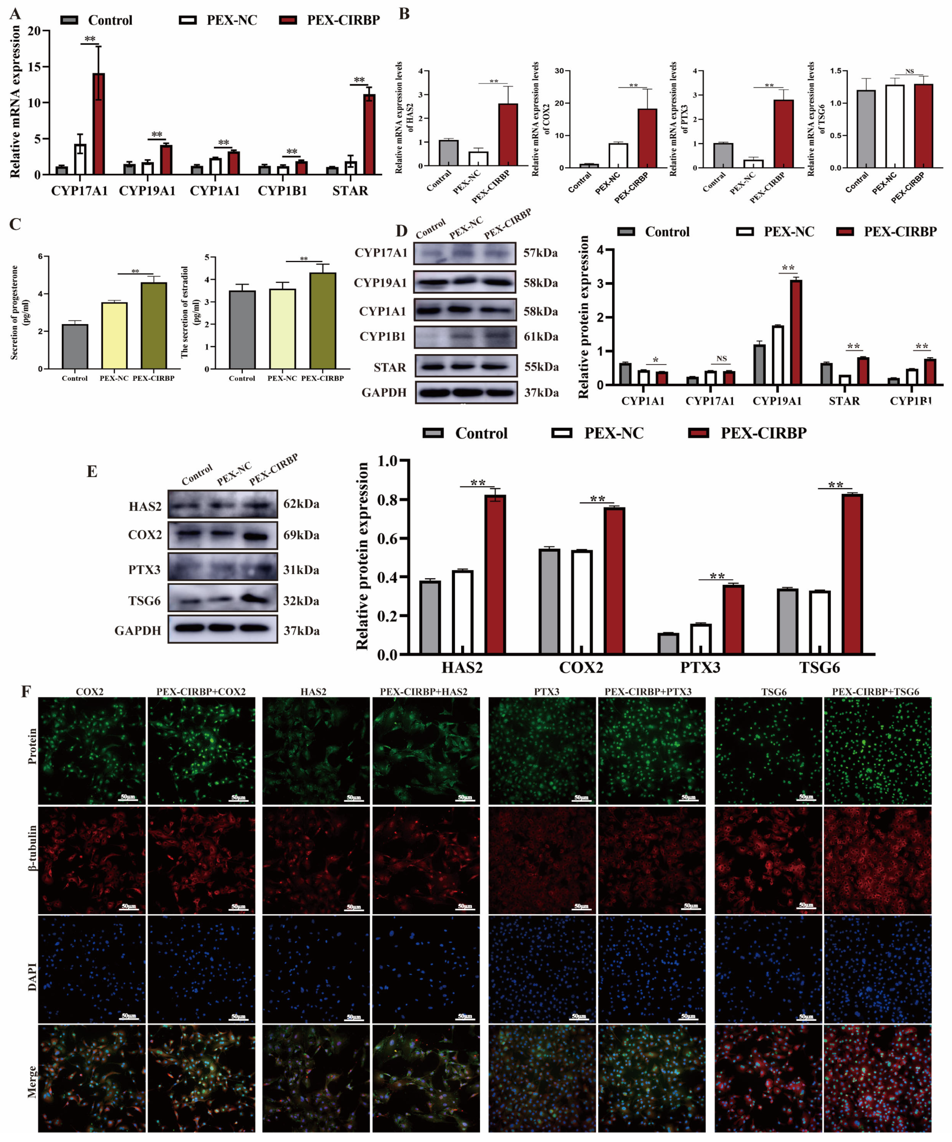
3.3. Overexpression of CIRBP Improves YCC Function and Inhibits Apoptosis

CCs provide energy and a microenvironment for oocyte development. Cumulus diffusion and the secretion of ovarian steroid hormones are essential for regulating oocyte development and maturation [29]. Therefore, we explored the effects of CIRBP overexpression on the synthesis and secretion of E2 and P4 and cumulus diffusion in YCCs. qRT-PCR revealed that CIRBP overexpression significantly up-regulated the mRNA expression levels of CYP1B1, CYP17A1, CYP19A1, STAR, CYP1A1, HAS2, COX2 and PTX3, and there was no significant difference in the expression level of TSG-6 mRNA (Figure 3A,B). The ELISA results showed that overexpression of CIRBP increased the secretion of E2 and P4 (Figure 3C). In addition, overexpression of CIRBP up-regulated the synthesis of E2 and P4 and the expression of cumulus-diffusion-related proteins (Figure 3D,E). The immunofluorescence results were consistent with the WB results, and up-regulated CIRBP induced the translation of cumulus-diffusion-related proteins (Figure 3F). The results suggested that the overexpression of CIRBP improved YCC function.
Previous studies have suggested that apoptosis is a mechanism leading to follicular atresia. Therefore, we further examined the effect of CIRBP on YCC apoptosis. The results showed that overexpression of CIRBP reduced the apoptosis rate. (Figure 4A,B). The expression levels of apoptosis-related proteins were consistent with the trend in mRNA expression levels. The overexpression of CIRBP significantly promoted the expression level of Bcl-2 and reduced Bax. In summary, this study found that CIRBP overexpression promoted the synthesis and secretion of E2 and P4 in YCCs, promoted cumulus diffusion, and inhibited YCC apoptosis.

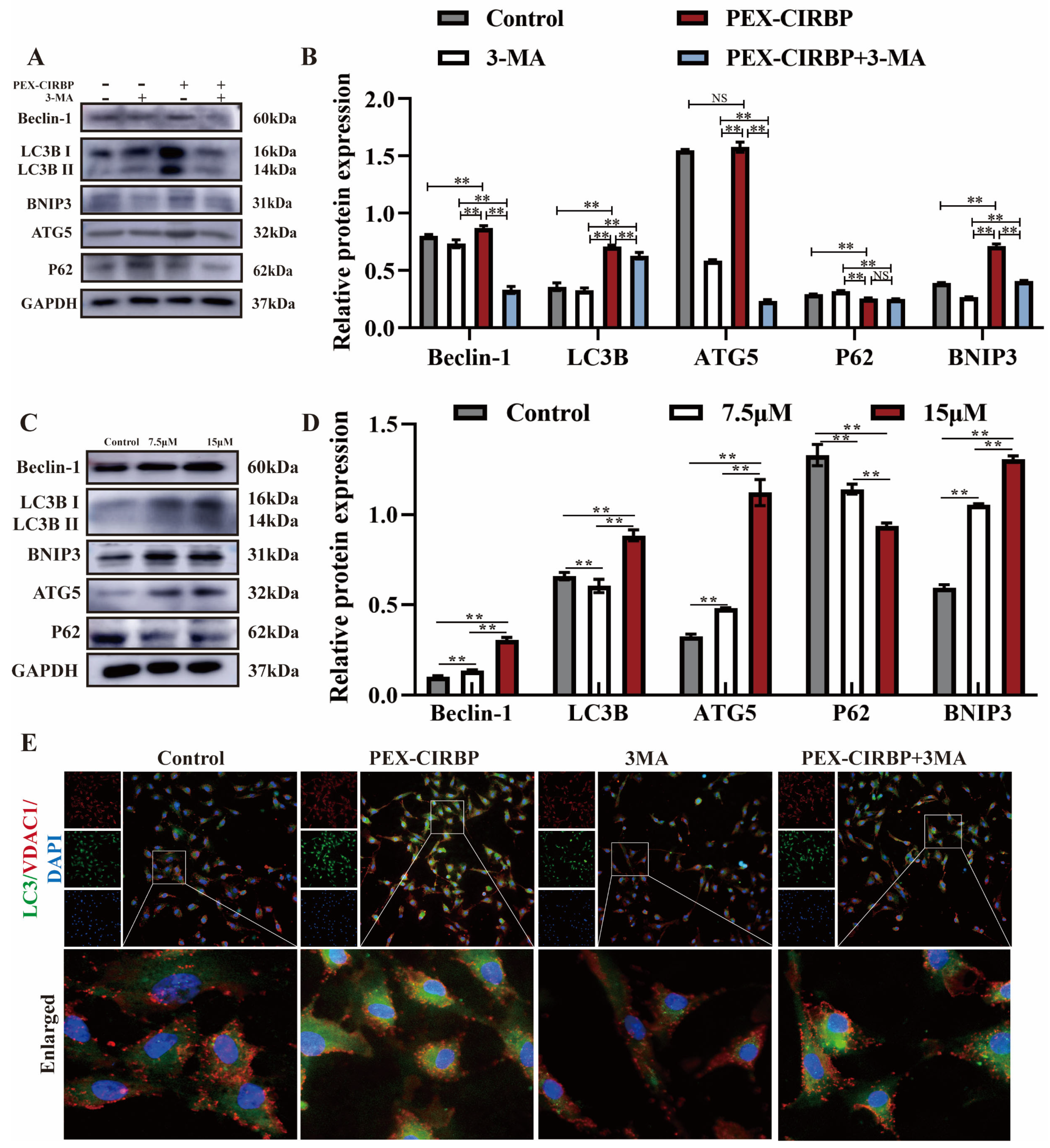
3.4. CIRBP Enhances YCC Function and Inhibits Apoptosis by Activating Mitophagy

To determine whether CIRBP affects YCC function and apoptosis by regulating mitophagy, YCCs were treated with PEX-CIRBP and mitophagy inhibitor 3MA to detect the regulation of CIRBP on YCC mitophagy. Research has found that 3-MA treatment inhibits the expression of mitochondrial-autophagy-related proteins, while the overexpression of CIRBP alleviated the down-regulation effect of 3-MA on mitochondrial-autophagy-related proteins (Figure 5A,B). Further treatment of YCCs with the mitophagy activator RAPA showed similar results to those of CIRBP overexpression (Figure 5C,D). In addition, immunofluorescence co-staining of LC3 and the mitochondrial membrane protein VDAC1 showed that when CIRBP was overexpressed, the co-localization of LC3 and VDAC1 was enhanced. The co-treatment group could alleviate the decreased co-localization of LC3 and VDAC1 caused by 3MA. These results suggested that CIRBP was involved in the regulation of mitophagy in YCCs.
To illustrate the effect of CIRBP on the function of YCCs by regulating mitophagy, we co-treated YCCs with PEX-CIRBP and 3MA and detected the expression of E2 and P4 synthesis-, cumulus-diffusion- and apoptosis-related proteins. We found that mitophagy inhibits significant hypocretin hormone synthesis and cumulus diffusion. CIRBP overexpression reversed the decreased expression of E2 and P4 biosynthesis- and cumulus-diffusion-related proteins caused by mitophagy inhibition (Figure 6A–D). Meanwhile, CIRBP overexpression significantly alleviated the mitophagy-inhibition-induced down-regulation of Bcl-2/BAX ratio. The ELISA results were consistent with the WB results. CIRBP overexpression significantly repaired 3MA-inhibited E2 and P4 secretion. In addition, we treated YCCs with RAPA and found that mitophagy activation showed similar results to those of CIRBP overexpression (Figure 6G,H). In summary, our data showed that CIRBP improve YCC function by activating mitophagy to promote cumulus diffusion, increasing steroid hormone synthesis and secretion, and inhibiting apoptosis.

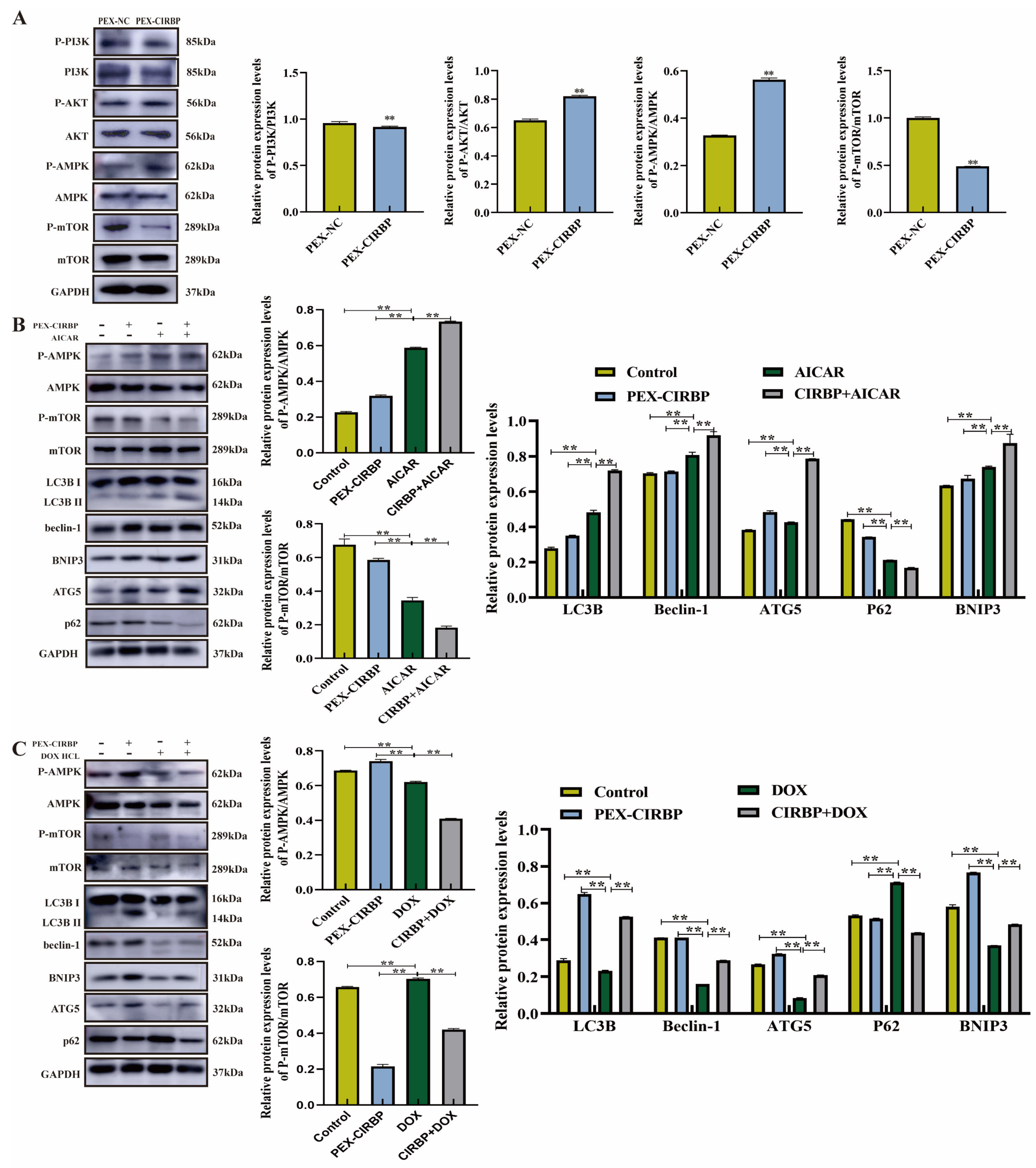
3.5. CIRBP Activated YCC Mitophagy Through the AMPK/mTOR Signaling Pathway

AMPK, PI3K, AKT and mTOR are the main regulators of autophagy and mitophagy at the post-translational level. The AMPK/mTOR and PI3K/AKT signaling pathways are essential for the regulation of mitophagy and apoptosis [30]. To explore whether AMPK/mTOR- and PI3K/AKT-mediated mitophagy was activated by CIRBP overexpression, we further explored whether CIRBP overexpression activates AMPK/mTOR- and PI3K/AKT-mediated mitophagy. We found that the up-regulation of CIRBP induced p-AKT and p-AMPK and inhibited p-PI3K and p-mTOR, while the total protein levels of AKT, AKMP, PI3K and mTOR remained unchanged (Figure 7A). This suggests that CIRBP’s regulation of mitophagy may involve the AMPK/mTOR pathway rather than the PI3K/AKT signaling pathway.
To verify that CIRBP-activated mitophagy was mediated by the AMPK/mTOR pathway, we co-treated YCCs with the AMPK/mTOR pathway activators AICAR and PEX-CIRBP to detect mitophagy. The results showed that the AMPK/mTOR pathway was activated after AICAR treatment; the expression levels of mitophagy-related proteins LC3B, Beclin-1, ATG5, and BNIP3 increased; and the expression level of P62 was continuously inhibited (Figure 7B). CIRBP overexpression enhanced the role of AICAR in activating mitophagy. In addition, we added AMPK inhibitor DOX HCL to co-treat with PEX-CIRBP and found that DOX HCL inhibited the AMPK/mTOR pathway (Figure 7C), and pathway inhibition significantly down-regulated the expression levels of LC3B, Beclin-1 and BNIP3. Overexpression of CIRBP significantly alleviated the down-regulation of mitophagy-related proteins via AMPK/mTOR pathway inhibition, in contrast to the effects of AICAR. Our results showed that the AMPK/mTOR signaling pathway mediates CIRBP’s regulation of mitophagy in YCCs.

3.6. PI3K/AKT Signaling Pathway Mediates CIRBP’s Regulation of Apoptosis in YCCs

Previous studies have shown that overexpression of CIRBP inhibits apoptosis and activates the PI3K/AKT pathway. We speculate that the anti-apoptotic effect of CIRBP may be related to its activation of the PI3K/AKT pathway. Therefore, we regulated the PI3K/AKT pathway on the basis of CIRBP overexpression and detected the level of apoptosis to verify the speculation above. We found that when the PI3K/AKT pathway was inhibited, BAX expression was up-regulated, Bcl-2 expression was down-regulated, and apoptosis was activated (Figure 8A,B). However, overexpression of CIRBP significantly alleviated the down-regulation of Bcl-2 by PI3K signal transduction pathway inhibition, compared to the blank control group, when the PI3K/AKT pathway was activated by 740-YP treatment; thus, CIRBP overexpression showed a synergistic effect with 740 Y-P treatment. Overexpression of CIRBP increased the induction of Bcl-2 via activation of the PI3K/AKT pathway and significantly inhibited Bax expression. In addition, we found that overexpression of CIRBP significantly alleviated the apoptosis induced by ly294002 and increased the anti-apoptotic effect of 740Y-P (Figure 8C,D). These data suggest that the regulation of CIRBP of the apoptosis of YCCs involves the PI3K/AKT signaling pathway.

4. Discussion

CIRBP is one of the RNA-binding proteins that can quickly respond to the up-regulation of low-temperature stress and participate in the stable cell state [31]; the optimal expression temperature range of CIRBP is about 32–34 °C. It is mainly located in the nucleus, and it participates in regulating gene transcription or binding with mRNA for post-transcriptional regulation [32]. However, under physiological or stress conditions, the expression site of CIRBP can migrate and even be released into the extracellular environment to regulate the transcription of target genes [32]. Research shows that under low-temperature conditions, transcription factor specificity protein 1 (Sp1) binds to the CIRBP promoter and activates its expression, while overexpression of Sp1 leads to the translocation of CIRBP from the cell nucleus to the cytoplasm [33]. This indicates that the function of CIRBP is determined by its expression location. As a functionally stable mRNA-binding protein, its expression location is related to its transcription method and the conditions regulating its expression level. It has been reported that a therapeutic low temperature (32 °C) can exert neuroprotective effects by activating CIRBP expression and inhibiting the production of mitochondrial apoptotic factors [34,35]. Expression of the CIRBP gene in bovine oocytes and cumulus cells is induced to be up-regulated by mild hypothermia [11]. Consistent with the findings above, we found that a small amount of CIRBP protein expression sites migrated and that CIRBP expression was induced to be up-regulated in cumulus cells after a mild low-temperature (32 °C) treatment. Autophagy is an important mechanism for maintaining cellular homeostasis; it is a complex process that can engulf and recycle cellular debris in lysosomes to adapt to the cellular environment. Mitophagy refers to the quality control mechanism that selectively remove damaged mitochondria through autophagosomes. Mild hypothermia has been shown to activate cellular mitophagy. Weng w et al. [36] showed that mild hypothermia treatment down-regulated lactonization of Tufm (Tu translation elongation factor, mitochondrial), a key regulator of mitophagy, in mouse neuronal cells, and activated mitophagy by facilitating its interaction with mitochondrial membrane proteins. Tan YL et al. [37] found that mild hypothermia treatment accelerated mitochondrial biogenesis in damaged hepatocytes by activating mitophagy to improve mitochondrial function. In our research, mild hypothermia activated mitophagy in YCCs. In contrast, studies have shown that prolonged hypothermia inhibits autophagy in mouse cardiac cells, but autophagy levels are repaired upon recovery from hypothermia [38]. This indicates that the regulation of mitophagy by hypothermia is dynamic and may be related to the degree of hypothermia exposure, hypothermia time, and cell type. In addition, our data show that hypothermia significantly increases the secretion levels of E2 and P4 in YCC. Cholesterol is the basis for steroid hormone biosynthesis. The inhibition of autophagy can damage the biosynthesis of steroid hormones by reducing the phagocytosis and transport of cholesterol by lipid droplets [39]. Ti3C2 nanosheets activate autophagy in mouse GCs to promote the ability of ovarian hormone secretion, while RAPA treatment of granulosa cells exhibited similar results [40]. Notably, scholars pointed out that the expression trends of CIRBP and LC3B showed a correlation in the cells treated with hypothermia followed by rewarming, and LC3B was regulated by CIRBP [41]. In summary, we propose hypothermia-induced mitophagy activation and increased E2 and P4 secretion in YCCs. It is hypothesized that this change may be CIRBP-mediated.
We verified the speculation above and found that up-regulation of CIRBP was accompanied by increased mitophagy activation and E2 and P4 synthesis and secretion in YCCs. Evidence suggests that as a protective factor for mitochondrial homeostasis, CIRBP eliminates endogenous ROS to promote mitochondrial metabolism [42]. CIRBP can induce ROS release and mtDNA fragmentation through TLR4 signaling, thereby activating macrophage mitophagy and inhibiting apoptosis [21]. CIRBP down-regulation can reduce mitophagy by inhibiting the PINK1/Parkin pathway [43]. Under stressful conditions, CIRBP activates ferritin autophagy by interacting with ELAVL1 to alleviate necroptosis [7]. In our study, BNIP3, LC3B and Beclin-1 were all up-regulated by CIRBP, highlighting the regulation of CIRBP on mitophagy in YCCs. This evidence also suggests that CIRBP has a regulatory role in apoptosis. Related studies found that down-regulation of CIRBP resulted in impaired ROS metabolism and activation of the PHD3/HIF-1α pathway to promote apoptosis [15]. CIRBP alleviates chemotherapy-induced cardiomyocyte apoptosis by inhibiting the OGF/OGFR axis [44]. We found that up-regulation of CIRBP inhibited YCC apoptosis. In addition, our results revealed that CIRBP overexpression increased the expression of cumulus-diffusion-related proteins HAS2, PTGS2, PTX3, and TNFAIP6, demonstrating for the first time that CIRBP is a regulator of cumulus diffusion. The cumulus diffusion process affects oocyte development and ovulation efficiency, which are important for oocyte maturation and ovulation. Notably, the synthesis and metabolism of estradiol in cumulus cells are regulated by autophagy activated by exosomes from yak follicular fluid [45]. The inhibition of apoptosis and activation of autophagy alleviate the decrease in the gene expression levels of sheep cumulus-diffusion-related factors caused by zearalenone (ZEN) [46]. This indicates that autophagy regulates steroid hormone synthesis, apoptosis, and cumulus diffusion. Therefore, we speculated that the effects of CIRBP on E2 and P4 biosynthesis, cumulus diffusion, and apoptosis in YCCs may be mediated by mitophagy.
In this study, we investigated whether CIRBP can regulate the function of YCCs through mitosis. We found that overexpression of CIRBP could reverse the inhibitory effect of mitophagy inhibitor on YCC mitophagy. CIRBP can alleviate the decrease in E2 and P4 secretion and the decrease in cumulus-diffusion-related protein expression caused by mitophagy inhibition. In addition, the inhibition of mitophagy significantly increased the expression level of Bax, whereas the overexpression of CIRBP alleviated the induction of apoptosis by inhibiting autophagy. Simultaneously, the changes in the function of YCCs after mitophagy activation showed the same results as when CIRBP was overexpressed. Bovine-follicular-fluid-derived exosomes promote steroid hormone secretion and inhibit apoptosis by inducing mitophagy in GCs [47]. Benzopyrene (BaP) can inhibit CL cell mitophagy through the ANT1-PINK1-Parkin pathway, thereby inhibiting P4 synthesis [48]. Consistent with these findings, we found that mitophagy activation promotes the synthesis and secretion of steroid hormones. In contrast, Ma Q et al. [49] found that dehydroepiandrosterone (DHEA) improved the function of rat ovarian granulosa cells by inhibiting mitophagy and AMPK-SIRT1 signaling. Cd exposure inhibits P4 synthesis in placental trophoblasts by activating PERK-regulated mitophagy [50]. This indicates that the regulation of mitophagy on steroid hormone biosynthesis is dynamic, which may be related to the feedback regulation between the two [51]. We have previously reported that CIRBP inhibits apoptosis by activating mitophagy in other cells, and CIRBP also plays this role in yak YCCs. In addition, we found that CIRBP promoted cumulus diffusion by activating YCC mitophagy. A recent study showed that HIF-1α regulates the function of YCCs and participates in oocyte development [26]. HIF-1α has been proven to be one of the target genes of CIRBP [52]. However, whether the effect of CIRBP on cumulus diffusion is mediated by HIF-1α and its specific mechanism need to be further explored. In summary, our data show that CIRBP affected the biosynthesis and secretion of E2 and P4, cumulus diffusion, and apoptosis of YCCs by regulating mitophagy, thereby improving YCC function; these results emphasize the importance of exploring the regulation of cellular functions by CIRBP.
AMPK/mTOR and PI3K/Akt pathways are two of the main pathways regulating mitophagy and apoptosis [53,54]. CIRBP promotes wound healing by activating AMPK [55] and regulates apoptosis and proliferation through the PI3K/AKT signaling pathway [56], suggesting that CIRBP can regulate cell function through the AMPK/mTOR and PI3K/AKT pathways. Our study found that CIRBP activated the AMPK/mTOR and PI3K/AKT signaling pathways. Further studies found that overexpression of CIRBP significantly alleviated the decrease in mitophagy-related protein expression caused by AMPK/mTOR pathway inhibition, while overexpression of CIRBP further enhanced the activation of mitophagy by AMPK/mTOR pathway activation. This indicates that the regulation of CIRBP of mitophagy of YCCs is mediated by AMPK/mTOR signaling pathway. BNIP3 directly binds to LC3B to initiate mitophagy [57]. Moreover, the overexpression of BNIP3 significantly induces the up-regulation of AMPK; however, there is evidence that the expression level of BNIP3 does not change significantly when AMPK is inhibited or activated [58]. In our study, CIRBP overexpression significantly up-regulated BNIP3 protein level, and activation of the AMPK pathway up-regulated BNIP3 expression. BNIP3 is a target gene directly regulated by HIF-1α [59], and HIF-1α is a target gene of CIRBP. Therefore, we speculate that the HIF-1α/BNIP3 signaling pathway may mediate CIRBP regulation of mitophagy. Berberine inhibits cardiomyocyte apoptosis induced by regulating HIF-1α/BNIP3-pathway-mediated mitophagy [60]. This suggests that the inhibitory effect of CIRBP overexpression on the apoptosis of YCCs may be related to an increase in BNIP3 expression. In addition, our results indicate that the regulation of CIRBP on YCC cell apoptosis is mediated by the AKT signaling pathway. Low-temperature exposure promotes platelet activation by up-regulating CIRBP to activate the P-AKT signaling pathway, thereby increasing the risk of ischemic stroke [61]. Furthermore, CIRBP alleviates hepatocyte apoptosis induced by low-temperature exposure by activating the AKT signaling pathway [62]. In this study, we found that the AMPK/mTOR pathway mediated the regulation of mitochondrial autophagy in YCCs by CIRBP, and the AKT signaling pathway mediated the regulation of apoptosis in YCCs by CIRBP; combined with the previous results of this study, we demonstrated that CIRBP can regulate mitophagy through the AMPK/mTOR pathway to affect YCC function and alleviate apoptosis. These findings provide a reference for further exploration of CIRBP regulation of programmed cell death and the role of CIRBP in reproductive medicine.

5. Conclusions

In summary, this study found that CIRBP improved cumulus cell function by induction of AMPK/mTOR-pathway-mediated mitophagy and inhibiting AKT-pathway-mediated YCC apoptosis.

Supplementary Materials

The following supporting information can be downloaded at: https://www.mdpi.com/article/10.3390/biom15060759/s1. Original Western blot images are provided in the Supplementary Materials.

Author Contributions

Y.P., S.Y., R.Z. and M.W. conceived and designed the experiment. R.Z., R.X., W.M. and J.W. contributed to sample collection and cell culture. D.Z. and Z.J. provided help in data visualization. R.Z. wrote the manuscript and Y.C. revised the manuscript. All authors have read and agreed to the published version of the manuscript.

Funding

This work was supported by the National Natural Science Foundation of China (32160859) and the Gansu Provincial Key R & D Program—Rural Areas (24YFNA015).

Institutional Review Board Statement

All procedures involving animals were approved by the Animal Care and Use Committee of the Gansu Agricultural University (GSAU-Eth-VMC-2024-009, 15 March 2024).

Informed Consent Statement

Not applicable.

Data Availability Statement

All the data obtained in this study can be obtained from the corresponding author under reasonable requirement.

Conflicts of Interest

The authors declare no conflicts of interest in this paper.

List of Abbreviations

PCDProgrammed cell death
GCsGranulosa cells
ANOVAAnalysis of variance
YCCsYak cumulus cells
Sp1Specific protein 1
MAPKMitogen-activated protein kinase
CYPCytochrome P450 enzyme
CIRBPCold-induced RNA-binding protein
HIF-1αHypoxia-inducible factor 1α
AMPKAMP-activated protein kinase
TLR4Toll-Like Receptor 4
BNIP3BCL2 interacting protein 3
ROSReactive oxygen species
3-MA3-Methyladenine
RAPARapamycin
AICARAcadesine
DOXDoxorubicin

References

  1. Lin, T.Y.; Jia, J.S.; Luo, W.R.; Lin, X.L.; Xiao, S.J.; Yang, J.; Xia, J.W.; Zhou, C.; Zhou, Z.H.; Lin, S.J.; et al. ThermomiR-377-3p-induced suppression of Cirbp expression is required for effective elimination of cancer cells and cancer stem-like cells by hyperthermia. J. Exp. Clin. Cancer Res. 2024, 43, 62. [Google Scholar] [CrossRef] [PubMed]
  2. Aziz, M.; Brenner, M.; Wang, P. Extracellular CIRP (eCIRP) and inflammation. J. Leukoc. Biol. 2019, 106, 133–146. [Google Scholar] [CrossRef] [PubMed]
  3. Wang, X.; Che, H.; Zhang, W.; Wang, J.; Ke, T.; Cao, R.; Meng, S.; Li, D.; Weiming, O.; Chen, J.; et al. Effects of Mild Chronic Intermittent Cold Exposure on Rat Organs. Int. J. Biol. Sci. 2015, 11, 1171–1180. [Google Scholar] [CrossRef]
  4. Kim, S.Y.; Ban, H.J.; Lee, S.; Jin, H.J. Regulation of CIRP by genetic factors of SP1 related to cold sensitivity. Front. Immunol. 2022, 13, 994699. [Google Scholar] [CrossRef] [PubMed]
  5. Rao, M.; Ke, D.; Cheng, G.; Hu, S.; Wu, Y.; Wang, Y.; Zhou, F.; Liu, H.; Zhu, C.; Xia, W. The regulation of CIRBP by transforming growth factor beta during heat shock-induced testicular injury. Andrology 2019, 7, 244–250. [Google Scholar] [CrossRef]
  6. Corre, M.; Lebreton, A. Regulation of cold-inducible RNA-binding protein (CIRBP) in response to cellular stresses. Biochimie 2024, 217, 3–9. [Google Scholar] [CrossRef]
  7. Sui, M.; Xu, D.; Zhao, W.; Lu, H.; Chen, R.; Duan, Y.; Li, Y.; Zhu, Y.; Zhang, L.; Zeng, L. CIRBP promotes ferroptosis by interacting with ELAVL1 and activating ferritinophagy during renal ischaemia-reperfusion injury. J. Cell. Mol. Med. 2021, 25, 6203–6216. [Google Scholar] [CrossRef]
  8. Morf, J.; Rey, G.; Schneider, K.; Stratmann, M.; Fujita, J.; Naef, F.; Schibler, U. Cold-inducible RNA-binding protein modulates circadian gene expression posttranscriptionally. Science 2012, 338, 379–383. [Google Scholar] [CrossRef]
  9. Xia, Z.; Zheng, X.; Zheng, H.; Liu, X.; Yang, Z.; Wang, X. Cold-inducible RNA-binding protein (CIRP) regulates target mRNA stabilization in the mouse testis. FEBS Lett. 2012, 586, 3299–3308. [Google Scholar] [CrossRef]
  10. Masuda, T.; Itoh, K.; Higashitsuji, H.; Higashitsuji, H.; Nakazawa, N.; Sakurai, T.; Liu, Y.; Tokuchi, H.; Fujita, T.; Zhao, Y.; et al. Cold-inducible RNA-binding protein (Cirp) interacts with Dyrk1b/Mirk and promotes proliferation of immature male germ cells in mice. Proc. Natl. Acad. Sci. USA 2012, 109, 10885–10890. [Google Scholar] [CrossRef]
  11. Gardela, J.; Ruiz-Conca, M.; Olvera-Maneu, S.; Lopez-Bejar, M.; Alvarez-Rodriguez, M. The mRNA expression of the three major described cold-inducible proteins, including CIRBP, differs in the bovine endometrium and ampulla during the estrous cycle. Res. Vet. Sci. 2022, 152, 181–189. [Google Scholar] [CrossRef] [PubMed]
  12. Peng, Y.; Yang, P.H.; Tanner, J.A.; Huang, J.D.; Li, M.; Lee, H.F.; Xu, R.H.; Kung, H.F.; Lin, M.C. Cold-inducible RNA binding protein is required for the expression of adhesion molecules and embryonic cell movement in Xenopus laevis. Biochem. Biophys. Res. Commun. 2006, 344, 416–424. [Google Scholar] [CrossRef] [PubMed]
  13. Zhou, Y.; Jin, Y.; Wang, Y.; Wu, R. Hypoxia activates the unfolded protein response signaling network: An adaptive mechanism for endometriosis. Front. Endocrinol. 2022, 13, 945578. [Google Scholar] [CrossRef] [PubMed]
  14. Goldblatt, Z.E.; Cirka, H.A.; Billiar, K.L. Mechanical Regulation of Apoptosis in the Cardiovascular System. Ann. Biomed. Eng. 2021, 49, 75–97. [Google Scholar] [CrossRef] [PubMed]
  15. Zhang, P.; Bai, L.; Tong, Y.; Guo, S.; Lu, W.; Yuan, Y.; Wang, W.; Jin, Y.; Gao, P.; Liu, J. CIRP attenuates acute kidney injury after hypothermic cardiovascular surgery by inhibiting PHD3/HIF-1alpha-mediated ROS-TGF-beta1/p38 MAPK activation and mitochondrial apoptotic pathways. Mol. Med. 2023, 29, 61. [Google Scholar] [CrossRef]
  16. Zhang, J.; Liu, T.; Wei, Y.; Peng, J.; Zeng, G.; Zhong, P. Upregulation of CIRP by its agonist prevents the development of heart failure in myocardial infarction rats. BMC Cardiovasc. Disord. 2024, 24, 185. [Google Scholar] [CrossRef]
  17. Su, F.; Yang, s.; Wang, H.; Qiao, Z.; Zhao, H.; Qu, Z. CIRBP Ameliorates Neuronal Amyloid Toxicity via Antioxidative and Antiapoptotic Pathways in Primary Cortical Neurons. Oxid. Med. Cell Longev. 2020, 2020, 2786139. [Google Scholar] [CrossRef]
  18. Yang, W.L.; Sharma, A.; Wang, Z.; Li, Z.; Fan, J.; Wang, P. Cold-inducible RNA-binding protein causes endothelial dysfunction via activation of Nlrp3 inflammasome. Sci. Rep. 2016, 6, 26571. [Google Scholar] [CrossRef]
  19. Liu, W.; Fan, Y.; Ding, H.; Han, D.; Yan, Y.; Wu, R.; Lv, Y.; Zheng, X. Normothermic machine perfusion attenuates hepatic ischaemia-reperfusion injury by inhibiting CIRP-mediated oxidative stress and mitochondrial fission. J. Cell. Mol. Med. 2021, 25, 11310–11321. [Google Scholar] [CrossRef]
  20. Zhou, J.; Peng, X.; Mei, S. Autophagy in Ovarian Follicular Development and Atresia. Int. J. Biol. Sci. 2019, 15, 726–737. [Google Scholar] [CrossRef]
  21. Li, Z.; Fan, E.K.; Liu, J.; Scott, M.J.; Li, Y.; Li, S.; Xie, W.; Billiar, T.R.; Wilson, M.A.; Jiang, Y.; et al. Cold-inducible RNA-binding protein through TLR4 signaling induces mitochondrial DNA fragmentation and regulates macrophage cell death after trauma. Cell Death Dis. 2017, 8, e2775. [Google Scholar] [CrossRef] [PubMed]
  22. Robichaud, S.; Fairman, G.; Vijithakumar, V.; Mak, E.; Cook, D.P.; Pelletier, A.R.; Huard, S.; Vanderhyden, B.C.; Figeys, D.; Lavallee-Adam, M.; et al. Identification of novel lipid droplet factors that regulate lipophagy and cholesterol efflux in macrophage foam cells. Autophagy 2021, 17, 3671–3689. [Google Scholar] [CrossRef] [PubMed]
  23. Warzych, E.; Lipinska, P. Energy metabolism of follicular environment during oocyte growth and maturation. J. Reprod. Dev. 2020, 66, 1–7. [Google Scholar] [CrossRef] [PubMed]
  24. Zhu, Q.; Zhu, S.; Li, Q.; Hu, C.; Pan, C.; Li, H.; Zhu, Y.; Li, X.; Tang, Y.; Ge, R.S. Prenatal diethylhexylphthalate exposure disturbs adult Leydig cell function via epigenetic downregulation of METTL4 expression in male rats. Ecotoxicol. Environ. Saf. 2024, 277, 116391. [Google Scholar] [CrossRef]
  25. Shao, T.; Ke, H.; Liu, R.; Xu, L.; Han, S.; Zhang, X.; Dang, Y.; Jiao, X.; Li, W.; Chen, Z.J.; et al. Autophagy regulates differentiation of ovarian granulosa cells through degradation of WT1. Autophagy 2022, 18, 1864–1878. [Google Scholar] [CrossRef]
  26. Ma, X.; Wang, M.; Wang, J.; Han, X.; Yang, X.; Zhang, H.; Zhong, D.; Qiu, S.; Yu, S.; Wang, L.; et al. Hypoxia-Inducible Factor 1alpha Affects Yak Oocyte Maturation and Early Embryonic Development by Regulating Autophagy. Antioxidants 2024, 13, 840. [Google Scholar] [CrossRef]
  27. Wan, R.; Zhao, Z.; Zhao, M.; Hu, K.; Zhai, J.; Yu, H.; Wei, Q. Characteristics of pulmonary microvascular structure in postnatal yaks. Sci. Rep. 2021, 11, 18265. [Google Scholar] [CrossRef]
  28. Gotic, I.; Schibler, U. Posttranscriptional mechanisms controlling diurnal gene expression cycles by body temperature rhythms. RNA Biol. 2017, 14, 1294–1298. [Google Scholar] [CrossRef]
  29. Sutton-McDowall, M.L.; Gilchrist, R.B.; Thompson, J.G. The pivotal role of glucose metabolism in determining oocyte developmental competence. Reproduction 2010, 139, 685–695. [Google Scholar] [CrossRef]
  30. Belousov, D.M.; Mikhaylenko, E.V.; Somasundaram, S.G.; Kirkland, C.E.; Aliev, G. The Dawn of Mitophagy: What Do We Know by Now? Curr. Neuropharmacol. 2021, 19, 170–192. [Google Scholar] [CrossRef]
  31. Aguilar-Garrido, P.; Otero-Sobrino, A.; Navarro-Aguadero, M.A.; Velasco-Estevez, M.; Gallardo, M. The Role of RNA-Binding Proteins in Hematological Malignancies. Int. J. Mol. Sci. 2022, 23, 9552. [Google Scholar] [CrossRef] [PubMed]
  32. Thandapani, P.; O’Connor, T.R.; Bailey, T.L.; Richard, S. Defining the RGG/RG motif. Mol. Cell 2013, 50, 613–623. [Google Scholar] [CrossRef] [PubMed]
  33. Merchant, J.L.; Du, M.; Todisco, A. Sp1 phosphorylation by Erk 2 stimulates DNA binding. Biochem. Biophys. Res. Commun. 1999, 254, 454–461. [Google Scholar] [CrossRef]
  34. Sun, Y.J.; Ma, S.; Fan, B.; Wang, Y.; Wang, S.R.; Li, G.Y. Therapeutic hypothermia protects photoreceptors through activating Cirbp pathway. Neurochem. Int. 2019, 126, 86–95. [Google Scholar] [CrossRef]
  35. Wu, L.; Sun, H.L.; Gao, Y.; Hui, K.L.; Xu, M.M.; Zhong, H.; Duan, M.L. Therapeutic Hypothermia Enhances Cold-Inducible RNA-Binding Protein Expression and Inhibits Mitochondrial Apoptosis in a Rat Model of Cardiac Arrest. Mol. Neurobiol. 2017, 54, 2697–2705. [Google Scholar] [CrossRef]
  36. Weng, W.; He, Z.; Ma, Z.; Huang, J.; Han, Y.; Feng, Q.; Qi, W.; Peng, Y.; Wang, J.; Gu, J.; et al. Tufm lactylation regulates neuronal apoptosis by modulating mitophagy in traumatic brain injury. Cell Death Differ. 2025, 32, 530–545. [Google Scholar] [CrossRef] [PubMed]
  37. Chen, Y.L.; Tao, J.; Zhao, P.J.; Tang, W.; Xu, J.P.; Zhang, K.Q.; Zou, C.G. Adiponectin receptor PAQR-2 signaling senses low temperature to promote C. elegans longevity by regulating autophagy. Nat. Commun. 2019, 10, 2602. [Google Scholar] [CrossRef]
  38. Ruperez, C.; Blasco-Roset, A.; Kular, D.; Cairo, M.; Ferrer-Curriu, G.; Villarroya, J.; Zamora, M.; Crispi, F.; Villarroya, F.; Planavila, A. Autophagy is Involved in Cardiac Remodeling in Response to Environmental Temperature Change. Front. Physiol. 2022, 13, 864427. [Google Scholar] [CrossRef]
  39. Texada, M.J.; Malita, A.; Christensen, C.F.; Dall, K.B.; Faergeman, N.J.; Nagy, S.; Halberg, K.A.; Rewitz, K. Autophagy-Mediated Cholesterol Trafficking Controls Steroid Production. Dev. Cell 2019, 48, 659–671. [Google Scholar] [CrossRef]
  40. Yang, L.; He, Z.; Hu, L.; Tang, H.; Geng, Y.; Tan, Q.; Zhang, Y.; Wen, Y.; Wu, W.; Gu, H.; et al. Ti3C2 nanosheet-induced autophagy derails ovarian functions. J. Nanobiotechnology 2024, 22, 242. [Google Scholar] [CrossRef]
  41. Neutelings, T.; Lambert, C.A.; Nusgens, B.V.; Colige, A.C. Effects of mild cold shock (25 degrees C) followed by warming up at 37 degrees C on the cellular stress response. PLoS ONE 2013, 8, e69687. [Google Scholar] [CrossRef] [PubMed]
  42. Liu, Y.; Xue, C.; Lu, H.; Zhou, Y.; Guan, R.; Wang, J.; Zhang, Q.; Ke, T.; Aschner, M.; Zhang, W.; et al. Hypoxia causes mitochondrial dysfunction and brain memory disorder in a manner mediated by the reduction of Cirbp. Sci. Total. Environ. 2022, 806 Pt 3, 151228. [Google Scholar] [CrossRef] [PubMed]
  43. Wu, S.; Li, Z.; Li, H.; Liao, K. Dihydroartemisinin Reduces Irradiation-Induced Mitophagy and Radioresistance in Lung Cancer A549 Cells via CIRBP Inhibition. Life 2022, 12, 1129. [Google Scholar] [CrossRef]
  44. Liu, C.; Cheng, X.; Xing, J.; Li, J.; Li, Z.; Jian, D.; Wang, Y.; Wang, S.; Li, R.; Zhang, W.; et al. CIRBP-OGFR axis safeguards against cardiomyocyte apoptosis and cardiotoxicity induced by chemotherapy. Int. J. Biol. Sci. 2022, 18, 2882–2897. [Google Scholar] [CrossRef]
  45. Xu, R.; Wang, J.; Wang, M.; Gao, L.; Zhang, R.; Zhao, L.; Liu, B.; Han, X.; Baloch, A.R.; Cui, Y.; et al. Exosomes Derived from Yak Follicular Fluid Increase 2-Hydroxyestradiol Secretion by Activating Autophagy in Cumulus Cells. Animals 2022, 12, 3174. [Google Scholar] [CrossRef] [PubMed]
  46. Li, Z.; Ma, T.; Liu, Y.; Liu, W.; Zhao, X.; Zhang, G.; Wang, J.; Zhang, Y. Screening and Mechanism Study of Three Antagonistic Drugs, Oxysophoridine, Rutin, and Phellodendrine, against Zearalenone-Induced Reproductive Toxicity in Ovine Oocytes. Antioxidants 2024, 13, 752. [Google Scholar] [CrossRef] [PubMed]
  47. Wang, Y.; Zhao, Y.; Ling, Z.; Luan, D.; Kang, J.; Dong, X.; Quan, F. HD-sEVs in bovine follicular fluid regulate granulosa cell apoptosis and estradiol secretion through the autophagy pathway. Theriogenology 2023, 212, 91–103. [Google Scholar] [CrossRef]
  48. Li, N.; Xu, H.; Liu, X.; Gao, R.; He, J.; Ding, Y.; Li, F.; Geng, Y.; Mu, X.; Chen, X. Exposure to benzo(a)pyrene suppresses mitophagy via ANT1-PINK1-Parkin pathway in ovarian corpus luteum during early pregnancy. Sci. Total. Environ. 2022, 814, 152759. [Google Scholar] [CrossRef]
  49. Ma, Q.; Shen, M.; Wu, J.; Ye, C.; Tan, Y. Mechanism Research of DHEA Treatment Improving Diminished Ovarian Reserve by Attenuating the AMPK-SIRT1 Signaling and Mitophagy. Reprod. Sci. 2024, 31, 2059–2072. [Google Scholar] [CrossRef]
  50. Zhu, H.L.; Shi, X.T.; Xu, X.F.; Xiong, Y.W.; Yi, S.J.; Zhou, G.X.; Liu, W.B.; Huang, M.M.; Gao, L.; Zhang, C.; et al. Environmental cadmium exposure induces fetal growth restriction via triggering PERK-regulated mitophagy in placental trophoblasts. Environ. Int. 2021, 147, 106319. [Google Scholar] [CrossRef]
  51. Zhang, L.; Zhao, Y.; Guo, L. 17beta-estradiol protects INS-1 insulinoma cells from mitophagy via G protein-coupled estrogen receptors and the PI3K/Akt signaling pathway. Int. J. Mol. Med. 2018, 41, 2839–2846. [Google Scholar] [CrossRef]
  52. Lu, M.; Ge, Q.; Wang, G.; Luo, Y.; Wang, X.; Jiang, W.; Liu, X.; Wu, C.L.; Xiao, Y.; Wang, X. CIRBP is a novel oncogene in human bladder cancer inducing expression of HIF-1alpha. Cell Death Dis. 2018, 9, 1046. [Google Scholar] [CrossRef]
  53. Yin, H.; Wang, C.; Guo, H.; Li, X.; Liu, J. The mechanism of nickel-induced autophagy and its role in nephrotoxicity. Ecotoxicol. Environ. Saf. 2024, 273, 116150. [Google Scholar] [CrossRef]
  54. Wu, C.; Chen, H.; Zhuang, R.; Zhang, H.; Wang, Y.; Hu, X.; Xu, Y.; Li, J.; Li, Y.; Wang, X.; et al. Betulinic acid inhibits pyroptosis in spinal cord injury by augmenting autophagy via the AMPK-mTOR-TFEB signaling pathway. Int. J. Biol. Sci. 2021, 17, 1138–1152. [Google Scholar] [CrossRef] [PubMed]
  55. Higashitsuji, H.; Fujita, T.; Higashitsuji, H.; Fujita, J. Mammalian cold-inducible RNA-binding protein facilitates wound healing through activation of AMP-activated protein kinase. Biochem. Biophys. Res. Commun. 2020, 533, 1191–1197. [Google Scholar] [CrossRef]
  56. Li, L.; Liu, D.; Chen, T.; Wei, C.; Qiao, Y.; Liu, W.; Liang, Y.; Liang, Z.; Chen, C.; Li, D.; et al. Hypoxia-enhanced YAP1-EIF4A3 interaction drives circ_0007386 circularization by competing with CRIM1 pre-mRNA linear splicing and promotes non-small cell lung cancer progression. J. Exp. Clin. Cancer Res. 2024, 43, 200. [Google Scholar] [CrossRef] [PubMed]
  57. Xie, Y.; Liu, J.; Kang, R.; Tang, D. Mitophagy Receptors in Tumor Biology. Front. Cell Dev. Biol. 2020, 8, 594203. [Google Scholar] [CrossRef] [PubMed]
  58. Wang, S.; Cheng, H.; Li, M.; Gao, D.; Wu, H.; Zhang, S.; Huang, Y.; Guo, K. BNIP3-mediated mitophagy boosts the competitive growth of Lenvatinib-resistant cells via energy metabolism reprogramming in HCC. Cell Death Dis. 2024, 15, 484. [Google Scholar] [CrossRef]
  59. Zhang, Y.; Liu, D.; Hu, H.; Zhang, P.; Xie, R.; Cui, W. HIF-1alpha/BNIP3 signaling pathway-induced-autophagy plays protective role during myocardial ischemia-reperfusion injury. Biomed. Pharmacother. 2019, 120, 109464. [Google Scholar] [CrossRef]
  60. Zhu, N.; Li, J.; Li, Y.; Zhang, Y.; Du, Q.; Hao, P.; Li, J.; Cao, X.; Li, L. Berberine Protects Against Simulated Ischemia/Reperfusion Injury-Induced H9C2 Cardiomyocytes Apoptosis In Vitro and Myocardial Ischemia/Reperfusion-Induced Apoptosis In Vivo by Regulating the Mitophagy-Mediated HIF-1alpha/BNIP3 Pathway. Front. Pharmacol. 2020, 11, 367. [Google Scholar] [CrossRef]
  61. Chen, Z.; Liu, P.; Xia, X.; Cao, C.; Ding, Z.; Li, X. Low ambient temperature exposure increases the risk of ischemic stroke by promoting platelet activation. Sci. Total. Environ. 2024, 912, 169235. [Google Scholar] [CrossRef] [PubMed]
  62. Liu, P.; Yao, R.; Shi, H.; Liu, Y.; Lian, S.; Yang, Y.; Yang, H.; Li, S. Effects of Cold-inducible RNA-binding Protein (CIRP) on Liver Glycolysis during Acute Cold Exposure in C57BL/6 Mice. Int. J. Mol. Sci. 2019, 20, 1470. [Google Scholar] [CrossRef] [PubMed]
Figure 1. Effect of low temperature on CIRBP expression, mitophagy and E2 and P4 secretion in cells. (A) Subcellular localization of CIRBP in YCCs after low-temperature treatment. (B) Expression levels of LC3, BNIP3 and CIRBP after low-temperature treatment detected by qRT-PCR, n = 4. (C) Expression levels of LC3, BNIP3 and CIRBP after low-temperature treatment detected by WB, n = 3. (D) Secretion of E2 (D1) and P4 (D2) after low-temperature treatment, n = 4t. Values are mean ± SE, **: p < 0.01. Original Western blot images are provided in the Supplementary Materials.
Biomolecules 15 00759 g001
Figure 2. Overexpression of CIRBP activates mitophagy in YCCs. (A) Evaluation of CIRBP overexpression modeling in YCCs, n = 3. (B) The mRNA expression level of mitophagy-related factors, n = 4. (C,D) The protein expression level of mitophagy-related factors, n = 3. (E) Flow cytometry detection of mitochondrial membrane potential, n = 3. (F) Ad-mCherry-GFP-LC3B labeling assay Bar = 10 μm. (G) The amount of GFP-LC3B and autophagosome/autolysosome. (H) The amount of autophagosomes and autolysosomes. (I) Detection of mitochondrial membrane potential by JC-I staining. Values are mean ± SE, **: p < 0.01. Original Western blot images are provided in the Supplementary Materials.
Biomolecules 15 00759 g002
Figure 3. Overexpression of CIRBP improves YCC function. (A) Steroid-synthesis-related factors expression level after CIRBP overexpression, n = 4. (B) The mRNA expression level of cumulus-diffusion-related factors, n = 4. (C) The secretion of E2 and P4, n = 4. (D) E2 and P4 synthesis-related protein bands and analysis, n = 3. (E) Cumulus-diffusion-related protein bands and analysis, n = 3. (F) Immunofluorescence detection of cumulus-diffusion-related proteins. Values are mean ± SE, *: p < 0.05, **: p < 0.01, NS: no difference. Original Western blot images are provided in the Supplementary Materials.
Biomolecules 15 00759 g003
Figure 4. CIRBP inhibits apoptosis of YCCs. (A,B) The apoptosis level of YCCs after overexpression of CIRBP was detected, n = 3. (C) Apoptosis-related factor mRNA expression level, n = 4. (D) Apoptosis-related protein bands and analysis, n = 3. Values are mean ± SE, **: p < 0.01,. Original Western blot images are provided in the Supplementary Materials.
Biomolecules 15 00759 g004
Figure 5. CIRBP regulates YCCs mitophagy. (A,B) Mitophagy-related protein bands and analysis after co-treatment of PEX-CIRBP and 3MA, n = 3. (C,D) Mitophagy-related protein bands and analysis after RAPA treatment (E) LC3 and VDAC1 were detected by immunofluorescence co-staining, n = 3. Values are mean ± SE, **: p < 0.01, NS: no difference. Original Western blot images are provided in the Supplementary Materials.
Biomolecules 15 00759 g005
Figure 6. CIRBP enhances YCC function and inhibits apoptosis by activating mitophagy. (AD) After co-treatment of PEX-CIRBP and 3MA, n = 3. (A) Steroid-synthesis-related protein bands and analysis. (B) Levels of estradiol and progesterone secretion. (C,D) Cumulus-diffusion- and apoptosis-related protein bands and analysis. (EH) After RAPA treatment, n = 3. (E) Steroid-synthesis-related protein bands and analysis. (F) Levels of estradiol and progesterone secretion. (G,H) Cumulus-diffusion- and apoptosis-related protein bands and analysis, n = 3. Values are mean ± SE, *: p < 0.05, **: p < 0.01, NS: no difference. Original Western blot images are provided in the Supplementary Materials.
Biomolecules 15 00759 g006
Figure 7. AMPK/mTOR signaling pathway mediates CIRBP regulation of mitophagy in YCCs. (A) AMPK/mTOR and PI3K/AKT pathways’ protein bands and analysis, n = 3. (B) Mitophagy-related protein bands and analysis after activation of AMPK/mTOR pathway, n = 3. (C) Mitophagy-related protein bands and analysis after inhibition of AMPK/mTOR pathway, n = 3. Values are mean ± SE, **: p < 0.01. Original Western blot images are provided in the Supplementary Materials.
Biomolecules 15 00759 g007
Figure 8. PI3K/AKT signaling pathway mediates CIRBP regulation of apoptosis in YCCs. (A) Associated protein bands and analysis after co-treatment of PEX-CIRBP with LY 294002, n = 3. (B) Associated protein bands and analysis after co-treatment of PEX-CIRBP with 740 Y-P. (C,D) Flow of apoptosis after co-treatment of PEX-CIRBP with LY 294002 and 740 Y-P assay, n = 3; Values are mean ± SE, **: p < 0.01,. Original Western blot images are provided in the Supplementary Materials.
Biomolecules 15 00759 g008
Table 1. Specific primer sequences.
PrimerPrimer Sequence (5′-3′)BpAnnealing Temperature
CIRBPF: TGGATACCGTGGTGGCTCT
R: GGCTGCTGTAGTAGTCTCTGG
16160 °C
LC3BF: TGTTAGGTCAGGCAGTCA
R: GTAGTAGGAAGCACTCGTTA
15056 °C
P62F: ATCAGCCTCTGGTCCATC
R: TTCTCTTGCCTCCGTGTT
12856 °C
ATG5F: ATCAATCGGAAACTCATG
R: AGATGTTCACTCAGCCAC
27256 °C
Beclin-1F: AACCTCAGCCGAAGACTA
R: TCAGCCTCTCCTCCTCTA
25356 °C
STARF: GGACCTTGATCTCCTTGAC
R: CCACACTCTATGAGGAGATG
17856 °C
CYP1A1F: GTCCCCTTCACCATCCCA
R: CCAAGCCGAAAATAATCACC
19456 °C
CYP1B1F: GCTTCCGTCTTGGGCTAC
R: GGTCAAAGTCCTCTGGGTTC
19656 °C
CYP11A1F: TTTGCCTTTGAGTCCATC
R: CCTAAATTCTGTTTTCCGTC
27356 °C
CYP17A1F: GCCCAAGACCAAGCACTC
R: GGAACCCAAACGAAAGGA
16056 °C
CYP19A1F: GAAGCACAGTCACTACATATC
R: ATGGAATCAGCACAGATGG
18356 °C
β-actinF: CGTCCGTGACATCAAGGAGAAGC
R: GGAACCGCTCATTGCCGATGG
14156 °C
HAS2F: ACAGGCATCTAACGAACCGAG
R: AGTAGGACTTGCTCCAGCGG
13556 °C
PTX3F: GCTATCGGTCCATAATGCTTG
R: CCACCGAGTCACCATTTACC
15056 °C
COX2F: CGTTTTCTCGTGAAGCCCTAT
R: CAGTACTCGGGAGAGCATATAGG
23056 °C
TSG6F: AGCAGTTAGAGGCAGCCAGAAA
R: AACACACCACCACACTCCTTTG
21156 °C
Disclaimer/Publisher’s Note: The statements, opinions and data contained in all publications are solely those of the individual author(s) and contributor(s) and not of MDPI and/or the editor(s). MDPI and/or the editor(s) disclaim responsibility for any injury to people or property resulting from any ideas, methods, instructions or products referred to in the content.

