Review

Interactions Between Non-Coding RNAs and HIF-1alpha in the Context of Colorectal Cancer

School of Basic Medicine, Health Science Center, Yangtze University, Jingzhou 434023, China
*
Authors to whom correspondence should be addressed.
These authors contributed equally to this work.
Biomolecules 2025, 15(4), 510; https://doi.org/10.3390/biom15040510
Submission received: 10 February 2025 / Revised: 17 March 2025 / Accepted: 30 March 2025 / Published: 1 April 2025
(This article belongs to the Section Molecular Medicine)

Abstract

:
Hypoxia-inducible factor-1α (HIF-1α), a master regulator of cellular adaptation to hypoxia, drives colorectal cancer (CRC) progression by fueling angiogenesis, metastasis, and therapy resistance. Emerging evidence delineates intricate crosstalk between non-coding RNAs (ncRNAs)—including microRNAs, long non-coding RNAs, and circular RNAs—and HIF-1α, forming bidirectional regulatory networks that orchestrate CRC pathogenesis. By interacting with HIF-1α, these non-coding RNAs contribute to the orchestration of the aggressive hypoxic tumor microenvironment. Recent studies have evaluated the clinical potential of lncRNAs and miRNAs in the realms of non-invasive liquid biopsies and RNA-targeted therapies. This review offers a comprehensive synthesis of recent investigations into the mechanisms by which lncRNAs and miRNAs interact with HIF-1α to modulate CRC progression. Additionally, we further explore the clinical implications of ncRNA/HIF-1α crosstalk, emphasizing their potential as diagnostic biomarkers and therapeutic targets, while also spotlighting intriguing and promising areas of ncRNA research. Methods: In this study, our search strategy employed in databases such as PubMed, Web of Science, and EMBASE is as follows: we will specify search terms, including combinations of “non-coding RNA”, “HIF-1α”, and “colorectal cancer”, along with a date range for the literature search (for example, from 2000 to 2025) to capture the most relevant and up-to-date research.

1. Introduction

1.1. Non-Coding RNAs
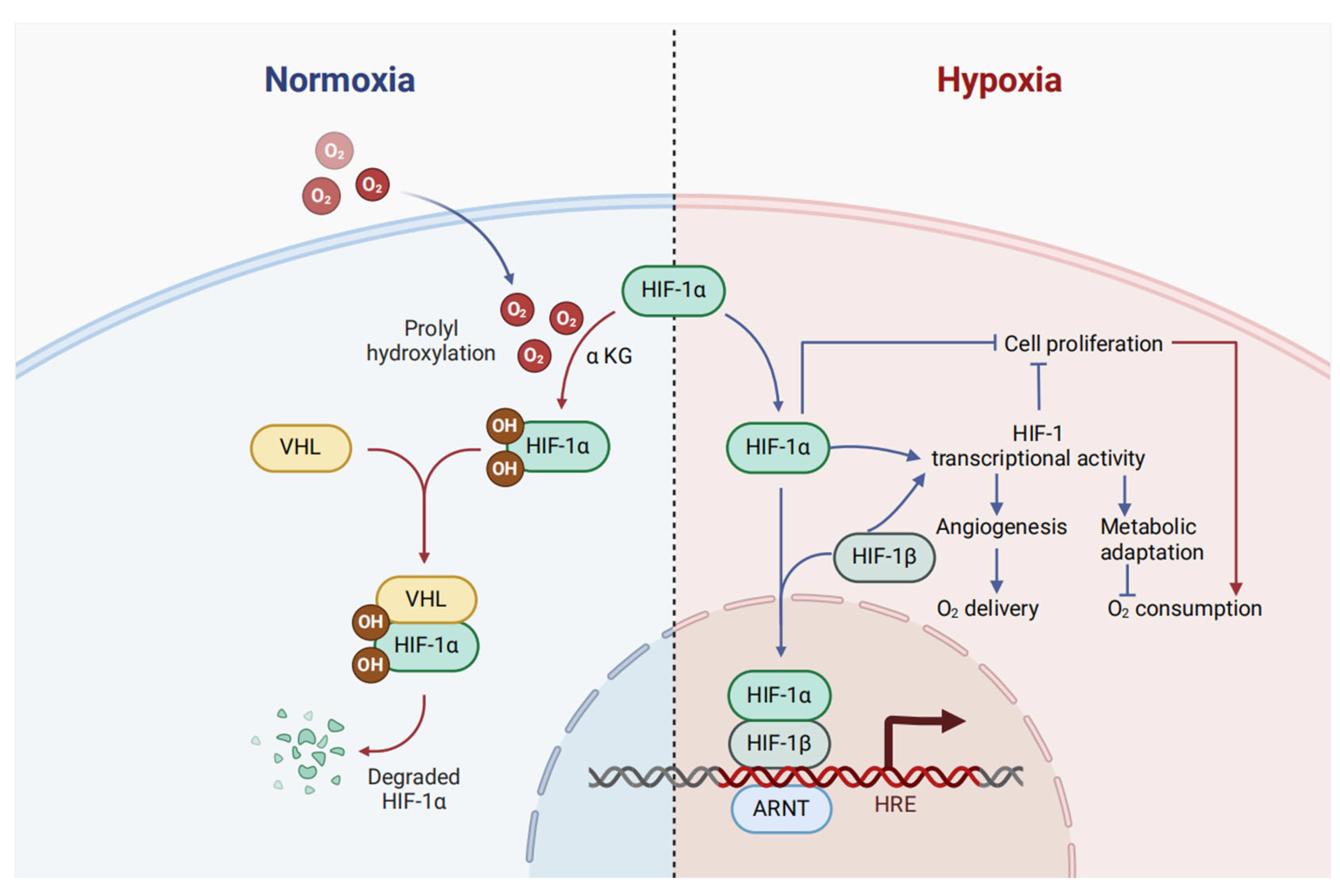
Approximately 75 percent of the human genome undergoes transcription into RNA, in contrast to the mere 3 percent that is transcribed into protein-coding mRNAs, as documented in the literature [1]. The class of non-coding RNAs (ncRNAs) encompasses long ncRNAs (lncRNAs), circular RNAs (circRNAs), microRNAs (miRNAs), and piwi-interacting RNAs (piRNAs) [2]. MicroRNAs are small non-coding RNAs, typically 20–22 nucleotides in length, that regulate gene expression post-transcriptionally. They bind to the 3′ untranslated regions (3′UTRs) of target mRNAs, usually leading to mRNA degradation or translational repression [3,4]. Long ncRNAs are longer non-coding transcripts, often more than 200 nucleotides. They can act at various levels of gene regulation, including chromatin remodeling, transcriptional regulation, and post-transcriptional regulation [5,6]. Circular RNAs are covalently closed circular molecules formed by back-splicing events. They can function as miRNA sponges, sequestering miRNAs and preventing their interaction with target mRNAs [7,8].
Extensive bodies of evidence have highlighted the pivotal roles played by ncRNAs in human malignancies, with these insights actively being translated into clinical practice. While the specific biological functions of certain ncRNAs are still being unraveled, a compelling theme has emerged: ncRNAs constitute a hierarchical, higher-order gene regulatory domain, with the potential to theoretically modulate the expression of numerous downstream mRNA targets. Notably, some ncRNAs exhibit remarkable stability, persisting in the bloodstream and presenting opportunities for the highly accurate and sensitive screening of major human cancers using minimal blood samples [9,10]. Furthermore, ncRNAs can serve as therapeutic targets, with delivery strategies drawing upon established knowledge regarding the delivery of RNAi and oligonucleotides targeting protein-coding mRNAs [11]. The identification and functional annotation of CDR1as [8], in conjunction with the initial profiling of circRNAs [7], sparked a surge in circRNA research. Accumulating evidence underscores the significant roles played by circRNAs in various diseases, including cancers [12,13,14,15,16]. Recently, multiple circRNAs have been found to be overexpressed in colorectal cancer (CRC) and to facilitate proliferation, migration, and metastasis through the upregulation of HIF-1α [17,18,19,20]. In addition to circRNAs, miRNAs and lncRNAs are relatively newer entities within the ncRNA landscape and are less well characterized. However, due to their prevalent occurrence, specific expression patterns, functionally significant roles in diseases, and potential clinical applications, they are garnering substantial attention [21,22]. This review will compile existing research findings and discuss the specific mechanisms of ncRNA and HIF-1α interactions, along with their potential applications in CRC treatment.

1.2. HIF-1α

The microenvironment of solid tumors commonly exhibits hypoxia. This condition is a consequence of the rapid and unregulated growth of tumor cells, which leads to restricted oxygen supply [23]. Hypoxia-inducible factor 1 (HIF-1) plays a pivotal role in the regulation of hypoxic responses. Elevated levels of HIF-1 are frequently linked to poor prognoses in diverse cancer types [24]. Structurally, HIF-1 is a heterodimeric complex composed of a constantly active HIF-1β subunit and an oxygen-sensitive HIF-1α subunit [25,26]. This complex can recognize the consensus sequence 5′-RCGTG-3′, which is either present within or close to genes under the control of HIF-1 [27]. The stability of HIF-1α is regulated in an oxygen-dependent manner through prolyl hydroxylation. This process promotes the association between HIF-1α and the von Hippel–Lindau tumor suppressor protein (VHL), a crucial part of an E3 ubiquitin ligase. Such an interaction triggers the ubiquitination and subsequent degradation of HIF-1α via the proteasome [28] (Figure 1). Furthermore, the stability of HIF-1α is influenced by the cell’s metabolic state. Alpha-ketoglutarate, a metabolite of the tricarboxylic acid cycle, acts as a substrate for prolyl hydroxylases in the presence of oxygen. These enzymes incorporate one oxygen atom into a proline residue (Pro-564 or Pro-403 in human HIF-1α specifically) and the other oxygen atom into α-ketoglutarate, transforming it into succinate and carbon dioxide (Figure 1). HIF-1α controls numerous adaptive responses to hypoxia, such as cell proliferation, metabolic changes, and angiogenesis. The transcriptional activity of the HIF-1α/HIF-1β heterodimer regulates angiogenesis and metabolic reprogramming. By contrast, cell proliferation is regulated by a combination of transcriptional and non-transcriptional processes mediated by HIF-1α [29].
HIF-1α serves as a pivotal regulator in the spectrum of cancer-related processes, encompassing metabolic reprogramming, angiogenesis, metastasis, and resistance to apoptosis. Specifically, in metabolic reprogramming, HIF-1α orchestrates the hypoxia-driven metabolic alterations in tumor cells, impacting both glycolysis and lipid metabolism [30,31,32]. In the realm of angiogenesis, HIF-1α stimulates VEGF production, thereby enhancing vascular density and reducing oxygen diffusion distance, ultimately disrupting local blood flow dynamics within the tumor microenvironment [33]. Concerning resistance to apoptosis, HIF-1α acts to inhibit apoptotic pathways, promoting the adaptive survival of cancer cells [34]. Regarding metastasis, HIF-1α exerts its influence by directly or indirectly modulating the expression of key genes at various stages of the metastatic cascade [35,36].

1.3. Colorectal Cancer

Variability in CRC prevalence across nations is attributed to multiple contributing factors, including socio-economic status, with lower socio-economic levels correlating with heightened CRC susceptibility. Advancements in screening, early interventions, and improved therapeutic strategies have led to a reduction in CRC mortality by approximately 35% from 1990 to 2007, with a further decrease of around 50% from peak mortality rates [37]. Nonetheless, it must be recognized that the overall decline in colorectal cancer (CRC) mortality might have concealed the mortality trend among young adult CRC patients. Data from the SEER database indicate that although the mortality rate for young adults (aged 20–54 years, white) dropped from 6.3 per 100,000 in 1970 to 3.9 in 2004, it increased to 4.3 per 100,000 in 2014.
While progress in evidence-based therapies and early diagnostic strategies for colorectal cancer is commendable, substantial knowledge deficits persist. These gaps necessitate further investigation to enhance our comprehension of the disease’s pathogenesis, to refine therapeutic efficacy, and to extend patient lifespan without compromising their quality of life. Emerging research has unveiled a complex and functional landscape of ncRNAs in CRC [38]. Numerous ncRNAs exhibit differential expression across normal tissue, primary, and metastatic castration-resistant CRCs, with some capable of independently promoting proliferation [39,40,41]. These individual ncRNAs often carry prognostic implications. Furthermore, ncRNAs exhibit enhanced stability when contrasted to their linear forms and are extensively present in human plasma exosomes, positioning them as prime candidates for minimally or non-invasive biomarkers [42,43]. Of greater significance, ncRNAs are capable of engaging with the hypoxic tumor microenvironment, a hallmark of solid tumors, particularly with HIF-1α, to synergistically modulate the progression of colorectal cancer [44,45].
In 2023, an interesting review on the interactions between ncRNAs and HIF-1α in cancer was published in the European Journal of Pharmacology. This article precisely focuses on the context of colorectal cancer, delving deep into the interactions between these ncRNAs and HIF-1α [39]. It particularly emphasizes highlighting the clinical relevance of HIF-1α-related ncRNAs in CRC, aiming to bridge the existing gap between the results obtained from basic research and the clinical implementation of HIF-1α-related ncRNAs as biomarkers for diagnosis, prognosis, and treatment in CRC. Moreover, this review centers around the core theme that, in future large-scale clinical trials, it is necessary to comprehensively evaluate the therapeutic potential and safety of HIF-1α-related ncRNAs.

2. HIF-1α Interacting ncRNAs in Colorectal Cancer

2.1. Long ncRNA

Previously regarded as transcriptional noise, lncRNAs have emerged as a ubiquitous class of ncRNAs with significant biological functions over the past decade [46]. Despite their lower abundance among ncRNAs, the context-specific expression patterns and the altered transcriptome levels of lncRNAs in CRC contexts render them viable candidates for biomarkers. A comprehensive study employing whole-transcriptome sequencing of 10 CRC and para-cancerous specimens identified 2852 lncRNA transcripts that displayed dysregulation when compared to normal control samples [47]. Research has shown that lncRNAs can modulate gene expression through various mechanisms and perform several common functions. In the realm of epigenetics, circRNAs can recruit a range of epigenetic factors to enhance signal transduction and transcription [48]. At the transcriptional level, lncRNAs interact with gene promoters, transcription factors (TFs), and co-factors to regulate transcription [49]. At the post-transcriptional level, lncRNAs engage with multiple splicing factors, such as miRNAs and RNA-binding proteins (RBPs), to influence the precursor mRNAs of numerous genes [50]. Moreover, lncRNAs regulate a wide array of RNA and protein modifications, thereby affecting their activity and stability [51]. Notably, peptides derived from lncRNAs exert pivotal functions within a multitude of biological pathways [52].
In the past decade, an extensive body of research has revealed the functional contributions of diverse individual lncRNAs in CRC, achieved through their engagement with HIF-1α, leveraging both cellular and animal model systems [39,44,45,46,47,48,49,50,51,52,53,54,55,56,57] (Table 1). One investigation highlights the hypoxic induction of HIF-1α, which leads to the transcriptional augmentation of lncRNA STEAP3-AS1, competing with YTHDF2 for binding and thereby increasing the mRNA stability of STEAP3, which in turn enhances STEAP3 protein levels. This upregulation of STEAP3 results in the generation of cellular Fe2+, activating the phosphorylation of Ser 9 and inactivating GSK3β, facilitating β-catenin nuclear translocation and the subsequent activation of Wnt signaling, thereby facilitating CRC progression [58]. A plethora of studies have also demonstrated that the HIF-1α/lncRNA interface is pivotal in CRC, modulating chemotherapy responsiveness (Figure 2). For example, lnc-RP11-536 K7.3 is linked to oxaliplatin resistance and poor prognosis. The deletion of lnc-RP11-536 K7.3 suppresses proliferation, glycolysis, and angiogenesis, and improves chemosensitivity in both in vitro chemo-resistant organoids and in vivo CC cells. Moreover, lnc-RP11-536 K7.3 recruits SOX2, which then promotes the transactivation of USP7 mRNA. This process results in the deubiquitination and stabilization of HIF-1α, ultimately playing a role in the development of oxaliplatin resistance [59]. Similarly, the knockdown of NORAD might reduce hypoxia-induced malignancy in colorectal cancer. It achieves this by impeding VM and chemoresistance. The underlying mechanism involves sequestering miR-495-3p/HIF-1α, which in turn regulates EMT [60]. Moreover, LINC00511 and HITT stimulate CRC cell proliferation and inhibit apoptosis. Notably, a robust association between HIF-1α mRNA and HITT has been substantiated in human colon cancer tissues [61,62]. Significantly, elevated serum levels of lncRNA/HIF1A-AS1 have emerged as a novel prognostic biomarker for unfavorable clinical outcomes [63]. In recent investigations, the significant functions of lncRNAs in the progression of CRC and the development of chemoresistance have been revealed. Notably, these functions are often mediated through interactions with HIF-1α. This research underscores the potential of lncRNAs as innovative diagnostic and prognostic biomarkers, as well as promising therapeutic targets.

2.2. MicroRNAs

MiRNAs are compact, evolutionarily conserved ncRNAs with a length of roughly 22 nucleotides, which are pivotal in the modulatory processes of gene expression [79]. The first miRNA, lin4, was identified from Caenorhabditis elegans in 1993 [80]. In 2000, Reinhart et al. reported the first mammalian miRNA, let-7, which was found to suppress the expression of the heterochronic gene lin-41 by means of sequence-specific RNA–RNA interactions with the 3′-UTR of its mRNA [81,82]. In 2002, Dr. Calin’s research team was the first to establish the role of miRNAs in human cancer development, revealing that miR-16-1 and miR-15a, situated on chromosome 13q14, are frequently deleted in B-cell chronic lymphocytic leukemia (CLL) [83]. Over the last decade, a multitude of investigations have clarified the roles of miRNAs in a variety of pathologies, with a particular emphasis on cancer. A transcriptomic study of plasma from 1382 CRC patients has highlighted the potential integration of plasma miRNAs into fecal immunochemical test-based CRC screening protocols. This integration aims to optimize colonoscopy scheduling by focusing on individuals most likely to benefit significantly from the procedure [84]. The regulation of gene expression by miRNAs is a universal feature among all recognized cancer cell types. Throughout tumor development, miRNAs exhibit distinct biological characteristics, suggesting their potential for cancer classification and improved prognosis.
Interactions between HIF-1α and miRNAs collectively influence the progression of CRC, including aspects of drug resistance and chemosensitivity (Figure 2 and Table 1). CRC cells exhibit enhanced exosome production, and the miRNAs contained within these exosomes play a significant role in disease progression, potentially facilitating metastasis. For instance, exosomes derived from hypoxic conditions promote the proliferation and metastasis of CRC by exporting HIF-1α-induced miR-4299 and regulating its target gene ZBTB4 [65]. Additionally, hypoxic repression of miR-338-5p stabilizes a HIF-1α-driven feedback loop via direct targeting of IL-6/STAT3/Bcl2 signaling, driving chemoresistance in colorectal cancer. Preclinical models demonstrate that combinatorial miR-338-5p restoration and HIF-1α inhibition disrupt this axis to re-sensitize tumors to oxaliplatin [73]. In CRC, miR-20a is significantly downregulated under hypoxic conditions. Overexpression of miR-20a has been found to mitigate hypoxia-induced autophagy. Mechanistically, miR-20a inhibits the hypoxia-induced autophagic flux by targeting multiple key autophagy regulators, such as ATG5 and FIP200. Through dual-luciferase assays, it has been demonstrated that miR-20a directly binds to the 3′-untranslated region of ATG5 and FIP200 mRNAs, thereby regulating their mRNA and protein levels [85]. Another study in this type of cancer has shown the HIF-1α induces an elevation in the levels of miR-361-3p within hypoxic extracellular vesicles (EVs) in the context of CRC. The upregulated miR-361-3p in CRC exerts its effects by directly targeting TNF receptor-associated factor 3. This targeting event inhibits cell apoptosis and promotes cell growth, ultimately leading to the activation of the noncanonical NF-κB pathway [86]. Importantly, the presence of these miRNAs in body fluids, derived from exosomes, underscores their substantial value as diagnostic markers for CRC.
Angiogenesis constitutes a pivotal factor in tumor progression. The importance of this process, encompassing the generation of novel blood vessels, in the expansion of solid neoplasms is extensively recognized. MicroRNAs have been shown to play a crucial role in the progression of CRC by modulating angiogenesis through interactions with HIF-1α. The microRNA miR-148a markedly inhibits angiogenesis by suppressing VEGF through downregulating the PERK/HIF-1α/VEGF axis, potentially leading to the inhibition of angiogenesis. Conversely, the downregulation of miR-148a is associated with an increased risk of early recurrence in CRC [72]. Furthermore, miR-148a represses HIF-1α/VEGF and Mcl-1 by directly targeting ROCK1/c-Met, thereby diminishing angiogenesis and enhancing apoptosis in colon cancer cells. Notably, patients with metastatic CRC (mCRC) displaying elevated levels of serum miR-148a demonstrate a more favorable therapeutic response to the combination of chemotherapy and bevacizumab (standard dose; 5 mg/kg) compared to those without such overexpression [68]. The microRNAs miR-206, miR-22, and miR-199a may exert anti-angiogenic effects in colon cancer [74,77,78]. Beyond angiogenesis, the interaction between miRNAs and HIF-1α also promotes glycolysis, thereby advancing the progression of CRC. The HIF-1α-induced miR-23a∼27a∼24 cluster (comprising miR-24, miR-27a, and miR-23a) is a critical regulator that redirects CRC metabolism from oxidative phosphorylation to glycolysis and, by controlling their expression, holds promise for the suppression of CRC progression [70].
Accumulating studies indicate that miRNAs are central to the molecular underpinnings of drug resistance and radiosensitivity through interactions with HIF-1α. Research has proposed that the suppression of miR-210-3p subsequent to 5-FU administration maintains DNA damage repair and metabolic adaptations, thereby mitigating the effects of the drug [71]. Additionally, HIF-1α activates miRNA-210, which potentiates autophagy and diminishes radiation sensitivity by repressing Bcl-2 expression in colon cancer cells [76]. A further investigation has unveiled an HIF-1α/miR-338-5p/IL-6 feedback circuitry that is essential for hypoxic-induced resistance to chemotherapy in CRC [73]. Furthermore, miR-6887-3p and miR-200b can also influence the invasion and proliferation of CRC cells by interacting with HIF-1α [69,75].
The interaction of HIF-1α with miRNAs is pivotal in the evolution of CRC, modulating therapeutic resistance and responsiveness, stimulating angiogenesis, and orchestrating metabolic cascades. CRC cells augment the secretion of exosomes, within which miRNAs are integral to the advancement of the disease, and may further facilitate metastatic dissemination. Collectively, the intricate web of interactions involving HIF-1α and a constellation of miRNAs profoundly influences CRC at multiple biological dimensions, encompassing proliferation, dissemination, angiogenesis, metabolism, and therapeutic outcomes. This elucidation offers novel insights and potential therapeutic avenues for CRC diagnosis and intervention.

3. New Areas of ncRNA Research in Colorectal Cancer

Given the significant heterogeneity characteristic of colorectal cancer, it is imperative to investigate cell-type-specific ncRNAs within the tumor microenvironment. Following the pioneering identification of non-coding RNAs over three decades ago, recent technological breakthroughs have facilitated the exploration of critical dimensions of non-coding RNA research that were hitherto inaccessible.

3.1. RNA Vaccines Based on ncRNA

The remarkable effectiveness of SARS-CoV-2 mRNA vaccines in the fight against the COVID-19 pandemic has rekindled interest in RNA-based immunotherapies. Drew Weissman and Katalin Karikó were awarded the 2023 Nobel Prize in Physiology or Medicine for their groundbreaking work on nucleoside base editing [87]. This discovery has been crucial in facilitating the development of revolutionary vaccines. An important advantage of circRNA lies in its covalently closed-loop structure. This structure protects circRNA from nuclease-mediated degradation, thereby improving its stability and persistence. Hypothetically, circRNAs might achieve enhanced therapeutic effects even when administered at low dosages [88]. Chen et al.’s team achieved a significant increase in protein yield from cyclic RNA translation through optimized designs, enabling efficient and sustained in vivo protein production [89]. Furthermore, RNA therapies based on miRNAs and lncRNAs have garnered considerable attention [90,91]. For instance, a research endeavor has engineered a bio-scaffold that incorporates biomimetic nanosystems designed to target the lncRNA Pvt1. This scaffold is instrumental in orchestrating a tri-functional immune response aimed at preventing the postoperative recurrence of CRC. This scaffold elicits a vigorous immune memory, effectively quelling ectopic tumor rechallenges and concurrent metastatic dissemination (with a success rate of 100%). Furthermore, the bio-scaffold demonstrates a 70.8% decrease in synchronous distant metastasis [92]. Notably, the number of ncRNA-based therapies undergoing clinical trials for various genetic, metabolic, and oncological conditions is steadily growing.

3.2. Non-Coding RNA and Disulfidptosis

The recently characterized phenomenon of disulfidptosis, a caspase-independent mode of programmed cell death, constitutes a novel frontier in cancer research, driven by its pathogenesis rooted in disulfide stress. Its relevance in the realm of cancer biology is underpinned by two primary considerations [93]. Initially, it embodies a unique mode of regulated cell death, distinct from other known forms [94]. Secondly, it holds promise as a potent therapeutic target for future cancer treatments. In a study by Dong et al., TCGA datasets were meticulously analyzed to construct a prognostic disulfidptosis-related lncRNA signature for colon cancer. Four lncRNAs were pinpointed as the most prognostically relevant indicators for patient survival, with a notable survival advantage observed in the low-risk subgroup compared to the high-risk subgroup. Bioinformatics analysis revealed distinct metabolic pathways and immune profiles. The model was further validated using independent datasets, and the differential expression of the lncRNAs was confirmed through rt-qPCR in tissue and cellular samples [95]. These findings underscore the importance of understanding the roles of various ncRNAs in disulfidptosis, providing crucial insights into their potential mechanisms of action.

3.3. Single Cell and Spatial Transcriptomics

Non-coding RNA expression is characterized by its specificity and stringent regulation. Within the context of cancer, a plethora of lncRNAs and miRNAs display expression profiles that are distinct to specific tumor types or subtypes [96]. While bulk transcriptomic analyses typically reveal low ncRNA expression, recent research has detected elevated expression in specific cellular subpopulations [97], presenting opportunities to explore intratumoral heterogeneity. Consequently, the examination of ncRNA expression signatures at the single-cell resolution or across spatially distinct tumor compartments could reveal novel molecular subtypes or infrequent cell populations within prostate cancer, thereby providing valuable insights into tumor complexity.
Traditional cancer research has primarily centered on detailed molecular and clinical investigations of key pathways and genes. In recent years, advancements in high-throughput technologies have significantly accelerated the collection of extensive cancer genomics data, encompassing genomics, transcriptomics, epigenomics, and other genomic fields, with the goal of thoroughly and systematically assessing various aspects of tumor characteristics [98]. As histological data rapidly expand, numerous bioinformatics databases and tools have been developed, greatly aiding in the storage, efficient retrieval, comprehensive integration, and in-depth analysis of these data [99]. The integration and detailed analysis of these datasets have fundamentally transformed the cancer research landscape, offering deeper insights into the molecular mechanisms driving tumorigenesis, elucidating the tumor microenvironment (TME) characteristics, accurately identifying potential biomarkers for early detection and prognostic assessment, and guiding the development of precision-targeted therapies for individual patients [100]. For instance, an investigation elucidated the involvement of TM4SF1 in facilitating EMT and cancer stem cell characteristics in CRC through the Wnt/β-catenin/SOX2 signaling axis, leveraging data resources from The Cancer Genome Atlas and the Gene Expression Omnibus repositories [101]. This approach significantly enhances the efficiency and accuracy of research.

4. Clinical Relevance of HIF-1α-Related ncRNAs in Colorectal Cancer

Many ncRNAs associated with HIF-1α have been found to be dysregulated in CRC. Moreover, these ncRNAs exhibit high specificity and are readily identifiable across a spectrum of biological matrices, including tissues, serum, saliva, urine, and plasma. Consequently, HIF-1α-associated ncRNAs hold significant promise as both diagnostic and prognostic biomarkers, as well as potential therapeutic endpoints, within the context of CRC (Figure 3).

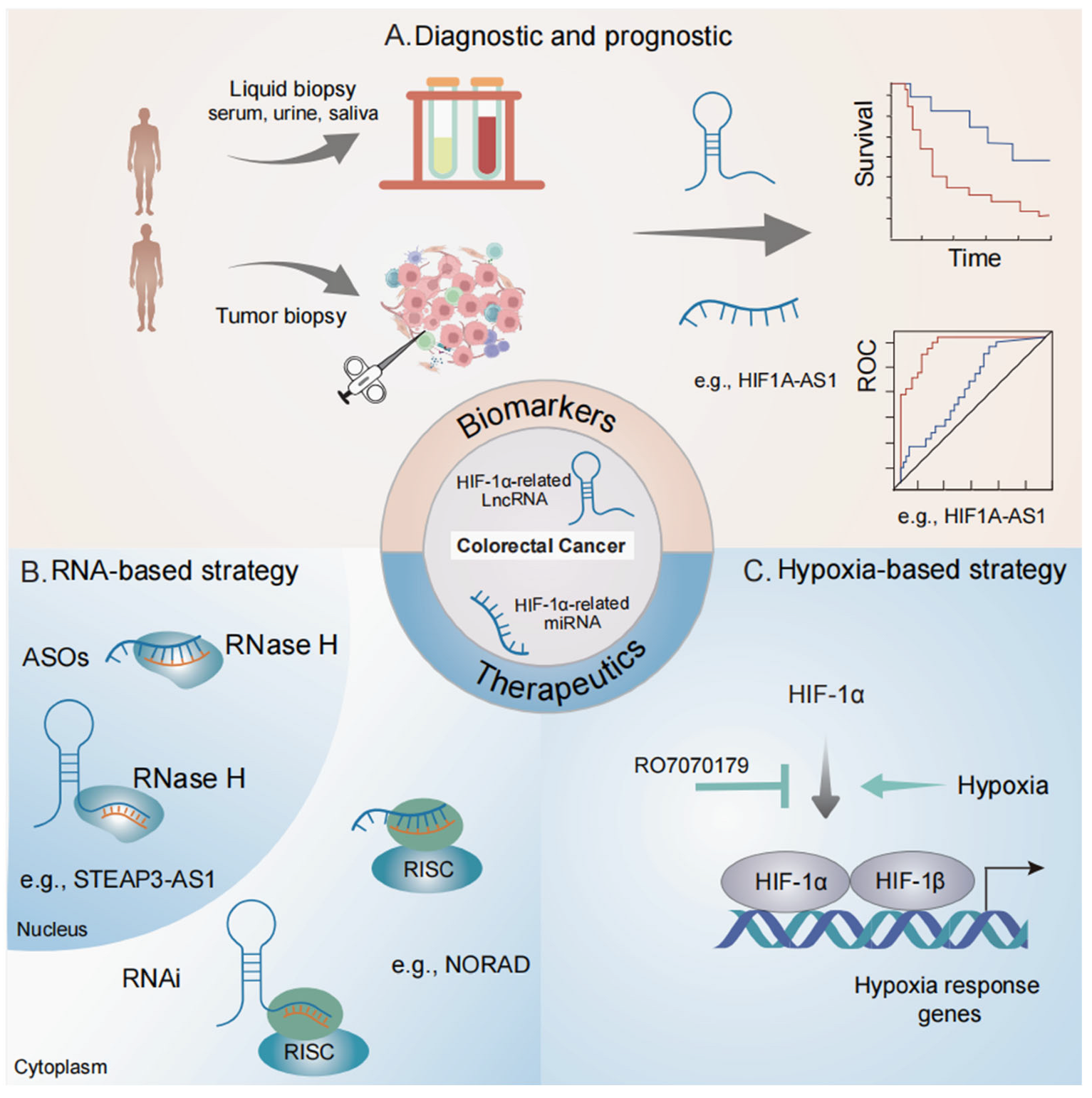
4.1. Biomarker Potentials

The serum levels of HIF1A-AS1 were significantly higher in CRC patients compared to healthy controls (p < 0.05). An ROC curve analysis indicated that HIF1A-AS1 has a high diagnostic potential for differentiating CRC patients from healthy individuals, with an AUC of 0.960. A Kaplan–Meier survival analysis revealed that factors such as differentiation grade, tumor size, TNM classification, T stage, N stage, M stage, and serum HIF1A-AS1 levels were all associated with the prognosis of CRC patients (all p < 0.05). Based on these findings, a serum-based biomarker panel centered on HIF1A-AS1 has been developed. This panel can be used to assess the need for rebiopsy in cases where carcinoembryonic antigen and carbohydrate antigen 19-9 levels are elevated, despite initially negative biopsies [63]. It has been reported that bioinformatic analyses of the CRC-transcriptomes indicated that the lncRNA/PVT1 locus might exert a far-reaching impact on the expression and functions of other key genes within two vital CRC-related signaling pathways, specifically the TGFβ/SMAD and Wnt/β-Catenin pathways. Furthermore, PVT1 acts as a novel oncogenic enhancer of MYC, with its activity being epigenetically regulated through aberrant methylation in CRC. The findings also suggested that the expression of PVT1/lncRNA could be a promising prognostic biomarker and a potential therapeutic target in colorectal cancer [102]. In addition to HIF1A-AS1, several other lncRNAs, including lncRNA HITT, CircMYH9, and miR-206, show promise as candidates for diagnostic and prognostic biomarkers in CRC [18,19,20,21,22,23,24,25,26,27,28,29,30,31,32,33,34,35,36,37,38,39,40,41,42,43,44,45,46,47,48,49,50,51,52,53,54,55,56,57,58,59,60,61,62,63,64,65,68,69,70,71,72,73,74,75,76,77,78,79,80,81,82,83,84,85,86].
Currently, several ongoing clinical trials are recruiting subjects to evaluate the value of ncRNAs as diagnostic and prognostic markers in CRC (Table 2). The research on HIF-1α-associated lncRNAs and miRNAs shows great promise for future exploration. Consequently, hypoxia-responsive lncRNAs and miRNAs are becoming increasingly attractive as potential cancer biomarkers for both diagnosis and prognosis (Figure 3). For instance, a clinical trial (NCT04269746) is now examining the potential of lncRNA CCAT1 as a CRC biomarker. The area of liquid biopsies is attracting significant attention. Liquid biopsies are less invasive than traditional tissue biopsies and are highly suitable for continuous monitoring of the disease. Due to their unique expression profiles, lncRNAs are emerging as feasible candidates for liquid biopsy-based biomarkers, with preclinical testing having been carried out in plasma (NCT04269746 and NCT06307249). In metastatic CRC [72], exosomes offer another source of extracellular lncRNAs, with miR-21-5p and miR-4299 being highly expressed in exosomes derived from CRC cells [64,65,66,68,69,70,71,72,73,74,75,76,77,78,85,86].

4.2. Therapeutic Potential in Targeting HIF Signaling

The hypoxia condition, characterized by its pivotal involvement in the onset, advancement, dissemination, and therapeutic resistance of neoplasms, has garnered attention as a highly attractive therapeutic niche [103]. Among the numerous avenues of investigation, the modulation of HIF signaling stands out as a focal point. The tumor’s adaptive response to hypoxia is predominantly governed by HIF signaling, which is initiated through a meticulously controlled signaling cascade. Each component of this pathway presents a potential target, encompassing the suppression of HIF-α protein aggregation with PHD inhibitors [104,105], the inhibition of PI3K/mTOR [106,107], the targeting of topoisomerases [108,109], the interference with microtubule dynamics, the disruption of thioredoxin activity [110], the attenuation of HSP90 [111], and the inhibition of HDACs [112]. Moreover, inhibitors that prevent the HIF-α–HIF-β heterodimerization, such as PAS domain inhibitors [113,114], those that interfere with HIF–HRE interaction, including HRE inhibitors [115,116], and those that disrupt coactivator engagement, such as TAD inhibitors, are also being explored [117,118]. At present, multiple clinical trials are underway to explore hypoxia-targeting strategies in cancer using investigational drugs. Significantly, a clinical investigation (NCT02564614) is currently evaluating the impacts of the HIF-1α mRNA antagonist RO7070179 in adult liver cancer patients. Additionally, a phase 3 clinical trial (NCT04195750) is assessing the effectiveness of the HIF-2α inhibitor MK-6482 in patients diagnosed with advanced renal clear cell carcinoma. A more in-depth comprehension of the regulatory mechanisms of HIF signaling pathways will contribute to the development of innovative drugs for targeting hypoxic tumors.

4.3. Therapeutic Potential in Targeting ncRNAs

Inhibiting HIF-1α and HIF-2α is a potential cancer treatment strategy. However, resistance is a common challenge. For example, extended use of PT2399, a specific HIF-2 inhibitor, can lead to resistance characterized by increased tumor blood vessel formation and higher VEGF levels [119]. One study has found that lncRNA, NORAD, and HIF-1α are elevated in CRC tissues and positively associated. The NORAD knockdown reduces hypoxia-induced VM formation, 5-FU resistance, and HIF-1α-mediated EMT by ponging miR-495-3p, suggesting NORAD as a potential therapeutic target for CRC [60]. The downregulation of miR-210-3p empowers resistant cells to mitigate the toxic impact of the drug. This is achieved by upregulating the expression of the RAD-52 protein, which is accountable for DNA damage repair. Additionally, the downregulation of miR-210-3p augments oxidative phosphorylation (OXPHOS). It performs as such by elevating the expression levels of succinate dehydrogenase subunits D, reducing intracellular succinate levels, and suppressing HIF-1α expression. Collectively, these adaptive changes result in enhanced cell survival upon drug exposure [71].Therefore, the development of novel and more efficacious treatment modalities is of great significance for enhancing clinical outcomes. One feasible approach to counteract hypoxia-induced cancer invasion is to target the downstream elements of the HIF pathway. The association between hypoxia-related ncRNAs and cancer progression indicates their potential as therapeutic targets. As a result, precisely targeting ncRNAs within the hypoxic TME presents a promising strategy for the treatment of diverse and complex cancer types.
The employment of efficient gene modulation techniques, such as overexpression or knockdown, can augment the precision of targeting hypoxia-associated lncRNAs and circRNAs for therapeutic purposes (Figure 3). Currently, RNA-based therapeutic strategies predominantly rely on RNAi and antisense oligonucleotides (ASOs) to specifically target particular RNA segments and molecules. Investigations in murine models have shown that the utilization of ASOs and RNAi to target oncogenic hypoxia-related lncRNAs and circRNAs can efficiently inhibit tumor growth. Non-coding RNAs that display substantial oncogenic potential in CRC emerge as attractive candidates for innovative therapeutic approaches. In preclinical investigations, lncRNAs like STEAP3-AS1, NORAD, and LINC00511 have been discovered to facilitate CRC cell growth and malignancy [58,59,60,61]. Additionally, the lnc-RP11-536 K7.3 is associated with the chemosensitivity of CRC [59]. MicroRNAs, like miR-148a, reduce angiogenesis and enhance apoptosis in colon cancer cells [72]. Considering their indispensable roles in pathogenesis, these oncogenic ncRNAs represent a compelling focus for the development of novel therapeutic strategies.

5. Concluding Remarks and Perspectives

The high mortality rate of CRC is attributed to its rapid progression and the absence of early diagnostic markers. Therefore, an imperative requirement exists for the discovery of novel tumor biomarkers and therapeutic endpoints to facilitate early diagnosis, precise prognostication, and efficacious therapeutic interventions. The altered expression of HIF-1α-related ncRNAs in tumor samples, coupled with their easy detection in bodily fluids, makes them promising candidates for non-invasive liquid biopsy approaches. Furthermore, these HIF-1α-related ncRNAs have been shown to significantly impact various cancer hallmarks, including tumor metabolism, immune evasion, angiogenesis, migration/invasion, and growth/proliferation. Consequently, HIF-1α-related ncRNAs hold potential as biomarkers or therapeutic targets in CRC.
Notwithstanding the remarkable progress achieved in elucidating the functions of ncRNAs within the hypoxic TME, numerous challenges and limitations persist and demand resolution. Moreover, several key questions remain unanswered. (i) When targeting HIF-1α-associated ncRNAs in cancer, how can we safeguard against affecting the expression of essential genes in normal tissues? The identification of target ligands characterized by high affinity and stability might be beneficial in minimizing potential side effects and toxicity. Additionally, the development of refined delivery systems tailored for the accurate targeting of circRNAs is of utmost importance. (ii) What are the factors responsible for the disparities in hypoxic responses witnessed between cell lines and animal models? The majority of current studies have predominantly concentrated on two-dimensional cell culture models. As such, future research endeavors should integrate three-dimensional tumor organ models that incorporate the TME to gain more comprehensive insights [120]. (iii) Is there an augmentation of therapeutic efficacy when HIF-1α-associated ncRNA-targeting therapies are combined with other anti-tumor approaches? A recent investigation demonstrated that the combination of ASO-induced downregulation of lncRNAs and traditional chemotherapeutic drugs was more efficacious than chemotherapy alone in the treatment of CRC [64]. Consequently, the integration of therapies that target HIF-1α-associated ncRNAs with other anti-cancer modalities is likely to enhance treatment effectiveness. (iv) The utilization of ncRNAs within bodily fluids as prospective biomarkers for cancer diagnostics and prognostication presents a formidable challenge. Recent progress in RNA analytical methodologies has enabled the creation of innovative platforms, which leverage machine learning algorithms to discern complex or composite biomarker profiles [121]. Notably, circRNAs, known for their stability and high translation efficiency, have attracted considerable attention [122].
Notably, a divergence exists between the results obtained from basic research and the clinical implementation of HIF-1α-related ncRNAs as diagnostic, prognostic, and therapeutic biomarkers. As a result, in future large-scale clinical trials, it is essential to comprehensively evaluate the therapeutic potential and safety of HIF-1α-related ncRNAs.

Author Contributions

R.X. conceived the review article; X.W. designed the overall research framework, provided overall supervision, and critically revised the manuscript; L.G. contributed to collecting relevant papers and the overall editing of the manuscript; H.Z. created the table and corresponding visualizations; Y.L. helped to modify the manuscript. All authors have read and agreed to the published version of the manuscript.

Funding

This review was supported by multiple funding sources. The Key Project of Tibet Autonomous Region Natural Science Foundation (Grant No. XZ202501ZR0140) provided essential financial support. Additionally, it received funds from the Shannan Science and Technology Plan Project (Grant No. SNSBJKJJHXM2024023). The Hubei Province Key R & D Program Aid Project to Tibet also contributed to this study. Moreover, the research was partly funded by the Hubei Province Natural Science Foundation (Grant No. JCZRYB202501311).

Institutional Review Board Statement

Not applicable.

Informed Consent Statement

Not applicable.

Data Availability Statement

Not applicable.

Conflicts of Interest

The authors declare no conflicts of interest.

References

  1. Mattick, J.S.; Makunin, I.V. Non-coding RNA. Hum. Mol. Genet. 2006, 15, R17–R29. [Google Scholar] [CrossRef]
  2. Nemeth, K.; Bayraktar, R.; Ferracin, M.; Calin, G.A. Non-coding RNAs in disease: From mechanisms to therapeutics. Nat. Rev. Genet. 2024, 25, 211–232. [Google Scholar] [CrossRef]
  3. Bartel, D. MicroRNAs: Genomics, biogenesis, mechanism, and function. Cell 2004, 116, 281–297. [Google Scholar] [CrossRef]
  4. Filipowicz, W.; Bhattacharyya, S.; Sonenberg, N. Mechanisms of post-transcriptional regulation by microRNAs: Are the answers in sight? Nat. Rev. Genet. 2008, 9, 102–114. [Google Scholar] [CrossRef]
  5. Ponting, C.; Oliver, P.; Reik, W. Evolution and functions of long noncoding RNAs. Cell 2009, 136, 629–641. [Google Scholar] [CrossRef]
  6. Mercer, T.; Dinger, M.; Mattick, J. Long non-coding RNAs: Insights into functions. Nat. Rev. Genet. 2009, 10, 155–159. [Google Scholar] [CrossRef]
  7. Memczak, S.; Jens, M.; Elefsinioti, A.; Torti, F.; Krueger, J.; Rybak, A.; Maier, L.; Mackowiak, S.; Gregersen, L.; Munschauer, M.; et al. Circular RNAs are a large class of animal RNAs with regulatory potency. Nature 2013, 495, 333–338. [Google Scholar] [CrossRef]
  8. Hansen, T.; Jensen, T.; Clausen, B.; Bramsen, J.; Finsen, B.; Damgaard, C.; Kjems, J. Natural RNA circles function as efficient microRNA sponges. Nature 2013, 495, 384–388. [Google Scholar] [CrossRef] [PubMed]
  9. Imaoka, H.; Toiyama, Y.; Fujikawa, H.; Hiro, J.; Saigusa, S.; Tanaka, K.; Inoue, Y.; Mohri, Y.; Mori, T.; Kato, T.; et al. Circulating microRNA-1290 as a novel diagnostic and prognostic biomarker in human colorectal cancer. Ann. Oncol. Off. J. Eur. Soc. Med. Oncol. 2016, 27, 1879–1886. [Google Scholar] [CrossRef]
  10. Yaman Agaoglu, F.; Kovancilar, M.; Dizdar, Y.; Darendeliler, E.; Holdenrieder, S.; Dalay, N.; Gezer, U. Investigation of miR-21, miR-141, and miR-221 in blood circulation of patients with prostate cancer. Tumour Biol. 2011, 32, 583–588. [Google Scholar] [CrossRef]
  11. Levin, A.A. Treating Disease at the RNA Level with Oligonucleotides. N. Engl. J. Med. 2019, 380, 57–70. [Google Scholar] [CrossRef]
  12. Du, W.W.; Fang, L.; Yang, W.; Wu, N.; Awan, F.M.; Yang, Z.; Yang, B.B. Induction of tumor apoptosis through a circular RNA enhancing Foxo3 activity. Cell Death Differ. 2017, 24, 357–370. [Google Scholar] [CrossRef]
  13. Zhou, X.; Natino, D.; Qin, Z.; Wang, D.; Tian, Z.; Cai, X.; Wang, B.; He, X. Identification and functional characterization of circRNA-0008717 as an oncogene in osteosarcoma through sponging miR-203. Oncotarget 2018, 9, 22288–22300. [Google Scholar] [CrossRef]
  14. Guarnerio, J.; Bezzi, M.; Jeong, J.C.; Paffenholz, S.V.; Berry, K.; Naldini, M.M.; Lo-Coco, F.; Tay, Y.; Beck, A.H.; Pandolfi, P.P. Oncogenic Role of Fusion-circRNAs Derived from Cancer-Associated Chromosomal Translocations. Cell 2016, 166, 1055–1056. [Google Scholar] [CrossRef]
  15. Yang, Q.; Du, W.W.; Wu, N.; Yang, W.; Awan, F.M.; Fang, L.; Ma, J.; Li, X.; Zeng, Y.; Yang, Z.; et al. A circular RNA promotes tumorigenesis by inducing c-myc nuclear translocation. Cell Death Differ. 2017, 24, 1609–1620. [Google Scholar] [CrossRef]
  16. Du, W.W.; Yang, W.; Li, X.; Awan, F.M.; Yang, Z.; Fang, L.; Lyu, J.; Li, F.; Peng, C.; Krylov, S.N.; et al. A circular RNA circ-DNMT1 enhances breast cancer progression by activating autophagy. Oncogene 2018, 37, 5829–5842. [Google Scholar] [CrossRef]
  17. Xu, Y.; Zhang, Y.; Hao, W.; Zhao, W.; Yang, G.; Jing, C. Hypoxia-induced Circular RNA hsa_circ_0006508 Promotes the Warburg Effect in Colorectal Cancer Cells. Balk. Med. J. 2023, 40, 21–27. [Google Scholar] [CrossRef]
  18. Liu, X.; Liu, Y.; Liu, Z.; Lin, C.; Meng, F.; Xu, L.; Zhang, X.; Zhang, C.; Zhang, P.; Gong, S.; et al. CircMYH9 drives colorectal cancer growth by regulating serine metabolism and redox homeostasis in a p53-dependent manner. Mol. Cancer 2021, 20, 114. [Google Scholar] [CrossRef]
  19. Chen, L.Y.; Wang, L.; Ren, Y.X.; Pang, Z.; Liu, Y.; Sun, X.D.; Tu, J.; Zhi, Z.; Qin, Y.; Sun, L.N.; et al. The circular RNA circ-ERBIN promotes growth and metastasis of colorectal cancer by miR-125a-5p and miR-138-5p/4EBP-1 mediated cap-independent HIF-1α translation. Mol. Cancer 2020, 19, 164. [Google Scholar] [CrossRef]
  20. Zhou, P.; Xie, W.; Huang, H.L.; Huang, R.Q.; Tian, C.; Zhu, H.B.; Dai, Y.H.; Li, Z.Y. circRNA_100859 functions as an oncogene in colon cancer by sponging the miR-217-HIF-1α pathway. Aging 2020, 12, 13338–13353. [Google Scholar] [CrossRef]
  21. Iyer, M.K.; Niknafs, Y.S.; Malik, R.; Singhal, U.; Sahu, A.; Hosono, Y.; Barrette, T.R.; Prensner, J.R.; Evans, J.R.; Zhao, S.; et al. The landscape of long noncoding RNAs in the human transcriptome. Nat. Genet. 2015, 47, 199–208. [Google Scholar] [CrossRef] [PubMed]
  22. Rupaimoole, R.; Slack, F.J. MicroRNA therapeutics: Towards a new era for the management of cancer and other diseases. Nat. Rev. Drug Discov. 2017, 16, 203–222. [Google Scholar] [CrossRef] [PubMed]
  23. Shao, C.; Yang, F.; Miao, S.; Liu, W.; Wang, C.; Shu, Y.; Shen, H. Role of hypoxia-induced exosomes in tumor biology. Mol. Cancer 2018, 17, 120. [Google Scholar] [CrossRef] [PubMed]
  24. Cowman, S.J.; Koh, M.Y. Revisiting the HIF switch in the tumor and its immune microenvironment. Trends Cancer 2022, 8, 28–42. [Google Scholar] [CrossRef]
  25. Wang, G.L.; Semenza, G.L. Purification and characterization of hypoxia-inducible factor 1. J. Biol. Chem. 1995, 270, 1230–1237. [Google Scholar] [CrossRef]
  26. Wang, G.L.; Jiang, B.H.; Rue, E.A.; Semenza, G.L. Hypoxia-inducible factor 1 is a basic-helix-loop-helix-PAS heterodimer regulated by cellular O2 tension. Proc. Natl. Acad. Sci. USA 1995, 92, 5510–5514. [Google Scholar] [CrossRef]
  27. Semenza, G.L.; Jiang, B.H.; Leung, S.W.; Passantino, R.; Concordet, J.P.; Maire, P.; Giallongo, A. Hypoxia response elements in the aldolase A, enolase 1, and lactate dehydrogenase A gene promoters contain essential binding sites for hypoxia-inducible factor 1. J. Biol. Chem. 1996, 271, 32529–32537. [Google Scholar] [CrossRef]
  28. Kaelin, W.G., Jr.; Ratcliffe, P.J. Oxygen sensing by metazoans: The central role of the HIF hydroxylase pathway. Mol. Cell 2008, 30, 393–402. [Google Scholar] [CrossRef]
  29. Semenza, G.L. Hypoxia-inducible factors in physiology and medicine. Cell 2012, 148, 399–408. [Google Scholar] [CrossRef]
  30. Semenza, G.L. HIF-1 mediates metabolic responses to intratumoral hypoxia and oncogenic mutations. J. Clin. Investig. 2013, 123, 3664–3671. [Google Scholar] [CrossRef]
  31. Vander Heiden, M.G.; Cantley, L.C.; Thompson, C.B. Understanding the Warburg effect: The metabolic requirements of cell proliferation. Science 2009, 324, 1029–1033. [Google Scholar] [CrossRef] [PubMed]
  32. Huang, D.; Li, T.; Li, X.; Zhang, L.; Sun, L.; He, X.; Zhong, X.; Jia, D.; Song, L.; Semenza, G.L.; et al. HIF-1-mediated suppression of acyl-CoA dehydrogenases and fatty acid oxidation is critical for cancer progression. Cell Rep. 2014, 8, 1930–1942. [Google Scholar] [CrossRef] [PubMed]
  33. Hicklin, D.J.; Ellis, L.M. Role of the vascular endothelial growth factor pathway in tumor growth and angiogenesis. J. Clin. Oncol. Off. J. Am. Soc. Clin. Oncol. 2005, 23, 1011–1027. [Google Scholar] [CrossRef] [PubMed]
  34. Semenza, G.L. Targeting HIF-1 for cancer therapy. Nat. Rev. Cancer 2003, 3, 721–732. [Google Scholar] [CrossRef]
  35. Wen, X.; Liu, X.; Mao, Y.P.; Yang, X.J.; Wang, Y.Q.; Zhang, P.P.; Lei, Y.; Hong, X.H.; He, Q.M.; Ma, J.; et al. Long non-coding RNA DANCR stabilizes HIF-1α and promotes metastasis by interacting with NF90/NF45 complex in nasopharyngeal carcinoma. Theranostics 2018, 8, 5676–5689. [Google Scholar] [CrossRef]
  36. Zeng, Z.; Xu, F.Y.; Zheng, H.; Cheng, P.; Chen, Q.Y.; Ye, Z.; Zhong, J.X.; Deng, S.J.; Liu, M.L.; Huang, K.; et al. LncRNA-MTA2TR functions as a promoter in pancreatic cancer via driving deacetylation-dependent accumulation of HIF-1α. Theranostics 2019, 9, 5298–5314. [Google Scholar] [CrossRef]
  37. Siegel, R.L.; Miller, K.D.; Fedewa, S.A.; Ahnen, D.J.; Meester, R.G.S.; Barzi, A.; Jemal, A. Colorectal cancer statistics, 2017. CA A Cancer J. Clin. 2017, 67, 177–193. [Google Scholar] [CrossRef]
  38. Ma, B.; Wang, S.; Wu, W.; Shan, P.; Chen, Y.; Meng, J.; Xing, L.; Yun, J.; Hao, L.; Wang, X.; et al. Mechanisms of circRNA/lncRNA-miRNA interactions and applications in disease and drug research. Biomed. Pharmacother. 2023, 162, 114672. [Google Scholar] [CrossRef]
  39. Ghafouri-Fard, S.; Hussen, B.M.; Shoorei, H.; Abak, A.; Poornajaf, Y.; Taheri, M.; Samadian, M. Interactions between non-coding RNAs and HIF-1α in the context of cancer. Eur. J. Pharmacol. 2023, 943, 175535. [Google Scholar] [CrossRef]
  40. Lan, J.; Sun, L.; Xu, F.; Liu, L.; Hu, F.; Song, D.; Hou, Z.; Wu, W.; Luo, X.; Wang, J.; et al. M2 Macrophage-Derived Exosomes Promote Cell Migration and Invasion in Colon Cancer. Cancer Res. 2019, 79, 146–158. [Google Scholar] [CrossRef]
  41. Yin, J.; Qi, W.; Ji, C.G.; Zhang, D.X.; Xie, X.L.; Ding, Q.; Jiang, X.Y.; Han, J.; Jiang, H.Q. Small RNA sequencing revealed aberrant piRNA expression profiles in colorectal cancer. Oncol. Rep. 2019, 42, 263–272. [Google Scholar] [CrossRef] [PubMed]
  42. Hu, B.; Zhang, Y.; Jiang, B.; Li, A. Prognostic value of circulating long non-coding RNAs in colorectal cancer patients: A meta-analysis. Expert. Rev. Anticancer. Ther. 2024, 24, 1249–1259. [Google Scholar] [CrossRef] [PubMed]
  43. Li, T.; Zhou, T.; Wu, J.; Lv, H.; Zhou, H.; Du, M.; Zhang, X.; Wu, N.; Gong, S.; Ren, Z.; et al. Plasma exosome-derived circGAPVD1 as a potential diagnostic marker for colorectal cancer. Transl. Oncol. 2023, 31, 101652. [Google Scholar] [CrossRef] [PubMed]
  44. Meng, F.; Luo, X.; Li, C.; Wang, G. LncRNA LINC00525 activates HIF-1α through miR-338-3p/UBE2Q1/β-catenin axis to regulate the Warburg effect in colorectal cancer. Bioengineered 2022, 13, 2554–2567. [Google Scholar] [CrossRef]
  45. Li, X.; Wang, J.; Lin, W.; Yuan, Q.; Lu, Y.; Wang, H.; Chen, Y.; Chen, L.; Dai, P.; Long, H.; et al. circEXOC6B interacting with RRAGB, an mTORC1 activator, inhibits the progression of colorectal cancer by antagonizing the HIF1A-RRAGB-mTORC1 positive feedback loop. Mol. Cancer 2022, 21, 135. [Google Scholar] [CrossRef]
  46. Du, Z.; Fei, T.; Verhaak, R.G.; Su, Z.; Zhang, Y.; Brown, M.; Chen, Y.; Liu, X.S. Integrative genomic analyses reveal clinically relevant long noncoding RNAs in human cancer. Nat. Struct. Mol. Biol. 2013, 20, 908–913. [Google Scholar] [CrossRef]
  47. Zhang, L.; Li, Y.; Fu, C.; Yang, L.; Li, G.; Wu, Y.; Tong, H.; Tian, G.; Wang, K.; Wang, J.; et al. Exploration and validation of ceRNA regulatory networks in colorectal cancer based on associations whole transcriptome sequencing. Sci. Rep. 2024, 14, 20446. [Google Scholar] [CrossRef]
  48. Tsai, M.C.; Manor, O.; Wan, Y.; Mosammaparast, N.; Wang, J.K.; Lan, F.; Shi, Y.; Segal, E.; Chang, H.Y. Long noncoding RNA as modular scaffold of histone modification complexes. Science 2010, 329, 689–693. [Google Scholar] [CrossRef]
  49. McHugh, C.A.; Chen, C.K.; Chow, A.; Surka, C.F.; Tran, C.; McDonel, P.; Pandya-Jones, A.; Blanco, M.; Burghard, C.; Moradian, A.; et al. The Xist lncRNA interacts directly with SHARP to silence transcription through HDAC3. Nature 2015, 521, 232–236. [Google Scholar] [CrossRef]
  50. Gong, C.; Maquat, L.E. lncRNAs transactivate STAU1-mediated mRNA decay by duplexing with 3′ UTRs via Alu elements. Nature 2011, 470, 284–288. [Google Scholar] [CrossRef]
  51. Yoon, J.H.; Abdelmohsen, K.; Kim, J.; Yang, X.; Martindale, J.L.; Tominaga-Yamanaka, K.; White, E.J.; Orjalo, A.V.; Rinn, J.L.; Kreft, S.G.; et al. Scaffold function of long non-coding RNA HOTAIR in protein ubiquitination. Nat. Commun. 2013, 4, 2939. [Google Scholar] [CrossRef]
  52. Anderson, D.M.; Anderson, K.M.; Chang, C.L.; Makarewich, C.A.; Nelson, B.R.; McAnally, J.R.; Kasaragod, P.; Shelton, J.M.; Liou, J.; Bassel-Duby, R.; et al. A micropeptide encoded by a putative long noncoding RNA regulates muscle performance. Cell 2015, 160, 595–606. [Google Scholar] [CrossRef]
  53. Zhang, W.; Yuan, W.; Song, J.; Wang, S.; Gu, X. LncRNA CPS1-IT1 suppresses EMT and metastasis of colorectal cancer by inhibiting hypoxia-induced autophagy through inactivation of HIF-1α. Biochimie 2018, 144, 21–27. [Google Scholar] [CrossRef]
  54. Qin, M.; Liu, Q.; Yang, W.; Wang, Q.; Xiang, Z. IGFL2-AS1-induced suppression of HIF-1α degradation promotes cell proliferation and invasion in colorectal cancer by upregulating CA9. Cancer Med. 2023, 12, 8415–8432. [Google Scholar] [CrossRef]
  55. Yang, X.; Luo, Y.; Li, M.; Jin, Z.; Chen, G.; Gan, C. Long non-coding RNA NBR2 suppresses the progression of colorectal cancer by downregulating miR-19a to regulate M2 macrophage polarization. Chin. J. Physiol. 2023, 66, 546–557. [Google Scholar] [CrossRef]
  56. Zhao, L.; Jiang, L.; Zhang, M.; Zhang, Q.; Guan, Q.; Li, Y.; He, M.; Zhang, J.; Wei, M. NF-κB-activated SPRY4-IT1 promotes cancer cell metastasis by downregulating TCEB1 mRNA via Staufen1-mediated mRNA decay. Oncogene 2021, 40, 4919–4929. [Google Scholar] [CrossRef]
  57. Xu, L.; Huan, L.; Guo, T.; Wu, Y.; Liu, Y.; Wang, Q.; Huang, S.; Xu, Y.; Liang, L.; He, X. LncRNA SNHG11 facilitates tumor metastasis by interacting with and stabilizing HIF-1α. Oncogene 2020, 39, 7005–7018. [Google Scholar] [CrossRef]
  58. Zhou, L.; Jiang, J.; Huang, Z.; Jin, P.; Peng, L.; Luo, M.; Zhang, Z.; Chen, Y.; Xie, N.; Gao, W.; et al. Hypoxia-induced lncRNA STEAP3-AS1 activates Wnt/β-catenin signaling to promote colorectal cancer progression by preventing m(6)A-mediated degradation of STEAP3 mRNA. Mol. Cancer 2022, 21, 168. [Google Scholar] [CrossRef]
  59. Li, Q.; Sun, H.; Luo, D.; Gan, L.; Mo, S.; Dai, W.; Liang, L.; Yang, Y.; Xu, M.; Li, J.; et al. Lnc-RP11-536 K7.3/SOX2/HIF-1α signaling axis regulates oxaliplatin resistance in patient-derived colorectal cancer organoids. J. Exp. Clin. Cancer Res. 2021, 40, 348. [Google Scholar] [CrossRef]
  60. Zhang, L.; Wu, H.; Zhang, Y.; Xiao, X.; Chu, F.; Zhang, L. Induction of lncRNA NORAD accounts for hypoxia-induced chemoresistance and vasculogenic mimicry in colorectal cancer by sponging the miR-495-3p/hypoxia-inducible factor-1α (HIF-1α). Bioengineered 2022, 13, 950–962. [Google Scholar] [CrossRef]
  61. Sun, S.; Xia, C.; Xu, Y. HIF-1α induced lncRNA LINC00511 accelerates the colorectal cancer proliferation through positive feedback loop. Biomed. Pharmacother. 2020, 125, 110014. [Google Scholar] [CrossRef]
  62. Wang, X.; Wang, Y.; Li, L.; Xue, X.; Xie, H.; Shi, H.; Hu, Y. A lncRNA coordinates with Ezh2 to inhibit HIF-1α transcription and suppress cancer cell adaption to hypoxia. Oncogene 2020, 39, 1860–1874. [Google Scholar] [CrossRef]
  63. Gong, W.; Tian, M.; Qiu, H.; Yang, Z. Elevated serum level of lncRNA-HIF1A-AS1 as a novel diagnostic predictor for worse prognosis in colorectal carcinoma. Cancer Biomark. 2017, 20, 417–424. [Google Scholar] [CrossRef]
  64. Wang, X.; Li, L.; Zhao, K.; Lin, Q.; Li, H.; Xue, X.; Ge, W.; He, H.; Liu, D.; Xie, H.; et al. A novel LncRNA HITT forms a regulatory loop with HIF-1α to modulate angiogenesis and tumor growth. Cell Death Differ. 2020, 27, 1431–1446. [Google Scholar] [CrossRef]
  65. Wu, L.; Xue, M.; Lai, S.; Chen, J.; Lin, Y.; Ding, N.; Zhong, J.; Chen, S.; Wang, L. Hypoxia derived exosomes promote the proliferation and metastasis of colorectal cancer through the regulation of HIF-1α/miR-4299/ZBTB4. Life Sci. 2023, 329, 121872. [Google Scholar] [CrossRef] [PubMed]
  66. Han, S.; Wang, D.; Huang, Y.; Zeng, Z.; Xu, P.; Xiong, H.; Ke, Z.; Zhang, Y.; Hu, Y.; Wang, F.; et al. A reciprocal feedback between colon cancer cells and Schwann cells promotes the proliferation and metastasis of colon cancer. J. Exp. Clin. Cancer Res. 2022, 41, 348. [Google Scholar] [CrossRef]
  67. Islam, S.U.; Ahmed, M.B.; Sonn, J.K.; Jin, E.J.; Lee, Y.S. PRP4 Induces Epithelial-Mesenchymal Transition and Drug Resistance in Colon Cancer Cells via Activation of p53. Int. J. Mol. Sci. 2022, 23, 3092. [Google Scholar] [CrossRef]
  68. Tsai, H.L.; Tsai, Y.C.; Chen, Y.C.; Huang, C.W.; Chen, P.J.; Li, C.C.; Su, W.C.; Chang, T.K.; Yeh, Y.S.; Yin, T.C.; et al. MicroRNA-148a induces apoptosis and prevents angiogenesis with bevacizumab in colon cancer through direct inhibition of ROCK1/c-Met via HIF-1α under hypoxia. Aging 2022, 14, 6668–6688. [Google Scholar] [CrossRef]
  69. Li, H.; Liang, J.; Wang, J.; Han, J.; Li, S.; Huang, K.; Liu, C. Mex3a promotes oncogenesis through the RAP1/MAPK signaling pathway in colorectal cancer and is inhibited by hsa-miR-6887-3p. Cancer Commun. 2021, 41, 472–491. [Google Scholar] [CrossRef]
  70. Jin, F.; Yang, R.; Wei, Y.; Wang, D.; Zhu, Y.; Wang, X.; Lu, Y.; Wang, Y.; Zen, K.; Li, L. HIF-1α-induced miR-23a∼27a∼24 cluster promotes colorectal cancer progression via reprogramming metabolism. Cancer Lett. 2019, 440–441, 211–222. [Google Scholar] [CrossRef]
  71. Pranzini, E.; Leo, A.; Rapizzi, E.; Ramazzotti, M.; Magherini, F.; Giovannelli, L.; Caselli, A.; Cirri, P.; Taddei, M.L.; Paoli, P. miR-210-3p mediates metabolic adaptation and sustains DNA damage repair of resistant colon cancer cells to treatment with 5-fluorouracil. Mol. Carcinog. 2019, 58, 2181–2192. [Google Scholar] [CrossRef]
  72. Tsai, H.L.; Miao, Z.F.; Chen, Y.T.; Huang, C.W.; Yeh, Y.S.; Yang, I.P.; Wang, J.Y. miR-148a inhibits early relapsed colorectal cancers and the secretion of VEGF by indirectly targeting HIF-1α under non-hypoxia/hypoxia conditions. J. Cell Mol. Med. 2019, 23, 3572–3582. [Google Scholar] [CrossRef]
  73. Xu, K.; Zhan, Y.; Yuan, Z.; Qiu, Y.; Wang, H.; Fan, G.; Wang, J.; Li, W.; Cao, Y.; Shen, X.; et al. Hypoxia Induces Drug Resistance in Colorectal Cancer through the HIF-1α/miR-338-5p/IL-6 Feedback Loop. Mol. Ther. 2019, 27, 1810–1824. [Google Scholar] [CrossRef]
  74. Xu, Z.; Zhu, C.; Chen, C.; Zong, Y.; Feng, H.; Liu, D.; Feng, W.; Zhao, J.; Lu, A. CCL19 suppresses angiogenesis through promoting miR-206 and inhibiting Met/ERK/Elk-1/HIF-1α/VEGF-A pathway in colorectal cancer. Cell Death Dis. 2018, 9, 974. [Google Scholar] [CrossRef]
  75. Shang, Y.; Chen, H.; Ye, J.; Wei, X.; Liu, S.; Wang, R. HIF-1α/Ascl2/miR-200b regulatory feedback circuit modulated the epithelial-mesenchymal transition (EMT) in colorectal cancer cells. Exp. Cell Res. 2017, 360, 243–256. [Google Scholar] [CrossRef]
  76. Sun, Y.; Xing, X.; Liu, Q.; Wang, Z.; Xin, Y.; Zhang, P.; Hu, C.; Liu, Y. Hypoxia-induced autophagy reduces radiosensitivity by the HIF-1α/miR-210/Bcl-2 pathway in colon cancer cells. Int. J. Oncol. 2015, 46, 750–756. [Google Scholar] [CrossRef]
  77. Ye, H.; Pang, L.; Wu, Q.; Zhu, Y.; Guo, C.; Deng, Y.; Zheng, X. A critical role of mir-199a in the cell biological behaviors of colorectal cancer. Diagn. Pathol. 2015, 10, 65. [Google Scholar] [CrossRef]
  78. Yamakuchi, M.; Yagi, S.; Ito, T.; Lowenstein, C.J. MicroRNA-22 regulates hypoxia signaling in colon cancer cells. PLoS ONE 2011, 6, e20291. [Google Scholar] [CrossRef]
  79. Bartel, D.P. Metazoan MicroRNAs. Cell 2018, 173, 20–51. [Google Scholar] [CrossRef]
  80. Wightman, B.; Ha, I.; Ruvkun, G. Posttranscriptional regulation of the heterochronic gene lin-14 by lin-4 mediates temporal pattern formation in C. elegans. Cell 1993, 75, 855–862. [Google Scholar] [CrossRef]
  81. Reinhart, B.J.; Slack, F.J.; Basson, M.; Pasquinelli, A.E.; Bettinger, J.C.; Rougvie, A.E.; Horvitz, H.R.; Ruvkun, G. The 21-nucleotide let-7 RNA regulates developmental timing in Caenorhabditis elegans. Nature 2000, 403, 901–906. [Google Scholar] [CrossRef]
  82. Pasquinelli, A.E.; Reinhart, B.J.; Slack, F.; Martindale, M.Q.; Kuroda, M.I.; Maller, B.; Hayward, D.C.; Ball, E.E.; Degnan, B.; Müller, P.; et al. Conservation of the sequence and temporal expression of let-7 heterochronic regulatory RNA. Nature 2000, 408, 86–89. [Google Scholar] [CrossRef]
  83. Calin, G.A.; Dumitru, C.D.; Shimizu, M.; Bichi, R.; Zupo, S.; Noch, E.; Aldler, H.; Rattan, S.; Keating, M.; Rai, K.; et al. Frequent deletions and down-regulation of micro- RNA genes miR15 and miR16 at 13q14 in chronic lymphocytic leukemia. Proc. Natl. Acad. Sci. USA 2002, 99, 15524–15529. [Google Scholar] [CrossRef]
  84. Zanutto, S.; Ciniselli, C.M.; Belfiore, A.; Lecchi, M.; Masci, E.; Delconte, G.; Primignani, M.; Tosetti, G.; Dal Fante, M.; Fazzini, L.; et al. Plasma miRNA-based signatures in CRC screening programs. Int. J. Cancer 2020, 146, 1164–1173. [Google Scholar] [CrossRef]
  85. Che, J.; Wang, W.; Huang, Y.; Zhang, L.; Zhao, J.; Zhang, P.; Yuan, X. miR-20a inhibits hypoxia-induced autophagy by targeting ATG5/FIP200 in colorectal cancer. Mol. Carcinog. 2019, 58, 1234–1247. [Google Scholar] [CrossRef]
  86. Li, J.; Yang, P.; Chen, F.; Tan, Y.; Huang, C.; Shen, H.; Peng, C.; Feng, Y.; Sun, Y. Hypoxic colorectal cancer-derived extracellular vesicles deliver microRNA-361-3p to facilitate cell proliferation by targeting TRAF3 via the noncanonical NF-κB pathways. Clin. Transl. Med. 2021, 11, e349. [Google Scholar] [CrossRef]
  87. Karikó, K.; Buckstein, M.; Ni, H.; Weissman, D. Suppression of RNA recognition by Toll-like receptors: The impact of nucleoside modification and the evolutionary origin of RNA. Immunity 2005, 23, 165–175. [Google Scholar] [CrossRef] [PubMed]
  88. Li, M.; Wang, Y.; Wu, P.; Zhang, S.; Gong, Z.; Liao, Q.; Guo, C.; Wang, F.; Li, Y.; Zeng, Z.; et al. Application prospect of circular RNA-based neoantigen vaccine in tumor immunotherapy. Cancer Lett. 2023, 563, 216190. [Google Scholar] [CrossRef]
  89. Chen, R.; Wang, S.K.; Belk, J.A.; Amaya, L.; Li, Z.; Cardenas, A.; Abe, B.T.; Chen, C.K.; Wender, P.A.; Chang, H.Y. Engineering circular RNA for enhanced protein production. Nat. Biotechnol. 2023, 41, 262–272. [Google Scholar] [CrossRef]
  90. Seyhan, A.A. Trials and Tribulations of MicroRNA Therapeutics. Int. J. Mol. Sci. 2024, 25, 1469. [Google Scholar] [CrossRef]
  91. Khorkova, O.; Stahl, J.; Joji, A.; Volmar, C.H.; Zeier, Z.; Wahlestedt, C. Long non-coding RNA-targeting therapeutics: Discovery and development update. Expert. Opin. Drug Discov. 2023, 18, 1011–1029. [Google Scholar] [CrossRef] [PubMed]
  92. Liu, F.; Dai, Z.; Cheng, Q.; Xu, L.; Huang, L.; Liu, Z.; Li, X.; Wang, N.; Wang, G.; Wang, L.; et al. LncRNA-targeting bio-scaffold mediates triple immune effects for postoperative colorectal cancer immunotherapy. Biomaterials 2022, 284, 121485. [Google Scholar] [CrossRef]
  93. Zheng, T.; Liu, Q.; Xing, F.; Zeng, C.; Wang, W. Disulfidptosis: A new form of programmed cell death. J. Exp. Clin. Cancer Res. 2023, 42, 137. [Google Scholar] [CrossRef]
  94. Zheng, P.; Zhou, C.; Ding, Y.; Duan, S. Disulfidptosis: A new target for metabolic cancer therapy. J. Exp. Clin. Cancer Res. 2023, 42, 103. [Google Scholar] [CrossRef]
  95. Dong, X.; Liao, P.; Liu, X.; Yang, Z.; Wang, Y.; Zhong, W.; Wang, B. Construction and Validation of a Reliable Disulfidptosis-Related LncRNAs Signature of the Subtype, Prognostic, and Immune Landscape in Colon Cancer. Int. J. Mol. Sci. 2023, 24, 12915. [Google Scholar] [CrossRef]
  96. Jung, G.; Hernández-Illán, E.; Moreira, L.; Balaguer, F.; Goel, A. Epigenetics of colorectal cancer: Biomarker and therapeutic potential. Nat. Rev. Gastroenterol. Hepatol. 2020, 17, 111–130. [Google Scholar] [CrossRef]
  97. Liu, S.J.; Nowakowski, T.J.; Pollen, A.A.; Lui, J.H.; Horlbeck, M.A.; Attenello, F.J.; He, D.; Weissman, J.S.; Kriegstein, A.R.; Diaz, A.A.; et al. Single-cell analysis of long non-coding RNAs in the developing human neocortex. Genome Biol. 2016, 17, 67. [Google Scholar] [CrossRef]
  98. Connor, A.A.; Gallinger, S. Pancreatic cancer evolution and heterogeneity: Integrating omics and clinical data. Nat. Rev. Cancer 2022, 22, 131–142. [Google Scholar] [CrossRef]
  99. Liu, C.J.; Hu, F.F.; Xie, G.Y.; Miao, Y.R.; Li, X.W.; Zeng, Y.; Guo, A.Y. GSCA: An integrated platform for gene set cancer analysis at genomic, pharmacogenomic and immunogenomic levels. Brief. Bioinform. 2023, 24, bbac558. [Google Scholar] [CrossRef]
  100. Jiménez-Santos, M.J.; García-Martín, S.; Fustero-Torre, C.; Di Domenico, T.; Gómez-López, G.; Al-Shahrour, F. Bioinformatics roadmap for therapy selection in cancer genomics. Mol. Oncol. 2022, 16, 3881–3908. [Google Scholar] [CrossRef]
  101. Tang, Q.; Chen, J.; Di, Z.; Yuan, W.; Zhou, Z.; Liu, Z.; Han, S.; Liu, Y.; Ying, G.; Shu, X.; et al. TM4SF1 promotes EMT and cancer stemness via the Wnt/β-catenin/SOX2 pathway in colorectal cancer. J. Exp. Clin. Cancer Res. 2020, 39, 232. [Google Scholar] [CrossRef]
  102. Shigeyasu, K.; Toden, S.; Ozawa, T.; Matsuyama, T.; Nagasaka, T.; Ishikawa, T.; Sahoo, D.; Ghosh, P.; Uetake, H.; Fujiwara, T.; et al. The PVT1 lncRNA is a novel epigenetic enhancer of MYC, and a promising risk-stratification biomarker in colorectal cancer. Mol. Cancer 2020, 19, 155. [Google Scholar] [CrossRef]
  103. Schito, L.; Semenza, G.L. Hypoxia-Inducible Factors: Master Regulators of Cancer Progression. Trends Cancer 2016, 2, 758–770. [Google Scholar] [CrossRef]
  104. Ivan, M.; Haberberger, T.; Gervasi, D.C.; Michelson, K.S.; Günzler, V.; Kondo, K.; Yang, H.; Sorokina, I.; Conaway, R.C.; Conaway, J.W.; et al. Biochemical purification and pharmacological inhibition of a mammalian prolyl hydroxylase acting on hypoxia-inducible factor. Proc. Natl. Acad. Sci. USA 2002, 99, 13459–13464. [Google Scholar] [CrossRef]
  105. Terzuoli, E.; Puppo, M.; Rapisarda, A.; Uranchimeg, B.; Cao, L.; Burger, A.M.; Ziche, M.; Melillo, G. Aminoflavone, a ligand of the aryl hydrocarbon receptor, inhibits HIF-1alpha expression in an AhR-independent fashion. Cancer Res. 2010, 70, 6837–6848. [Google Scholar] [CrossRef]
  106. Mohlin, S.; Hamidian, A.; von Stedingk, K.; Bridges, E.; Wigerup, C.; Bexell, D.; Påhlman, S. PI3K-mTORC2 but not PI3K-mTORC1 regulates transcription of HIF2A/EPAS1 and vascularization in neuroblastoma. Cancer Res. 2015, 75, 4617–4628. [Google Scholar] [CrossRef]
  107. Lee, S.H.; Jee, J.G.; Bae, J.S.; Liu, K.H.; Lee, Y.M. A group of novel HIF-1α inhibitors, glyceollins, blocks HIF-1α synthesis and decreases its stability via inhibition of the PI3K/AKT/mTOR pathway and Hsp90 binding. J. Cell Physiol. 2015, 230, 853–862. [Google Scholar] [CrossRef]
  108. Kummar, S.; Chen, A.; Ji, J.; Zhang, Y.; Reid, J.M.; Ames, M.; Jia, L.; Weil, M.; Speranza, G.; Murgo, A.J.; et al. Phase I study of PARP inhibitor ABT-888 in combination with topotecan in adults with refractory solid tumors and lymphomas. Cancer Res. 2011, 71, 5626–5634. [Google Scholar] [CrossRef]
  109. Norris, R.E.; Shusterman, S.; Gore, L.; Muscal, J.A.; Macy, M.E.; Fox, E.; Berkowitz, N.; Buchbinder, A.; Bagatell, R. Phase 1 evaluation of EZN-2208, a polyethylene glycol conjugate of SN38, in children adolescents and young adults with relapsed or refractory solid tumors. Pediatr. Blood Cancer 2014, 61, 1792–1797. [Google Scholar] [CrossRef]
  110. Welsh, S.J.; Williams, R.R.; Birmingham, A.; Newman, D.J.; Kirkpatrick, D.L.; Powis, G. The thioredoxin redox inhibitors 1-methylpropyl 2-imidazolyl disulfide and pleurotin inhibit hypoxia-induced factor 1alpha and vascular endothelial growth factor formation. Mol. Cancer Ther. 2003, 2, 235–243. [Google Scholar]
  111. Isaacs, J.S.; Jung, Y.J.; Mimnaugh, E.G.; Martinez, A.; Cuttitta, F.; Neckers, L.M. Hsp90 regulates a von Hippel Lindau-independent hypoxia-inducible factor-1 alpha-degradative pathway. J. Biol. Chem. 2002, 277, 29936–29944. [Google Scholar] [CrossRef]
  112. Hutt, D.M.; Roth, D.M.; Vignaud, H.; Cullin, C.; Bouchecareilh, M. The histone deacetylase inhibitor, Vorinostat, represses hypoxia inducible factor 1 alpha expression through translational inhibition. PLoS ONE 2014, 9, e106224. [Google Scholar] [CrossRef]
  113. Park, J.E.; Kong, D.; Fisher, R.; Cardellina, J.; Shoemaker, R.H.; Melillo, G. Targeting the PAS-A domain of HIF-1α for development of small molecule inhibitors of HIF-1. Cell Cycle 2006, 5, 1847–1853. [Google Scholar] [PubMed]
  114. Scheuermann, T.H.; Li, Q.; Ma, H.W.; Key, J.; Zhang, L.; Chen, R.; Garcia, J.A.; Naidoo, J.; Longgood, J.; Frantz, D.E.; et al. Allosteric inhibition of hypoxia inducible factor-2 with small molecules. Nat. Chem. Biol. 2013, 9, 271–276. [Google Scholar] [CrossRef]
  115. Kong, D.; Park, E.J.; Stephen, A.G.; Calvani, M.; Cardellina, J.H.; Monks, A.; Fisher, R.J.; Shoemaker, R.H.; Melillo, G. Echinomycin, a small-molecule inhibitor of hypoxia-inducible factor-1 DNA-binding activity. Cancer Res. 2005, 65, 9047–9055. [Google Scholar] [CrossRef]
  116. Olenyuk, B.Z.; Zhang, G.J.; Klco, J.M.; Nickols, N.G.; Kaelin, W.G., Jr.; Dervan, P.B. Inhibition of vascular endothelial growth factor with a sequence-specific hypoxia response element antagonist. Proc. Natl. Acad. Sci. USA 2004, 101, 16768–16773. [Google Scholar] [CrossRef]
  117. Reece, K.M.; Richardson, E.D.; Cook, K.M.; Campbell, T.J.; Pisle, S.T.; Holly, A.J.; Venzon, D.J.; Liewehr, D.J.; Chau, C.H.; Price, D.K.; et al. Epidithiodiketopiperazines (ETPs) exhibit in vitro antiangiogenic and in vivo antitumor activity by disrupting the HIF-1α/p300 complex in a preclinical model of prostate cancer. Mol. Cancer 2014, 13, 91. [Google Scholar] [CrossRef]
  118. Yeo, E.J.; Ryu, J.H.; Cho, Y.S.; Chun, Y.S.; Huang, L.E.; Kim, M.S.; Park, J.W. Amphotericin B blunts erythropoietin response to hypoxia by reinforcing FIH-mediated repression of HIF-1. Blood 2006, 107, 916–923. [Google Scholar] [CrossRef]
  119. Chen, W.; Hill, H.; Christie, A.; Kim, M.S.; Holloman, E.; Pavia-Jimenez, A.; Homayoun, F.; Ma, Y.; Patel, N.; Yell, P.; et al. Targeting renal cell carcinoma with a HIF-2 antagonist. Nature 2016, 539, 112–117. [Google Scholar] [CrossRef]
  120. Tuveson, D.; Clevers, H. Cancer modeling meets human organoid technology. Science 2019, 364, 952–955. [Google Scholar] [CrossRef]
  121. Banijamali, M.; Höjer, P.; Nagy, A.; Hååg, P.; Gomero, E.P.; Stiller, C.; Kaminskyy, V.O.; Ekman, S.; Lewensohn, R.; Karlström, A.E.; et al. Characterizing single extracellular vesicles by droplet barcode sequencing for protein analysis. J. Extracell. Vesicles 2022, 11, e12277. [Google Scholar] [CrossRef] [PubMed]
  122. Qu, L.; Yi, Z.; Shen, Y.; Lin, L.; Chen, F.; Xu, Y.; Wu, Z.; Tang, H.; Zhang, X.; Tian, F.; et al. Circular RNA vaccines against SARS-CoV-2 and emerging variants. Cell 2022, 185, 1728–1744.e1716. [Google Scholar] [CrossRef] [PubMed]
Figure 1. The HIF-1 transcription factor regulates the balance between oxygen supply and utilization. In environments rich in oxygen, PHD domain-containing proteins harness O2 and α-ketoglutarate to catalyze the hydroxylation of HIF-1α, subsequent to which the protein interacts with VHL, becomes ubiquitinated, and is then degraded via the proteasomal machinery. By contrast, during hypoxic conditions, hydroxylation is suppressed, culminating in an increase in HIF-1α levels. Subsequently, HIF-1α can either exert direct effects on cell proliferation or form a heterodimer with HIF-1β, thereby activating the expression of a diverse array of target genes. A significant proportion of these genes encode enzymes and transporters that are pivotal in regulating cellular metabolism. The red and blue arrows represent pathways that are predominant under normoxic and hypoxic conditions, respectively.
Biomolecules 15 00510 g001
Figure 2. The emerging roles of HIF-1α-related lncRNAs and miRNAs in colorectal cancer. HIF-1α-related lncRNAs and miRNAs execute a variety of functions in CRC, such as proliferation, metastasis, chemoresistance, radiosensitivity, drug resistance, and angiogenesis. Upregulated lncRNAs are denoted by the blue arrow, while downregulated lncRNAs are marked by the red arrow.
Biomolecules 15 00510 g002
Figure 3. Potential applications of HIF-1α-associated lncRNAs and miRNAs as biomarkers and therapeutic targets in colorectal cancer. (A) Diagnostic and prognostic potential of HIF-1α-associated lncRNAs and miRNAs. These ncRNAs can be consistently detected in various biospecimens, including both traditional tumor biopsy samples and liquid biopsy samples (such as blood and urine). (B) Therapeutic potential of HIF-1α-associated lncRNAs and miRNAs. Modulating the expression and function of HIF-1α-associated lncRNAs and miRNAs through RNAi and ASO therapies, specifically targeting the cytoplasm and nucleus, respectively. (C) Inhibitors effectively regulate the expression and function of HIF-1α. The HIF-1α mRNA antagonist RO7070179 suppresses the expression of hypoxia response genes.
Biomolecules 15 00510 g003
Table 1. Summary of HIF-1α-related lncRNAs and miRNAs in CRC.
NcRNAsExpressionTarget GenesFunctionsCancersReference
lncRNA IGFL2- AS1UpregulateHIF-1αPromotes CRC cell proliferation and invasionCRC[54]
lncRNA NBR2DownregulateHIF-1αSuppresses the progressionCRC[55]
LINC00525 UpregulateHIF-1αRegulates the Warburg effectCRC[44]
lncRNA NORADUpregulateHIF-1αEnhances vasculogenic mimicry and contributes to resistance against 5-fluorouracilCRC[60]
lncRNA STEAP3-AS1UpregulateSTEAP3Facilitates CRC progression by inhibiting m(6)A-mediated STEAP3 mRNA degradation.CRC[58]
Lnc-RP11-536 K7.3UpregulateHIF-1αEnhances progression and chemoresistance via the SOX2/USP7/HIF-1α signaling pathway.CRC[59]
LncRNA SNHG11UpregulateTCEB1Facilitates metastasis through the downregulation of TCEB1 mRNA.CRC[57]
lncRNA HITTDownregulateHIF-1αInhibits cancer cell adaptation to hypoxia by collaborating with Ezh2 to repress HIF-1α transcription.CRC[64]
LncRNA CPS1-IT1DownregulateHIF-1αInhibits EMT and metastasis by suppressing autophagy through the inactivation of HIF-1α.CRC[53]
lncRNA/HIF1A-AS1Upregulate Facilitates tumor proliferation and metastasis CRC[63]
miR-4299UpregulateZBTB4Enhances the proliferation and metastasis by modulating the HIF-1α/miR-4299/ZBTB4 pathwayCRC[65]
miR-21-5pUpregulateHIF-1αFacilitates the proliferation and migration of CRC cellsCRC[66]
miR-210UpregulateP53Induces EMT and drug resistance via activation of p53CRC[67]
MicroRNA-148aDownregulateHIF-1αTriggers apoptosis and inhibits angiogenesis with bevacizumab by suppressing ROCK1/c-Met through HIF-1αCRC[68]
miR-6887-3pDownregulateRAP1/MAPKFacilitates oncogenesis via the RAP1/MAPK signaling pathway and is suppressed by miR-6887-3p.CRC[69]
miR-24UpregulateHIF-1αPromotes CRC progression via reprogramming metabolismCRC[70]
miR-210-3pDownregulateHIF-1αFacilitates metabolic adaptation and maintains DNA damage repair in CRC cells resistant to 5-fluorouracil treatment.CRC[71]
miR-148aDownregulate Inhibits VEGF production and early relapse by targeting HIF-1α CRC[72]
miR-338-5pDownregulateIL-6Triggers drug resistance via the HIF-1α/miR-338-5p/IL-6 feedback loop.CRC[73]
miR-206 UpregulateHIF-1αinhibits the Met/ERK/Elk-1/HIF-1α/VEGF-A pathway to prevent angiogenesis. CRC[74]
miR-200b DownregulateHIF-1αAffects the EMT-MET plasticityCRC[75]
miR-210UpregulateBcl-2Decreases radiosensitivity via the HIF-1α/miR-210/Bcl-2 pathwayCRC[76]
miR-199aDownregulateHIF-1αInhibits CRC progression via the HIF-1α/VEGF pathway.CRC[77]
MicroRNA-22DownregulateHIF-1αSuppresses angiogenesisCRC[78]
Table 2. Example of preclinical and clinical investigations examining the exploitation of HIF-1α-related ncRNAs in CRC.
NCT NumberNcRNAsSampleBiomarkerStudy StatusDiseaseStudy Type
NCT06351384miRNAPeripheral bloodDiagnosticRecruitingCRCObservational
NCT02635087miRNAPostoperative specimenTherapeutic response RecruitingCRCObservational
NCT02466113miRNAPostoperative specimenTherapeutic responseNACRCInterventional
NCT01828918miRNAPeripheral bloodDiagnosticPhase 1CRCInterventional
NCT04269746lncRNAPeripheral bloodDiagnostic CompleteCRCObservational
NCT06432413lncRNAPeripheral bloodDiagnosticCompleteCRCObservational
NCT06307249lncRNABlood or tissueDiagnostic Phase 1CRCInterventional
Disclaimer/Publisher’s Note: The statements, opinions and data contained in all publications are solely those of the individual author(s) and contributor(s) and not of MDPI and/or the editor(s). MDPI and/or the editor(s) disclaim responsibility for any injury to people or property resulting from any ideas, methods, instructions or products referred to in the content.

