Article

Platelet Polyphosphate Signals Through NFκB to Induce Myofibroblast Differentiation

by
Patrick M. Suess
1,
Chanel C. La
2,3,
Sreeparna Vappala
2,4,
Jayachandran N. Kizhakkedathu
2,3,4,5 and
James H. Morrissey
1,*
1
Department of Biological Chemistry, University of Michigan Medical School, Ann Arbor, MI 48109, USA
2
Centre for Blood Research, Life Sciences Institute, University of British Columbia, Vancouver, BC V6T 1Z3, Canada
3
Department of Chemistry, University of British Columbia, Vancouver, BC V6T 1Z3, Canada
4
The School of Biomedical Engineering, University of British Columbia, Vancouver, BC V6T 1Z3, Canada
5
Department of Pathology and Laboratory Medicine, University of British Columbia, Vancouver, BC V6T 1Z3, Canada
*
Author to whom correspondence should be addressed.
Biomolecules 2025, 15(10), 1441; https://doi.org/10.3390/biom15101441 (registering DOI)
Submission received: 28 August 2025 / Revised: 5 October 2025 / Accepted: 10 October 2025 / Published: 12 October 2025
(This article belongs to the Special Issue Polyphosphate (PolyP) in Health and Disease)

Abstract

Myofibroblasts drive wound healing and fibrotic disease through generation of contractile force to promote wound closure and production of matrix proteins to generate scar tissue. Platelets secrete many pro-wound healing molecules, including cytokines and growth factors. We previously reported that inorganic polyphosphate, secreted by activated platelets, is chemotactic for fibroblasts and induces a myofibroblast phenotype. Using NIH-3T3 cells and primary human fibroblasts, we examined the impact of inhibitors of cell-surface receptors and intracellular signaling molecules on polyphosphate-induced myofibroblast differentiation. We now report that polyphosphate-induced differentiation of fibroblasts to myofibroblasts occurs through a signaling pathway mediated by the receptor for advanced glycation end products (RAGE) and nuclear factor kappa B (NFκB) transcription factor. Inhibition of these signaling components ablated the effects of polyphosphate on fibroblasts. Platelet releasates also induced NFκB signaling and myofibroblast differentiation. Blocking the polyphosphate content of platelet releasates with a biocompatible polyP inhibitor rendered the releasates unable to induce myofibroblast differentiation. These results identify a cell-surface receptor and intracellular transcription factor utilized by platelet polyphosphate to promote wound healing through myofibroblast differentiation and may provide targets for promoting wound healing or altering the disease progression of fibrosis.

1. Introduction

Inorganic polyphosphate (polyP), found in all kingdoms of life, consists of linear chains of phosphates linked by high-energy phosphoanhydride bonds [1]. PolyP has been studied most extensively in prokaryotes and unicellular eukaryotes, where it has roles in virulence, biofilm formation, stress endurance, phosphate storage, heavy metal resistance, migration, germination, and proliferation [2,3,4,5,6,7,8,9,10]. Investigating the roles of polyP in mammalian cells has been hindered by the lack of identification of the enzymes responsible for synthesizing polyP, and therefore polyP synthesis cannot currently be blocked by genetic means. Despite this, extracellular polyP has recently emerged as an important regulatory and signaling molecule. Platelets store polyP in their dense granules and secrete this molecule upon platelet activation [11], while other mammalian cell types have also been shown to secrete polyP under a variety of circumstances [12,13]. In mammals, polyP has now been shown to play (or has been implicated in playing) roles in blood coagulation, inflammation, differentiation, migration, biomineralization, and wound healing [11,14,15,16,17,18,19,20,21,22,23].
We previously reported that polyP secreted by activated platelets acts as a signaling molecule to recruit fibroblasts via chemotaxis, and to induce them to transition to myofibroblasts [23]. We proposed that this could play an important role in the normal process of wound healing, in which platelet activation at a wound site not only participates in conferring hemostasis in the short term, but also serves, via polyP secretion, to recruit nearby fibroblasts into the wound site to initiate scar formation locally. Myofibroblasts are contractile cells that promote wound closure and resolution through contractile force and the secretion of matrix proteins, which are essential components of wound healing. On the other hand, myofibroblasts can also drive fibrotic disease through the overaccumulation of pathologic levels of scar tissue in otherwise healthy organs such as lung, liver and bone marrow [24,25]. In the United States, chronic wounds affect over 6 million individuals and place a large financial burden on the healthcare industry [26,27]. It has been estimated that fibrotic diseases are responsible for 45% of all deaths in the industrialized world; however, no cure exists, and treatments remain largely ineffective [28].
Here we report that extracellular polyP induces differentiation in fibroblasts through a signaling pathway mediated by the receptor for advanced end glycation products (RAGE) and nuclear factor kappa B (NFκB). This signaling is required for fibroblasts to accumulate alpha-smooth muscle actin (α-SMA) and several matrix proteins, which are hallmarks of myofibroblast differentiation.

2. Materials and Methods

2.1. Materials

Antibody to α-SMA (14-9760-82), goat anti-mouse IgG2a secondary antibody (A-21133), Dulbecco’s Modified Eagle Medium (DMEM), ProLong Gold Antifade Mountant, TRIzol, SYBR Green I, bovine calf serum (BCS), bovine serum albumin (BSA), 4-(2-hydroxyethyl)piperazine-1-ethanesulfonic acid (HEPES), Tween-20, and Triton X-100 were from Thermo Fisher Scientific (Waltham, MA, USA). Antibodies to phospho-NFκB p65 (Ser536) (3033) and the alpha-1 chain of type I collagen (COL1A1; 84336) were from Cell Signaling Technology (Danvers, MA, USA). The PAR-1 selective activating peptide, TRAP (S1820), prostaglandin E1, theophylline, and FPS-ZM1 were from Sigma-Aldrich (St. Louis, MO, USA). Cy3 labeled goat anti-rabbit secondary antibody was from Jackson ImmunoResearch Labs (West Grove, PA, USA). JSH-23 was from Abcam (Cambridge, UK). Bengamide B was from Tocris Bioscience (Bristol, UK). 384-well PCR plates were from Bio-Rad (Hercules, CA, USA). Luna Universal One-Step RT-qPCR Kit was from New England Biolabs (Ipswich, MA, USA). The dendrimer-like polyP inhibitor, MPI 8, was synthesized and characterized as described [29].
Heterogeneous preparations of approximately platelet-sized polyP (polyP75) and long-chain polyP (polyP700) were prepared as previously described [30], except that polyP polymer lengths were estimated by polyacrylamide gel electrophoresis in reference to DNA ladders, as described [31]. In this paper, polyP concentrations are given in terms of the concentration of phosphate monomer.

2.2. Cell Culture

Fibroblast growth medium (termed here, supplemented DMEM) consisted of DMEM containing 4 mM L-glutamine, 10 mM streptomycin, 100 I.U./mL penicillin, 25 mM HEPES, plus the indicated concentration of BCS. The established mouse fibroblast line, NIH-3T3 (American Type Culture Collection, ATCC CRL-1658), and primary fetal human lung fibroblasts, GM05387 (Coriell Institute, NIGMS Human Genetic Cell Repository), were routinely cultured at 37 °C in a 5% CO2 environment in supplemented DMEM (10% BCS). Cells were cultured for no more than 2 weeks at a time and maintained at less than 90% confluency.

2.3. Immunofluorescence

For immunofluorescence studies, cells were allowed to attach for 24 h on glass coverslips in supplemented DMEM (2.5% BCS), seeded at 1.6 × 10⁴ cells/cm2 if cells were to be stimulated 1 h, or at 0.5 × 10⁴ cells/cm2 if cells were to be stimulated 48 h. The next day, the medium was replaced with supplemented DMEM (1.5% BCS) plus the desired stimulant for 1 or 48 h, after which the cells were fixed, permeabilized and stained at room temperature as follows. First, cells on cover slips were washed once with TBS (50 mmol/L Tris-HCl pH 7.5, 150 mmol/L NaCl), then fixed for 10 min with 4% paraformaldehyde in TBS followed by three TBS washes. Fixed cells were then permeabilized with 0.2% Triton X-100 in TBS for 5 min, followed by three TBS washes and 1 h blocking with 0.05% Tween-20, 5% BSA in TBS. Cells were then incubated 1 h with primary antibody in 0.05% Tween-20, 1% BSA in TBS, washed three times with TBS, and then incubated 1 h in the dark with secondary antibody and SYBR Green 1 (to stain DNA; 1:500,000 dilution) in 0.05% Tween-20, 1% BSA in TBS. Cells were washed three times with TBS, briefly rinsed with water, mounted onto glass microscope slides with Antifade Mountant, and allowed to sit overnight at room temperature, after which they were stored dark at 4 °C until imaging. Primary antibodies were diluted 1:1000 for phospho-NFκB, 1:1000 for α-SMA, and 1:500 for COL1A1. Secondary antibodies were diluted 1:500. Images were obtained on a Stellaris 5 confocal microscope (Leica Microsystems, Wetzlar, Germany). A minimum of 50 individual cells from a minimum of 5 representative images were analyzed for each experimental condition. Integrated density (defined as product of cell area and mean fluorescence intensity) was determined using ImageJ version 1.53 and the average integrated density for each experiment was determined [32].

2.4. Reverse Transcription Quantitative Polymerase Chain Reaction (RT-qPCR)

NIH-3T3 cells were stimulated with polyP in the presence or absence of the indicated inhibitors for 1 h in supplemented DMEM (1.5% BCS) and RNA was immediately extracted using TRIzol according to the manufacturer’s instructions. One ng of RNA was used as template for RT-qPCR with 150 nM of each primer, using a CFX384 Touch Real-Time PCR Detection System (Bio-Rad). The housekeeping gene, ribosomal protein L13a (RPL13A), was used as the reference target. Primers were from Integrated DNA Technologies (Newark, NJ, USA), as follows (5′ to 3′): transforming growth factor beta 1 (TGF-β1) forward: ACAGGGCTTTCGATTCAGCG, reverse: GGAAGGGCCGGTTCATGTC; alpha-1 chain of type II collagen (COL2A1) forward: TATCTGTGAAGACCCAGACTGCC, reverse: CCTTTGGCCCTAATTTTCCACTG; alpha-1 chain of type III collagen (COL3A1) forward: CACGTAAGCACTGGTGGACA, reverse: CAGGAGGGCCATAGCTGAAC; fibronectin (FN1) forward: GCCACCATTACTGGTCTGGAG, reverse: GGGGTGTGGAAGGGTAACCA; and RPL13A forward: CTGAAGCCTACCAGAAAGTTTGC, reverse: CACTGCCTGGTACTTCCACC. Fold changes in transcription levels were calculated as 2−(−ΔΔCt), as described [33].

2.5. Platelet Releasates

Recently expired human platelet units were kindly provided without patient identifying information by the University of Michigan Blood Bank. Platelet releasates were prepared as described [23].

3. Results

3.1. Role of NFκB in the Response of Fibroblasts to PolyP

Platelet-derived polyP has been shown to activate endothelial cells via NFκB signaling [16,34]. We therefore examined the role of NFκB in the responses of fibroblasts to polyP by incubating NIH-3T3 cells with platelet-sized polyP, after which the levels of phospho-NFκB in nuclei were assessed. We found that physiologically relevant (low micromolar) concentrations of polyP induced nuclear translocation of phospho-NFκB in NIH-3T3 cells (Figure 1A,B). PolyP can have differential effects based on size (polymer length) in some mammalian systems [35,36]. We found that both platelet-sized and long-chain polyP induced nuclear localization of phospho-NFκB (Figure 1C). NIH-3T3 cells are an immortalized mouse cell line with fibroblast properties [37], so to extended this study to primary human fibroblasts we repeated selected experiments with primary human fetal lung fibroblasts (GM05387 cells [38]). When incubated with polyP we found, as with NIH-3T3 cells, that micromolar concentrations of polyP induced phospho-NFκB accumulation in nuclei of GM05387 cells (Figure 1D). These data indicate that in addition to endothelial cells, polyP induces NFκB signaling in fibroblasts.
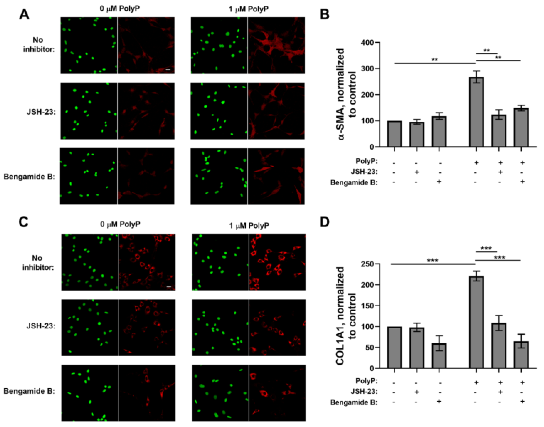
We previously reported that polyP induces myofibroblast differentiation in murine and human fibroblasts [23]. Since NFκB is known to promote myofibroblast differentiation and survival [39,40], we investigated whether polyP-induced differentiation of fibroblasts into myofibroblasts required the participation of NFκB. Accordingly, we cultured NIH-3T3 cells with polyP in the presence or absence of the NFκB inhibitors, JSH-23 [41] or Bengamide B [42], then assessed two markers of myofibroblast differentiation: α-SMA and COL1A1. Both of the NFκB inhibitors abrogated the polyP-induced increase in α-SMA (Figure 2A,B) and COL1A1 (Figure 2C,D) in these cells. This indicates that signal transduction via NFκB is essential for polyP to induce the differentiation of fibroblasts into the myofibroblast phenotype.

3.2. RAGE Mediates the Fibroblast Response to PolyP

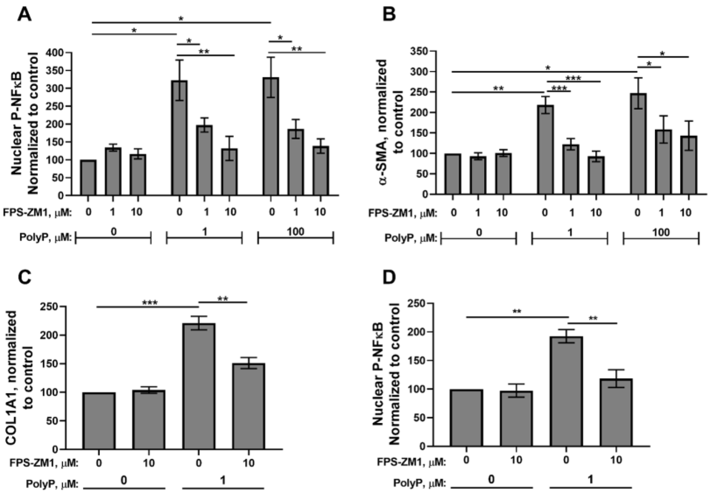
Although the receptors utilized by polyP for cellular signaling in mammalian systems are relatively little explored, recent studies have demonstrated that the purinergic receptor, P2Y1, and the receptor for advanced glycation end products (RAGE) can act as polyP cell surface receptors on endothelial, neuronal, and fibroblastic cells [23,43,44]. We previously reported that polyP-induced differentiation of fibroblasts into myofibroblasts was independent of P2Y1 signaling [23]. We therefore assessed whether polyP-induced myofibroblast differentiation was dependent on RAGE. We now report that the high-affinity RAGE antagonist, FPS-ZM1 [45], abrogated the nuclear translocation of phospho-NFκB in polyP-treated NIH-3T3 cells (Figure 3A). RAGE antagonism also inhibited the accumulation of two markers of myofibroblast differentiation, α-SMA and COL1A1, in NIH-3T3 cells (Figure 3B,C). Furthermore, RAGE inhibition also abrogated NFκB activation in primary human fibroblast cells (GM05387 cells; Figure 3D). These studies indicate that extracellular polyP signaling in fibroblasts is mediated by RAGE, leading to NFκB activation and myofibroblast differentiation.
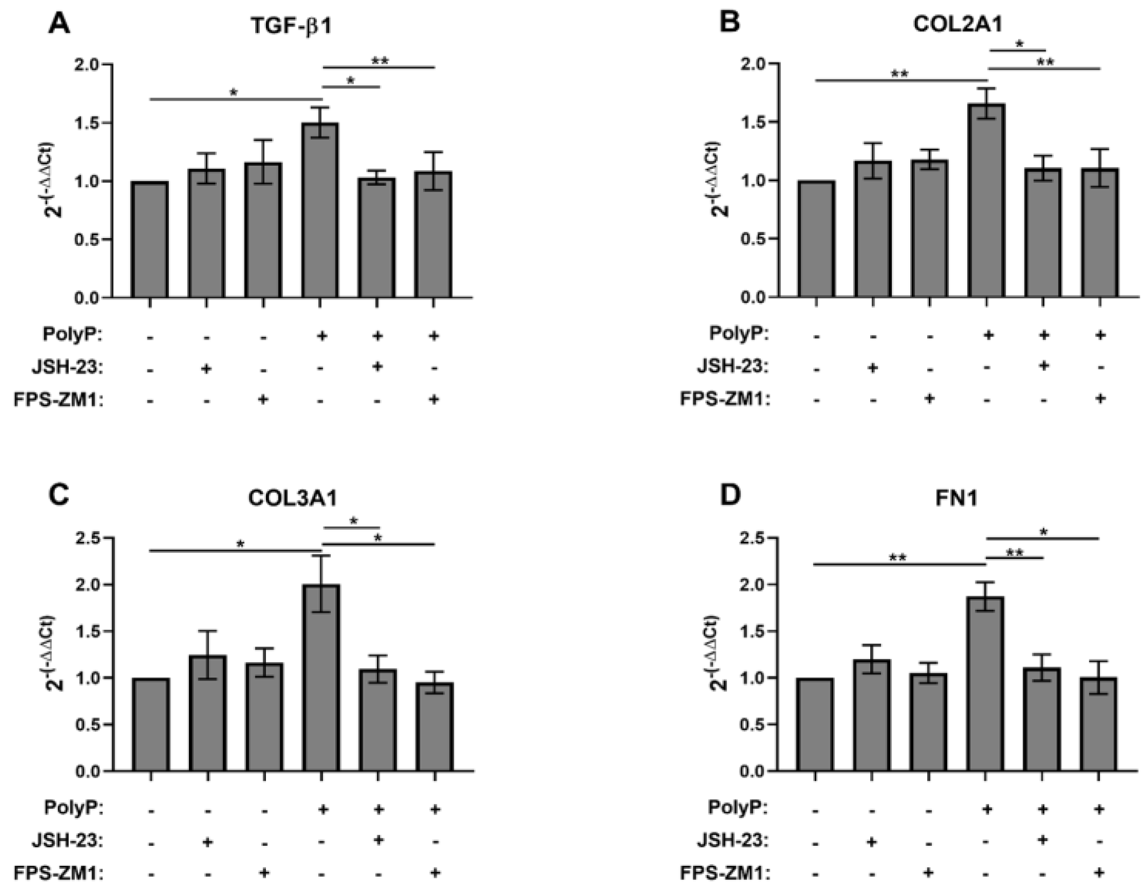
We next assessed transcript levels of additional myofibroblast markers induced by polyP using RT-qPCR with RNA from NIH-3T3 cells stimulated with platelet-sized polyP. We also examined the dependency of these transcripts on signaling via RAGE and NFκB. We found that 1 μM polyP increased the transcript levels of TGF-β1 (known to be a major driver of myofibroblast differentiation [46,47]) as well as the transcript levels of the matrix proteins, COL2A1, COL3A1, and FN1 (Figure 4A–D). Inhibitors of RAGE and NFκB abrogated the polyP-mediated induction of transcripts for all of these proteins (Figure 4A–D).
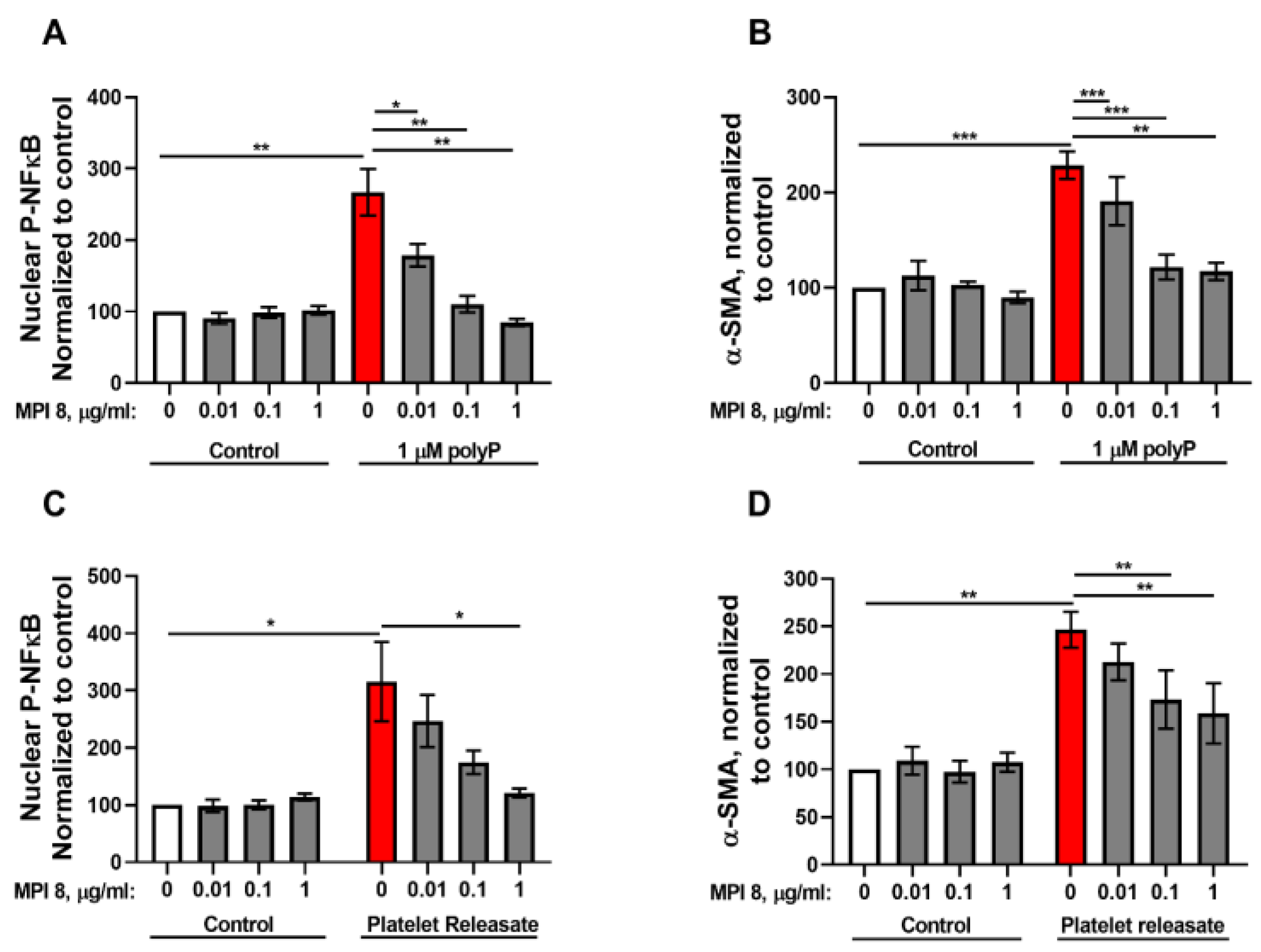
We previously reported the synthesis and characterization of a family of highly biocompatible, dendrimer-like cationic compounds, including agents that safely and potently reverse the anticoagulant activity of heparin [48,49,50], or block the prothrombotic activity of polyP [50,51,52], both in vitro and in vivo. Our newest-generation polyP inhibitor, MPI 8, is an especially potent polyP inhibitor while showing even better biocompatibility than our earlier-generation compounds [29]. In this study, we tested the ability of MPI 8 to abrogate myofibroblast differentiation of NIH-3T3 cells induced by platelet-sized polyP and found that 1 μg/mL MPI 8 drastically reduced NFκB activation and α-SMA accumulation in these cells (Figure 5A,B). We previously showed that releasates harvested from activated human platelets will induce differentiation of fibroblasts into myofibroblasts (visualized by α-SMA accumulation), and by using a polyP-degrading enzyme we further demonstrated that the polyP content of such releasates was the major activating component [23]. We now report that platelet releasates promoted NFκB activation in NIH-3T3 cells, and that this activity was completely abrogated by adding 1 μg/mL MPI 8 to the releasates (Figure 5C). Similarly, we found that adding 1 μg/mL MPI 8 to platelet releasates severely blunted their ability to induce α-SMA accumulation in NIH-3T3 cells (Figure 5D). These results suggest that polyP released from the dense granules of activated platelets induces NFκB signaling to promote myofibroblast differentiation, and that the use of MPI 8 is a unique way of blocking the action of polyP, even in complex mixtures like platelet releasates.

4. Discussion

Platelet-secreted polyP is an emerging cell signaling molecule with activity reported for endothelial cells, neutrophils, monocytes, macrophages, osteoblasts, neurons, astrocytes [15,17], and keratinocytes [19,20,21,43,44]. Despite these numerous findings, the signaling mechanisms utilized by polyP have not been extensively studied. Platelet polyP has previously been shown to activate NFκB in endothelial cells in a pathway mediated by the RAGE and P2Y1 receptors [43], while NFκB signaling has also been linked to myofibroblast differentiation [53]. Activation of RAGE by other ligands has also been shown to activate fibroblast proliferation and induce fibrosis [54,55]. Here, we show that polyP promotes myofibroblast differentiation of fibroblasts in a signaling pathway mediated by RAGE and NFκB. Platelet-sized polyP induced NIH-3T3 cell nuclear translocation of phospho-NFκB in a concentration range that matches polyP-induced myofibroblast differentiation [23]. Like polyP-induced myofibroblast differentiation, both short and long-chain polyP induce NFκB activation [23]. PolyP caused fibroblasts to increase the levels of protein and/or mRNA for α-SMA, TGF-β1, fibronectin and types I, II and III collagen, while inhibition of RAGE or NFκB signaling abrogated these effects of polyP on fibroblasts.
Chronic wounds such as diabetic ulcers represent a large burden to millions of patients and present financial burdens to the healthcare industry in the United States [26]. Platelets produce components that promote wound healing [56,57], and exogenously applied polyP has been shown to promote the wound healing process in vivo [18,21]. In our previous study we showed that it was the polyP component of platelet releasates that was essential for promoting the differentiation of fibroblasts into a myofibroblast phenotype [23], while in the present study we extended these observations to show that platelet releasates promote NFκB activation, revealing more of the molecular mechanism by which polyP and platelets modulate this process. We also show that the highly biocompatible, dendrimeric polyP inhibitor MPI 8 can effectively block the differentiation-inducing action of polyP in platelet releasates.
The mechanism by which polyP elicits signaling via RAGE in fibroblasts is not yet clear. A previous study by Dinarvand et al. [43] with cultured endothelial cells found that direct signaling of polyP via RAGE required very high polyP concentrations (50 μM), but when endothelial cells were incubated with polyP together with either HMGB1 or histone H4, the required polyP concentration dropped to just 2.5 μM (similar to the polyP concentrations we found to elicit RAGE-dependent signaling in fibroblasts). They also showed that polyP binds with high affinity to both HMGB1 and histone H4. Their findings indicated that it was likely that complexes of polyP with HMGB1 or histone H4 (which are known RAGE ligands) were much more effective at inducing signals than was polyP alone. On the other hand, Dinarvand et al. found that this signaling also required P2Y1 and provided evidence that polyP/HMBG1 complexes signaled in endothelial cells in a mechanism in which the polyP bridged between both RAGE and P2Y1. Not surprisingly, this required very long-chain polyP [43]. We previously showed that the ability of polyP to elicit differentiation of fibroblasts into myofibroblasts was independent of P2Y1, and furthermore that it was maximal at polyP polymer lengths equivalent to platelet-sized polyP [23]. Although the mechanism of polyP signaling through RAGE on fibroblasts appears to be different from that of endothelial cells, it nevertheless seems possible that the true ligand that is binding to RAGE in our studies could be a complex of polyP and another protein. Since our experiments were conducted in the presence of calf serum, it is possible that polyP could be activating RAGE via association with such a serum protein. It is also possible that polyP is interacting with a protein secreted from fibroblasts (such as TFG-β), amplifying an autocrine signaling mechanism. Examining these ideas is beyond the scope of the present study but should be addressed in future research.
Further investigation of polyP as a potential therapeutic for chronic wounds is warranted. While myofibroblasts are essential for the wound healing process by promoting wound closure, wound resolution, and scar tissue production, this cell type is also the primary effector cell in fibrotic diseases [58]. Platelet polyP also promotes differentiation in vitro of another pro-fibrotic cell type, the fibrocyte [20]. The possible role of platelet-derived polyP in various pathologies has yet to be investigated, which can largely be attributed to a lack of genetic means by which to inhibit polyP synthesis in mammalian organisms. MPI 8 may therefore be a useful tool with which to inhibit the effects of extracellular polyP in vivo, including examining the potential role of platelet-derived polyP in inflammation, thrombosis and fibrotic diseases [50,52]. Myelofibrosis in bone marrow is thought to be driven, at least in part, by megakaryocytes, the cells responsible for platelet production in the bone marrow [59,60]. Idiopathic pulmonary fibrosis patients have been shown to have increased platelet reactivity and activation attributed to an unknown plasma factor [61]. MPI 8 should allow for investigation of the role of polyP in vivo in such disease states.

5. Conclusions

This study shows that polyP promotes differentiation of fibroblasts in vitro into a myofibroblast phenotype via a signaling pathway mediated by RAGE and NFκB. It also shows that polyP induces fibroblasts to increase production of α-SMA, TGF-β1, fibronectin and types I, II and III collagen, also in a manner that requires RAGE and NFκB signaling. It also shows that a highly biocompatible polyP inhibitor (MPI 8) is an effective tool for blocking the differentiation-inducing action of polyP that is present in complex platelet releasates.

Author Contributions

P.M.S. designed and performed the research, collected data, analyzed, and interpreted the data, and wrote the paper. C.C.L. synthesized and characterized MPI 8. S.V. characterized MPI-8. J.N.K. synthesized MPI 8 and revised the paper. J.H.M. coordinated and conceived the study, acquired funding, interpreted the data, and revised the paper. All authors have read and agreed to the published version of the manuscript.

Funding

This work was supported by National Institutes of Health grants R35 HL135823, R35 HL171334, and T32 HL007622. JNK acknowledges fundings from Canadian Institutes of Health Research and Natural Sciences and Engineering Research Council of Canada. J.N.K. is a Tier 1 Canada Research Chair in Immunomodulating Materials and Immunotherapy. C.C.L acknowledges the NSERC CREATE NanoMat Program.

Institutional Review Board Statement

Not applicable.

Informed Consent Statement

Not applicable.

Data Availability Statement

The original contributions presented in this study are included in the article. Further inquiries can be directed to the corresponding author.

Acknowledgments

The infrastructure facility at the Centre for Blood Research is supported by the Canada Foundation for Innovation (CFI) and the British Columbia Knowledge Development Fund (BCKDF).

Conflicts of Interest

P.M.S., C.C.L., S.V., J.N.K., and J.H.M. are inventors on patents and patent applications for medical uses of polyP and polyP inhibitors. The laboratory of J.H.M. receives research funding from sales of polyP and polyP derivatives through Kerafast, Inc. The funders had no role in the design of the study; in the collection, analyses, or interpretation of data; in the writing of the manuscript; or in the decision to publish the results.

Abbreviations

α-SMAalpha-smooth muscle actin
BCSbovine calf serum
BSAbovine serum albumin
COL1A1alpha-1chain of type I collagen
COL2A1alpha-1 chain of type II collagen
COL3A1alpha-1 chain of type III collagen
DMEMDulbecco’s modified Eagle medium
FN1fibronectin
HEPES4-(2-hydroxyethyl)piperazine-1-ethanesulfonic acid
NFκBnuclear factor kappa B
polyPpolyphosphate
polyP75platelet-sized polyphosphate
polyP700long-chain polyphosphate
RAGEreceptor for advanced glycation end products (RAGE)
RPL13Aribosomal protein L13a
RT-qPCRreverse transcription quantitative polymerase chain reaction
TBS50 mmol/L Tris-HCl pH 7.5, 150 mmol/L NaCl
TGF-β1transforming growth factor beta 1

References

  1. Brown, M.R.; Kornberg, A. Inorganic polyphosphate in the origin and survival of species. Proc. Natl. Acad. Sci. USA 2004, 101, 16085–16087. [Google Scholar] [CrossRef] [PubMed]
  2. Rashid, M.H.; Rao, N.N.; Kornberg, A. Inorganic polyphosphate is required for motility of bacterial pathogens. J. Bacteriol. 2000, 182, 225–227. [Google Scholar] [CrossRef] [PubMed]
  3. Rashid, M.H.; Rumbaugh, K.; Passador, L.; Davies, D.G.; Hamood, A.N.; Iglewski, B.H.; Kornberg, A. Polyphosphate kinase is essential for biofilm development, quorum sensing, and virulence of Pseudomonas aeruginosa. Proc. Natl. Acad. Sci. USA 2000, 97, 9636–9641. [Google Scholar] [CrossRef] [PubMed]
  4. Zhang, H.; Gómez-García, M.R.; Brown, M.R.; Kornberg, A. Inorganic polyphosphate in Dictyostelium discoideum: Influence on development, sporulation, and predation. Proc. Natl. Acad. Sci. USA 2005, 102, 2731–2735. [Google Scholar] [CrossRef]
  5. Rao, N.N.; Gómez-García, M.R.; Kornberg, A. Inorganic polyphosphate: Essential for growth and survival. Annu. Rev. Biochem. 2009, 78, 605–647. [Google Scholar] [CrossRef]
  6. Nikel, P.I.; Chavarría, M.; Martínez-García, E.; Taylor, A.C.; de Lorenzo, V. Accumulation of inorganic polyphosphate enables stress endurance and catalytic vigour in Pseudomonas putida KT2440. Microb. Cell Fact. 2013, 12, 50. [Google Scholar] [CrossRef]
  7. Livermore, T.M.; Chubb, J.R.; Saiardi, A. Developmental accumulation of inorganic polyphosphate affects germination and energetic metabolism in Dictyostelium discoideum. Proc. Natl. Acad. Sci. USA 2016, 113, 996–1001. [Google Scholar] [CrossRef]
  8. Singh, M.; Tiwari, P.; Arora, G.; Agarwal, S.; Kidwai, S.; Singh, R. Establishing virulence associated polyphosphate kinase 2 as a drug target for Mycobacterium tuberculosis. Sci. Rep. 2016, 6, 26900. [Google Scholar] [CrossRef]
  9. Suess, P.M.; Gomer, R.H. Extracellular polyphosphate inhibits proliferation in an autocrine negative feedback loop in Dictyostelium discoideum. J. Biol. Chem. 2016, 291, 20260–20269. [Google Scholar] [CrossRef]
  10. Kulakovskaya, T. Inorganic polyphosphates and heavy metal resistance in microorganisms. World J. Microbiol. Biotechnol. 2018, 34, 139. [Google Scholar] [CrossRef]
  11. Ruiz, F.A.; Lea, C.R.; Oldfield, E.; Docampo, R. Human platelet dense granules contain polyphosphate and are similar to acidocalcisomes of bacteria and unicellular eukaryotes. J. Biol. Chem. 2004, 279, 44250–44257. [Google Scholar] [CrossRef]
  12. Moreno-Sanchez, D.; Hernandez-Ruiz, L.; Ruiz, F.A.; Docampo, R. Polyphosphate is a novel pro-inflammatory regulator of mast cells and is located in acidocalcisomes. J. Biol. Chem. 2012, 287, 28435–28444. [Google Scholar] [CrossRef]
  13. Arredondo, C.; Cefaliello, C.; Dyrda, A.; Jury, N.; Martinez, P.; Díaz, I.; Amaro, A.; Tran, H.; Morales, D.; Pertusa, M.; et al. Excessive release of inorganic polyphosphate by ALS/FTD astrocytes causes non-cell-autonomous toxicity to motoneurons. Neuron 2022, 110, 1656–1670.e12. [Google Scholar] [CrossRef]
  14. Smith, S.A.; Mutch, N.J.; Baskar, D.; Rohloff, P.; Docampo, R.; Morrissey, J.H. Polyphosphate modulates blood coagulation and fibrinolysis. Proc. Natl. Acad. Sci. USA 2006, 103, 903–908. [Google Scholar] [CrossRef]
  15. Usui, Y.; Uematsu, T.; Uchihashi, T.; Takahashi, M.; Takahashi, M.; Ishizuka, M.; Doto, R.; Tanaka, H.; Komazaki, Y.; Osawa, M.; et al. Inorganic polyphosphate induces osteoblastic differentiation. J. Dent. Res. 2010, 89, 504–509. [Google Scholar] [CrossRef]
  16. Hassanian, S.M.; Dinarvand, P.; Smith, S.A.; Rezaie, A.R. Inorganic polyphosphate elicits proinflammatory responses through activation of the mammalian target of rapamycin complexes 1 and 2 in vascular endothelial cells. J. Thromb. Haemost. 2015, 13, 860–871. [Google Scholar] [CrossRef] [PubMed]
  17. Chrysanthopoulou, A.; Kambas, K.; Stakos, D.; Mitroulis, I.; Mitsios, A.; Vidali, V.; Angelidou, I.; Bochenek, M.; Arelaki, S.; Arampatzioglou, A.; et al. Interferon lambda1/IL-29 and inorganic polyphosphate are novel regulators of neutrophil-driven thromboinflammation. J. Pathol. 2017, 243, 111–122. [Google Scholar] [CrossRef]
  18. Müller, W.E.G.; Relkovic, D.; Ackermann, M.; Wang, S.; Neufurth, M.; Paravic Radicevic, A.; Ushijima, H.; Schröder, H.C.; Wang, X. Enhancement of wound healing in normal and diabetic mice by topical application of amorphous polyphosphate. Superior effect of a host–guest composite material composed of collagen (host) and polyphosphate (guest). Polymers 2017, 9, 300. [Google Scholar] [CrossRef] [PubMed]
  19. Müller, W.E.G.; Ackermann, M.; Wang, S.; Neufurth, M.; Muñoz-Espí, R.; Feng, Q.; Schröder, H.C.; Wang, X. Inorganic polyphosphate induces accelerated tube formation of HUVEC endothelial cells. Cell. Mol. Life Sci. 2018, 75, 21–32. [Google Scholar] [CrossRef]
  20. Suess, P.M.; Chinea, L.E.; Pilling, D.; Gomer, R.H. Extracellular polyphosphate promotes macrophage and fibrocyte differentiation, inhibits leukocyte proliferation, and acts as a chemotactic agent for neutrophils. J. Immunol. 2019, 203, 493–499. [Google Scholar] [CrossRef] [PubMed]
  21. Carney, B.C.; Simbulan-Rosenthal, C.M.; Gaur, A.; Browne, B.J.; Moghe, M.; Crooke, E.; Moffatt, L.T.; Shupp, J.W.; Rosenthal, D.S. Inorganic polyphosphate in platelet rich plasma accelerates re-epithelialization in vitro and in vivo. Regen. Ther. 2020, 15, 138–148. [Google Scholar] [CrossRef] [PubMed]
  22. Rijal, R.; Cadena, L.A.; Smith, M.R.; Carr, J.F.; Gomer, R.H. Polyphosphate is an extracellular signal that can facilitate bacterial survival in eukaryotic cells. Proc. Natl. Acad. Sci. USA 2020, 117, 31923–31934. [Google Scholar] [CrossRef]
  23. Suess, P.M.; Smith, S.A.; Morrissey, J.H. Platelet polyphosphate induces fibroblast chemotaxis and myofibroblast differentiation. J. Thromb. Haemost. 2020, 18, 3043–3052. [Google Scholar] [CrossRef]
  24. Bochaton-Piallat, M.L.; Gabbiani, G.; Hinz, B. The myofibroblast in wound healing and fibrosis: Answered and unanswered questions. F1000Research 2016, 5, F1000 Faculty Rev-752. [Google Scholar] [CrossRef] [PubMed]
  25. Hinz, B. Myofibroblasts. Exp. Eye Res. 2016, 142, 56–70. [Google Scholar] [CrossRef]
  26. Powers, J.G.; Higham, C.; Broussard, K.; Phillips, T.J. Wound healing and treating wounds: Chronic wound care and management. J. Am. Acad. Dermatol. 2016, 74, 607–625, quiz 625–606. [Google Scholar] [CrossRef] [PubMed]
  27. Bowers, S.; Franco, E. Chronic wounds: Evaluation and management. Am. Fam. Physician 2020, 101, 159–166. [Google Scholar]
  28. Henderson, N.C.; Rieder, F.; Wynn, T.A. Fibrosis: From mechanisms to medicines. Nature 2020, 587, 555–566. [Google Scholar] [CrossRef]
  29. La, C.C.; Smith, S.A.; Vappala, S.; Adili, R.; Luke, C.E.; Abbina, S.; Luo, H.D.; Chafeeva, I.; Drayton, M.; Creagh, L.A.; et al. Smart thrombosis inhibitors without bleeding side effects via charge tunable ligand design. Nat. Commun. 2023, 14, 2177. [Google Scholar] [CrossRef]
  30. Smith, S.A.; Baker, C.J.; Gajsiewicz, J.M.; Morrissey, J.H. Silica particles contribute to the procoagulant activity of DNA and polyphosphate isolated using commercial kits. Blood 2017, 130, 88–91. [Google Scholar] [CrossRef]
  31. Smith, S.A.; Wang, Y.; Morrissey, J.H. DNA ladders can be used to size polyphosphate resolved by polyacrylamide gel electrophoresis. Electrophoresis 2018, 39, 2454–2459. [Google Scholar] [CrossRef]
  32. Schneider, C.A.; Rasband, W.S.; Eliceiri, K.W. NIH Image to ImageJ: 25 years of image analysis. Nat. Methods 2012, 9, 671–675. [Google Scholar] [CrossRef]
  33. Livak, K.J.; Schmittgen, T.D. Analysis of relative gene expression data using real-time quantitative PCR and the 2(-Delta Delta C(T)) Method. Methods 2001, 25, 402–408. [Google Scholar] [CrossRef]
  34. Bae, J.S.; Lee, W.; Rezaie, A.R. Polyphosphate elicits pro-inflammatory responses that are counteracted by activated protein C in both cellular and animal models. J. Thromb. Haemost. 2012, 10, 1145–1151. [Google Scholar] [CrossRef] [PubMed]
  35. Smith, S.A.; Choi, S.H.; Davis-Harrison, R.; Huyck, J.; Boettcher, J.; Rienstra, C.M.; Morrissey, J.H. Polyphosphate exerts differential effects on blood clotting, depending on polymer size. Blood 2010, 116, 4353–4359. [Google Scholar] [CrossRef]
  36. Wang, Y.; Ivanov, I.; Smith, S.A.; Gailani, D.; Morrissey, J.H. Polyphosphate, Zn2+ and high molecular weight kininogen modulate individual reactions of the contact pathway of blood clotting. J. Thromb. Haemost. 2019, 17, 2131–2140. [Google Scholar] [CrossRef] [PubMed]
  37. Jainchill, J.L.; Aaronson, S.A.; Todaro, G.J. Murine sarcoma and leukemia viruses: Assay using clonal lines of contact-inhibited mouse cells. J. Virol. 1969, 4, 549–553. [Google Scholar] [CrossRef]
  38. Hershberger, R.P.; Culp, L.A. Cell-type-specific expression of alternatively spliced human fibronectin IIICS mRNAs. Mol. Cell. Biol. 1990, 10, 662–671. [Google Scholar] [CrossRef] [PubMed]
  39. Watson, M.R.; Wallace, K.; Gieling, R.G.; Manas, D.M.; Jaffray, E.; Hay, R.T.; Mann, D.A.; Oakley, F. NF-kappaB is a critical regulator of the survival of rodent and human hepatic myofibroblasts. J. Hepatol. 2008, 48, 589–597. [Google Scholar] [CrossRef]
  40. Pan, H.W.; Cui, Y.H.; Zeng, J.W. NF-kappaB mediates the survival of corneal myofibroblast induced by angiotensin II. Investig. Ophthalmol. Vis. Sci. 2014, 55, 4220–4228. [Google Scholar] [CrossRef]
  41. Shin, H.M.; Kim, M.H.; Kim, B.H.; Jung, S.H.; Kim, Y.S.; Park, H.J.; Hong, J.T.; Min, K.R.; Kim, Y. Inhibitory action of novel aromatic diamine compound on lipopolysaccharide-induced nuclear translocation of NF-κB without affecting IκB degradation. FEBS Lett. 2004, 571, 50–54. [Google Scholar] [CrossRef]
  42. Johnson, T.A.; Sohn, J.; Vaske, Y.M.; White, K.N.; Cohen, T.L.; Vervoort, H.C.; Tenney, K.; Valeriote, F.A.; Bjeldanes, L.F.; Crews, P. Myxobacteria versus sponge-derived alkaloids: The bengamide family identified as potent immune modulating agents by scrutiny of LC-MS/ELSD libraries. Bioorg. Med. Chem. 2012, 20, 4348–4355. [Google Scholar] [CrossRef]
  43. Dinarvand, P.; Hassanian, S.M.; Qureshi, S.H.; Manithody, C.; Eissenberg, J.C.; Yang, L.; Rezaie, A.R. Polyphosphate amplifies proinflammatory responses of nuclear proteins through interaction with receptor for advanced glycation end products and P2Y1 purinergic receptor. Blood 2014, 123, 935–945. [Google Scholar] [CrossRef]
  44. Holmström, K.M.; Marina, N.; Baev, A.Y.; Wood, N.W.; Gourine, A.V.; Abramov, A.Y. Signalling properties of inorganic polyphosphate in the mammalian brain. Nat. Commun. 2013, 4, 1362. [Google Scholar] [CrossRef] [PubMed]
  45. Deane, R.; Singh, I.; Sagare, A.P.; Bell, R.D.; Ross, N.T.; LaRue, B.; Love, R.; Perry, S.; Paquette, N.; Deane, R.J.; et al. A multimodal RAGE-specific inhibitor reduces amyloid β-mediated brain disorder in a mouse model of Alzheimer disease. J. Clin. Investig. 2012, 122, 1377–1392. [Google Scholar] [CrossRef] [PubMed]
  46. Tarbit, E.; Singh, I.; Peart, J.N.; Rose’Meyer, R.B. Biomarkers for the identification of cardiac fibroblast and myofibroblast cells. Heart Fail. Rev. 2019, 24, 1–15. [Google Scholar] [CrossRef]
  47. Vallée, A.; Lecarpentier, Y. TGF-beta in fibrosis by acting as a conductor for contractile properties of myofibroblasts. Cell Biosci. 2019, 9, 98. [Google Scholar] [CrossRef] [PubMed]
  48. Shenoi, R.A.; Kalathottukaren, M.T.; Travers, R.J.; Lai, B.F.L.; Creagh, A.L.; Lange, D.; Yu, K.; Weinhart, M.; Chew, B.H.; Du, C.; et al. Affinity-based design of a synthetic universal reversal agent for heparin anticoagulants. Sci. Transl. Med. 2014, 6, 260ra150. [Google Scholar] [CrossRef]
  49. Kalathottukaren, M.T.; Creagh, A.L.; Abbina, S.; Lu, G.; Karbarz, M.J.; Pandey, A.; Conley, P.B.; Kizhakkedathu, J.N.; Haynes, C. Comparison of reversal activity and mechanism of action of UHRA, andexanet, and PER977 on heparin and oral FXa inhibitors. Blood Adv. 2018, 2, 2104–2114. [Google Scholar] [CrossRef]
  50. Kalathottukaren, M.T.; Abraham, L.; Kapopara, P.R.; Lai, B.F.; Shenoi, R.A.; Rosell, F.I.; Conway, E.M.; Pryzdial, E.L.; Morrissey, J.H.; Haynes, C.A.; et al. Alteration of blood clotting and lung damage by protamine are avoided using the heparin and polyphosphate inhibitor UHRA. Blood 2017, 129, 1368–1379. [Google Scholar] [CrossRef]
  51. Mafi, A.; Abbina, S.; Kalathottukaren, M.T.; Morrissey, J.H.; Haynes, C.; Kizhakkedathu, J.N.; Pfaendtner, J.; Chou, K.C. Design of polyphosphate inhibitors: A molecular dynamics investigation on polyethylene glycol-linked cationic binding groups. Biomacromolecules 2018, 19, 1358–1367. [Google Scholar] [CrossRef]
  52. Travers, R.J.; Shenoi, R.A.; Kalathottukaren, M.T.; Kizhakkedathu, J.N.; Morrissey, J.H. Nontoxic polyphosphate inhibitors reduce thrombosis while sparing hemostasis. Blood 2014, 124, 3183–3190. [Google Scholar] [CrossRef]
  53. Xu, A.; Deng, F.; Chen, Y.; Kong, Y.; Pan, L.; Liao, Q.; Rao, Z.; Xie, L.; Yao, C.; Li, S.; et al. NF-κB pathway activation during endothelial-to-mesenchymal transition in a rat model of doxorubicin-induced cardiotoxicity. Biomed. Pharmacother. 2020, 130, 110525. [Google Scholar] [CrossRef] [PubMed]
  54. Zhao, L.M.; Zhang, W.; Wang, L.P.; Li, G.R.; Deng, X.L. Advanced glycation end products promote proliferation of cardiac fibroblasts by upregulation of KCa3.1 channels. Pflügers Arch. 2012, 464, 613–621. [Google Scholar] [CrossRef]
  55. Liang, B.; Zhou, Z.; Yang, Z.; Liu, J.; Zhang, L.; He, J.; Li, H.; Huang, Y.; Yang, Q.; Xian, S.; et al. AGEs-RAGE axis mediates myocardial fibrosis via activation of cardiac fibroblasts induced by autophagy in heart failure. Exp. Physiol. 2022, 107, 879–891. [Google Scholar] [CrossRef] [PubMed]
  56. Golebiewska, E.M.; Poole, A.W. Platelet secretion: From haemostasis to wound healing and beyond. Blood Rev. 2014, 29, 153–162. [Google Scholar] [CrossRef] [PubMed]
  57. Nurden, A.T.; Nurden, P.; Sanchez, M.; Andia, I.; Anitua, E. Platelets and wound healing. Front. Biosci. 2008, 13, 3532–3548. [Google Scholar] [CrossRef]
  58. Darby, I.A.; Zakuan, N.; Billet, F.; Desmouliere, A. The myofibroblast, a key cell in normal and pathological tissue repair. Cell. Mol. Life Sci. 2016, 73, 1145–1157. [Google Scholar] [CrossRef]
  59. Malara, A.; Abbonante, V.; Zingariello, M.; Migliaccio, A.; Balduini, A. Megakaryocyte contribution to bone marrow fibrosis: Many arrows in the quiver. Mediterr. J. Hematol. Infect. Dis. 2018, 10, e2018068. [Google Scholar] [CrossRef]
  60. Papadantonakis, N.; Matsuura, S.; Ravid, K. Megakaryocyte pathology and bone marrow fibrosis: The lysyl oxidase connection. Blood 2012, 120, 1774–1781. [Google Scholar] [CrossRef]
  61. Crooks, M.G.; Fahim, A.; Naseem, K.M.; Morice, A.H.; Hart, S.P. Increased platelet reactivity in idiopathic pulmonary fibrosis is mediated by a plasma factor. PLoS ONE 2014, 9, e111347. [Google Scholar] [CrossRef] [PubMed]
Figure 1. PolyP induces nuclear translocation of phospho-NFκB in fibroblasts. (A) Representative confocal microscopy images of NIH-3T3 cells incubated 1 h without (Control) or with 1 μM polyP75, then stained for DNA (green) and phospho-NFκB (red); bar = 20 µm. (B) Integrated intensity of phospho-NFκB (P-NFκB) staining in NIH-3T3 cell nuclei as a function of polyP75 concentration (after 1 h incubation), normalized to cells not exposed to polyP (with the normalized signal set at 100). (C) Normalized intensity of nuclear phospho-NFκB staining in NIH-3T3 cells exposed for 1 h to the indicated concentrations of polyP75 or polyP700. (D) Normalized intensity of nuclear phospho-NFκB staining in GM05387 cells exposed for 1 h to the indicated concentrations of polyP75. Nuclear antibody staining for phospho-NFκB was quantified by confocal microscopy, and all values are mean ± standard error of the mean (SEM; n = 5). Asterisks highlight statistical significance relative to the no-polyP controls: * p < 0.05, ** p < 0.01, *** p < 0.001 (paired t-tests).
Biomolecules 15 01441 g001
Figure 2. NFκB inhibitors block myofibroblast differentiation induced by polyP in NIH 3T3 cells. NIH-3T3 cells were incubated for 48 h in 1.5% BCS without or with 1 µM polyP75, in the presence or absence of the NFκB inhibitors, JSH-23 (10 µM) or Bengamide B (100 nM), after which antibody staining was performed. (A) Representative images after staining for DNA (green) and α-SMA (red); bar = 20 µm. (B) Normalized staining intensity for α-SMA in cells incubated without or with polyP75, in the presence or absence of JSH-23 or bengamide B, as indicated. (C) Representative images after staining for DNA (green) and COL1A1 (red); bar = 20 µm. (D) Normalized staining intensity for COL1A1 in cells incubated without or with polyP75, in the presence or absence of JSH-23 or bengamide B, as indicated. All values are mean ± SEM (n = 5). Asterisks highlight statistical significance of the indicated comparisons: ** p < 0.01, *** p < 0.001 (paired t-tests).
Biomolecules 15 01441 g002
Figure 3. RAGE inhibitors diminish polyP-induced differentiation of fibroblasts into a myofibroblast phenotype, and reduce phospho-NFκB accumulation in their nuclei. (A) NIH-3T3 cells were incubated for 1 h in 1.5% BCS with the indicated concentration of polyP75 and the RAGE antagonist, FPS-ZM1, then stained for phospho-NFκB, after which nuclear antibody staining for phospho-NFκB was quantified. (B,C) NIH-3T3 cells were incubated for 48 h in 1.5% BCS with the indicated concentrations of polyP75 and FPS-ZM1, then stained for the indicated antigens, quantified using confocal microscopy. (D) GM05387 cells were incubated for 1 h in 1.5% BCS with the indicated concentrations of polyP75 and FPS-ZM1, after which nuclear antibody staining for phospho-NFκB was quantified. All values are mean ± SEM (n = 5). Asterisks highlight statistical significance of the indicated comparisons: * p < 0.05, ** p < 0.01, *** p < 0.001 (paired t-tests).
Biomolecules 15 01441 g003
Figure 4. Effect of RAGE and NFκB inhibitors on polyP-induced transcription of genes for TGF-β1and the matrix proteins, COL2A1, COL3A1, and FN1. NIH-3T3 cells were incubated in 1.5% BCS with 1 µM polyP75 in the presence or absence of 10 µM JSH-23 or 1 µM FPS-ZM1 for 1 h and RNA was extracted and used as a template for RT-qPCR for the indicated genes (AD). All values are mean ± SEM (n = 5). Asterisks highlight statistical significance of the indicated comparisons: * p < 0.05, ** p < 0.01 (paired t-tests).
Biomolecules 15 01441 g004
Figure 5. The polyP inhibitor, MPI 8, abrogates fibroblast differentiation and phospho-NFκB nuclear translocation induced by synthetic polyP or by platelet releasates. (A,B) NIH-3T3 cells were incubated in 1.5% serum with 1 µM polyP75 in the presence or absence of the indicated concentration of MPI 8. (A) After one hour, nuclear antibody staining for phospho-NFκB was quantified using confocal microscopy. (B) After 48 h antibody staining for α-SMA was quantified using confocal microscopy. (C,D) NIH-3T3 cells were incubated in 1.5% serum with releasates from activated human platelets (diluted to 15 μg/mL protein) in the presence or absence of the indicated concentration of MPI 8. (C) After one hour, nuclear antibody staining for phospho-NFκB was quantified using confocal microscopy. (D) After 48 h antibody staining for α-SMA was quantified using confocal microscopy. All values are mean ± SEM (n = 5). Asterisks highlight statistical significance of the indicated comparisons: * p < 0.05, ** p < 0.01, *** p <0.001 (paired t-tests).
Biomolecules 15 01441 g005
Disclaimer/Publisher’s Note: The statements, opinions and data contained in all publications are solely those of the individual author(s) and contributor(s) and not of MDPI and/or the editor(s). MDPI and/or the editor(s) disclaim responsibility for any injury to people or property resulting from any ideas, methods, instructions or products referred to in the content.