Article

Menthol Pretreatment Alleviates Campylobacter jejuni-Induced Enterocolitis in Human Gut Microbiota-Associated IL-10−/− Mice

Gastrointestinal Microbiology Research Group, Institute of Microbiology, Infectious Diseases and Immunology, Charité-Universitätsmedizin Berlin, Corporate Member of Freie Universität Berlin, Humboldt-Universität zu Berlin, and Berlin Institute of Health, 12203 Berlin, Germany
*
Author to whom correspondence should be addressed.
Biomolecules 2024, 14(3), 290; https://doi.org/10.3390/biom14030290
Submission received: 23 January 2024 / Revised: 21 February 2024 / Accepted: 28 February 2024 / Published: 29 February 2024
(This article belongs to the Special Issue The Value of Natural Compounds as Therapeutic Agents II)

Abstract

:
Human Campylobacter jejuni infections are of worldwide importance and represent the most commonly reported bacterial enteritis cases in middle- and high-income countries. Since antibiotics are usually not indicated and the severity of campylobacteriosis is directly linked to the risk of developing post-infectious complications, non-toxic antibiotic-independent treatment approaches are highly desirable. Given its health-promoting properties, including anti-microbial and anti-inflammatory activities, we tested the disease-alleviating effects of oral menthol in murine campylobacteriosis. Therefore, human gut microbiota-associated IL-10−/− mice were orally subjected to synthetic menthol starting a week before C. jejuni infection and followed up until day 6 post-infection. Whereas menthol pretreatment did not improve campylobacteriosis symptoms, it resulted in reduced colonic C. jejuni numbers and alleviated both macroscopic and microscopic aspects of C. jejuni infection in pretreated mice vs. controls. Menthol pretreatment dampened the recruitment of macrophages, monocytes, and T lymphocytes to colonic sites of infection, which was accompanied by mitigated intestinal nitric oxide secretion. Furthermore, menthol pretreatment had only marginal effects on the human fecal gut microbiota composition during the C. jejuni infection. In conclusion, the results of this preclinical placebo-controlled intervention study provide evidence that menthol application constitutes a promising way to tackle acute campylobacteriosis, thereby reducing the risk for post-infectious complications.

1. Introduction

Campylobacter are microaerophilic and rod-shaped Gram-negative bacilli that typically reside as commensal members of the intestinal tract in warm-blooded vertebrates [1]. Frequently colonized with Campylobacter jejuni, birds, in particular chickens and turkeys, rarely display clinical signs [2,3,4]. During the livestock slaughter process, the derived meat products might become contaminated with bacteria [5]. Upon human consumption of undercooked or even raw contaminated meat products, unpasteurized milk or surface water, C. jejuni infection can occur, leading to acute campylobacteriosis [6,7]. After a latency of 2 to 5 days, infected individual presents symptoms of varying severity, including general malaise, tenesmus, nausea, vomiting, and fever accompanied by enteritis with watery or bloody diarrhea accompanied by mucous discharge [8,9]. The severity of the C. jejuni-induced disease depends on both the arsenal of virulence factors of the enteropathogen and the immune competence of infected individuals [10,11]. Following successful passage of the stomach and duodenum, the motile C. jejuni traverse the viscous mucous barrier, reach the subepithelial layers by invasion of epithelial cells, and initiate macrophage activation, resulting in the recruitment of monocytes, neutrophilic granulocytes, and leukocytes to the infected sites [12,13]. Furthermore, the endotoxin lipo-oligosacchride (LOS) leads to a Toll-like receptor-4 (TLR-4)-dependent overstimulation of the host immune system [11]. Consequently, pro-inflammatory mediators aim to limit the infection but cause damage to intestinal tissues and induce even systemic complications [11]. The oxidative stress results in gut epithelial cell apoptosis, compromised epithelial barrier function, malabsorption and diarrhea with electrolyte loss (“leaky gut”) [14,15,16]. Typically, the treatment of symptomatic C. jejuni-infected individuals involves non-steroidal antiphlogistic compounds and the substitution of fluids and electrolytes [9]. In rather critical cases, particularly in patients with immune-compromising co-morbidities, however, antibiotics such as macrolides or fluoroquinolones may be prescribed [9,17]. Generally, enteritis symptoms completely resolve within two weeks post-infection (p.i.). However, on rare occasions, autoimmune disorders might manifest with a latency of a few weeks to months p.i. [18]. These post-infectious complications of C. jejuni infection can affect the joints (reactive arthritis), the intestines (irritable bowel syndrome, inflammatory bowel diseases), or the nervous system (Guillain–Barré syndrome) [19,20,21] and are known to depend on the sialylation status of the C. jejuni-LOS [18,22]. Notably, there is a direct correlation between the risk of developing such autoimmune diseases and the severity of the preceding enteritis [22]. In addition, given the rising prevalence of multi-drug-resistant C. jejuni strains, which limits effective antibacterial treatment of severely ill patients [17,23], it is important to identify non-toxic compounds with potential anti-C. jejuni and anti-inflammatory properties that alleviate the disease and, in turn, reduce the risk of post-infectious diseases. This urgency is even further emphasized by the increasing global incidence of campylobacteriosis cases, which represents the most commonly reported cause of bacterial enteritis, with unreported cases far exceeding the reported ones [24]. In 2021, more than 127,000 campylobacteriosis cases were confirmed in the European Union alone, for instance [25], and the associated expenses for society, including the economies and health care systems, have been shown to be significant [26,27,28].
Menthol, a monoterpene with a high abundance of essential oils originating from the peppermint plant Mentha piperita L. (Lamiaceae), is widely employed as a flavoring agent and a remedy in traditional folk medicine due to its various health-promoting properties [29]. In fact, menthol has been shown to exhibit potent anti-microbial, anti-inflammatory, anti-oxidant, and anti-apoptotic effects [30,31,32,33,34], positioning the compound as a promising candidate for combating C. jejuni-induced morbidities.
Therefore, we tested the potential of prophylactic menthol treatment in experimental campylobacteriosis, focusing on its pathogen-reducing, immunomodulatory and consequent disease-alleviating effects. To address this, we utilized human microbiota-associated (hma) IL-10−/− mice. Given that the commensal gut microbiota of murine (as opposed to human) origin protects against C. jejuni infection [35], we initially depleted the murine gut microbiota by antibiotic treatment and subsequently introduced a complex human gut microbiota instead by fecal microbiota transplantation (FMT). Interestingly, mice demonstrate higher resistance to bacterial TLR-4 ligands, including lipo-polysaccharide (LPS) and LOS [36]; however, the il10 gene deficiency, encoding the anti-inflammatory cytokine IL-10, can overcome this LPS and LOS resistance [37]. In fact, microbiota-depleted wildtype mice were shown not to develop overt C. jejuni-induced enteritis [35]. In contrast, within less than a week, both microbiota-depleted and hma IL-10−/− mice succumbed to C. jejuni-induced disease affecting not only the intestinal tract but also extra-intestinal and systemic organs [38,39]. Hence, the hma IL-10−/− mouse model serves as a valuable experimental tool to explore the effects of the compound of interest on the interactions among (i) the enteropathogen (i.e., C. jejuni), (ii) the human gut microbial communities, and (iii) the vertebrate host immunity during acute campylobacteriosis [39]. This was the major driver behind subjecting hma IL-10−/− mice to peroral menthol treatment starting a week before C. jejuni infections to monitor intestinal pathogen burdens, clinical, macroscopic, and microscopic complications of infection, as well as pro-inflammatory immune responses in the intestinal tract and beyond.

2. Materials and Methods

2.1. Mice, Microbiota Depletion

In the same unit of the Forschungseinrichtungen für Experimentelle Medizin (FEM, Charité-University Medicine Berlin), IL-10−/− mice (of the C57BL/6 background) were bred under specific pathogen-free (SPF) and standardized conditions (i.e., 22–24 °C room temperature, 55 ± 15% humidity, and a 12 h light/12 dark cycle). In an experimental semi-barrier, mice were housed in cages with filter tops covering them and were allowed unrestricted access to autoclaved standard chow (food pellets: sniff R/M-H, V1534-300, Sniff, Soest, Germany). After placing 3-week-old littermate animals in sterile cages (two to four mice per cage), ampicillin and sulbactam (2 g/L and 1 g/L, respectively; Dr. Friedrich Eberth Arzneimittel, Ursensollen, Germany) were administered via the drinking water for eight weeks (ad libitum) as stated earlier [38,39]. The mice were handled under rigorous aseptic conditions to prevent microbial contamination and ensure successful microbiota depletion. Two days before human FMT (day-16; Figure 1A), the antibiotics were replaced with sterile tap water (ad libitum).

2.2. Human Fecal Microbiota Transplantation

Fresh fecal samples were voluntarily donated by healthy subjects, suspended in sterile phosphate-buffered saline (PBS; Thermo Fisher Scientific, Waltham, MA, USA), aliquoted, and preserved at −80 °C. Prior to the human FMT, fecal aliquots were thawed and pooled immediately. To establish hma mice, human fecal microbiota transplantation (hFMT) was performed over three consecutive days (days-14, -13, and -12; Figure 1A). Therefore, mice were orally subjected to 0.3 mL of the human fecal donor suspension via orogastric gavage, as outlined earlier [35].

2.3. Prophylactic Regimen

Commencing seven days prior to C. jejuni infection (day-7; Figure 1A), mice were treated with synthetic menthol (purchased from Sigma-Alderich, München, Germany). Menthol was administered to mice by autoclaved tap water (ad libitum). The final menthol concentration in the drinking solution was 500 mg/L, which meant that 100 mg of treatment dosage per kilogram of body weight was applied each day. Placebo-control mice received autoclaved tap water.

2.4. Campylobacter jejuni Infection

Bacterial cryo stocks of C. jejuni strain 81–176 were preserved at −80 °C. Following thawing, the bacteria were streaked out and cultured on selective karmali agar plates (obtained from Oxoid, Wesel, Germany) under microaerophilic conditions for 48 h at 37 °C, as previously reported [35]. The bacteria were then resuspended in sterile PBS (Thermo Fisher Scientific, Waltham, MA, USA), and the hma IL-10−/− mice (female and male 3-month-old littermates) were infected with 109 colony-forming units (CFU) of the enteropathogen on days 0 and 1 via orogastric gavage (Figure 1A).

2.5. Summary of Mouse Cohorts

Naive group (6 female/6 male): human FMT, non-treated, non-infected;
Placebo group (9 female/8 male): human FMT, non-treated, infected;
Menthol group (11 female/10 male): human FMT, verum-treated, infected.

2.6. Measurement of Gastrointestinal C. jejuni Loads

To assess gastrointestinal pathogen levels, the counts of live C. jejuni bacteria were monitored daily in fecal samples and additionally, upon necropsy in intraluminal gastrointestinal biopsies taken from the stomach, duodenum, ileum, and colon lumen that were homogenized in sterile PBS (Thermo Fisher Scientific, Waltham, MA, USA). C. jejuni quantification involved counting CFU after cultivating serial dilutions of intestinal samples on karmali agar for minimum 48 h at 37 °C under microaerophilic conditions as previously described in details [35]. The detection limit for viable pathogens was in the range of 100 CFU of C. jejuni bacteria per gram fecal material.

2.7. Fecal Microbiota Composition

For the culture-independent molecular analysis of the fecal microbiota composition, genomic DNA was isolated from the human fecal transplants and the colonic luminal murine samples first. In a nutshell, the Quant-iT PicoGreen reagent (Invitrogen, Paisley, UK) was used to quantify the DNA content, which was then adjusted to 1 ng of DNA per µL. Using species-, genera-, or group-specific 16S rRNA gene primers (Tib MolBiol, Berlin, Germany), the most prominent bacterial groups that were abundant in the intestinal microbiota of the hma mice were evaluated using quantitative real-time polymerase chain reaction (qRT-PCR), as previously described [40].

2.8. Clinical Conditions of Mice

We used a standardized cumulative clinical score on a daily basis to record the clinical status of mice before and after enteropathogen application as reported earlier [41] (Supplementary Table S1).

2.9. Sampling Procedures

Following C. jejuni infection, mice were sacrificed on day 6 p.i. by inhalation of carbon dioxide. Luminal gastrointestinal samples were obtained under sterile conditions from the stomach, duodenum, ileum, and colon, in addition to ex vivo biopsies from the mesenteric lymph nodes (MLN) and the distal colon, for subsequent microbiological, immunohistopathological, and immunological measurements. Cardiac blood was taken to generate serum samples.

2.10. Histopathology

Colon explants were promptly fixed in 5% formalin, embedded in paraffin, and 5-µm-sections were subjected to hematoxylin and eosin (H&E) staining. To evaluate the severity of histological alterations of the colonic mucosa, the tissue samples were assessed using light microscopy (100-times magnification), and the histopathological sequelae of infection were quantified based on an established scoring system [42] (Supplementary Table S2).

2.11. Quantitative In Situ Immunohistochemistry

As previously outlined in detail [43], quantitative in situ immunohistochemistry investigations were conducted on large intestinal explants that had been promptly subjected to tissue fixation in 5% formalin paraffin embedding. Subsequently, 5-µm-sections of paraffin-embedded tissues were stained with distinct primary antibodies (Supplementary Table S3). A blinded and independent researcher used light microscopy (400-times magnification) to enumerate the positively stained cells. For each sample, the mean count of specifically stained cells was determined across a minimum of six high-power fields (HPFs, 0.287 mm2, 400-times magnification).

2.12. Pro-Inflammatory Mediators

Terminal ileal and colonic explants (approximately 1 cm2 each) were longitudinally cut and the tissues cleansed in sterile PBS (Thermo Fisher Scientific, Waltham, MA, USA). Then, in addition to the intestinal samples, ex vivo biopsies derived from the MLN (3 nodes) were transferred to 24-flat-bottom well culture plates (Thermo Fisher Scientific, Waltham, MA, USA) containing 500 µL serum-free RPMI 1640 medium (Thermo Fisher Scientific, Waltham, MA, USA), penicillin (100 µg/mL; Biochrom, Berlin, Germany) and streptomycin (100 µg/mL; Biochrom, Berlin, Germany). Following an 18 h incubation period at 37 °C, culture supernatants were tested for monocyte chemoattractant protein-1 (MCP-1) by the Mouse Inflammation Cytometric Bead Assay (BD Biosciences, Heidelberg, Germany) in a BD FACSCanto II flow cytometer (BD Biosciences). Nitric oxide (NO) levels were determined by the Griess reaction [44].

2.13. Statistics

GraphPad Prism (version 9; San Diego, CA, USA) was used to compute medians and significance levels following the pooling of data from three independent experiments. The normal distribution of the data was evaluated using the Anderson-Darling test. Pairwise comparisons of regularly distributed and non-normally distributed data were performed using the Student’s t-test and the Mann–Whitney test, respectively. The one-way ANOVA with Tukey post hoc test (for regularly distributed data) and the Kruskal–Wallis test with Dunn’s post hoc test (for non-normally distributed data) were used for multiple comparisons. Significant two-sided probability (p) values were defined as <0.05. Significant outliers were determined by using the Grubb’s test (α = 0.001).

3. Results

3.1. Clinical Conditions over Time following C. jejuni Infection of hma IL-10−/− Mice with Menthol Pretreatment

First, we tested whether menthol pretreatment would improve the clinical conditions in hma IL10−/− mice following C. jejuni infection. Our daily assessments revealed that mice from the menthol and placebo cohorts presented with comparable campylobacteriosis-related conditions at individual days p.i. (not significant (n.s.); Figure 2). This was also the case when focusing on clinical scores specifying wasting symptoms, diarrhea, and fecal flood (n.s.; Supplementary Figure S1). Notably, the standard deviations of the respective clinical scores were very high within both infected groups. Hence, menthol pretreatment did not alleviate campylobacteriosis symptoms.

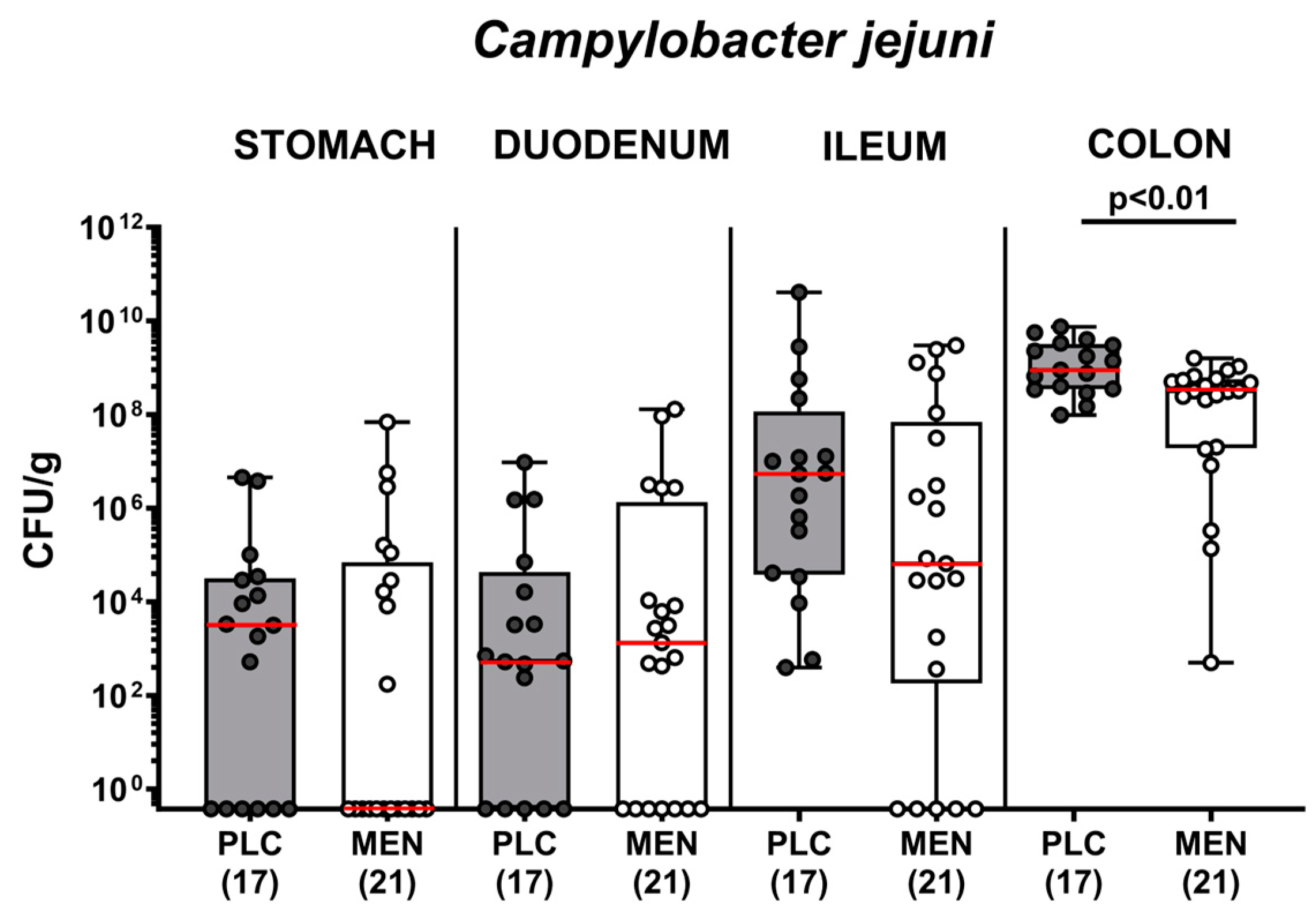
3.2. Pathogen Numbers in Gastrointestinal Luminal Samples from C. jejuni Infected hma IL-10−/− Mice with Menthol Pretreatment

Next, we addressed whether oral menthol would interfere with intestinal C. jejuni colonization. Results from daily cultural analyses indicated comparably high C. jejuni numbers in fecal samples over time following infection of mice from both cohorts (n.s.; Supplementary Figure S2). On the day of necropsy (i.e., day 6 p.i.), luminal C. jejuni loads did not differ in the stomach, duodenum, and ileum of menthol and placebo-pretreated mice (n.s.), whereas less than one order of magnitude lower median pathogen numbers were detected in the colonic lumen of the former vs. the latter (p < 0.01; Figure 3). Hence, menthol pretreatment slightly but significantly lowered colonic C. jejuni numbers.

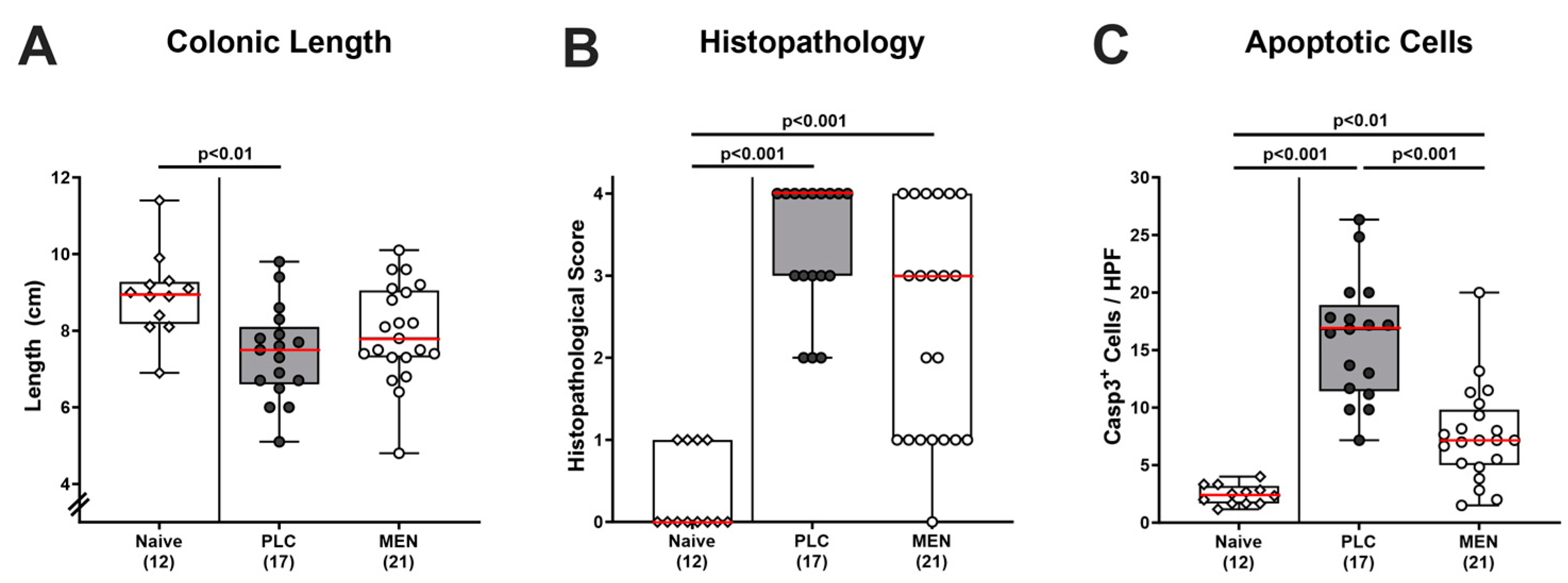
3.3. Inflammatory Changes in the Colon of C. jejuni Infected hma IL-10−/− Mice with Menthol Pretreatment

Further, we tested whether menthol pretreatment could mitigate C. jejuni-induced macroscopic and microscopic inflammatory changes. As a macroscopic inflammatory parameter, we recorded the absolute lengths of the large intestines upon necropsy. On day 6 p.i., we found shorter colons in mice from the placebo (p < 0.01) as opposed to the menthol cohort (n.s.) when compared to naive (i.e., non-treated and non-infected) control animals (Figure 4A), indicative of mitigated macroscopic inflammatory signs of C. jejuni-infection. When addressing the severity of microscopic inflammatory sequelae in the colon, we revealed comparable histopathological scores in mice from both treatment cohorts but observed a trend towards lower values in the menthol group as compared to the placebo group that did not reach statistical significance due to high standard deviations in the former (Figure 4B). We further investigated the extent of C. jejuni-induced intestinal apoptosis and found increased numbers of apoptotic cells in colonic epithelia in both infected groups (p < 0.01–0.001), but considerably fewer apoptotic cell counts in menthol as compared to placebo-treated mice on day 6 p.i. (p < 0.001; Figure 4C). Hence, menthol pretreatment alleviated both macroscopic and microscopic complications of the C. jejuni infection.

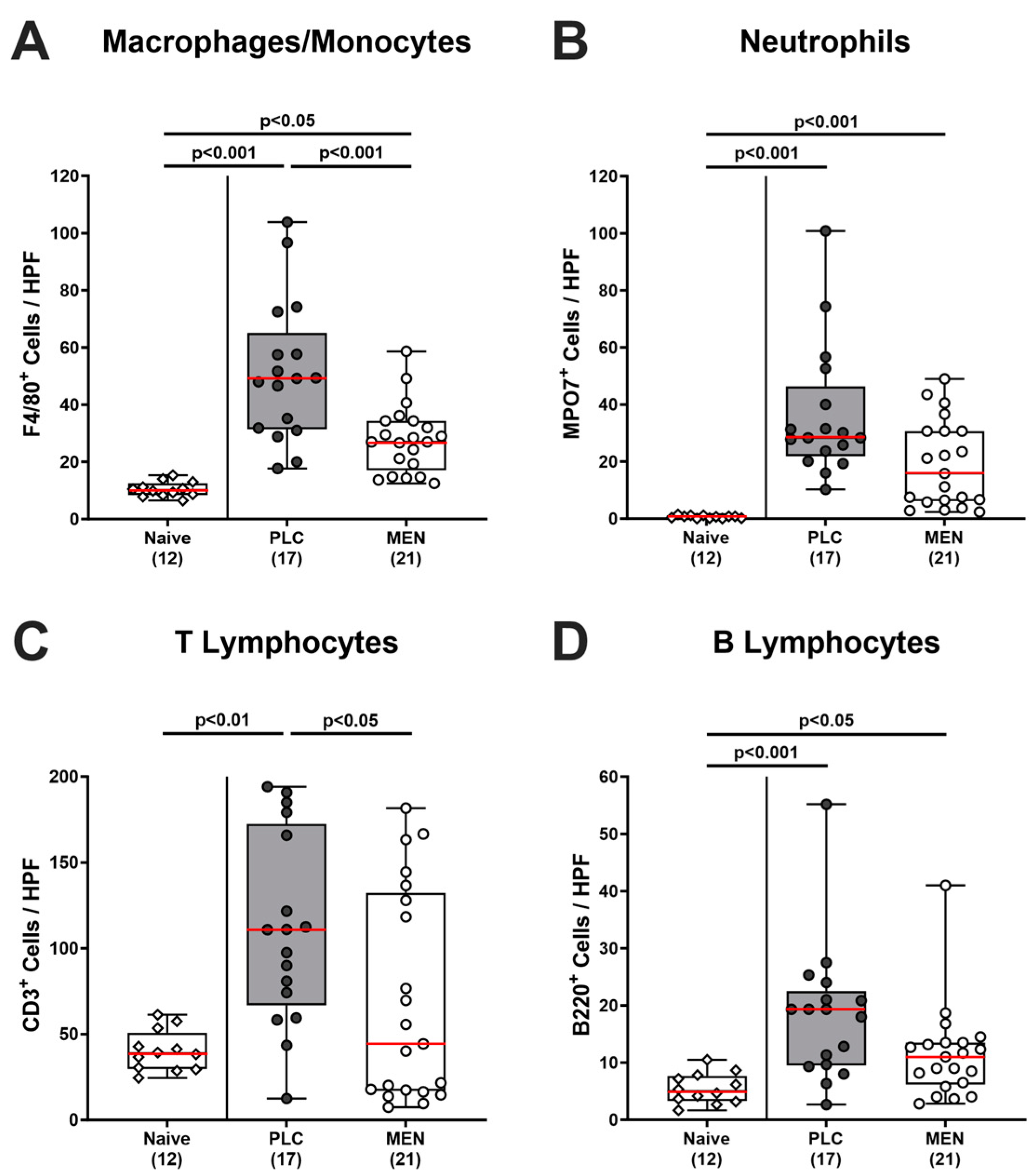
3.4. Colonic Immune Cells in C. jejuni Infected hma IL-10−/− Mice with Menthol Pretreatment

Potential menthol-dependent immune cell responses upon C. jejuni infection were investigated by in situ immunohistochemical analyses in colonic paraffin sections. Results demonstrated that C. jejuni infection was accompanied by increases in innate immune cell populations such as F4/80+ macrophages and monocytes (p < 0.05–0.001; Figure 5A) and MPO7+ neutrophilic granulocytes (p < 0.001; Figure 5B) in the colonic mucosa and lamina propria. The increase in the former was, however, less pronounced in menthol as compared to placebo-pretreated mice (p < 0.001; Figure 5A). In the case of the neutrophils, at least a trend towards fewer cell counts could be observed upon menthol challenge on day 6 p.i. (n.s. vs. placebo; Figure 5B). When assessing C. jejuni-induced adaptive immune cell responses, we detected increased colonic numbers of CD3+ T lymphocytes in placebo (p < 0.01 vs. naive) but not in menthol-pretreated mice (p < 0.05 vs. placebo; n.s. vs. naive) on day 6 p.i. (Figure 5C). Like neutrophils, C. jejuni-infected mice displayed elevated B220+ B lymphocyte numbers in their colonic mucosa and lamina propria (p < 0.05–0.001; Figure 5D), whereas a trend towards lower counts was detected in menthol-treated mice as compared to placebo-treated mice on day 6 p.i. (n.s.; Figure 5D). Hence, menthol pretreatment resulted in attenuated recruitment of macrophages/monocytes and of T lymphocytes into the colon upon C. jejuni infection.

3.5. Intestinal Proinflammatory Mediator Secretion in C. jejuni Infected hma IL-10−/− Mice with Menthol Pretreatment

Next, we determined NO concentrations in distinct intestinal organs. Whereas enhanced NO secretion could be detected in ex vivo biopsies derived from the colon, ileum, and MLN of placebo control animals on day 6 p.i. (p < 0.005 vs. naive), menthol-pretreated mice displayed basal values (n.s. vs. naive; Figure 6), indicative of diminished C. jejuni-induced oxidative stress in respective intestinal compartments. Furthermore, C. jejuni infection resulted in increased colonic secretion of the pro-inflammatory chemokine MCP-1 in mice from the placebo group (p < 0.05 vs. naive), but not in the menthol cohort (n.s. vs. naive; Supplementary Figure S3). Hence, menthol pretreatment resulted in attenuated C. jejuni-induced intestinal secretion of pro-inflammatory mediators, thereby reducing oxidative stress in the intestinal organs.

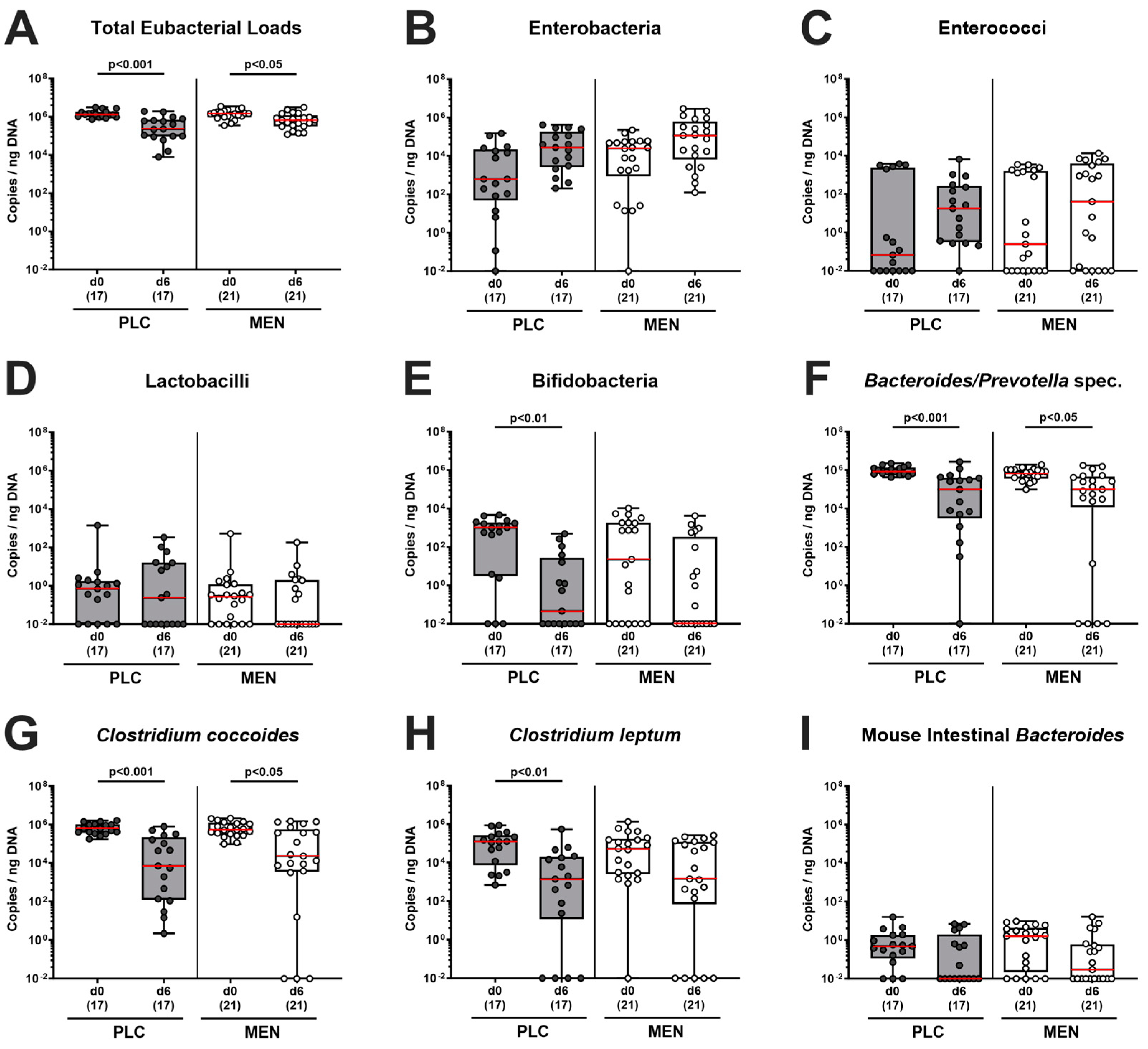
3.6. Fecal Microbiota during C. jejuni Infection of hma IL-10−/− Mice with Menthol Pretreatment

Furthermore, we tested for fecal microbiota changes during C. jejuni infection in menthol-treated mice. Our quantitative culture-independent analyses revealed comparable fecal microbiota compositions in both cohorts on day 0, and hence, immediately before oral C. jejuni challenge of mice (n.s. menthol vs. placebo; Figure 7). In the course of C. jejuni infection, total eubacterial loads and gene numbers of Bacteroides/Prevotella species and of the Clostridium coccoides group decreased irrespective of the pretreatment (p < 0.05–0.001, day 6 vs. day 0; Figure 7A,F,G). As opposed to menthol-pretreated mice, the gene copies of bifidobacteria (p < 0.01; Figure 7E) and of the Clostridium leptum group (p < 0.01; Figure 7H) were lower in fecal samples taken from placebo counterparts on day 6 p.i. if compared to day 0. Hence, oral menthol pretreatment had (if at all) only minor effects on the human fecal gut microbiota composition during C. jejuni infection of hma IL-10−/− mice.

4. Discussion

In our present preclinical placebo-controlled intervention study, we tested the disease-mitigating, anti-pathogenic, anti-inflammatory, anti-oxidant and anti-apoptotic effects of oral menthol in acute experimental campylobacteriosis employing hma IL-10−/− mice. At first, it was rather disappointing that menthol pretreatment did not significantly improve overt C. jejuni-induced clinical signs such as wasting symptoms and bloody diarrhea (Figure 2). One needs to take into consideration, however, that the severity of the observed symptoms ranged from almost no observable clinical signs at all to full-blown disease in both cohorts. In our previous in vivo study employing a different murine campylobacteriosis model, we found that oral menthol of the same concentration therapeutically given to secondary abiotic IL-10−/− mice from day 2 until day 6 p.i. resulted in an improved clinical outcome [45].
In our actual trial, oral menthol pretreatment could prevent inflammation-induced colonic shrinkage, whereas placebo-treated mice displayed shorter colons upon C. jejuni infection if compared to untreated and non-infected controls (Figure 4A). The anti-inflammatory effects of exogenous menthol were also evident on the microscopic level, given that treated animals displayed diminished pathogen-induced colonic epithelial apoptosis (Figure 4C). In support, previous in vivo studies reported potent anti-apoptotic effects upon menthol treatment, given decreases in pro-apoptotic molecules such as cleaved caspase-3 and Bax protein and/or increases in anti-apoptotic molecules including Bcl-2 and HSP-70 [32,33,46,47]. The dampened apoptotic responses upon C. jejuni infection were accompanied by lower numbers of macrophages and monocytes in the colonic mucosa in menthol-pretreated mice on day 6 p.i. (Figure 5A). In support, menthol application could decrease infiltration of infected brain tissues with respective innate immune cell subsets during brain ischemia [48]. Furthermore, C. jejuni-induced increases in colonic T lymphocytes could be reversed to basal values in menthol-treated mice (Figure 5C). In support, a recent in vitro study revealed that menthol inhibited lymphocytic proliferation in a dose-dependent manner and reduced the numbers of T cells expressing pro-inflammatory cytokines [49]. Moreover, in our study, oral menthol could effectively reduce pathogen-induced oxidative stress in different intestinal compartments, as shown by NO concentrations measured in the colon, ileum, and MLN of treated mice on day 6 p.i. that did not differ from those obtained from naive mice (Figure 6). In parallel, enhanced colonic secretion of the pro-inflammatory chemokine MCP-1 secretion could be assessed in the infected placebo as opposed to the verum cohort upon necropsy (Supplementary Figure S3).
The potent anti-oxidant and anti-inflammatory properties of exogenous menthol were shown in vitro to inhibit NO and pro-inflammatory mediator secretion in LPS-stimulated monocytes [50,51]. Furthermore, in vivo studies revealed that menthol mitigated acute intestinal morbidities. Intraperitoneal injection of synthetic menthol before colitis induction, for instance, could exert potent anti-colitogenic effects in rats [52], which also held true for an improved clinical outcome and attenuated macroscopic and microscopic disease due to diminished oxidative stress and inflammation in rats suffering from acute colitis [53].
When addressing the anti-pathogenic effects of the compounds, we found less than one order of magnitude lower C. jejuni numbers in the colon lumen of menthol as compared to placebo treated mice on day 6 p.i. (Figure 3). It is, however, questionable whether the (statistically significant) slightly lower pathogen burden might explain the observed biological effects. In our previous study, we did not observe significant differences in colonic C. jejuni counts in menthol- vs. placebo-treated mice with acute campylobacteriosis. In that study, however, we applied a therapeutic regimen to secondary abiotic IL-10−/− mice from day 2 until day 6 p.i. only [45]. Furthermore, the menthol concentration in the drinking solutions given in our previous and actual studies (i.e., 500 mg/L) was far below the minimum inhibitory concentration (MIC) of the compound measured against the applied C. jejuni 81–176 strain (16,000 mg/L). Anyhow, it is much more likely that the mitigated enterocolitis following menthol pretreatment of C. jejuni-infected mice was due to immune-modulatory, including anti-inflammatory, anti-oxidant, and anti-apoptotic effects, as, in support, had been reported in a murine gastritis model earlier [32].
Furthermore, we surveyed the impact of menthol pretreatment on influencing the (human) commensal gut microbiota composition following human FMT but found comparable fecal microbiota conditions on day 0 (i.e., immediately before C. jejuni infection) (Figure 7). This result points towards comparably effective engraftments of fecal human transplants and speaks against the significant microbiota-shifting effects of exogenous menthol that had already been applied for one week by that time point. In addition, loads of human bacterial communities were comparable in fecal samples derived at the end of the experiment on day 6 p.i. (Figure 7). When focusing on microbiota shifts during C. jejuni infection within respective treatment cohorts (i.e., changes between day 0 and day 6 p.i.), we found statistically significant decreases in fecal gene numbers of potentially probiotic bifidobacteria and Clostridium leptum in the placebo, but only a trend towards lower counts of respective phyla in the menthol cohort (n.s.; Figure 7). Given these rather marginal differences, one should not overemphasize these observations.
Menthol is generally considered a safe compound by the United States Food and Drug Administration and the Flavoring Extract Manufacturer’s Association, and only very rarely have intoxications resulting in dizziness, agitation, hallucination, convulsion, and coma been reported [54,55]. A medical integrative international organization (Natural Standards) has indicated a dose of up to 1 g per kg of body weight as fatal [54], whereas in our trial, mice received one tenth.
In summary, our actual preclinical trial provides the first evidence that oral pretreatment of C. jejuni-infected IL-10−/− mice harboring a commensal human gut microbiota mitigates acute enterocolitis due to its immune-modulatory, including anti-inflammatory, anti-oxidant, and anti-apoptotic modes of action.

5. Conclusions

Menthol constitutes a promising antibiotic-independent option to fight acute campylobacteriosis, thereby decreasing the risk for the development of post-infectious autoimmune morbidities given that the severity of the enteritis is associated with the risk of complications including Guillain–Barré syndrome, reactive arthritis, and chronic intestinal inflammatory morbidities later on. Furthermore, menthol might be a useful measure to prevent C. jejuni infection in individuals with an increased risk of exposure to the enteropathogen. Future research should focus on developing antibiotic-independent methods for the prevention and/or treatment of food-borne illnesses brought on by enteropathogens besides Campylobacter species.

Supplementary Materials

The following supporting information can be downloaded at: https://www.mdpi.com/article/10.3390/biom14030290/s1, Table S1: Clinical scores; Table S2: Histopathological scores; Table S3: Primary antibodies for in situ immunohistochemical analyses; Figure S1: Clinical signs of campylobacteriosis over time following C. jejuni infection of hma IL-10−/− mice with menthol pre-treatment. Figure S2: Pathogen numbers in fecal samples taken following C. jejuni infection of hma IL-10−/− mice with menthol pre-treatment. Figure S3: Colonic MCP-1 secretion in C. jejuni infected hma IL-10−/− mice with menthol pretreatment.

Author Contributions

Conceptualization, supervision, and methodology: M.M.H. and S.B.; Investigation, M.M.H., L.Q.L., N.S., N.W.S. and S.M.; Visualization: L.Q.L. and S.M.; Evaluation and interpretation of data: M.M.H., L.Q.L., S.M. and S.B.; Writing—original draft preparation: M.M.H.; Writing—review and editing: N.W.S., S.M. and S.B. All authors have read and agreed to the published version of the manuscript.

Funding

Grants were provided from the German Federal Ministries of Education and Research (BMBF) in the framework of the PAC-Campylobacter zoonoses research consortium to M.M.H. and S.B. (IP7/01KI2007D) and from the Federal Ministry for Economic Affairs and Energy in response to a resolution of the Deutscher Bundestag, the German National Parliament, to M.M.H. and S.B. (ZIM, ZF4117908 AJ8) supporting this work. The funders were not involved in the design of the study, the gathering and analysis of data, the publishing decision, or the writing of the manuscript.

Institutional Review Board Statement

Mouse experiments were approved by the commission for animal experiments headed by the “Landesamt für Gesundheit und Soziales” (LaGeSo, Berlin; registration numbers G0104/19, approved on 15 July 2019) and carried out in accordance with the European Guidelines for animal welfare (2010/63/EU) and the ARRIVE guidelines.

Informed Consent Statement

Not applicable.

Data Availability Statement

The data presented in this study are available on request from the corresponding author.

Acknowledgments

We acknowledge the great technical help and animal breeding provided by Alexandra Bittroff-Leben, Ines Puschendorf, Sumaya Abdul-Rahman, Gernot Reifenberger, and the personnel of the animal research facility at FEM of Charité-University Medicine Berlin, respectively.

Conflicts of Interest

The funders had no role in the design of the study; in the collection, analyses, or interpretation of data; in the writing of the manuscript; or in the decision to publish the results.

References

  1. Oyarzabal, O.A.; Backert, S. Food Safety Resources. In Microbial Food Safety; Springer: Berlin/Heidelberg, Germany, 2012; pp. 235–239. [Google Scholar]
  2. Wilson, D.J.; Gabriel, E.; Leatherbarrow, A.J.; Cheesbrough, J.; Gee, S.; Bolton, E.; Fox, A.; Fearnhead, P.; Hart, C.A.; Diggle, P.J. Tracing the source of campylobacteriosis. PLoS Genet. 2008, 4, e1000203. [Google Scholar] [CrossRef] [PubMed]
  3. Pielsticker, C.; Glünder, G.; Rautenschlein, S. Colonization properties of Campylobacter jejuni in chickens. Eur. J. Microbiol. Immunol. 2012, 2, 61–65. [Google Scholar] [CrossRef]
  4. Fitzgerald, C. Campylobacter. Clin. Lab. Med. 2015, 35, 289–298. [Google Scholar] [CrossRef]
  5. Ellis-Iversen, J.; Ridley, A.; Morris, V.; Sowa, A.; Harris, J.; Atterbury, R.; Sparks, N.; Allen, V. Persistent environmental reservoirs on farms as risk factors for Campylobacter in commercial poultry. Epidemiol. Infect. 2012, 140, 916–924. [Google Scholar] [CrossRef]
  6. Skirrow, M. Campylobacter enteritis: A” new” disease. Br. Med. J. 1977, 2, 9–11. [Google Scholar] [CrossRef]
  7. Silva, J.; Leite, D.; Fernandes, M.; Mena, C.; Gibbs, P.A.; Teixeira, P. Campylobacter spp. as a foodborne pathogen: A review. Front. Microbiol. 2011, 2, 200. [Google Scholar] [CrossRef]
  8. Black, R.E.; Levine, M.M.; Clements, M.L.; Hughes, T.P.; Blaser, M.J. Experimental Campylobacter jejuni infection in humans. J. Infect. Dis. 1988, 157, 472–479. [Google Scholar] [CrossRef]
  9. Allos, B.M. Campylobacter jejuni Infections: Update on emerging issues and trends. Clin. Infect. Dis. 2001, 32, 1201–1206. [Google Scholar] [CrossRef]
  10. Tegtmeyer, N.; Sharafutdinov, I.; Harrer, A.; Esmaeili, D.S.; Linz, B.; Backert, S. Campylobacter Virulence Factors and Molecular Host–Pathogen Interactions. In Fighting Campylobacter Infections: Towards a One Health Approach; Springer: Cham, Switzerland, 2021; Volume 431, pp. 169–202. [Google Scholar] [CrossRef]
  11. Callahan, S.M.; Dolislager, C.G.; Johnson, J.G. The host cellular immune response to infection by Campylobacter spp. and its role in disease. Infect. Immun. 2021, 89, e00116–e00121. [Google Scholar] [CrossRef]
  12. Walker, R.I.; Caldwell, M.B.; Lee, E.C.; Guerry, P.; Trust, T.J.; Ruiz-Palacios, G.M. Pathophysiology of Campylobacter enteritis. Microbiol. Rev. 1986, 50, 81–94. [Google Scholar] [CrossRef] [PubMed]
  13. Wassenaar, T.M.; Blaser, M.J. Pathophysiology of Campylobacter jejuni infections of humans. Microbes Infect. 1999, 1, 1023–1033. [Google Scholar] [CrossRef]
  14. Young, K.T.; Davis, L.M.; Dirita, V.J. Campylobacter jejuni: Molecular biology and pathogenesis. Nat. Rev. Microbiol. 2007, 5, 665–679. [Google Scholar] [CrossRef]
  15. Butkevych, E.; Lobo de Sá, F.D.; Nattramilarasu, P.K.; Bücker, R. Contribution of Epithelial Apoptosis and Subepithelial Immune Responses in Campylobacter jejuni-Induced Barrier Disruption. Front. Microbiol. 2020, 11, 344. [Google Scholar] [CrossRef]
  16. Lobo de Sá, F.; Schulzke, J.-D.; Bücker, R. Diarrheal mechanisms and the role of intestinal barrier dysfunction in Campylobacter infections. Curr. Top. Microbiol. Immunol. 2021, 431, 203–231. [Google Scholar] [CrossRef]
  17. Dai, L.; Sahin, O.; Grover, M.; Zhang, Q. New and alternative strategies for the prevention, control, and treatment of antibiotic-resistant Campylobacter. Transl. Res. 2020, 223, 76–88. [Google Scholar] [CrossRef] [PubMed]
  18. Finsterer, J. Triggers of Guillain-Barré Syndrome: Campylobacter jejuni Predominates. Int. J. Mol. Sci. 2022, 23, 14222. [Google Scholar] [CrossRef]
  19. Nachamkin, I.; Allos, B.M.; Ho, T. Campylobacter species and Guillain-Barre syndrome. Clin. Microbiol. Rev. 1998, 11, 555–567. [Google Scholar] [CrossRef]
  20. Pope, J.E.; Krizova, A.; Garg, A.X.; Thiessen-Philbrook, H.; Ouimet, J.M. Campylobacter reactive arthritis: A systematic review. In Seminars in Arthritis and Rheumatism; Elsevier: Amsterdam, The Netherlands, 2007; pp. 48–55. [Google Scholar]
  21. Omarova, S.; Awad, K.; Moos, V.; Püning, C.; Gölz, G.; Schulzke, J.-D.; Bücker, R. Intestinal Barrier in Post-Campylobacter jejuni Irritable Bowel Syndrome. Biomolecules 2023, 13, 449. [Google Scholar] [CrossRef]
  22. Mortensen, N.P.; Kuijf, M.L.; Ang, C.W.; Schiellerup, P.; Krogfelt, K.A.; Jacobs, B.C.; van Belkum, A.; Endtz, H.P.; Bergman, M.P. Sialylation of Campylobacter jejuni lipo-oligosaccharides is associated with severe gastro-enteritis and reactive arthritis. Microbes Infect. 2009, 11, 988–994. [Google Scholar] [CrossRef]
  23. Mouftah, S.F.; Cobo-Díaz, J.F.; Álvarez-Ordóñez, A.; Elserafy, M.; Saif, N.A.; Sadat, A.; El-Shibiny, A.; Elhadidy, M. High-throughput sequencing reveals genetic determinants associated with antibiotic resistance in Campylobacter spp. from farm-to-fork. PLoS ONE 2021, 16, e0253797. [Google Scholar] [CrossRef]
  24. World Health Organisation. Campylobacter. Available online: https://www.who.int/news-room/fact-sheets/detail/campylobacter (accessed on 9 January 2024).
  25. European Food Safety Authority; European Centre for Disease Prevention and Control. The European Union Summary Report on Antimicrobial Resistance in zoonotic and indicator bacteria from humans, animals and food in 2019–2020. EFSA J. 2022, 20, e07209. [Google Scholar] [CrossRef]
  26. Kaakoush, N.O.; Castano-Rodriguez, N.; Mitchell, H.M.; Man, S.M. Global Epidemiology of Campylobacter Infection. Clin. Microbiol. Rev. 2015, 28, 687–720. [Google Scholar] [CrossRef]
  27. Kirk, M.D.; Pires, S.M.; Black, R.E.; Caipo, M.; Crump, J.A.; Devleesschauwer, B.; Döpfer, D.; Fazil, A.; Fischer-Walker, C.L.; Hald, T. World Health Organization estimates of the global and regional disease burden of 22 foodborne bacterial, protozoal, and viral diseases, 2010: A data synthesis. PLoS Med. 2015, 12, e1001921. [Google Scholar]
  28. Lackner, J.; Weiss, M.; Müller-Graf, C.; Greiner, M. The disease burden associated with Campylobacter spp. in Germany, 2014. PLoS ONE 2019, 14, e0216867. [Google Scholar] [CrossRef]
  29. MacDougall, J.M.; Fandrick, K.; Zhang, X.; Serafin, S.V.; Cashman, J.R. Inhibition of human liver microsomal (S)-nicotine oxidation by (−)-menthol and analogues. Chem. Res. Toxicol. 2003, 16, 988–993. [Google Scholar] [CrossRef]
  30. Trombetta, D.; Castelli, F.; Sarpietro, M.G.; Venuti, V.; Cristani, M.; Daniele, C.; Saija, A.; Mazzanti, G.; Bisignano, G. Mechanisms of antibacterial action of three monoterpenes. Antimicrob. Agents Chemother. 2005, 49, 2474–2478. [Google Scholar] [CrossRef]
  31. Sabzghabaee, A.M.; Nili, F.; Ghannadi, A.; Eizadi-Mood, N.; Anvari, M. Role of menthol in treatment of candidial napkin dermatitis. World J. Pediatr. 2011, 7, 167–170. [Google Scholar] [CrossRef]
  32. Rozza, A.L.; Meira de Faria, F.; Souza Brito, A.R.; Pellizzon, C.H. The gastroprotective effect of menthol: Involvement of anti-apoptotic, antioxidant and anti-inflammatory activities. PLoS ONE 2014, 9, e86686. [Google Scholar] [CrossRef]
  33. Matouk, A.I.; El-Daly, M.; Habib, H.A.; Senousy, S.; Naguib Abdel Hafez, S.M.; Kasem, A.W.; Almalki, W.H.; Alzahrani, A.; Alshehri, A.; Ahmed, A.-S.F. Protective effects of menthol against sepsis-induced hepatic injury: Role of mediators of hepatic inflammation, apoptosis, and regeneration. Front. Pharmacol. 2022, 13, 952337. [Google Scholar] [CrossRef]
  34. Goudarzi, M.A.; Radfar, M.; Goudarzi, Z. Peppermint as a promising treatment agent in inflammatory conditions: A comprehensive systematic review of literature. Phytother. Res. 2023, 38, 187–195. [Google Scholar] [CrossRef]
  35. Bereswill, S.; Fischer, A.; Plickert, R.; Haag, L.M.; Otto, B.; Kuhl, A.A.; Dasti, J.I.; Zautner, A.E.; Munoz, M.; Loddenkemper, C.; et al. Novel murine infection models provide deep insights into the “menage a trois” of Campylobacter jejuni, microbiota and host innate immunity. PLoS ONE 2011, 6, e20953. [Google Scholar] [CrossRef]
  36. Warren, H.S.; Fitting, C.; Hoff, E.; Adib-Conquy, M.; Beasley-Topliffe, L.; Tesini, B.; Liang, X.; Valentine, C.; Hellman, J.; Hayden, D. Resilience to bacterial infection: Difference between species could be due to proteins in serum. J. Infect. Dis. 2010, 201, 223–232. [Google Scholar] [CrossRef] [PubMed]
  37. Lippert, E.; Karrasch, T.; Sun, X.; Allard, B.; Herfarth, H.H.; Threadgill, D.; Jobin, C. Gnotobiotic IL-10; NF-κB mice develop rapid and severe colitis following Campylobacter jejuni infection. PLoS ONE 2009, 4, e7413. [Google Scholar] [CrossRef]
  38. Heimesaat, M.M.; Mousavi, S.; Bandick, R.; Bereswill, S. Campylobacter jejuni infection induces acute enterocolitis in IL-10-/- mice pretreated with ampicillin plus sulbactam. Eur. J. Microbiol. Immunol. 2022, 12, 73–83. [Google Scholar] [CrossRef]
  39. Shayya, N.W.; Foote, M.S.; Langfeld, L.Q.; Du, K.; Bandick, R.; Mousavi, S.; Bereswill, S.; Heimesaat, M.M. Human microbiota associated IL-10−/− mice: A valuable enterocolitis model to dissect the interactions of Campylobacter jejuni with host immunity and gut microbiota. Eur. J. Microbiol. Immunol. 2023, 12, 107–122. [Google Scholar] [CrossRef]
  40. Weschka, D.; Mousavi, S.; Biesemeier, N.; Bereswill, S.; Heimesaat, M.M. Survey of Pathogen-Lowering and Immuno-Modulatory Effects Upon Treatment of Campylobacter coli-Infected Secondary Abiotic IL-10−/− Mice with the Probiotic Formulation Aviguard®. Microorganisms 2021, 9, 1127. [Google Scholar] [CrossRef]
  41. Heimesaat, M.M.; Alutis, M.; Grundmann, U.; Fischer, A.; Tegtmeyer, N.; Böhm, M.; Kühl, A.A.; Göbel, U.B.; Backert, S.; Bereswill, S. The role of serine protease HtrA in acute ulcerative enterocolitis and extra-intestinal immune responses during Campylobacter jejuni infection of gnotobiotic IL-10 deficient mice. Front. Cell. Infect. Microbiol. 2014, 4, 77. [Google Scholar] [CrossRef]
  42. Erben, U.; Loddenkemper, C.; Doerfel, K.; Spieckermann, S.; Haller, D.; Heimesaat, M.M.; Zeitz, M.; Siegmund, B.; Kühl, A.A. A guide to histomorphological evaluation of intestinal inflammation in mouse models. Int. J. Clin. Exp. Pathol. 2014, 7, 4557–4576. [Google Scholar]
  43. Heimesaat, M.M.; Giladi, E.; Kühl, A.A.; Bereswill, S.; Gozes, I. The octapetide NAP alleviates intestinal and extra-intestinal anti-inflammatory sequelae of acute experimental colitis. Peptides 2018, 101, 1–9. [Google Scholar] [CrossRef]
  44. Bryan, N.S.; Grisham, M.B. Methods to detect nitric oxide and its metabolites in biological samples. Free Radic. Biol. Med. 2007, 43, 645–657. [Google Scholar] [CrossRef]
  45. Bandick, R.; Busmann, L.V.; Mousavi, S.; Shayya, N.W.; Piwowarski, J.P.; Granica, S.; Melzig, M.F.; Bereswill, S.; Heimesaat, M.M. Therapeutic Effects of Oral Application of Menthol and Extracts from Tormentil (Potentilla erecta), Raspberry Leaves (Rubus idaeus), and Loosestrife (Lythrum salicaria) during Acute Murine Campylobacteriosis. Pharmaceutics 2023, 15, 2410. [Google Scholar] [CrossRef]
  46. Muruganathan, U.; Srinivasan, S.; Vinothkumar, V. Antidiabetogenic efficiency of menthol, improves glucose homeostasis and attenuates pancreatic β-cell apoptosis in streptozotocin-nicotinamide induced experimental rats through ameliorating glucose metabolic enzymes. Biomed. Pharmacother. 2017, 92, 229–239. [Google Scholar] [CrossRef]
  47. Santo, S.G.E.; Romualdo, G.R.; Santos, L.A.D.; Grassi, T.F.; Barbisan, L.F. Modifying effects of menthol against benzo(a)pyrene-induced forestomach carcinogenesis in female Swiss mice. Environ. Toxicol. 2021, 36, 2245–2255. [Google Scholar] [CrossRef]
  48. Huang, S.-S.; Su, H.-H.; Chien, S.-Y.; Chung, H.-Y.; Luo, S.-T.; Chu, Y.-T.; Wang, Y.-H.; MacDonald, I.J.; Lee, H.-H.; Chen, Y.-H. Activation of peripheral TRPM8 mitigates ischemic stroke by topically applied menthol. J. Neuroinflamm. 2022, 19, 192. [Google Scholar] [CrossRef]
  49. Bayat, M.; Kalantar, K.; Amirghofran, Z. Inhibition of interferon-γ production and T-bet expression by menthol treatment of human peripheral blood mononuclear cells. Immunopharmacol. Immunotoxicol. 2019, 41, 267–276. [Google Scholar] [CrossRef]
  50. Juergens, U.R.; Stöber, M.; Vetter, H. The anti-inflammatory activity of L-menthol compared to mint oil in human monocytes in vitro: A novel perspective for its therapeutic use in inflammatory diseases. Eur. J. Med. Res. 1998, 3, 539–545. [Google Scholar]
  51. Marcuzzi, A.; Tommasini, A.; Crovella, S.; Pontillo, A. Natural isoprenoids inhibit LPS-induced-production of cytokines and nitric oxide in aminobisphosphonate-treated monocytes. Int. Immunopharmacol. 2010, 10, 639–642. [Google Scholar] [CrossRef]
  52. Ghasemi-Pirbaluti, M.; Motaghi, E.; Bozorgi, H. The effect of menthol on acute experimental colitis in rats. Eur. J. Pharmacol. 2017, 805, 101–107. [Google Scholar] [CrossRef]
  53. Bastaki, S.M.; Adeghate, E.; Amir, N.; Ojha, S.; Oz, M. Menthol inhibits oxidative stress and inflammation in acetic acid-induced colitis in rat colonic mucosa. Am. J. Transl. Res. 2018, 10, 4210–4222. [Google Scholar]
  54. Baibars, M.; Eng, S.; Shaheen, K.; Alraiyes, A.H.; Alraies, M.C. Menthol toxicity: An unusual cause of coma. Case Rep. Med. 2012, 2012, 187039. [Google Scholar] [CrossRef]
  55. Kumar, A.; Baitha, U.; Aggarwal, P.; Jamshed, N. A fatal case of menthol poisoning. Int. J. Appl. Basic. Med. Res. 2016, 6, 137–139. [Google Scholar] [CrossRef]
Figure 1. Timeline of experimental procedures and the microbiota composition of human fecal transplants. (A) Human microbiota-associated (hma) IL-10−/− mice were generated by antibiotic gut microbial depletion (with ampicillin plus sulbactam) and subsequent triple human fecal microbiota transplantation (hFMT). Starting a week before oral Campylobacter jejuni strain 81–176 infection, hma mice were subjected to menthol treatment via the drinking water and followed up until necropsy on day (d) 6 post-infection. (B) Gut microbial communities were quantitated in human fecal transplants (n = 3 per experiment) by culture-independent (i.e., 16S rRNA-based molecular) analyses. Box plots (25th and 75th percentiles), whiskers (minimum and maximum values), and medians (red bar in boxes) are given (pooled from three independent experiments). TEL, total eubacterial loads; EB, enterobacteria; EC, enterococci; LB, lactobacilli; BB, bifidobacteria; BP, Bacteroides/Prevotella species; CC, Clostridium coccoides group; CL, Clostridium leptum (CL) group; MIB, Mouse Intestinal Bacteroides.
Biomolecules 14 00290 g001
Figure 2. Clinical conditions over time following C. jejuni infection of hma IL-10−/− mice with menthol pretreatment. Hma mice were orally pretreated with menthol (MEN, white bars) or placebo (PLC, grey bars) and infected with C. jejuni on day 0 (d0) and d1 by orogastric gavage. The clinical conditions of mice were surveyed daily until necropsy by using a clinical campylobacteriosis score recording wasting, diarrhea, and fecal blood (see methods). Box plots (25th and 75th percentiles), whiskers (minimum and maximum values), medians (red line in boxes), and numbers of analyzed mice (in parentheses) from three experiments are given.
Biomolecules 14 00290 g002
Figure 3. Pathogen numbers in gastrointestinal luminal samples from C. jejuni infected hma IL-10−/− mice with menthol pretreatment. Hma mice were orally pretreated with menthol (MEN, white bars) or placebo (PLC, grey bars) and infected with C. jejuni on days 0 and 1 by orogastric gavage. The luminal C. jejuni numbers were determined in gastrointestinal samples taken on day 6 post-infection by culture. Box plots (25th and 75th percentiles), whiskers (minimum and maximum values), medians (red line in boxes), significance levels (p values) determined by the Mann–Whitney test, and numbers of analyzed mice (in parentheses) from three experiments are given. CFU, colony-forming units.
Biomolecules 14 00290 g003
Figure 4. Inflammatory changes in the colon of C. jejuni infected hma IL-10−/− mice with menthol pretreatment. Hma IL-10−/− mice were orally pretreated with menthol (MEN, white bars) or placebo (PLC, grey bars) and infected with C. jejuni on days 0 and 1 by orogastric gavage. On day 6 post-infection, (A) the colonic lengths were measured. Furthermore, (B) the histopathological scores and (C) numbers of apoptotic epithelial cells positive for cleaved caspase-3 (Casp3+) were determined in colonic paraffin sections. Naive mice (non-infected without pretreatment) served as negative controls. Box plots (25th and 75th percentiles), whiskers (minimum and maximum values), medians (red line in boxes), significance levels (p values) determined by the one-way ANOVA test with Tukey post hoc test (A,C) and Kruskal–Wallis test with Dunn’s post hoc test (B), and numbers of analyzed mice (in parentheses) from three experiments are given. HPF, high power field.
Biomolecules 14 00290 g004
Figure 5. Colonic immune cells in C. jejuni infected hma IL-10−/− mice with menthol pretreatment. Hma IL-10−/− mice were orally pretreated with menthol (MEN, white bars) or placebo (PLC, grey bars) and infected with C. jejuni on days 0 and 1 by orogastric gavage. On day 6 post-infection, numbers of (A) macrophages and monocytes (F4/80+), (B) neutrophils (MPO7+), (C) T lymphocytes (CD3+), and (D) B lymphocytes (B220+) were determined in the colonic lamina propria (immunohistochemically stained paraffin sections). Naive mice (non-infected without pretreatment) served as negative controls. Box plots (25th and 75th percentiles), whiskers (minimum and maximum values), medians (red line in boxes), significance levels (p values) determined by the one-way ANOVA test with Tukey post hoc test (A) and Kruskal–Wallis test with Dunn’s post hoc test (BD), and numbers of analyzed mice (in parentheses) from three experiments are given. HPF, high power field.
Biomolecules 14 00290 g005
Figure 6. Intestinal nitric oxide secretion in C. jejuni infected hma IL-10−/− mice with menthol pretreatment. Hma IL-10−/− mice were orally pretreated with menthol (MEN, white bars) or placebo (PLC, grey bars) and infected with C. jejuni on days 0 and 1 by orogastric gavage. On day 6 post-infection, nitric oxide concentrations were measured in ex vivo biopsies sampled from the (A) colon, (B) ileum, and (C) mesenteric lymph nodes (MLN). Naive mice (non-infected without pretreatment) served as negative controls. Box plots (25th and 75th percentiles), whiskers (minimum and maximum values), medians (red line in boxes), significance levels (p values) determined by the Kruskal–Wallis test with Dunn’s post hoc test, and numbers of analyzed mice (in parentheses) from three experiments are given. The Grubb’s test was used to identify definite outliers.
Biomolecules 14 00290 g006
Figure 7. Fecal microbiota during C. jejuni infection of hma IL-10−/− mice with menthol pretreatment. Immediately before (i.e., day (d)0) and on d6 post C. jejuni infection, the fecal microbiota compositions were assessed in hma IL-10−/− mice with menthol (MEN; white bars) or placebo (PLC; grey bars) prophylaxis by culture-independent methods. The (A) total eubacterial loads, (B) enterobacteria, (C) enterococci, (D) lactobacilli, (E) bifidobacteria, (F) Bacteroides/Prevotella species, (G) Clostridium coccoides and (H) Clostridium leptum groups, and (I) Mouse Intestinal Bacteroides are expressed as copies per ng DNA. Box plots (25th and 75th percentiles), whiskers (minimum and maximum values), medians (red line in boxes), significance levels (p values) determined by the one-way ANOVA test with Tukey post hoc test (A) and the Kruskal–Wallis test with Dunn’s post hoc test (BI), and numbers of analyzed mice (in parentheses) from three experiments are given.
Biomolecules 14 00290 g007
Disclaimer/Publisher’s Note: The statements, opinions and data contained in all publications are solely those of the individual author(s) and contributor(s) and not of MDPI and/or the editor(s). MDPI and/or the editor(s) disclaim responsibility for any injury to people or property resulting from any ideas, methods, instructions or products referred to in the content.

