Review

Voltage-Gated Proton Channels in the Tree of Life

1
Center of Physiology, Pathophysiology and Biophysics, The Nuremberg Location, Paracelsus Medical University, 90419 Nuremberg, Germany
2
Center of Physiology, Pathophysiology and Biophysics, The Salzburg Location, Paracelsus Medical University, 5020 Salzburg, Austria
*
Author to whom correspondence should be addressed.
Biomolecules 2023, 13(7), 1035; https://doi.org/10.3390/biom13071035
Submission received: 14 May 2023 / Revised: 14 June 2023 / Accepted: 21 June 2023 / Published: 24 June 2023
(This article belongs to the Special Issue Proton and Proton-Coupled Transport)

Abstract

:
With a single gene encoding HV1 channel, proton channel diversity is particularly low in mammals compared to other members of the superfamily of voltage-gated ion channels. Nonetheless, mammalian HV1 channels are expressed in many different tissues and cell types where they exert various functions. In the first part of this review, we regard novel aspects of the functional expression of HV1 channels in mammals by differentially comparing their involvement in (1) close conjunction with the NADPH oxidase complex responsible for the respiratory burst of phagocytes, and (2) in respiratory burst independent functions such as pH homeostasis or acid extrusion. In the second part, we dissect expression of HV channels within the eukaryotic tree of life, revealing the immense diversity of the channel in other phylae, such as mollusks or dinoflagellates, where several genes encoding HV channels can be found within a single species. In the last part, a comprehensive overview of the biophysical properties of a set of twenty different HV channels characterized electrophysiologically, from Mammalia to unicellular protists, is given.

1. Proton Channels in Mammals

Voltage-gated proton currents were measured for the first time in eukaryotes using voltage-clamp. The investigation of an ion channel that exclusively conducts a subatomic particle, the proton, originates from a mollusk, the snail Helix aspersa [1]. Before its actual discovery in a living organism, the necessity of a functional voltage-gated proton-selective channel (HV) had been anticipated. Fogel and Hastings [2] predicted the voltage-gated proton channel to be the trigger for the bioluminescence reaction dinoflagellates, one of the first eukaryotes spawned in evolution. The second prediction had far-reaching consequences for human health. Henderson et al. [3] prognosticated the voltage-gated proton channel to be essential for the respiratory burst of human phagocytes. The respiratory burst is a greatly increased oxygen consumption of a cell where molecular oxygen is reduced to superoxide [4], and it is essential to kill pathogens. Yet, the first recording of HV in mammals was performed in respiratory tissue (alveolar type 2 cells) of rats by DeCoursey [5]. Type 2 cells, as far as we know, do not perform a respiratory burst. Shortly afterwards, the detection in human tissue was achieved by three groups simultaneously [6,7,8]. Here, two innate immune cells were investigated, freshly isolated human neutrophils and the HL-60 cell line representing human granulocytes. Bernheim et al., 1993, found the channel in human skeletal muscle tissue, which undoubtedly is a strongly metabolically active tissue not performing the respiratory burst. An intensive investigation on the connection between HV and the respiratory burst and the role of the channel in cell types lacking immunological functions in human and mammalian tissue followed. At present, there is no indication of HV channels in prokaryotes. In this first part of the review, we categorize the expression of voltage-gated proton channels in two sections. Firstly, cell types in mammals expressing HV capable of performing the respiratory burst, and secondly, mammalian tissue expressing HV although incapable of conducting a respiratory burst.

1.1. What Are the Pillars of the Respiratory Burst?

A prerequisite key element of the respiratory burst is the NADPH oxidase, a multiprotein complex that oxidizes NADPH and reduces molecular oxygen to superoxide or hyperoxide (O2). The functioning of the enzyme complex depends on assembly of its components: gp91, p47, p67, p40, p22, and Rac. The function of translocating electrons resides in the transmembrane heterodimer gp91 and p22 (NOX2), where the gp91 subunit harbors all parts needed for electron transfer including two heme groups. Of the four cytosolic components, Rac and p67 are essential to activate NOX2. The subunit p47 appears inessential for activation in some cell-free systems, similar to p40 [9]. As the name obviously states, NADPH is oxidized, and electrons are moved through gp91 across the membrane. This translocation of charge across the isolator plasma membrane generates an electrical potential. The reversal potential of the electrons depends on the midpoint potential of the two redox pairs NADPH/NADP+ + H+ (−320 mV), and the midpoint potential of O2 to O2 (−160 mV). Furthermore, it is subjected to the concentration of the charged particles. Ergo, depolarizations of the membrane up to +200 mV are potentially reachable. The human NADPH oxidase in eosinophils and polymorphonuclear leucocytes (PMN) is voltage-dependent [10], ceasing electron flux at high depolarizations. Therefore, charge compensation during the respiratory burst is fundamental, preventing the sudden end of the immune response due to massive depolarization. The second pilar of the respiratory burst is the voltage-gated proton channel, the main focus of this review.
HV1 is the most complementary protein of NADPH oxidase during the respiratory burst. HV1 opens by depolarization and is controlled by the proton gradient across the membrane. The NADPH oxidation during the respiratory burst accumulates protons inside the cytosol that decrease the intracellular pH (pHi) (Figure 1). The change of the pH outside the cell (pHo) is negligible due to the immense volume of the extracellular space. Accordingly, the pH gradient (ΔpH) increases and favors HV1 opening. Moreover, the outward conduction of protons repolarizes the cell membrane. Consequently, the membrane potential is regulated by the equilibrium between the electron and the proton outflux, avoiding the reduction in the translocation of electrons due to massive membrane depolarizations. Theoretically, the system is then running endlessly as long as there is supply of NADPH. In conclusion, two independent proteins, originated from distinct genes, work synergistically together in one of the most important immune responses of the human body.

1.2. Which Other Physiological Functions Are Affected by HV1?

Foremost, the cell’s pH homeostasis is affected. Proton channels prevent acidosis of the cytosol by conducting the protons out of the cell [7]. The conduction is driven by the electrochemical gradient without any additional requirement of energy [11]. Securing normal cellular pH prevents the cell from all the negative effects of proton accumulation [12].
Interestingly, HV additionally sustains Ca2+ influx due to store operated Ca2+ channels. The concept is comparable to the one found in the potassium channel functioning, especially in Ca2+ activated K+ channels. The Ca2+ influx depolarizes the cell membrane, while the efflux of H+ repolarizes it, balancing the charge [13]. Moreover, HV channels appear to be necessary for cell migration [13,14]. They play a role in the exocytosis machinery and granula release, both essential to the immune system [15,16]. In mouse eosinophils, they prevent cell death, shown by HV1-KO eosinophils’ decreased survivability [17]. Proton channels support the survival of cancer cells, most unwanted by the host [18]. Invasiveness of metastatic cancer is supported by the function of HV1 channels, and the channel itself is evidently a potential biomarker for metastatic cancer [19,20,21]. In microglia, the expression of HV1 regulates polarization towards an M1 proinflammatory and an M2 anti-inflammatory cell subset [22]. One of the most intriguing topics is their contribution to metabolic function. In pancreatic β-cells, Kupffer cells, and in adipocytes, they are involved in hormone release, glucose metabolism, and control of glucotoxicity [23,24,25]. In the immune system, B-cells and B-cell receptors are affected by proton channels [26,27], as well as dendritic cells where proton channels are involved in pattern recognition [28]. HV1 knock out causes T-cell-related autoimmune diseases [29]. HV1 is strongly expressed in lung tissue, and the function there ranges from acidification of the airways to CO2 homeostasis [30,31]. The channel is essential to sperm maturation [32,33] but also functional in human oocytes [34], affecting reproduction. Myeloid-derived suppressor cells are suggested to control the adaptive immune system via HV1 function [35].
A list of tissue expressing HV1 and its functions there is provided in Table 1. The investigation of tissue expression and the function of voltage-gated proton channels has just begun. Potential major breakthroughs are expected in future, especially in this thriving medically relevant field.

2. Voltage-Gated Proton Channels in Evolution

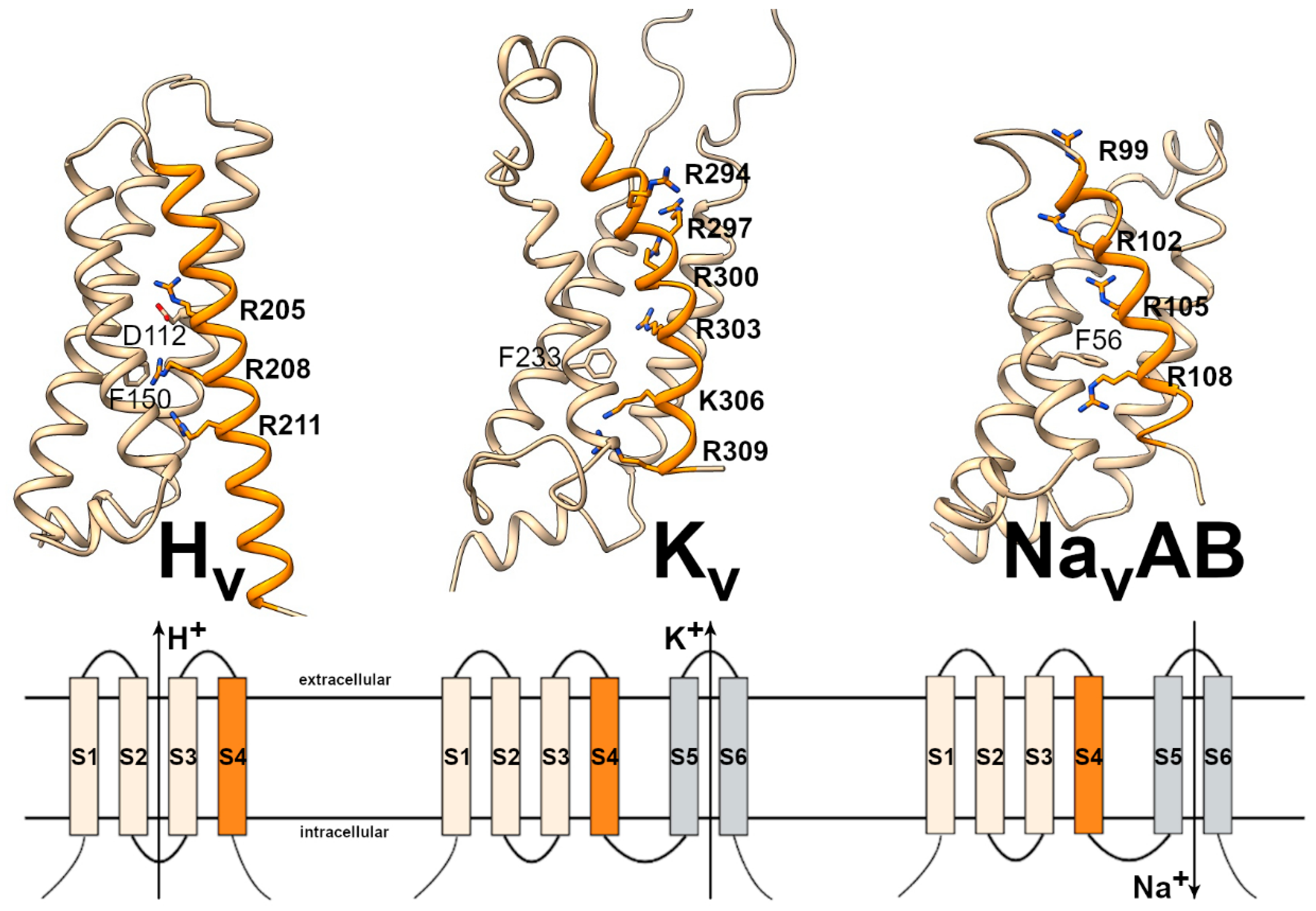
As a member of the superfamily of voltage-gated ion channels, proton channels share the membrane spanning domain including the voltage-sensor in S4 with other members of this superfamily, such as voltage-gated potassium (KV), sodium (NaV), and calcium channels (CaV), TRP channels, hyperpolarization-activated HCN channels, and cyclic-nucleotide-activated CNG channels [106]. Whereas the basic structure of these channels comprises six transmembrane segments, proton channels have lost the typical S5–S6 pore forming domain of other voltage-gated channels, establishing a new H+-selective permeation pathway within the first four transmembrane segments [107] (Figure 2).
Among single mammalian species, the diversity of voltage-gated ion channel is huge. For NaV and CaV channels, ten different genes have been found. Each NaV and CaV channel is formed by four domains of the voltage-gated superfamily (S1–S6), all located on one polypeptide chain and encoded by one single gene. Within the KV-family, up to 40 different genes have been described. Although the KV channels are also formed by four voltage-gated domains, only one S1–S6 domain is encoded per gene, opening the possibility for the formation of homo- or hetero-tetrameric ion channels. For the exploration of biophysical properties and pharmacological differences among these ion channels, the enormous diversity was of great benefit. Differences in channel function could easily be predicted from simple comparison of the primary amino acid sequence using multiple alignments with functional properties of the analyzed channels. Site-directed mutagenesis approaches, construction of chimeric ion channels, or the analysis of the formation of heteromeric channels (in KV) finally gave experimental proof of the investigated structure–function relationship.
For voltage-gated proton channels the situation in mammals is completely different as only one single proton channel (HV1) is encoded by one single gene (HVCN1). In humans, this gene is found on chromosome 12q24.11, and seven exons are encoding HV1 [110]. Although the existence of two different splice variants in humans has been described [27], the diversity of voltage-gated proton channels in mammals is extremely low, excluding the possibility of sequence comparison and biophysical and pharmacological analysis by “sequence exchange experiments” using paralogous channels. Therefore, the identification and functional analysis of more distantly related voltage-gated proton channels is of great importance. As described in the next chapter, HV channels are found in many eukaryotes from unicellular organisms to most animal species.

2.1. The HV Channel “Tree of Life”

Since the 1990’s, it is well established that protein structures typical for potassium, sodium, or calcium channels have already evolved in prokaryotes [111,112,113,114,115]. As a central structural feature, the S1–S4 transmembrane region, harboring the voltage-sensor and dominating the structure of eukaryotic HV1 channels, is found in many prokaryotic voltage-gated ion channels. However, as known so far (and verified by database searches), all these prokaryotic voltage-sensing domains are accompanied by a potassium, sodium, or calcium selective pore domain (S5–S6). There is no evidence of typical HV1-type proton channels in eubacteria and archaea. In addition, the totally conserved tryptophan in S4 of eukaryotic HV1 channels is not found in prokaryotic S1–S4 segments. HV channels found in protists, representing the “early HV evolution”, show significant identity (28–36%) and similarity (~50–60%) to eukaryotic voltage-gated sodium and calcium channels (Table 2). Hence, it can be concluded that HV channels have evolved in an early eukaryote by a split of a region encoding S1–S4 from a eukaryotic NaV or CaV gene. This hypothesis is supported functionally and structurally by mutations within voltage-sensing elements (arginine residues) of sodium channels rendering these channels permeable for cations including protons through this gating-pore element [116,117].
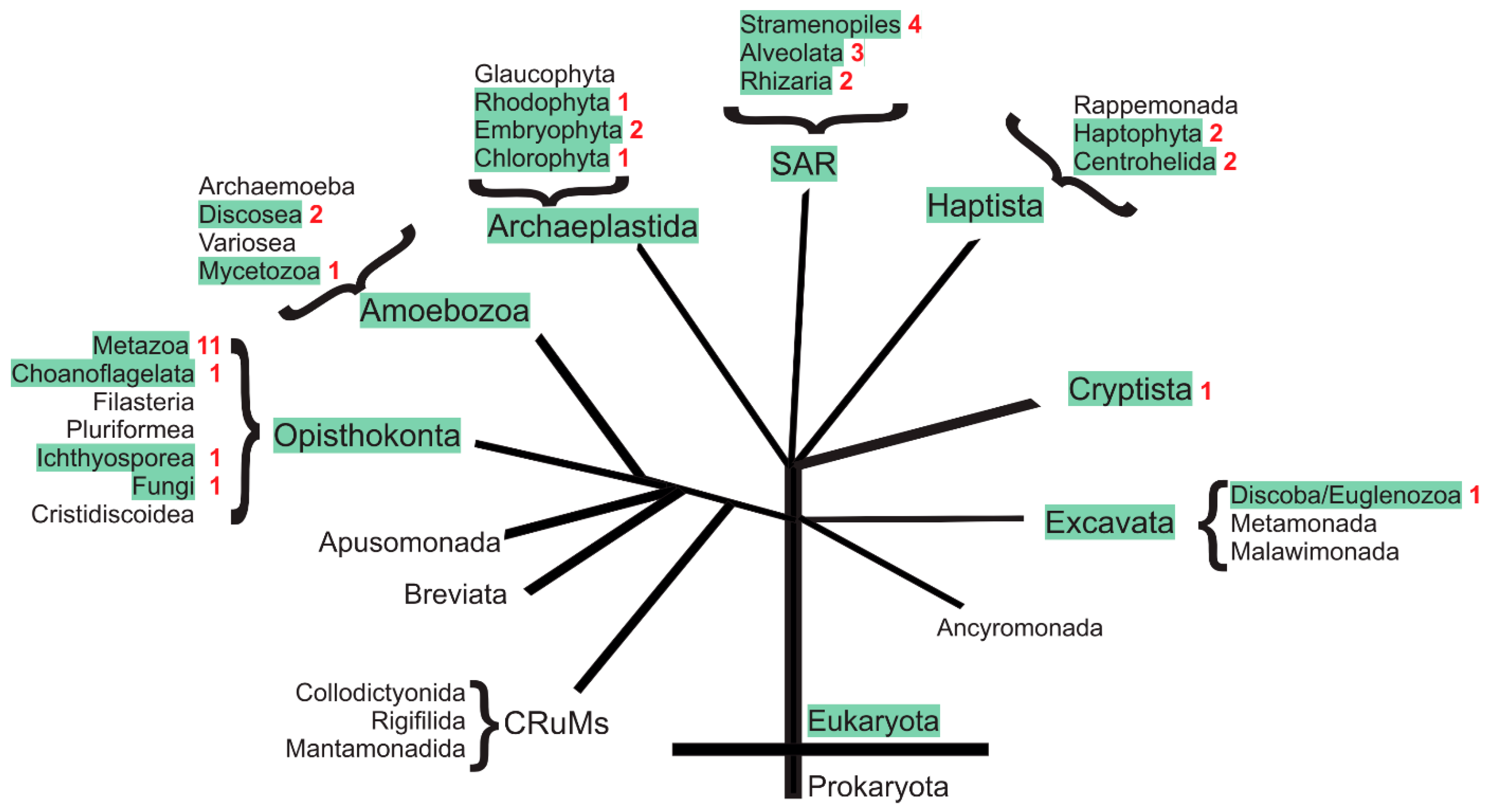
The appearance of HV channels within the phylogenetic tree of life is shown in Figure 3. A list summarizing the identified HV channel proteins and their GenBank accession numbers is given in Figure S1. For green-labeled clades, the presence of HV1 channels and their genes is supported by experimental data or database entries. Unlabeled clades in Figure 3 may either not possess HV channels or the respective data volume coverage may be yet too small to identify a proton channel. Many species within this tree of life lack any typical HV channel. This is exemplified in Table 3 where the presence of an HV channel and its respective gene was analyzed in selected and well-characterized model systems. In many scientific standard model systems, such as the fly Drosophila melanogaster, the yeast Saccharomyces cerevisiae, the nematode worm Caenorhabditis elegans, and the slime mold Dictyostelium discoideum, absolutely no indication for an expressed HV channel was found, nor was an HV channel gene found in the respective genomes. In these cases, HV channels might not be essential for life and many species survive without it. On the other hand, a majority of the eukaryotic species analyzed possess at least one HV channel gene and, for some of them, even several distinct HV channel genes have been identified. This, in turn, shows that HV channel genes may be of significant evolutionary advantage and that HV channels may have gathered many novel functions during evolution yet to be discovered and analyzed.

2.1.1. HV Channels in Chordata

HV1 channels genes were found initially in mammals. In 2006, Ramsey et al. and Sasaki et al. identified and characterized the first HV1 sequences in human and mice, respectively. Among the first publications, a proton channel homolog in the more distantly related invertebrate tunicate Ciona intestinalis was described 2006 [118], showing 46% identity to mammalian HV1 channels, and indicating for the first time, that HV channels are present in non-mammalian animals as well. In the following years, HV1 channels were also found within other vertebrates, such as zebra fish [119].
Curiously, in the clawed frog Xenopus laevis, a recent gene duplication gave rise to two independent genes (NC_054371 and NC_054372) located on separate loci of chromosome 1 (1L and 1S, respectively). As the encoded HV channels (XM_018249100 and XM_018244209) have 90% of identity and 94% of similarity (Figure S2), future experiments regarding cellular expression and biophysical properties will show if they have already evolved different functions.
Besides tunicates, Cephalochordata represents an evolutionary old group of chordates with the lancelet as the main species. Sequence analysis of Branchiostoma belcheri unambiguously identified three independent channels encoded by three different genes (Figure S3). Whereas one Branchiostoma channel is a typical HV channel (XR_002139895), the other two channels incorporated in their structures either a large N-terminal domain (XM_019760615) or a large C-terminal domain (XM_019764911) of unknown function.

2.1.2. HV Channels in Ecdysozoa

Ecdysozoa is a monophyletic superphylum of protostome animals characterized by their ability to grow by ecdysis and include, for example, Arthropoda, Nematoda, and Priapulida. As the largest group within Arthropoda, insects were long thought to lack a proton channel. Indeed, none of the early sequenced dipteran genomes of Drosophila, Anopheles, or Aedes contained a gene encoding a proton channel. In 2016, HV1 channels were however found in evolutionary “old” basal hexapodes (Zygentoma, Archeognatha, Diplura, and Protura), and one HV channel from the silver fish Nicoletia phytophila was functionally characterized [120]. Later, in the polyneopteran stick insects Extatosoma tiaratum, another HV channel was analyzed [121]. Whereas both channels share all typical structural and functional features of proton channels (see below), the Extatosoma HV channel was the first proton channel to be characterized harboring a glutamate residue within the S1 segment instead of the usual aspartate. A careful analysis of expression and genomic databases indeed showed that HV channel genes are absent from genomes of hemipteran or holometabolan insects, including all Diptera. Therefore, “modern” insects have lost their HV gene during evolution [121].
In the second major group of arthropodes, the Chelicerata, several species contain more than one HV gene. The tick Ixodes scapularis, the varroa mite Varroa destructor, and the common house spider Parasteatoda tepidariorum contain two different channels (Figure S1). In the Atlantic horseshoe crab Limulus polyphemus, even three distinct HV channels and their respective genes could be identified in TSA expression and in genome databases, respectively (Figure S4). The first identified Limulus HV channel is a typical, rather small (<250 amino acids) HV channel and has only ~30% identity with the other two closely related (~75% identity) and much larger (>450 amino acids) Limulus HV channels. As in the insect Extatosoma, the large Limulus HV channels possess a glutamate residue within the S1 selectivity filter.
In crustaceans, one typical HV channel is regularly observed, whereas many Myriapoda seem to possess two different HV channels as exemplified by Scolopocryptops rubiginosus (Figure S1). Finally, in the only Priapalida species analyzed (Priapulus caudatus), a single typical HV channel was identified.
The situation for the second major Ecdysozoa phylum, Nematoda, is slightly different as sporadic HV1 homologs were only identified in few Enoplea, including the pathogene Trichinella spiralis and three Chromadorea species. For most other Nematoda, including the model system Caenorhabditis elegans, no HV1 gene was found.

2.1.3. HV Channels in Other Metazoa

A closer look at GenBank TSA and genomic databases of other metazoan phylae unambiguously identified a single HV1-like proton channel also present in Hemichordata (Saccoglossus kowalevskii). In Echinodermata, a single HV channel was already described and functionally characterized in the sea urchin Strongylocentrotus purpuratus [122].
In Platyhelminthes, two different HV channels were identified in some species. For instance, in the bilharzia causing parasitic flatworms (Schistosoma), two related channels (~26% identity, ~56% similarity) share an unusual structural feature of an elongated S3-S4 loop. In addition, in the genome of Schistosoma haematobium, both genes are located on the same scaffold (NW_023366479) separated by only ~270,000 bp, indicating a gene duplication after Platyhelminthes and are separated from other metazoan organisms.
A similar situation is found in the marine free-living and non-parasitic Placozoa, where two different but closely related channels were identified in Trichoplax adhaerens (~39% identity, ~54% similarity), with their genes separated by less than 10,000 bp (NW_002060945).
In Porifera, most species seem to have only one HV channel. However, in the calcareous sponge Sycon ciliatum, three different HV channels have been identified in TSA databases (see Figure S1). Future genomic and expression studies on Porifera are still needed as actual sequences analysis is somewhat scattered.
In reef-building corals (Cnidaria) HV channels were identified and characterized in Acropora millepora where they play an important role in calcification [123].

2.1.4. HV Channels in Lophotrochozoa

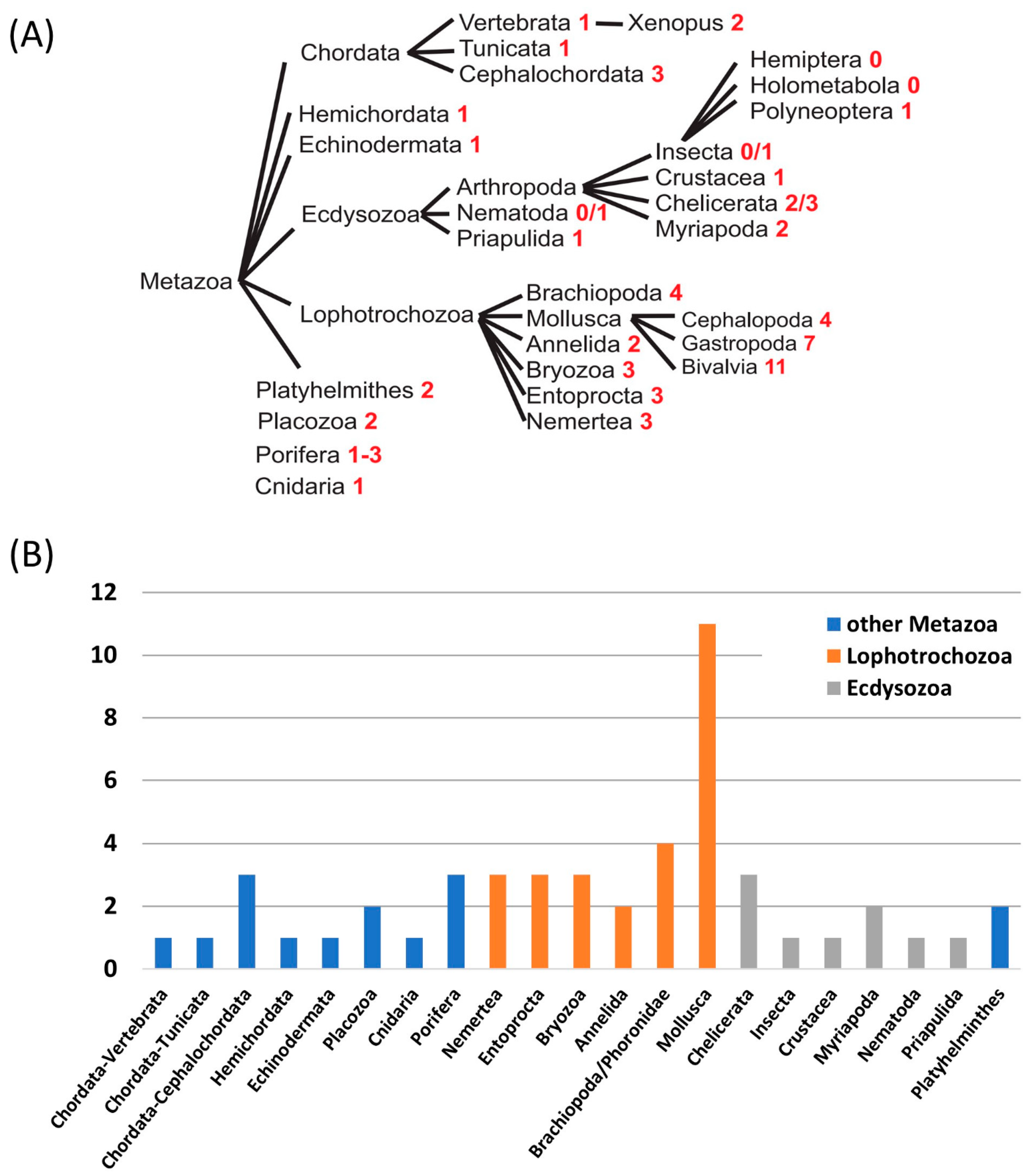
From the picture drawn so far within Metazoa, it could be concluded that most animal species have a single HV1 channel, some have none, and a few of them have two or even three separate genes. These changes are drastic, based on the analysis of the presence of putative HV channel homologs in Lophotrochozoa. In this monophyletic superphylum, the number of putative HV channels is utterly increasing, while the reasons for the presence of this large number of channels is still unknown. In Annelida (2), Entoprocta (3), Nemertea (3), and Bryozoa (3), Brachiopoda and Phoronidae (4), several gene duplications already increased HV channel diversity within individual species (Figure 4).
In the major Lophotrochozoan phylum, mollusks, up to eleven different HV channels and their respective gene were found in a bivalvian species, the scallop Mizuhopecten yessoensis. The diversity is lower in the gastropode Aplysia californica (seven putative HV channels) and in the cephalopode Octopus bimaculoides (four putative HV channels) than in bivalvia. So far, four of these channels have been characterized: three in the gastropode Aplysia californica and one in the bilaterian Crassostrea gigas [124,125] (see below). Structurally, they can be divided into two groups: (1) conventional HV channels (AcHV1, AcHV2, and CgHV4), characterized as rather small molecules (200–250 amino acids in length) with typical conserved residues for the selectivity filter in S1 (Asp64 in AcHV1) and voltage-sensor in S4 (RxWRxxR/K), and (2) unconventional HV channels (AcHV3) with much larger intracellular domains, a very large extracellular S1–S2 loop and a slightly modified voltage-sensor motif (xPWRxxR). Database analysis showed that this later unconventional HV channel family comprises a large proton channel family within the mollusk phylum, with huge variation in the length of the S1–S2 loop and intracellular domains and up to eight different members per species in bivalvia (manuscript in preparation). Outside the mollusk phylum, unconventional channels harboring a typical proline residue within S4 are found in a few species (Brachiopoda and Phoronida). In Annelida, Bryozoa, and Nemertea, HV channels with large extracellular loops and large intracellular domains are present. However, the typical S4 proline residue of mollusk HV3 channels is missing. This leads to an evolutionary picture, where HV gene duplication events and incorporation of large S1–S2 loops and intracellular domains took place in an early lophotrochozoan species, whereas the unusual proline residue entered the S4 segment only in a common ancestor of mollusks, brachiopodes and phoronides.
Speculatively, reasons why mollusks have so many different HV channels might encompass: (1) As most mollusks live in a marine environment, a need for rapid adaptation to pH changes in their surrounding aqueous space may be of advantage. (2) The biomineralization process (calcification) during the formation of the exoskeleton in mollusks strongly depends on pH, and HV channels might work in close conjunction with Ca2+-ATPases in this respect [126]. Indeed, even the calcification of mammalian otoconia is strongly dependent on a proton channel (Otopetrin-1) although this channel is structurally not related to HV channels [127,128]. (3) A mollusk-specific function in the generation of action potentials maybe suggested as all three functionally analyzed Aplysia channels (AcHv1-3) are expressed in CNS/ganglia. A special need for HV channel in mollusk may be caused by a higher metabolic rate in nerve fibers due to the lack of myelin sheath or even in a direct electrical participation of HV channels in action potential formation. (4) As motifs for intracellular localization can be found within the sequence of several molluscan HV channels, a putative function in the acidification of intracellular compartments was proposed.

2.1.5. HV Channels in Fungi, Ichthyosporea, and Choanoflagelates

Beside Metazoa, HV channels have been found in three other opisthokonta kingdoms: the fungi, where they also have been characterized electrophysiologically [129], in choanoflagelates, and in ichthyosporea. In all these kingdoms, only one HV channel per species was found so far. Several fungi however lack voltage-gated proton channels, including yeast and Neurospora crassa. In identified fungal HV channels, the consensus voltage-sensor motif (RxWRxxR) is somewhat modified as in these channels the third arginine is substituted by a lysine residue (RxWRxxK). The only channel found in ichtyosporea (Amoebidium parasiticum) has an interesting extension of the voltage-sensor motif reminiscent to HV channels in protists, with a histidine residue three amino acids downstream the last arginine (RxWRxxRxxH) (Figure S1).

2.1.6. HV Channels in Plants

Proton channels are found in many red algae (Rhodophyta), green algae (Chlorophyta), and land plants (Embryophyta). As recently reviewed by Taylor et al. [130], these proton channels may have important functions in cellular pH homeostasis as well as in sensory biology, depending on the adaptation to different environments. Indeed, marine phytoplankton, freshwater algae, and land plants may have very different needs in their regulation of pHi and H+ fluxes. Interestingly, proton channel genes are absent from many freshwater algae (e.g., Chlamydomonas reinhardtii or Volvox) and marine phytoplankton (Ostreococcus, Micromonas, and Aureococcus), whereas other members of the same order harbor typical HV channels (see Figure S1; Ref. [130]). Whereas in red and green algae, only one HV channel per species is found, at least some land plants, such as the spreading earthmoss Physcomitrella patens, may contain two separate genes (Figure S1).

2.1.7. HV Channels in Protists

Protists represent a large and diverse group of unicellular eukaryotes generally accepted to be at the origin of the evolution of eukaryotes. More than 1.8 billion years of eukaryotic evolution gave rise to a large diversity of HV channels within protists. Whereas some protists (e.g., CRuMs) have no HV channel homolog, some others have up to four different genes (Stramenopiles, see below). As a common sequence feature and in contrast to animal HV channels, protist share the presence of a conserved histidine residue as an elongation of the voltage-sensor motif in S4 (RxWRxxRxxH) with most plants and Ichtyosporea. Only a few protist HV channels have been described and characterized so far. In the coccolithophores Emiliania huxleyi and Coccolithus pelagicus ssp braarudii (Haptophyta), two HV channels homologs were initially identified and physiologically characterized [131]. Their role in the process of calcification may be of general importance as ocean acidification is one of our main environmental problems to date. A database analysis showed that in Emiliania and two other members of the haptophyte order Isochrysidales two different proton channels exist (Figure S1). Similarly, the related Haptiste Choanocystis sp. (Centroplasthelida) possesses two distinct HV channels. A bit unusual for protist HV channels. The two Choanocystis HVs are a slightly larger and show N-terminal domains (>200 amino acids) and C-terminal domains (>150 amino acids) of significant length.
In dinoflagellates (Alveolata), HV channels were characterized in Karlodinium veneficum [132] and in Lingulodinium polyedrum [133] where they are thought to trigger the bioluminescence flash (Lingulodinium). Subsequently, three major HV subfamilies were identified in dinoflagellates, raising the possibility for additional proton channel functions beyond bioluminescence [134]. Database analysis identified HV channel homologs also in Stramenopiles and Rhizaria (forming the supergroup SAR with Alveolata). Whereas in Rhizaria maximally two HV channels were found per species (Amorphochlora amoebiformis, Figure S1) in at least one Stramenopiles, the marine diatom Odontella aurita, up to four different HV channels and their respective genes may exist.
Finally, database analysis identified single protist HV channels in Excavata (e.g., Euglena gracilis), Cryptista (e.g., Chroomonas sp.), and Amoebozoa/Eumycetozoa (e.g., Raperostelium potamoides) and up to two HV channel homologs in a second Amoebozoa class (Discosea, Balamuthia mandrillaris). Although both Balamuthia HV’s share ~50% sequence identity within the S1–S4 core region, the different selectivity filter residue in S1 (Asp79 in GISS01003879 and Glu139 in GISS01013796) may indicate differing physiological properties and functions within these species. In Figure S6, a multiple sequence alignment combines the main HV channels found in animals and protists.

2.1.8. Summary

In summary, HV channels are widely expressed among eukaryotes, whereas no HV channels are found in eubacteria and archaea. Many eukaryotes have at least one Hv channel gene. In several marine species, especially in species performing some form of biomineralization, such as mollusks, brachiopoda, stramenopiles, or haptophyta, the number of different HV channels per species increases significantly. Bioluminescence, in alveolata, or the acidification of specialized intracellular compartments might be reasons for the existence of more than one Hv gene in some species. On the other hand, many species, including all modern insects (Diptera) and most of the nematodes, have no HV channel, indicating that Hv channels are not a prerequisite for life.

2.2. Evolution beyond the Typical Proton Channels

Within the tree of life many gene duplication events gave rise to a huge amount of different proton channels with a vast variety of different structural features, such as different intracellular domains, extracellular loops of variable length, and characteristic point mutations within the selectivity filter motif or in the voltage-sensor. Most of the mutations should preserve proton channel function as long as the major HV channel consensus sequences are conserved. However, it may not be surprising that in some species structures evolved from HV channels have lost typical HV channel consensus elements and therefore may not represent proton channels anymore. On the other hand, as they are expressed and do not represent pseudo genes, these channels may have gathered functions beyond just proton channels. In Figure S1, some examples are highlighted in red. A well-characterized example is the Ciona intestinalis voltage-sensor containing phosphates (Ci-VSP). Similar to HV channels, the Ci-VSP has four transmembrane regions and a voltage-sensor in S4, The Ci-VSP sequence however misses a typical negatively charged residue in S1, serving as selectivity filter in HV channels, and also has no tryptophan residue within the S4 segment. Not surprisingly, electrophysiological experiments showed gating currents underlying the movement of the voltage-sensor, but no typical proton selective current could be measured in WT Ci-VSP [135]. With the coupling of the Ci-VSP S1-S4 segment to an intracellular phosphatase domain, a new structural element arises combining functionality of both domains resulting in a voltage-gated phosphatase. At least one more structure with the same coupling of S1–S4 to a phosphatase was found in the ascidian tunicate Styela clava (XM_039409759).
Another example is the pelagic tunicate Oikopleura dioica. Here, two different but similar sequences (32% identity) where found, both harboring a five amino acid deletion within the S4 selectivity filter, including one of the voltage-sensing arginines and the conserved tryptophan residue (Figure S5). Some prediction algorithms do not recognize this shortened sequence element as a transmembrane region anymore, resulting in a protein with three transmembrane segments and an extracellular C-terminal domain (Figure S5).
In the hemichordate Saccoglossus kowalevskii, two sequences could be extracted from the GenBank database with the predicted definition “voltage-gated hydrogen channel” (see Figure S1). Indeed, both channels showed significant overall homology to HV channels; however, one channel is a typical proton channel, and the second structure lacks a typical selectivity filter residue (Gln61 instead of Asp or Glu) as well as the consensus tryptophan in S4. Regarding these changes, a function as a proton-selective channel is unlikely.
Finally, also mollusks structures have evolved from the massive HV gene duplications that may not resemble proton channels anymore. At first sight, even the members of the HV3 channel family were suspected to have lost proton channel function by their incorporation of large extracellular loops as S1–S2 linkers and their large intracellular domains. However, the functional expression of the Aplysia AcHV3 channel clearly showed that proton channel function is still preserved in these channels. On the other hand, some of the identified mollusk sequences have even lost further conserved sequence elements. For example, the sea snail Limacina antarctica has a S4 segment with voltage-sensing arginine residues and a S1 aspartate as selectivity filter; however, the HV-typical S4 tryptophan is missing. Future experiments will have to show if this “tryptophan-free” structure still retains its proton channel function since this residue has critical implications in HV functioning [136,137].

3. Biophysical Properties of Functionally Tested HV Channels among Species

The biophysical properties of HV channels determine their physiological functions in different organisms. Although these channels exhibit common hallmark features such as perfect proton selectivity, pH- and voltage-regulated H+ conduction, strong temperature-dependent H+ permeation, inhibition by divalent cations (e.g., Zn2+), among others, there are subtle yet important differences intrinsic to the function of each channel. In this section, we focus on twenty proton channels characterized electrophysiologically from various groups of organisms and describe three main biophysical features: selectivity, voltage-dependent gating, and pH-dependent gating. The compiled data for the selected HV channels is summarized in Table 4.

3.1. Proton Selectivity

The selectivity of HV channels for protons is perhaps the most striking feature of these molecules and proved to be very high. The selectivity of a voltage-activated ion channel is described by the Goldman–Hodgkin–Katz (GHK) equation (Equation (1)). Accordingly, the ion with the higher concentration and/or greater permeability is likely to dominate the electrochemical equilibrium or reversal potential (Vrev).
V rev = R T F   ln ( i n P M i + [ M i + ] o u t   + j m P A j [ A j ] i n   i n P M i + [ M i + ] i n   + j m P A j [ A j ] o u t   )
Vrev = reversal potential (in V or J ∙ C−1); R = ideal gas constant (in J ∙ mol−1 ∙ K−1); T = temperature (in K); F = Faraday constant (in C ∙ mol−1); [M+]out = extracellular concentration of the cation M+ (in M); [M+]in = intracellular concentration of the cation M+ (in M); PM+ = permeability for the cation M+ (m ∙ s−1); [A]out = extracellular concentration of the anion A (in M); [A]in = intracellular concentration of the anion A (M); PA = permeability for the anion A (in m ∙ s−1).
E H = R T z F   ln [ H + ] o u t [ H + ] i n
EH = Nernst potential for protons (in V); z = ion charge (1+); [H+]out = extracellular concentration of protons (in M); [H+]in = intracellular concentration of protons (in M).
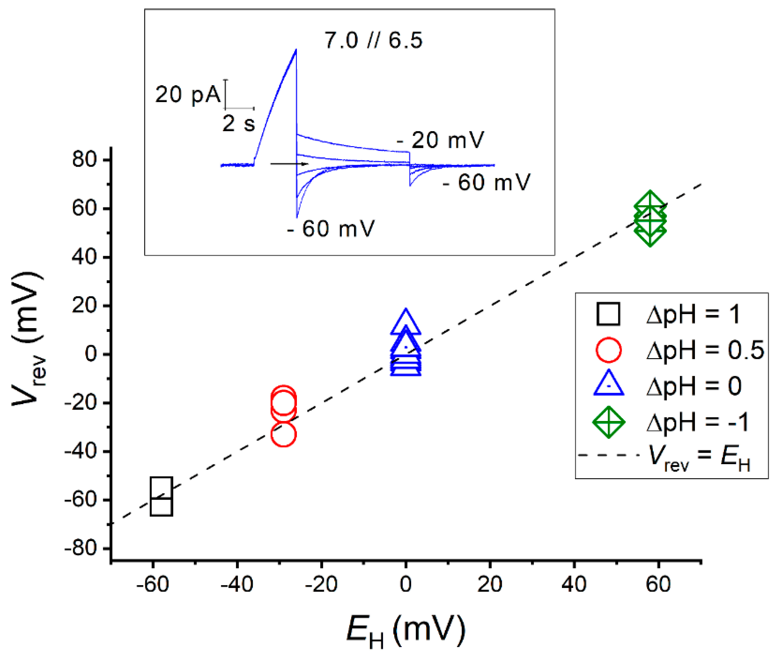
The selectivity of an HV channel can then be tested by measuring the reversal potential experimentally, at different proton gradients, and comparing with the Nernst potential for protons, EH (Equation (2)). Figure 5 illustrates an example of the experimental determination of the selectivity of a HV channel using electrophysiology.
Deviations of Vrev from EH values are associated to poor experimental control of the pHi, which can occur when there is depletion of the protonated buffer [86]. Hence, in small cells, such as those used as expression systems in patch-clamp studies, proton depletion is common at large depolarization. Protons conducted through HV channels leave the cell faster than the buffer can be replenished, due to the diffusion time from the pipette to the cell and to the dissociation time in the cytoplasmic bulk solution. Proton depletion leads to a rise in pHi and a consequent shift in Vrev from its nominal value (EH) to more positive potentials. Conversely, negative deviations of Vrev from EH may occur in rare cases where HV channels permit inward H+ conduction that acidifies the cytosol, making the accurate determination of Vrev challenging.
The direction of H+ flux is determined by both the pH- and voltage-dependence of gating, which is a characteristic of each channel. Inward directed conduction is possible at large inward pH gradients (pHo < pHi) for most of HV channels, even those considered as proton extruders. However, for a few HV channels, H+ influx occurs even at symmetrical pH conditions. For instance, the channel of the insect Extatosoma tiaratum (EtHV1) exhibits robust inward H+ currents during depolarization and slow closing times after hyperpolarization, leading to the acidification of the internal milieu that rapidly changes Vrev during electrophysiological protocols [121]. Furthermore, the aberrant type-3 HV channel from the mollusk Aplysia Californica (AcHV3) leaks H+ selective currents in the closed-state, constantly acidifying the cytoplasm and inducing new equilibriums at different voltage-pulses [124].
Although there are some experimental limitations in evaluating the selectivity of a proton channel, all HV channels tested to date have demonstrated perfect selectivity regardless of their ionic environment. The minimal H+/ion permeability rates are given in Table 4. The values depicted are either as reported by the authors or calculated from the experimental conditions as the quotient between the minimal proton concentration (higher pH) and the ion present in higher concentration.
Under physiological conditions, the concentration of protons is <106 times lower than that of other ions, e.g. the intracellular concentration of potassium ([K+]i) is around 150 mM, whereas the intracellular concentration of protons ([H+]i) is approximately 100 nM. Given this, the selectivity mechanism of proton channels must be considered extremely efficient.
By comparing the amino acid sequences of HV channels to the voltage-sensor domains (VSD) of other voltage-activated cation channels and of the non-conducting but voltage-sensing protein C15orf27 (TMEM266) [149], Musset et al. identified five amino acid residues potentially responsible for the exclusive proton conduction in HV channels. Single mutation of each of those candidates in the human proton channel (hHV1) to those found at equivalent positions in C15orf27, permitted to identified Asp112 as the selectivity filter of HV channels [141]. Furthermore, substitution of this aspartate to non-acidic amino acids made hHV1 permeable to anions, an observation that was also confirmed in dinoflagellate and insect HV channels [120,132].
Asp112 is located directly above a constriction of the channel that separates the internal and external solutions, approximately in the middle of the S1 alpha helix [142,150,151], and is highly conserved among HV molecules. To date, the only tested HV channel that presents a naturally occurring variation at this position is EtHV1. EtHV1 possesses a glutamate residue as a selectivity filter instead of the typical aspartate, a feature common to HV channels from stick and gladiator insects [121]. Despite this peculiarity, EtHV1 elicited H+ currents that were consistent with EH along different pH gradients (ΔpH = pHo − pHi), as it was observed by experimentally mutating the selectivity filter Asp to Glu in channels from other species [120,132,141].
Multiple evidence as the high temperature dependence of HV [47,87], the diminished deuterium conductance [83], the H+ hopping between pairs of charged residues lining HV pore detected in QM/MM molecular dynamics simulations [152], or more recently the proposed existence of an interrupted water wire within HV1 pore [153], suggest that a protonable carboxyl group is essential for the selectivity of HV channels. However, the mechanism underlying this selectivity involves also specific physicochemical microenvironments where H+ conduction and selectivity are coupled [120,132,141,154,155,156,157,158]. Thus, previous studies have shown that moving Asp112 along S1 helix of the channel affects proton selectivity [156], and further quantum mechanical calculations have revealed that an Asp can interact with a counter charge carried by the Arg sidechain to create an electrostatic barrier that repels other ions besides H+ [157]. The selectivity is, nevertheless, maintained when Arg is replaced with Lys [157]. While some studies support the interaction between Asp112 and Arg208 in S4 [120,142,150,156,159], others propose an alternative explanation where Asp112 interacts with Arg211 instead [158,160,161,162]. A 3D structure of hHV1 showing Asp112 in S1, and R208 and R2011 in S4, is displayed in Figure 2 (left).
According to the analysis of a vast collection of mutant HV channels, DeCoursey et al. proposed the minimal requirements of the selectivity filter of HV channels: a carboxyl group (Asp or Glu) facing the pore in a narrow region of the channel and the ability to interact with a counter charge (Arg or Lys) [163].

3.2. Molecule Architecture and the Voltage-Dependent Gating

Voltage-gated proton channels can detect electrochemical changes across the cell membrane and consequently open or close their conduction pathway; a process called gating. With membrane depolarization, HV channels open their gates and generate time-dependent H+ currents, while membrane hyperpolarization leads to channel closure. HV channels are formed by a bundle of four membrane-spanning alpha helices that are structurally similar to the VSD of other voltage-sensitive ion channels such as KV and NaV channels (Figure 2). Despite its resemblance to other cation channels, the constitutive pore segments, S5 and S6, are absent in HV channels. Proton channels are VSDs that selectively conduct H+ [110,118].
Experiments aimed at determining the stoichiometry of the channel have confirmed its dimeric nature across several species, including mammals, insects, ascidians, sea urchins, and reef-building corals [41,122,123,138,139,143,145,147,159] (Table 4). HV subunits are held together by a coiled-coil structure at the cytosolic C-terminal [138,139,143,159,164]. The only possible exceptions to HV oligomerization come from the unicellular organisms Karlodinium veneficum and Phaeodactylum tricornutum, where informatic tools did not predict a coiled-coil region at the C-terminal, thus enabling a potential monomer expression [132,165]. Dimerization is important for the regulation of other properties, such as channel kinetics or the inhibition by divalent cations, e.g., HV monomers activate faster and are less sensitive to Zn2+ than dimers [166]. Moreover, HV gating is also modulated by the cooperativity between the channel subunits [145,167,168,169,170]. Despite the importance of oligomerization for the function of HV channels, when the C-terminal is truncated to force the expression of monomeric forms, the resulting subunits demonstrate their own functional pore, conserving properties such as selectivity and voltage- and pH-dependence of gating [138,143,154].
Depolarization induces conformational changes in HV that precede proton conduction and drive the channel from a closed to an open configuration. It is commonly agreed that the S4 alpha helix moves upward before HV conduction, as proved by voltage-clamp fluorometry and patch clamp recordings [161,168,171,172]. Other studies suggest that other parts of the channel, such as S1, may also move during the transition from closed to open configuration [161]. Furthermore, these conformational changes are regulated not only by the membrane potential but also by the ΔpH [81,173,174,175,176] (Figure 6).
In order to provide a comprehensive understanding of HV gating, numerous mathematical models have been proposed. Cherny et al. generated the first model in 1995, for the rat channel RnHV1, which described gating regulation by pH with a three-state model [81]. Patch clamp measurements of dimeric and monomeric HVs suggested multiple voltage-dependent conformational changes confirming the Cole–Moore effect [96,146]. The voltage-dependent gating of Ciona intestinalis proton channel (CiHV1) was described by Gonzalez et al. using also a three-state model, with each transition translocating two effective gating charges [172]. In CiHV1 monomers, Carmona et al. measured gating currents and proposed that a model describing HV activation should have more than two-state kinetic steps (≥5 states) [146]. Analyzing the kinetics of human hHV1, Villalba-Galea proposed that the activation and the deactivation processes have different transitions, and suggested that a voltage-independent transition occurs before HV opening [177]. Chaves et al. produced a simplified model based on two-state kinetics for the Nicoletia channel (NpHV1) that described the channel activation kinetic well using parameters representing a dimer (power [n = 1.5–2.1]), similar to the ones used by Fujiwara et al. (power [n = 1.7]) in their kinetics analysis of the mouse mHV1 [147,178]. More recently, Suárez-Delgado et al. used a non-natural fluorescent amino acid set on the top of S4 of hHV1 and suggested the possibility of further movements in the VSD, even after channel opening. The authors generated a four-state model for channel activation similar to the voltage-dependent kinetic proposed by Villalba-Galea in 2014 [174]. Rangel-Yescas et al. conducted studies on the reef-building coral AmHV1 and created an allosteric eight-state model that couples proton binding to opening transitions [123].
Upon careful examination of the gating mechanism of proton channels, it becomes clear that the process is complex and lacks consensus among researchers. However, it is widely accepted that the voltage-sensing of HV channels works similarly to the other canonical VSDs [179,180,181]. In these VSDs, positive charges located at the S4 segment move across a narrow hydrophobic region of the VSD that separates the external and internal milieu. This hydrophobic region focuses the electric field across the cell membrane to form the charge transfer center (CTC) and it can be delimited by a conserved phenylalanine residue in S2 [181,182,183,184] (Figure 2).
Similarly, HV channels have a conserved Phe residue in S2 (Phe150 in hHV1) that belongs to a ring of hydrophobic residues located approximately in the middle of the membrane, called hydrophobic gasket (HG) [185] or hydrophobic plug [186]. The hydrophobic gasket regulates HV1 activation [187], and consequently, HV activation can be defined by the relative movement of charged amino acids across a point demarcated by amino acids at positions equivalent to Phe150 (Figure 6). In the S4 segment, HV channels possesses a series of three positively-charged arginine residues (R1, R2, and R3) organized in an archetypal RxWRxxR voltage-sensor motif (VSM) that are responsible for voltage sensing [172]. This characteristic VSM motif is highly conserved among most HV channels and, together with the selectivity filter in S1, has been used to identify HV homology sequences in TSA databases [120,121,123,124,125,132,147].
The movement of charged residues through the CTC during HV gating can be studied by determining the elementary charges (e0) that theoretically may correspond to arginine residues crossing the entire electrical field [188] or negatively charged residues moving in the other direction. In this regard, the estimation of effective gating charges in HV channels has been achieved by different methods, such as the limiting slope method of the G–V relationship in semi-log plots [147,172,189], Boltzmann fittings of normalized G–V curves [110,145], or direct measurement of gating currents [140,146]. As seen in Table 4, estimations in different species harboring HV channels show elementary charges ranging from 1.1 to 2.7 e0 (monomer) and 4.3 to 6.1 e0 (dimer), corresponding to three arginines in S4 per monomer.
The function of each charge carrier in voltage-sensing and HV-functioning channel has been assessed separately. Neutralization of the first arginine (R1) in hHV1 (R205A mutant) ( = 0.57, V0.5 = 99.5, ΔpH = −1) reduced the apparent voltage-dependence of gating compared to the WT channel ( = 0.90, V0.5 = 58.0, ΔpH = −1), indicating a drop in the effective charge moved [110]. Mutation of R1 to glutamine accelerates the activation kinetics and shifts the gHV relationship to more negative potentials, enabling the mouse HV1 to activate at more negative values than Vrev, thus allowing H+ influx [122]. In hHV1, R1 interacts with Asp185 (in S3) during gating, suggesting a change in the residue’s position during the process [190]. R1 is well conserved among HV channels with the exception of some unconventional channels, as the type-3 HV of A. californica (AcHV3) that displays a natural substitution to a hydrophobic leucine to generate an atypical LPWRxxR VSM. Interestingly, AcHV3 voltage-dependent gating appears to be altered, with reduced steepness of gHV curves (~60 mV/e-fold change) compared to AcHV1 and AcHV2 channels (4–6 mV/e-fold change), which in contrast retain the RxWRxxR VSM typical of regular HV channels [124]. The reduced voltage-dependence of gating of AcHV3 is hypothesized to be related to the leak of protons in the channel’s closed state and/or the inability of being measured at very negative potentials. Similarly, alterations of the physicochemical properties of the hydrophobic gasket (HG) of hHV1 generate H+ leak in the closed state [185].
Notably, the second arginine (R2) of the VSM is fully conserved in all HV channels found so far. R2 appears to have a dual role in HV channels, both acting as a charge carrier and interacting with Asp112 in the conducting state [157,176], to become an essential component of the proton-selectivity mechanism. In hHV1, arginine to histidine mutants were tested with Zn2+ in patch clamp experiments to reveal state-dependent interactions. During the essays, in the open state, both R2H and R1H were accessible to externally applied Zn2+. In the closed state, R2H mutant was internally accessible but not R1H, indicating a journey of R2 from the inner to the external water vestibule during HV activation [150]. Jardin et al., 2022, [176] suggested that the outward S4 movement focuses on the central region of the VSM, where R2 shuttles between the inner and outer vestibules of hHV1. Wu et al., 2022, proposed that Phe150 in the HG interacts with R2 in the closed state but with R3 in the open state [187].
Water accessibility studies have provided evidence that the third arginine (R3) is exposed to the intracellular medium in the closed configuration [142,150,153,156,159,172,176]. However, controversy arises regarding the journey of R3 and its final position in the open configuration. Different research groups have suggested that R3 interacts with Asp112 in the open state configuration, which supports a two-click hypothesis in Ciona and human HVs [158,160,161,162]. In contrast, other studies support a one-click hypothesis [150,157,159,176]. Neutralization of R3 to alanine in NpHV1 did not impair H+ selectivity at pHo = pHi = 5.5 [120]. R3K charge-conserved mutations in hHV1 conducted by Wu et al. revealed that the mutation affected H+ conduction by slowing the activation kinetics and shifting the channel conductance to more positive potentials. The study showed that R3–Phe150 interaction is important for the stabilization of the open state, as R2 moves further upwards during the closed to open transition [187]. Moreover, truncation of mHV1 between R2 and R3 did not prevent HV voltage-dependence [159].
The conservation of R3 among all HV channels is not fully established, and natural variations have been recently discovered. For instance, fungal HV channels and the newly discovered CgHV4 from the pacific oyster display a natural variation to a lysine (K3) at positions equivalent to R3, which generates a RxWRxxK signature [125,129]. This naturally occurring K3 is conserved in all HV4 channels of mollusks and in fungal HV1. The only exception is found in the fungi P. brasilianum, which has a glutamine variation. Gating charge calculations obtained from gHV relationship slopes resulted in ~5 e0 for AoHV1 and SlHV1 fungal channels, consistent with the ~6 e0 characteristic of dimeric HVs from other species (Table 4). There is no report of gating charge calculations or CgHV4 stoichiometry in Chaves et al., 2023b. However, CgHV4, similar to AoHV1 and SlHV1, displays a lag phase in the onset of H+ currents that may indicate cooperativity between HV subunits [145,166]. The exposition of R3 to the intracellular solution would provoke the pKa values for both R3 and K3 to be close to those reported for bulk solution (>10). Therefore, it is likely that K3 is protonated at physiological conditions and effectively serves as a gating charge carrier. The fact that a lysine residue can act as an effective gating charge carrier in VSDs is uncommon, as there is a peculiar prevalence of arginines over lysines. The reason for such a phenomenon is still not completely understood [191].

3.3. The pH-Dependent Gating and Its Physiological Implications

The modulation of the proton conductance (gH) by pH is a ubiquitous feature of HV channels and is fundamental to their physiological function. For most of the cases and particularly in mammals, the channel functions like an overpressure valve that opens as the concentration of H+ in the interior of the cell rises, thereby regulating cellular pHi homeostasis.
The pH dependence of gating of HV channels has been commonly described by the rule of forty, established by Cherny et al. for rat RnHV1 in 1995 [81], and apply to native channels in other species [192]. This rule states that HV channels shift their gH to more positive or negative potentials in ~40 mV per unit pH. The modulation of gH is independent of the side of the membrane where the pH change is applied (pHi or pHo) [81]. Therefore, a cellular process that acidifies the cytosol by a unit of pH (decreasing pHi) or alkalizes the extracellular space (increasing pHo) by a unit of pH will set the proton conductance to a membrane potential 40 mV more negative than its nominal value. Since most of native HV channels open ~20 mV positively to EH at symmetrical pH [97,192], the rule of forty explains why HV channels are considered proton extruders.
The amino acid residues responsible of ΔpH sensing remain a mystery. Ramsey et al., 2010, extensively studied single mutations of ionizable residues in hHV1, but none of the neutralized residues affected substantially the Δ40 mV/pH unit sensitivity [155], potentially pointing to the involvement of multiple groups of amino acids [180]. Moreover, the underlying molecular mechanism is independent of the characteristic position of the gHV curve along the voltage-axis of each channel. For example, the rule applies for both the Danio rerio channel (DrHV1) and Mus musculus channel (mHV1), although the first one activates more negatively than the latter [144].
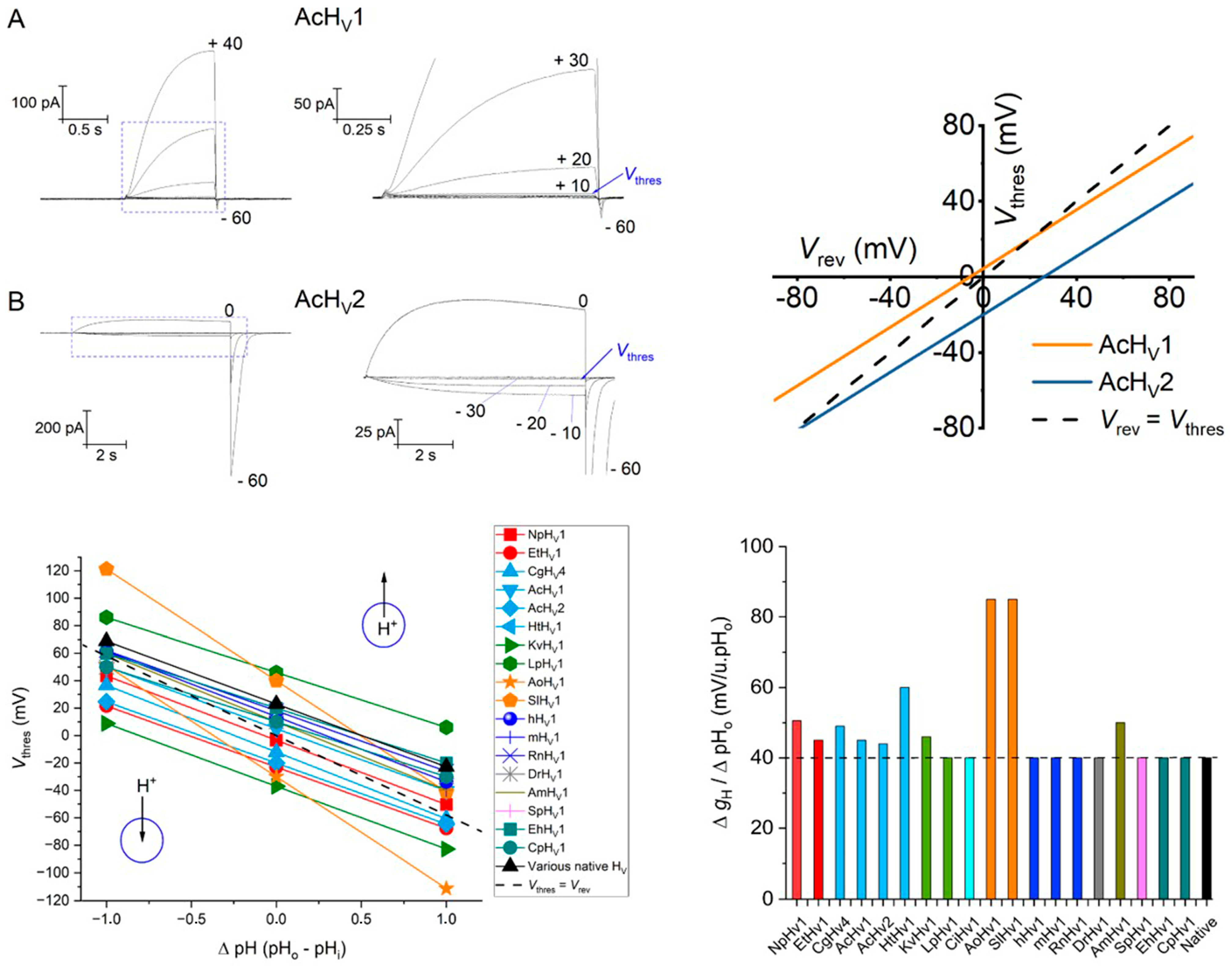
Nevertheless, a few exceptions to the rule of forty have been reported in the last years (Figure 7, bottom-right). For instance, the proton channel from the snail Helisoma trivolvis HtHV1 displays an anomalous ΔpH-dependent gating with weak pHi sensitivity (~15 mV/unit pHi) while its pHo sensitivity is increased (~60 mV/unit pHo) [148]. Other molluscan HV channels also present alterations in ΔpH-dependent gating. In A. Californica, AcHV1 and AcHV2 channels have identical pHo dependence of gating, which still falls within the range of the rule of forty (~43–45 mV/pHo unit). Yet, AcHV1 has a weak pHi sensitivity similar to that of HtHV1, while AcHV2 pHi sensitivity is normal (~40 mV/pHi unit) [124]. V0.5 vs. ΔpH plots, displayed by the channel from the reef-building coral Acropora millepora (AmHV1), show a slope steeper than 40 mV/ΔpH that deviates from linearity to saturate at high ΔpH [123]. Remarkably, fungal AoHV1 and SlHV1 exhibit the greatest deviation from the rule of forty (80–90 mV/ΔpH unit). Both channels have similar ΔpH-dependent gating, but SlHV1 displays an abnormal pHi sensitivity within the pH range from 5.5 to 6.5 (~18 mV/pHi unit), whereas for the same pH range, conduction of AoHV1 appears to be regulated exclusively by ΔpH instead of the absolute pH [129]. The NpHV1 and EtHV1 insect channels, the molluscan CgHV4, and the dinoflagellate kHV1 have all greater pH-dependent gating ranging between 45–50 mV/ΔpH [120,121,125,132]. There is also loss of linearity in the ΔgHV/ΔpH relationship at certain pH values in some species. For instance, the gHV curves of LpHV1 show saturation at pHo > 8 and pHi > 7.0 [133]. Similarly, the pH-dependence of gating of mammalian hHV1 and RnHV1, the coccolithophore channel from EhHV1, and the dinoflagellate kHV1 show deviations at pHo > 8.0 [81,148]. In HtHV1, there is no sign of saturation of gHV curves, but the slope of the VgH,max/10/ΔpH relationship increases at larger ΔpH [148]. The deviations from the rule of forty are hypothetically related to the strength of allosteric coupling between the voltage sensor and the proton-binding sites [123], while the saturation of gHV curves at large ΔpH are thought to be indicative of the pka values of those proton binding sites [81].
A molecular mechanism coupling the pH- and voltage-sensing would be of great help to understand the pH-dependent gating of HV channels. Despite this mechanism is yet unknown, several mathematical and atomistic models have been proposed to describe it. Markin established the first mathematical model for RnHV1 [81], and recently, Rangel-Yescas et al. developed a model for the reef coral building [123]. However, both models lack a clear explanation regarding how protonation of internal and external sites affect HV gating. In Sokolov et al. [193], two molecular mechanisms are proposed to regulate simultaneously: an electrostatic mechanism and a countercharge model. In the electrostatic mechanism, protons binding to the channel affect the way HV sense voltage, while in the countercharge model, protonation of acidic residues within the S1–S3 segments destabilizes salt bridge interactions that promote HV active or inactive states. Ramsey et al. [155] proposed that the pH gradient and the voltage-dependence of activation are regulated by the interaction of protonated waters with the S4 arginine residues in the pore of the channel. Jardin et al., 2022 [176] used a constant-pH molecular dynamics approach to obtain the protonation states of putative ionizable residues and the conformational changes in hHV1 driven solely by the influence of the pH. Similarly, Carmona et al. [175] suggested that the ΔpH-dependent gating of CiHV1 is a consequence of the motions of the voltage sensor movements in a state-dependent manner, regulated by pH. Using patch-clamp fluorometry and proton uncaging, Schladt and Berger proposed that ΔpH provokes changes in the VSM in S4 that couple voltage and pH sensing in CiHV1 [173]. Han et al. [194] also mentioned the affectation of pH on S4 movement of hHV1. The authors concluded that from three different conformations acquired by S4, two in the deactivated (intracellular and middle) and one in the activated (extracellular) configuration, solely the conformational transition between deactivated-intracellular to activated-extracellular displays both voltage- and pH-dependence. To date, the only residue that has been identified to alter pH sensing significantly is His168 (in hHV1 nomenclature) [195]. This His residue, located at the bottom of S3, close to the S2-S3 linker, and below Phe150, is critical for pHi sensing. The residue also accounts for the slow activation kinetics displayed by mammalian HV channels, as other faster channels from other species, such as H. trivolvis, L. stagnalis and S. purpuratus, show natural variations to Gln or Ser [195].
The pH-dependent gating of HV channels can be more easily described by a linear equation of the form:
Vthres = a·Vrev + b
a = slope (Vthres/Vrev) and b = Voffset (mV)
Thus, the pH dependence of a proton channel can be characterized by measuring the threshold voltage of activation (Vthres) and the reversal potentials (Vrev) in a wide range of ΔpH. While Vthres is an arbitrary parameter (the conductance of a channel is determined by the open probability as function of voltage), it can be generally estimated during patch-clamp protocols as the most negative voltage where the onset of H+ currents are observed, typically identified by the appearance of a first distinctive tail current after depolarizing pulses [97].
In order to compare the selected HV channels, we used reports from studies that have evaluated the pH-dependence of gating according to Equation (3). In cases where this information was not directly provided, we used Vthres at symmetrical pH conditions close to neutrality (e.g., pH 6.5–7.0), either explicitly stated by the authors or deduced from gHV relationships at different ΔpH, to obtain the linear function. The modulation of gH by pH as described before is restricted to certain pH ranges though. Figure 7 (bottom, left) displays the results for a ΔpHo range from −1 to +1 for 18 HV channels based on the information compiled in Table 4. The dotted line in Figure 7 shows parity between Vrev and Vthres. A channel activation negative to EH (Vthres < Vrev, data fitting below the dotted line) implies a driving force moving protons inwardly, and vice versa. In the absence of a pH gradient (ΔpH = 0), the direction of H+ flux is readily described by Voffset in Equation (3). This method permits a quick visualization of the direction of the H+ flux for the different channels.
The H+ flux direction, in conjunction with the physicochemical environment of the channel in physiological conditions, might indicate the role of HV channels in the different species. For example, the type-1 and type-2 HV channels from the mollusk A.californica have identical voltage-dependent gating (same Vthres/Vrev slope) but differ markedly in Voffset (Figure 7, upper right). While AcHV1 is a regular proton extruder, AcHV2 displays vigorous H+ inward currents to presumably generate action potentials related to the respiratory pumping reflex of the animal [124]. The AoHV1 from the fungus Aspergillus oryzae opens, when the electrochemical H+ gradient is inward, to consequently acidify and depolarize the cell. Nevertheless, its huge voltage-dependent gating (~90 mV pH/unit) would make the channel to act as a H+ extruder under suitable pH conditions [129]. Likewise, the abnormal ΔpH-dependent gating of HtHV1 might theoretically allow H+ influx under specific conditions [148]. The dinoflagellate LpHV1 displays a very positive Voffset (+46 mV), theoretically pointing to a proton extrusion function. However, LpHV1 carries inward H+ currents to trigger bioluminescence, as a consequence of large proton gradients found in Lingulodinium vacuoles (ΔpH = 3.5) [133]. In contrast, the other dinoflagellate channel, kHV1, has a significantly negative Voffset (−37 mV) and shows robust inward H+ currents even at symmetrical pH conditions. Karlodinium kHV1 conducts inward H+ currents over a wide pH-range, whose function is presumably related to feeding, as Karlodinium is not bioluminescent [132]. The channel from the insect Extatosoma (EtHV1) also presents robust H+ influx which may be related to different functions, including neuronal depolarization, regulation of ventilation rates, or the control of the acid–base transport systems [121]. Curiously, the other insect channel, NpHV1, displays pH-dependent gating identical to EtHV1, in the same voltage range, but its more positive Voffset makes NpHV1 a regular proton extruder [121].

3.4. Summary: Biophyscial Properties of HV Channels

The selectivity for protons in HV channels is a distinctive characteristic. Experimental determination involves comparing the reversal potential with the Nernst potential for protons, although deviations can occur due to poor control of intracellular proton concentration. Despite experimental limitations, HV channels have demonstrated an exceptional proton selectivity of at least 106 PH+/Pother ion, indicating a highly efficient mechanism. Specific residues, particularly Asp112, play a critical role in proton selectivity, and mutations of this residue can alter the channel permeability. Neutralizing Asp112 renders HV channels from humans, dinoflagellates, and insects permeable to anions. Asp112 is conserved and, in a proposed mechanism, interacts via salt bridge with a counter charge arginine near the channel constriction, establishing the selectivity filter. To date, the only variation of Asp112 is found in some polyneopteran insects, where a glutamate residue replaces it. Nevertheless, the charge is conserved and proton selectivity in such HV channels remains unaffected.
HV channels undergo gating in response to changes in membrane potential. They consist of four membrane-spanning alpha helices similar to the voltage-sensing domain (VSD) of other voltage-sensitive ion channels but lack specific pore segments. In nature, HV channels exist as dimers, and this oligomerization is important for regulating various properties. The gating mechanism of HV channels is complex and involves conformational changes in response to depolarization and/or pH differences. Several mathematical models have been proposed to describe HV gating, with the consensus being that the movement of the S4 segment is crucial. Three arginine residues in the S4 segment (R1, R2, and R3), organized in a typical RxWRxxR voltage-sensing motif (VSM), play a key role in voltage sensing and are thought to serve as the charge carriers. These charged residues move through a hydrophobic region in the middle of the channel (delimited by a conserved Phe residue) and their individual contribution has been studied. The first charge, R1, is exposed to the extracellular solution during gating and is naturally substituted by a lysine residue only in HV3 channels. HV3 channels have an atypical LPWRxxR VSM and display poor voltage sensing and H+ leakage in the closed state. R2 is fully conserved in all species and interacts with Asp112 in the open state to form the selectivity filter. Nevertheless, R3 is also proposed to interact with Asp112 in the open state, and there are different opinions regarding its final position in the open configuration. R3 is exposed to the intracellular solution in the closed configuration, and natural substitutions to lysine (K3) have been discovered recently in molluskan HV4 and fungal HV channels.
The modulation of proton conductance (gH) by pH in HV channels determines their physiological significance. This regulation has been commonly described by the “rule of forty” and applies for most HV channels. The property states that the channel shifts gH by approximately 40 mV per unit pH change, independently of the side of the membrane where the pH change occurs. However, several exceptions to this rule have been recently reported in different species, indicating variations in pH sensing. Some channels exhibit deviations from linearity or gH/ΔpH saturation at certain pH values. These deviations are thought to be related to the strength of allosteric coupling between the voltage sensor and proton-binding sites as well as an indication of the pka values of these binding sites. The molecular mechanism coupling pH and voltage sensing is not yet fully understood, and the amino acid residues responsible for pH sensing remain unknown. Various mathematical and atomistic models have been proposed, but none provide a clear explanation of how protonation of internal and external sites affects HV gating. Some models suggest that proton binding affects voltage sensing or destabilizes salt bridge interactions involved in channel activation. By using a linear relationship involving the threshold voltage of activation (Vthres) and reversal potentials (Vrev), the pH-dependent gating of HV channels can be compared and evaluated. Moreover, the H+ flux direction can be determined, and in conjunction with the physicochemical environment of the channels, insights into the functional roles of HV channels in different species can be gained. For example, most HV channels act as proton extruders, particularly in mammals, where their functioning is important to avoid the accumulation of acid in the cytosol or extreme depolarization of the cell membrane. In contrast, other species such as mollusks, insects, fungi, and dinoflagellates have HV channels that allow H+ influx under specific conditions. In these species, HV functions may range from triggering action potentials to initiating bioluminescent flashes.

Supplementary Materials

The following supporting information can be downloaded at: https://www.mdpi.com/article/10.3390/biom13071035/s1, Figure S1: Tree of life; Figure S2: Alignment of HV1 and HV2 from Xenopus; Figure S3: Cephalochordata sequences; Figure S4: Chelicerata sequences; Figure S5: Sequence alignment of two putative Oikopleura dioica HV channels with Ciona intestinalis; Figure S6: Multiple alignment of HV channels.

Author Contributions

Writing—original draft preparation, G.C., C.J., C.D. and B.M.; writing—review and ed-iting, G.C., C.J., C.D. and B.M. All authors have read and agreed to the published version of the manuscript.

Funding

This research received no external funding.

Data Availability Statement

All data is included in the manuscript or supplement.

Conflicts of Interest

The authors declare no conflict of interest.

References

  1. Thomas, R.C.; Meech, R.W. Hydrogen ion currents and intracellular pH in depolarized voltage-clamped snail neurones. Nature 1982, 299, 826–828. [Google Scholar] [CrossRef]
  2. Fogel, M.; Hastings, J.W. Bioluminescence: Mechanism and Mode of Control of Scintillon Activity. Proc. Natl. Acad. Sci. USA 1972, 69, 690–693. [Google Scholar] [CrossRef] [PubMed] [Green Version]
  3. Henderson, L.M.; Chappell, J.B.; Jones, O.T.G. The superoxide-generating NADPH oxidase of human neutrophils is electrogenic and associated with an H+ channel. Biochem. J. 1987, 246, 325–329. [Google Scholar] [CrossRef] [PubMed]
  4. Baldridge, C.W.; Gerard, R.W. The extra respiration of phagocytosis. Am. J. Physiol. Content 1932, 103, 235–236. [Google Scholar] [CrossRef] [Green Version]
  5. DeCoursey, T. Hydrogen ion currents in rat alveolar epithelial cells. Biophys. J. 1991, 60, 1243–1253. [Google Scholar] [CrossRef] [PubMed] [Green Version]
  6. DeCoursey, T.; Cherny, V. Potential, pH, and arachidonate gate hydrogen ion currents in human neutrophils. Biophys. J. 1993, 65, 1590–1598. [Google Scholar] [CrossRef] [Green Version]
  7. Bernheim, L.; Krause, R.M.; Baroffio, A.; Hamann, M.; Kaelin, A.; Bader, C.R. A voltage-dependent proton current in cultured human skeletal muscle myotubes. J. Physiol. 1993, 470, 313–333. [Google Scholar] [CrossRef] [Green Version]
  8. Demaurex, N.; Grinstein, S.; Jaconi, M.; Schlegel, W.; Lew, D.P.; Krause, K.H. Proton currents in human granulocytes: Regulation by membrane potential and intracellular pH. J. Physiol. 1993, 466, 329–344. [Google Scholar]
  9. Pick, E. Cell-Free NADPH Oxidase Activation Assays: “In Vitro Veritas”. Methods Mol. Biol. 2014, 1124, 339–403. [Google Scholar] [CrossRef]
  10. DeCoursey, T.E.; Morgan, D.; Cherny, V.V. The voltage dependence of NADPH oxidase reveals why phagocytes need proton channels. Nature 2003, 422, 531–534. [Google Scholar] [CrossRef]
  11. Ma, J.; Gao, X.; Li, Y.; DeCoursey, T.E.; Shull, G.E.; Wang, H. The HVCN1 voltage-gated proton channel contributes to pH regulation in canine ventricular myocytes. J. Physiol. 2022, 600, 2089–2103. [Google Scholar] [CrossRef] [PubMed]
  12. Morgan, D.; Capasso, M.; Musset, B.; Cherny, V.V.; Ríos, E.; Dyer, M.J.S.; DeCoursey, T.E. Voltage-gated proton channels maintain pH in human neutrophils during phagocytosis. Proc. Natl. Acad. Sci. USA 2009, 106, 18022–18027. [Google Scholar] [CrossRef] [PubMed] [Green Version]
  13. El Chemaly, A.; Okochi, Y.; Sasaki, M.; Arnaudeau, S.; Okamura, Y.; Demaurex, N. VSOP/Hv1 proton channels sustain calcium entry, neutrophil migration, and superoxide production by limiting cell depolarization and acidification. J. Exp. Med. 2010, 207, 129–139. [Google Scholar] [CrossRef] [PubMed] [Green Version]
  14. Okochi, Y.; Umemoto, E.; Okamura, Y. Hv1/VSOP regulates neutrophil directional migration and ERK activity by tuning ROS production. J. Leukoc. Biol. 2020, 107, 819–831. [Google Scholar] [CrossRef]
  15. Okochi, Y.; Aratani, Y.; Adissu, H.A.; Miyawaki, N.; Sasaki, M.; Suzuki, K.; Okamura, Y. The voltage-gated proton channel Hv1/VSOP inhibits neutrophil granule release. J. Leukoc. Biol. 2016, 99, 7–19. [Google Scholar] [CrossRef]
  16. Musset, B.; Morgan, D.; Cherny, V.V.; MacGlashan, D.W.; Thomas, L.L.; Ríos, E.; DeCoursey, T.E. A pH-stabilizing role of voltage-gated proton channels in IgE-mediated activation of human basophils. Proc. Natl. Acad. Sci. USA 2008, 105, 11020–11025. [Google Scholar] [CrossRef] [Green Version]
  17. Zhu, X.; Mose, E.; Zimmermann, N. Proton channel HVCN1 is required for effector functions of mouse eosinophils. BMC Immunol. 2013, 14, 24. [Google Scholar] [CrossRef] [Green Version]
  18. Wang, Y.; Zhang, S.; Li, S.J. Zn2+ induces apoptosis in human highly metastatic SHG-44 glioma cells, through inhibiting activity of the voltage-gated proton channel Hv1. Biochem. Biophys. Res. Commun. 2013, 438, 312–317. [Google Scholar] [CrossRef]
  19. Wang, Y.; Wu, X.; Li, Q.; Zhang, S.; Li, S.J. Human Voltage-Gated Proton Channel Hv1: A New Potential Biomarker for Diagnosis and Prognosis of Colorectal Cancer. PLoS ONE 2013, 8, e70550. [Google Scholar] [CrossRef] [Green Version]
  20. Wang, Y.; Li, S.J.; Pan, J.; Che, Y.; Yin, J.; Zhao, Q. Specific expression of the human voltage-gated proton channel Hv1 in highly metastatic breast cancer cells, promotes tumor progression and metastasis. Biochem. Biophys. Res. Commun. 2011, 412, 353–359. [Google Scholar] [CrossRef]
  21. Wang, Y.; Li, S.J.; Wu, X.; Che, Y.; Li, Q. Clinicopathological and Biological Significance of Human Voltage-gated Proton Channel Hv1 Protein Overexpression in Breast Cancer. J. Biol. Chem. 2012, 287, 13877–13888. [Google Scholar] [CrossRef] [PubMed] [Green Version]
  22. Li, X.; Liu, R.; Yu, Z.; He, D.; Zong, W.; Wang, M.; Xie, M.; Wang, W.; Luo, X. Microglial Hv1 exacerbates secondary damage after spinal cord injury in mice. Biochem. Biophys. Res. Commun. 2020, 525, 208–215. [Google Scholar] [CrossRef]
  23. Kawai, T.; Kayama, K.; Tatsumi, S.; Akter, S.; Miyawaki, N.; Okochi, Y.; Abe, M.; Sakimura, K.; Yamamoto, H.; Kihara, S.; et al. Regulation of hepatic oxidative stress by voltage-gated proton channels (Hv1/VSOP) in Kupffer cells and its potential relationship with glucose metabolism. FASEB J. 2020, 34, 15805–15821. [Google Scholar] [CrossRef] [PubMed]
  24. Pang, H.; Li, J.; Du, H.; Gao, Y.; Lv, J.; Liu, Y.; Li, S.J. Loss of voltage-gated proton channel Hv1 leads to diet-induced obesity in mice. BMJ Open Diabetes Res. Care 2020, 8, e000951. [Google Scholar] [CrossRef]
  25. Wang, X.; Gao, Y.-T.; Jiang, D.; Wang, Y.; Du, H.; Lv, J.; Li, S.J. Hv1-deficiency protects β cells from glucotoxicity through regulation of NOX4 level. Biochem. Biophys. Res. Commun. 2019, 513, 434–438. [Google Scholar] [CrossRef]
  26. Capasso, M.; Bhamrah, M.K.; Henley, T.; Boyd, R.S.; Langlais, C.; Cain, K.; Dinsdale, D.; Pulford, K.; Khan, M.; Musset, B.; et al. HVCN1 modulates BCR signal strength via regulation of BCR-dependent generation of reactive oxygen species. Nat. Immunol. 2010, 11, 265–272. [Google Scholar] [CrossRef] [Green Version]
  27. Hondares, E.; Brown, M.A.; Musset, B.; Morgan, D.; Cherny, V.V.; Taubert, C.; Bhamrah, M.K.; Coe, D.; Marelli-Berg, F.; Gribben, J.G.; et al. Enhanced activation of an amino-terminally truncated isoform of the voltage-gated proton channel HVCN1 enriched in malignant B cells. Proc. Natl. Acad. Sci. USA 2014, 111, 18078–18083. [Google Scholar] [CrossRef] [Green Version]
  28. Montes-Cobos, E.; Huscher, B.; Engler, J.B.; Woo, M.S.; Binkle, L.; Bauer, S.; Kursawe, N.; Moles, M.; Friese, M.A.; Ufer, F. Voltage-Gated Proton Channel Hv1 Controls TLR9 Activation in Plasmacytoid Dendritic Cells. J. Immunol. 2020, 205, 3001–3010. [Google Scholar] [CrossRef] [PubMed]
  29. Sasaki, M.; Tojo, A.; Okochi, Y.; Miyawaki, N.; Kamimura, D.; Yamaguchi, A.; Murakami, M.; Okamura, Y. Autoimmune disorder phenotypes in Hvcn1-deficient mice. Biochem. J. 2013, 450, 295–301. [Google Scholar] [CrossRef] [PubMed] [Green Version]
  30. Murphy, R.; Cherny, V.V.; Morgan, D.; DeCoursey, T.E. Voltage-gated proton channels help regulate pHiin rat alveolar epithelium. Am. J. Physiol. Cell Mol. Physiol. 2005, 288, L398–L408. [Google Scholar] [CrossRef] [Green Version]
  31. Schwarzer, C.; Machen, T.E.; Illek, B.; Fischer, H. NADPH Oxidase-dependent Acid Production in Airway Epithelial Cells. J. Biol. Chem. 2004, 279, 36454–36461. [Google Scholar] [CrossRef] [Green Version]
  32. Lishko, P.V.; Botchkina, I.L.; Fedorenko, A.; Kirichok, Y. Acid Extrusion from Human Spermatozoa Is Mediated by Flagellar Voltage-Gated Proton Channel. Cell 2010, 140, 327–337. [Google Scholar] [CrossRef] [PubMed] [Green Version]
  33. Zhao, R.; Dai, H.; Arias, R.J.; De Blas, G.A.; Orta, G.; Pavarotti, M.A.; Shen, R.; Perozo, E.; Mayorga, L.S.; Darszon, A.; et al. Direct activation of the proton channel by albumin leads to human sperm capacitation and sustained release of inflammatory mediators by neutrophils. Nat. Commun. 2021, 12, 3855. [Google Scholar] [CrossRef]
  34. Smith, R.Y.; Morgan, D.; Sharma, L.; Cherny, V.V.; Tidswell, N.; Molo, M.W.; DeCoursey, T.E. Voltage-gated proton channels exist in the plasma membrane of human oocytes. Hum. Reprod. 2019, 34, 1974–1983. [Google Scholar] [CrossRef]
  35. Alvear-Arias, J.J.; Carrillo, C.; Villar, J.P.; Garcia-Betancourt, R.; Peña-Pichicoi, A.; Fernandez, A.; Fernandez, M.; Carmona, E.M.; Pupo, A.; Neely, A.; et al. Expression of H(v)1 proton channels in myeloid-derived suppressor cells (MDSC) and its potential role in T cell regulation. Proc. Natl. Acad. Sci. USA 2022, 119, e2104453119. [Google Scholar] [CrossRef]
  36. Gordienko, D.V.; Tare, M.; Parveen, S.; Fenech, C.J.; Robinson, C.; Bolton, T.B. Voltage-activated proton current in eosinophils from human blood. J. Physiol. 1996, 496 (Pt 2), 299–316. [Google Scholar] [CrossRef] [Green Version]
  37. Kovács, I.; Horváth, M.; Kovács, T.; Somogyi, K.; Tretter, L.; Geiszt, M.; Petheő, G.L. Comparison of proton channel, phagocyte oxidase, and respiratory burst levels between human eosinophil and neutrophil granulocytes. Free. Radic. Res. 2014, 48, 1190–1199. [Google Scholar] [CrossRef] [PubMed] [Green Version]
  38. Morgan, D.; Cherny, V.V.; Finnegan, A.; Bollinger, J.; Gelb, M.H.; DeCoursey, T.E. Sustained activation of proton channels and NADPH oxidase in human eosinophils and murine granulocytes requires PKC but not cPLA2α activity. J. Physiol. 2007, 579, 327–344. [Google Scholar] [CrossRef]
  39. Morgan, D.; Cherny, V.V.; Murphy, R.; Katz, B.Z.; DeCoursey, T.E. The pH dependence of NADPH oxidase in human eosinophils. J. Physiol. 2005, 569, 419–431. [Google Scholar] [CrossRef]
  40. Morgan, D.; Cherny, V.V.; Murphy, R.; Xu, W.; Thomas, L.L.; DeCoursey, T. Temperature dependence of NADPH oxidase in human eosinophils. J. Physiol. 2003, 550, 447–458. [Google Scholar] [CrossRef] [PubMed]
  41. Petheő, G.L.; Orient, A.; Baráth, M.; Kovács, I.; Réthi, B.; Lányi, A.; Rajki, A.; Rajnavölgyi, E.; Geiszt, M. Molecular and Functional Characterization of Hv1 Proton Channel in Human Granulocytes. PLoS ONE 2010, 5, e14081. [Google Scholar] [CrossRef] [Green Version]
  42. Schrenzel, J.; Lew, D.P.; Krause, K.H. Proton currents in human eosinophils. Am. J. Physiol. Physiol. 1996, 271, C1861–C1871. [Google Scholar] [CrossRef]
  43. Schrenzel, J.; Serrander, L.; Bánfi, B.; Nüße, O.; Fouyouzi, R.; Lew, D.P.; Demaurex, N.; Krause, K.-H. Electron currents generated by the human phagocyte NADPH oxidase. Nature 1998, 392, 734–737. [Google Scholar] [CrossRef]
  44. DeCoursey, T.E.; Cherny, V.V.; DeCoursey, A.G.; Xu, W.; Thomas, L.L. Interactions between NADPH oxidase-related proton and electron currents in human eosinophils. J. Physiol. 2001, 535, 767–781. [Google Scholar] [CrossRef]
  45. Femling, J.K.; Cherny, V.V.; Morgan, D.; Rada, B.; Davis, A.P.; Czirják, G.; Enyedi, P.; England, S.K.; Moreland, J.G.; Ligeti, E.; et al. The Antibacterial Activity of Human Neutrophils and Eosinophils Requires Proton Channels but Not BK Channels. J. Gen. Physiol. 2006, 127, 659–672. [Google Scholar] [CrossRef] [PubMed] [Green Version]
  46. Cherny, V.V.; Henderson, L.M.; Xu, W.; Thomas, L.L.; DeCoursey, T.E. Activation of NADPH oxidase-related proton and electron currents in human eosinophils by arachidonic acid. J. Physiol. 2001, 535, 783–794. [Google Scholar] [CrossRef]
  47. DeCoursey, T.E.; Cherny, V.V. Temperature dependence of voltage-gated H+ currents in human neutrophils, rat alveolar epithelial cells, and mammalian phagocytes. J. Gen. Physiol. 1998, 112, 503–522. [Google Scholar] [CrossRef] [PubMed] [Green Version]
  48. DeCoursey, T.E.; Cherny, V.V.; Zhou, W.; Thomas, L.L. Simultaneous activation of NADPH oxidase-related proton and electron currents in human neutrophils. Proc. Natl. Acad. Sci. USA 2000, 97, 6885–6889. [Google Scholar] [CrossRef] [Green Version]
  49. DeCoursey, T.E.; Cherny, V.V.; Morgan, D.; Katz, B.Z.; Dinauer, M.C. The gp91 Component of NADPH Oxidase Is Not the Voltage-gated Proton Channel in Phagocytes, but It Helps. J. Biol. Chem. 2001, 276, 36063–36066. [Google Scholar] [CrossRef] [PubMed] [Green Version]
  50. Okochi, Y.; Sasaki, M.; Iwasaki, H.; Okamura, Y. Voltage-gated proton channel is expressed on phagosomes. Biochem. Biophys. Res. Commun. 2009, 382, 274–279. [Google Scholar] [CrossRef]
  51. Ramsey, I.S.; Ruchti, E.; Kaczmarek, J.S.; Clapham, D.E. Hv1 proton channels are required for high-level NADPH oxidase-dependent superoxide production during the phagocyte respiratory burst. Proc. Natl. Acad. Sci. USA 2009, 106, 7642–7647. [Google Scholar] [CrossRef] [Green Version]
  52. El Chemaly, A.; Nunes, P.; Jimaja, W.; Castelbou, C.; Demaurex, N. Hv1 proton channels differentially regulate the pH of neutrophil and macrophage phagosomes by sustaining the production of phagosomal ROS that inhibit the delivery of vacuolar ATPases. J. Leukoc. Biol. 2014, 95, 827–839. [Google Scholar] [CrossRef] [PubMed]
  53. Droste, A.; Chaves, G.; Stein, S.; Trzmiel, A.; Schweizer, M.; Karl, H.; Musset, B. Zinc accelerates respiratory burst termination in human PMN. Redox Biol. 2021, 47, 102133. [Google Scholar] [CrossRef] [PubMed]
  54. Musset, B.; Cherny, V.V.; DeCoursey, T.E. Strong glucose dependence of electron current in human monocytes. Am. J. Physiol. Physiol. 2012, 302, C286–C295. [Google Scholar] [CrossRef] [Green Version]
  55. DeCoursey, T.E.; Kim, S.Y.; Silver, M.R.; Quandt, F.N. Ion channel expression in PMA-differentiated human THP-1 macrophages. J. Membr. Biol. 1996, 152, 141–157. [Google Scholar] [CrossRef] [PubMed]
  56. Kapus, A.; Romanek, R.; Qu, A.Y.; Rotstein, O.D.; Grinstein, S. A pH-sensitive and voltage-dependent proton conductance in the plasma membrane of macrophages. J. Gen. Physiol. 1993, 102, 729–760. [Google Scholar] [CrossRef] [Green Version]
  57. Mori, H.; Sakai, H.; Morihata, H.; Kawawaki, J.; Amano, H.; Yamano, T.; Kuno, M. Regulatory Mechanisms and Physiological Relevance of a Voltage-Gated H+ Channel in Murine Osteoclasts: Phorbol Myristate Acetate Induces Cell Acidosis and the Channel Activation. J. Bone Miner. Res. 2003, 18, 2069–2076. [Google Scholar] [CrossRef]
  58. Mori, H.; Sakai, H.; Morihata, H.; Yamano, T.; Kuno, M. A voltage-gated H+ channel is a powerful mechanism for pH homeostasis in murine osteoclasts. Kobe J. Med. Sci. 2002, 48, 87–96. [Google Scholar]
  59. Nordström, T.; Rotstein, O.D.; Romanek, R.; Asotra, S.; Heersche, J.N.M.; Manolson, M.F.; Brisseau, G.F.; Grinstein, S. Regulation of Cytoplasmic pH in Osteoclasts. J. Biol. Chem. 1995, 270, 2203–2212. [Google Scholar] [CrossRef] [Green Version]
  60. Sakai, H.; Kawawaki, J.; Moriura, Y.; Mori, H.; Morihata, H.; Kuno, M. pH dependence and inhibition by extracellular calcium of proton currents via plasmalemmal vacuolar-type H+-ATPase in murine osteoclasts. J. Physiol. 2006, 576, 417–425. [Google Scholar] [CrossRef]
  61. Sakai, H.; Li, G.; Hino, Y.; Moriura, Y.; Kawawaki, J.; Sawada, M.; Kuno, M. Increases in intracellular pH facilitate endocytosis and decrease availability of voltage-gated proton channels in osteoclasts and microglia. J. Physiol. 2013, 591, 5851–5866. [Google Scholar] [CrossRef]
  62. Eder, C.; Fischer, H.-G.; Hadding, U.; Heinemann, U. Properties of voltage-gated currents of microglia developed using macrophage colony-stimulating factor. Pflugers Arch. 1995, 430, 526–533. [Google Scholar] [CrossRef]
  63. McLarnon, J.; Xu, R.; Lee, Y.; Kim, S. Ion channels of human microglia in culture. Neuroscience 1997, 78, 1217–1228. [Google Scholar] [CrossRef]
  64. Morihata, H.; Nakamura, F.; Tsutada, T.; Kuno, M. Potentiation of a Voltage-Gated Proton Current in Acidosis-Induced Swelling of Rat Microglia. J. Neurosci. 2000, 20, 7220–7227. [Google Scholar] [CrossRef] [PubMed] [Green Version]
  65. Visentin, S.; Agresti, C.; Patrizio, M.; Levi, G. Ion channels in rat microglia and their different sensitivity to lipopolysaccharide and interferon? J. Neurosci. Res. 1995, 42, 439–451. [Google Scholar] [CrossRef] [PubMed]
  66. Peng, J.; Yi, M.-H.; Jeong, H.; McEwan, P.P.; Zheng, J.; Wu, G.; Ganatra, S.; Ren, Y.; Richardson, J.R.; Oh, S.B.; et al. The voltage-gated proton channel Hv1 promotes microglia-astrocyte communication and neuropathic pain after peripheral nerve injury. Mol. Brain 2021, 14, 99. [Google Scholar] [CrossRef]
  67. Wu, L.-J.; Wu, G.; Sharif, M.R.A.; Baker, A.; Jia, Y.; Fahey, F.H.; Luo, H.R.; Feener, E.P.; Clapham, D.E. The voltage-gated proton channel Hv1 enhances brain damage from ischemic stroke. Nat. Neurosci. 2012, 15, 565–573. [Google Scholar] [CrossRef] [PubMed] [Green Version]
  68. Wu, L.-J. Voltage-gated proton channel HV1 in microglia, The Neuroscientist: A review journal bringing neurobiology, neurology and psychiatry. Neuroscientist 2014, 20, 599–609. [Google Scholar] [CrossRef] [PubMed]
  69. Ritzel, R.M.; He, J.; Li, Y.; Cao, T.; Khan, N.; Shim, B.; Sabirzhanov, B.; Aubrecht, T.; Stoica, B.A.; Faden, A.I.; et al. Proton extrusion during oxidative burst in microglia exacerbates pathological acidosis following traumatic brain injury. Glia 2020, 69, 746–764. [Google Scholar] [CrossRef]
  70. Tian, D.-S.; Li, C.-Y.; Qin, C.; Murugan, M.; Wu, L.-J.; Liu, J.-L. Deficiency in the voltage-gated proton channel Hv1 increases M2 polarization of microglia and attenuates brain damage from photothrombotic ischemic stroke. J. Neurochem. 2016, 139, 96–105. [Google Scholar] [CrossRef] [Green Version]
  71. Liu, J.; Tian, D.; Murugan, M.; Eyo, U.B.; Dreyfus, C.F.; Wang, W.; Wu, L. Microglial Hv1 proton channel promotes cuprizone-induced demyelination through oxidative damage. J. Neurochem. 2015, 135, 347–356. [Google Scholar] [CrossRef] [Green Version]
  72. Chen, M.; Yang, L.-L.; Hu, Z.-W.; Qin, C.; Zhou, L.-Q.; Duan, Y.-L.; Bosco, D.B.; Wu, L.-J.; Zhan, K.-B.; Xu, S.-B.; et al. Deficiency of microglial Hv1 channel is associated with activation of autophagic pathway and ROS production in LPC-induced demyelination mouse model. J. Neuroinflammation 2020, 17, 333. [Google Scholar] [CrossRef]
  73. Yu, Y.; Luo, X.; Li, C.; Ding, F.; Wang, M.; Xie, M.; Yu, Z.; Ransom, B.R.; Wang, W. Microglial Hv1 proton channels promote white matter injuries after chronic hypoperfusion in mice. J. Neurochem. 2020, 152, 350–367. [Google Scholar] [CrossRef] [PubMed]
  74. Murugan, M.; Zheng, J.; Wu, G.; Mogilevsky, R.; Zheng, X.; Hu, P.; Wu, J.; Wu, L.-J. The voltage-gated proton channel Hv1 contributes to neuronal injury and motor deficits in a mouse model of spinal cord injury. Mol. Brain 2020, 13, 143. [Google Scholar] [CrossRef] [PubMed]
  75. Li, X.; Yu, Z.; Zong, W.; Chen, P.; Li, J.; Wang, M.; Ding, F.; Xie, M.; Wang, W.; Luo, X. Deficiency of the microglial Hv1 proton channel attenuates neuronal pyroptosis and inhibits inflammatory reaction after spinal cord injury. J. Neuroinflammation 2020, 17, 263. [Google Scholar] [CrossRef]
  76. Li, Y.; Ritzel, R.M.; He, J.; Cao, T.; Sabirzhanov, B.; Li, H.; Liu, S.; Wu, L.-J.; Wu, J. The voltage-gated proton channel Hv1 plays a detrimental role in contusion spinal cord injury via extracellular acidosis-mediated neuroinflammation. Brain Behav. Immun. 2021, 91, 267–283. [Google Scholar] [CrossRef]
  77. Zhang, Z.; Zheng, X.; Zhang, X.; Zhang, Y.; Huang, B.; Luo, T. Aging alters Hv1-mediated microglial polarization and enhances neuroinflammation after peripheral surgery. CNS Neurosci. Ther. 2020, 26, 374–384. [Google Scholar] [CrossRef] [PubMed]
  78. El Chemaly, A.; Guinamard, R.; Demion, M.; Fares, N.; Jebara, V.; Faivre, J.-F.; Bois, P. A voltage-activated proton current in human cardiac fibroblasts. Biochem. Biophys. Res. Commun. 2006, 340, 512–516. [Google Scholar] [CrossRef]
  79. Zhao, R.; Kennedy, K.; De Blas, G.A.; Orta, G.; Pavarotti, M.A.; Arias, R.J.; de la Vega-Beltrán, J.L.; Li, Q.; Dai, H.; Perozo, E.; et al. Role of human Hv1 channels in sperm capacitation and white blood cell respiratory burst established by a designed peptide inhibitor. Proc. Natl. Acad. Sci. USA 2018, 115, E11847–E11856. [Google Scholar] [CrossRef] [Green Version]
  80. Musset, B.; Clark, R.A.; DeCoursey, T.E.; Petheo, G.L.; Geiszt, M.; Chen, Y.; Cornell, J.E.; Eddy, C.A.; Brzyski, R.G.; El Jamali, A. NOX5 in human spermatozoa: Expression, function, and regulation. J. Biol. Chem. 2012, 287, 9376–9388. [Google Scholar] [CrossRef] [Green Version]
  81. DeCoursey, T.; Cherny, V.V. Na(+)-H+ antiport detected through hydrogen ion currents in rat alveolar epithelial cells and human neutrophils. J. Gen. Physiol. 1994, 103, 755–785. [Google Scholar] [CrossRef] [Green Version]
  82. Cherny, V.V.; Markin, V.S.; DeCoursey, T.E. The voltage-activated hydrogen ion conductance in rat alveolar epithelial cells is determined by the pH gradient. J. Gen. Physiol. 1995, 105, 861–896. [Google Scholar] [CrossRef] [PubMed] [Green Version]
  83. DeCoursey, T.E.; Cherny, V.V. Voltage-activated proton currents in membrane patches of rat alveolar epithelial cells. J. Physiol. 1995, 489 (Pt 2), 299–307. [Google Scholar] [CrossRef] [Green Version]
  84. DeCoursey, T.E.; Cherny, V.V. Deuterium Isotope Effects on Permeation and Gating of Proton Channels in Rat Alveolar Epithelium. J. Gen. Physiol. 1997, 109, 415–434. [Google Scholar] [CrossRef] [Green Version]
  85. DeCoursey, T.E. Four varieties of voltage-gated proton channels. Front. Biosci. 1998, 3, d477–d482. [Google Scholar] [CrossRef] [PubMed] [Green Version]
  86. Cherny, V.V.; DeCoursey, T.E. Ph-Dependent Inhibition of Voltage-Gated H+ Currents in Rat Alveolar Epithelial Cells by Zn2+ and Other Divalent Cations. J. Gen. Physiol. 1999, 114, 819–838. [Google Scholar] [CrossRef] [PubMed] [Green Version]
  87. DeCoursey, T.; Cherny, V. Effects of buffer concentration on voltage-gated H+ currents: Does diffusion limit the conductance? Biophys. J. 1996, 71, 182–193. [Google Scholar] [CrossRef] [PubMed] [Green Version]
  88. Kuno, M.; Kawawaki, J.; Nakamura, F. A Highly Temperature-sensitive Proton Current in Mouse Bone Marrow–derived Mast Cells. J. Gen. Physiol. 1997, 109, 731–740. [Google Scholar] [CrossRef] [Green Version]
  89. Schilling, T.; Gratopp, A.; DeCoursey, T.; Eder, C. Voltage-activated proton currents in human lymphocytes. J. Physiol. 2002, 545, 93–105. [Google Scholar] [CrossRef]
  90. Musset, B.; Capasso, M.; Cherny, V.V.; Morgan, D.; Bhamrah, M.; Dyer, M.J.S.; DeCoursey, T.E. Identification of Thr29 as a Critical Phosphorylation Site That Activates the Human Proton Channel Hvcn1 in Leukocytes. J. Biol. Chem. 2010, 285, 5117–5121. [Google Scholar] [CrossRef] [Green Version]
  91. Asuaje, A.; Martín, P.; Enrique, N.; Zegarra, L.A.D.; Smaldini, P.; Docena, G.; Milesi, V. Diphenhydramine inhibits voltage-gated proton channels (Hv1) and induces acidification in leukemic Jurkat T cells- New insights into the pro-apoptotic effects of antihistaminic drugs. Channels 2017, 12, 58–64. [Google Scholar] [CrossRef] [Green Version]
  92. Asuaje, A.; Smaldini, P.; Martín, P.; Enrique, N.; Orlowski, A.; Aiello, E.A.; León, C.G.; Docena, G.; Milesi, V. The inhibition of voltage-gated H+ channel (HVCN1) induces acidification of leukemic Jurkat T cells promoting cell death by apoptosis. Pflugers Arch. 2017, 469, 251–261. [Google Scholar] [CrossRef] [PubMed]
  93. Coe, D.; Poobalasingam, T.; Fu, H.; Bonacina, F.; Wang, G.; Morales, V.; Moregola, A.; Mitro, N.; Cheung, K.C.; Ward, E.J.; et al. Loss of voltage-gated hydrogen channel 1 expression reveals heterogeneous metabolic adaptation to intracellular acidification by T cells. JCI Insight 2022, 7, 147814. [Google Scholar] [CrossRef] [PubMed]
  94. Cherny, V.V.; Thomas, L.L.; DeCoursey, T.E. Voltage-gated proton currents in human basophils. Biol. Membrany. 2001, 18, 458–465. [Google Scholar]
  95. Cherny, V.V.; Henderson, L.M.; DeCoursey, T.E. Proton and chloride currents in Chinese hamster ovary cells. Membr. Cell Biol. 1997, 11, 337–347. [Google Scholar]
  96. Bare, D.J.; Cherny, V.V.; DeCoursey, T.E.; Abukhdeir, A.M.; Morgan, D. Expression and function of voltage gated proton channels (Hv1) in MDA-MB-231 cells. PLoS ONE 2020, 15, e0227522. [Google Scholar] [CrossRef] [PubMed]
  97. DeCoursey, T.E.; Cherny, V.V. Voltage-activated hydrogen ion currents. J. Membr. Biol. 1994, 141, 203–223. [Google Scholar] [CrossRef]
  98. Musset, B.; Cherny, V.V.; Morgan, D.; Okamura, Y.; Ramsey, I.S.; Clapham, D.E.; DeCoursey, T.E. Detailed comparison of expressed and native voltage-gated proton channel currents. J. Physiol. 2008, 586, 2477–2486. [Google Scholar] [CrossRef]
  99. Fischer, H.; Widdicombe, J.H.; Illek, B. Acid secretion and proton conductance in human airway epithelium. Am. J. Physiol. Cell Physiol. 2002, 282, C736–C743. [Google Scholar] [CrossRef] [Green Version]
  100. Fischer, H. Function of proton channels in lung epithelia. Wiley Interdiscip. Rev. Membr. Transp. Signal. 2012, 1, 247–258. [Google Scholar] [CrossRef] [PubMed] [Green Version]
  101. Ribeiro-Silva, L.; Queiroz, F.O.; da Silva, A.M.B.; Hirata, A.E.; Arcisio-Miranda, M. Voltage-Gated Proton Channel in Human Glioblastoma Multiforme Cells. ACS Chem. Neurosci. 2016, 7, 864–869. [Google Scholar] [CrossRef] [PubMed]
  102. Wu, X.; Li, Y.; Maienschein-Cline, M.; Feferman, L.; Wu, L.; Hong, L. RNA-Seq Analyses Reveal Roles of the HVCN1 Proton Channel in Cardiac pH Homeostasis. Front. Cell Dev. Biol. 2022, 10, 860502. [Google Scholar] [CrossRef] [PubMed]
  103. Zheng, Z.; Zhang, Z.; Wang, M. Hv1 proton channel possibly promotes atherosclerosis by regulating reactive oxygen species production. Med. Hypotheses 2020, 141, 109724. [Google Scholar] [CrossRef] [PubMed]
  104. Du, H.; Pang, H.; Gao, Y.; Zhou, Y.; Li, S.J. Deficiency of voltage-gated proton channel Hv1 aggravates ovalbumin-induced allergic lung asthma in mice. Int. Immunopharmacol. 2021, 96, 107640. [Google Scholar] [CrossRef]
  105. Pang, H.; Wang, X.; Zhao, S.; Xi, W.; Lv, J.; Qin, J.; Zhao, Q.; Che, Y.; Chen, L.; Li, S.J. Loss of the voltage-gated proton channel Hv1 decreases insulin secretion and leads to hyperglycemia and glucose intolerance in mice. J. Biol. Chem. 2020, 295, 3601–3613. [Google Scholar] [CrossRef] [Green Version]
  106. Wang, X.; Xi, W.; Qin, J.; Lv, J.; Wang, Y.; Zhang, T.; Li, S.J. Deficiency of voltage-gated proton channel Hv1 attenuates streptozotocin-induced β-cell damage. Biochem. Biophys. Res. Commun. 2018, 498, 975–980. [Google Scholar] [CrossRef]
  107. Nau, C. Voltage-Gated Ion Channels. In Modern Anesthetics; Springer: Berlin/Heidelberg, Germany, 2008; Volume 182, pp. 85–92. [Google Scholar] [CrossRef]
  108. Hille, B. Ion Channels of Excitable Membranes, 3rd ed.; Sinauer Associates, Inc.: Sunderland, UK, 2001. [Google Scholar]
  109. Ramsey, I.S.; Moran, M.M.; Chong, J.A.; Clapham, D.E. A voltage-gated proton-selective channel lacking the pore domain. Nature 2006, 440, 1213–1216. [Google Scholar] [CrossRef] [PubMed] [Green Version]
  110. Chen, X.; Wang, Q.; Ni, F.; Ma, J. Structure of the full-length Shaker potassium channel Kv1.2 by normal-mode-based X-ray crystallographic refinement. Proc. Natl. Acad. Sci. USA 2010, 107, 11352–11357. [Google Scholar] [CrossRef] [Green Version]
  111. Payandeh, J.; Scheuer, T.; Zheng, N.; Catterall, W.A. The crystal structure of a voltage-gated sodium channel. Nature 2011, 475, 353–358. [Google Scholar] [CrossRef] [Green Version]
  112. Derst, C.; Karschin, A. Evolutionary link between prokaryotic and eukaryotic K+ channels. J. Exp. Biol. 1998, 201, 2791–2799. [Google Scholar] [CrossRef]
  113. MacKinnon, R.; Cohen, S.L.; Kuo, A.; Lee, A.; Chait, B.T. Structural Conservation in Prokaryotic and Eukaryotic Potassium Channels. Science 1998, 280, 106–109. [Google Scholar] [CrossRef] [Green Version]
  114. Kuo, M.M.-C.; Haynes, W.J.; Loukin, S.H.; Kung, C.; Saimi, Y. Prokaryotic K+ channels: From crystal structures to diversity. FEMS Microbiol. Rev. 2005, 29, 961–985. [Google Scholar] [CrossRef] [PubMed] [Green Version]
  115. Ren, D.; Navarro, B.; Xu, H.; Yue, L.; Shi, Q.; Clapham, D.E. A Prokaryotic Voltage-Gated Sodium Channel. Science 2001, 294, 2372–2375. [Google Scholar] [CrossRef] [PubMed] [Green Version]
  116. Shimomura, T.; Yonekawa, Y.; Nagura, H.; Tateyama, M.; Fujiyoshi, Y.; Irie, K. A native prokaryotic voltage-dependent calcium channel with a novel selectivity filter sequence. Elife 2020, 9, e52828. [Google Scholar] [CrossRef] [PubMed]
  117. Sokolov, S.; Scheuer, T.; Catterall, W.A. Ion Permeation through a Voltage- Sensitive Gating Pore in Brain Sodium Channels Having Voltage Sensor Mutations. Neuron 2005, 47, 183–189. [Google Scholar] [CrossRef] [Green Version]
  118. Jiang, D.; El-Din, T.M.G.; Ing, C.; Lu, P.; Pomès, R.; Zheng, N.; Catterall, W.A. Structural basis for gating pore current in periodic paralysis. Nature 2018, 557, 590–594. [Google Scholar] [CrossRef]
  119. Sasaki, M.; Takagi, M.; Okamura, Y. A Voltage Sensor-Domain Protein Is a Voltage-Gated Proton Channel. Science 2006, 312, 589–592. [Google Scholar] [CrossRef]
  120. Sutton, K.A.; Jungnickel, M.K.; Jovine, L.; Florman, H.M. Evolution of the Voltage Sensor Domain of the Voltage-Sensitive Phosphoinositide Phosphatase VSP/TPTE Suggests a Role as a Proton Channel in Eutherian Mammals. Mol. Biol. Evol. 2012, 29, 2147–2155. [Google Scholar] [CrossRef] [Green Version]
  121. Chaves, G.; Derst, C.; Franzen, A.; Mashimo, Y.; Machida, R.; Musset, B. Identification of an HV1 voltage-gated proton channel in insects. FEBS J. 2016, 283, 1453–1464. [Google Scholar] [CrossRef] [Green Version]
  122. Chaves, G.; Derst, C.; Jardin, C.; Franzen, A.; Musset, B. Voltage-gated proton channels in polyneopteran insects. FEBS Open Bio 2022, 12, 523–537. [Google Scholar] [CrossRef]
  123. Sakata, S.; Miyawaki, N.; McCormack, T.J.; Arima, H.; Kawanabe, A.; Özkucur, N.; Kurokawa, T.; Jinno, Y.; Fujiwara, Y.; Okamura, Y. Comparison between mouse and sea urchin orthologs of voltage-gated proton channel suggests role of S3 segment in activation gating. Biochim. Biophys. Acta 2016, 1858, 2972–2983. [Google Scholar] [CrossRef]
  124. Rangel-Yescas, G.; Cervantes, C.; Cervantes-Rocha, M.A.; Suárez-Delgado, E.; Banaszak, A.T.; Maldonado, E.; Ramsey, I.S.; Rosenbaum, T.; Islas, L.D. Discovery and characterization of Hv1-type proton channels in reef-building corals. eLife 2021, 10, e69248. [Google Scholar] [CrossRef] [PubMed]
  125. Chaves, G.; Ayuyan, A.G.; Cherny, V.V.; Morgan, D.; Franzen, A.; Fieber, L.; Nausch, L.; Derst, C.; Mahorivska, I.; Jardin, C.; et al. Unexpected expansion of the voltage-gated proton channel family. FEBS J. 2023, 290, 1008–1026. [Google Scholar] [CrossRef]
  126. Clark, M.S. Molecular mechanisms of biomineralization in marine invertebrates. J. Exp. Biol. 2020, 223, 206961. [Google Scholar] [CrossRef] [PubMed]
  127. Schwarz, H.; Geisler, R.; Nicolson, T. Mutated otopetrin 1 affects the genesis of otoliths and the localization of Starmaker in zebrafish. Dev. Genes Evol. 2004, 214, 582–590. [Google Scholar] [CrossRef]
  128. Ramsey, I.S.; DeSimone, J.A. Otopetrin-1: A sour-tasting proton channel. J. Gen. Physiol. 2018, 150, 379–382. [Google Scholar] [CrossRef] [PubMed] [Green Version]
  129. Zhao, C.; Tombola, F. Voltage-gated proton channels from fungi highlight role of peripheral regions in channel activation. Commun. Biol. 2021, 4, 261. [Google Scholar] [CrossRef]
  130. Taylor, A.R.; Brownlee, C.; Wheeler, G.L. Proton channels in algae: Reasons to be excited. Trends Plant Sci. 2012, 17, 675–684. [Google Scholar] [CrossRef]
  131. Taylor, A.R.; Chrachri, A.; Wheeler, G.; Goddard, H.; Brownlee, C. A Voltage-Gated H+ Channel Underlying pH Homeostasis in Calcifying Coccolithophores. PLoS Biol. 2011, 9, e1001085. [Google Scholar] [CrossRef]
  132. Smith, S.M.E.; Morgan, D.; Musset, B.; Cherny, V.V.; Place, A.R.; Hastings, J.W.; DeCoursey, T.E. Voltage-gated proton channel in a dinoflagellate. Proc. Natl. Acad. Sci. USA 2011, 108, 18162–18167. [Google Scholar] [CrossRef] [Green Version]
  133. Rodriguez, J.D.; Haq, S.; Bachvaroff, T.; Nowak, K.F.; Nowak, S.J.; Morgan, D.; Cherny, V.V.; Sapp, M.M.; Bernstein, S.; Bolt, A.; et al. Identification of a vacuolar proton channel that triggers the bioluminescent flash in dinoflagellates. PLoS ONE 2017, 12, e0171594. [Google Scholar] [CrossRef] [Green Version]
  134. Kigundu, G.; Cooper, J.L.; Smith, S.M.E. Hv 1 Proton Channels in Dinoflagellates: Not Just for Bioluminescence? J. Eukaryot. Microbiol. 2018, 65, 928–933. [Google Scholar] [CrossRef]
  135. Shen, R.; Meng, Y.; Roux, B.; Perozo, E. Mechanism of voltage gating in the voltage-sensing phosphatase Ci-VSP. Proc. Natl. Acad. Sci. USA 2022, 119, e2206649119. [Google Scholar] [CrossRef]
  136. Cherny, V.V.; Morgan, D.; Musset, B.; Chaves, G.; Smith, S.M.; DeCoursey, T.E. Tryptophan 207 is crucial to the unique properties of the human voltage-gated proton channel, hHV1. J. Gen. Physiol. 2015, 146, 343–356. [Google Scholar] [CrossRef] [PubMed] [Green Version]
  137. Okuda, H.; Yonezawa, Y.; Takano, Y.; Okamura, Y.; Fujiwara, Y. Direct Interaction between the Voltage Sensors Produces Cooperative Sustained Deactivation in Voltage-gated H+ Channel Dimers. J. Biol. Chem. 2016, 291, 5935–5947. [Google Scholar] [CrossRef] [PubMed] [Green Version]
  138. Tombola, F.; Ulbrich, M.H.; Isacoff, E.Y. The Voltage-Gated Proton Channel Hv1 Has Two Pores, Each Controlled by One Voltage Sensor. Neuron 2008, 58, 546–556. [Google Scholar] [CrossRef] [Green Version]
  139. Lee, S.-Y.; Letts, J.A.; MacKinnon, R. Dimeric subunit stoichiometry of the human voltage-dependent proton channel Hv1. Proc. Natl. Acad. Sci. USA 2008, 105, 7692–7695. [Google Scholar] [CrossRef] [Green Version]
  140. De La Rosa, V.; Ramsey, I.S. Gating Currents in the Hv1 Proton Channel. Biophys. J. 2018, 114, 2844–2854. [Google Scholar] [CrossRef] [Green Version]
  141. Musset, B.; Smith, S.M.E.; Rajan, S.; Morgan, D.; Cherny, V.V.; DeCoursey, T.E. Aspartate 112 is the selectivity filter of the human voltage-gated proton channel. Nature 2011, 480, 273–277. [Google Scholar] [CrossRef] [Green Version]
  142. Li, Q.; Shen, R.; Treger, J.S.; Wanderling, S.S.; Milewski, W.; Siwowska, K.; Bezanilla, F.; Perozo, E. Resting state of the human proton channel dimer in a lipid bilayer. Proc. Natl. Acad. Sci. USA 2015, 112, E5926–E5935. [Google Scholar] [CrossRef] [PubMed] [Green Version]
  143. Koch, H.P.; Kurokawa, T.; Okochi, Y.; Sasaki, M.; Okamura, Y.; Larsson, H.P. Multimeric nature of voltage-gated proton channels. Proc. Natl. Acad. Sci. USA 2008, 105, 9111–9116. [Google Scholar] [CrossRef] [PubMed] [Green Version]
  144. Ratanayotha, A.; Kawai, T.; Higashijima, S.; Okamura, Y. Molecular and functional characterization of the voltage-gated proton channel in zebrafish neutrophils. Physiol. Rep. 2017, 5, 13345. [Google Scholar] [CrossRef] [PubMed]
  145. Gonzalez, C.; Koch, H.P.; Drum, B.M.; Larsson, H.P. Strong cooperativity between subunits in voltage-gated proton channels. Nat. Struct. Mol. Biol. 2010, 17, 51–56. [Google Scholar] [CrossRef]
  146. Carmona, E.M.; Larsson, H.P.; Neely, A.; Alvarez, O.; Latorre, R.; Gonzalez, C. Gating charge displacement in a monomeric voltage-gated proton (H(v)1) channel. Proc. Natl. Acad. Sci. USA 2018, 115, 9240–9245. [Google Scholar] [CrossRef] [Green Version]
  147. Chaves, G.; Bungert-Plümke, S.; Franzen, A.; Mahorivska, I.; Musset, B. Zinc modulation of proton currents in a new voltage-gated proton channel suggests a mechanism of inhibition. FEBS J. 2020, 287, 4996–5018. [Google Scholar] [CrossRef]
  148. Thomas, S.; Cherny, V.V.; Morgan, D.; Artinian, L.R.; Rehder, V.; Smith, S.M.; DeCoursey, T.E. Exotic properties of a voltage-gated proton channel from the snail Helisoma trivolvis. J. Gen. Physiol. 2018, 150, 835–850. [Google Scholar] [CrossRef]
  149. Papp, F.; Lomash, S.; Szilagyi, O.; Babikow, E.; Smith, J.; Chang, T.-H.; Bahamonde, M.I.; Toombes, G.E.S.; Swartz, K.J. TMEM266 is a functional voltage sensor regulated by extracellular Zn2+. eLife 2019, 8, e42372. [Google Scholar] [CrossRef]
  150. Kulleperuma, K.; Smith, S.M.; Morgan, D.; Musset, B.; Holyoake, J.; Chakrabarti, N.; Cherny, V.V.; DeCoursey, T.E.; Pomès, R. Construction and validation of a homology model of the human voltage-gated proton channel hHV1. J. Gen. Physiol. 2013, 141, 445–465. [Google Scholar] [CrossRef] [PubMed] [Green Version]
  151. Takeshita, K.; Sakata, S.; Yamashita, E.; Fujiwara, Y.; Kawanabe, A.; Kurokawa, T.; Okochi, Y.; Matsuda, M.; Narita, H.; Okamura, Y.; et al. X-ray crystal structure of voltage-gated proton channel. Nat. Struct. Mol. Biol. 2014, 21, 352–357. [Google Scholar] [CrossRef] [PubMed]
  152. van Keulen, S.C.; Gianti, E.; Carnevale, V.; Klein, M.L.; Rothlisberger, U.; Delemotte, L. Does Proton Conduction in the Voltage-Gated H+ Channel hHv1 Involve Grotthuss-Like Hopping via Acidic Residues? J. Phys. Chem. B 2017, 121, 3340–3351. [Google Scholar] [CrossRef]
  153. Boytsov, D.; Brescia, S.; Chaves, G.; Koefler, S.; Hannesschlaeger, C.; Siligan, C.; Goessweiner-Mohr, N.; Musset, B.; Pohl, P. Trapped Pore Waters in the Open Proton Channel H(V) 1. Small 2023, 19, e2205968. [Google Scholar] [CrossRef] [PubMed]
  154. Musset, B.; DeCoursey, T. Biophysical properties of the voltage-gated proton channel HV1. Wiley Interdiscip. Rev. Membr. Transp. Signal. 2012, 1, 605–620. [Google Scholar] [CrossRef] [PubMed] [Green Version]
  155. Ramsey, I.S.; Mokrab, Y.; Carvacho, I.; Sands, Z.A.; Sansom, M.; Clapham, D. An aqueous H+ permeation pathway in the voltage-gated proton channel Hv1. Nat. Struct. Mol. Biol. 2010, 17, 869–875. [Google Scholar] [CrossRef] [Green Version]
  156. Morgan, D.; Musset, B.; Kulleperuma, K.; Smith, S.M.; Rajan, S.; Cherny, V.V.; Pomès, R.; DeCoursey, T.E. Peregrination of the selectivity filter delineates the pore of the human voltage-gated proton channel hHV1. J. Gen. Physiol. 2013, 142, 625–640. [Google Scholar] [CrossRef] [PubMed] [Green Version]
  157. Dudev, T.; Musset, B.; Morgan, D.; Cherny, V.V.; Smith, S.M.E.; Mazmanian, K.; DeCoursey, T.E.; Lim, C. Selectivity Mechanism of the Voltage-gated Proton Channel, HV1. Sci. Rep. 2015, 5, 10320. [Google Scholar] [CrossRef] [Green Version]
  158. Wood, M.L.; Schow, E.V.; Freites, J.A.; White, S.H.; Tombola, F.; Tobias, D.J. Water wires in atomistic models of the Hv1 proton channel. Biochim. Biophys. Acta. 2012, 1818, 286–293. [Google Scholar] [CrossRef] [Green Version]
  159. Sakata, S.; Kurokawa, T.; Nørholm, M.H.H.; Takagi, M.; Okochi, Y.; von Heijne, G.; Okamura, Y. Functionality of the voltage-gated proton channel truncated in S4. Proc. Natl. Acad. Sci. USA 2009, 107, 2313–2318. [Google Scholar] [CrossRef] [Green Version]
  160. Berger, T.K.; Isacoff, E.Y. The Pore of the Voltage-Gated Proton Channel. Neuron 2011, 72, 991–1000. [Google Scholar] [CrossRef] [Green Version]
  161. Chamberlin, A.; Qiu, F.; Wang, Y.; Noskov, S.Y.; Larsson, H.P. Mapping the Gating and Permeation Pathways in the Voltage-Gated Proton Channel Hv1. J. Mol. Biol. 2015, 427, 131–145. [Google Scholar] [CrossRef] [PubMed] [Green Version]
  162. Mony, L.; Berger, T.K.; Isacoff, E.Y. A specialized molecular motion opens the Hv1 voltage-gated proton channel. Nat. Struct. Mol. Biol. 2015, 22, 283–290. [Google Scholar] [CrossRef] [PubMed] [Green Version]
  163. DeCoursey, T.E.; Morgan, D.; Musset, B.; Cherny, V.V. Insights into the structure and function of HV1 from a meta-analysis of mutation studies. J. Gen. Physiol. 2016, 148, 97–118. [Google Scholar] [CrossRef] [PubMed] [Green Version]
  164. Li, S.J.; Zhao, Q.; Zhou, Q.; Unno, H.; Zhai, Y.; Sun, F. The Role and Structure of the Carboxyl-terminal Domain of the Human Voltage-gated Proton Channel Hv1. J. Biol. Chem. 2010, 285, 12047–12054. [Google Scholar] [CrossRef] [PubMed] [Green Version]
  165. DeCoursey, T.E. Voltage-Gated Proton Channels: Molecular Biology, Physiology, and Pathophysiology of the HV Family. Physiol. Rev. 2013, 93, 599–652. [Google Scholar] [CrossRef]
  166. Musset, B.; Smith, S.M.E.; Rajan, S.; Cherny, V.V.; Sujai, S.; Morgan, D.; DeCoursey, T.E. Zinc inhibition of monomeric and dimeric proton channels suggests cooperative gating. J. Physiol. 2010, 588, 1435–1449. [Google Scholar] [CrossRef]
  167. Mony, L.; Stroebel, D.; Isacoff, E.Y. Dimer interaction in the Hv1 proton channel. Proc. Natl. Acad. Sci. USA 2020, 117, 20898–20907. [Google Scholar] [CrossRef]
  168. Qiu, F.; Rebolledo, S.; Gonzalez, C.; Larsson, H.P. Subunit Interactions during Cooperative Opening of Voltage-Gated Proton Channels. Neuron 2013, 77, 288–298. [Google Scholar] [CrossRef] [PubMed] [Green Version]
  169. Hong, L.; Singh, V.; Wulff, H.; Tombola, F. Interrogation of the intersubunit interface of the open Hv1 proton channel with a probe of allosteric coupling. Sci. Rep. 2015, 5, 14077. [Google Scholar] [CrossRef] [PubMed] [Green Version]
  170. Musset, B.; Smith, S.M.; Rajan, S.; Cherny, V.V.; Morgan, D.; DeCoursey, T.E. Oligomerization of the voltage gated proton channel. Channels 2010, 4, 260–265. [Google Scholar] [CrossRef] [Green Version]
  171. Qiu, F.; Chamberlin, A.; Watkins, B.M.; Ionescu, A.; Perez, M.E.; Barro-Soria, R.; González, C.; Noskov, S.Y.; Larsson, H.P. Molecular mechanism of Zn 2+ inhibition of a voltage-gated proton channel. Proc. Natl. Acad. Sci. USA 2016, 113, E5962–E5971. [Google Scholar] [CrossRef] [Green Version]
  172. Gonzalez, C.; Rebolledo, S.; Perez, M.E.; Larsson, H.P. Molecular mechanism of voltage sensing in voltage-gated proton channels. J. Gen. Physiol. 2013, 141, 275–285. [Google Scholar] [CrossRef] [Green Version]
  173. Schladt, T.M.; Berger, T.K. Voltage and pH difference across the membrane control the S4 voltage-sensor motion of the Hv1 proton channel. Sci. Rep. 2020, 10, 21293. [Google Scholar] [CrossRef] [PubMed]
  174. Suárez-Delgado, E.; Orozco-Contreras, M.; Rangel-Yescas, G.E.; Islas, L.D. Activation-pathway transitions in human voltage-gated proton channels revealed by a non-canonical fluorescent amino acid. eLife 2023, 12, e85836. [Google Scholar] [CrossRef] [PubMed]
  175. Carmona, E.M.; Fernandez, M.; Alvear-Arias, J.J.; Neely, A.; Larsson, H.P.; Alvarez, O.; Garate, J.A.; Latorre, R.; Gonzalez, C. The voltage sensor is responsible for ΔpH dependence in H v 1 channels. Proc. Natl. Acad. Sci. USA 2021, 118, e2025556118. [Google Scholar] [CrossRef]
  176. Jardin, C.; Ohlwein, N.; Franzen, A.; Chaves, G.; Musset, B. The pH-dependent gating of the human voltage-gated proton channel from computational simulations. Phys. Chem. Chem. Phys. 2022, 24, 9964–9977. [Google Scholar] [CrossRef]
  177. Villalba-Galea, C.A. Hv1 Proton Channel Opening Is Preceded by a Voltage-independent Transition. Biophys. J. 2014, 107, 1564–1572. [Google Scholar] [CrossRef] [PubMed] [Green Version]
  178. Fujiwara, Y.; Kurokawa, T.; Takeshita, K.; Nakagawa, A.; Larsson, H.P.; Okamura, Y. Gating of the designed trimeric/tetrameric voltage-gated H+channel. J. Physiol. 2013, 591, 627–640. [Google Scholar] [CrossRef]
  179. DeCoursey, T.; Hosler, J. Philosophy of voltage-gated proton channels. J. R. Soc. Interface 2014, 11, 20130799. [Google Scholar] [CrossRef] [Green Version]
  180. DeCoursey, T.E. Voltage and pH sensing by the voltage-gated proton channel, H(V)1. J. R. Soc. Interface 2018, 15, 20180108. [Google Scholar] [CrossRef] [Green Version]
  181. Li, Q.; Wanderling, S.; Paduch, M.; Medovoy, D.; Singharoy, A.; McGreevy, R.; Villalba-Galea, C.A.; Hulse, R.E.; Roux, B.; Schulten, K.; et al. Structural mechanism of voltage-dependent gating in an isolated voltage-sensing domain. Nat. Struct. Mol. Biol. 2014, 21, 244–252. [Google Scholar] [CrossRef] [Green Version]
  182. Tao, X.; Lee, A.; Limapichat, W.; Dougherty, D.A.; MacKinnon, R. A Gating Charge Transfer Center in Voltage Sensors. Science 2010, 328, 67–73. [Google Scholar] [CrossRef] [Green Version]
  183. Aggarwal, S.K.; MacKinnon, R. Contribution of the S4 Segment to Gating Charge in the Shaker K+ Channel. Neuron 1996, 16, 1169–1177. [Google Scholar] [CrossRef] [Green Version]
  184. Starace, D.M.; Bezanilla, F. A proton pore in a potassium channel voltage sensor reveals a focused electric field. Nature 2004, 427, 548–553. [Google Scholar] [CrossRef] [PubMed]
  185. Banh, R.; Cherny, V.V.; Morgan, D.; Musset, B.; Thomas, S.; Kulleperuma, K.; Smith, S.M.E.; Pomès, R.; DeCoursey, T.E. Hydrophobic gasket mutation produces gating pore currents in closed human voltage-gated proton channels. Proc. Natl. Acad. Sci. USA 2019, 116, 18951–18961. [Google Scholar] [CrossRef] [Green Version]
  186. Chamberlin, A.; Qiu, F.; Rebolledo, S.; Wang, Y.; Noskov, S.Y.; Larsson, H.P. Hydrophobic plug functions as a gate in voltage-gated proton channels. Proc. Natl. Acad. Sci. USA 2014, 111, E273–E282. [Google Scholar] [CrossRef] [Green Version]
  187. Wu, X.; Zhang, L.; Hong, L. The role of Phe150 in human voltage-gated proton channel. iScience 2022, 25, 105420. [Google Scholar] [CrossRef] [PubMed]
  188. Armstrong, C.M.; Bezanilla, F. Currents Related to Movement of the Gating Particles of the Sodium Channels. Nature 1973, 242, 459–461. [Google Scholar] [CrossRef]
  189. Fujiwara, Y.; Kurokawa, T.; Takeshita, K.; Kobayashi, M.; Okochi, Y.; Nakagawa, A.; Okamura, Y. The cytoplasmic coiled-coil mediates cooperative gating temperature sensitivity in the voltage-gated H+ channel Hv1. Nat. Commun. 2012, 3, 816. [Google Scholar] [CrossRef] [PubMed] [Green Version]
  190. Cherny, V.V.; Musset, B.; Morgan, D.; Thomas, S.; Smith, S.M.; DeCoursey, T.E. Engineered high-affinity zinc binding site reveals gating configurations of a human proton channel. J. Gen. Physiol. 2020, 152. [Google Scholar] [CrossRef]
  191. Armstrong, C.T.; Mason, P.E.; Anderson, J.L.R.; Dempsey, C.E. Arginine side chain interactions and the role of arginine as a gating charge carrier in voltage sensitive ion channels. Sci. Rep. 2016, 6, 21759. [Google Scholar] [CrossRef] [Green Version]
  192. DeCoursey, T.E. Voltage-Gated Proton Channels and Other Proton Transfer Pathways. Physiol. Rev. 2003, 83, 475–579. [Google Scholar] [CrossRef] [Green Version]
  193. Sokolov, V.S.; Cherny, V.V.; Ayuyan, A.G.; DeCoursey, T.E. Analysis of an electrostatic mechanism for ΔpH dependent gating of the voltage-gated proton channel, H(V)1, supports a contribution of protons to gating charge. Biochim. Biophys. Acta Bioenerg. 2021, 1862, 148480. [Google Scholar] [CrossRef] [PubMed]
  194. Han, S.; Peng, S.; Vance, J.; Tran, K.; Do, N.; Bui, N.; Gui, Z.; Wang, S. Structural dynamics determine voltage and pH gating in human voltage-gated proton channel. eLife 2022, 11, e73093. [Google Scholar] [CrossRef] [PubMed]
  195. Cherny, V.V.; Morgan, D.; Thomas, S.; Smith, S.M.; DeCoursey, T.E. Histidine168 is crucial for ΔpH-dependent gating of the human voltage-gated proton channel, hHV1. J. Gen. Physiol. 2018, 150, 851–862. [Google Scholar] [CrossRef] [PubMed] [Green Version]
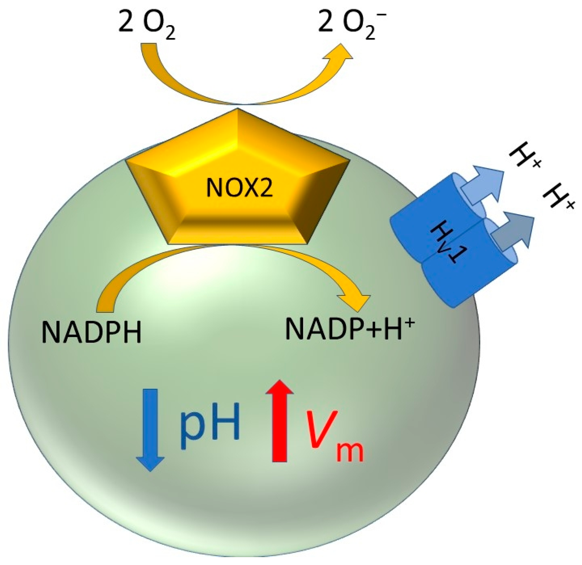
Figure 1. Simplified scheme of the respiratory burst in phagocytes. The activity of the NADPH oxidase reduces intracellular pH and depolarizes the plasma membrane. The voltage-gated proton channel HV1 compensates for the charge by conducting protons out of the cell. Additionally, the outflux of protons counteracts the decreased pH. Therefore, HV1 and NADPH oxidase are perfect synergistic partners during the respiratory burst.
Biomolecules 13 01035 g001
Figure 2. Architecture of the voltage-sensing and conduction pore domains of HV, KV, and NaV channels. As examples, a homology model of the human HV1, the crystal structure of the KV1.2 (PDB: 3LUT [108]), and the bacterial NaVAB channels (PDB 3RVY [109]) are shown. The voltage-sensing domain of all voltage-gated channels consists of four transmembrane helices (S1–S4). In HV channels, the voltage-sensing domain is also the ion conduction pore. In KV, NaV, and also CaV channels, the ion conduction pore is formed by a tetrameric assembly of two further transmembrane helices S5 and S6. The voltage-sensing arginines, three in HV, four in NaV, and five plus a lysine in KV are shown as sticks. Upward, resp. downward movement of the S4 (orange) helix is coupled to the activation, resp. deactivation, of the channels upon membrane depolarization, resp. hyperpolarization. A phenylalanine, shown as sticks, in the hydrophobic gasket that separates the intracellular and extracellular vestibules of the channels is conserved among voltage-gated ion channels.
Biomolecules 13 01035 g002
Figure 3. HV channels within the eukaryotic tree of life. Phylae and Subphylae containing at least one species harboring an HV channel gene are marked in green. The maximal number of different HV channel genes per species is indicated in red.
Biomolecules 13 01035 g003
Figure 4. Hv channels in Metazoa. (A) Tree of metazoan life. The maximal number of different HV channel genes per species is indicated in red. (B) Maximal number of HV channels genes per species in different phylae.
Biomolecules 13 01035 g004
Figure 5. Single Vrev measurements of the HV4 channel from Crassostrea gigas are plotted against the EH predicted value at different proton gradients (ΔpH = pHo – pHi). The dotted line shows equality between Vrev and EH. The inset depicts an example of a Vrev experimental determination by the tail currents method in a whole-cell patch clamp configuration at ΔpH = 0.5. Repolarization of the cell membrane after a depolarizing pulse was conducted in Δ10 mV/step from −60 mV to −20 mV) with a holding potential of −60 mV. The arrow indicates the point where H+ currents reverse (Vrev), here ~33 mV. Taken from [125].
Biomolecules 13 01035 g005
Figure 6. ΔpH-gating of the human HV1 channel from molecular dynamics simulations. The ΔpH-dependent gating from a deactivated (left, brown) to an activated (right, blue) conformation moves the three gating charges R1 to R3 in S4 (orange ribbon) outwards, passing the second arginine R2 through the hydrophobic gasket (HG). Changes of the location and orientation of the gating charges in the two conformations result in different interaction networks both in the internal and in the external vestibules, as illustrated for the selectivity filter aspartate (D112 in hHV1, here in red) that interact with R1 in the deactivated but with R2 in the activated conformation. For more details, please see [176].
Biomolecules 13 01035 g006
Figure 7. pH-dependent gating of HV channels in different organisms. The evaluation of the pH-dependent gating of HV channels can be described by a linear function of the form Vthres = slope·Vrev + offset from data obtained at different ΔpH conditions. (Upper panel, left) Families of H+ currents elicited by two different proton channels from Aplysia californica at symmetrical pH = 6.5. A zoom-in of dashed areas for each family is shown on the right side. The membrane potential where currents are first detected (Vthres) differs for both channels. AcHV1 activates positive to Vrev (here at +10 mV) and H+ currents are then outwardly directed (A). In contrast, AcHV2 activates negative to Vrev (here at −30 mV), permitting H+ influx (B). (Upper panel, right) pH-dependent gating of AcHV1 (orange line) and AcHV2 (blue line) represented according to Equation (3). Both channels present identical pH-sensing with the same slope (Vthres/Vrev) but have distinct offsets. The dotted line represents equality between Vthres and Vrev and delimits the equilibrium between outward and inward driving forces. Data over and below the dotted line indicate outwardly and inwardly directed H+ driving force, respectively. While AcHV1 extrudes H+ alkalinizing the cytosol, AcHV2 conducts inward currents that acidify the cell. From [124]. (Lower panel, left) Vthres vs. ΔpH (pHo − pHi) plot determined for some functionally tested HV channels from different species in a ΔpH ranging from −1 to +1. The pH-dependent gating of native HV channels of 15 cell types (from distinct organisms including rat, hamster, mouse, human, frog, and snail) described by [192] is displayed for comparison (black solid triangles). The dotted line shows equality between Vrev and Vthres. Most of HV channels are proton extruders but there are few notable exceptions, i.e., the channels from the dinoflagellate K. veneficum (kHV1), the fungi A. oryzae (AoHV1), the stick insect E. tiaratum (EtHV1), or the type-2 channel from sea hare A. californica (AcHV2). (Lower panel, right) the pH-dependent gating on the external pHo for several HV channels is represented as ΔgH/ΔpH. The values indicate the shift of the conductance–voltage curves expected once pHo is changed in one unit while maintaining pHi constant. The dotted line shows the 40 mV/unit of pH thumb rule describing the pH-dependent gating for most of HV channels [192]. The channels are grouped by organism in insects (red), mollusks (light blue), dinoflagellates (green), ascidians (light cyan), fungi (orange), mammals (blue), fishes (grey), corals (dark green), sea urchins (pink), coccolithophores (dark cyan), and 15 native HV from different species. Np: Nicoletia phytophila, Et: Extatosoma tiaratum, Cg: Crassostrea gigas, Ac: Aplysia californica, Ht: Helisoma trivolvis, Kv: Karlodinium veneficum, Lp: Lingulodinium polyedrum, Ci: Ciona intestinalis, Ao: Aspergillus oryzae, Sl: Suillus luteus, h: homo sapiens, m: Mus musculus, Rn: Rattus norvegicus, Dr: Danio rerio, Am: Acropora millepora, Sp: Strongylocentrotus purpuratus, Eh: Emiliania huxleyi, Cp: Coccolithus pelagicus ssp braarudii.
Biomolecules 13 01035 g007
Table 1. HV1 in human tissue and other mammalian tissue.
Respiratory BurstCell TypeFunction of HV1References
YesEosinophil
(human)
(rodent)
Charge compensation, prevention of cell death.[10,12,17,36,37,38,39,40,41,42,43,44,45,46]
YesNeutrophil
(human)
PLB-985 (hcl)
HL-60 (hcl)
K-562 (hcl)
Neutrophil (rodent)
Charge compensation, migration, granula release, calcium homeostasis, pH homeostasis, ERK activity, phagosomal pH homeostasis.[6,8,10,12,13,14,15,41,45,47,48,49,50,51,52,53]
YesMonocyte
(human)
Charge compensation.[54]
Yes, smallMacrophage
(human)
THP-1 (Hcl)
Macrophage (mice)
Charge compensation, phagosome acidification.[52,55,56]
Yes, smallOsteoclast (rodent/Leporidae)pH homeostasis, charge compensation, ROS production.[57,58,59,60,61]
Yes, smallMicroglia (rodent)
Microglia culture (human)
BV-2 (rcl)
GM1-R1 (rcl)
MLS-9 (rcl)
pH homeostasis, charge compensation, ROS production, microglia-astrocyte communication, neuropathic pain promotion, brain damage enhancing, acidosis exacerbation, M2 polarization reduction, demyelination promotion, white matter injuries promotion, secondary spinal cord damage enhancing, neuroinflammation promotion, pyroptosis increase, motor deficit expansion, autophagy increase, M1 polarization promotion in aged mice.[22,61,62,63,64,65,66,67,68,69,70,71,72,73,74,75,76,77]
YesKupffer cell (mice/rodent)Glucose metabolism, ROS production suppression, hyperglycaemia, and hyperinsulinemia prevention. [23]
NoCardiac fibroblast
(human)
pH homeostasis, membrane potential, potentially beneficial in ischemia.[78]
Yes, smallDendritic cells (rodent/human)TLR9 activation.[28]
NoSperm cell
(human)
Capacitation, acid extrusion. [32,33,79,80]
YesOocyte
(human)
pH homeostasis. [34]
NoType 2 alveolar cells (rodent)pH regulation.[30,47,81,82,83,84,85,86]
NoMast cell (mouse)pH homeostasis.[87]
Yes, tinyB cells (human)
(rodent)
LK35.2 (rodent)
B cell receptor signalling, migration and proliferation enhancing (short isoform).[26,27,88,89]
NoT cells (human)
Jurkat (human)
T cells (rodent)
Apoptosis prevention, pH homeostasis, autoimmune disorders prevention. [29,88,90,91,92]
NoCardiomyocytes (canine)pH homeostasis.[11]
NoSHG-44 glioma cells (human)Apoptosis prevention.[18]
NoColorectal cancer
(human)
SW620 (hcl)
HT29 (hcl)
LS174T (hcl)
Colo205 (hcl)
Prevention of cellular acidosis, support of cancer cell metabolism, pH homeostasis, potential biomarker, and drug target[19]
NoBasophils
(human)
Exocytosis (histamine release), pH homeostasis.[16,93]
NoOvary cells (Hamster)pH homeostasis.[94]
NoBreast cancer cells (human primary)
MDA-BA-231(hcl)
MCF-7 (hcl)
MDA-MB-468 (hcl)
MDA-MB-453 (hcl)
T-47D (hcl)
SK-BR-3 (hcl)
Tumor growth, metastasis and invasiveness promotion, (expression predicts prognosis of tumor).[20,21,95]
NoLung cancer cell
A549
(human)
No information.[96]
NoProstate cancer cell
PC-3 (human)
No information.[96]
NoKidney (human)
HEK-293
No information.[96,97]
Yes, smallNasal epithelium
(human primary culture)
JME/CF 15
(human)
Cystic fibrosis genotype
Airway surface epithelium acidification, proton extrusion. [98]
Yes, smallCiliated tracheal cells (human)NADPH oxidase activity driven proton extrusion.[31,98]
Yes, smalllung epithelium fetal (human)DUOX driven proton release, acid extrusion.[99]
Yes, tinySerous gland cell line Calu-3 (human)Airway surface epithelium acidification, proton extrusion (to a lesser extent than airway epithelium)[31]
NoSkeletal muscle myocyte (human)pH homeostasis. [7]
NoGlioblastoma cell line (human)
T98G
Cell’s survival and migration.[100]
NoWhole heart (rodent)NOXs transcription and CO2 homeostasis control, electrophysiological remodelling. [101]
No/YesVascular system, Immune systemAtherosclerosis advancement (hypothetical).[102]
No/Yes Whole tissueLung (rodent)Goblet cell hyperplasia prevention. Depression expression of IL-4, IL-5, and IL-13. Reduction of the expression levels of NOX2, NOX4, and DUOX1. Promotion of the expression of SOD2 and catalase. Reduction of the development of allergic asthma through ROS production enhancing.[103]
YesMyeloid derived suppressor cells (MDSC) (rodent)T-cells regulation (via ROS production). [35]
Noepididymal adipose tissue (rodent)Diet obesity induction.[24]
Yes, tinyPancreatic β cells (rodent)Insulin secretion, ROS production, NOX4 upregulation, glucotoxicity induction. [25,104,105]
hcl = human cell line; rcl = rodent cell line.
Table 2. Sequence homology in HV channels. Sequence identities/similarities of ten selected protist HV channels to eukaryotic sodium and calcium channels are shown. In brackets, the length of the respective sequence as retrieved by BLAST is shown.
SpeciesPhylae/SubphylaeNaVCaV
Euglena gracilisExcavata28/52% (86)35/50% (91)
Raperostelium potamoidesAmoebozoa36/51% (106)35/56% (86)
Balamuthia mandrillarisAmoebozoa34/52% (74)<25%
Paramoeba aestuarinaAmoebozoa27/50% (64)<25%
Emiliania huxleyiHaptista37/51% (79)32/57% (73)
Amorphochlora amoebiformisSAR-Rhizaria<25%28/55% (79)
Odontella auritaSAR-Stramenopiles36/58% (67) *<25%
Karlodinium veneficumSAR-Alveolata<25%34/55% (80)
Scrippsiella hangoeiSAR-Alveolata29/51% (87)29/56% (85)
Gracilaria vermiculophyllaRhodophyta30/63% (69)<25%
* In one case to a prokaryotic sodium channel.
Table 3. Identified HV channels. GenBank accession numbers of cDNA and genes of indicated model organisms are listed.
SpeciesKingdomcDNA (mRNA)Gene
Escherichia coliProkaryota--
Dictyostelium discoideumProtist--
Tetrahymena thermophila --
Naegleria gruberi --
Emiliania huxleyi HBNU01018021
GIZZ01010784
NW_005196830
NW_005202428
Thalassiosira pseudonana NC_012076
Aspergillus nigerFungiXM_001390088NT_166520
Neurospora crassa --
Saccharomyces cerevisiae --
Arabidopsis thalianaPlantaeNP_001321473NC_003070
Zea mays --
Oryza sativa --
Physcomitrella patens XM_024508236
XM_024525718
NC_037254
NC_037259
Marchantia polymorpha -AP019868
AP019873
AP019871
Chlamydomonas reinhardtiiArchaeplastida--
Aplysia californicaInvertebrataXM_005100609
XM_005093050
XM_005094218
XM_013086351
XM_013080089
XM_013090418
XM_013082371
NW_004797523
NW_004797327
NW_004797348
NW_004797727
NW_004797344
NW_004798539
NW_004797441
Branchiostoma belcheri XR_002139895
XM_019760615
XM_019764911
NW_017802379

NW_017803191
Caenorhabditis elegans --
Ciona intestinalis NM_001078469NW_004190496
Daphnia pulex XM_046604416NC_060027
Drosophila melanogaster --
Hydra vulgaris XM_047284425-
Lymnaea stagnalis FX197150
FX190227
FX196339
nys
Nematostella vectensis XP_001626501NC_064038
Strongylocentrotus purpuratus XM_030990962NW_022145605
Trichoplax adhaerens XM_002110878
XM_002110360
NW_002060945
Ambystoma mexicanumVertebrataGFZP01114012JXRH01463164
Gallus gallus NM_001396354NC_052587
Mesocricetus auratus XM_040731183NW_024429206
Cavia porcellus XM_003462980NT_176304
Mus musculus NM_028752NC_000071
Rattus norvegicus XM_017598517NC_051347
Macaca mulatta XM_028829869NC_041764
Takifugu rubripes XM_003977031NC_042298
Xenopus laevis XM_018249100
XM_018244209
NC_054371
NC_054372
Danio rerio NM_001002346NC_007121
Homo sapiens NM_001040107NC_000012
nys = not yet sequenced.
Table 4. Biophysical properties of some HV channels.
OrganismSpeciesChannelOligomerization?SelectivityGating Charges, e0Slope
Vthres/Vrev
Vthres at ΔpH = 0
(mV)
ΔgH-V/ΔpH
(mV/pHo)
H+ Influx at Relevant Physiological pH?References
MammalsH. sapienshHV1confirmed f,g,j,k>106 PH+/PTMA+ d,i
>106 PH+/PCH3SO3- i
>106 PH+/PCl- i
~ 5 h,δ
~ 6 l
0.82 d
0.67–0.71 (expressed) l
0.71
(native) l
13.8 d
−9 to −11
(expressed) l
+27
(native) l
40 lno
(native)
yes
(if expressed) l
[110] d, [138] f, [139] g, [140] h, [141] i, [41] j, [142] k, [97] l
M. musculusmHV1confirmed n>107 PH+/PNMDG+
>107 PH+/PNa+
>107 PH+/PK+
~6 m0.86 * (expressed)
0.69 m
(expressed)
+10 to +20
−15
(expressed) m
50
40 m
no
(native)
yes
(if expressed) m
[118], [97] m, [143] n
R. norvegicusRnHV1possibly>107 PH+/PTMA+ o
>108 PD+/PTMA+ p
5.4 p0.76+1844
40 o,p
no[5], [81] o, [83] p
FishD. rerioDrHV1possibly>107 PH+/PNMDG+n.d.0.69 *~+10 mV ε~ 40 εno[144]
Sea squirtC. intestinalisCiHV1confirmed bn.d.4.4–5.9 (dimer) b
1.6–2.7 (monomer) c
n.d.n.d.~ 40 cno[118], [145] c, [146] d
InsectsN. phytophilaNpHV1confirmed a>108 PH+/PTMA+
>104 PH+/PNa+
>104 PH+/PCl-
4.7–6.1 a0.81 a−3.4 a47–54 ano[120] a, [147] b
E. tiaratumEtHV1n.d.>106 PH+/PTMA+n.d.0.77−2345yes[121]
MollusksC. gigasCgHV4possibly>107 PH+/PTMA+n.d.0.84−1249no [125]
A. californicaAcHV1possibly>107 PH+/PTMA+
>106 PH+/PNa+
>106 PH+/PK+
5.70.78543–45no[124]
A. californicaAcHV2possibly>107 PH+/PTMA+
>106 PH+/PK+
5.30.77−2044
40 (pHi)
yes[124]
A. californicaAcHV3possibly +>107 PH+/PTMA+n.d.n.d.n.d.n.d.yes §[124]
H. trivolvisHtHV1possibly>107 PH+/PTMA+5.51.03 *
0.26 (pHi) *
n.d.60.0
15.3 (pHi)
no[148]
CoralsA. milleporaAmHV1confirmed>107 PH+/PTMA+2 λ0.86 *~ +10 mV θ~ 50 θno[123]
Sea UrchinS. purpuratusSpHV1confirmed>107 PH+/PK+4.3
(dimer)
1.1 (monomer)
0.69 *~ +10 mV~ 40 βno[122]
FungiA. oryzaeAoHV1possibly +>105 PH+/PTEA+ #51.40–1.55 *~ −30
(pH 5.5) γ
~ −30
(pH 6.5) γ
80–90yes[129]
S. luteusSlHV1possibly +>105 PH+/PTEA+ #51.40–1.55 *~ +20
(pH 5.5) γ
~ +40
(pH 6.5) γ
80–90no[129]
DinoflagellatesK. veneficumkHV1possibly not φ>107 PH+/PTMA+
>105 PH+/PCl-
n.d.0.79−3746yes[132]
L. polyedrumLpHV1possibly +>109 PH+/PTMA+n.d.0.69 *4640 **yes α[133]
PhytoplanktonE. huxleyiEhHV1possibly>106 PH+/PK+
>106 PH+/PCl-
n.d.0.69 μ
(expressed)
~+20 mV (expressed) μ~40 Ωno[131]
C. pelagicusCpHV1possibly>106 PH+/PK+
>106 PH+/PCl-
n.d.0.69 μ
(native)
+10 mV
(native) μ
~40 Ωno[131]
* Calculated from conductance shifts (∆gH-V/∆pH) and EH = 58 mV·pH−1. § AcHV3 leaks protons at the closed state [124]. # Calculated from pH = 6.0 and working solution containing 30 mM of TEA+ as main cation. CgHV4 has an activation of −12 mV which permits proton influx at symmetrical pH (physiological) conditions. Nevertheless, the proximity of the offset value to Vrev makes the inward H+ electrochemical gradient small. Thus, H+ influx is small at ∆pH = 0 where currents rectify rapidly with depolarization, behaving more like a typical HV proton extruder. During measurements, strong and/or consistent H+ influx currents were not detected. φ kHV1 lacks the predicted coiled-coil which is important for HV dimerization, a potential monomeric expression is discussed by the authors [132]. + Protein sequence analysis predicts a C-terminal coiled-coil domain. ** LpHV1 pH-dependent gating saturates above pHo 8.0 [133] similar to rat, human, Karlodinium, and Emiliania HV channels [136]. α Inward H+ currents are onset at large inward pH gradients (1–3 ∆pH units) [133]. β estimated from gH-V curves shift between pHo 6.5 and 7.0 (~20 mV), at pHi = 7.0 (Figure 2; Ref. [122]). The gH-V shifts between pHo 6.0 to 6.5 and 6.5 to 7.0 are not identical due to experimental limitations reported by the authors. The calculated value on pHo-dependent gating agrees with ΔgH-V/pHi unit (Figure S1; Ref. [122]). γ from normalized gH-V curves (Figure 3; Ref. [129]). δ from Qon of dimeric W207A-N214R mutant ([140]). θ from Vthres-ΔpH and slope of V0.5-ΔpH curves (Figure 6; Ref. [125]). λ apparent from steepness of normalized gH-V (Figure 6; Ref. [123]). Ω from current densities (Figure 1; Ref. [131]). μ the slope Vthres/Vrev was calculated from shifts of IH onsets and EH. Voffset was estimated shifts of IH activation, EH, and Equation (3) (Figure 4; [131]). PH+ = proton permeability, PTMA+ = tetramethylammonium permeability, PTEA+ = tetraethylammonium permeability, PK+ = potassium permeability, PNa+ = sodium permeability, PCl- = chloride permeability, PNMDG+ = N-methyl-d-glucamine permeability, PCH3SO3- = methanesulfonate permeability, n.d. = no data, Vthres = threshold potential, Vrev = reversal potential, ∆pH = proton gradient (pHoutside – pHinside), gH-V = proton conductance – voltage relationship, pHo = external pH, pHi = internal pH.
Disclaimer/Publisher’s Note: The statements, opinions and data contained in all publications are solely those of the individual author(s) and contributor(s) and not of MDPI and/or the editor(s). MDPI and/or the editor(s) disclaim responsibility for any injury to people or property resulting from any ideas, methods, instructions or products referred to in the content.