Review

The Autoimmune Rheumatic Disease Related Dry Eye and Its Association with Retinopathy

Department of Ophthalmology, Qilu Hospital of Shandong University, Jinan 250012, China
*
Author to whom correspondence should be addressed.
Biomolecules 2023, 13(5), 724; https://doi.org/10.3390/biom13050724
Submission received: 15 October 2022 / Revised: 19 April 2023 / Accepted: 20 April 2023 / Published: 23 April 2023
(This article belongs to the Special Issue New Discoveries in Retinal Cell Degeneration and Retinal Diseases)

Abstract

:
Dry eye disease is a chronic disease of the ocular surface characterized by abnormal tear film composition, tear film instability, and ocular surface inflammation, affecting 5% to 50% of the population worldwide. Autoimmune rheumatic diseases (ARDs) are systemic disorders with multi-organ involvement, including the eye, and play a significant role in dry eye. To date, most studies have focused on Sjögren’s syndrome (one of the ARDs) since it manifests as two of the most common symptoms–dry eyes and a dry mouth-and attracts physicians to explore the relationship between dry eye and ARDs. Many patients complained of dry eye related symptoms before they were diagnosed with ARDs, and ocular surface malaise is a sensitive indicator of the severity of ARDs. In addition, ARD related dry eye is also associated with some retinal diseases directly or indirectly, which are described in this review. This review also summarizes the incidence, epidemiological characteristics, pathogenesis, and accompanying ocular lesions of ARD’s related dry eye, emphasizing the potential role of dry eye in recognition and monitoring among ARDs patients.

1. Introduction

Dry eye is a multifactorial disease of the ocular surface characterized by a loss of homeostasis of the tear film and accompanied by ocular symptoms, in which tear film instability and hyperosmolarity, ocular surface inflammation and damage, and neurosensory abnormalities play etiological roles [1]. It can be manifested as decreased vision and eye discomfort, accompanied by eye inflammation and ocular nerve damage. The vision-related quality of life and work efficiency of patients are significantly reduced by those malaises [2]. Epidemiological surveys reported that the prevalence of dry eye differed greatly among regions, ranging from 5% to 50% worldwide [3]. The incidence of dry eye has increased significantly in recent years due to the prolonged use of digital screens [4], air pollution [5], and population aging. Predisposing factors of dry eye include the environment, increased use of electronic products, systemic diseases, ocular surface diseases, eye surgeries, application of eye drops and ointment, etc. Systemic diseases, especially rheumatic immune diseases are closely related to dry eye [2].
Autoimmune rheumatic diseases (ARDs), also named rheumatic immune diseases or immune-mediated rheumatic diseases, are a group of diseases related to the overactivation of the immune system, including primary Sjögren’s syndrome (SS), rheumatoid arthritis (RA), systemic lupus erythematosus (SLE), systemic sclerosis (SSc) or scleroderma, idiopathic inflammatory myositis (IIM), and the systemic vasculitides (SV) [6].
As a systemic disorder with multi-organ involvement, ARDs lead to various clinical manifestations including skin lesions, joint pain, vascular damage, and dry eye. Furthermore, treatments for ARDs, including methotrexate and cyclophosphamide, usually cause or exacerbate symptoms of dry eye. Under normal conditions, the intrinsic immune mechanisms of the eyes could maintain the microenvironment homeostasis. When the eye becomes the target of the immune response of ARDs, the ocular immune system is excessively stimulated by relevant immunoregulatory molecules and the balance of the immunoregulatory mechanisms is therefore disrupted. Additionally, chronic ocular surface inflammation is a result of dysregulated balance of the innate and adaptive immune systems, leading to chronic dry eye [7]. Ocular surface symptoms could be sensitive manifestations of the severity of the systemic condition of ARDs. In addition, a considerable proportion of dry eye patients have ARDs but are not diagnosed and treated in a timely manner [8]. However, the reported literature mainly focused on specific kinds of ARDs (SS, RA, or SLE), and the number of cases is limited. Moreover, we retrieved a few studies exploring the correlation between other or overall ARDs and dry eye. Hence, this review aims to summarize the characteristics and pathogenesis of dry eye in various kinds of ARDs. For the lack of studies reporting IIM or SV-related dry eye, this article mainly focuses on the relationship between other kinds of ARDs (SS, RA, SLE, and SSc) and dry eye. Dry eye is related to some retinal diseases directly or indirectly. The retina is often involved in ARDs, commonly associated with autoimmunity and systemic inflammatory cell infiltration. Consequently. the retinal diseases related to dry eye and ARDs are also illuminated.

2. Epidemiology of ARD Related Dry Eye

It is reported that ARDs are significant causes of dry eye. Approximately 10–95% of patients with immune system disorders are accompanied by dry eye. For example, 38–47% of patients with RA have dry eye [9,10], 13.4–39.5% in SLE [11,12], 95% in SS [13,14], and 37–79% in SSc [15]. Consequently, the prevalence of dry eye in patients with SS is higher than those in other diseases of ARDs. In addition, the proportion of females with ARDs is much higher than that of males (appropriately 9:1) [16]. Hence, ARD related dry eye is more likely to affect female patients.
The relationship between SS and dry eye was emphasized in the guidelines reported by the American Academy of Ophthalmology. The guidelines showed that about 10% of patients with dry eye have SS [8]. According to a randomized controlled study that included 535 patients with moderate to severe dry eye disease, SS was significantly associated with more severe dry eye signs [17]. Moreover, the guidelines also report that RA is a risk factor for dry eye [8]. Similarly, Paulsen et.al suggested that arthritis significantly correlated with dry eye symptoms [18]. A recent prospective multicenter study involving more than 400 patients with primary SS showed that 94% of cases had dry eye symptoms [19]. Abd-Allah et.al. found that RA can be accompanied by severe dry eye symptoms even when it is not accompanied by secondary SS [20]. Similarly, the incidence of dry eye in SLE patients was much higher than that in healthy individuals [21].

3. Mechanisms of ARD Related Dry Eye

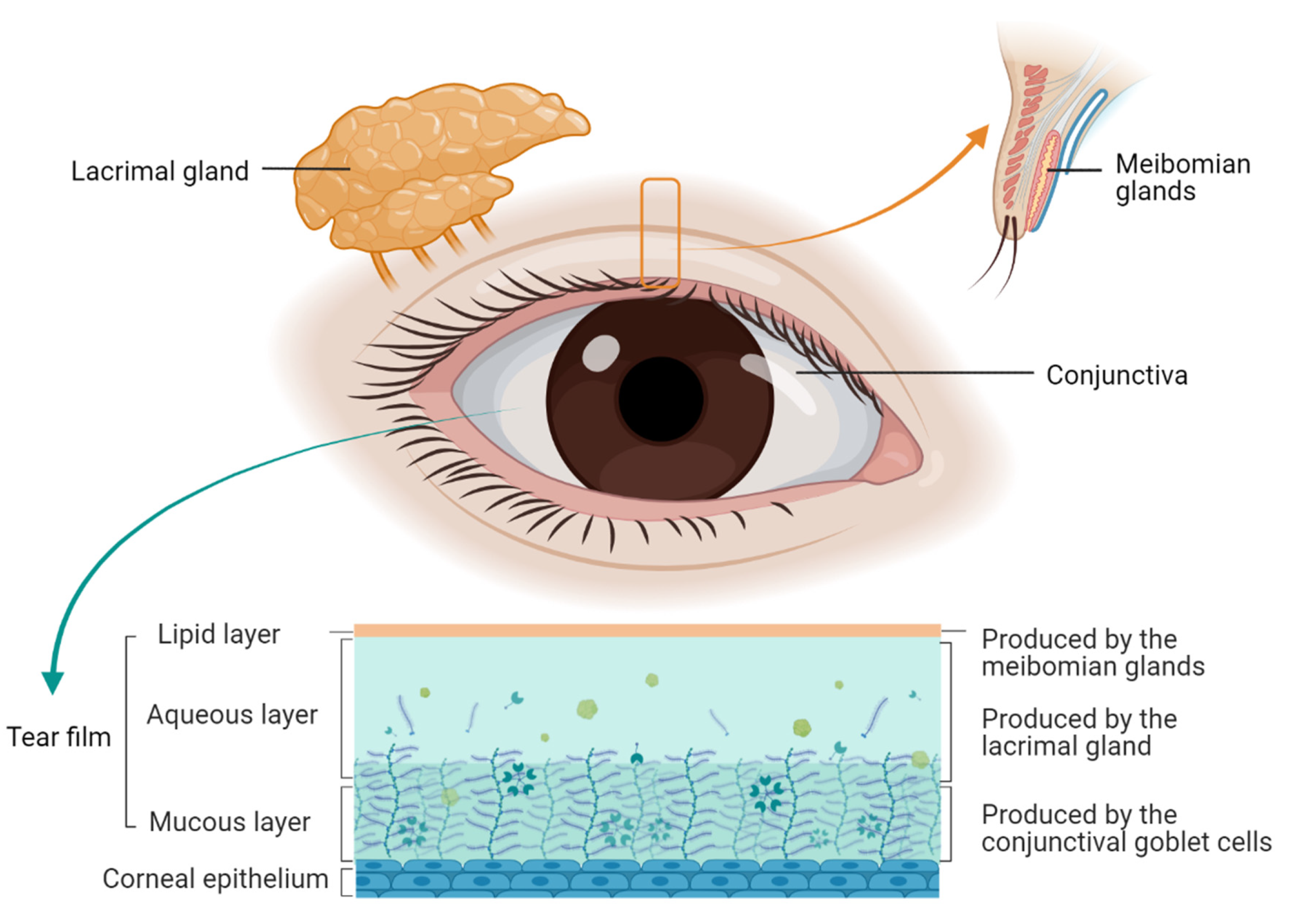
3.1. Pathogenesis of Dry Eye

The pathogenesis of dry eye is complex and involves a group of units, such as the lacrimal gland, meibomian gland, tear film, conjunctival membrane, cornea, and tissue structures [22]. Pathological changes include inflammatory factors, tear hyperosmotic pressure, lipids, mucins, microorganisms, etc. Dry eye is commonly classified into aqueous-deficient dry eye and evaporative dry eye [2]. The main mechanisms are evaporation and water loss, leading to hyperosmotic tissue damage. Ocular surface inflammation is recognized as the critical process of dry eye pathogenesis. Induced by a variety of risk factors such as hyperosmolarity, UV light, and desiccation, ocular surface epithelial cells release various mediators, including metalloproteinases, inflammatory cytokines, and chemokines. These lead to relevant immune-cell activation and associated inflammation through stress signal transduction pathways [22]. Ocular structures related to dry eye are presented in Figure 1.
Both innate and adaptive immunity are involved in the pathogenesis of dry eye. Previous studies based on clinical and laboratory research have found that Th1 and Th17 cells could modulate the immune response on the ocular surface [23]. Combined with exposure to autoantigens, these innate inflammatory mediators can lead to an adaptive T cell-mediated response. Additionally, after the activation of the immune response, the dysfunction and death of conjunctival goblet cells is promoted by the Th1 cytokine IFN-γ, which aggravates dry eye [24]. Studies on human and animal models have shown that loss of ocular surface epithelial cells and goblet cells could lead to shorter tear break-up time [25]. Interferon-gamma (IFN-γ), produced by NK cells, is responsible for upregulating Th1 cells to recruit chemokines (CXCL9, CXCL10, and CXCL11) in conjunctival and corneal epithelial cells. Meanwhile, increasing expression of activating NK cell receptor ligands and activation of innate immune pathways can lead to enhancement of T cell infiltration, involving the lysis of epithelial tight junctions. In consequence, the cornea barrier is destroyed and the phenotype of the epithelial cells changes, which destabilizes the tear film, amplifies inflammation and creates a vicious cycle [22].

3.2. Pathogenesis of ARD Related Dry Eye

The immune systems in patients with rheumatic immune diseases are impaired, resulting in abnormal immune responses and imbalanced immune regulation. Substantial immune cells and inflammatory factors imperil the lacrimal gland, conjunctival membrane, cornea, and meibomian glands, resulting in tissue destruction and dysfunction [26]. A large number of immune cells, most of which are T lymphocytes, infiltrate the lacrimal duct and accessory lacrimal gland in patients with rheumatic and immune diseases, causing autophagy and apoptosis of acinar, ductal and myoepithelial cells, disruption of lacrimal gland function and a decrease in tear secretion [25].
Due to the presence of innate immune cells and the secretion of inflammatory response factors, the signal transduction caused by abnormal antibodies leads to chronic fibrosis of the lacrimal gland and further aggravation of dysfunction. The infiltration of T cells in the conjunctival tissue results in squamous metaplasia, decreased goblet cell density, and reduced mucin secretion. The deposition of autoantibodies and antigen-antibody complexes in corneal tissues leads to corneal dystrophy [27]. An increased number of corneal dendritic cells, as well as decreased nerve density and sensitivity are revealed by confocal microscopy through focusing [28]. Villani et.al. found that 35 patients with SS-related dry eye had significantly low corneal nerve counts (3.34 ± 0.76 vs. 5.10 ± 0.79, p < 0.0001) and an increased degree of tortuosity (2.62 ± 0.94 vs. 1.20 ± 0.70, p < 0.0001), compared with the control group [29], which was consistent with the study by Benítez del Castillo et al. [30]. Moreover, Xu et al. [31] reported that the corneal nerve sensitivity of SS-related dry eye patients was significantly reduced (4.5 ± 1.2 cm vs. 5.8 ± 0.4 cm, p < 0.01). Additionally, immune cell infiltration in the meibomian glands and consequent mechanical obstruction of its ducts were very common in patients with rheumatic immune disease related dry eye (57.9%) [32]. Atrophy and occlusion of some meibomian gland ducts can be revealed by meibomian gland imaging. Moreover, recent studies have shown that gut dysbiosis (Firmicutes/Bacteroidetes ratio and genus Faecalibacterium) is closely related to dry eye diseases caused by autoimmune diseases such as SS. However, the key communication routes of the “gut dysbiosis–ocular surface–lacrimal gland axis” still need to be further elucidated [33].
In terms of genetic heredity, RA is a complex disease and is correlated with multiple genetic loci, each of which usually has only one modest association with a specific condition [34]. According to a recent study, 39 variants were identified in immune-related genes across SLE, RA, and SS families. Among this gene set, regulation of T cell activation and T cell receptor signaling pathways were particularly concentrated [35], which might correlate with symptoms and signs of dry eye. Consequently, gene analysis is a powerful tool for understanding the pathogenesis of ARD related dry eye [36].

3.2.1. Primary Sjogren’s Syndrome & Dry Eye

Primary SS is an autoimmune and systemic disorder. Globally, the prevalence of primary SS ranges from 0.1% to 4.8% depending on age [37,38], and 90% of patients are females. Sicca symptoms (dry mouth and dry eyes) are common oral and ocular complaints. Nevertheless, SS is often underdiagnosed, misdiagnosed, or delayed in being diagnosed since the symptoms are usually vague for many years [39] until irreversible organ damage has occurred [40]. As an autoimmune disease that mainly affects exocrine glands (such as the salivary and lacrimal glands), primary SS is most closely related to dry eye among the various types of ARDs. Primary SS is also a possible diagnosis in patients with dry eye symptoms. Staining of the temporal conjunctiva and severe dry mouth symptoms were the major factors in distinguishing primary SS from dry eye disease [41].
SS is a chronic inflammatory autoimmune disease, histopathologically characterized by lymphocytic infiltration of exocrine (mainly salivary and lacrimal) glands with remarkable B-cell hyperactivity and the presence of autoantibodies against the ribonucleoprotein particles SSA/Ro and SSB/La. The glandular function is impaired by the infiltrating cells in several ways, including local production of autoantibodies, destruction of glandular structures by cell-mediated mechanisms, and secretion of cytokines that activate pathways related to interferons (IFNs) [13]. The salivary and lacrimal glands are the principal targets of a proposed T cell-mediated chronic inflammation, followed by glandular atrophy and deficient function. Primary SS is considered to arise from an interplay between environmental factors and genetic susceptibility. Activation of B cells, the presence of anti-Ro-52 antibody and the “triggers” of viral infections result in the activation of the innate and adaptive immune systems, which perpetuate self-reactive activity against healthy tissues [42,43].
Due to the invasive process of lacrimal gland biopsy, most studies of SS related dry eye were carried out on mouse models, such as the Aec and IL-12 transgenic mice [44], TSP-1KO mice [45], CD25KO mice [46], BXSB/MpJ-Yaa mice [47], and lupus-prone female NZB×NZWF1 (B/WF1) mice [48] (secondary SS), and indicated lymphocytic infiltrations as well as loss of secretory functions. Noteworthily, Hayashi et al. [48] reported substantial destruction of the myoepithelial cells and lysis of basement membranes within the inflammatory lymphocytic foci in the lacrimal gland. Hawley et al. found substantial changes in the myoepithelial cell morphology and contractile protein expression in these relatively normal areas (between the lymphocytic foci) of the lacrimal gland, which is likely due to decreased expression of the contractile proteins of smooth muscle actin and calponin, and significant loss of the oxytocin receptor [49]. Hayashi et al. suggested that acini destruction in lacrimal glands may be induced by systemic and local Th1 cell-dominant reactions [50].
In addition, the corneal nerves play a critical role in the pathophysiology of dry eye because they contribute to the reflex control of basal tear production and blinking [51]. Compared to healthy subjects, both the density and function of sensory nerve fiber terminals in the cornea declined in primary SS [52,53], leading to a vicious circle of corneal hypoesthesia. Moreover, primary SS also results in damage to secretomotor innervation and inflammatory destruction of acini and ducts [54]. SS dry eye is generally described as aqueous-deficient dry eye and much attention has been paid to the lack of tear production [55,56]. However, as a result of the meibomian glands being under immune attack, meibomian glands dysfunction (MGD) is captured in primary SS patients [57], with a diffuse abnormality of the meibomian glands, commonly characterized by chronic terminal duct obstructions and qualitative or quantitative changes in the glandular secretion [58]. Lymphocytic infiltration was also found in the conjunctiva of patients with SS related dry eye [59]. By using a Sjogren’s syndrome model MRL/MpJ-Fas(lpr) mouse, Diebold et al. found lymphocytic infiltration and goblet cell marker alteration in the conjunctiva [60]. Furthermore, desiccating stress caused by SS promotes the maturation of monocytes to antigen-presenting cells with elevated levels of inflammatory genes in conjunctiva [61]. Keratinization of the conjunctival and corneal epithelia, squamous metaplasia, and goblet cell loss are observed in severe primary SS.
In conclusion, SS dry eye is a multifactorial disease, comprising aqueous, lipid, and mucin components, which is comprised of a series of disorders of the lacrimal gland, conjunctiva, corneal and meibomian glands, causing aqueous-deficient or evaporative dry eye.

3.2.2. Rheumatoid Arthritis & Dry Eye

RA is a common systemic inflammatory disease, characterized by persistent synovitis, systemic inflammation, and autoantibodies (particularly rheumatoid factor and citrullinated peptide) [62]. The morbidity ranges from 0.5% to 2% in the overall population [63]. RA affects the joints, but the systemic inflammatory process could affect other tissues and organs, for example, the eyes. Most relevant studies showed that dry eye was the most frequent ocular complication in RA patients, while others include scleritis, episcleritis, keratitis, etc. [64,65,66]. Although some patients with RA can develop secondary SS with the prevalence varying from 4% to 50% [67], dry eye is common even in RA patients without secondary SS [10]. No matter what kind of RA (RA-SS or RA-nonSS), dry eye is the most common ocular manifestation. As for gender characteristics of dry eye in RA, it is more common in women, with a female-to-male ratio of 9:1 [68]. The probability of ocular involvement increases with the duration of the disease and it may represent the main clinical manifestation of RA in patients with long-standing disease. In addition, sometimes ocular complaints are the first symptom of RA patients [64]. The severity of eye dryness was verified to be highly correlated with juvenile RA [69]. However, there was no significant correlation between the severity of dry eye and RA activity. Therefore, the deterioration of the systemic condition of RA does not one hundred percent lead to the aggravation of dry eye symptoms, and dry eye cannot be excluded even in patients with mild RA [10].
Mechanisms of RA are the repeated activation of innate immunity, especially at mucosal surfaces. Innate immunity could activate fibroblast-like synoviocytes, dendritic cells, and macrophages during the earliest phases in individuals with underlying immune hyperreactivity. The overproduction of pro-inflammatory cytokines, such as tumor necrosis factor (TNF) and interleukin-6 (IL-6), lead to the proliferation of synovial cells in joints and subsequent pannus formation, cartilage destruction, and bone erosions [62]. Among ARDs other than SS, the association is the closest between dry eye and RA [70]. As mentioned above, increased deterioration of RA seems not to lead to the aggravation of dry eye. Consequently, the etiology of dry eye is different in SS and non-SS patients with RA [10].
The mechanism of dry eye in RA patients is different from SS and might be a result of local pathology affecting the tear fluid, conjunctiva, or cornea. The pathological changes of the ocular surface in RA-SS are dissimilar to RA-nonSS as discovered by Villani et al. after they detected the concentration of human IL-1a, IL-6, IL-8, and TNF-a (substantial in the synovial fluid and tissue of RA patients) in tear samples [66]. After intervention therapy using blockers against TNF-a, IL-1a, and IL-6, there were remarkable therapeutic efficacies in RA. Additionally, the concentration of IL-1a and IL-6 in the tear ducts of RA-SS patients declined significantly, while there were no evident changes of those cytokines in RA-nonSS patients [66]. However, Usuba et al. demonstrated the evaporative component disturbance in RA patients, with the presence of meibomian gland dysfunction (associated with alterations in the lipid layer of tear film) as well as decreased goblet cell numbers (associated with the mucin layer of the tear film), in addition to the traditional aqueous tear deficiency related to dry eye disease. Furthermore, treatment with TNF inhibitors improved conjunctival goblet cell numbers in RA patients with mild dry eye, which is conducive to relieving dry eye by increasing the secretion of mucin [71]. This is consistent with Tong’s findings that TNF-inhibitor treatment controlled inflammatory disease activity concomitantly to conjunctival cytology, especially the goblet cell density, implying a common underlying inflammatory mechanism for ocular and articular activity [72]. In addition, in a clinical trial by Schargus et al., tear film hyperosmolarity highly correlates (>316 mOsmol/L) with undiagnosed dry eye in RA patients [73].
Most of the current studies demonstrated that dry eye in RA tends to correlate with local pathogenic mechanisms other than with systemic inflammation [10,73]. In summary, mixing aqueous-deficient dry eye and evaporative dry eye, RA-nonSS dry eye comprises disorders of the conjunctiva and meibomian gland, resulting from a decrease of aqueous, lipid, and mucin components. Findings of note include that the meibomian gland dysfunction is associated with higher disease activity parameters, and that TNF-inhibitor therapy improves ocular surface health by promoting the recovery of goblet cells.

3.2.3. Systemic Lupus Erythematosus & Dry Eye

SLE is a multisystem, chronic, autoimmune disease that mostly affects women (female/male ratio ranging from 6:1 to 10:1) who are of child-bearing age [74]. Ocular symptoms can be detected in approximately one-third of SLE patients. The incidence of SS secondary to SLE is about 13.96% [75]. Dry eye complaints are usually ignored in SLE patients without secondary SS. However, dry eye is the most common ocular manifestation of SLE [11], and the same is true of juvenile systemic lupus erythematosus [76]. As a significant “window” into SLE, dry eye is diagnosed with a prevalence of 5% in early-onset SLE patients, 16% in the duration stage, and 33% in late-onset [77].
Elevated levels of anti-double-stranded DNA antibodies (anti-dsDNA) and erythrocyte sedimentation rate [78], as well as the depression in complement levels (C3 and C4) [79,80] are commonly used to predict lupus’s flare and assess its activity. Chen et al. found that in SLE patients without secondary SS, the progress of dry eye severity was consistent with anti-dsDNA titers and low C3 levels, but not with the low C4 levels, erythrocyte sedimentation rate, and antinuclear antibodies (ANAs) [81]. SLE alters not only the density and the morphology of the corneal Langerhans cells, but it also interferes with corneal homeostasis and might contribute to the development of dry eye [82].
As significant risk factors for dry eye, poorer meibomian gland function and worse tear film lipid layers were also found in SLE patients without secondary SS, leading to decreased tear volume and faster evaporation [83]. SLE tends to present with multiple factors of induced dry eye, including disorders of the lacrimal gland, cornea, and meibomian glands.

3.2.4. Systemic Sclerosis & Dry Eye

Scleroderma, also called SSc, is an immune-mediated rheumatic disease that is characterized by fibrosis and vasculopathy of the skin and internal organs [84]. The proportion of females with SSc is higher than that of males (female/male = 3:1) [85]. This disease involves different organs, including the eyes and periocular tissues. Dry eye is the most common ocular manifestation of SSc.
SSc related dry eye is likely owing to the fibrosis of the conjunctiva and lacrimal glands [86]. Based on a histological study, the fibrosis of the conjunctiva is associated with degranulating mast cells. Dry eye from primary SS and dry eye from SSc share a similar mechanism since the primary lacrimal gland duct is involved in both. However, the lymphoid infiltration in the gland is sparser in SSc [15], which can be used to differentiate SSc and primary SS related dry eye. The presence of fibrosis suggests SSc, whereas lymphocytic infiltration is a critical sign of primary SS [59]. Because SSc is related to peripheral neuropathy, de A. F. Gomes et al. held the opinion that patients with SSc may have a decreased corneal sensation, which can explain the lack of correlation between signs and symptoms of dry eye [15].
In addition to the impairment of the aqueous layer of the tear film due to lacrimal gland fibrosing [86], blepharitis and MGD are other factors for SSc related dry eye. The lacrimal gland, conjunctiva, cornea, and meibomian glands are all involved in SSc related dry eye. Characteristics of primary SS, RA, SLE, and SSc related dry eye disease are shown in Table 1.

4. ARDs Related Dry Eye and Its Association with Retinopathy

4.1. How Dry Eye Disease Affects Retinal Diseases

Dry eye is related to some retinal diseases directly or indirectly. Systemic diseases or exposure to hazardous substances should be considered if the dry eye and retinal diseases are concurrent.
Dry eye and retinal diseases both belong to the catalog of age-related ocular diseases, including age-related macular degeneration, cataracts, and glaucoma. These above-mentioned changes may be attributed to similar pathophysiological mechanisms, including dysregulated mitochondrial metabolism, reprogrammed glucose metabolism, and impaired methylation in the aging eye [87]. The overlap of endocrine (hormonal withdrawal) and neuronal dysfunction with aging contribute to age-related ocular diseases [88]. The age-related ocular diseases can be improved in various parameters of visual function by using L-carnitine and its derivatives [89].
The environment is one of the most significant risks for dry eye. Endocrine disrupting chemicals (EDCs) are a group of chemical compounds that can interfere with endocrine hormone homeostasis, including about 1000 synthesized chemicals. Humans are exposed to EDCs from their diet, thermal receipts, personal care products, antimicrobial soaps, household or agricultural pesticides, and cleaning products, leading to physiological abnormalities in the body, such as ocular surface disorders, including dry eye [90]. The induction of chronic physiological abnormalities caused by EDCs may subsequently result in retina diseases, including age-related macular degeneration, retinitis, retinal detachment, and diabetic retinopathy [91].
In addition, the ocular surface and retina are often involved in diabetes. Diabetes is associated with progressive damage to corneal nerves and epithelial cells, which can increase the risk of dry eye [92]. Up to 54% of type 2 diabetic patients suffer from dry eye syndrome, which is more frequent in diabetic patients with diabetic retinopathy [93].
Alpha-lipoic acid showed similar therapeutic effects in the treatment of both dry eye disease and diabetic retinopathy [94].

4.2. How ARDs Affect Retinal Diseases

The retina is often involved in ARDs and commonly associated with systemic inflammatory cell infiltration, which may constitute the initial presentation of systemic immune-mediated disorders. Retinal vasculitis may also represent the most common retinal manifestations of the rheumatic disease and it is often accompanied by uveitis, scleritis, or macular edema [95]. It was reported that about 51% of SSc patients had different forms of retinal diseases, including mild retinal pigment epithelial atrophy, drusen, choroidal scar formation, and severe age-related macular degeneration [96]. Retinal involvement, the second most common ocular manifestation among SLE patients, has a prevalence of 7–29% and is one of the most common indicators of a highly-active period of SLE [97].
Since a case of optic neuropathy secondary to SS was first reported in 1990, more and more studies are focusing on the relationship between SS and retinopathy [98]. With the continuous improvement of retinal detecting technology, more details of the mechanisms have been uncovered by recent studies. According to a clinical study of patients with SS, anti-SSB autoantibodies might be a useful marker to predict abnormally reduced peripapillary retinal nerve fiber layers and macular ganglion cell–inner plexiform layer thickness [99]. Furthermore, as reported recently, optical coherence tomography angiography was used to investigate the differences in retinal thickness and superficial vascular density between patients with SS and healthy controls, suggesting the retinal thinning of the macular area can also reflect the severity of dry eye in SS and has clinical value for assisted imaging diagnosis [100]. Similarly, in RA, retinal capillary plexus density in the macula is lower than in normal individuals [101]. In addition, even if no clinical and ophthalmoscopic signs are present, retinal vessel inflammation is present in 18% of RA patients and can represent one of the possible extra-articular manifestations of RA [102].

5. Clinical Attention–Dry Eye Is the “Window” of ARDs

First, dry eye is a challenge for patients and may be difficult for clinicians to diagnose and manage effectively [103]. Moreover, patients ultimately diagnosed with an autoimmune rheumatic disease are also a diagnostic challenge [6]. We should devote more attention to symptoms of ARD related dry eye. Second, there is a sequence of other ocular disorders that ARDs patients may have, e.g., scleritis, episcleritis, keratitis, etc. However, the symptoms and signs of these ocular diseases are easy to notice, unlike the latent dry eye. Third, the eye could be a surrogate marker for the onset or aggravation of an immune reactivation in ARDs since dry eye complaints can antedate the diagnosis of rheumatic diseases. It is possible to avoid the delay of many long-term sequelae by recognizing dry eye manifestations of ARDs.
Of note, a significant percentage of dry eye diagnoses can be missed because of the number of symptoms just for diagnosis of dry eye as well as isolated inclusion criteria. Severe dry eye is usually common in patients with ARDs. In addition, the decreased corneal sensation is a feature of severe dry eye which might alter the patient’s perception of symptoms of ocular irritation. The reduced corneal sensation could provide inadequate feedback through the ophthalmic nerve to the central nervous system, resulting in less efferent stimulation to the lacrimal gland, reduced tear production, and the promotion of a vicious cycle [104]. Overall, examines related to dry eye are significant and should be implemented in patients with ARDs, even if there is no specific ocular complaint.

6. Conclusions

Dry eye is the most common ocular complication in ARDs. ARD related dry eye, caused by functional disorders of the lacrimal gland, lacrimal duct, conjunctiva, cornea, and meibomian glands, includes the two kinds of aqueous-deficient and evaporative dry eye, simultaneously. Severe symptoms of dry eye are usually presented in patients with ARDs, which might enable the dry eye to be a significant indicator of ARDs. In the future, the research on ARD related dry eye might contribute to the investigation of systemically immunological and molecular mechanisms in ARDs. Significantly, because of the direct or indirect associations of ARD related dry eye with some retinal diseases, a comprehensive understanding of dry eye, ARDs, and relative complications is necessary.

Author Contributions

Conceptualization, K.P.; Methodology, K.P. and H.S.; Writing—original draft preparation, H.S.; Writing—review and editing, K.P., W.L. and Y.L.; visualization, H.S.; Supervision, K.P. All authors have read and agreed to the published version of the manuscript.

Funding

This research received no external funding.

Institutional Review Board Statement

Not applicable.

Informed Consent Statement

Not applicable.

Data Availability Statement

Not applicable.

Acknowledgments

We thank BioRender (BioRender.com, accessed on 24 September 2022) for the creation of the figure. The authors thank Xiaoyan Li, Department of Ophthalmology, Qilu Hospital of Shandong University, Jinan, Shandong, China, and Liqun Chen, Department of Ophthalmology, The First Clinical Medical College, Xinjiang Medical University, Urumqi, Xinjiang, China for their assistance in editing and proofreading the manuscript.

Conflicts of Interest

The authors declare no conflict of interest.

References

  1. Craig, J.P.; Nichols, K.K.; Akpek, E.K.; Caffery, B.; Dua, H.S.; Joo, C.K.; Liu, Z.; Nelson, J.D.; Nichols, J.J.; Tsubota, K.; et al. TFOS DEWS II Definition and Classification Report. Ocul. Surf. 2017, 15, 276–283. [Google Scholar] [CrossRef] [PubMed]
  2. Clayton, J.A. Dry Eye. N. Engl. J. Med. 2018, 378, 2212–2223. [Google Scholar] [CrossRef]
  3. Stapleton, F.; Alves, M.; Bunya, V.Y.; Jalbert, I.; Lekhanont, K.; Malet, F.; Na, K.S.; Schaumberg, D.; Uchino, M.; Vehof, J.; et al. TFOS DEWS II Epidemiology Report. Ocul. Surf. 2017, 15, 334–365. [Google Scholar] [CrossRef]
  4. Mehra, D.; Galor, A. Digital Screen Use and Dry Eye: A Review. Asia Pac. J. Ophthalmol. 2020, 9, 491–497. [Google Scholar] [CrossRef]
  5. Mandell, J.T.; Idarraga, M.; Kumar, N.; Galor, A. Impact of Air Pollution and Weather on Dry Eye. J. Clin. Med. 2020, 9, 3740. [Google Scholar] [CrossRef] [PubMed]
  6. Goldblatt, F.; O′Neill, S.G. Clinical aspects of autoimmune rheumatic diseases. Lancet 2013, 382, 797–808. [Google Scholar] [CrossRef] [PubMed]
  7. Periman, L.M.; Perez, V.L.; Saban, D.R.; Lin, M.C.; Neri, P. The Immunological Basis of Dry Eye Disease and Current Topical Treatment Options. J. Ocul. Pharmacol. Ther. 2020, 36, 137–146. [Google Scholar] [CrossRef] [PubMed]
  8. Akpek, E.K.; Amescua, G.; Farid, M.; Garcia-Ferrer, F.J.; Lin, A.; Rhee, M.K.; Varu, D.M.; Musch, D.C.; Dunn, S.P.; Mah, F.S. Dry Eye Syndrome Preferred Practice Pattern®. Ophthalmology 2019, 126, P286–P334. [Google Scholar] [CrossRef] [PubMed]
  9. Kemeny-Beke, A.; Szodoray, P. Ocular manifestations of rheumatic diseases. Int. Ophthalmol. 2020, 40, 503–510. [Google Scholar] [CrossRef]
  10. Fujita, M.; Igarashi, T.; Kurai, T.; Sakane, M.; Yoshino, S.; Takahashi, H. Correlation between dry eye and rheumatoid arthritis activity. Am. J. Ophthalmol. 2005, 140, 808–813. [Google Scholar] [CrossRef]
  11. Dammacco, R. Systemic lupus erythematosus and ocular involvement: An overview. Clin. Exp. Med. 2018, 18, 135–149. [Google Scholar] [CrossRef]
  12. El-Shereef, R.R.; Mohamed, A.S.; Hamdy, L. Ocular manifestation of systemic lupus erythematosus. Rheumatol. Int. 2013, 33, 1637–1642. [Google Scholar] [CrossRef] [PubMed]
  13. Mavragani, C.P.; Moutsopoulos, H.M. Sjögren syndrome. CMAJ 2014, 186, E579–E586. [Google Scholar] [CrossRef] [PubMed]
  14. Ramos-Casals, M.; Brito-Zeron, P.; Siso-Almirall, A.; Bosch, X. Primary Sjogren syndrome. BMJ 2012, 344, e3821. [Google Scholar] [CrossRef]
  15. de AF Gomes, B.; Santhiago, M.R.; de Azevedo, M.N.; Moraes, H.V., Jr. Evaluation of dry eye signs and symptoms in patients with systemic sclerosis. Graefes Arch. Clin. Exp. Ophthalmol. 2012, 250, 1051–1056. [Google Scholar] [CrossRef]
  16. Helmick, C.G.; Felson, D.T.; Lawrence, R.C.; Gabriel, S.; Hirsch, R.; Kwoh, C.K.; Liang, M.H.; Kremers, H.M.; Mayes, M.D.; Merkel, P.A.; et al. Estimates of the prevalence of arthritis and other rheumatic conditions in the United States. Part I. Arthritis Rheum. 2008, 58, 15–25. [Google Scholar] [CrossRef]
  17. Yu, K.; Bunya, V.; Maguire, M.; Asbell, P.; Ying, G.-S. Systemic Conditions Associated with Severity of Dry Eye Signs and Symptoms in the Dry Eye Assessment and Management Study. Ophthalmology 2021, 128, 1384–1392. [Google Scholar] [CrossRef] [PubMed]
  18. Paulsen, A.J.; Cruickshanks, K.J.; Fischer, M.E.; Huang, G.-H.; Klein, B.E.K.; Klein, R.; Dalton, D.S. Dry eye in the beaver dam offspring study: Prevalence, risk factors, and health-related quality of life. Am. J. Ophthalmol. 2014, 157, 799–806. [Google Scholar] [CrossRef]
  19. Rosas, J.; Sánchez-Piedra, C.; Fernández-Castro, M.; Andreu, J.L.; Martínez-Taboada, V.; Olivé, A. ESSDAI activity index of the SJÖGRENSER cohort: Analysis and comparison with other European cohorts. Rheumatol. Int. 2019, 39, 991–999. [Google Scholar] [CrossRef]
  20. Abd-Allah, N.M.; Hassan, A.A.; Omar, G.; Hamdy, M.; Abdelaziz, S.T.A.; Abd El Hamid, W.M.; Moussa, R.A. Dry eye in rheumatoid arthritis: Relation to disease activity. Immunol. Med. 2020, 43, 92–97. [Google Scholar] [CrossRef]
  21. Arnaud, L.; Mathian, A.; Boddaert, J.; Amoura, Z. Late-onset systemic lupus erythematosus: Epidemiology, diagnosis and treatment. Drugs Aging 2012, 29, 181–189. [Google Scholar] [CrossRef]
  22. Pflugfelder, S.C.; de Paiva, C.S. The Pathophysiology of Dry Eye Disease: What We Know and Future Directions for Research. Ophthalmology 2017, 124, S4–S13. [Google Scholar] [CrossRef]
  23. De Paiva, C.S.; Chotikavanich, S.; Pangelinan, S.B.; Pitcher, J.D.; Fang, B.; Zheng, X.; Ma, P.; Farley, W.J.; Siemasko, K.F.; Niederkorn, J.Y.; et al. IL-17 disrupts corneal barrier following desiccating stress. Mucosal. Immunol. 2009, 2, 243–253. [Google Scholar] [CrossRef] [PubMed]
  24. García-Posadas, L.; Hodges, R.R.; Li, D.; Shatos, M.A.; Storr-Paulsen, T.; Diebold, Y.; Dartt, D.A. Interaction of IFN-γ with cholinergic agonists to modulate rat and human goblet cell function. Mucosal. Immunol. 2016, 9, 206–217. [Google Scholar] [CrossRef] [PubMed]
  25. Bron, A.J.; de Paiva, C.S.; Chauhan, S.K.; Bonini, S.; Gabison, E.E.; Jain, S.; Knop, E.; Markoulli, M.; Ogawa, Y.; Perez, V.; et al. TFOS DEWS II pathophysiology report. Ocul. Surf. 2017, 15, 438–510. [Google Scholar] [CrossRef]
  26. Wieczorek, R.; Jakobiec, F.A.; Sacks, E.H.; Knowles, D.M. The immunoarchitecture of the normal human lacrimal gland. Relevancy for understanding pathologic conditions. Ophthalmology 1988, 95, 100–109. [Google Scholar] [CrossRef]
  27. Bozic, B.; Pruijn, G.J.; Rozman, B.; van Venrooij, W.J. Sera from patients with rheumatic diseases recognize different epitope regions on the 52-kD Ro/SS-A protein. Clin. Exp. Immunol. 1993, 94, 227–235. [Google Scholar] [CrossRef]
  28. Patel, S.; Hwang, J.; Mehra, D.; Galor, A. Corneal Nerve Abnormalities in Ocular and Systemic Diseases. Exp. Eye Res. 2021, 202, 108284. [Google Scholar] [CrossRef] [PubMed]
  29. Villani, E.; Galimberti, D.; Viola, F.; Mapelli, C.; Ratiglia, R. The cornea in Sjogren’s syndrome: An in vivo confocal study. Investig. Ophthalmol. Vis. Sci. 2007, 48, 2017–2022. [Google Scholar] [CrossRef]
  30. Benítez del Castillo, J.M.; Wasfy, M.A.S.; Fernandez, C.; Garcia-Sanchez, J. An in vivo confocal masked study on corneal epithelium and subbasal nerves in patients with dry eye. Investig. Ophthalmol. Vis. Sci. 2004, 45, 3030–3035. [Google Scholar] [CrossRef] [PubMed]
  31. Xu, K.P.; Yagi, Y.; Tsubota, K. Decrease in corneal sensitivity and change in tear function in dry eye. Cornea 1996, 15, 235–239. [Google Scholar] [CrossRef]
  32. Ogawa, Y. Sjögren’s Syndrome, Non-Sjögren’s Syndrome, and Graft-Versus-Host Disease Related Dry Eye. Investig. Ophthalmol. Vis. Sci. 2018, 59, DES71–DES79. [Google Scholar] [CrossRef] [PubMed]
  33. Moon, J.; Yoon, C.H.; Choi, S.H.; Kim, M.K. Can Gut Microbiota Affect Dry Eye Syndrome? Int. J. Mol. Sci. 2020, 21, 8443. [Google Scholar] [CrossRef] [PubMed]
  34. Kowalski, E.N.; Qian, G.; Vanni, K.M.M.; Sparks, J.A. A Roadmap for Investigating Preclinical Autoimmunity Using Patient-Oriented and Epidemiologic Study Designs: Example of Rheumatoid Arthritis. Front. Immunol. 2022, 13, 890996. [Google Scholar] [CrossRef]
  35. Wang, Y.; Chen, S.; Chen, J.; Xie, X.; Gao, S.; Zhang, C.; Zhou, S.; Wang, J.; Mai, R.; Lin, Q.; et al. Germline genetic patterns underlying familial rheumatoid arthritis, systemic lupus erythematosus and primary Sjögren’s syndrome highlight T cell-initiated autoimmunity. Ann. Rheum. Dis. 2020, 79, 268–275. [Google Scholar] [CrossRef]
  36. de Paiva, C.S.; Trujillo-Vargas, C.M.; Schaefer, L.; Yu, Z.; Britton, R.A.; Pflugfelder, S.C. Differentially Expressed Gene Pathways in the Conjunctiva of Sjögren Syndrome Keratoconjunctivitis Sicca. Front. Immunol. 2021, 12, 702755. [Google Scholar] [CrossRef]
  37. Mavragani, C.P.; Moutsopoulos, H.M. The geoepidemiology of Sjogren’s syndrome. Autoimmun. Rev. 2010, 9, A305–A310. [Google Scholar] [CrossRef] [PubMed]
  38. Haugen, A.J.; Peen, E.; Hulten, B.; Johannessen, A.C.; Brun, J.G.; Halse, A.K.; Haga, H.J. Estimation of the prevalence of primary Sjogren’s syndrome in two age-different community-based populations using two sets of classification criteria: The Hordaland Health Study. Scand. J. Rheumatol. 2008, 37, 30–34. [Google Scholar] [CrossRef]
  39. Gøransson, L.G.; Haldorsen, K.; Brun, J.G.; Harboe, E.; Jonsson, M.V.; Skarstein, K.; Time, K.; Omdal, R. The point prevalence of clinically relevant primary Sjögren’s syndrome in two Norwegian counties. Scand. J. Rheumatol. 2011, 40, 221–224. [Google Scholar] [CrossRef]
  40. Jonsson, R.; Vogelsang, P.; Volchenkov, R.; Espinosa, A.; Wahren-Herlenius, M.; Appel, S. The complexity of Sjogren’s syndrome: Novel aspects on pathogenesis. Immunol. Lett. 2011, 141, 1–9. [Google Scholar] [CrossRef]
  41. Caffery, B.; Simpson, T.; Wang, S.; Bailey, D.; McComb, J.; Rutka, J.; Slomovic, A.; Bookman, A. Rose bengal staining of the temporal conjunctiva differentiates Sjogren’s syndrome from keratoconjunctivitis sicca. Investig. Ophthalmol. Vis. Sci. 2010, 51, 2381–2387. [Google Scholar] [CrossRef] [PubMed]
  42. Ronnblom, L. The type I interferon system in the etiopathogenesis of autoimmune diseases. Upsala J. Med. Sci. 2011, 116, 227–237. [Google Scholar] [CrossRef] [PubMed]
  43. Fayyaz, A.; Kurien, B.T.; Scofield, R.H. Autoantibodies in Sjogren’s Syndrome. Rheum. Dis. Clin. N. Am. 2016, 42, 419–434. [Google Scholar] [CrossRef] [PubMed]
  44. Vosters, J.L.; Landek-Salgado, M.A.; Yin, H.; Swaim, W.D.; Kimura, H.; Tak, P.P.; Caturegli, P.; Chiorini, J.A. Interleukin-12 induces salivary gland dysfunction in transgenic mice, providing a new model of Sjogren’s syndrome. Arthritis Rheum. 2009, 60, 3633–3641. [Google Scholar] [CrossRef]
  45. Turpie, B.; Yoshimura, T.; Gulati, A.; Rios, J.D.; Dartt, D.A.; Masli, S. Sjögren’s Syndrome-Like Ocular Surface Disease in Thrombospondin-1 Deficient Mice. Am. J. Pathol. 2009, 175, 1136–1147. [Google Scholar] [CrossRef]
  46. De Paiva, C.S.; Hwang, C.S.; Pitcher, J.D., 3rd; Pangelinan, S.B.; Rahimy, E.; Chen, W.; Yoon, K.C.; Farley, W.J.; Niederkorn, J.Y.; Stern, M.E.; et al. Age-related T-cell cytokine profile parallels corneal disease severity in Sjogren’s syndrome-like keratoconjunctivitis sicca in CD25KO mice. Rheumatology 2010, 49, 246–258. [Google Scholar] [CrossRef]
  47. Kosenda, K.; Ichii, O.; Otsuka, S.; Hashimoto, Y.; Kon, Y. BXSB/MpJ-Yaa mice develop autoimmune dacryoadenitis with the appearance of inflammatory cell marker messenger RNAs in the lacrimal fluid. Clin. Exp. Ophthalmol. 2013, 41, 788–797. [Google Scholar] [CrossRef]
  48. Hayashi, T.; Hayashi, H.; Fujii, T.; Adachi, C.; Hasegawa, K. Ultrastructure of myoepithelial cells as a target cell in sialoadenitis of submandibular glands of lupus-prone female NZBxNZWF1 mice. Virchows Arch. 2008, 453, 177–188. [Google Scholar] [CrossRef]
  49. Hawley, D.; Tang, X.; Zyrianova, T.; Shah, M.; Janga, S.; Letourneau, A.; Schicht, M.; Paulsen, F.; Hamm-Alvarez, S.; Makarenkova, H.P.; et al. Myoepithelial cell-driven acini contraction in response to oxytocin receptor stimulation is impaired in lacrimal glands of Sjögren’s syndrome animal models. Sci. Rep. 2018, 8, 9919. [Google Scholar] [CrossRef]
  50. Hayashi, T.; Shimoyama, N.; Mizuno, T. Destruction of salivary and lacrimal glands by Th1-polarized reaction in a model of secondary Sjogren’s syndrome in lupus-prone female NZB x NZWF(1) mice. Inflammation 2012, 35, 638–646. [Google Scholar] [CrossRef]
  51. Belmonte, C.; Nichols, J.J.; Cox, S.M.; Brock, J.A.; Begley, C.G.; Bereiter, D.A.; Dartt, D.A.; Galor, A.; Hamrah, P.; Ivanusic, J.J.; et al. TFOS DEWS II pain and sensation report. Ocul. Surf. 2017, 15, 404–437. [Google Scholar] [CrossRef] [PubMed]
  52. Villani, E.; Magnani, F.; Viola, F.; Santaniello, A.; Scorza, R.; Nucci, P.; Ratiglia, R. In vivo confocal evaluation of the ocular surface morpho-functional unit in dry eye. Optom. Vis. Sci. 2013, 90, 576–586. [Google Scholar] [CrossRef]
  53. Li, F.; Zhang, Q.; Ying, X.; He, J.; Jin, Y.; Xu, H.; Cheng, Y.; Zhao, M. Corneal nerve structure in patients with primary Sjogren’s syndrome in China. BMC Ophthalmol. 2021, 21, 211. [Google Scholar] [CrossRef] [PubMed]
  54. Zeng, M.; Szymczak, M.; Ahuja, M.; Zheng, C.; Yin, H.; Swaim, W.; Chiorini, J.A.; Bridges, R.J.; Muallem, S. Restoration of CFTR Activity in Ducts Rescues Acinar Cell Function and Reduces Inflammation in Pancreatic and Salivary Glands of Mice. Gastroenterology 2017, 153, 1148–1159. [Google Scholar] [CrossRef]
  55. Akpek, E.K.; Bunya, V.Y.; Saldanha, I.J. Sjögren’s Syndrome: More Than Just Dry Eye. Cornea 2019, 38, 658–661. [Google Scholar] [CrossRef]
  56. Liew, M.S.; Zhang, M.; Kim, E.; Akpek, E.K. Prevalence and predictors of Sjogren’s syndrome in a prospective cohort of patients with aqueous-deficient dry eye. Br. J. Ophthalmol. 2012, 96, 1498–1503. [Google Scholar] [CrossRef] [PubMed]
  57. Zi, C.; Huang, Q.; Ren, Y.; Yao, H.; He, T.; Gao, Y. Meibomian gland dysfunction and primary Sjogren’s syndrome dry eye: A protocol for systematic review and meta-analysis. BMJ Open 2021, 11, e048336. [Google Scholar] [CrossRef]
  58. Menzies, K.L.; Srinivasan, S.; Prokopich, C.L.; Jones, L. Infrared imaging of meibomian glands and evaluation of the lipid layer in Sjogren’s syndrome patients and nondry eye controls. Investig. Ophthalmol. Vis. Sci. 2015, 56, 836–841. [Google Scholar] [CrossRef]
  59. Mancel, E.; Janin, A.; Gosset, D.; Hatron, P.Y.; Gosselin, B. Conjunctival Biopsy in Scleroderma and Primary Sjögren’s Syndrome. Am. J. Ophthalmol. 1993, 115, 792–799. [Google Scholar] [CrossRef]
  60. Diebold, Y.; Chen, L.-L.; Tepavcevic, V.; Ferdman, D.; Hodges, R.R.; Dartt, D.A. Lymphocytic infiltration and goblet cell marker alteration in the conjunctiva of the MRL/MpJ-Fas(lpr) mouse model of Sjögren’s syndrome. Exp. Eye Res. 2007, 84, 500–512. [Google Scholar] [CrossRef]
  61. Alam, J.; de Paiva, C.S.; Pflugfelder, S.C. Desiccation Induced Conjunctival Monocyte Recruitment and Activation-Implications for Keratoconjunctivitis. Front. Immunol. 2021, 12, 701415. [Google Scholar] [CrossRef] [PubMed]
  62. Scott, D.L.; Wolfe, F.; Huizinga, T.W.J. Rheumatoid arthritis. Lancet 2010, 376, 1094–1108. [Google Scholar] [CrossRef] [PubMed]
  63. Minichiello, E.; Semerano, L.; Boissier, M.C. Time trends in the incidence, prevalence, and severity of rheumatoid arthritis: A systematic literature review. Jt. Bone Spine 2016, 83, 625–630. [Google Scholar] [CrossRef] [PubMed]
  64. Conforti, A.; Di Cola, I.; Pavlych, V.; Ruscitti, P.; Berardicurti, O.; Ursini, F.; Giacomelli, R.; Cipriani, P. Beyond the joints, the extra-articular manifestations in rheumatoid arthritis. Autoimmun. Rev. 2021, 20, 102735. [Google Scholar] [CrossRef] [PubMed]
  65. Murray, P.I.; Rauz, S. The eye and inflammatory rheumatic diseases: The eye and rheumatoid arthritis, ankylosing spondylitis, psoriatic arthritis. Best Pract. Res. Clin. Rheumatol. 2016, 30, 802–825. [Google Scholar] [CrossRef] [PubMed]
  66. Villani, E.; Galimberti, D.; Del Papa, N.; Nucci, P.; Ratiglia, R. Inflammation in dry eye associated with rheumatoid arthritis: Cytokine and in vivo confocal microscopy study. Innate Immun. 2013, 19, 420–427. [Google Scholar] [CrossRef] [PubMed]
  67. Theander, E.; Jacobsson, L.T. Relationship of Sjogren’s syndrome to other connective tissue and autoimmune disorders. Rheum. Dis. Clin. N. Am. 2008, 34, 935–947, viii-ix. [Google Scholar] [CrossRef]
  68. Choudhary, M.M.; Hajj-Ali, R.A.; Lowder, C.Y. Gender and ocular manifestations of connective tissue diseases and systemic vasculitides. J. Ophthalmol. 2014, 2014, 403042. [Google Scholar] [CrossRef]
  69. El-Shazly, A.A.; Mohamed, A.A. Relation of dry eye to disease activity in juvenile rheumatoid arthritis. Eur. J. Ophthalmol. 2012, 22, 330–334. [Google Scholar] [CrossRef]
  70. Lemp, M.A. Dry eye (Keratoconjunctivitis Sicca), rheumatoid arthritis, and Sjogren’s syndrome. Am. J. Ophthalmol. 2005, 140, 898–899. [Google Scholar] [CrossRef]
  71. Usuba, F.S.; de Medeiros-Ribeiro, A.C.; Novaes, P.; Aikawa, N.E.; Bonfiglioli, K.; Santo, R.M.; Bonfa, E.; Alves, M.R. Dry eye in rheumatoid arthritis patients under TNF-inhibitors: Conjunctival goblet cell as an early ocular biomarker. Sci. Rep. 2020, 10, 14054. [Google Scholar] [CrossRef]
  72. Tong, L.; Thumboo, J.; Tan, Y.K.; Wong, T.Y.; Albani, S. The eye: A window of opportunity in rheumatoid arthritis? Nat. Rev. Rheumatol. 2014, 10, 552–560. [Google Scholar] [CrossRef]
  73. Schargus, M.; Wolf, F.; Tony, H.-P.; Meyer-Ter-Vehn, T.; Geerling, G. Correlation between tear film osmolarity, dry eye disease, and rheumatoid arthritis. Cornea 2014, 33, 1257–1261. [Google Scholar] [CrossRef]
  74. Somers, E.C.; Marder, W.; Cagnoli, P.; Lewis, E.E.; DeGuire, P.; Gordon, C.; Helmick, C.G.; Wang, L.; Wing, J.J.; Dhar, J.P.; et al. Population-based incidence and prevalence of systemic lupus erythematosus: The Michigan Lupus Epidemiology and Surveillance program. Arthritis Rheumatol. 2014, 66, 369–378. [Google Scholar] [CrossRef]
  75. Alani, H.; Henty, J.R.; Thompson, N.L.; Jury, E.; Ciurtin, C. Systematic review and meta-analysis of the epidemiology of polyautoimmunity in Sjogren’s syndrome (secondary Sjogren’s syndrome) focusing on autoimmune rheumatic diseases. Scand. J. Rheumatol. 2018, 47, 141–154. [Google Scholar] [CrossRef]
  76. Gawdat, G.; El-Fayoumi, D.; Marzouk, H.; Farag, Y. Ocular Manifestations in Children with Juvenile-Onset Systemic Lupus Erythematosus. Semin. Ophthalmol. 2018, 33, 470–476. [Google Scholar] [CrossRef]
  77. Cervera, R.; Khamashta, M.A.; Font, J.; Sebastiani, G.D.; Gil, A.; Lavilla, P.; Doménech, I.; Aydintug, A.O.; Jedryka-Góral, A.; de Ramón, E. Systemic lupus erythematosus: Clinical and immunologic patterns of disease expression in a cohort of 1,000 patients. The European Working Party on Systemic Lupus Erythematosus. Medicine 1993, 72, 113–124. [Google Scholar] [CrossRef]
  78. Farland, L.V.; Missmer, S.A.; Bijon, A.; Gusto, G.; Gelot, A.; Clavel-Chapelon, F.; Mesrine, S.; Boutron-Ruault, M.C.; Kvaskoff, M. Associations among body size across the life course, adult height and endometriosis. Hum. Reprod. 2017, 32, 1732–1742. [Google Scholar] [CrossRef]
  79. Mirzayan, M.J.; Schmidt, R.E.; Witte, T. Prognostic parameters for flare in systemic lupus erythematosus. Rheumatology 2000, 39, 1316–1319. [Google Scholar] [CrossRef]
  80. Smith, P.P.; Gordon, C. Systemic lupus erythematosus: Clinical presentations. Autoimmun. Rev. 2010, 10, 43–45. [Google Scholar] [CrossRef]
  81. Chen, A.; Chen, H.T.; Hwang, Y.H.; Chen, Y.T.; Hsiao, C.H.; Chen, H.C. Severity of dry eye syndrome is related to anti-dsDNA autoantibody in systemic lupus erythematosus patients without secondary Sjogren syndrome: A cross-sectional analysis. Medicine 2016, 95, e4218. [Google Scholar] [CrossRef]
  82. Resch, M.D.; Marsovszky, L.; Nemeth, J.; Bocskai, M.; Kovacs, L.; Balog, A. Dry eye and corneal langerhans cells in systemic lupus erythematosus. J. Ophthalmol. 2015, 2015, 543835. [Google Scholar] [CrossRef]
  83. Gu, Z.; Lu, Q.; Zhang, A.; Shuai, Z.W.; Liao, R. Analysis of Ocular Surface Characteristics and Incidence of Dry Eye Disease in Systemic Lupus Erythematosus Patients Without Secondary Sjogren’s Syndrome. Front. Med. 2022, 9, 833995. [Google Scholar] [CrossRef]
  84. Denton, C.P.; Khanna, D. Systemic sclerosis. Lancet 2017, 390, 1685–1699. [Google Scholar] [CrossRef]
  85. Chifflot, H.; Fautrel, B.; Sordet, C.; Chatelus, E.; Sibilia, J. Incidence and prevalence of systemic sclerosis: A systematic literature review. Semin. Arthritis Rheum. 2008, 37, 223–235. [Google Scholar] [CrossRef]
  86. Tailor, R.; Gupta, A.; Herrick, A.; Kwartz, J. Ocular manifestations of scleroderma. Surv. Ophthalmol. 2009, 54, 292–304. [Google Scholar] [CrossRef]
  87. Wang, Y.; Grenell, A.; Zhong, F.; Yam, M.; Hauer, A.; Gregor, E.; Zhu, S.; Lohner, D.; Zhu, J.; Du, J. Metabolic signature of the aging eye in mice. Neurobiol. Aging 2018, 71, 223–233. [Google Scholar] [CrossRef]
  88. Cascio, C.; Deidda, I.; Russo, D.; Guarneri, P. The estrogenic retina: The potential contribution to healthy aging and age-related neurodegenerative diseases of the retina. Steroids 2015, 103, 31–41. [Google Scholar] [CrossRef]
  89. Pescosolido, N.; Imperatrice, B.; Karavitis, P. The aging eye and the role of L-carnitine and its derivatives. Drugs R D 2008, 9 (Suppl. S1), 3–14. [Google Scholar] [CrossRef]
  90. Pontelli, R.C.N.; Rocha, B.A.; Garcia, D.M.; Pereira, L.A.; Souza, M.C.O.; Barbosa, F.; Rocha, E.M. Endocrine disrupting chemicals associated with dry eye syndrome. Ocul. Surf. 2020, 18, 487–493. [Google Scholar] [CrossRef]
  91. Li, M.; Yang, T.; Gao, L.; Xu, H. An inadvertent issue of human retina exposure to endocrine disrupting chemicals: A safety assessment. Chemosphere 2021, 264, 128484. [Google Scholar] [CrossRef]
  92. Han, S.B.; Yang, H.K.; Hyon, J.Y. Influence of diabetes mellitus on anterior segment of the eye. Clin. Interv. Aging 2019, 14, 53–63. [Google Scholar] [CrossRef]
  93. Manaviat, M.R.; Rashidi, M.; Afkhami-Ardekani, M.; Shoja, M.R. Prevalence of dry eye syndrome and diabetic retinopathy in type 2 diabetic patients. BMC Ophthalmol. 2008, 8, 10. [Google Scholar] [CrossRef]
  94. Ajith, T.A. Alpha-lipoic acid: A possible pharmacological agent for treating dry eye disease and retinopathy in diabetes. Clin. Exp. Pharmacol. Physiol. 2020, 47, 1883–1890. [Google Scholar] [CrossRef]
  95. Androudi, S.; Dastiridou, A.; Symeonidis, C.; Kump, L.; Praidou, A.; Brazitikos, P.; Kurup, S.K. Retinal vasculitis in rheumatic diseases: An unseen burden. Clin. Rheumatol. 2013, 32, 7–13. [Google Scholar] [CrossRef]
  96. Szucs, G.; Szekanecz, Z.; Aszalos, Z.; Gesztelyi, R.; Zsuga, J.; Szodoray, P.; Kemeny-Beke, A. A Wide Spectrum of Ocular Manifestations Signify Patients with Systemic Sclerosis. Ocul. Immunol. Inflamm. 2021, 29, 81–89. [Google Scholar] [CrossRef]
  97. Conigliaro, P.; Cesareo, M.; Chimenti, M.S.; Triggianese, P.; Canofari, C.; Barbato, C.; Giannini, C.; Salandri, A.G.; Nucci, C.; Perricone, R. Take a look at the eyes in Systemic Lupus Erythematosus: A novel point of view. Autoimmun. Rev. 2019, 18, 247–254. [Google Scholar] [CrossRef]
  98. Berman, J.L.; Kashii, S.; Trachtman, M.S.; Burde, R.M. Optic neuropathy and central nervous system disease secondary to Sjögren’s syndrome in a child. Ophthalmology 1990, 97, 1606–1609. [Google Scholar] [CrossRef]
  99. Yang, J.M.; Heo, H.; Park, S.W. Relationship between retinal morphological findings and autoantibody profile in primary Sjögren’s syndrome. Jpn. J. Ophthalmol. 2014, 58, 359–368. [Google Scholar] [CrossRef]
  100. Liu, R.; Wang, Y.; Li, Q.; Xia, Q.; Xu, T.; Han, T.; Cai, S.; Luo, S.; Wu, R.; Shao, Y. Optical Coherence Tomography Angiography Biomarkers of Retinal Thickness and Microvascular Alterations in Sjogren’s Syndrome. Front. Neurol. 2022, 13, 853930. [Google Scholar] [CrossRef]
  101. Ayar, K.; Can, M.E.; Koca, N.; Çelik, D.Ş. Evaluation of retinal vascularization by optical coherence tomography angiography (OCTA) in rheumatoid arthritis, and its relationship with disease activity. Mod. Rheumatol. 2021, 31, 817–826. [Google Scholar] [CrossRef]
  102. Giordano, N.; D’Ettorre, M.; Biasi, G.; Fioravanti, A.; Moretti, L.; Marcolongo, R. Retinal vasculitis in rheumatoid arthritis: An angiographic study. Clin. Exp. Rheumatol. 1990, 8, 121–125. [Google Scholar] [CrossRef]
  103. Hakim, F.E.; Farooq, A.V. Dry Eye Disease: An Update in 2022. JAMA 2022, 327, 478–479. [Google Scholar] [CrossRef]
  104. Vitale, S.; Goodman, L.A.; Reed, G.F.; Smith, J.A. Comparison of the NEI-VFQ and OSDI questionnaires in patients with Sjogren’s syndrome-related dry eye. Health Qual. Life Outcomes 2004, 2, 44. [Google Scholar] [CrossRef]
Figure 1. Ocular structures related to dry eye. The three main components of the tear film are the lipid layer, the aqueous layer, and the mucous layer, which are produced by the meibomian glands, the lacrimal gland, and the conjunctival goblet cells, respectively. Decreased tear production by the lacrimal gland results in less eye surface lubrication. Additionally, decreased oil secretion by meibomian glands leads to excess tear evaporation.
Biomolecules 13 00724 g001
Table 1. Characteristics of autoimmune rheumatic diseases related dry eye disease.
Primary Sjogren’s SyndromeRheumatoid ArthritisSystemic Lupus ErythematosusSystemic Sclerosis
Incidence of Dry Eye95%38–47%13.4–39.5%37–79%
Incidence of Secondary Sjogren’s Syndrome-4–50%13.96%14–60%
The Most Common Ocular Manifestationdry eyedry eyedry eyedry eye
Correlation between Dry Eye and ARDs Activitypositive relationshipuncertain in adults, a positive relationship between juvenile RA and dry eyepositive relationshippositive relationship
Ocular Structures Involved
lacrimal gland
acini and ducts of lacrimal gland
conjunctiva
corneal
meibomian gland
conjunctiva
cornea
meibomian gland
lacrimal gland
cornea
meibomian gland
lacrimal gland
conjunctiva
cornea
especially meibomian gland
Type of Dry Eyeaqueous-deficient and evaporative dry eyeaqueous-deficient and evaporative dry eyeaqueous-deficient and evaporative dry eyeaqueous-deficient and mainly evaporative dry eye
Changes in Tear Filmaqueous, lipid, and mucinaqueous, lipid, and mucinaqueous, lipid, and mucinaqueous, mucin especially lipid
Note: ARDs, autoimmune rheumatic diseases.
Disclaimer/Publisher’s Note: The statements, opinions and data contained in all publications are solely those of the individual author(s) and contributor(s) and not of MDPI and/or the editor(s). MDPI and/or the editor(s) disclaim responsibility for any injury to people or property resulting from any ideas, methods, instructions or products referred to in the content.

