Article

Repurposing an Antioxidant to Kill Mycobacterium tuberculosis by Targeting the 50S Subunit of the Ribosome

1
National Key Laboratory of Agricultural Microbiology, College of Veterinary Medicine, Huazhong Agricultural University, Wuhan 430070, China
2
The Cooperative Innovation Center for Sustainable Pig Production, Wuhan 430070, China
3
Key Laboratory of Preventive Veterinary Medicine in Hubei Province, Wuhan 430070, China
4
Hubei Hongshan Laboratory, Wuhan 430070, China
*
Author to whom correspondence should be addressed.
These authors contributed equally to this work.
Biomolecules 2023, 13(12), 1793; https://doi.org/10.3390/biom13121793
Submission received: 25 October 2023 / Revised: 1 December 2023 / Accepted: 3 December 2023 / Published: 14 December 2023

Abstract

:
Tuberculosis and drug-resistant TB remain serious threats to global public health. It is urgent to develop novel anti-TB drugs in order to control it. In addition to redesigning and developing new anti-TB drugs, drug repurposing is also an innovative way to develop antibacterial drugs. Based on this method, we discovered SKQ-1 in the FDA-approved drug library and evaluated its anti-TB activity. In vitro, we demonstrated that SKQ-1 engaged in bactericidal activity against drug-sensitive and -resistant Mtb and confirmed the synergistic effects of SKQ1 with RIF and INH. Moreover, SKQ-1 showed a significant Mtb-killing effect in macrophages. In vivo, both the SKQ-1 treatment alone and the treatment in combination with RIF were able to significantly reduce the bacterial load and improve the survival rate of G. mellonella infected with Mtb. We performed whole-genome sequencing on screened SKQ-1-resistant strains and found that the SNP sites were concentrated in the 50S ribosomal subunit of Mtb. Furthermore, we proved that SKQ-1 can inhibit protein translation. In summary, from the perspective of drug repurposing, we discovered and determined the anti-tuberculosis effect of SKQ-1, revealed its synergistic effects with RIF and INH, and demonstrated its mechanism of action through targeting ribosomes and disrupting protein synthesis, thus making it a potential treatment option for DR-TB.

1. Introduction

Tuberculosis (TB) causes an estimated 1.4 million deaths annually [1]. The emergence of drug-resistant tuberculosis (DR-TB) has increased the difficulty of controlling and eliminating TB. It is urgently necessary to develop and find new drugs for TB treatment. The development of new drugs requires sufficient time for clinical trials. Therefore, in recent years, old drugs have been reused to find anti-TB drugs [2,3,4,5]. The anti-TB effects of linezolid, clofazimine, and meropenem were discovered through repurposing old drugs [6]. Nonsteroidal anti-inflammatory drugs (NSAIDs) have attracted attention in the repurposing of anti-TB drugs due to their versatility and low cost. Hydroxyphenylbutazone demonstrated antibacterial activity against replicating, dormant, and drug-resistant Mycobacterium tuberculosis (Mtb) [7]. Aspirin and ibuprofen synergized with the first-line anti-tuberculosis drug pyrazinamide to significantly reduce the tissue bacterial burden in a mouse model infected with Mtb [8,9]. The non-antibiotic metformin, which is derived from leguminous plants and is mainly used in the treatment of type 2 diabetes, was revealed to have a characteristic anti-tuberculosis activity that can reduce the risk of developing active TB [10,11]. We identified the antimycobacterial activity of SKQ-1, a mitochondria-targeted antioxidant, which can reduce the production of mitochondrial reactive oxygen species [12,13]. It can also be used to treat dry eye and light-induced retinal degeneration [14,15,16,17]. However, its antimycobacterial activity has not been reported.
Here, we report the in vitro antibacterial activity of SKQ-1 against Mtb H37Rv and clinically isolated drug-resistant Mtb. We explored the effects of SKQ-1 in combination with the first-line antimycobacterial drug RIF in vivo and in vitro. We revealed the antibacterial mechanism of SKQ-1 through sequencing the genome of SKQ-1-drug-resistant strains and detecting protein transcription/translation efficiency.

2. Materials and Methods

2.1. Drugs and Reagents

SKQ-1 and bedaquiline (BDQ) were obtained from TargetMol (Shanghai, China). Isoniazid (INH), ethambutol (EMB), LNZ (linezolid), and rifampicin (RIF) were obtained from Selleck (Shanghai, China). Dulbecco’s modified Eagle’s (DMEM) medium and fetal bovine serum (FBS) were purchased from Gibco (New York, NY, USA). Middlebrook 7H9 medium, Middlebrook 7H11 medium 7H9, and oleic acid, albumin, and dextrose (OADC) were purchased from Becton Dickinson (Franklin Lakes, NJ, USA). Glycerol, Tween 80, and dimethyl sulfoxide (DMSO) were purchased from Sigma-Aldrich (St. Louis, MO, USA). A WST-1 cell proliferation and cytotoxicity assay kit and an E. coli S30 circular DNA assay kit (L1020) were purchased from Beyotime (Shanghai, China) and Promega (Wisconsin, DC, USA), respectively.
Glycerol, Tween 80, and dimethyl sulfoxide (DMSO), Dulbecco’s modified Eagle’s (DMEM) medium and fetal bovine serum (FBS), and a WST-1 cell proliferation and cytotoxicity detection kit and an E. coli S30 circular DNA detection kit (L1020) were purchased from Sigma-Aldrich, Gibco, Biyuntian, and Promega (Madison, WI, USA), respectively.

2.2. M. tuberculosis Strains and Cell Culture

DR-Mtb strains were donated by Heze Fifth People’s Hospital. The reference strain H37Rv of Mtb (ATCC 27294) and the isolated DR-Mtb strains were resuscitated on solid Middlebrook 7H11 medium (10% OADC and 0.5% glycerol) and cultured in liquid Middlebrook 7H9 medium (10% OADC, 0.5% glycerol, and 0.05% Tween 80). Experiments related to Mtb were all carried out in a Biosafety Level 3 facility at Huazhong Agricultural University (Wuhan, China). THP-1 human monocyte-macrophage cells and Vero cells were cultured in RPMI-1640 medium and DMEM medium supplemented with 10% FBS, respectively, and they were cultured at 37 °C with 5% CO2.

2.3. MIC Determination

The minimum inhibitory concentration (MIC) was determined using a resazurin-reducing microplate assay (REMA) [18]. Drugs were serially diluted twofold in 96-well plates supplemented with 100 μL of 7H9 medium. A negative control without a drug and a positive control with RIF were set up at the same time. Then, 1 × 10⁵ logarithmic-phase bacteria were added to each well and cultured at 37 °C in an incubator for 7 days. Subsequently, 30 μL of 0.01% resazurin was added to each well of plates, followed by incubation at 37 °C for 24 h. A change from blue to pink indicates bacterial growth. Therefore, the MIC was defined as the lowest drug concentration that prevented color change.

2.4. Cytotoxicity Determination

A total of 1 × 10⁴ Vero cells were seeded into 96-well cell plates and cultured in DMEM (10% FBS) containing different concentrations of SKQ-1 at 37 °C with 5% CO2. Vero cells were cultured for 24 h and another 2 h after adding 10 μL of WST-1 to each well. The 96-well cell plate was shaken for 1 min before the measurement of the fluorescence value with a fluorescence microplate reader (TECAN SPARK 10M, TECAN, Grödig, Austria) at OD450. The minimum concentration that inhibited at least 50% of cell growth was determined and defined as IC50.

2.5. Kill Curve Assay

An analysis of the bactericidal kinetics was carried out according to a previous description [18]. We diluted the log-phase H37Rv to an OD600 of 0.05 and cultured the bacteria at final concentrations of 1 μg/mL, 4 μg/mL, 8 μg/mL, and 16 μg/mL SKQ-1 in 7H9 medium. The log-phase bacteria cultured without a drug and those with a final concentration of 0.25 μg/mL INH were used as the negative control and positive control, respectively. The cultures were serially diluted tenfold and plated on 7H11 agar medium at 0 days, 2 days, 4 days, 8 days, and 12 days post-incubation. The plates were cultured at 37 °C for 4 weeks, and the colonies were counted.

2.6. Drug Synergy according to the Checkerboard Method

The combined effects of SKQ-1 and anti-tuberculosis drugs, including RIF, INH, EMB, LNZ, and BDQ, on M.tb H37Rv were determined using the checkerboard method [18]. Serial twofold dilutions of SKQ-1 and one anti-tuberculosis drug were added to 96-well plates, and then 50 μL of logarithmic-phase Mtb diluted to an OD600 of 0.05 was added. The determination of MIC was carried out according to the above-described method. The fractional inhibitory concentration index (FICI) and the total fractional inhibitory concentration (FIC) of the two drugs were used to indicate the combined effects of the two drugs. The FIC was calculated with the following formula: FIC = MIC of the drug used alone/MIC of the drug used in combination. FICI ≤ 0.5, FICI = 0.5–4.0, and FICI ≥ 4.0, respectively, indicated that the combination of the two drugs had a synergistic effect, an additive effect, or different antagonistic effects [19].

2.7. Intracellular Antibacterial Activity Assay

A total of 4 × 10⁵ THP-1 cells were seeded per well in 24-well plates and induced for 48 h with complete RPMI-1640 medium at a final concentration of 100 ng/mL PMA so that they would differentiate into macrophages. Subsequently, the macrophages were cultured in RPMI-1640 medium for 12 h and then infected with log-phase H37Rv at an MOI of 5 for 4 h [20]. Cells were washed 3 times with PBS to remove extracellular bacteria and were treated with RPMI-1640 medium (2% FBS) containing different final concentrations of the drugs until 48 h after infection. Macrophages were lysed with 0.025% Triton X-100, and the lysates were diluted into an appropriate gradient in PBS, plated on 7H11 agar medium, and incubated at 37 °C for intracellular bacterial survival assays.

2.8. Protective Effect of SKQ-1 on Galleria mellonella In Vivo

Due to the limitations of the conditions of the Animal Biosafety Level 3 laboratory, we chose the Galleria mellonella (G. mellonella) model to evaluate the in vivo protective effect of SKQ-1. To determine the appropriate amount of H37Rv in the G. mellonella infection model, we used various amounts of bacteria to infect G. mellonella and observe its survival. The minimum infectious dose that killed all G. mellonella within three days was determined as the infectious dose for the subsequent experiments.
A total of 45 G. mellonella with good growth and flexible movement were selected and injected with the minimum lethal dose of H37Rv; then, they were randomly divided into three groups, with 15 G. mellonella in each group. At 24 h post-infection, the G. mellonella were separately treated with different drugs. The RIF group and the SKQ-1 group were treated with 10 mg/kg of RIF and 10 mg/kg of SKQ-1, respectively. The SKQ-1+RIF group involved a combination of 10 mg/kg of RIF and 10 mg/kg of SKQ-1. The G. mellonella were cultured at 37 °C, and their survival was continuously observed [21]. We fixed them in 4% paraformaldehyde before death or 5 days post-treatment and embedded them in paraffin. The embedded G. mellonella were cut into sections that were 2–3 μm thick. We observed the M. tuberculosis burden in G. mellonella using light microscopy (OLYMPUS, BX41-12P02 Tokyo, Japan) on acid-fast stained sections [22].

2.9. Mutation Frequency and Acquisition of Resistant Mutants

A total of 1 × 10⁹ log-phase H37Rv cells were seeded on 7H11 plates at concentrations of 8×, 16×, and 32× the MIC, respectively. Single colonies were picked from plates incubated at 37 °C for 1 month and inoculated into 7H9 medium containing SKQ-1. The genomes of the resistant mutant strains and the wild-type laboratory strain were extracted and subjected to whole-genome sequencing. The sequencing data were obtained based on the Illumina sequencing platform, and variant detection was performed through alignment with the H37Rv reference genome. The genomic DNA underwent quality inspection, library construction, detection, and sequencing to obtain clean data. The clean data were aligned with the Mtb reference genome (H37Rv NC_000962.3) to obtain the sequencing depth and coverage statistics (BWA 0.7.17) [23].

2.10. The Coupled Transcription/Translation Assay

An E. coli S30 circular DNA assay kit was used to detect the inhibition of compounds in bacterial translation. The 50 µL reaction mixture contained 20 µL of S30 premix without amino acids, 15 µL of S30 extract, 5 µL of a complete amino acid mixture, 2 µg of pBESTluc™ DNA, nuclease-free water, and the detected drug (1 μg/mL, 2 μg/mL, 4 μg/mL and 8 μg/mL SKQ-1; 0.5 μg/mL LNZ was used as a positive control, and DMSO was used as a negative control). After gentle vortexing and brief centrifugation, the mixture was incubated at 37 °C for 1 h, followed by an ice bath for 5 min to terminate the reaction. Then, 50 µL of the luciferase assay reagent was diluted fivefold in luciferase dilution reagent and added to a 96-well black plate, and the fluorescence value was measured after adding the previous reaction systems [24].

2.11. Statistical Analysis

We used GraphPad Prism 8.0 for statistical analysis and defined p < 0.05 as *, p < 0.01 as **, p < 0.005 as ***, and ns as not significant. The data were assessed using one-way ANOVA with the Dunnet correction for multiple comparisons.

3. Results

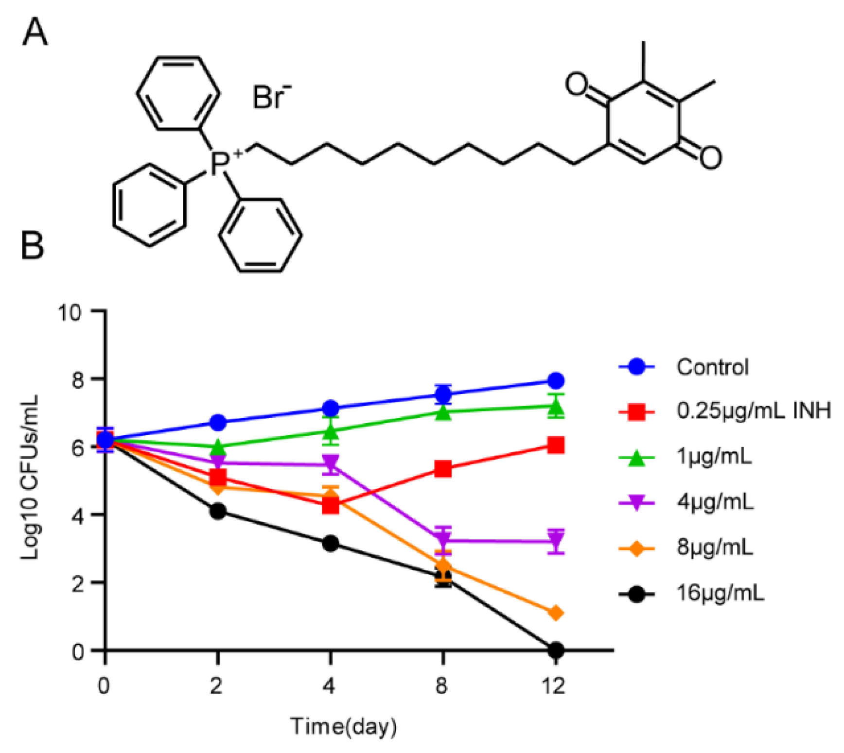
3.1. Activity of SKQ-1 against Mtb

We preliminarily determined the antibacterial effect of SKQ-1 on the BCG strain using the method of whole-cell screening. Subsequently, we used the resazurin detection method to detect the 90% MICs of SKQ-1 against Mtb H37Rv, the attenuated strain H37Ra, the virulent strain M. bovis, and the vaccine strain BCG. The results showed that the MICs for both H37Rv and H37Ra were 1 μg/mL, and the MICs for M. bovis and BCG were both 2 μg/mL. We examined the antimicrobial activity of SKQ-1 against drug-resistant strains, where the MICs for mono-drug-resistant strains against first-line anti-tuberculosis drugs (RIF, INH, and EMB at 1–2 µg/mL) and the MICs of SKQ-1 against the multidrug-resistant strain at 4 μg/mL (Table 1), proved the strong antibacterial effect on drug-resistant strains. The cytotoxicity of SKQ-1 in Vero cells was determined using the WST-1, and the results showed that the IC50 of SKQ-1 in Vero cells was 32 µg/mL.

3.2. Kill Curve Dynamics

To determine the killing kinetics of SKQ-1 against Mtb H37Rv, we performed a CFU analysis of bacteria that were cultured in 7H9 medium at concentrations of 1 μg/mL, 4 μg/mL, 8 μg/mL, and 16 μg/mL SKQ-1 for 0, 2, 4, 8, and 12 days. The results showed that SKQ-1 had a concentration-dependent killing effect on Mtb. The bacteria were reduced by 1 log10 CFU/mL and 4 log10 CFU/mL at 2 days and 12 days, respectively, after treatment with 8 μg/mL SKQ-1. At a concentration of 16 μg/mL, SKQ-1 killed all bacteria on the 12th day (Figure 1). These results indicated that the compound had a significant bactericidal effect on Mtb.

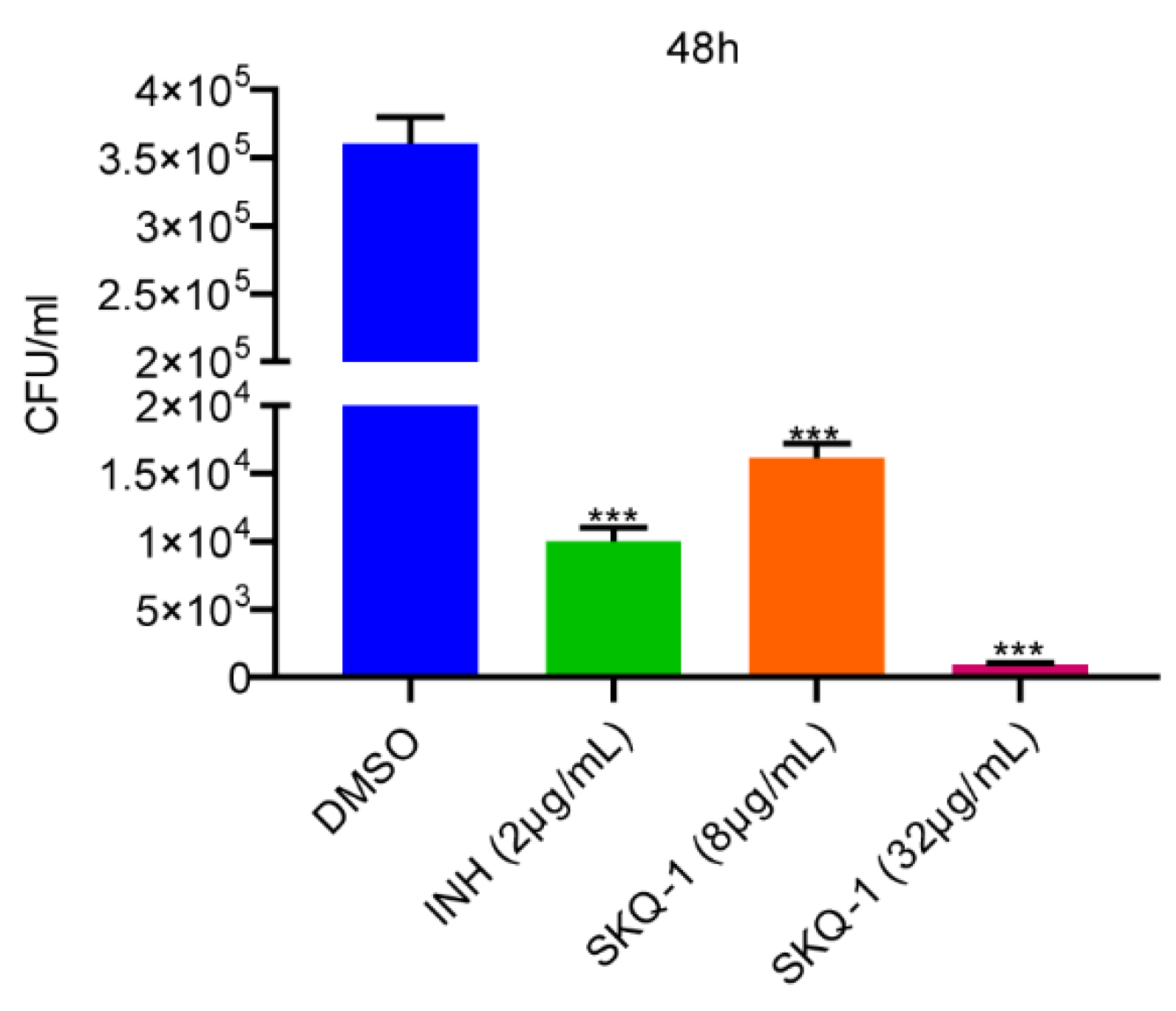
3.3. Activity of SKQ-1 against Intracellular Mtb

The effect of SKQ-1 on intracellular Mtb was examined through the THP-1 macrophage infection model. Macrophages were incubated with RPMI-1640 medium supplemented with 2% FBS containing 8 μg/mL or 32 μg/mL SKQ-1 until 48 h post-infection. A group cultured with a medium supplemented with DMSO served as a negative control, and a group cultured with a medium with a final concentration of 2 μg/mL INH served as a positive control. At 48 h post-infection, the cells were lysed, and CFU assays were performed to determine the number of viable intracellular bacteria. The bactericidal effect of INH on intracellular H37Rv was consistent with that in previous reports. The effects of 8 μg/mL SKQ-1 and 2 μg/mL INH were similar, and the clearance of H37Rv in macrophages was enhanced with the increase in SKQ-1 concentration. Compared with the negative control group, the group of SKQ-1 at 32 μg/mL exhibited a reduction in intracellular bacteria by 360 times (Figure 2). The above results indicate that SKQ-1 can kill H37Rv in macrophages.

3.4. The Antibacterial Synergy of SKQ-1

The TB treatments are long, and long-term use of a single drug would accelerate the development of drug-resistant Mtb strains. The development of effective combination therapy regimens is crucial in reducing the emergence of drug-resistant strains and shortening the duration of treatments. We determined that the MICs of RIF, INH, EMB, LNZ, and BDQ against the H37Rv strain were 0.0156 μg/mL, 0.0625 μg/mL, 1 μg/mL, 0.5 μg/mL, and 0.0625 μg/mL, respectively. Subsequently, we explored the effects of combining SKQ-1 with RIF, INH, EMB, LNZ, and BDQ using the checkerboard method. The results in terms of drug synergy showed that the synergistic effects of SKQ-1 combined with RIF and INH on Mtb H37Rv and the FICIs were 0.1875 and 0.0626, respectively. In combination with SKQ-1, the MICs of RIF and INH in H37Rv decreased from 0.0156 and 0.0625 μg/mL to 0.002 μg/mL; thus, they decreased by 7.8 and 31.25 times, respectively. In addition, there were no synergistic effects of SKQ-1 when combined with EMB, LNZ, or BDQ in Mtb H37Rv, but there was an additive effect; the FICIs were 1.004, 1.0156, and 0.5156, respectively. Furthermore, in combination with SKQ-1, the MIC of BDQ in H37Rv decreased from 0.0625 μg/mL to 0.0312 μg/mL, a twofold decrease (Table 2).

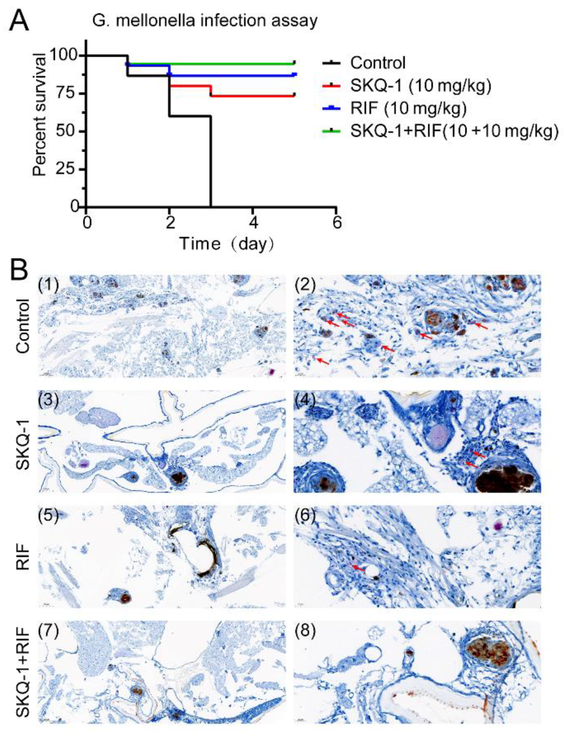
3.5. SKQ-1 in Combination with RIF Protects G. mellonella from Mtb

We used different doses to infect G. mellonella; the lowest dose of 5 × 10⁶, which killed all the G. mellonella within 3 days, was set as the infectious dose for the subsequent protective experiments. The anti-tubercular effects of SKQ-1 and SKQ-1 combined with RIF were evaluated through observing the survival rate of G. mellonella and the results of acid-fast staining. In the control group, the G. mellonella without any drug treatment, all died at 3 days post-infection, and multiple aggregates of Mtb were observed using acid-fast staining. The G. mellonella survival rates in the SKQ-1 and RIF groups were 75% and 80%, respectively. Compared with those in the control group, Mtb aggregates were significantly reduced in the single-drug treatment groups in vivo. The combination of SKQ-1 and RIF increased the survival rate of G. mellonella to 90% (Figure 3), and Mtb was not observed in this group (Figure 3). The above results indicate that SKQ-1 is active against Mtb in vivo, and its use in combination with RIF can enhance its therapeutic effect.

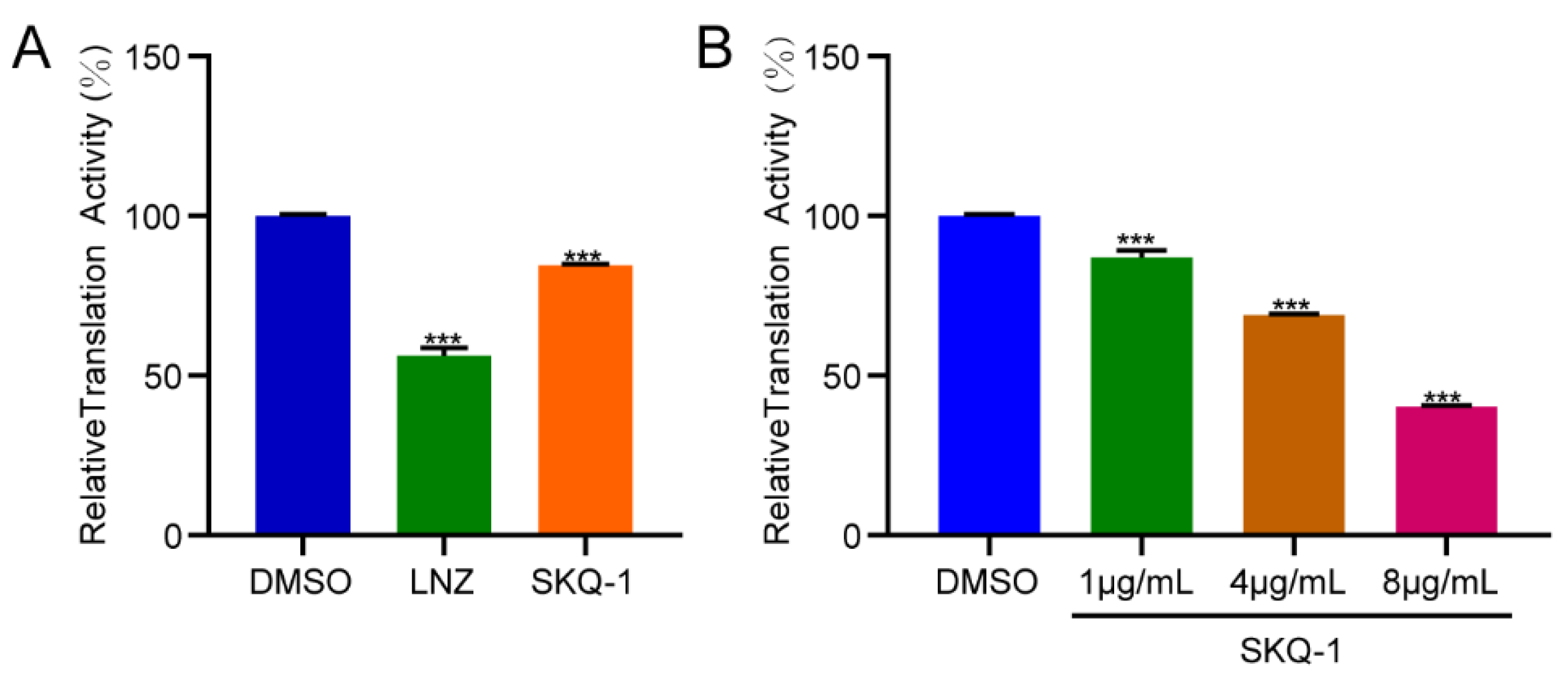
3.6. SKQ-1 Targets Ribosomes in Mtb

To reveal the mutation frequency, we collected 1 × 10⁹ log-phase H37Rv cells and coated them on 8×, 16×, and 32× MIC plates; then, they were cultured at 37 °C for one month. We obtained two single colonies on the 32× MIC plate, but no colonies were observed on the 8× and 16× MIC plates. We calculated the resistance frequency of Mtb to SKQ-1 to be 0.5 × 10⁻⁸/CFU from the initial inoculation and the number of mutant colonies obtained. Previous studies indicated that the mutation frequencies of INH and RIF were 10−6 and 10−7 to 10−8, respectively [25]. The results indicated that SKQ-1 had a lower mutation frequency during use.
To explore the target of SKQ-1, we performed whole-genome sequencing of two SKQ-1-resistant strains and a wild-type laboratory strain. Through aligning the results with the sequence of the reference genome, we found that the genomic sequence changes that were present in both the mutant strains but not in the wild-type laboratory strain were located on the 50S subunits of Mtb. There were 14 single-nucleotide polymorphisms (SNPs) localized in the 50S ribosome (Table 3), suggesting that SKQ-1 targets the ribosome in Mtb. The normal function of the ribosome is an indispensable prerequisite for ensuring protein synthesis in Mtb. Linezolid, which is an oxazolidinone, kills Mtb through interacting with the 23S rRNA of the 50S ribosome and interfering with protein synthesis [26,27].
To confirm the effect of SKQ-1 on protein synthesis, we performed a coupled transcription/translation analysis. DMSO and 0.5 μg/mL LNZ were added to the reaction systems as a negative control and positive control, respectively. We detected the inhibitory effect of 2 μg/mL SKQ-1 on translation. The results showed that LNZ significantly inhibits protein translation, which was consistent with the findings of previous studies, and SKQ-1 exhibited the same effect as that of LNZ (Figure 4A). Subsequently, we measured the inhibition of translation after treatment with different concentrations of SKQ-1 (1 μg/mL, 4 μg/mL, and 8 μg/mL), and the results showed that the inhibition of protein synthesis was enhanced as the concentration increased. It was also found that 8 μg/mL SKQ-1 was able to reduce the translation activity to 40% (Figure 4B).

4. Discussion

The development of new drugs for TB therapy requires high costs and a long time, but repurposing previously discovered TB drugs is faster and more effective. Drugs that have been approved for use have relatively complete safety data and clinical experience. SKQ-1 is an approved drug for the treatment of dry eye syndrome in the FDA Drug Library. The relatively low cytotoxicity of SKQ-1 in cells is a prerequisite for TB treatment.
The survival of Mtb in host macrophages is why it is difficult to clear it. The ability to kill intracellular Mtb is also an important indicator for evaluating anti-tuberculosis drugs. We found that SKQ-1 showed a significant killing ability against Mtb in macrophages. Thus, SKQ-1 has the potential to become a clinical treatment for tuberculosis.
INH, RIF, and EMB play a vital role in the clinical treatment of TB [28]. BDQ is a selective Mtb ATP synthase inhibitor; it is active against replicating and dormant Mtb and can be used to treat MDR-TB [29,30]. The oxazolidinone antibiotic linezolid, which inhibits protein synthesis via binding to the bacterial 50S ribosomal subunit, was discovered through drug repurposing and is recommended by the WHO for the treatment of patients infected with MDR-TB or XDR-TB infection [31,32]. We measured the combined effects of SKQ-1 and these anti-tuberculosis drugs. The results showed that SKQ-1 had a synergistic effect when used in combination with RIF and INH, and it had an additive effect when used with EMB, BDQ, and LNZ. We identified the single-nucleotide mutations of SKQ-1-resistant strains located in the 23S rRNA, which is an important component of the ribosomal 50S subunit, and we demonstrated that SKQ-1 impairs protein synthesis through inhibiting translation. Since both SKQ-1 and LNZ target the 50S subunit, there were no synergistic effects when the two drugs were used in combination. SKQ-1 synergizes with RIF, which targets RNA polymerase to repress Mtb transcription [33], thus impairing protein synthesis. It also had good synergistic effects both in vivo and in vitro. We detected the MIC of SKQ-1 against clinically isolated resistant strains. The results showed that the MICs of RIF, INH, and EMB single-drug-resistant tuberculosis were 1–2 μg/mL, and the MICs for the two MDR-TB strains were 4 and 8 μg/mL, respectively.
In summary, we identified an FDA-approved drug, SKQ-1, which targets the 50S subunit of the ribosome of Mtb to inhibit protein synthesis. We demonstrated the effect of SKQ-1 on clinically isolated drug-resistant strains and the synergistic effect of SKQ-1 and RIF in vitro and in vivo. It is suggested that SKQ-1 can be a candidate drug for drug-susceptible tuberculosis and mono-drug-resistant tuberculosis treatment.

Author Contributions

Conceptualization, G.W. and C.T.; methodology, Y.B. and C.W.; software, Z.Z.; validation, Y.L; formal analysis, W.D., Y.L. and L.Z.; investigation, G.W. and Y.B.; resources, H.L.; data curation, Y.B. and W.L.; writing—original draft preparation, W.D.; writing—review and editing, G.W. and C.T.; visualization, W.L.; supervision, X.W. and H.C.; project administration, X.W., H.C. and C.T.; funding acquisition, C.T. All authors have read and agreed to the published version of the manuscript.

Funding

This work was funded by the National Key Research and Development Program (2021YFD1800402) and the Natural Science Foundation of Hubei Province (2021CFA016).

Institutional Review Board Statement

Not applicable.

Informed Consent Statement

Not applicable.

Data Availability Statement

The data are not publicly available due to privacy.

Acknowledgments

We thank Chuan-You Li from the Beijing Tuberculosis & Thoracic Tumor Research Institute for kindly providing the M. tuberculosis.

Conflicts of Interest

The authors declare no conflict of interest.

References

  1. Bagcchi, S. WHO’s global tuberculosis report 2022. Lancet Microbe 2023, 4, e20. [Google Scholar] [CrossRef] [PubMed]
  2. Wolff, K.A.; Nguyen, L. Strategies for potentiation of ethionamide and folate antagonists against Mycobacterium tuberculosis. Expert. Rev. Anti. Infect. Ther. 2012, 10, 971–981. [Google Scholar] [CrossRef] [PubMed]
  3. Zumla, A.I.; Gillespie, S.H.; Hoelscher, M.; Philips, P.P.; Cole, S.T.; Abubakar, I.; McHugh, T.D.; Schito, M.; Maeurer, M.; Nunn, A.J. New antituberculosis drugs, regimens, and adjunct therapies: Needs, advances, and future prospects. Lancet Infect. Dis. 2014, 14, 327–340. [Google Scholar] [CrossRef] [PubMed]
  4. Zitko, J.; Doležal, M. Old Drugs and New Targets as an Outlook for the Treatment of Tuberculosis. Curr. Med. Chem. 2018, 25, 5142–5167. [Google Scholar] [CrossRef] [PubMed]
  5. An, Q.; Li, C.; Chen, Y.; Deng, Y.; Yang, T.; Luo, Y. Repurposed drug candidates for antituberculosis therapy. Eur. J. Med. Chem. 2020, 192, 112175. [Google Scholar] [CrossRef]
  6. Zumla, A.; Nahid, P.; Cole, S.T. Advances in the development of new tuberculosis drugs and treatment regimens. Nat. Rev. Drug Discov. 2013, 12, 388–404. [Google Scholar] [CrossRef]
  7. Gold, B.; Pingle, M.; Brickner, S.J.; Shah, N.; Roberts, J.; Rundell, M.; Bracken, W.C.; Warrier, T.; Somersan, S.; Venugopal, A.; et al. Nonsteroidal anti-inflammatory drug sensitizes Mycobacterium tuberculosis to endogenous and exogenous antimicrobials. Proc. Natl. Acad. Sci. USA 2012, 109, 16004–16011. [Google Scholar] [CrossRef]
  8. Byrne, S.T.; Denkin, S.M.; Zhang, Y. Aspirin and ibuprofen enhance pyrazinamide treatment of murine tuberculosis. J. Antimicrob. Chemother. 2007, 59, 313–316. [Google Scholar] [CrossRef]
  9. Maitra, A.; Bates, S.; Shaik, M.; Evangelopoulos, D.; Abubakar, I.; McHugh, T.D.; Lipman, M.; Bhakta, S. Repurposing drugs for treatment of tuberculosis: A role for non-steroidal anti-inflammatory drugs. Br. Med. Bull. 2016, 118, 138–148. [Google Scholar] [CrossRef]
  10. Vashisht, R.; Brahmachari, S.K. Metformin as a potential combination therapy with existing front-line antibiotics for Tuberculosis. J. Transl. Med. 2015, 13, 83. [Google Scholar] [CrossRef]
  11. Oglesby, W.; Kara, A.M.; Granados, H.; Cervantes, J.L. Metformin in tuberculosis: Beyond control of hyperglycemia. Infection 2019, 47, 697–702. [Google Scholar] [CrossRef] [PubMed]
  12. Antonenko, Y.N.; Roginsky, V.A.; Pashkovskaya, A.A.; Rokitskaya, T.I.; Kotova, E.A.; Zaspa, A.A.; Chernyak, B.V.; Skulachev, V.P. Protective effects of mitochondria-targeted antioxidant SkQ in aqueous and lipid membrane environments. J. Membr. Biol. 2008, 222, 141–149. [Google Scholar] [CrossRef] [PubMed]
  13. Shabalina, I.G.; Vyssokikh, M.Y.; Gibanova, N.; Csikasz, R.I.; Edgar, D.; Hallden-Waldemarson, A.; Rozhdestvenskaya, Z.; Bakeeva, L.E.; Vays, V.B.; Pustovidko, A.V.; et al. Improved health-span and lifespan in mtDNA mutator mice treated with the mitochondrially targeted antioxidant SkQ1. Aging 2017, 9, 315–339. [Google Scholar] [CrossRef] [PubMed]
  14. Novikova, Y.P.; Gancharova, O.S.; Eichler, O.V.; Philippov, P.P.; Grigoryan, E.N. Preventive and therapeutic effects of SkQ1-containing Visomitin eye drops against light-induced retinal degeneration. Biochemistry 2014, 79, 1101–1110. [Google Scholar] [CrossRef] [PubMed]
  15. Brzheskiy, V.V.; Efimova, E.L.; Vorontsova, T.N.; Alekseev, V.N.; Gusarevich, O.G.; Shaidurova, K.N.; Ryabtseva, A.A.; Andryukhina, O.M.; Kamenskikh, T.G.; Sumarokova, E.S.; et al. Results of a Multicenter, Randomized, Double-Masked, Placebo-Controlled Clinical Study of the Efficacy and Safety of Visomitin Eye Drops in Patients with Dry Eye Syndrome. Adv. Ther. 2015, 32, 1263–1279. [Google Scholar] [CrossRef]
  16. Qu, M.; Wan, L.; Dong, M.; Wang, Y.; Xie, L.; Zhou, Q. Hyperglycemia-induced severe mitochondrial bioenergetic deficit of lacrimal gland contributes to the early onset of dry eye in diabetic mice. Free Radic. Biol. Med. 2021, 166, 313–323. [Google Scholar] [CrossRef] [PubMed]
  17. Chistyakov, D.V.; Baksheeva, V.E.; Tiulina, V.V.; Goriainov, S.V.; Azbukina, N.V.; Gancharova, O.S.; Arifulin, E.A.; Komarov, S.V.; Chistyakov, V.V.; Tikhomirova, N.K.; et al. Mechanisms and Treatment of Light-Induced Retinal Degeneration-Associated Inflammation: Insights from Biochemical Profiling of the Aqueous Humor. Int. J. Mol. Sci. 2020, 21, 704. [Google Scholar] [CrossRef]
  18. Wang, G.; Dong, W.; Lu, H.; Lu, W.; Feng, J.; Wang, X.; Chen, H.; Liu, M.; Tan, C. Enniatin A1, A Natural Compound with Bactericidal Activity against Mycobacterium tuberculosis In Vitro. Molecules 2019, 25, 38. [Google Scholar] [CrossRef]
  19. Odds, F.C. Synergy, antagonism, and what the chequerboard puts between them. J. Antimicrob. Chemother. 2003, 52, 1. [Google Scholar] [CrossRef]
  20. Dong, W.; Wang, G.; Feng, J.; Li, P.; Wang, R.; Lu, H.; Lu, W.; Wang, C.; Wang, X.; Chen, H.; et al. MiR-25 blunts autophagy and promotes the survival of Mycobacterium tuberculosis by regulating NPC1. iScience 2022, 25, 104279. [Google Scholar] [CrossRef]
  21. Asai, M.; Li, Y.; Spiropoulos, J.; Cooley, W.; Everest, D.; Robertson, B.D.; Langford, P.R.; Newton, S.M. A novel biosafety level 2 compliant tuberculosis infection model using a DeltaleuDDeltapanCD double auxotroph of Mycobacterium tuberculosis H37Rv and Galleria mellonella. Virulence 2020, 11, 811–824. [Google Scholar] [CrossRef] [PubMed]
  22. Asai, M.; Li, Y.; Khara, J.S.; Robertson, B.D.; Langford, P.R.; Newton, S.M. Galleria mellonella: An Infection Model for Screening Compounds against the Mycobacterium tuberculosis Complex. Front. Microbiol. 2019, 10, 2630. [Google Scholar] [CrossRef] [PubMed]
  23. Wilson, R.; Kumar, P.; Parashar, V.; Vilcheze, C.; Veyron-Churlet, R.; Freundlich, J.S.; Barnes, S.W.; Walker, J.R.; Szymonifka, M.J.; Marchiano, E.; et al. Antituberculosis thiophenes define a requirement for Pks13 in mycolic acid biosynthesis. Nat. Chem. Biol. 2013, 9, 499–506. [Google Scholar] [CrossRef] [PubMed]
  24. Lemieux, M.R.; Siricilla, S.; Mitachi, K.; Eslamimehr, S.; Wang, Y.; Yang, D.; Pressly, J.D.; Kong, Y.; Park, F.; Franzblau, S.G.; et al. An antimycobacterial pleuromutilin analogue effective against dormant bacilli. Bioorganic. Med. Chem. 2018, 26, 4787–4796. [Google Scholar] [CrossRef] [PubMed]
  25. Siddiqi, S.; Takhar, P.; Baldeviano, C.; Glover, W.; Zhang, Y. Isoniazid induces its own resistance in nonreplicating Mycobacterium tuberculosis. Antimicrob. Agents Chemother. 2007, 51, 2100–2104. [Google Scholar] [CrossRef] [PubMed]
  26. Zhang, M.; Sala, C.; Dhar, N.; Vocat, A.; Sambandamurthy, V.K.; Sharma, S.; Marriner, G.; Balasubramanian, V.; Cole, S.T. In vitro and in vivo activities of three oxazolidinones against nonreplicating Mycobacterium tuberculosis. Antimicrob. Agents Chemother. 2014, 58, 3217–3223. [Google Scholar] [CrossRef] [PubMed]
  27. Leach, K.L.; Brickner, S.J.; Noe, M.C.; Miller, P.F. Linezolid, the first oxazolidinone antibacterial agent. Ann. N. Y. Acad. Sci. 2011, 1222, 49–54. [Google Scholar] [CrossRef]
  28. Wang, X.; Zhang, H.; Han, Y.; Huo, L.; Cao, Y.; Xu, X.; Ai, L. Rapid and simultaneous determination of ten anti-tuberculosis drugs in human plasma by UPLC-MS/MS with applications in therapeutic drug monitoring. J. Chromatogr. B 2020, 1152, 122246. [Google Scholar] [CrossRef]
  29. Luo, M.; Zhou, W.; Patel, H.; Srivastava, A.P.; Symersky, J.; Bonar, M.M.; Faraldo-Gómez, J.D.; Liao, M.; Mueller, D.M. Bedaquiline inhibits the yeast and human mitochondrial ATP synthases. Commun. Biol. 2020, 3, 452. [Google Scholar] [CrossRef]
  30. Zhao, Y.; Fox, T.; Manning, K.; Stewart, A.; Tiffin, N.; Khomo, N.; Leslie, J.; Boulle, A.; Mudaly, V.; Kock, Y.; et al. Improved Treatment Outcomes with Bedaquiline When Substituted for Second-line Injectable Agents in Multidrug-resistant Tuberculosis: A Retrospective Cohort Study. Clin. Infect. Dis. 2019, 68, 1522–1529. [Google Scholar] [CrossRef]
  31. Zhang, X.; Falagas, M.E.; Vardakas, K.Z.; Wang, R.; Qin, R.; Wang, J.; Liu, Y. Systematic review and meta-analysis of the efficacy and safety of therapy with linezolid containing regimens in the treatment of multidrug-resistant and extensively drug-resistant tuberculosis. J. Thorac. Dis. 2015, 7, 603–615. [Google Scholar] [CrossRef] [PubMed]
  32. Hashemian, S.M.; Farhadi, T.; Ganjparvar, M. Linezolid: A review of its properties, function, and use in critical care. Drug Des. Dev. Ther. 2018, 12, 1759–1767. [Google Scholar] [CrossRef] [PubMed]
  33. Campbell, E.A.; Korzheva, N.; Mustaev, A.; Murakami, K.; Nair, S.; Goldfarb, A.; Darst, S.A. Structural mechanism for rifampicin inhibition of bacterial rna polymerase. Cell 2001, 104, 901–912. [Google Scholar] [CrossRef] [PubMed]
Figure 1. The killing kinetics of SKQ-1 against Mtb. (A) Structures of SKQ-1. (B) Mtb was treated with 1 μg/mL, 4 μg/mL, 8 μg/mL, and 16 μg/mL SKQ-1, and 0.25 μg/mL. INH was used as a positive control. The CFU was measured at different time points after treatment. Experiments were performed in triplicate, and at least two biological replicates were included. Data are shown as mean ± SD.
Biomolecules 13 01793 g001
Figure 2. Intracellular bactericidal activity of SKQ-1 against Mtb. THP-1 macrophages were infected with Mtb and treated with SKQ-1 at final concentrations of 8× and 32× the minimum inhibitory concentration (MIC). Macrophages treated with DMSO were used as a negative control. Macrophages treated with isoniazid (INH) at 32× the MIC were used as positive controls. The CFU was measured after 2 days of treatment and recalculated as CFU/mL. Experiments were performed in triplicate, and at least two biological replicates were included. Data were shown as mean ± SD and unpaired Student’s t-test or one-way ANOVA was used to conduct statistical significance analysis. (***, p < 0.001).
Biomolecules 13 01793 g002
Figure 3. Combined effects of SKQ-1 and rifampicin (RIF) in a G. mellonella larvae infection assay. G. mellonella larvae (n = 15 per group) were infected with H37Rv (5 × 10⁶ CFU). G. mellonella larvae were then treated with SKQ-1 (10 mg/kg) or RIF (10 mg/kg) alone or in combination (10 + 10 mg/kg). (A) Survival rates of the G. mellonella larvae. (B) Acid-fast staining of H37Rv in the G. mellonella larvae. The scale bars in images (1), (3), (5), and (7) represent 100 μm; the scale bars in images (2), (4), (6), and (8) represent 20 µm. Red arrow points to bacteria.
Biomolecules 13 01793 g003
Figure 4. The inhibition of translation by SKQ-1. The 50 µL reaction systems contained S30 premix, S30 extract, a complete amino acid mixture, pBESTluc™ DNA, and different concentrations of drugs; these were used to detect the inhibition of protein synthesis. (A) The relative translation activity of linezolid (LNZ) at 0.5 μg/mL and SKQ-1 at 2 μg/mL. (B) The relative translation activity of SKQ-1 at 1 μg/mL, 4 μg/mL, and 8 μg/mL. Experiments were performed in triplicate, and at least two biological replicates were included. Data were shown as mean ± SD and unpaired Student’s t-test or one-way ANOVA was used to conduct statistical significance analysis. (***, p < 0.001).
Biomolecules 13 01793 g004
Table 1. MICs of SKQ-1 against strains of M. tuberculosis.
StrainsMIC (μg/mL)
H37Rv (Susceptible)1
M. bovis (Susceptible)2
H37Ra (Susceptible)1
BCG (Susceptible)2
CR1 (RIF-R)2
CR2 (RIF-R)2
CR3 (INH-R)2
CR4 (INH-R)1
CR5 (EMB-R)1
CR6 (MDR)4
MIC: the minimum inhibitory concentration; M. bovis: Mycobacterium bovis; RIF: rifampicin; INH: isoniazid; EMB: ethambutol.
Table 2. Effects of Combining SKQ-1 with RIF, INH, EMB, LNZ, and BDQ to treat Mtb.
DrugMIC (μg/mL)FICFICIRemarks
CombinationAloneCombination
SKQ-110.06250.06250.1875synergy
RIF0.01560.0020.125
SKQ-110.03120.03120.0624synergy
INH0.06250.0020.0312
SKQ-110.0040.0041.004additive
EMB111
SKQ-110.01560.01561.0156additive
LNZ0.50.51
SKQ-110.01560.01560.5156additive
BDQ0.06250.03120.5
LNZ: linezolid; BDQ: bedaquiline.
Table 3. Single-nucleotide polymorphisms were detected in the two SKQ-1-resistant Mtb strains.
TypeLocationReferenceAfterGeneAnnotation
SNP1476234GArrl23S ribosomal RNA
SNP1476260AGrrl23S ribosomal RNA
SNP1476268ATrrl23S ribosomal RNA
SNP1476332GCrrl23S ribosomal RNA
SNP1476353GTrrl23S ribosomal RNA
SNP1476358TCrrl23S ribosomal RNA
SNP1476408GArrl23S ribosomal RNA
SNP1476466CTrrl23S ribosomal RNA
SNP1476481TCrrl23S ribosomal RNA
SNP1476506TCrrl23S ribosomal RNA
SNP1476530CTrrl23S ribosomal RNA
SNP1476547CTrrl23S ribosomal RNA
SNP1476567CTrrl23S ribosomal RNA
SNP1476584CTrrl23S ribosomal RNA
Disclaimer/Publisher’s Note: The statements, opinions and data contained in all publications are solely those of the individual author(s) and contributor(s) and not of MDPI and/or the editor(s). MDPI and/or the editor(s) disclaim responsibility for any injury to people or property resulting from any ideas, methods, instructions or products referred to in the content.

