
Thiazole Derivatives as Modulators of GluA2 AMPA Receptors: Potent Allosteric Effects and Neuroprotective Potential


Abstract
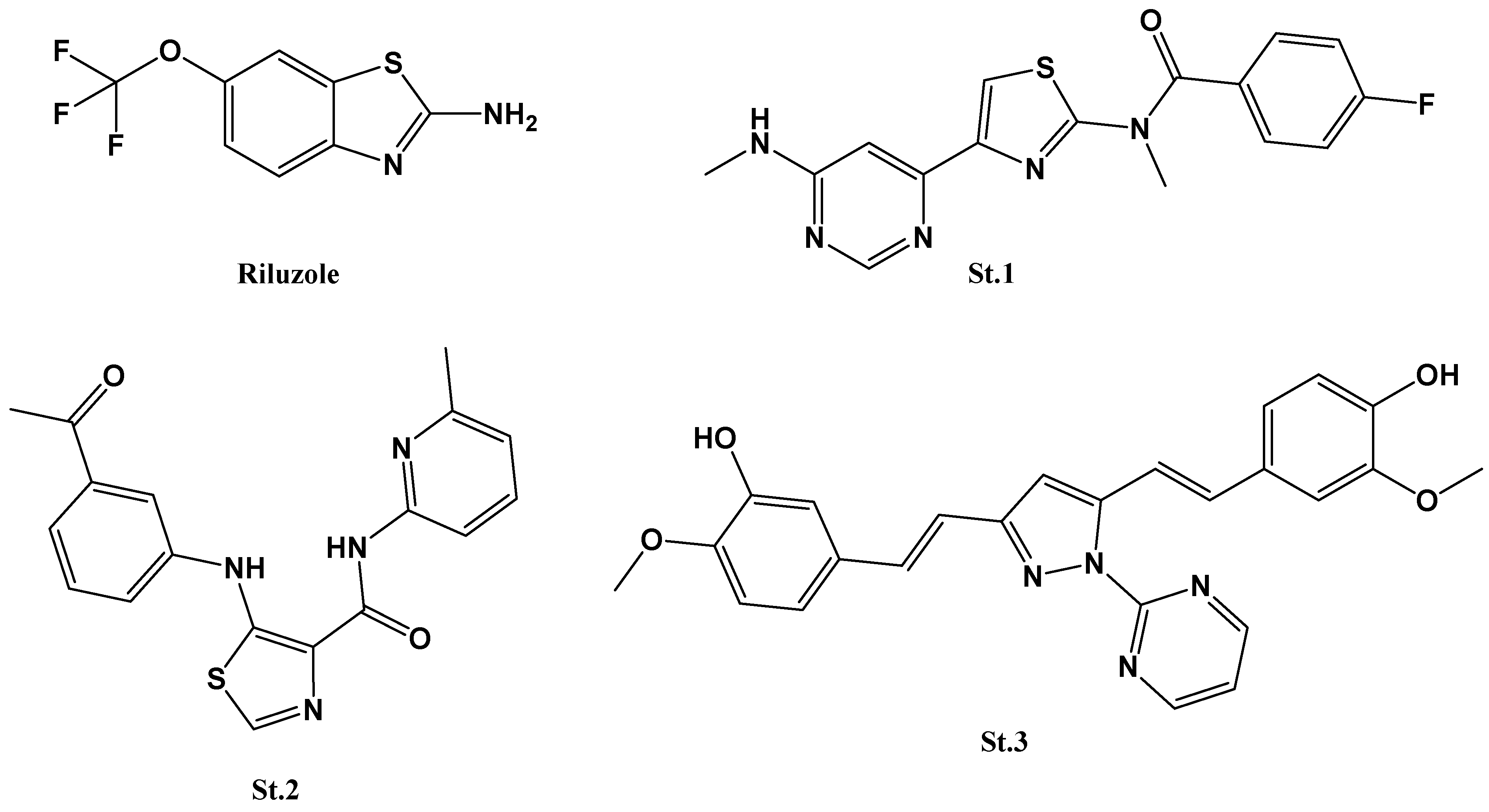
:1. Introduction
2. Materials and Methods
2.1. Materials
2.2. Instrumentation
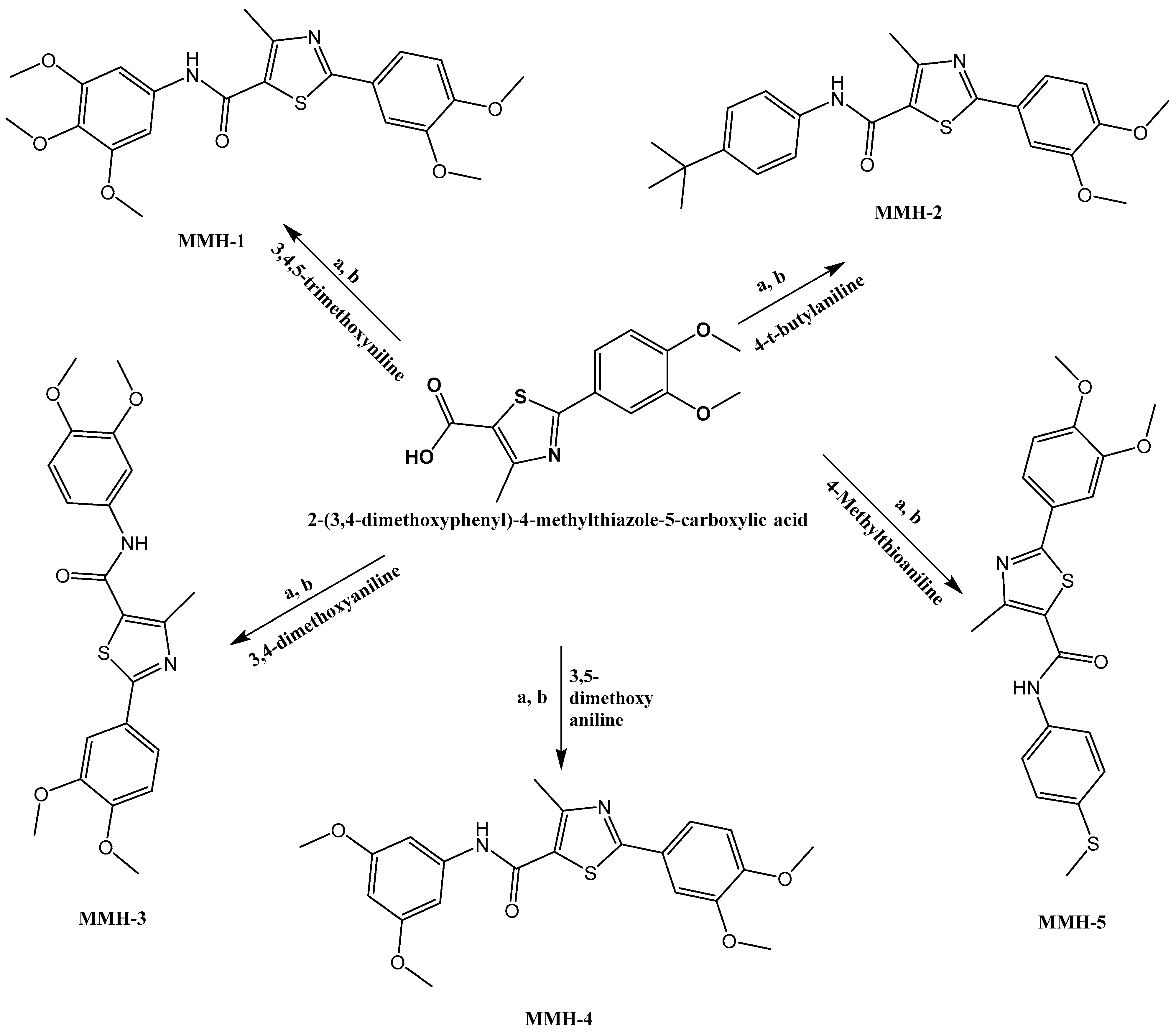
2.3. General Procedure for the Synthesis of Thiazole Carboxamide (MMH1-5)
2.4. HEK293T Cell Patch Clamp Recordings
2.5. Molecular Docking Studies
2.6. Cell Culture and Cytotoxicity Assay (MTS)
3. Results and Discussion
3.1. Chemistry
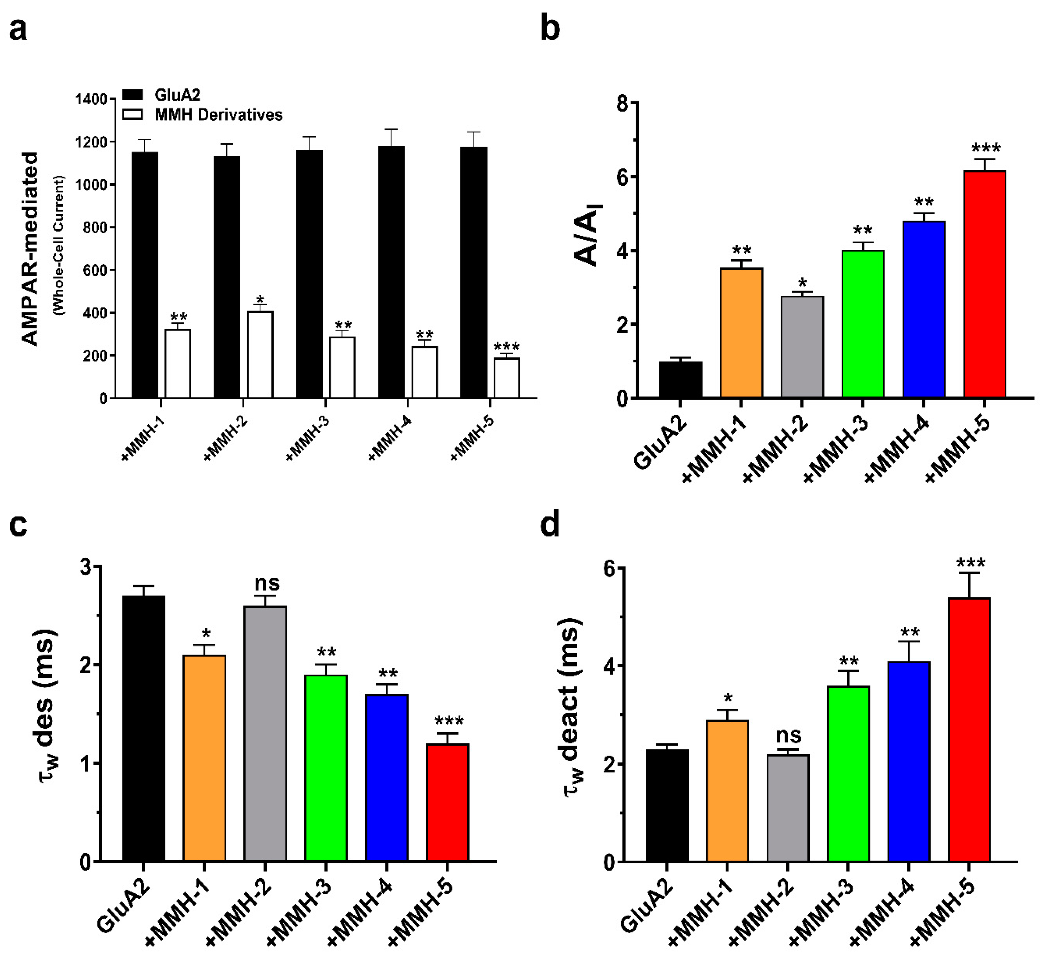
3.2. Thiazole Derivatives’ Effects on GluA2 Currents and Kinetics: Unveiling Desensitization and Deactivation Dynamics
3.3. Molecular Docking and SAR
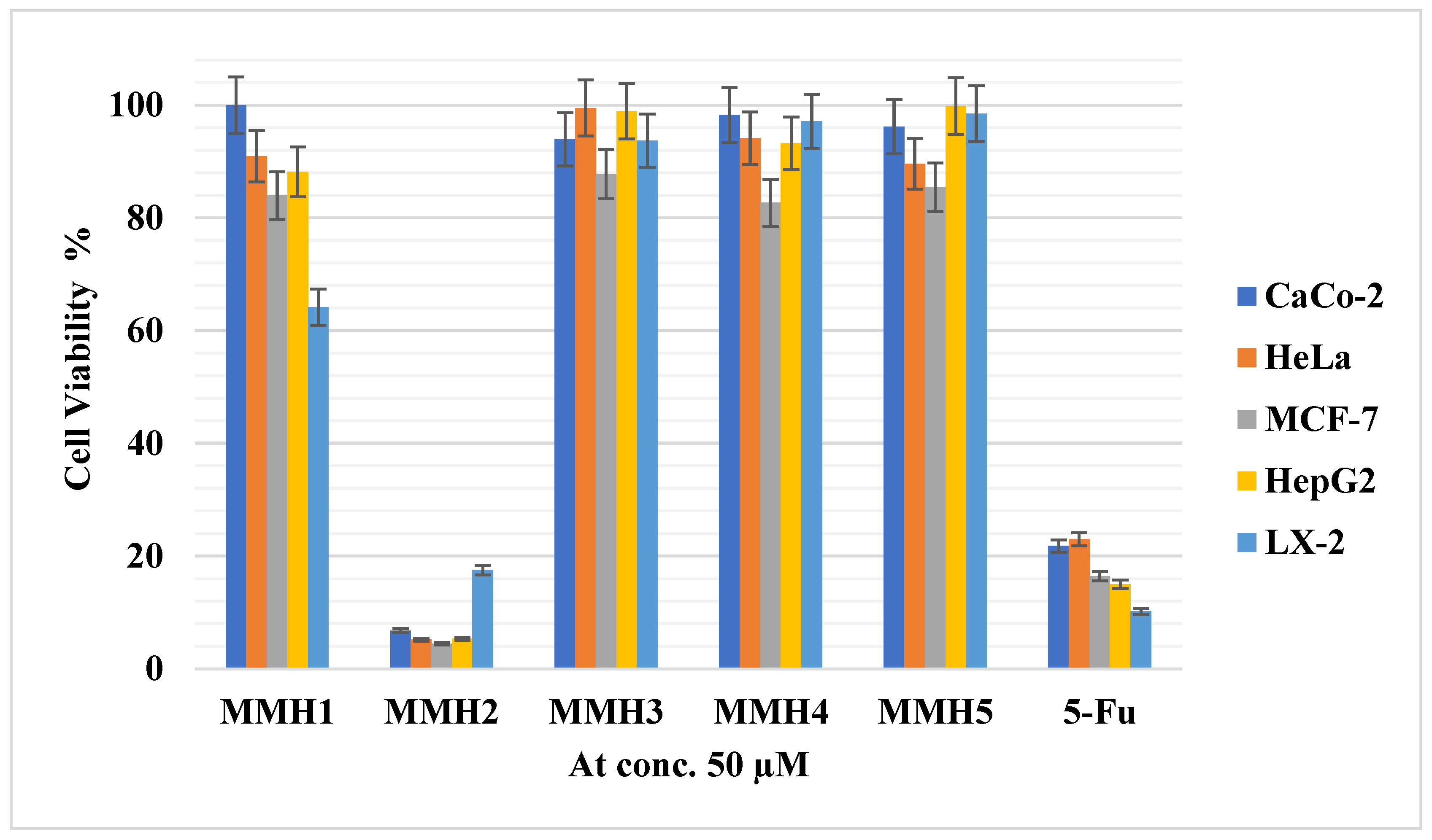
3.4. Cytotoxicity Results
4. Conclusions
Supplementary Materials
Funding
Institutional Review Board Statement
Informed Consent Statement
Data Availability Statement
Acknowledgments
Conflicts of Interest
References
- Stroebel, D.; Paoletti, P. Architecture and function of NMDA receptors: An evolutionary perspective. J. Physiol. 2021, 599, 2615–2638. [Google Scholar] [CrossRef] [PubMed]
- Narisawa-Saito, M.; Silva, A.J.; Yamaguchi, T.; Hayashi, T.; Yamamoto, T.; Nawa, H. Growth factor-mediated Fyn signaling regulates α-amino-3-hydroxy-5-methyl-4-isoxazolepropionic acid (AMPA) receptor expression in rodent neocortical neurons. Proc. Natl. Acad. Sci. USA 1999, 96, 2461–2466. [Google Scholar] [CrossRef] [PubMed]
- Zhao, Y.; Chen, S.; Swensen, A.C.; Qian, W.-J.; Gouaux, E. Architecture and subunit arrangement of native AMPA receptors elucidated by cryo-EM. Science 2019, 364, 355–362. [Google Scholar] [CrossRef] [PubMed]
- Greger, I.H.; Watson, J.F.; Cull-Candy, S.G. Structural and functional architecture of AMPA-type glutamate receptors and their auxiliary proteins. Neuron 2017, 94, 713–730. [Google Scholar] [CrossRef] [PubMed]
- Wright, A.; Vissel, B. The essential role of AMPA receptor GluR2 subunit RNA editing in the normal and diseased brain. Front. Mol. Neurosci. 2012, 5, 34. [Google Scholar] [CrossRef]
- Ptak, C.P.; Ahmed, A.H.; Oswald, R.E. Probing the allosteric modulator binding site of GluR2 with thiazide derivatives. Biochemistry 2009, 48, 8594–8602. [Google Scholar] [CrossRef]
- Krintel, C.; Frydenvang, K.; Olsen, L.; Kristensen, M.T.; de Barrios, O.; Naur, P.; Francotte, P.; Pirotte, B.; Gajhede, M.; Kastrup, J.S. Thermodynamics and structural analysis of positive allosteric modulation of the ionotropic glutamate receptor GluA2. Biochem. J. 2012, 441, 173–178. [Google Scholar] [CrossRef]
- Bigge, C.F.; Nikam, S.S. AMPA receptor agonists, antagonists and modulators: Their potential for clinical utility. Expert Opin. Ther. Pat. 1997, 7, 1099–1114. [Google Scholar] [CrossRef]
- Di Bonaventura, C.; Labate, A.; Maschio, M.; Meletti, S.; Russo, E. AMPA receptors and perampanel behind selected epilepsies: Current evidence and future perspectives. Expert Opin. Pharmacother. 2017, 18, 1751–1764. [Google Scholar] [CrossRef]
- Lazzaro, J.; Paternain, A.V.; Lerma, J.; Chenard, B.; Ewing, F.; Huang, J.; Welch, W.; Ganong, A.; Menniti, F.S. Functional characterization of CP-465,022, a selective, noncompetitive AMPA receptor antagonist. Neuropharmacology 2002, 42, 143–153. [Google Scholar] [CrossRef]
- Zwart, R.; Sher, E.; Ping, X.; Jin, X.; Sims, J.; Chappell, A.; Gleason, S.; Hahn, P.; Gardinier, K.; Gernert, D. Perampanel, an antagonist of α-amino-3-hydroxy-5-methyl-4-isoxazolepropionic acid receptors, for the treatment of epilepsy: Studies in human epileptic brain and nonepileptic brain and in rodent models. J. Pharmacol. Exp. Ther. 2014, 351, 124–133. [Google Scholar] [CrossRef] [PubMed]
- Hattab, S.; Kittana, N.; Qasarweh, L.; Tayem, Y. Prevalence and factors associated with polypharmacy among patients treated for psychiatric disorders in Palestine. Palest. Med. Pharm. J. 2023, 8, 1. [Google Scholar] [CrossRef]
- Cull-Candy, S.G.; Farrant, M. Ca2+-permeable AMPA receptors and their auxiliary subunits in synaptic plasticity and disease. J. Physiol. 2021, 599, 2655–2671. [Google Scholar] [CrossRef]
- Wu, Q.-L.; Gao, Y.; Li, J.-T.; Ma, W.-Y.; Chen, N.-H. The role of AMPARs composition and trafficking in synaptic plasticity and diseases. Cell. Mol. Neurobiol. 2022, 42, 2489–2504. [Google Scholar] [CrossRef]
- Qneibi, M.; Hamed, O.; Jaradat, N.; Hawash, M.; Al-Kerm, R.; Al-Kerm, R.; Sobuh, S.; Tarazi, S. The AMPA receptor biophysical gating properties and binding site: Focus on novel curcumin-based diazepines as non-competitive antagonists. Bioorg. Chem. 2021, 116, 105406. [Google Scholar] [CrossRef]
- Hosaka, T.; Yamashita, T.; Tamaoka, A.; Kwak, S. Extracellular RNAs as biomarkers of sporadic amyotrophic lateral sclerosis and other neurodegenerative diseases. Int. J. Mol. Sci. 2019, 20, 3148. [Google Scholar] [CrossRef]
- Balannik, V.; Menniti, F.S.; Paternain, A.V.; Lerma, J.; Stern-Bach, Y. Molecular mechanism of AMPA receptor noncompetitive antagonism. Neuron 2005, 48, 279–288. [Google Scholar] [CrossRef]
- Zhang, W.; Cho, Y.; Lolis, E.; Howe, J.R. Structural and single-channel results indicate that the rates of ligand binding domain closing and opening directly impact AMPA receptor gating. J. Neurosci. 2008, 28, 932–943. [Google Scholar] [CrossRef]
- Qneibi, M.; Hawash, M.; Jaradat, N.; Bdir, S. Affecting AMPA receptor biophysical gating properties with negative allosteric modulators. Mol. Neurobiol. 2022, 59, 5264–5275. [Google Scholar] [CrossRef]
- Roth, R.H.; Zhang, Y.; Huganir, R.L. Dynamic imaging of AMPA receptor trafficking in vitro and in vivo. Curr. Opin. Neurobiol. 2017, 45, 51–58. [Google Scholar] [CrossRef]
- Pkn, S.; Sahoo, J.; Paidesetty, S.K.; Mohanta, G.P. Thiazoles as potent anticancer agents: A review. Indian Drugs 2016, 53, 5–11. [Google Scholar]
- Bryson, H.M.; Fulton, B.; Benfield, P. Riluzole: A review of its pharmacodynamic and pharmacokinetic properties and therapeutic potential in amyotrophic lateral sclerosis. Drugs 1996, 52, 549–563. [Google Scholar] [CrossRef]
- Calabrese, E.J.; Calabrese, V.; Giordano, J. Demonstrated hormetic mechanisms putatively subserve riluzole-induced effects in neuroprotection against amyotrophic lateral sclerosis (ALS): Implications for research and clinical practice. Ageing Res. Rev. 2021, 67, 101273. [Google Scholar] [CrossRef]
- Satoh, A.; Nagatomi, Y.; Hirata, Y.; Ito, S.; Suzuki, G.; Kimura, T.; Maehara, S.; Hikichi, H.; Satow, A.; Hata, M. Discovery and in vitro and in vivo profiles of 4-fluoro-N-[4-[6-(isopropylamino) pyrimidin-4-yl]-1, 3-thiazol-2-yl]-N-methylbenzamide as novel class of an orally active metabotropic glutamate receptor 1 (mGluR1) antagonist. Bioorg. Med. Chem. Lett. 2009, 19, 5464–5468. [Google Scholar] [CrossRef]
- Mishra, C.B.; Kumari, S.; Tiwari, M. Thiazole: A promising heterocycle for the development of potent CNS active agents. Eur. J. Med. Chem. 2015, 92, 1–34. [Google Scholar] [CrossRef]
- Kretschmer, B.D.; Kratzer, U.; Schmidt, W.J. Riluzole, a glutamate release inhibitor, and motor behavior. Naunyn-Schmiedeberg’s Arch. Pharmacol. 1998, 358, 181–190. [Google Scholar] [CrossRef]
- Agarwal, S.; Kalal, P.; Gandhi, D.; Prajapat, P. Thiazole containing Heterocycles with CNS activity. Curr. Drug Discov. Technol. 2018, 15, 178–195. [Google Scholar] [CrossRef]
- Singh, A.; Malhotra, D.; Singh, K.; Chadha, R.; Bedi, P.M.S. Thiazole derivatives in medicinal chemistry: Recent advancements in synthetic strategies, structure activity relationship and pharmacological outcomes. J. Mol. Struct. 2022, 1266, 133479. [Google Scholar] [CrossRef]
- Iwaszkiewicz-Grzes, D.; Cholewinski, G.; Kot-Wasik, A.; Trzonkowski, P.; Dzierzbicka, K. Synthesis and biological activity of mycophenolic acid-amino acid derivatives. Eur. J. Med. Chem. 2013, 69, 863–871. [Google Scholar] [CrossRef]
- Kim, I.H.; Lee, I.H.; Nishiwaki, H.; Hammock, B.D.; Nishi, K. Structure–activity relationships of substituted oxyoxalamides as inhibitors of the human soluble epoxide hydrolase. Bioorg. Med. Chem. 2014, 22, 1163–1175. [Google Scholar] [CrossRef]
- Qneibi, M.; Nassar, S.; Bdir, S.; Hidmi, A. α-Lipoic Acid Derivatives as Allosteric Modulators for Targeting AMPA-Type Glutamate Receptors’ Gating Modules. Cells 2022, 11, 3608. [Google Scholar] [CrossRef]
- Jaradat, N.; Hawash, M.; Qneibi, M.; Shtayeh, T.; Sobuh, S.; Arar, M.; Bdir, S. The effect of novel negative allosteric 2,3-benzodiazepine on glutamate AMPA receptor and their cytotoxicity. J. Mol. Struct. 2022, 1261, 132936. [Google Scholar] [CrossRef]
- Jaradat, N.; Qneibi, M.; Hawash, M.; Al-Maharik, N.; Qadi, M.; Abualhasan, M.N.; Ayesh, O.; Bsharat, J.; Khadir, M.; Morshed, R. Assessing Artemisia arborescens essential oil compositions, antimicrobial, cytotoxic, anti-inflammatory, and neuroprotective effects gathered from two geographic locations in Palestine. Ind. Crops Prod. 2022, 176, 114360. [Google Scholar] [CrossRef]
- Adasme, M.F.; Linnemann, K.L.; Bolz, S.N.; Kaiser, F.; Salentin, S.; Haupt, V.J.; Schroeder, M. PLIP 2021: Expanding the scope of the protein–ligand interaction profiler to DNA and RNA. Nucleic Acids Res. 2021, 49, W530–W534. [Google Scholar] [CrossRef]
- Schrödinger, Inc. The PyMOL Molecular Graphics System, version 1.8; Schrödinger, Inc.: New York, NY, USA, 2015. [Google Scholar]
- Jaradat, N.; Al-Maharik, N. Fingerprinting, Antimicrobial, Antioxidant, Anticancer, Cyclooxygenase and Metabolic Enzymes Inhibitory Characteristic Evaluations of Stachys viticina Boiss. Essential Oil. Molecules 2019, 24, 3880. [Google Scholar] [CrossRef]
- Özcan, G.; Akman, G.; Khalilia, W. Induction of Apoptosis by Hypoxia in C-4 I Human Cervical Cancer Cells. Palest. Med. Pharm. J. 2020, 7, 2. [Google Scholar]
- Qneibi, M.; Hawash, M.; Bdir, S.; Nacak Baytas, S. Targeting the kinetics mechanism of AMPA receptor inhibition by 2-oxo-3H-benzoxazole derivatives. Bioorg. Chem. 2022, 129, 106163. [Google Scholar] [CrossRef]
- Szymańska, E.; Nielsen, B.; Johansen, T.N.; Cuñado Moral, A.M.; Pickering, D.S.; Szczepańska, K.; Mickowska, A.; Kieć-Kononowicz, K. Pharmacological characterization and binding modes of novel racemic and optically active phenylalanine-based antagonists of AMPA receptors. Eur. J. Med. Chem. 2017, 138, 874–883. [Google Scholar] [CrossRef]
- Armstrong, N.; Gouaux, E. Mechanisms for Activation and Antagonism of an AMPA-Sensitive Glutamate Receptor: Crystal Structures of the GluR2 Ligand Binding Core. Neuron 2000, 28, 165–181. [Google Scholar] [CrossRef]
- El-Helby, A.-G.A.; Ayyad, R.R.; El-Adl, K.; Elwan, A. Quinoxalin-2 (1H)-one derived AMPA-receptor antagonists: Design, synthesis, molecular docking and anticonvulsant activity. Med. Chem. Res. 2017, 26, 2967–2984. [Google Scholar] [CrossRef]
- Qneibi, M.; Jaradat, N.; Hawash, M.; Olgac, A.; Emwas, N. Ortho versus Meta Chlorophenyl-2, 3-Benzodiazepine Analogues: Synthesis, Molecular Modeling, and Biological Activity as AMPAR Antagonists. ACS Omega 2020, 5, 3588–3595. [Google Scholar] [CrossRef] [PubMed]


| Hydrogen Bonds | Bond Distance (Å) | Hydrophobic Bonds | Bond Distance (Å) | Docking Score |
|---|---|---|---|---|
| GLN-392 ASP-473 PHE-438 | 2.7 2.8 2.8–3.0 | VAL-395 PHE-438 LEU-742 ASP-473 | 2.9 2.6–2.9 2.9 2.8 | −7.3 |
Disclaimer/Publisher’s Note: The statements, opinions and data contained in all publications are solely those of the individual author(s) and contributor(s) and not of MDPI and/or the editor(s). MDPI and/or the editor(s) disclaim responsibility for any injury to people or property resulting from any ideas, methods, instructions or products referred to in the content. |
© 2023 by the author. Licensee MDPI, Basel, Switzerland. This article is an open access article distributed under the terms and conditions of the Creative Commons Attribution (CC BY) license (https://creativecommons.org/licenses/by/4.0/).
Share and Cite
Hawash, M. Thiazole Derivatives as Modulators of GluA2 AMPA Receptors: Potent Allosteric Effects and Neuroprotective Potential. Biomolecules 2023, 13, 1694. https://doi.org/10.3390/biom13121694
Hawash M. Thiazole Derivatives as Modulators of GluA2 AMPA Receptors: Potent Allosteric Effects and Neuroprotective Potential. Biomolecules. 2023; 13(12):1694. https://doi.org/10.3390/biom13121694
Chicago/Turabian StyleHawash, Mohammed. 2023. "Thiazole Derivatives as Modulators of GluA2 AMPA Receptors: Potent Allosteric Effects and Neuroprotective Potential" Biomolecules 13, no. 12: 1694. https://doi.org/10.3390/biom13121694
APA StyleHawash, M. (2023). Thiazole Derivatives as Modulators of GluA2 AMPA Receptors: Potent Allosteric Effects and Neuroprotective Potential. Biomolecules, 13(12), 1694. https://doi.org/10.3390/biom13121694