
Vitreous Levels of Vascular Endothelial Growth Factor and Platelet-Derived Growth Factor in Patients with Proliferative Diabetic Retinopathy: A Clinical Correlation

 ,
,
Abstract
:1. Introduction
2. Materials and Methods
2.1. Patient Recruitment and Sample Acquisition
2.2. Operative Details and Sample Handling
2.3. Sample Processing and Biomarker Quantification
2.4. Statistical Analysis
3. Results
3.1. Study Population
3.2. Patient Characteristics: DR (Case) vs. N-DR (Control) Groups
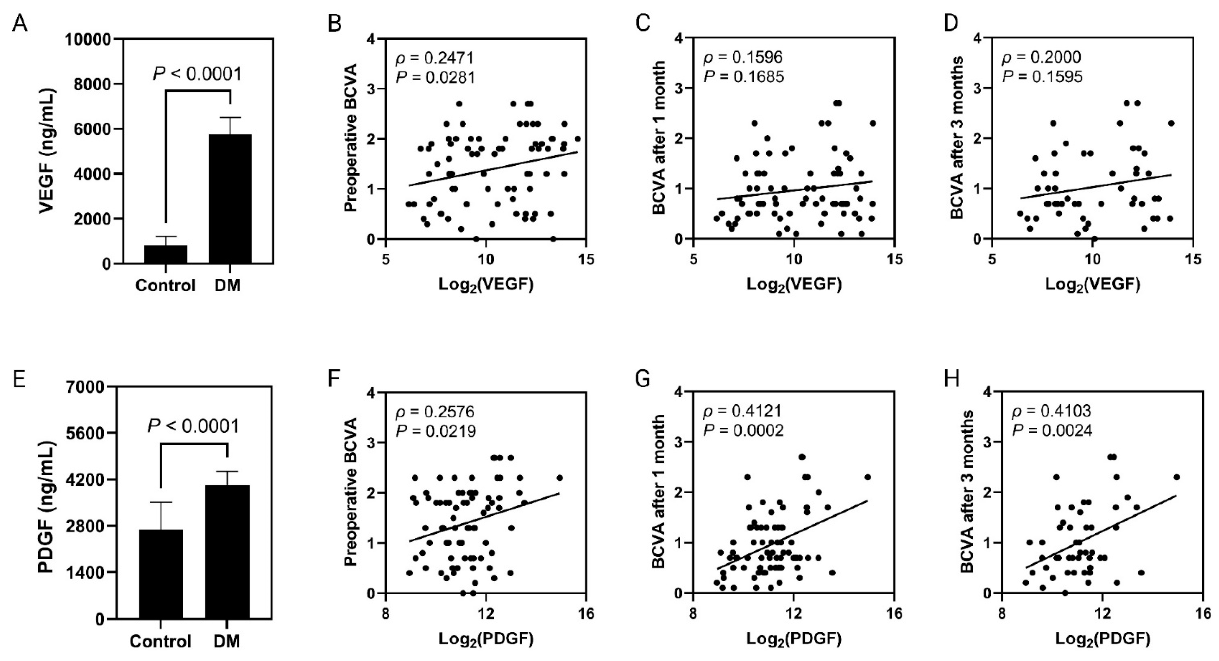
3.3. VEGF and PDGF-AA Concentrations in Vitreous Samples
3.4. Factors Affecting the Vitreous Levels of VEGF and PDGF-AA
3.5. Visual Outcomes
4. Discussion
5. Conclusions
Author Contributions
Funding
Institutional Review Board Statement
Informed Consent Statement
Data Availability Statement
Conflicts of Interest
References
- Cheung, N.; Mitchell, P.; Wong, T.Y. Diabetic retinopathy. Lancet 2010, 376, 124–136. [Google Scholar] [CrossRef]
- Hosseini, S.M.; Boright, A.P.; Sun, L.; Canty, A.J.; Bull, S.B.; Klein, B.E.; Klein, R.; Paterson, A.D. The association of previously reported polymorphisms for microvascular complications in a meta-analysis of diabetic retinopathy. Hum. Genet. 2015, 134, 247–257. [Google Scholar] [CrossRef] [PubMed]
- Zheng, Y.; He, M.; Congdon, N. The worldwide epidemic of diabetic retinopathy. Indian J. Ophthalmol. 2012, 60, 428–431. [Google Scholar] [PubMed]
- Rajavi, Z.; Safi, S.; Javadi, M.A.; Azarmina, M.; Moradian, S.; Entezari, M.; Nourinia, R.; Ahmadieh, H.; Shirvani, A.; Shahraz, S.; et al. Diabetic Retinopathy Clinical Practice Guidelines: Customized for Iranian Population. J. Ophthalmic Vis. Res. 2016, 11, 394–414. [Google Scholar]
- Nathan, D.M.; Genuth, S.; Lachin, J.; Cleary, P.; Crofford, O.; Davis, M.; Rand, L.; Siebert, C. The effect of intensive treatment of diabetes on the development and progression of long-term complications in insulin-dependent diabetes mellitus. N. Engl. J. Med. 1993, 329, 977–986. [Google Scholar]
- Han, L.; Zhang, L.; Xing, W.; Zhuo, R.; Lin, X.; Hao, Y.; Wu, Q.; Zhao, J. The associations between VEGF gene polymorphisms and diabetic retinopathy susceptibility: A meta-analysis of 11 case-control studies. J. Diabetes Res. 2014, 2014, 805801. [Google Scholar] [CrossRef]
- Ferrara, N.; Davis-Smyth, T. The biology of vascular endothelial growth factor. Endocr. Rev. 1997, 18, 4–25. [Google Scholar] [CrossRef] [PubMed]
- Pierce, E.A.; Avery, R.L.; Foley, E.D.; Aiello, L.P.; Smith, L.E. Vascular endothelial growth factor/vascular permeability factor expression in a mouse model of retinal neovascularization. Proc. Natl. Acad. Sci. USA 1995, 92, 905–909. [Google Scholar] [CrossRef]
- Stone, J.; Chan-Ling, T.; Pe’er, J.; Itin, A.; Gnessin, H.; Keshet, E. Roles of vascular endothelial growth factor and astrocyte degeneration in the genesis of retinopathy of prematurity. Investig. Ophthalmol. Vis. Sci. 1996, 37, 290–299. [Google Scholar]
- Stone, J.; Itin, A.; Alon, T.; Pe’er, J.; Gnessin, H.; Chan-Ling, T.; Keshet, E. Development of retinal vasculature is mediated by hypoxia-induced vascular endothelial growth factor (VEGF) expression by neuroglia. J. Neurosci. Off. J. Soc. Neurosci. 1995, 15, 4738–4747. [Google Scholar] [CrossRef]
- Miller, J.W.; Adamis, A.P.; Aiello, L.P. Vascular endothelial growth factor in ocular neovascularization and proliferative diabetic retinopathy. Diabetes/Metab. Rev. 1997, 13, 37–50. [Google Scholar] [CrossRef]
- Shibuya, M. Vascular endothelial growth factor and its receptor system: Physiological functions in angiogenesis and pathological roles in various diseases. J. Biochem. 2013, 153, 13–19. [Google Scholar] [CrossRef] [PubMed]
- Behl, T.; Kotwani, A. Exploring the various aspects of the pathological role of vascular endothelial growth factor (VEGF) in diabetic retinopathy. Pharmacol. Res. 2015, 99, 137–148. [Google Scholar] [CrossRef]
- Penn, J.S.; Madan, A.; Caldwell, R.B.; Bartoli, M.; Caldwell, R.W.; Hartnett, M.E. Vascular endothelial growth factor in eye disease. Prog. Retin. Eye Res. 2008, 27, 331–371. [Google Scholar] [CrossRef]
- Kusari, J.; Zhou, S.X.; Padillo, E.; Clarke, K.G.; Gil, D.W. Inhibition of Vitreoretinal VEGF Elevation and Blood–Retinal Barrier Breakdown in Streptozotocin-Induced Diabetic Rats by Brimonidine. Investig. Ophthalmol. Vis. Sci. 2010, 51, 1044–1051. [Google Scholar] [CrossRef] [PubMed]
- Boyd, S.R.; Advani, A.; Altomare, F.; Stockl, F. Retinopathy. Can. J. Diabetes 2013, 37 (Suppl. S1), S137–S141. [Google Scholar] [CrossRef]
- Kim, J.; Park, J.Y.; Kong, J.S.; Lee, H.; Won, J.Y.; Cho, D.W. Development of 3D printed Bruch’s membrane-mimetic substance for the maturation of retinal pigment epithelial cells. Int. J. Mol. Sci. 2021, 22, 1095. [Google Scholar] [CrossRef]
- Saint-Geniez, M.; Kurihara, T.; Sekiyama, E.; Maldonado, A.E.; D’Amore, P.A. An essential role for RPE-derived soluble VEGF in the maintenance of the choriocapillaris. Proc. Natl. Acad. Sci. USA 2009, 106, 18751–18756. [Google Scholar] [CrossRef]
- Ferrara, N. Vascular endothelial growth factor: Basic science and clinical progress. Endocr. Rev. 2004, 25, 581–611. [Google Scholar] [CrossRef]
- Praidou, A.; Papakonstantinou, E.; Androudi, S.; Georgiadis, N.; Karakiulakis, G.; Dimitrakos, S. Vitreous and serum levels of vascular endothelial growth factor and platelet-derived growth factor and their correlation in patients with non-proliferative diabetic retinopathy and clinically significant macula oedema. Acta Ophthalmol. 2011, 89, 248–254. [Google Scholar] [CrossRef] [PubMed]
- Freyberger, H.; Bröcker, M.; Yakut, H.; Hammer, J.; Effert, R.; Schifferdecker, E.; Schatz, H.; Derwahl, M. Increased levels of platelet-derived growth factor in vitreous fluid of patients with proliferative diabetic retinopathy. Exp. Clin. Endocrinol. Diabetes Off. J. Ger. Soc. Endocrinol. Ger. Diabetes Assoc. 2000, 108, 106–109. [Google Scholar] [CrossRef]
- Cassidy, L.; Barry, P.; Shaw, C.; Duffy, J.; Kennedy, S. Platelet derived growth factor and fibroblast growth factor basic levels in the vitreous of patients with vitreoretinal disorders. Br. J. Ophthalmol. 1998, 82, 181–185. [Google Scholar] [CrossRef] [PubMed]
- Endo, H.; Naito, T.; Asahara, T.; Kajima, M.; Shiota, H. Cytokines in the vitreous fluid of patients with proliferative diabetic retinopathy—Vascular endothelial growth factor and platelet-derived growth factor are elevated in proliferative diabetic retinopathy. Nippon Ganka Gakkai Zasshi 2000, 104, 711–716. [Google Scholar] [PubMed]
- Diabetic Retinopathy Study. Report Number 6. Design, methods, and baseline results. Report Number 7. A modification of the Airlie House classification of diabetic retinopathy. Prepared by the Diabetic Retinopathy. Investig. Ophthalmol. Vis. Sci. 1981, 21, 1–226.
- Grading diabetic retinopathy from stereoscopic color fundus photographs—An extension of the modified Airlie House classification. ETDRS report number 10. Early Treatment Diabetic Retinopathy Study Research Group. Ophthalmology 1991, 98, 786–806. [CrossRef]
- Schulze-Bonsel, K.; Feltgen, N.; Burau, H.; Hansen, L.; Bach, M. Visual Acuities “Hand Motion” and “Counting Fingers” Can Be Quantified with the Freiburg Visual Acuity Test. Investig. Ophthalmol. Vis. Sci. 2006, 47, 1236–1240. [Google Scholar] [CrossRef] [PubMed]
- Alshari, O.; Al Zu’bi, Y.O.; Al Sharie, A.H.; Wafai, F.H.; Aleshawi, A.J.; Atawneh, F.H.; Obeidat, H.A.; Daoud, M.N.; Khrais, M.Z.; Albals, D.; et al. Evaluating the Prognostic Role of Monocytopenia in Chemotherapy-Induced Febrile Neutropenia Patients Treated with Granulocyte Colony-Stimulating Factor. Ther. Clin. Risk Manag. 2021, 17, 963–973. [Google Scholar] [CrossRef]
- Al Sharie, A.H.; Al Zu’bi, Y.O.; El-Elimat, T.; Al-Kammash, K.; Abu Lil, A.; Isawi, I.H.; Al Sharie, S.; Abu Mousa, B.M.; Al Malkawi, A.A.; Alali, F.Q. ANO4 Expression Is a Potential Prognostic Biomarker in Non-Metastasized Clear Cell Renal Cell Carcinoma. J. Pers. Med. 2023, 13, 295. [Google Scholar] [CrossRef]
- Wang, J.; Chen, S.; Jiang, F.; You, C.; Mao, C.; Yu, J.; Han, J.; Zhang, Z.; Yan, H. Vitreous and plasma VEGF levels as predictive factors in the progression of proliferative diabetic retinopathy after vitrectomy. PLoS ONE 2014, 9, e110531. [Google Scholar] [CrossRef]
- Simó, R.; Carrasco, E.; García-Ramírez, M.; Hernández, C. Angiogenic and antiangiogenic factors in proliferative diabetic retinopathy. Curr. Diabetes Rev. 2006, 2, 71–98. [Google Scholar] [CrossRef]
- Miller, J.W.; Le Couter, J.; Strauss, E.C.; Ferrara, N. Vascular endothelial growth factor a in intraocular vascular disease. Ophthalmology 2013, 120, 106–114. [Google Scholar] [CrossRef] [PubMed]
- Funatsu, H.; Yamashita, H.; Mimura, T.; Noma, H.; Nakamura, S.; Hori, S. Risk evaluation of outcome of vitreous surgery based on vitreous levels of cytokines. Eye 2007, 21, 377–382. [Google Scholar] [CrossRef]
- Wakabayashi, Y.; Usui, Y.; Okunuki, Y.; Ueda, S.; Kimura, K.; Muramatsu, D.; Kezuka, T.; Goto, H. Intraocular VEGF Level as a Risk Factor for Postoperative Complications after Vitrectomy for Proliferative Diabetic Retinopathy. Investig. Ophthalmol. Vis. Sci. 2012, 53, 6403–6410. [Google Scholar] [CrossRef]
- Yan, H.; Cui, J.; Yu, J.G.; Han, J.D.; Chen, S.; Zhang, J.K.; Xu, Y.H. The expression of vascular endothelial growth factor of vitreous in patients with proliferative diabetic retinopathy. [Zhonghua Yan Ke Za Zhi] Chin. J. Ophthalmol. 2009, 45, 206–209. [Google Scholar]
- Oshima, Y.; Shima, C.; Wakabayashi, T.; Kusaka, S.; Shiraga, F.; Ohji, M.; Tano, Y. Microincision vitrectomy surgery and intravitreal bevacizumab as a surgical adjunct to treat diabetic traction retinal detachment. Ophthalmology 2009, 116, 927–938. [Google Scholar] [CrossRef] [PubMed]
- Yoshida, S.; Nakama, T.; Ishikawa, K.; Arima, M.; Tachibana, T.; Nakao, S.; Sassa, Y.; Yasuda, M.; Enaida, H.; Oshima, Y.; et al. Antiangiogenic shift in vitreous after vitrectomy in patients with proliferative diabetic retinopathy. Investig. Ophthalmol. Vis. Sci. 2012, 53, 6997–7003. [Google Scholar] [CrossRef]
- Itakura, H.; Kishi, S.; Kotajima, N.; Murakami, M. Persistent secretion of vascular endothelial growth factor into the vitreous cavity in proliferative diabetic retinopathy after vitrectomy. Ophthalmology 2004, 111, 1880–1884. [Google Scholar] [CrossRef]
- Baharivand, N.; Zarghami, N.; Panahi, F.; Dokht Ghafari, M.Y.; Mahdavi Fard, A.; Mohajeri, A. Relationship between vitreous and serum vascular endothelial growth factor levels, control of diabetes and microalbuminuria in proliferative diabetic retinopathy. Clin. Ophthalmol. 2012, 6, 185–191. [Google Scholar]
- Chernykh, V.; Varvarinsky, E.; Smirnov, E.; Chernykh, D.; Trunov, A. Proliferative and inflammatory factors in the vitreous of patients with proliferative diabetic retinopathy. Indian J. Ophthalmol. 2015, 63, 33–36. [Google Scholar]
- Nakashima, N.; Umeda, F.; Yamauchi, T.; Ishii, H.; Hisatomi, A.; Nawata, H.; Masuko, H.; Nakayama, K.; Tatematsu, A. Platelet-derived growth factor and growth-promoting activity in the serum samples and platelets of patients with non-insulin-dependent diabetes mellitus. J. Lab. Clin. Med. 1992, 120, 78–85. [Google Scholar]
- Muhiddin, H.S.; Kamaruddin, M.I.; Ichsan, A.M.; Budu. Vitreous and Serum Concentrations of Vascular Endothelial Growth Factor and Platelet-Derived Growth Factor in Proliferative Diabetic Retinopathy. Clin. Ophthalmol. 2020, 14, 1547–1552. [Google Scholar] [CrossRef]
- Jaffe, G.J.; Ciulla, T.A.; Ciardella, A.P.; Devin, F.; Dugel, P.U.; Eandi, C.M.; Masonson, H.; Monés, J.; Pearlman, J.A.; Quaranta-El Maftouhi, M.; et al. Dual Antagonism of PDGF and VEGF in Neovascular Age-Related Macular Degeneration: A Phase IIb, Multicenter, Randomized Controlled Trial. Ophthalmology 2017, 124, 224–234. [Google Scholar] [CrossRef]
- Pennock, S.; Haddock, L.J.; Mukai, S.; Kazlauskas, A. Vascular endothelial growth factor acts primarily via platelet-derived growth factor receptor α to promote proliferative vitreoretinopathy. Am. J. Pathol. 2014, 184, 3052–3068. [Google Scholar] [CrossRef] [PubMed]
- Lei, H.; Velez, G.; Hovland, P.; Hirose, T.; Gilbertson, D.; Kazlauskas, A. Growth factors outside the PDGF family drive experimental PVR. Investig. Ophthalmol. Vis. Sci. 2009, 50, 3394–3403. [Google Scholar] [CrossRef] [PubMed]
- Ichsan, A.M.; Windy, D.A.; Muhiddin, H.S.; Budu Massi, M.N.; Islam, I.C. Comparison between Vascular Endothelial Growth Factor and Platelet-Derived Growth Factor Levels in Rhegmatogenous Retinal Detachment. J. Ophthalmol. 2021, 2021, 2688837. [Google Scholar] [CrossRef] [PubMed]
- Rasier, R.; Gormus, U.; Artunay, O.; Yuzbasioglu, E.; Oncel, M.; Bahcecioglu, H. Vitreous levels of VEGF, IL-8, and TNF-alpha in retinal detachment. Curr. Eye Res. 2010, 35, 505–509. [Google Scholar] [CrossRef] [PubMed]
- Su, C.Y.; Chen, M.T.; Wu, W.S.; Wu, W.C. Concentration of vascular endothelial growth factor in the subretinal fluid of retinal detachment. J. Ocul. Pharmacol. Ther. Off. J. Assoc. Ocul. Pharmacol. Ther. 2000, 16, 463–469. [Google Scholar] [CrossRef]
- Yalcinbayir, O.; Buyukuysal, R.L.; Gelisken, O.; Buyukuysal, C.; Can, B. Amino acid and vascular endothelial growth factor levels in subretinal fluid in rhegmatogenous retinal detachment. Mol. Vis. 2014, 20, 1357–1365. [Google Scholar]
- Mori, K.; Gehlbach, P.; Ando, A.; Dyer, G.; Lipinsky, E.; Chaudhry, A.G.; Hackett, S.F.; Campochiaro, P.A. Retina-specific expression of PDGF-B versus PDGF-A: Vascular versus nonvascular proliferative retinopathy. Investig. Ophthalmol. Vis. Sci. 2002, 43, 2001–2006. [Google Scholar]
- El-Ghrably, I.A.; Dua, H.S.; Orr, G.M.; Fischer, D.; Tighe, P.J. Intravitreal invading cells contribute to vitreal cytokine milieu in proliferative vitreoretinopathy. Br. J. Ophthalmol. 2001, 85, 461–470. [Google Scholar] [CrossRef]
- Campochiaro, P.A.; Hackett, S.F.; Vinores, S.A.; Freund, J.; Csaky, C.; LaRochelle, W.; Henderer, J.; Johnson, M.; Rodriguez, I.R.; Friedman, Z.; et al. Platelet-derived growth factor is an autocrine growth stimulator in retinal pigmented epithelial cells. J. Cell Sci. 1994, 107, 2459–2469. [Google Scholar] [CrossRef] [PubMed]
- WHO Guidelines Approved by the Guidelines Review Committee. Use of Glycated Haemoglobin (HbA1c) in the Diagnosis of Diabetes Mellitus: Abbreviated Report of a WHO Consultation; World Health Organization: Geneva, Switzerland, 2011. [Google Scholar]
- Chico, A.; Pérez, A.; Córdoba, A.; Arcelús, R.; Carreras, G.; de Leiva, A.; González-Sastre, F.; Blanco-Vaca, F. Plasma homocysteine is related to albumin excretion rate in patients with diabetes mellitus: A new link between diabetic nephropathy and cardiovascular disease? Diabetologia 1998, 41, 684–693. [Google Scholar] [CrossRef] [PubMed]
- Xu, C.; Wu, Y.; Liu, G.; Liu, X.; Wang, F.; Yu, J. Relationship between homocysteine level and diabetic retinopathy. a systematic review and meta-analysis. Diagn. Pathol. 2014, 9, 16. [Google Scholar] [CrossRef] [PubMed]
- Castellarin, A.; Grigorian, R.; Bhagat, N.; Del Priore, L.; Zarbin, M.A. Vitrectomy with silicone oil infusion in severe diabetic retinopathy. Br. J. Ophthalmol. 2003, 87, 318–321. [Google Scholar] [CrossRef] [PubMed]
- Lu, X.; Chen, W.; Xia, H.; Zheng, K.; Jin, C.; Ng, D.S.C.; Chen, H. Atrophy of retinal inner layers is associated with poor vision after endophthalmitis: A spectral domain optical coherence tomography study. Eye 2017, 31, 1488–1495. [Google Scholar] [CrossRef] [PubMed]

| Variables | Number (Percentage) or Mean ± SEM | |||
|---|---|---|---|---|
| Overall (n = 80) | DR Case Group (n = 42) | N-DR Control Group (n = 38) | p-Value | |
| Age (years) | 52.5 ± 1.5 | 55.3 ± 1.4 | 49.3 ± 2.7 | 0.046 |
| Gender | ||||
| Male | 45 (56.3) | 20 (47.6) | 25 (65.8) | NS |
| Female | 35 (43.8) | 22(52.4) | 13 (34.2) | |
| Eye laterality | ||||
| Right eye (OD) | 42 (52.5) | 20 (47.6) | 22 (57.9) | NS |
| Left eye (OS) | 38 (47.5) | 22 (52.4) | 16 (42.1) | |
| Comorbidities | ||||
| Diabetes mellitus | 52 (65.0) | 42 (100) | 10 (26.3) | 0.001 |
| Hypertension | 52 (65.0) | 36 (58.7) | 16 (42.1) | 0.001 |
| Chronic kidney disease | 5 (6.3) | 4 (6.6) | 12 (10.9) | NS |
| Treatment of DM | ||||
| Oral hypoglycemic agents | 20 (39.2) | 15 (36.6) | 5 (50) | NS |
| Oral hypoglycemic agents and/or insulin | 31 (60.8) | 26 (36.4) | 5 (50) | |
| Duration of diabetes mellitus (years) | 14.3 ± 1.1 | 15.1 ± 1.1 | 10.6 ± 2.8 | NS |
| Type of intravitreal injection | ||||
| Aflibercept | 12 (31.6) | 10 (29.4) | 2 (50) | NS |
| Ranibizumab | 26 (28.4) | 24 (70.6) | 2 (50) | |
| Number of intravitreal injections | 2.5 ± 0.4 | 4.2 ± 0.7 | 0.7 ± 0.4 | 0.00001 |
| Time elapsed since last intravitreal injection (days) | 26.9 ± 9.8 | 18.2 ± 8.2 | 91.8 ± 50.4 | 0.013 |
| Preoperative diagnosis | ||||
| Advanced PDR (FVM and VH) | 42 (52.5) | 42 (100) | 0 (0) | 0.0000 |
| Rhegmatogenous retinal detachment | 23 (28.7) | 0 (0) | 23 (60.5) | |
| Vitreomacular interface diseases (ERM/MH/VMT) | 9 (11.3) | 0 (0) | 9 (23.7) | |
| Endophthalmitis | 3 (3.8) | 0 (0) | 3 (7.9) | |
| Dropped IOL or crystalline lens | 3 (3.8) | 0 (0) | 3 (7.9) | |
| Associated cataract surgerywith the primary PPV | ||||
| Yes | 39 (49.3) | 30 (71.4) | 11 (28.9) | 0.0001 |
| No | 41 (50.7) | 12 (28.6) | 27 (71.1) | |
| Type of vitreous tamponade | ||||
| Silicone oil | 38 (47.5) | 15 (35.7) | 23 (60.5) | 0.035 |
| Gas | 22 (27.5) | 12 (28.6) | 10 (26.3) | |
| Air | 20 (25) | 15 (35.7) | 5 (13.2) | |
| Postoperative AG use | 25 (32.5) | 11 (28.2) | 14 (36.8) | NS |
| Follow-up period (months) | 17.2 ± 1.3 | 16.0 ± 2.1 | 17.8 ± 1.8 | NS |
| Visual outcomes (Log MAR) | ||||
| Preoperative BCVA | 1.40 ± 0.1 | 1.5 ± 0.1 | 1.3 ± 0.1 | NS |
| BCVA after 1 month | 0.97 ± 0.1 | 1.00 ± 0.1 | 0.9 ± 0.1 | NS |
| BCVA after 3 months | 1.03 ± 0.1 | 1.2 ± 0.1 | 0.9 ± 0.1 | NS |
| BCVA at the last follow-up | 0.95 ± 0.1 | 1.0 ± 0.1 | 0.9 ± 0.1 | NS |
| HbA1c (%) | 8.2 ± 0.3 | 9.23 ± 0.3 | 6.06 ± 0.5 | 0.001 |
| Homocysteine (mcmol/L) | 18.5 ± 1.9 | 18.0 ± 2.0 | 19.22 ± 3.5 | NS |
| Measured Markers | Number (Percentage) or Mean ± SEM | ||
|---|---|---|---|
| DR Case Group (n = 42) | N-DR Control Group (n = 38) | p-Value | |
| VEGF (pg/mL) | 5744.06 ± 761.5 | 817.94 ± 403.1 | 0.0001 |
| VEGF/Protein (pg/mg) | 4133.21 ± 488.2 | 388.95 ± 52.0 | 0.0001 |
| PDGF-AA (pg/mL) | 4031.51 ± 410.2 | 2691.46 ± 821.0 | 0.001 |
| PDGF-AA/protein (pg/mg) | 2873.08 ± 293.5 | 2007.87 ± 252.3 | 0.03 |
| Protein content (mg/mL) | 1.56 ± 0.1 | 2.00 ± 0.4 | NS |
| Variables | Mean ± SEM * or B Regression Coefficient ± SEM ** | |
|---|---|---|
| VEGF (pg/mL) | p-Value | |
| Age (years) ** | −18.1 ± 3.8 | NS |
| Gender * | ||
| Male | 2832.30 ± 674.9 | NS |
| Female | 4139.40 ± 807.7 | |
| Eye laterality * | ||
| Right eye (OD) | 3438.86 ± 740.1 | NS |
| Left eye (OS) | 3365.80 ± 648.2 | |
| Comorbidities | ||
| Diabetes mellitus * | ||
| Yes | 4711.95 ± 681.5 | 0.00001 |
| No | 975.39 ± 545.8 | |
| Hypertension * | ||
| Yes | 3908.65 ± 647.4 | NS |
| No | 2467 ± 456. 6 | |
| Treatment of DM * | ||
| Oral hypoglycemic agents | 3769.47 ± 828.2 | NS |
| Oral hypoglycemic agents and/or insulin | 5202.27 ± 1001.3 | |
| Duration of diabetes mellitus (years) ** | 165.30 ± 12.1 | NS |
| Type of intravitreal injection * | ||
| Aflibercept | 4682.29 ± 1030.5 | NS |
| Ranibizumab | 5899.54 ± 1092.6 | |
| Number of intravitreal injections ** | 330 ± 13.0 | 0.013 |
| Time elapsed since last intravitreal injection (days) ** | −16.30 ± 1.3 | NS |
| Preoperative diagnosis * | ||
| Advanced PDR (FVM and VH) | 5744.06 ± 760.1 ↑ | 0.0001 |
| Rhegmatogenous retinal detachment | 418.16 ± 57.2 | |
| Vitreomacular interface diseases (ERM/MH/VMT) | 259.43 ± 46.1 | |
| Endophthalmitis | 6181.67 ± 90.7 ↑↑ | |
| Dropped IOL or crystalline lens | 194.67 ± 22.1 | |
| Associated cataract surgerywith the primary PPV * | ||
| Yes | 4558.67 ± 456.9 | 0.028 |
| No | 2261.05 ± 789.0 | |
| Type of vitreous tamponade * | ||
| Silicone oil | 2025.74 ± 520.3 | 0.036 |
| Gas | 4373.68 ± 1045.1 | |
| Air | 4956.66 ± 1391.6 | |
| Postoperative AG use * | 2405.15 ± 567.4 | NS |
| HbA1c (%) ** | 771.01 ± 29.8 | 0.013 |
| Homocysteine ** | −10.36 ± 4.2 | NS |
| Variables | Mean ± SEM * or B Regression Coefficient ± SEM ** | |
|---|---|---|
| PDGF-AA (pg/mL) | p-Value | |
| Age (years) ** | −57.23 ± 3.2 | NS |
| Gender * | ||
| Male | 2879.33 ± 314.8 | NS |
| Female | 4057.99 ± 939.3 | |
| Eye laterality * | ||
| Right eye (OD) | 3016.33 ± 567.2 | NS |
| Left eye (OS) | 3813.52 ± 346.8 | |
| Comorbidities | ||
| Diabetes mellitus * | ||
| Yes | 3583.70 ± 360.7 | NS |
| No | 3044.5 ±990.7 | |
| Hypertension * | ||
| Yes | 3629.90 ± 613.1 | NS |
| No | 2958.7 ± 679.1 | |
| Treatment of DM * | ||
| Oral hypoglycemic agents | 2556.37 ± 516.2 | 0.02 |
| Oral hypoglycemic agents and/or insulin | 4281.68 ± 470.3 | |
| Duration of diabetes mellitus (years) ** | 93.73 ± 6.2 | NS |
| Type of intravitreal injection * | ||
| Aflibercept | 4066.29 ± 723.1 | NS |
| Ranibizumab | 3540.43 ± 546.1 | |
| Number of intravitreal injections ** | −12.78 ± 11.6 | NS |
| Time elapsed since last intravitreal injection (days) ** | −6.74 ± 0.6 | NS |
| Preoperative diagnosis * | ||
| Advanced PDR (FVM and VH) | 4031.52 ± 530.8 ↑ | 0.0001 |
| Rhegmatogenous retinal detachment | 1641.44 ± 101.1 | |
| Vitreomacular interface diseases (ERM/MH/VMT) | 1830.65 ± 83.1 | |
| Endophthalmitis | 15,301.89 ± 830.4 ↑↑ | |
| Dropped IOL or crystalline lens | 713.61 ± 99.6 | |
| Associated cataract surgerywith the primary PPV * | ||
| Yes | 4298.03 ± 546.6 | 0.049 |
| No | 2517.79 ± 789.1 | |
| Type of vitreous tamponade * | ||
| Silicone oil | 3766.45 ± 643.9 | NS |
| Gas | 2571.87 ± 567.1 | |
| Air | 3594.66 ± 989.5 | |
| HbA1c (%) ** | 6.06 ± 0.5 | 0.001 |
| Homocysteine ** | 19.22 ± 3.5 | NS |
Disclaimer/Publisher’s Note: The statements, opinions and data contained in all publications are solely those of the individual author(s) and contributor(s) and not of MDPI and/or the editor(s). MDPI and/or the editor(s) disclaim responsibility for any injury to people or property resulting from any ideas, methods, instructions or products referred to in the content. |
© 2023 by the authors. Licensee MDPI, Basel, Switzerland. This article is an open access article distributed under the terms and conditions of the Creative Commons Attribution (CC BY) license (https://creativecommons.org/licenses/by/4.0/).
Share and Cite
Al-Dwairi, R.; El-Elimat, T.; Aleshawi, A.; Al Sharie, A.H.; Abu Mousa, B.M.; Al Beiruti, S.; Alkazaleh, A.; Mohidat, H. Vitreous Levels of Vascular Endothelial Growth Factor and Platelet-Derived Growth Factor in Patients with Proliferative Diabetic Retinopathy: A Clinical Correlation. Biomolecules 2023, 13, 1630. https://doi.org/10.3390/biom13111630
Al-Dwairi R, El-Elimat T, Aleshawi A, Al Sharie AH, Abu Mousa BM, Al Beiruti S, Alkazaleh A, Mohidat H. Vitreous Levels of Vascular Endothelial Growth Factor and Platelet-Derived Growth Factor in Patients with Proliferative Diabetic Retinopathy: A Clinical Correlation. Biomolecules. 2023; 13(11):1630. https://doi.org/10.3390/biom13111630
Chicago/Turabian StyleAl-Dwairi, Rami, Tamam El-Elimat, Abdelwahab Aleshawi, Ahmed H. Al Sharie, Balqis M. Abu Mousa, Seren Al Beiruti, Ahmad Alkazaleh, and Hasan Mohidat. 2023. "Vitreous Levels of Vascular Endothelial Growth Factor and Platelet-Derived Growth Factor in Patients with Proliferative Diabetic Retinopathy: A Clinical Correlation" Biomolecules 13, no. 11: 1630. https://doi.org/10.3390/biom13111630
APA StyleAl-Dwairi, R., El-Elimat, T., Aleshawi, A., Al Sharie, A. H., Abu Mousa, B. M., Al Beiruti, S., Alkazaleh, A., & Mohidat, H. (2023). Vitreous Levels of Vascular Endothelial Growth Factor and Platelet-Derived Growth Factor in Patients with Proliferative Diabetic Retinopathy: A Clinical Correlation. Biomolecules, 13(11), 1630. https://doi.org/10.3390/biom13111630