Article

Association of Cytokine Gene Polymorphisms and Their Impact on Active and Latent Tuberculosis in Brazil’s Amazon Region

by
Ednelza da Silva Graça Amoras
,
Thais Gouvea de Morais
,
Rafaella do Nascimento Ferreira
,
Samara Tatielle Monteiro Gomes
,
Francisca Dayse Martins de Sousa
,
Iury de Paula Souza
,
Ricardo Ishak
,
Antonio Carlos Rosário Vallinoto
and
Maria Alice Freitas Queiroz
*
Virus Laboratory, Institute of Biological Sciences, Federal University of Pará, Belém 66075-110, Brazil
*
Author to whom correspondence should be addressed.
Biomolecules 2023, 13(10), 1541; https://doi.org/10.3390/biom13101541
Submission received: 29 August 2023 / Revised: 10 October 2023 / Accepted: 13 October 2023 / Published: 18 October 2023
(This article belongs to the Special Issue Tuberculosis: Immunopathogenesis and Therapeutic Strategies)

Abstract

:
Some genetic variations in cytokine genes can alter their expression and influence the evolution of Mycobacterium tuberculosis (Mtb) infection. This study aimed to investigate the association of polymorphisms in cytokine genes and variability in plasma levels of cytokines with the development of tuberculosis (TB) and latent tuberculosis infection (LTBI). Blood samples from 245 patients with TB, 80 with LTBI, and healthy controls (n = 100) were included. Genotyping of the IFNG +874A/T, IL6 -174G/C, IL4 -590C/T, and IL10 -1082A/G polymorphisms was performed by real-time PCR, and cytokine levels were determined by flow cytometry. Higher frequencies of genotypes AA (IFNG +874A/T), GG (IL6 -174G/C), TT (IL4 -590C/T), and GG (IL10 -1082A/G) were associated with an increased risk of TB compared to that of LTBI (p = 0.0027; p = 0.0557; p = 0.0286; p = 0.0361, respectively) and the control (p = <0.0001, p = 0.0021; p = 0.01655; p = 0.0132, respectively). In combination, the A allele for IFNG +874A/T and the T allele for IL4 -590C/T were associated with a higher chance of TB (p = 0.0080; OR = 2.753 and p < 0.0001; OR = 3.273, respectively). The TB group had lower levels of IFN-γ and higher concentrations of IL-6, IL-4, and IL-10. Cytokine levels were different between the genotypes based on the polymorphisms investigated (p < 0.05). The genotype and wild-type allele for IFNG +874A/T and the genotype and polymorphic allele for IL4 -590C/T appear to be more relevant in the context of Mtb infection, which has been associated with the development of TB among individuals infected by the bacillus and with susceptibility to active infection but not with susceptibility to latent infection.

1. Introduction

Tuberculosis (TB) is caused by Mycobacterium tuberculosis (Mtb), which is acquired through the respiratory tract and can cause a primary infection of alveolar macrophages or establish a latent infection [1]. The World Health Organization (WHO) estimates that approximately 10 million people become ill with TB per year worldwide [2].
The evolution of Mtb infection to TB results from a variety of combined factors, such as the virulence mechanisms of the bacillus, the host’s genetics and nutritional status, and vaccination history [3]. Thus, infected individuals may develop latent tuberculosis infection (LTBI), which is asymptomatic and nontransmissible, or TB, which is characterized by the presence of clinical symptoms resulting from lung infection and spread to multiple organs [4]. The World Health Organization (WHO) defines LTBI as a state of persistent immune response to stimulation by Mtb antigens without evidence of clinical disease [5,6]. Approximately one-quarter of the global population has LTBI [5]. Thus, individuals with LTBI represent a reservoir for new cases of active TB [7]. The probability of progression from latent infection to active clinical tuberculosis is determined by bacterial, host, and environmental factors, including: the initial bacillary load, related to the severity of the disease in an index case and the proximity of contact; suppression of cellular immunity induced by the human immunodeficiency virus (HIV); tumor necrosis factor α (TNF-α) inhibitors; glucocorticoids; and organ transplantation [6].
Mtb infection is regulated by two distinct patterns of T helper CD4+ cell responses, which result in the production of pro-inflammatory (T helper 1, Th1) and anti-inflammatory (T helper 2, Th2) mediators [8]. Initially, a protective immune response is mounted by natural killer (NK) cells and antigen-specific Th1 cells with the production of IFN-γ, which stimulates the antimicrobial activity of infected alveolar macrophages while organizing the formation of the protective granuloma [8,9,10]. However, Mtb may modulate Th1 cell activation, inducing the synthesis of T helper 2 (Th2) cytokines, which are associated with progressive disease [8,11]. The marked production of suppressive cytokines, such as TGFβ and IL-4, during Mtb infection inhibits phago-lysosome fusion in macrophages and reduces the expression of T lymphocyte-activating molecules [10]. Although the host immune response has been associated with the development of TB, the identification of specific biomarkers that can characterize LTBI is still lacking, as many mediators related to the latency stage also correlate with the acute phase of Mtb infection [12].
Single-nucleotide polymorphisms (SNPs) in the promoter and coding regions of key genes have been associated with susceptibility and/or resistance to Mtb infection. The wild-type IFNG +874A/T polymorphism allele is associated with lower IFN-γ synthesis [13], and its presence may impair macrophage activation, thus affecting Mtb infection control [14]. High levels of interleukin 6 (IL-6) are produced in response to Mtb infection by activated monocytes and macrophages [15]. The wild-type IL6 -174G/C polymorphism is associated with higher levels of IL-6 and has been described as a risk factor for the development of TB [16]. IL-4 and IL-10 are Th2 cytokines involved in the downregulation of IFN-γ expression and therefore have a deleterious effect in patients with Mtb infection [17,18]. IL4 -560C/T polymorphisms may influence gene transcription and have been associated with increased IL-4 levels [19]. The SNP in IL10 -1082A⁄G alters the level of IL-10 and interferes with the Th1/Th2 balance [20].
The influence of polymorphisms on the development of diseases seems to depend on factors such as the type of genetic variation (the affected gene and the position where the genetic alteration occurs), the interaction of the effects of various mutations, and, mainly, the ethnic composition of a population [21,22]. The present study aimed to investigate the association of IFNG +874A/T, IL6 -174G/C, IL4 -590C/T, and IL10 -1082A/G polymorphisms and plasma levels of cytokines with susceptibility to Mtb, the presence of LTBI, and the development of TB.

2. Materials and Methods

2.1. Study Population

The study included a group of patients diagnosed with clinical TB (n = 245) attending the João de Barros Barreto University Hospital (HUJBB) in Belém Pará, Brazil. Individuals co-infected with HIV were excluded from the study. Infection was confirmed by X-ray, sputum smear, specific culture of bronchial lavage fluid, and positive biopsy results for Mtb. Patients were examined before starting treatment. The LTBI group was formed of 80 contact individuals with a positive result in the tuberculin skin test (TST) but who never had TB.
The control group consisted of 100 health professionals from the HUJBB who had never presented with TB. These professionals are subjected to periodic occupational examinations every six months. Those who had a negative TST and presented normal results in the following tests were included in the study: chest X-ray, sputum smear microscopy, complete blood count, laboratory test for venereal diseases (VDRL), routine urine analysis, and plasma glucose assessment. All participants in the groups reported having been vaccinated and showing scars from the BCG vaccine.

2.2. Sample Collection and Storage

Blood samples (10 mL) were collected by intravenous puncture using a vacuum collection system containing EDTA as an anticoagulant. Samples were centrifuged to isolate plasma and leukocytes, which were used for genomic DNA extraction to investigate gene polymorphisms. Samples were stored in a freezer at −70 °C until use.

2.3. DNA Extraction

Genomic DNA was extracted from peripheral blood leukocytes using a Puregene kit (Puregene, Gentra Systems, Inc., Minneapolis, MN, USA) according to the manufacturer’s protocol, which included the following steps: cell lysis, protein precipitation, DNA precipitation, and hydration. After extraction, the DNA obtained was quantified by spectrophotometry in a NanoDrop® system (Thermo Fisher Scientific, Wilmington, DE, USA) following the manufacturer’s recommendations.

2.4. Genotyping

Polymorphism genotyping was performed by real-time PCR using the StepOnePlus™ Real-Time PCR System (Thermo Fisher, Carlsbad, CA, USA). Assay systems containing primers and probes specific for amplification of the sequence of each target were used (Thermo Fisher, Carlsbad, CA, USA). The following polymorphisms with their respective commercial assays were investigated: IL4 -590C/T (rs2243250; C__16176216_10); IL6 -174G/C (rs1800795; C_1839697_20); and IL10 -1082A/G (rs1800896; C_1747360_10). For identification of IFNG +874A/T genotypes, primers and probes were used [23]: primers (IFNG-F: 5′-TTC AGA CAT TCA CAA TTG ATT TTA TTC T-3; IFNG-R: 5′−CCC CCA ATG GTA CAG GTT TC-3′); and probes (FAM-AAAATCAAATCTCACACACACA-MGB; VIC-AAAATCAAATCACACACACACA-MGB).
For each reaction, TaqMan® Universal PCR Master Mix [2X], [20X] TaqMan® Assay, and 20 ng of DNA were used in a final reaction volume of 10 µL. The following temperature cycles were used in the amplification reactions: 60 °C for 30 s, followed by 95 °C for 10 min, and 50 cycles of 92 °C for 30 s and 60 °C for 1 min and 30 s.

2.5. Plasma Cytokine Measurement

Cytokine levels were quantified by flow cytometry using a Human Th1/Th2/Th17 Cytometric Bead Array (CBA) Kit (BD Biosciences, San Diego, CA, USA) in a BD FACS Canto II instrument. All procedures followed the manufacturer’s instructions; the method is based on beads conjugated to a capture antibody, i.e., six populations of beads with different fluorescence intensities, conjugated to a capture antibody specific for each cytokine, mixed to form the CBA, and read in channel FL-3 in the flow cytometer. The bead populations were recorded on the basis of their respective fluorescence intensities, from the least bright to the brightest (IL-17 < IFN-γ < TNF-α < IL-10 < IL-6 < IL-4 < IL-2).

2.6. Tuberculin Skin Test (TST)

Control subjects submitted to a TST, performed by applying an intradermal injection of 0.1 mL (0.04 mcg) of PPD RT-23 (Mantoux, 2 UT/0.1 mL) in the middle third of the anterior surface of the left forearm, at an angle of 5 to 15 degrees, until the formation of a papule. The reading was performed 48 to 72 h after application using a specific millimeter ruler, measuring the largest transverse diameter of the induration perpendicular to the forearm. The results with induration greater than or equal to 5 mm were considered to indicate a positive PPD test [24].

2.7. Statistical Analysis

The data obtained was entered into a database in Microsoft Office Excel 2020 (Microsoft, Redmond, WA, USA). The genotypic and allelic frequencies were compared between the groups using the chi-square and G tests. The odds ratio (OR) was used to evaluate the homozygous genotypes for each polymorphism (wild-type and polymorphic, or vice versa) that represented a risk of developing the different clinical forms of TB. Variations in the plasma levels of the cytokines between the groups were evaluated using the nonparametric Kruskal-Wallis test and Dunn´s post-test. Violin charts were used to show the dispersion of cytokine levels between the groups investigated. All tests were performed using BioEstat version 5.3 and GraphPad Prism 8.0 (GraphPad Software, San Diego, CA, USA), and p values < 0.05 were considered to indicate significance.

3. Results

The characteristics of the studied groups showed that among patients with TB, 59.0% were male, 82.0% had the pulmonary form, and 18.0% had the extrapulmonary form of the disease, with a mean age of 41.2 years. In patients with LTBI, 57.5% were male, and the mean age was 38.4 years. In healthy controls, 58.0% were male, and the mean age was 39.7 years (Table 1).
The comparison of the allelic and genotypic frequencies of the IFNG +874A/T polymorphism between the TB and LTBI groups showed that the wild-type AA genotype (p = 0.0027; OR = 3.6104, 95% CI 1.36–9.60) and the A allele (p < 0.0001; OR = 2.1649, 95% CI 1.48–3.1) were more frequent in the TB group. In the comparison between the TB and control groups, an association of the wild-type AA genotype (p < 0.0001; OR: 5.5156, 95% CI 2.45–11.9) and the A allele (p < 0.0001; OR = 2.2790, 95% CI 1.60–3.22) with the TB group was observed. Between the LTBI and HC groups, there was no significant difference between genotypic and allele frequencies (Table 2).
The comparison of IL6 -174G/C genotypes between the evaluated groups showed that there was no significant difference in genotypic and allelic frequencies between the TB and LTBI groups (p > 0.05), but when compared to the control group, the TB group had a higher frequency of the GG genotype (p = 0.0021; OR = 4.3955, 95% CI 1.46–13.22) and G allele (p = 0.0003; OR = 2.1128, 95% CI 1.41–3.14). There was no significant difference in genotype and allele frequencies between the LTBI and HC groups (Table 2).
For the IL4 -590C/T polymorphism, a higher frequency of the TT genotype (p = 0.0286; OR = 2.800, 95% CI 1.26–6.22) and of the T allele (p = 0.0029; OR = 1.6300, 95% CI 1.12–2.35) was observed in the TB group than in the LTBI group. The comparison of the TB and control groups showed that the TT genotype and the T allele were significantly more frequent in the TB group (p = 0.01655; OR = 2.7097, 95% CI 1.31–5.60 and p = 0.0013; OR = 1.6213, CI 1.58–2.27, respectively). Between the LTBI and HC groups, differences between genotype and allele frequencies were not significant (Table 2).
Genotyping of the IL10 +1082G/A polymorphism showed that the GG genotype was significantly more frequent in the TB group than in the LTBI (p = 0.0132) and control groups (p = 0.0361). The allele frequencies did not show significant differences among the evaluated groups. Genotypic and allele frequencies did not show significant differences between the LTBI and HC groups (Table 2).
We evaluated whether allelic combinations for the IFNG +874A/T, IL6 -174G/C, IL4 -560C/T, and IL10 +1082A/G polymorphisms contribute to risk or protection from active Mtb infection. Of the 16 possible combinations, there was an association between the A for IFNG +874A/T and T for IL4 -560C/T alleles with TB compared to LTBI, especially in the combinations AGTA (p = 0.0008; OR  =  2.753; 95% CI 1.52–4.96). The A allele for IFNG +874A/T and T for IL4 -560C/T were associated with the risk of TB when compared to the control, mainly in the AGTA (p < 0.0001; OR = 3.273; 95% CI 1.85–5) (Table 3).
In contrast, some combinations of the T polymorphic allele for the IFNG +874A/T variation appeared to protect against TB when compared to the LTBI group, especially the combinations TGCA (p = 0.0103; OR = 0.396; 95% CI 0.20–0.77), TGCG (p = 0.0352; OR = 0.344; 95% CI 0.14–0.86), TGTG (OR = 0.357; 95% CI 0.16–0.78), and TCTA (p = 0.0157; OR = 0.266; 95% CI 0.09–0.80). Similarly, some combinations with the T allele for IFNG +874A/T appear associated with a lower risk of TB in comparison to healthy controls, including TCCA (p = 0.0200; OR = 0.245; 95% CI 0.08–0.75), TCTA (p = 0.0062; OR = 0.233; 95% CI 0.08–0.65), and TCTG (p = 0.0348; OR = 0.388; 95% CI 0.17–0.88; Table 3).
The comparison of the LTBI group and the control group showed no significant difference for any of the combinations evaluated (p > 0.05).
Assessment of differences in cytokine levels between groups showed that the LTBI group had higher levels of IFN-γ similar to the control group (p = 0.0408; Figure 1a). The levels of the cytokines IL-6, IL-4, and IL-10 were higher in the TB group than in the LTBI and control groups (p < 0.0001; Figure 1b–d).
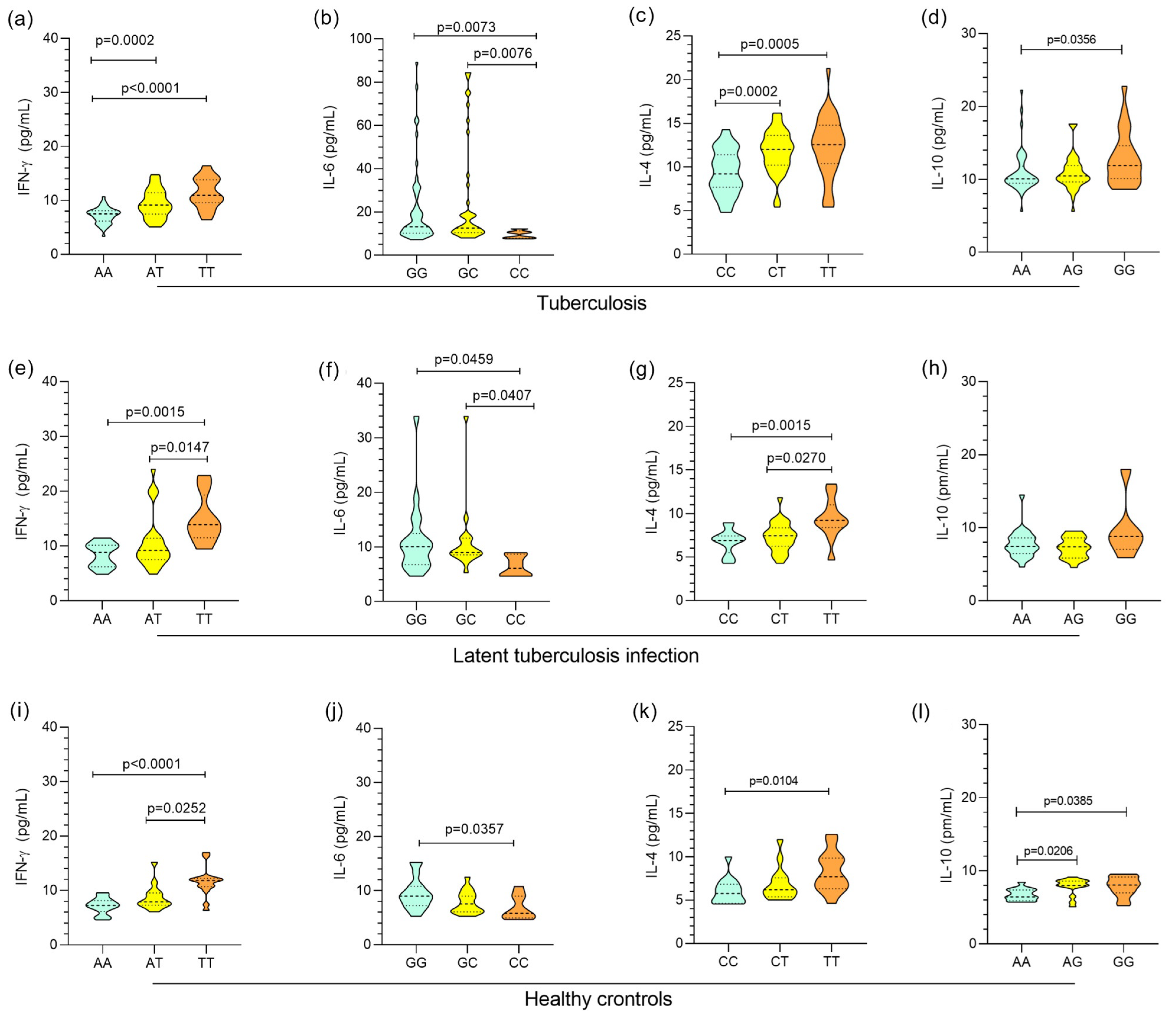
In Figure 2, the plasma levels of the cytokines IFN-γ, IL-6, IL-4, and IL-10 were grouped by genotype, and the levels showed a similar profile in the investigated groups. The AA, AT, and TT genotypes for IFNG +874A/T were associated with low, intermediate, and high levels of IFN-γ, respectively, in the active TB (Figure 2a), LTBI (Figure 2e), and control (Figure 2i) groups. IL-6 concentrations were significantly higher in carriers of the GG genotype for IL6 -174G/C than in individuals with the CC genotype in the active TB (Figure 2b), LTBI (Figure 2f), and control (Figure 2j) groups. A comparison of IL-4 levels showed that individuals with the TT polymorphic genotype for IL4 -560C/T had higher levels than carriers of the wild-type CC genotype in the active TB (Figure 2c), LTBI (Figure 2g), and control (Figure 2k) groups. Higher concentrations of IL-10 were observed in individuals with the polymorphic genotype GG for IL10 +1082A/G than in those with the wild-type AA genotype in the active TB (Figure 2d), LTBI (Figure 2h), and control (Figure 2l) groups.
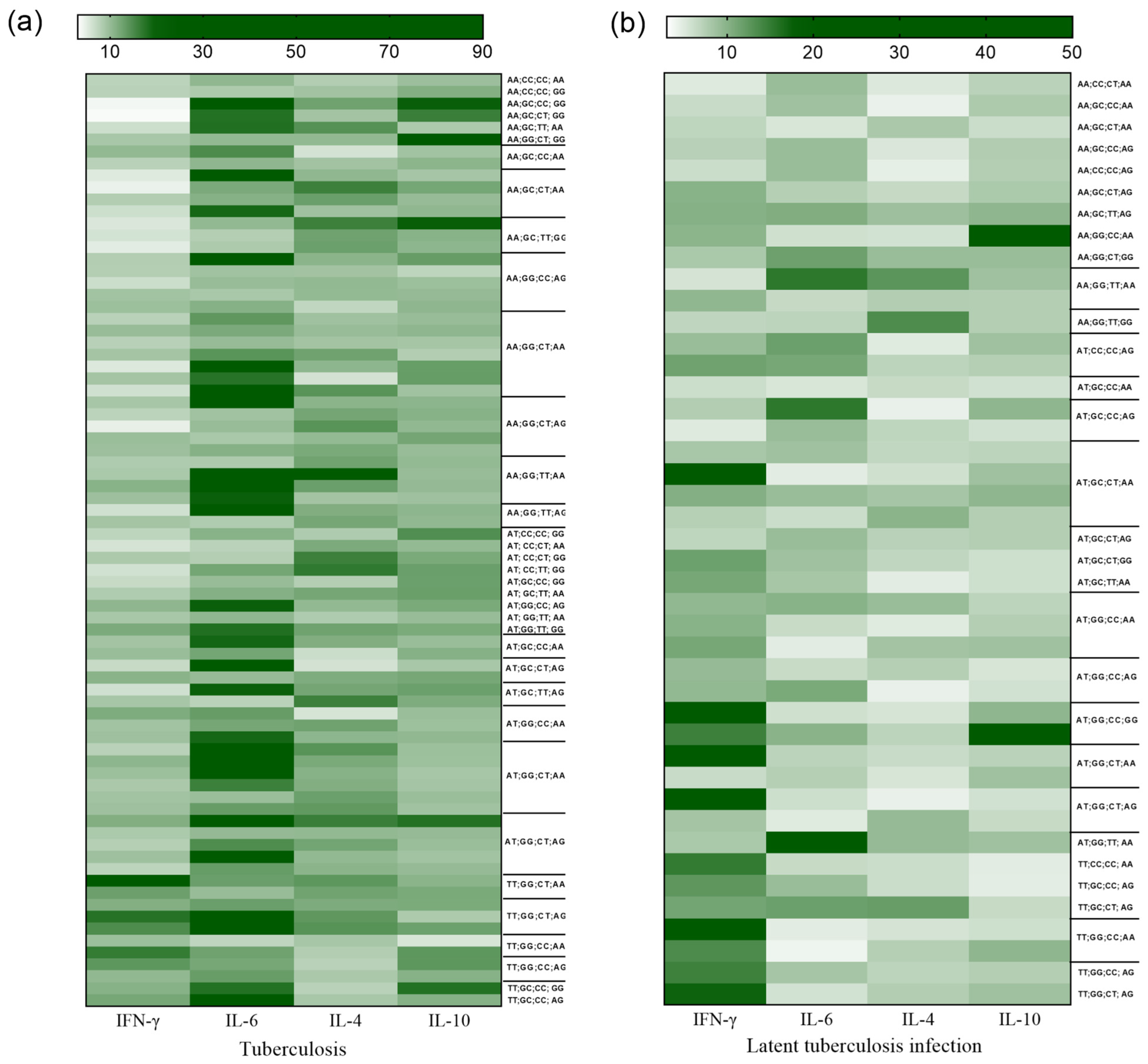
As shown in the heatmap, there is an inversion of Th1 and Th2 cytokine concentrations between the groups with active TB (Figure 3a) and LTBI (Figure 3b). However, the cytokine concentrations were related to certain genotypes in both evaluated groups. In the subsets plotted for active TB, an association of lower concentrations of IFN-γ with higher levels of the cytokines IL-6, IL-4, and IL-10 was observed (Figure 3a). Conversely, in the LTBI group, there was an association between higher levels of IFN-γ and lower concentrations of IL-6 and Th2 cytokines (Figure 3b).

4. Discussion

Variations in the levels of specific cytokines determine the susceptibility and progression of Mtb infection or containment of the infection. Some cytokines resulting from the activation of T lymphocytes and phagocytic cells, such as IFN-γ and TNF-α, are efficient in controlling infection, while others may contribute to the survival and growth of Mtb or mediate susceptibility to tuberculosis, including IL-4, TGF-β, and IL-10 [25]. As genes encoding cytokines such as IFNG, IL4, IL6, and IL10 are highly polymorphic, some genetic variations may influence the expression of cytokines and contribute to regulating resistance or susceptibility to TB [20,26].
The study showed an association between the frequencies of the wild genotype for IFNG +874A/T and IL6 -174G/C and the polymorphic genotype of IL4 -560C/T and IL10 +1082A/G with TB. IFN-γ levels were more reduced in individuals with TB and higher in the LTBI group, while IL-6, IL-4, and IL-10 levels were higher in individuals with TB and reduced in the LTBI group. There was no statistical difference in the frequencies of investigated polymorphisms and cytokine levels between the LTBI and control groups. IFN-γ levels were more reduced in individuals with the AA genotype for IFNG +874A/T, and the levels of IL-6, IL-4, and IL-10 were higher in carriers of the GG, TT, and GG genotypes for the variations IL6 -174G/C, IL4 -560C/T, and IL10 +1082A/G, respectively.
Although this study shows interesting results related to the isolated evaluation of each of the polymorphisms and their association and the influence of polymorphisms on cytokine levels in different groups, the small sample size of the LTBI and control groups represents a limiting factor in the study. The characterization of the LTBI group was also a limitation of the study, since this LTBI group was selected due to the positive result of the TST test associated with the absence of TB symptoms. The interferon-gamma release assay (IGRA) would be a more appropriate test to define the LTBI group, as it uses Mtb-specific antigens that induce the production of IFN-γ in individuals with previous contact with Mtb, and the result is not influenced by BCG vaccination or exposure to most non-tuberculous mycobacteria (NMT) infections [27,28]. However, according to the WHO, both the TST and IGRA can be used as screening tests for LTBI in countries with high TB endemicity, such as Brazil [29]. According to the Manual of Recommendations for Tuberculosis Control in Brazil (Ministry of Health), the diagnosis of LTBI can be made by TST or IGRA; however, IGRA is recommended only for specific groups, such as HIV-infected individuals and children, not being routinely used to identify LTBI in the general population, as it is less expensive, widely available, and simpler to perform for health professionals [30,31]. As the population included in the study does not meet the criteria for the use of IGRA, the diagnosis was made by TST.
The analysis of the IFNG +874A/T polymorphism between the TB and LTBI groups suggested that the increased frequency of the wild-type AA genotype represented a 3-fold greater chance of the individual developing TB among those infected with Mtb; between the TB and HC groups, the presence of the AA genotype was associated with a 5-fold greater chance of the individual acquiring infection and developing the disease.
The wild-type AA genotype is associated with lower cytokine levels and may contribute to the establishment of TB. Other studies have observed an association of the wild-type genotype and allele with TB in different populations [32,33,34]. IFN-γ is fundamental to the protective immune response against Mtb, which is mediated by CD4+ Th1 cells [35]. This cytokine has a major impact on the physiology of infected macrophages and on the biology of phagosomes [36]. Elevated cytokine levels may also promote greater FASL activation and enhance the apoptosis of infected cells [37]. However, a deficient IFN-γ response is associated with susceptibility to and severity of TB, as it reduces the activation of T-cell responses and the detection of infected cells [14,38].
The presence of the wild-type GG genotype and the G allele for the IL6 -174G/C polymorphism was associated with a 4-fold increased risk for infection and development of TB when comparing individuals with TB and the healthy control. The GG and CC genotypes were associated with higher and lower levels of cytokines, respectively. Our results corroborate a previous study in which the presence of the polymorphic genotype CC was associated with a lower risk of TB [39,40]. However, in a study performed in India, the CC genotype was associated with TB [41], in contrast to our results. It is possible that this divergence is related to the ethnic differences in the population. IL-6 has been identified as a key driver of TB severity and is associated with lung inflammation and lung damage. The cytokine, which is produced by macrophages, inhibits IFN-γ signaling, contributing to the severe disease phenotype [42,43].
The TT polymorphic genotype for IL4 -560C/T was associated with a greater chance (almost three times) of the individual developing TB among those infected by Mtb (TB vs. LTBI) and of having the infection and developing disease (comparison between the TB and control groups). As the TT genotype was related to higher levels of the cytokine IL-4, this result corroborates information previously described [19,44]. Because the polymorphism induces an increase in IL-4 production, it favors a Th2 immune response and antibody production, which is not an efficient response to control Mtb infection and inhibits the differentiation of Th1 cells, which are effective in combating the bacterium. The IL4 -590C/T polymorphism might affect the ability of macrophages to inhibit infection caused by the bacillus [17]. In addition, increased IL-4 (i) impaired antimicrobial activity due to reduced TNF-α-mediated apoptosis of infected cells and decreased activity of iNOS (and therefore decreased levels of nitrogen and oxygen intermediates), (ii) increased iron availability for intracellular Mtb, and (iii) increased proliferation of FOXP3+ regulatory T cells, favoring the immunopathogenesis of TB [45].
The GG polymorphic genotype of IL10 -1082A/G was associated with higher cytokine levels in all studied groups, and the highest frequency was observed in patients with TB. Other studies have observed this same association with TB in different populations [20,46]. IL-10 has been reported to limit the effector immune response to Mtb infection through immunosuppressive mechanisms [47]. Production of this cytokine by human macrophages infected with Mtb inhibits phagosome maturation, resulting in impaired bacterial clearance [48] and increased chances of developing active TB [49,50].
Considering that the SNPs are associated with changes in cytokine levels, our results suggest that these polymorphisms may interact synergistically, decreasing the signaling of the Th1 response and therefore interfering with effector mechanisms, leading to a significant enhancement of the pathogenesis of TB [51,52]. In this context, the combination of the allele related to low levels of IFN-γ (A) and high levels of IL-4 (T) was predominantly associated with the risk of being infected and developing TB (when comparing TB with the HC condition), especially in the AGTA (OR = 3.273) and ACTG (OR = 5.038) combinations. These combinations (AGTA; OR = 2.753) and (ACTG; OR = 4.000) were also associated with the risk of developing TB among those infected with Mtb (in the comparison between TB and LTBI groups). These results suggest that genetic variations may contribute to promoting changes in the levels of Th1 and Th2 cytokines.
An inversion of Th1 and Th2 cytokine concentrations was observed between the TB and LTBI groups. In TB, lower levels of IFN-γ and higher concentrations of IL-6, IL-4, and IL-10 were observed. Conversely, the LBTI condition was associated with higher levels of IFN-γ and lower concentrations of IL-6 and Th2 cytokines. Previous studies have shown that Mtb modulates the effector mechanisms of the immune response, such as decreasing IFN-γ production, impairing the activation of alveolar macrophages, and favoring the differentiation of Th2 cells with an increase in the levels of the primary cytokines IL-4 and IL-10 [52]. Furthermore, Mtb also increases IL-6 production in infected macrophages, reducing IFN-γ effector signaling and contributing to an impaired cellular immune response and impaired infection eradication [53,54].
LTBI appears to result in a balance between the host’s immune response and Mtb activity. Most infected people present a robust Th1 immune response, culminating in the formation of a granulomatous lung lesion that contains the infection and prevents the occurrence of active disease but is not sufficient to eliminate the bacillus [55]. This study showed that individuals with LTBI had a higher protective immune response against Mtb infection than those who had active disease, which was mainly influenced by the IFNG +874A/T and IL4 -590C/T polymorphisms. Furthermore, no differences were observed in the frequencies of polymorphisms and cytokine levels between the LTBI and control groups, suggesting that the genetic variations evaluated are not associated with susceptibility to LTBI. Thus, genetic variations in these cytokine genes may promote susceptibility to active infection and the establishment of TB symptom manifestations. These results reinforce that the immune responses generated against Mtb need to be adequately balanced to eliminate or promote the latency state of the bacillus [51,56,57]. Approximately 5 to 10% of individuals with LTBI progress to active infection during their lifetime [5]. The search for prognostic biomarkers that can predict the risk of active TB in individuals with LTBI is of enormous value for TB control. The identification of a transcriptional profile and gene expression signature that distinguished individuals with active TB from those with LTBI highlighted the expression of IFNG as a possible biomarker of differentiation between active and latent forms of Mtb infection [58].

5. Conclusions

The genetic variations investigated were shown to influence the levels of the cytokines IFN-γ, IL-6, IL-4, and IL-10; however, the genotype and wild-type allele for IFNG +874A/T and the genotype and polymorphic allele for IL4 -590C/T appear to be more relevant in the context of Mtb infection, which has been associated with the development of TB among individuals infected by the bacillus and with susceptibility to active infection but not with susceptibility to latent infection.

Author Contributions

Conception: E.d.S.G.A. and M.A.F.Q.; Formal analysis: E.d.S.G.A. and M.A.F.Q.; Funding acquisition: R.I., A.C.R.V. and M.A.F.Q.; Methodology: T.G.d.M., R.d.N.F., S.T.M.G., F.D.M.d.S. and I.d.P.S.; Resources: E.d.S.G.A., R.I. and A.C.R.V.; Supervision: M.A.F.Q.; Writing—original draft: E.d.S.G.A., R.I., A.C.R.V. and M.A.F.Q.; Writing—review and editing: E.d.S.G.A. and M.A.F.Q. All authors have read and agreed to the published version of the manuscript.

Funding

This work was supported by the Conselho Nacional de Desenvolvimento Científico e Tecnológico, CNPq (research grants: RI #312979/2018–5, AV #302935/2021–5, and MQ #304835/2022–6).

Institutional Review Board Statement

This project was approved by the Human Research Ethics Committee of the Health Sciences Institute (CAEE: 31446920.0.0000.0018; 30 May 2020) and the João de Barros Barreto University Hospital (CAEE: 31446920.0.3001.0017; 29 October 2020), both part of the Federal University of Pará.

Informed Consent Statement

Informed consent was obtained from all subjects involved in this study.

Data Availability Statement

The data analyzed in this study are included within the paper.

Acknowledgments

We thank all the participants for taking part in the study and the generous funding to develop it.

Conflicts of Interest

The authors declare no conflict of interest.

References

  1. Cohen, A.; Mathiasen, V.D.; Schön, T.; Wejse, C. The global prevalence of latent tuberculosis: A systematic review and meta-analysis. Eur. Respir. J. 2019, 54, 1900655. [Google Scholar] [CrossRef] [PubMed]
  2. World Health Organization (WHO). Global Tuberculosis Report 2022. Available online: https://www.who.int/teams/global-tuberculosis-programme/tb-reports/global-tuberculosis-report-2022 (accessed on 2 March 2023).
  3. Schorey, J.S.; Schlesinger, L.S. Innate Immune Responses to Tuberculosis. Microbiol. Spectr. 2016, 4, 1–27. [Google Scholar] [CrossRef] [PubMed]
  4. Barry, C.E., 3rd; Boshoff, H.I.; Dartois, V.; Dick, T.; Ehrt, S.; Flynn, J.; Schnappinger, D.; Wilkinson, R.J.; Young, D. The spectrum of latent tuberculosis: Rethinking the biology and intervention strategies. Nat. Rev. Microbiol. 2009, 7, 845–855. [Google Scholar] [CrossRef] [PubMed]
  5. World Health Organization (WHO). Latent Tuberculosis Infection: Updated and Consolidated Guidelines for Programmatic Management 2022. Available online: https://www.who.int/tb/publications/2018/latent-tuberculosis-infection/en/ (accessed on 21 December 2022).
  6. Getahun, H.; Matteelli, A.; Chaisson, R.E.; Raviglione, M. Latent Mycobacterium tuberculosis infection. N. Engl. J. Med. 2015, 372, 2127–2135. [Google Scholar] [CrossRef] [PubMed]
  7. Shea, K.M.; Kammerer, J.S.; Winston, C.A.; Navin, T.R.; Horsburgh, C.R., Jr. Estimated rate of reactivation of latent tuberculosis infection in the United States, overall and by population subgroup. Am. J. Epidemiol. 2014, 179, 216–225. [Google Scholar] [CrossRef]
  8. Flynn, J.L. Immunology of tuberculosis and implications in vaccine development. Tuberculosis. 2004, 84, 93–101. [Google Scholar] [CrossRef]
  9. Walzl, G.; Ronacher, K.; Hanekom, W.; Scriba, T.J.; Zumla, A. Immunological biomarkers of tuberculosis. Nat. Rev. Immunol. 2011, 11, 343–354. [Google Scholar] [CrossRef]
  10. Qin, Y.; Wang, Q.; Shi, J. Immune checkpoint modulating T cells and NK cells response to Mycobacterium tuberculosis infection. Microbiol. Res. 2023, 273, 127393. [Google Scholar] [CrossRef]
  11. Amelio, P.; Portevin, D.K.; Reither, F.; Mhimbira, M.; Mpina, A.; Tumbo, B.; Nickel, H.; Marti, S.; Knopp, S.; Ding, A.; et al. Babu. Mixed Th1 and Th2 Mycobacterium tuberculosis-specific CD4 T cell responses in patients with active pulmonary tubercu-losis from Tanzania. PLoS Negl. Trop. Dis. 2017, 11, e0005817. [Google Scholar] [CrossRef]
  12. Alvarez, A.H. Revisiting tuberculosis screening: An insight to complementary diagnosis and prospective molecular approaches for the recognition of the dormant TB infection in human and cattle hosts. Microbiol. Res. 2021, 252, 126853. [Google Scholar] [CrossRef]
  13. Pravica, V.; Perrey, C.; Stevens, A.; Lee, J.H.; Hutchinson, I.V. A single nucleotide polymorphism in the first intron of the human IFN-gamma gene: Absolute correlation with a polymorphic CA microsatellite marker of high IFN-gamma production. Hum. Immunol. 2000, 61, 863–866. [Google Scholar] [CrossRef] [PubMed]
  14. Ankley, L.; Thomas, S.; Olive, A.J. Fighting persistence: How chronic infections with Mycobacterium tuberculosis evade T cell-mediated clearance and new strategies to defeat them. Infect. Immun. 2020, 88, e00916-19. [Google Scholar] [CrossRef] [PubMed]
  15. Boni, F.G.; Hamdi, I.; Koundi, L.M.; Shrestha, K.; Xie, J. Cytokine storm in tuberculosis and IL-6 involvement. Infect. Genet. Evol. 2022, 97, 105166. [Google Scholar] [CrossRef]
  16. Mao, X.; Ke, Z.; Liu, S.; Tang, B.; Wang, J.; Huang, H.; Chen, S. IL-1β+3953C/T, -511T/C and IL-6 -174C/G polymorphisms in association with tuberculosis susceptibility: A meta-analysis. Gene 2015, 573, 75–83. [Google Scholar] [CrossRef] [PubMed]
  17. Pooran, A.; Davids, M.; Nel, A.; Shoko, A.; Blackburn, J.; Dheda, K. IL-4 subverts mycobacterial containment in Mycobacterium tuberculosis-infected human macrophages. Eur. Respir. J. 2019, 54, 1802242. [Google Scholar] [CrossRef]
  18. Moreira-Teixeira, L.; Redford, P.S.; Stavropoulos, E.; Ghilardi, N.; Maynard, C.L.; Weaver, C.T.; Freitas do Rosário, A.P.; Wu, X.; Langhorne, J.; O’Garra, A. T Cell-Derived IL-10 Impairs Host Resistance to Mycobacterium tuberculosis Infection. J. Immunol. 2017, 199, 613–623. [Google Scholar] [CrossRef]
  19. Singh, A.K.; Yadav, A.B.; Garg, R.; Misra, A. Single nucleotide polymorphic macrophage cytokine regulation by Mycobacterium tuberculosis and drug treatment. Pharmacogenomics 2014, 15, 497–508. [Google Scholar] [CrossRef]
  20. He, S.; Yang, S.; Zhao, Q.; Wang, L.; Liu, H.; Sheng, Y.; Yuan, D.; Jin, T. Association of IL4, IL6, and IL10 polymorphisms with pulmonary tuberculosis in a Tibetan Chinese population. Oncotarget 2018, 9, 16418–16426. [Google Scholar] [CrossRef]
  21. Hoffmann, S.C.; Stanley, E.M.; Cox, E.D.; DiMercurio, B.S.; Koziol, D.E.; Harlan, D.M.; Kirk, A.D.; Blair, P.J. Ethnicity greatly influences cytokine gene polymorphism distribution. Am. J. Transplant. 2002, 2, 560–567. [Google Scholar] [CrossRef]
  22. van Tong, H.; Velavan, T.P.; Thye, T.; Meyer, C.G. Human genetic factors in tuberculosis: An update. Trop. Med. Int. Health 2017, 22, 1063–1071. [Google Scholar] [CrossRef]
  23. Tso, H.W.; Ip, W.K.; Chong, W.P.; Tam, C.M.; Chiang, A.K.; Lau, Y.L. Association of interferon gamma and interleukin 10 genes with tuberculosis in Hong Kong Chinese. Genes. Immun. 2005, 6, 358–363. [Google Scholar] [CrossRef] [PubMed]
  24. Ministério Da Saúde (Brasil). Boletim Epidemiológico Tuberculose 2020. Available online: http://www.aids.gov.br/pt-br/pub/2020/boletim-epidemiologico-de-turbeculose-2020 (accessed on 15 July 2023).
  25. Domingo-Gonzalez, R.; Prince, O.; Cooper, A.; Khader, S.A. Cytokines and Chemokines in Mycobacterium tuberculosis Infection. Microbiol. Spectr. 2016, 4, 4–5. [Google Scholar] [CrossRef] [PubMed]
  26. Hill, A.V. The immunogenetics of human infectious diseases. Annu. Rev. Immunol. 1998, 16, 593–617. [Google Scholar] [CrossRef]
  27. Rossouw, M.; Nel, H.J.; Cooke, G.S.; van Helden, P.D.; Hoal, E.G. Association between tuberculosis and a polymorphic NFkappaB binding site in the interferon gamma gene. Lancet 2003, 361, 1871–1872. [Google Scholar] [CrossRef] [PubMed]
  28. Vallinoto, A.C.; Graça, E.S.; Araújo, M.S.; Azevedo, V.N.; Cayres-Vallinoto, I.; Machado, L.F.; Ishak, M.O.; Ishak, R. IFNG +874T/A polymorphism and cytokine plasma levels are associated with susceptibility to Mycobacterium tuberculosis infection and clinical manifestation of tuberculosis. Hum. Immunol. 2010, 71, 692–696. [Google Scholar] [CrossRef]
  29. Wei, Z.; Wenhao, S.; Yuanyuan, M.; Yang, L.; Daming, Z.; Jiangchun, X.; Jijun, J. A single nucleotide polymorphism in the interferon-γ gene (IFNG +874 T/A) is associated with susceptibility to tuberculosis. Oncotarget 2017, 8, 50415–50429. [Google Scholar] [CrossRef]
  30. Ministério Da Saúde (Brasil). Manual de Recomendações para o Controle da Tuberculose no Brasil. 2nd ed. Brasília, DF: Ministério da Saúde 2019. Available online: http://bvsms.saude.gov.br/bvs/publicacoes/manual_recomendacoes_controle_tuberculose_brasil_2_ed.pdf (accessed on 15 September 2023).
  31. Ministério Da Saúde (Brasil). Relatório de Recomendação. Teste de Liberação Interferon-Gama (Interferon Gamma Release Assay—IGRA) Para Detecção da Tuberculose em Pacientes Imunocomprometidos 2020. Available online: https://www.gov.br/conitec/pt-br/midias/consultas/relatorios/2020/20201113_relatorio_de_recomendacao_573_igra.pdf (accessed on 9 October 2023).
  32. Flynn, J.L.; Chan, J.; Triebold, K.J.; Dalton, D.K.; Stewart, T.A.; Bloom, B.R. An essential role for interferon gamma in resistance to Mycobacterium tuberculosis infection. J. Exp. Med. 1993, 178, 2249–2254. [Google Scholar] [CrossRef] [PubMed]
  33. Schnettger, L.; Rodgers, A.; Repnik, U.; Lai, R.P.; Pei, G.; Verdoes, M.; Wilkinson, R.J.; Young, D.B.; Gutierrez, M.G. A Rab20-Dependent Membrane Trafficking Pathway Controls M. tuberculosis Replication by Regulating Phagosome Spaciousness and Integrity. Cell Host Microbe 2017, 21, 619–628.e5. [Google Scholar] [CrossRef]
  34. Souza, I.d.P.; Amoras, E.d.S.G.; Sousa, F.D.M.d.; Sousa, P.V.N.R.d.; Lima, S.S.; Cayres-Vallinoto, I.M.V.; Ishak, R.; Vallinoto, A.C.R.; Queiroz, M.A.F. Plasma Levels of sFas-sFasL and FASL Gene Expression Are Associated with Tuberculosis. Biomolecules 2023, 13, 80. [Google Scholar] [CrossRef]
  35. Queiroz, M.A.F.; Lima, S.S.; Amoras, E.D.S.G.; Sousa, F.D.M.D.; Souza, I.D.P.; Nunes, J.A.L.; Brasil-Costa, I.; Cayres-Vallinoto, I.M.V.; Ishak, R.; Vallinoto, A.C.R. Epidemiological and Cytokine Profile of Patients with Pulmonary and Extrapulmonary Tuberculosis in a Population of the Brazilian Amazon. Microorganisms 2022, 10, 2075. [Google Scholar] [CrossRef]
  36. Wang, H.; Pang, C.; Zeng, N.; Wan, C.; Shen, Y.; Wen, F. Association between the IL-6 gene polymorphism and tuberculosis risk: A meta-analysis. Infect. Drug Resist. 2017, 10, 445–454. [Google Scholar] [CrossRef] [PubMed]
  37. Milano, M.; Moraes, M.O.; Rodenbusch, R.; Carvalho, C.X.; Delcroix, M.; Mousquer, G.; Laux da Costa, L.; Unis, G.; Dalla Costa, E.R.; Rossetti, M.L. Single Nucleotide Polymorphisms in IL17A and IL6 Are Associated with Decreased Risk for Pulmonary Tuberculosis in Southern Brazilian Population. PLoS ONE 2016, 11, e0147814. [Google Scholar] [CrossRef] [PubMed]
  38. Joshi, L.; Chelluri, L.K.; Valluri, V.; Gaddam, S. Association of TNF-α, IL-10 and IL-6 promoter polymorphisms in pulmonary tuberculosis patients and their household contacts of younger age group. Comp. Immunol. Microbiol. Infect. Dis. 2018, 56, 20–26. [Google Scholar] [CrossRef] [PubMed]
  39. Guzmán-Beltrán, S.; Carreto-Binaghi, L.E.; Carranza, C.; Torres, M.; Gonzalez, Y.; Muñoz-Torrico, M.; Juárez, E. Oxidative Stress and Inflammatory Mediators in Exhaled Breath Condensate of Patients with Pulmonary Tuberculosis. A Pilot Study with a Biomarker Perspective. Antioxidants 2021, 10, 1572. [Google Scholar] [CrossRef]
  40. Tang, S.; Cui, H.; Yao, L.; Hao, X.; Shen, Y.; Fan, L.; Sun, H.; Zhang, Z.; Huang, J.A. Increased Cytokines Response in Patients with Tuberculosis Complicated with Chronic Obstructive Pulmonary Disease. PLoS ONE 2013, 8, e62385. [Google Scholar] [CrossRef]
  41. Liu, Q.; Li, W.; Chen, Y. Association of IL-4 rs2243250 polymorphism with susceptibility to tuberculosis: A meta-analysis involving 6794 subjects. Microb. Pathog. 2021, 158, 104959. [Google Scholar] [CrossRef]
  42. Romero-Adrian, T.B.; Leal-Montiel, J.; Fernández, G.; Valecillo, A. Role of cytokines and other factors involved in the Mycobacterium tuberculosis infection. World J. Immunol. 2015, 5, 16–50. [Google Scholar] [CrossRef]
  43. Liang, B.; Guo, Y.; Li, Y.; Kong, H. Association between IL-10 gene polymorphisms and susceptibility of tuberculosis: Evidence based on a meta-analysis. PLoS ONE 2014, 9, e88448. [Google Scholar] [CrossRef]
  44. Redford, P.S.; Murray, P.J.; O’Garra, A. The role of IL-10 in immune regulation during M. tuberculosis infection. Mucosal Immunol. 2011, 4, 261–270. [Google Scholar] [CrossRef]
  45. O’Leary, S.; O’Sullivan, M.P.; Keane, J. IL-10 blocks phagosome maturation in mycobacterium tuberculosis-infected human macrophages. Am. J. Respir. Cell Mol. Biol. 2011, 45, 172–180. [Google Scholar] [CrossRef]
  46. Liu, Q.; Li, W.; Li, D.; Feng, Y.; Tao, C. The association of interleukin-10-1082, -819, -592 polymorphisms and tuberculosis risk. Saudi Med. J. 2015, 36, 407–417. [Google Scholar] [CrossRef] [PubMed]
  47. Hu, Y.; Wu, L.; Li, D.; Zhao, Q.; Jiang, W.; Xu, B. Association between cytokine gene polymorphisms and tuberculosis in a Chinese population in Shanghai: A case-control study. BMC Immunol. 2015, 16, 8. [Google Scholar] [CrossRef] [PubMed]
  48. Chandra, P.; Grigsby, S.J.; Philips, J.A. Immune evasion and provocation by Mycobacterium tuberculosis. Nat. Rev. Microbiol. 2022, 20, 750–766. [Google Scholar] [CrossRef] [PubMed]
  49. Queiroz, M.A.F.; Azevedo, V.N.; Amoras, E.D.S.G.; Moura, T.C.F.; Guimarães Ishak, M.O.; Ishak, R.; Vallinoto, A.C.R.; Martins Feitosa, R.N. IFNG +874A/T Polymorphism among Asymptomatic HTLV-1-Infected Individuals Is Potentially Related to a Worse Prognosis. Front. Microbiol. 2018, 9, 795. [Google Scholar] [CrossRef] [PubMed]
  50. Nagabhushanam, V.; Solache, A.; Ting, L.M.; Escaron, C.J.; Zhang, J.Y.; Ernst, J.D. Innate inhibition of adaptive immunity: Mycobacterium tuberculosis-induced IL-6 inhibits macrophage responses to IFN-γ. J. Immunol. 2003, 171, 4750–4757. [Google Scholar] [CrossRef]
  51. Martinez, A.N.; Mehra, S.; Kaushal, D. Role of interleukin 6 in innate immunity to Mycobacterium tuberculosis infection. J. Infect. Dis. 2013, 207, 1253–1261. [Google Scholar] [CrossRef]
  52. Rao, M.; Ippolito, G.; Mfinanga, S.; Ntoumi, F.; Yeboah-Manu, D.; Vilaplana, C.; Zumla, A.; Maeurer, M. Latent TB Infection (LTBI)-Mycobacterium tuberculosis pathogenesis and the dynamics of the granuloma battleground. Int. J. Infect. Dis. 2019, 80, S58–S61. [Google Scholar] [CrossRef]
  53. Martinot, A.J. Microbial Offense vs Host Defense: Who Controls the TB Granuloma? Vet. Pathol. 2018, 55, 14–26. [Google Scholar] [CrossRef]
  54. Gibson, S.E.R.; Harrison, J.; Cox, J.A.G. Modelling a Silent Epidemic: A Review of the In Vitro Models of Latent Tuberculosis. Pathogens 2018, 7, 88. [Google Scholar] [CrossRef]
  55. Natarajan, S.; Ranganathan, M.; Hanna, L.E.; Tripathy, S. Transcriptional Profiling and Deriving a Seven-Gene Signature That Discriminates Active and Latent Tuberculosis: An Integrative Bioinformatics Approach. Genes 2022, 13, 616. [Google Scholar] [CrossRef]
  56. Chin, K.L.; Anibarro, L.; Sarmiento, M.E.; Acosta, A. Challenges and the Way forward in Diagnosis and Treatment of Tuberculosis Infection. Trop. Med. Infect. Dis. 2023, 8, 89. [Google Scholar] [CrossRef] [PubMed]
  57. Carrère-Kremer, S.; Kolia-Diafouka, P.; Pisoni, A.; Bolloré, K.; Peries, M.; Godreuil, S.; Bourdin, A.; Van de Perre, P.; Tuaillon, E. QuantiFERON-TB Gold Plus Assay in Patients With Latent vs. Active Tuberculosis in a Low Incidence Setting: Level of IFN-γ, CD4/CD8 Responses, and Release of IL-2, IP-10, and MIG. Front. Microbiol. 2022, 13, 825021. [Google Scholar] [CrossRef] [PubMed]
  58. WHO. Latent Tuberculosis Infection: Updated and Consolidated Guidelines for Programmatic Management; World Health Organization: Geneva, Switzerland, 2018; Available online: https://apps.who.int/iris/handle/10665/260233 (accessed on 2 October 2023).
Figure 1. Comparison of the levels of the cytokines IFN-γ (a), IL-6 (b), IL-4 (c), and IL-10 (d) among the TB, LTBI, and healthy control (HC) groups. Kruskal-Wallis test and Dunn’s post-test.
Biomolecules 13 01541 g001
Figure 2. Comparison of the levels of the cytokines IFN-γ, IL-6, IL-4, and IL-10 among the genotypes of the polymorphisms IFNG +874A/T, IL6 -174G/C, IL4 -560C/T, and IL10 +1082A/G in the TB (ad), LTBI (eh), and healthy control (il) groups. Kruskal-Wallis test and Dunn´s post-test.
Biomolecules 13 01541 g002
Figure 3. Analysis of the concentrations of the cytokines IFN-γ, IL-6, IL-4, and IL-10 according to the genotypes of the polymorphisms IFNG +874A/T, IL6 -174G/C, IL4 -560C/T, and IL10 -1082A/G in groups with TB (a) and LTBI (b).
Biomolecules 13 01541 g003
Table 1. Characteristics of the study populations.
TB
n = 245
LTBI
n = 80
HC
n = 100
Age, mean (SD)41.2 (10.1)38.4 (9.4)35.7 (11.6)
Sex, n (%)
Female
Male

100 (41.0)
145 (59.0)

34 (42.5)
46 (57.5)

42 (42.0)
58 (58.0)
Location, n (%)
TB, pulmonary
TB, extrapulmonary

201 (82.0)
44 (18.0)
--
TB: patients with active tuberculosis; LTBI: subjects with latent tuberculosis infection; HC: healthy controls.
Table 2. Distribution of allelic and genotypic frequencies in the TB, LTBI, and healthy control groups.
Alleles and GenotypesTBLTBIHCpOR
n = 245n = 80n = 100(95% IC)
n (%)n (%)n (%)
IFNG +874A/T
AA139 (56.7) 22 (27.5)33 (33.0)0.0027 a∆3.6104 (1.36–9.60) a
p = 0.0168
AT92 (37.55)50 (62.5) 49 (49.0)<0.0001 b∆5.5156 (2.45–11.9) b
p < 0.0001
TT14 (5.71)8 (10.0)18 (18.0) 0.1436 c∆-
* A0.7550.5880.571<0.0001 a∆2.1649 (1.48–3.15) a
p < 0.0001
* T0.2450.4120.429<0.0001 b∆2.2790 (1.60–3.22) b
p < 0.0001
0.8112 c∆-
IL6 -174G/C
GG178 (72.7)53 (66.3)54 (54.0)0.0557 a∞-
GC61 (24.9)21 (26.2)38 (38.0)0.0021 b∞4.3955 (1.46–13.22) b
p = 0.0118
CC6 (2.4)6 (7.6)8 (8.0)0.2222 c∞-
* G0.8510.7940.730.9374-
* C0.1490.2060.270.0003 b∆2.1128 (1.41–3.14) b
p < 0.0001
0.1603 c∆-
IL4 -560C/T
CC62 (25.3)31 (38.8) 39 (39.0) 0.0286 a∆2.8000 (1.26–6.22) a
p = 0.0165
CT127 (51.8)39 (48.7)48 (48.0)0.01655 b∆2.7097 (1.31–5.60) b
p = 0.0099
TT56 (22.9) 10 (12.5)13 (13.0)0.9927 c∆-
* C0.5120.6310.630.0029 a∆1.6300 (1.12–2.35) a
p = 0.0114
* T0.4880.3690.370.0013 b∆1.6213 (1.58–2.27) b
p = 0.0062
0.9805 c∆-
IL10 -1082A/G
AA148 (60.4) 38 (47.5)50 (50.0)0.0361 a∆-
AG72 (29.4)36 (45.0) 45 (45.0) 0.0132 b∆-
GG25 (10.2)6 (7.5)5 (5.0)0.7758 c∆-
* A0.7460.7030.7380.3368 a∆-
* G0.2540.2970.2620.8903 b∆-
0.6020 c∆-
TB: patients with active tuberculosis; LTBI: subjects with latent tuberculosis infection; HC: healthy controls. n: number of individuals; * alleles; a TB vs. LTBI; b TB vs. HC; c LTBI vs. HC; chi-squared test; G test; residue analysis; OR: odds ratio.
Table 3. Association between allelic combinations of polymorphisms IFNG +874A/T, IL6 -174G/C, IL4 -560C/T, and IL10 -1082A/G in the TB, LTBI, and healthy control groups.
IFNG
+874
IL6
-174
IL4
-560
IL10
+1082
TBLTBIHCTB vs. LTBI
OR (95% CI)
TB vs. HC
OR (95% CI)
1AGCA16353640.998 (0.67–1.44)
p = 0.9965
1.042 (0.73–1.48)
p = 0.8900
2AGCG27671.473 (0.59–3.63)
p = 0.5256
1.588 (0.68–3.70)
p = 0.3764
3AGTA10414152.753 (1.52–4.96) *
p = 0.0008
3.273 (1.85–5.78) *
p < 0.0001
4AGTG358101.348 (0.61–2.98)
p = 0.5506
1.356 (0.65–2.81)
p = 0.5166
5ACCA14670.742 (0.28–1.96)
p = 0.7361
0.817 (0.31–2.01)
p = 0.8213
6ACCG7231.126 (0.23–5.47)
p = 0.8064
0.941 (0.24–3.67)
p = 0.7916
7ACTA14282.285 (0.51–10.10)
p = 0.4120
0.698 (0.28–1.69)
p = 0.5742
8ACTG24224.00 (0.93–17.11)
p = 0.0743
5.038 (1.17–21.52) *
p = 0.0280
9TGCA2116120.396 (0.20–0.77) *
p = 0.0103
0.693 (0.33–1.43)
p = 0.4282
10TGCG10970.344 (0.14–0.86) *
p = 0.0352
0.568 (0.21–1.51)
p = 0.3820
11TGTA369171.307 (0.62–2.77)
p = 0.6036
0.843 (0.46–1.54)
p = 0.6909
12TGTG1412110.357 (0.16–0.78) *
p = 0.0157
0.499 (0.22–1.12)
p = 0.1362
13TCCA5480.396 (0.11–1.49)
p = 0.3048
0.245 (0.08–0.75) *
p = 0.0200
14TCCG6580.378 (0.11–1.26)
p = 0.1958
0.294 (0.10–0.85) *
p = 0.0382
15TCTA67100.266 (0.09–0.80) *
p = 0.0292
0.233 (0.08–0.65) *
p = 0.0062
16TCTG125120.815 (0.28–2.34)
p = 0.9220
0.388 (0.17–0.88) *
p = 0.0348
TB: patients with active tuberculosis; LTBI: subjects with latent tuberculosis infection; HC: healthy controls. OR: Odds ratio. * p < 0.05.
Disclaimer/Publisher’s Note: The statements, opinions and data contained in all publications are solely those of the individual author(s) and contributor(s) and not of MDPI and/or the editor(s). MDPI and/or the editor(s) disclaim responsibility for any injury to people or property resulting from any ideas, methods, instructions or products referred to in the content.