Review

Advanced Omics Techniques for Understanding Cochlear Genome, Epigenome, and Transcriptome in Health and Disease

by
Annamaria Tisi
1,*,†,
Sakthimala Palaniappan
1,† and
Mauro Maccarrone
1,2,*
1
Department of Biotechnological and Applied Clinical Sciences, University of L’Aquila, 67100 L’Aquila, Italy
2
Laboratory of Lipid Neurochemistry, European Center for Brain Research (CERC), Santa Lucia Foundation IRCCS, 00143 Rome, Italy
*
Authors to whom correspondence should be addressed.
These authors share first authorship.
Biomolecules 2023, 13(10), 1534; https://doi.org/10.3390/biom13101534
Submission received: 24 August 2023 / Revised: 10 October 2023 / Accepted: 13 October 2023 / Published: 17 October 2023
(This article belongs to the Section Molecular Biology)

Abstract

:
Advanced genomics, transcriptomics, and epigenomics techniques are providing unprecedented insights into the understanding of the molecular underpinnings of the central nervous system, including the neuro-sensory cochlea of the inner ear. Here, we report for the first time a comprehensive and updated overview of the most advanced omics techniques for the study of nucleic acids and their applications in cochlear research. We describe the available in vitro and in vivo models for hearing research and the principles of genomics, transcriptomics, and epigenomics, alongside their most advanced technologies (like single-cell omics and spatial omics), which allow for the investigation of the molecular events that occur at a single-cell resolution while retaining the spatial information.

1. Introduction

According to the World Health Organization (WHO), 432 million adults and 32 million children are affected by disabling hearing loss, and it is estimated that this number will increase to 700 million by 2050 [1]. In particular, sensorineural hearing loss (SNHL) is characterized by the deterioration of the neuro-sensory structure of the inner ear—the cochlea—and leads to irreversible hearing loss that affects communication, speech, and cognition, with a clear impact on the quality of life and severe socio-economic consequences. SNHL can be caused by either congenital or acquired factors (noise exposure, ototoxic drugs, ageing, strial or metabolic dysfunctions) [2]. The severity of the aetiology can range from synaptic disconnectivity of the sensory epithelium [3]—composed of inner (IHCs)/outer hair cells (OHCs) and supporting cells (SCs)—to critical cases of the loss of hair cells (HCs). The latter process is often followed by the degeneration of the downstream spiral ganglion neurons (SGNs) [4], whose axons form the auditory nerve. Although cochlear implants and hearing aids exhibit some beneficial outcomes in deaf patients, they cannot entirely replace the cochlea’s functionality [5]. Thus, management-based approaches must give way to disease-modifying interventions. This strategy needs a more thorough understanding of the molecular events that could eventually become novel therapeutic targets and/or diagnostic biomarkers of SNHL, to be exploited also in cochlear regeneration strategies. Thanks to the technological advancements in the field of molecular biology, recent progress has been made in identifying and characterizing novel genes involved in hearing loss [6], as well as new molecular mechanisms of cochlear development [7], degeneration, and regeneration [8]. In this review, we aim to present cutting-edge molecular methodologies that have been used to investigate the genome, epigenome, and transcriptome in cochlear research, as well as methods that could be employed in the future to expand our understanding in the field, such as the latest sophisticated single-cell and spatial genomics, transcriptomics, and epigenomics.

2. Experimental Models in Inner Ear Research

Modelling inner ear disorders is important to understand the molecular basis of hearing, as well as the mechanisms of deafness in humans. Currently, it is only possible to study human inner ear disorders in cadavers [9] since sampling tissues from alive subjects would cause irreversible damage to the intricate inner ear structures. Hence, this is possible only in cases of inner ear tumors [10,11]. Moreover, non-invasive techniques, such as magnetic resonance imaging (MRI) and computerized tomography (CT), cannot lead to a detailed understanding of the inner ear pathogenesis [12]. Therefore, most of the models for studies on the cochlea are based on cell cultures from animals or on animal models. Figure 1 summarizes the experimental models that are currently available and used in cochlear research.

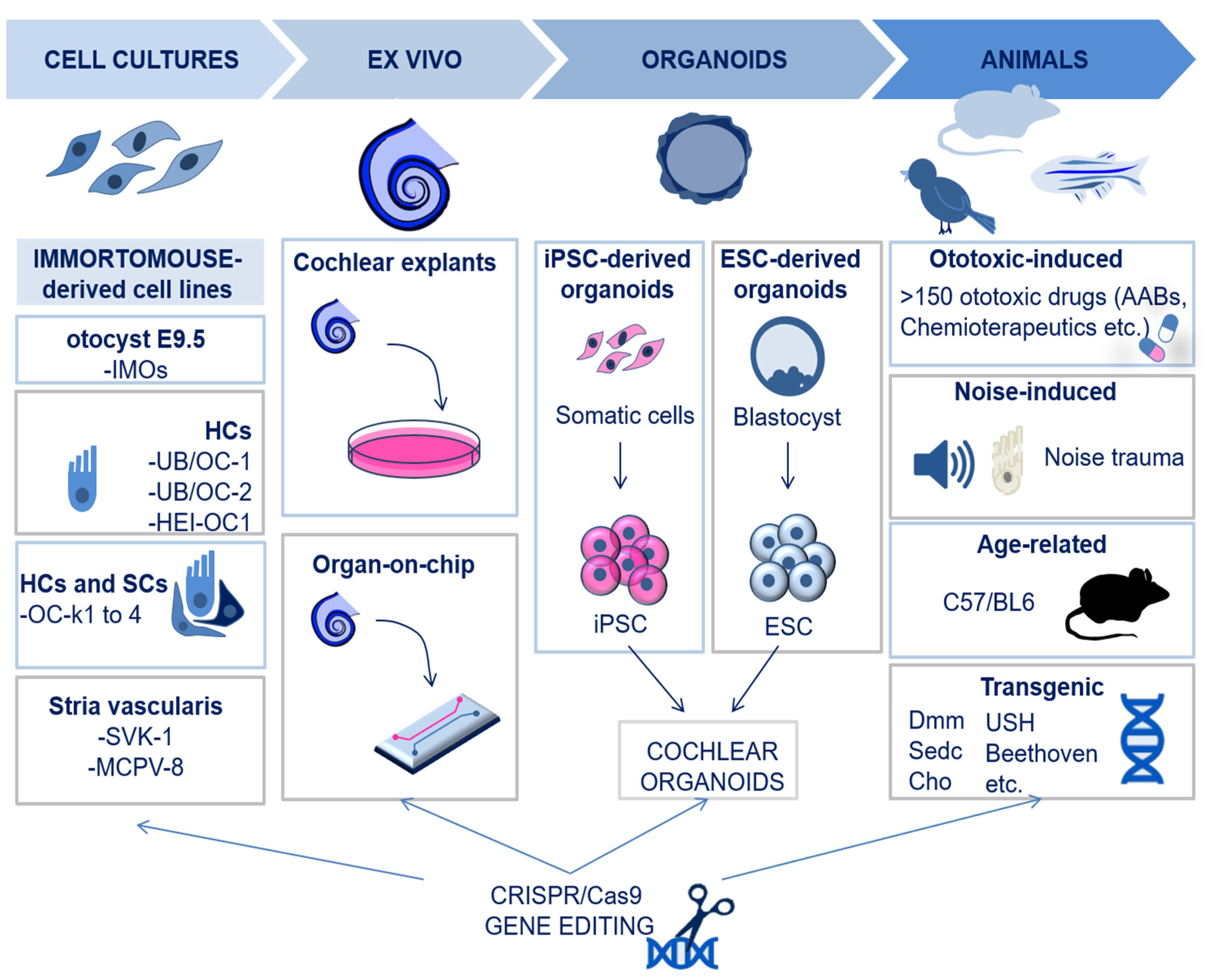
2.1. In Vitro and Ex Vivo Models: Cochlear Cell Lines, Organotypic Cultures, and Organoids

The establishment of cochlear cell cultures has been challenging for a long time due to the paucity of the tissue and the poor accessibility of the inner ear. With the advent of the Immortomouse™, a transgenic mouse model carrying the temperature-sensitive tsA58 variant of the SV40 T-antigen, it became possible to develop immortalized cell lines from the inner ear [13]. Therefore, to date, most of the available cell lines are derived from ImmunomortomouseTM, including the ImmunomortomouseTM otocysts E9.5 (IMOs), HC cultures (like UB/OC-1, UB/OC-2, HEI-OC1), and cells of the organ of Corti, which include either HCs or SCs (e.g., OC-k1to 4). The only human inner ear cell line developed so far is the immortalized endolymphatic sac (ES) cell line [14], while none have been developed yet for the human organ of Corti. In addition to cell cultures, cochlear explants (or organotypic cultures) are another efficient experimental tool to identify and characterize molecular and genetic pathways which play a role in the specification and patterning of cells in their natural environment [15]. A recent improvement in organotypic cochlear models is based on the use of microfluidic chambers for organ-on-chip culture, which allows us to reproduce a more controlled microenvironment [16]. However, cochlear explants require the use of a large number of animals, and there are technical issues with the isolation of the intact tissue to be cultured. Inner ear organoids, derived from induced pluripotent stem cells (iPSCs) or embryonic stem cells (ESCs), represent a relevant alternative to recapitulate the physiological dynamics of the cochlea in terms of cell type yield and functionality, particularly for HCs. However, it is only claimed that a small portion of the organoid cultures contain functional sensory HCs, and the reason why this happens is not yet fully understood [17]. It should be stressed that the optimization of inner ear organoid culture could lead to better drug screening programs and disease modelling opportunities.

2.2. In Vivo Models

Animal models used in hearing research are based on multiple species (rats, guinea pigs, mice, chinchillas, gerbils, birds, and zebrafish) that differ in the physiological and anatomical characteristics of the auditory system and offer different view angles to study the inner ear [18]. These animals include genetic models of spontaneous or inherited hearing loss, carrying mutations for specific genes associated with hearing impairment in humans. For instance, mutations in the genes encoding for collagen and non-collagen proteins (like α-tectorin) that are important for the structure of the basilar membrane (BM) and the tectorial membrane (TM) in the organ of Corti cause SNHL. These mutations successfully recapitulate cochlear degeneration in mice; for instance, disproportionate micromelia (Dmm) mice, spondyloepiphyseal dysplasia congenita (sedc) mice, Col2a1G574S mice, Col9a1 KO mice, and chondrodysplasia (Cho) mice all carry mutations in collagen genes; Tecta∆ENT/∆ENT and TectaY1870C/Y1870C both carry a mutation in the gene encoding for α-tectorin, Tecta [19,20]. Also, mutations in genes encoding for myosin and other proteins important for HC function and mechanotransduction, as well as genes encoding for endolymph proteins, are associated with SNHL, and multiple genetic models have been successfully developed: Beethoven mice (mutations in TMC1), Usher syndrome models, and Lcc and Ysb mice [21,22]. In addition to genetic predisposition, SNHL may also be caused by external noxious events. As of now, more than 150 established ototoxic substances have been identified, the most widely used being aminoglycoside antibiotics (AABs), loop diuretics, and antitumor medications [23]. The administration of these substances to animals recapitulates cochlear degenerative events observed in humans and is helpful in the study of degenerative mechanisms and neuroprotective strategies [18]. Additionally, exposure to loud sounds is an important risk factor for noise-induced hearing loss (NIHL); hence, noise trauma can be used to successfully reproduce NIHL in animals as well. Of note, rodents are more susceptible to noise trauma compared to non-human primates, suggesting different degenerative mechanisms with important implications from a translational point of view [24].

2.3. New Models Created by CRISPR/Cas9 Technology

The CRISPR/Cas9 technology has been developed following years of research on adaptive immunity in prokaryotes [25,26,27]. Engineering of this machinery has allowed the performance of gene editing in terms of base and prime editing, as well as the knock-out/ knock-in of genes. Therefore, the CRISPR/Cas9 technology represents a powerful tool for basic molecular studies in hearing research and a promising strategy for therapeutic approaches to SNHL [28,29,30]. In particular, CRISPR/Cas9 has been successfully applied to the creation of new in vitro and in vivo models of cochlear diseases [31]. For instance, this technology has allowed the study of genes associated with ototoxicity via the knock-out of Lim-domain only 4 (LMO4), for cisplatin, and HtrA Serine Peptidase 2 (htra2), for aminoglycosides, in vitro and in vivo models, respectively [32,33]. It has also allowed the study of inherited hearing loss genes, such as MYO7A, CIB2, and CDH23, for Usher syndrome [31]. Moreover, zebrafish models to study genes involved in the development of the auditory system, such as POU4F3, have recently been developed based on CRISPR/Cas9 [34].

3. Omics Techniques

3.1. Introduction to Omics: Principles and Advancements

The term omics refers to a rapidly evolving and expanding group of techniques aimed at investigating pools of biological molecules of an organism, including nucleic acids, proteins, and metabolites [35]. Hence, the main branches of omics techniques are known as genomics, epigenomics, transcriptomics, proteomics, and metabolomics. The most advanced omics techniques include single-cell omics and spatial omics, which allow the investigation of the molecular events occurring at a single-cell resolution and the retention of spatial information [36,37]. There are also other advanced and upcoming sequencing-based omics, such as epitranscriptomics, epiproteomics, and interactomics (DNA–RNA, RNA–RNA, RNA–protein, protein–protein, protein–metabolite), which give detailed information on the complex interactions and dynamics of regulation in a biological system [35]. The number of omics studies in cochlear research is relatively low compared to other sensory systems, since the sampling of the cochlear tissues has only recently advanced, and some techniques are incompatible with the small sample quantity obtained [38]. Nonetheless, the studies performed so far have significantly advanced the knowledge of cochlear physio-pathology.
In this review, we focus on the omics techniques that target nucleic acids, which are genomics, transcriptomics, and epigenomics in bulk, single, and spatial resolution. We also provide evidence that the availability of these techniques has been transformative in unraveling novel molecular signatures in hearing research, advancing our understanding of the treatment of cochlear degenerative diseases.

3.2. Principles of Single-Cell Omics

The term single-cell omics refers to the process of profiling the genome, transcriptome, epigenome, proteome, and metabolome in individual cells. As a consequence, single-cell techniques were shown to be useful in several biological fields, including cancer [39], developmental biology [40], stem cell research [41], neuroscience [42], and hearing [8].
The first step of all these technologies is the isolation of individual cells and the setting up of libraries. Multiple methodologies have been designed to isolate single cells from pooled cell populations/tissues through a variety of techniques [43] that span from the most straightforward—using pipettes and cell isolation by dilution—to the more sophisticated—using advanced microfluidic technologies [44]. The latter include hydrodynamic trapping, droplet-based isolation, valve-based isolation, microwell-based isolation and dielectrophoresis trapping [45], as well as magnetic-activated cell sorting (MACS), flow-activated cell sorting (FACS) [43], laser capture microdissection (LCM)—which also preserves spatial context—and nanowell-based cell sorting [46]. Details of the isolation methods for abundant or rare cells have been described by Wang and Navin [47].
After the isolation of single cells, the genome, the epigenome and the transcriptome can be profiled [8,48]. Notably, single-cell multi-omics approaches have recently been developed to investigate the molecular events that occur in individual cells under physiological or pathological conditions in a wider overview, at once. An example of this cutting-edge methodology is single-cell triple-omics sequencing (scTrio-seq), which simultaneously gathers data from the genome, DNA methylome, and transcriptome of a single cell [49].

3.3. Spatial Omics

The study of omics at a single-cell resolution has been transformative in the identification of novel biomarkers and molecular regulators of tissues, yet single-cell omics cannot deliver information on the tissue or sub-cellular localization of the isolated cells. For this reason, spatial omics have been developed with the aim of identifying molecular events while maintaining the spatial information. Multiple spatial omics approaches exist, and they vary depending on the biomolecules of interest. In the cochlea, spatial omics are of particular relevance due to its complex anatomical architecture. Indeed, the cochlea exhibits a tonotopic organization from its base (high-frequency perception) to the apex (low-frequency perception), which requires appropriate cellular structures and expression patterns [50]. Moreover, different cell types are also present from the medial (i.e., greater epithelial ridge (GER), IHCs, and their associated SCs) to the lateral (i.e., Deiters’ cells, pillar cells, and OHCs) compartment of the cochlea [50]. Hence, entering the spatial era can deepen our understanding of the cellular organization and interplay in regions of interest.

4. The Role of Bioinformatics in Analyzing Omics Data

In order to fill the knowledge gap between omics data acquisition and interpretation, bioinformatics is a critical field. Numerous computational techniques have been developed to this end, including machine learning, deep learning, data mining and statistical and metaheuristic approaches, to analyze, process, interpret, and integrate omics data for both single omics and integrative multi-omics [51,52,53,54,55]. Machine learning and deep learning are frequently used in the research community for decoding and analyzing data, predicting disease occurrence and recurrence, calculating survival rates, and finding potential biomarkers [51]. Deep learning models are a sub-set of machine learning tools of high utility since they are automated and analyze large high-dimensional data sets. Deep learning is primarily based on stratified artificial neural networks, providing diverse interpretations based on the fed data. The primary neural networks in deep learning include recursive neural networks (RvNNs), recurrent neural networks (RNNs), and convolutional neural networks (CNNs) [56]. Given the recent rapid advancements in omics and the accumulation of high-throughput omics data, future efforts should be aimed towards improving current machine learning and deep learning models for multi-omic data analysis. In this context, graph neural networks (GNNs) have gained attention in recent years [57]: the spatial relations within and between cells can be better represented with graph models, and graph-based artificial intelligence appears to hold promise, especially with regard to the most advanced omics (i.e, spatial omics). In this context, it should be mentioned that two relevant techniques have recently been developed to analyze spatial transcriptomics data: SPAcI [58] and SiGra [59]. They both have several technical advantages over existing methods, such as an improvement in accuracy, enhancing noisy gene expression data sets, and an increased ability to adapt [57,58].

5. Genomics

5.1. Principles of Sequencing

Genomics investigates somatic and germ-line inter-individual variations in the genome. The currently most used genomics are based on sequencing for the determination of the nucleic acid sequence. Genomics has been used to identify several genetic disorders and to disclose novel alleles in multiple inherited human diseases [35], including hearing loss [60,61,62,63,64,65]. The first sequencing method, known as the chain-termination method, was first developed by Sanger in 1977 and was based on the capillary electrophoresis of fragmented DNA bound to a single-stranded DNA template. The main drawback of Sanger sequencing is the ability to sequence only a low amount of DNA at a time [66]. To date, more advanced sequencing technologies have been developed and allow massive, faster, and more precise sequencing of nucleic acids. These are next generation sequencing (NGS)—more widely used—and third generation sequencing (TGS). The primary difference between these two techniques is the DNA read length. In NGS, the DNA is cleaved in small fragments (150–1000 bp), then amplified and sequenced; instead, TGS uses single-molecule sequencing without the need for prior amplification and reads long DNA sequences at a time. Moreover, it is possible to sequence different lengths of the genome depending on the experimental purpose: targeted genes (targeted panel sequencing), whole-exome sequencing (WES) [62,63] or whole-genome sequencing (WGS) [64,65] (Figure 2). In Table 1, we summarize details of the existing advanced sequencing methods and how they work.

5.2. Single-Cell and Spatial Genomics

Single-cell DNA sequencing (scDNAseq) allows the DNA profiling of individual cells [72] and is generally based on NGS. The whole genome of single cells can be primarily amplified using three methods: (i) the degenerate oligonucleotide-primed PCR (DOP-PCR), (ii) the multiple displacement amplification (MDA), and (iii) the multiple annealing and looping-based amplification cycles (MALBAC) [73]. Recently, a single-cell WGS method based on TGS was also developed in order to sequence long reads; this is known as “single-molecule real-time sequencing of long fragments amplified through transposon insertion” (SMOOTH-seq) [73]. SMOOTH-seq has greatly improved the identification of structural variants (SVs) and extra-chromosomal DNA compared to NGS [74]. Also, spatial genomics has recently been developed, but it is mostly used in cancer research to dissect the cellular genome heterogeneity of tumoral cells [75].

5.3. Genomic Studies Have Delivered Unprecedented Knowledge on the Genetic Background and Early Diagnosis of Inherited Hearing Loss

Genetic hearing loss affects any part of the auditory system and accounts for ~50% of the deaf population. It can be either non-syndromic (70%) [76,77] or syndromic (30%) [78]. The large heterogeneity of genes involved in deafness makes it difficult to study and diagnose it [62]. However, thanks to the advancements in genomics, to date several variants have been identified in genes associated with hearing loss. For instance, the combination of WES, qPCR, and TGS was able to unravel for the first-time novel SVs of centrosomal protein 78 (CEP78), a key gene responsible for hearing loss associated with cone–rod dystrophy (CRDHL) [65]. The applications of advanced genomics have also revealed new variants that have recently been outlined in important hearing loss-related genes, namely, myosin 15 A (MYO15A), otoferlin (OTOF), radixin (RDX) [79], TATA-box-binding protein-associated factor 1 (TAF1) [80], atonal BHLH transcription factor 1 (ATOH1) [81], and centrosomal protein 78 (CEP78) [65]. The discovery of novel variants represents a fundamental step forward in the understanding of the molecular basis of cochlear diseases, and indeed, it has improved the diagnosis of genetic hearing loss, as well as the prediction of its severity and prognosis. So far, several studies have benefitted from genome sequencing (via either WES or WGS) for the early detection of hearing loss [82,83,84]. For instance, a recent study has shown that the combination of conventional hearing screening and extended genetic sequencing improves the early diagnosis of inherited hearing loss in newborns, with important implications for their clinical management [85]. Yet, genetic conductivity and SNHL are a common occurrence among newborns, whose diagnosis is often missed due to the lack of proper genetic screening at birth.
Overall, the use of genomics has been useful in revealing novel gene variants linked to hearing loss, and thus, it represents a potent diagnostic tool for the genetic screening of inherited deafness.

6. Transcriptomics

Transcriptomics enable the analysis of gene expression at the RNA level, including messenger RNAs (mRNAs), transfer RNAs (tRNAs), ribosomal RNAs (rRNAs), and other non-coding RNAs (ncRNAs) (e.g., microRNAs (miRNAs), long-non-coding RNAs (lncRNAs), and circular RNAs (circRNAs)) [35,86]. As for genomics, the currently most used transcriptomics technologies are based on sequencing (described above in Section 5.1); however, transcriptome profiling is more challenging compared to genome sequencing due to the highly dynamic nature of the transcriptome in biological processes. The sequencing of the entire transcriptome in a tissue or cell population is known as bulk-RNAseq and can be performed either with direct RNA sequencing (dRNA-seq) or with cDNA sequencing [87]. Additionally, the transcriptome can also be investigated at a single-cell and spatial resolution, as detailed in the following paragraphs. A schematic overview of the transcriptomics techniques is shown in Figure 3.

6.1. Single-Cell Transcriptomics

Single-cell RNA sequencing (scRNA-seq) allows the RNA profiling of individual cells. Notably, most of the single-cell omics developed so far have been focused on the transcriptome [47,48,88]. The currently available high-throughput platforms for scRNAseq require retro-transcription of the RNA into cDNA. The cDNA is then amplified for the preparation of sequencing libraries. The amplification methods may be based on PCR or in vitro transcription (IVT) and are followed by sequencing through different platforms [88,89]. Among the existing platforms for scRNA-seq, it is noteworthy to mention SMART/SMARTseq2, CEL-seq/CEL-seq2, 10X Genomics, Drop-seq, inDrop, and seq-well [48,89]. To date, the most used commercial platform is 10X Genomics Chromium, which is a droplet-based scRNA-seq technology (10X Genomics, Pleasanton, CA, USA). An important improvement has been made in scRNA-seq which allows for the sequencing of dozens of thousands of cells individually, thanks to the development of microfluidics-based (e.g., Drop-seq [90], inDrop [43,91], and nanowell-based (such as the seq-well) methods [92]. Recently, single-nuclei RNA sequencing (snRNA-seq) has also been developed to improve the quality of scRNA-seq by mitigating the expression changes that can be induced by enzymatic cellular dissociation methods; snRNA-seq is also used to study gene expression under particular conditions, such as those where it is difficult to recover intact cells [93,94]. However, it is important to outline that snRNA-seq does not include cytoplasmatic RNAs; hence, it could hide important information needed to fully characterize the cell transcriptome. Although scRNA-seq has advanced our understanding of cell heterogeneity, it requires cell lysis, which hampers the follow-up molecular analysis on the same cell. To date, it has been difficult to track changes in a cell’s ground-state characteristics to its downstream signaling. Thanks to the advent of Live-cell omics, a state-of-the-art technique coupling Fluidforce microscopy (Fluid FM)-based cytoplasmic biopsy and low-input RNA-seq (as low as 1 picogram) workflow, it is now possible to profile the transcriptome, as well as the molecular/functional changes of the same cell at different time points, while preserving cell viability. This methodology is known as live-seq and can enhance the knowledge on cell dynamics and regulation [95].

6.2. Spatial Transcriptomics

The spatial omics for transcriptome studies are broadly divided into imaging-based technologies (where RNA is detected using fluorophores on intact tissues and then detected by microscopy) and sequencing-based technologies (based on RNA capturing from the tissue, followed by NGS) [75,96,97,98].

6.2.1. Imaging-Based Technologies

One of the primary imaging-based methodologies for spatial transcriptomics is fluorescent in situ hybridization (FISH), which includes SeqFISH [99], SeqFISH+, and MERFISH [100]. The principle of FISH is the hybridization of fluorescent gene-specific probes to nucleic acids on a tissue section directly, which is then analyzed through microscopy. A recent improvement in FISH is the enhanced electric FISH (EEL-FISH). In EEL-FISH, tissue mRNAs are electrophoretically transferred onto glass coverslips and are then hybridized. In this way, it is possible to accelerate data collection due to a reduced need for acquired images compared to the other FISH techniques [101]. Indeed, since the acquisition analysis is performed on coverslips and not on the tissue directly, the time of acquisition is shortened because it is not necessary to image the z axis of the tissue section. Notably, today it is also possible to visualize 3D gene expression in a tissue, thanks to the development of the expansion-assisted iterative fluorescence in situ hybridization (EASI-FISH) methodology [98,102]. Another imaging-based approach for spatial transcriptomics is in situ sequencing (ISS), where nucleic acids are first amplified (preserving spatial localization through rolling circle amplification). Differently from FISH, ISS does not use gene-specific probes; instead, it employs probes for 1–2 nucleotides at a time linked to distinct fluorophores and visualization through microscopy, which leads to the identification of the transcripts [97,98,103].

6.2.2. Sequencing-Based Technologies

Sequencing-based technologies allow the sequencing of the RNA from a tissue section through NGS. The mRNA is first captured in the tissue and then retrotranscribed to cDNA followed by calculating gene-specific sequences using NGS. Importantly, the spatial information is also retained because of the recording of the specific location where the RNA is captured. Sequencing-based techniques include “microdissection-based” and “array-based” methodologies [98]. Microdissection-based methods allow sequencing (via different platforms) of a specific portion of a tissue that is microdissected with dedicated instruments. Hence, the main limitation is the low spatial resolution. Microdissection-based technologies include laser capture microdissection combined with NGS (LCM-NGS) [104], Tomo-seq [105], Geo-seq [106], GeoMx DSP [107,108], and STRP-seq [109].
Differently, array-based technologies employ arrays with spatially barcoded probes: the RNAs are retro-transcribed in cDNAs that are then sequenced. In this case, the spatial resolution depends on the area of the barcode and is therefore higher compared to microdissection-based methods [98]. To date, Visium by 10X Genomics has achieved a spatial resolution of 2 μm [110,111], while the Stereo-seq is capable of achieving an even a lower resolution, up to 0.5 μm [112]. Other notable techniques for spatial transcriptomic analysis also include slide-seq and slide-seqV2 [75,113,114] and the deterministic barcoding in tissue for spatial omics sequencing (DBiT-seq), which employs microfluidic channels to print the array directly onto the tissue. Here, the spatial resolution depends on the diameter of the microfluidic channel used (no less than 10 μm) [115].

6.3. scRNA-seq Is a Key Tool for Deciphering the Complex Cellular Heterogeneity of the Cochlea

Previous studies on the auditory transcriptome were performed using microarray technologies and bulk RNA-seq [116], providing significant knowledge and information on the differential gene expression in physiological and pathological conditions of the cochlea, with important implications for the development of new therapies [117,118]. Studies on the transcriptome have also given insights into the developmental processes of the inner ear [119], the transcriptional changes associated with ageing [120], and cochlear cell damage/degeneration [121]. However, information on the specific cell populations is not possible with bulk RNA-seq, and this is a major limitation for studies on the cochlea due to its cellular heterogeneity [116].
In this context, scRNA-seq has given unprecedented information on cochlear cell diversity and alternative signaling mechanisms [122]. Profiling the transcriptome at a single-cell resolution helped to unravel novel populations of cells in the cochlea. For instance, subtypes of SCs (lateral and medial) with a distinct cluster of regenerative-associated markers were discovered in the avian cochlea [123]. Intriguingly, some of these discovered markers were also found to be expressed in specific regions of the mammalian cochlea [124], further supporting the stem cell-like potential of SCs [125]. Moreover, scRNA-seq has also allowed the identification of new specific markers of HCs, like sorcin (sri) for OHCs [126], which was then discovered to be implicated in calcium dynamics and the somatomotility of OHCs [126]. Another important application of scRNA-seq has been the study of exons and genes associated with deafness [126,127]. In this context, new genes associated with apoptosis, calcium regulation, and the extra cellular matrix (ECM) were found to be modulated in HCs of inner ear organoids in association with type II transmembrane protease 3 (tmprss3), a key gene for hearing loss [127]. Likewise, differential gene expression patterns among the cells of the lateral wall, the stria vascularis, and the immune system and SGNs have been identified in association with acoustic trauma by means of scRNA-seq, delineating a cell-specific transcriptomic map of the cochlea upon noise damage [128].
Overall, due to the complexity and heterogeneity of the cochlea, scRNA-seq has provided unmatched opportunities to broaden our current understanding of its molecular underpinnings in health and disease.

6.4. Spatial Transcriptomics Have Enabled Us to Understand the Cellular and Molecular Architecture of the Cochlea

The cochlea is spatially organized with distinct and localized functions. Thanks to the development of spatial transcriptomics methodologies, it is now possible to study the localization of specific gene expression patterns in relationship to the different anatomical structures of the cochlea [129,130]. For instance, the combination of scRNA-seq and FISH has allowed the identification of two subpopulations of SCs (named SC1 and SC2) retaining distinct transcriptomes in specific anatomic locations of the cochlea: medial for SC1 and lateral for SC2 [131]. Spatial transcriptomics are also particularly important for studies in the developing cochlea since the cochlea’s cellular organization during development is regulated by several spatiotemporal-dependent key signaling mechanisms. For instance, Munnamalai and co-workers investigated the spatiotemporal cadence of Wnt, NOTCH, and BMP signaling in the developing cochlea and found that they are differentially regulated depending on the cochlear location (from lateral to medial) and on the developmental stage. This study emphasizes the spatiotemporal signaling necessary to modulate the development of the cochlea in its radial axis and further supports the importance of spatial transcriptomics for cochlear research [132]. Another study used LCM-NGS to profile the transcriptome in different regions of the cochlea (e.g., the organ of Corti, spiral ganglion, lateral wall, and spiral limbus) and provided quantitative information on the transcripts of each region with important findings on deafness-associated genes [104].
To our knowledge, more advanced spatial transcriptomics technologies, like the Visium or the Stereo-seq technologies, have not yet been applied to cochlear research. However, it is expected that they could provide unparalleled opportunities for future studies in the field.

7. Epigenomics

7.1. Principles of Epigenomics

The term epigenomics refers to the techniques used to investigate the epigenome, which is the set of regulatory processes that modify the activity of gene expression without modifications in the DNA sequence. Epigenomics can be classified depending on the target: DNA methylation, histone modifications, chromatin accessibility, and chromosome interactions. The methodologies to study bulk epigenomics can be further classified as “array-based” and “sequencing-based” techniques [133]. Array-based technologies use hybridization with pre-designed microarrays, while sequencing-based methods use techniques like NGS. DNA methylation is an epigenetic mark where methyl groups are added to the cytosine bases of the DNA. It is important to highlight that to investigate DNA methylation, a required first step is the exposure of the methylated DNA through one of the following methods: (i) DNA digestion by methylation-sensitive restriction enzymes (MSREs) [134], (ii) affinity enrichment of DNA by antibodies targeting methylated CpGs [135], or (iii) conversion of unmethylated cytosines to uracil by bisulfite treatment [134]. To date, the bisulfite sequencing (BS-seq) method is considered the gold-standard technique for studies on DNA methylation because of its single-base resolution [133]. Histones can be modified primarily through acetylation, phosphorylation, methylation, and other miscellaneous modifications [136]. One of the most used techniques for monitoring histone modifications is chromatin immunoprecipitation (ChIP), in which the histone modifications of interest are targeted by antibodies. The cleavage under targets and release using nuclease (CUT&RUN) [137] and cleavage under targets and tagmentation (CUT&TAG) [138] methods are additional techniques used for the analysis of histone modifications; both rely on the same principle of recognizing DNA-bound proteins of interest through specific antibodies. The chromatin is highly dynamic, allowing regulators (enhancers, promoters, and chromatin-binding factors, among others) to have multiple physical interactions with DNA, thereby playing an important role in regulating gene expression. Multiple techniques for chromatin accessibility studies have also been developed. Among these, the most recent is the accessible chromatin using sequencing technology (ATAC-seq). It employs tagmentation (inserting adapter sequences by using the hyperactive mutant Tn5 transposase) to open target regions of the chromatin, which are then amplified and sequenced [139]. Other widely used techniques for chromatin accessibility include the DNase I hyper-sensitive sites sequencing (DNAse-seq) [140], the micrococcal nuclease digestion with deep sequencing (MNase-seq) [141], and the formaldehyde-assisted identification of regulatory elements followed by sequencing (FAIRE-seq) [142]. The higher-order organization of the nucleus is also important for the epigenetic regulation of cellular processes; hence, techniques able to analyze chromosomal interactions have also been developed. They include the chromatin conformation capture technique (3C), Hi-C, the chromatin interaction analysis by paired-end tag sequencing (ChIA PET), and the proximity ligation-assisted ChIP-seq (PLAC-seq) [133]. Further details of available epigenomics methodologies have already been extensively reviewed (see [133,143]), and those applied to cochlear research are summarized in Figure 4.

7.2. Single-Cell Epigenomics

Single-cell epigenomics enable a detailed analysis of the epigenetic regulation at a single-cell resolution, which includes single-cell DNA methylation profiling, single-cell chromatin mapping, single-cell Hi-C, and single-cell replication dynamics [144]. Several methods for single-cell DNA methylation profiling exist, the most recent of which are “single-cell combinatorial indexing for methylation analysis” (sci-MET) and “single-cell CGI methylation sequencing” (scCGI-seq) [133,145,146]. Histone modifications in single cells can also be studied using sc-ChIP-seq, single-cell droplet-based chromatin immunoprecipitation (drop-ChIP) [48], single-cell chromatin immune-cleavage sequencing technique (scChIC-seq), antibody-guided chromatin tagmentation sequencing (ACT-seq), combinatorial barcoding and targeted chromatin release (COBATCH), and single-cell chromatin integration labeling sequencing (scChIL-seq) [133]. Finally, single-cell chromatin accessibility can be investigated using scDNAse-seq and scATAC-seq. The available single-cell epigenetic methods have been recently reviewed in detail (see [144,147]).

7.3. Spatial Epigenomics

To fully appreciate the influence of epigenetic variations in patho-physiological processes, it is essential to know their spatial context. However, the development of spatial epigenomics techniques has been challenging for a long time due to the limited spatial resolution available [148,149]. The first spatial epigenomic technology was developed in 2021 and is now beginning to open new possibilities in the field of biology and medicine. The first spatial epigenomic technique that has been developed is the “high-spatial-resolution chromatin modification state profiling by sequencing” (hsrChST-seq). It is based on the spatial transcriptomic technique DBiT-seq, in which there is a combination of CUT&TAG and tissue deterministic barcoding with fluorescence microscopy [150]. Another technique developed later on to resolve chromatin accessibility spatially is the spatial-ATAC seq, which is based on the combination of in situ Tn5 transposase chemistry with microfluidic deterministic barcoding [151]. Recently, it has been possible to analyze the active and inactive promoters/enhancers associated with histone modifications in single cells while maintaining spatial information thanks to the advent of epigenomic MERFISH [151]. Epigenomic MERFISH combines CUT&TAG and MERFISH (a spatial epigenomic technique for the analysis of histone modifications) [152]. Furthermore, LCM can also be applied to epigenomics in order to spatially analyze modifications in the epigenome [153]. Finally, the most recent epigenomic technique is the spatial chromatin accessibility sequencing (SCA-seq), which provides simultaneous knowledge on the chromatin accessibility, epigenomics marks (e.g., CpG methylation), and higher-order genome architecture [154].

7.4. Epigenetic Profiling of the Cochlea Has Provided New Insights into the Mechanisms Whereby Genes Responsible for Auditory Function Are Regulated

Hearing loss can be caused by epigenetic alterations or by mutations in the genes encoding for the epigenetic machinery, affecting DNA methylation dynamics [155,156,157], histone modifications [158,159,160], and chromatin remodeling [156,161,162]. Thus, investigating epigenetic mechanisms could eventually pave the way towards new approaches to therapeutics. To date, most of the studies on the cochlear epigenome are based on bulk epigenomic profiling, and only a few were performed with single-cell epigenomics, namely scATAC-seq [163,164]. Instead, spatial epigenomics has not yet been applied in this field, though the epigenomics studies conducted until now have given us profound insights into the regulatory mechanisms of development, trans-differentiation, and regeneration of the auditory system. The application of ChIP-seq and ChIP-qPCR has led to the identification of fundamental epigenetic modifications in the promoters of two key genes involved in SGN differentiation (Cdk2 and NeuroD1), which affects the binding of the regulatory transcription factor neurogenin 1 (neurog1) [165]. Also, ChIP-qPCR allowed for the description of the histone modifications associated with the epigenetic regulation of atonal bHLH transcription factor 1 (Atoh1), which is an evolutionarily conserved transcription factor for the development of the auditory system [166]. Yet, histone modifications of Atoh1, which are characteristic of HCs during their development, are suppressed in the same cells after birth, but they persist in perinatal SCs. This is an important finding, which gives new information on the mechanisms underlying the regenerative potential of SCs [166]. Likewise, the application of ATAC-seq provided new findings on specific variations in chromatin accessibility during the reprogramming of SCs into HCs in cochlear organoids [167], and scATAC-seq has unraveled important information on the mechanisms which limit the capacity of SCs trans-differentiation into HCs in the adult mammalian cochlea [163]. Furthermore, the combination of scATAC-seq with scRNA-seq has recently allowed the identification of molecular regulators of key transcription factors (such as Sox and Six) involved in HC regeneration from SCs in the zebrafish’s inner ear [164].
Overall, the epigenome profiling conducted until now in the cochlea has given new insights into the regulatory mechanisms of cochlear development, regeneration, and disease. It is expected that the application of the most advanced spatial epigenomics techniques in the future could provide a much better understanding of these processes.

8. Discussion

The cochlea is a complex sensory organ, whose degeneration can be caused by multiple damaging conditions that lead to irreversible hearing loss. The neuro-sensory epithelium—the organ of Corti—is particularly susceptible to degeneration as a consequence of inherited or environmental conditions. Moreover, SGNs degenerate as a consequence of damage to the organ of Corti, resulting in the reduced performance of cochlear implants [168,169,170]. As of now, there are no approved pharmacological treatments for SNHL. Recently, a novel putative therapeutic drug (FX-322) in a phase 2 clinical trial has been shown to hold promise in inducing the regeneration of HCs, thereby providing improvements in hearing function in cases of chronic noise-induced/sudden SNHL [171,172]. Hence, an understanding of the detailed molecular mechanisms that underlie cochlear physiology and pathology can pave the way for the development of new therapeutics to treat SNHL. The molecular basis (both genetic and non-genetic) of hearing loss, the developmental processes of the cochlea, and the stem cell-like regenerative potential of SCs in the organ of Corti are among the most active fields of cochlear research. Significant improvements in these areas have been possible due to the advent of advanced omics, and Table 2 summarizes such advanced genomics, transcriptomics, and epigenomics techniques that have been applied in cochlear research, alongside the improvement that they have provided in the field. For instance, genome sequencing has allowed for the identification of new variants in genetic hearing loss and represents a great improvement in the diagnosis of genetic deafness in newborns [82,83,84]. Nonetheless, studies on the cochlea are particularly challenging compared to other sensory organs because of several practical limitations. Indeed, the cochlea is enveloped in the bony labyrinth and cannot be visualized directly, requiring imaging techniques like MRI and CT [173,174] or histological techniques on postmortem tissues of humans and animals [175,176]. The high heterogeneity of the tissue [177], as well as the low number of cells, especially in the organ of Corti (~3.500 IHCs and ~12.000 OHCs in the human ear) [178], represent additional major limitations for studies in the field, especially with conventional molecular techniques (like real-time PCR or Western blotting). In general, there is a limited quantity of cells, nucleic acids, and other molecules (proteins and metabolites), which makes it necessary to pool samples in order to reach a sufficient amount for analysis. Moreover, the heterogeneity of the tissue in location (several subpopulations of cells with varied morphologies and functions) and direction (same population of cells varying in expression and phenotype) makes it difficult to interpret molecular data derived from a whole tissue. In fact, bulk studies in the whole cochlea may easily hide some molecular information, eventually diluted in the pool of the whole genome/transcriptome/epigenome. More recently, due to the advancements in single-cell omics techniques, these limitations have been successfully overcome [8]. In cochlear research, most of the single-cell studies have been performed to investigate the transcriptome through scRNA-seq [8,179]. Emerging single-cell transcriptomic techniques on live cells, such as live-seq, can be particularly useful in cochlear research in the future for two main reasons: (i) the live-seq technique is currently used for low sample input (as low as 1 pg) and (ii) the follow-up of a cell from its ground-state transcriptome to its downstream signaling pathways can enhance our understanding of processes of auditory function and development in health and disease. Single-cell analysis does not allow us to retain the spatial information, and the application of spatial transcriptomics seems to be of particular relevance in the field because of the complex cellular heterogeneity of the cochlea [129,132]. Until now, among spatial genomic, epigenomic, and transcriptomic methods, only the latter has been applied in cochlear research, to the best of our knowledge. Thanks to spatial transcriptomics, it has been possible to identify differential gene expression patterns for the development and regeneration of the cochlea in distinct cell populations of specific anatomical locations [180]. For instance, Waldhaus and colleagues profiled SCs in the apex and base of the murine cochlea and found that SCs—especially pillar cells—express regenerative and proliferative genes potentially relevant to the regeneration of HCs in mouse apical cochlea [180].
However, the most advanced spatial transcriptomic techniques, such as visium or stereo-seq, have not yet been exploited in cochlear research. Therefore, it is expected that using high-throughput resolution spatial omics will enable a further dissection of the cochlear patho-physiology in more detail. In addition to advanced single spatial omic techniques, spatial multi-omic methodologies, such as simultaneous profiling of the transcriptome and epigenome (both imaging-based and microfluidic deterministic barcoding-based methods), have emerged recently [181]. The application of such innovative techniques could provide an integrated picture of the multiple underlying molecular events, allowing a more comprehensive understanding of the cochlear function in health and disease. For instance, the genotype–phenotype correlation holds promise for the identification of novel biomarkers and pharmacological targets.
Nonetheless, thanks to the application of scRNA-seq in species capable of self-regenerating HCs (like zebrafish and birds), researchers have identified key expression patterns in SCs, which could potentially induce their reprogramming into HCs also in the mammalian cochlea [182,183]. Although some HC-like cells have been successfully regenerated in the murine cochlea by forcing the expression of the identified patterns, fully differentiated and functional HCs in the mammalian cochlea have not yet been developed. Notably, recent studies seem to indicate that this is due to epigenetic patterns. Indeed, epigenomic data obtained through scATAC-seq identified specific epigenetic modifications in the zebrafish’s SCs essential for their reprogramming into HCs [164]. Likewise, scATAC-seq also revealed the epigenetic mechanisms responsible for the inability of SCs to trans-differentiate into HCs in the mammalian cochlea [163]. From recent studies, the crucial role of epigenetic regulators in cochlear dynamics is apparent, but the sub-cellular/nuclear location of these epigenetic marks remains unknown. Hence, the application of spatial epigenomic techniques (such as epigenomic MERFISH) in addition to bulk/single-cell epigenomics can broaden our understanding of molecular regulation in auditory function. Although promising, epigenomic MERFISH is a targeted approach, and hence, prior knowledge on the epigenetic regulators is required. This strongly suggests a need for basic research in this area before the application of such high-throughput techniques.
The role of bioinformatics is undoubtedly crucial in maximizing the knowledge gained from omics data. Despite the huge amount of information provided by high-throughput omics methodologies, a major limitation lies in the storage of data and their interpretation. Indeed, multiple platforms are used for data storage, and each retains a different format; thus, the pre-processing of data is necessary before analysis. Moreover, artifacts may be generated for multiple reasons, such as the low amount of input material (especially for genome sequencing due to the presence of only two DNA copies) and the induction of stress genes due to the dissociation methods used for cell isolation. Therefore, orthogonal validation with targeted approaches is needed, and specialized bioinformaticians are necessary to properly read and interpret the data [47,88]. Although the currently available computational techniques have provided innumerable insights into the gained omics data in the cochlea, there are currently challenges encountered in efficient interpretation due to heterogeneous and noisy data, imbalance, or missing data, among others [51]. The application of the state-of-the-art deep learning models to analyzing spatial transcriptomics data such as spaCI [58] or SiGra [59] can indeed provide valuable information on cochlear cell architecture and dynamics.
Table 2. Advanced genomics, epigenomics, and transcriptomics techniques that have been applied in cochlear research.
Omics CategoriesTechniquesApplications in Hearing ResearchModels UtilizedReference
GenomicsWGS, WESIdentification of novel structural variants and rare mutations in genes associated with deafnessHumans
(affected individuals with the CRDHL)
[65]
WESEarly detection of hearing loss for diagnostic purposesHumans (individuals with diagnosis of hearing loss)[83]
Target exome panelImprovement in the clinical diagnostic yield and thereby routine genetic screeningHumans
(deaf patients suspected with underlying genetic causes of deafness)
[84]
Humans (patients diagnosed with SNHL)[82]
TranscriptomicsTruSeq Identification of differential and preferential gene expression patterns and characterization of novel molecular pathways of the cochleaHumans (patients with tumors of the skull base with normal hearing)[118]
Engineered mouse models of genes related to circadian rhythm with noise damage [121]
Comprehension of mechanisms involved in hair cell regeneration Ototoxic (neomycin)-treated zebrafish[184]
SMART-Seq v4 Insights into the transcriptional changes in HCs during the process of ageing and damage1-, 9-, 18-, 22-, and 26-month-old CBA/J mice[120]
RNA-Seq V2 Unraveling the genes specific to SGNs and their dynamicity in developmental processes Mouse at different stages: E15.5, P1, P8, P14, and P30[185]
Single-cell transcriptomicsSMART-Seq2 Identification of novel subtypes of cochlear cellsChicken [123]
Identification of new markers of HCsMouse (C3HeB/FeJ)[126]
10x GenomicsIdentification of gene regulatory networks involved in HC regenerationZebrafish (transgenic model for HC ablation)[164]
Identification of genes associated with Tmprss3-related hearing lossMouse (Tmprss3-KO organoids)[127]
Delineation of key regulatory mechanisms in HC regeneration Rats[186]
Spatial transcriptomicsSingle-molecule FISH (smFISH)Annotating distinct transcriptome of SC populations in specific anatomic locations of the cochleaMouse[131]
Whole-mount ISHSpatiotemporal cadence of key signaling pathways in the context of developmental processes of the cochleaMouse organotypic cultures[132]
Genetically engineered mouse models of genes related to developmental processes[129]
Uncovering quantitative differential transcriptional profile in pre-mature and mature HCs, revealing novel role of genes in the differentiation processMice (P4 and 3 weeks old)[130]
LCM-NGSDiscovery of quantitative information of transcripts relevant in deafness in the organ of Corti, spiral ganglion, lateral wall, and spiral limbusMice (C57BL/6J)[104]
EpigenomicsChIP-seq and ChIP-qPCREpigenetic modifications in the promoters of genes involved in SGN differentiationIn vitro immortalized multipotent otic progenitors (iMOP cells)[165]
ATAC-seqIdentification of dynamics in chromatin accessibility of key transcriptional factors during the reprogramming of SCs into HCsMouse (Atoh1-nGFP, Sox2-GFP or Lgr5-GFP) and cochlear organoids[167]
Single-cell epigenomicsscATAC-seqRegulation of chromatin accessibility during the process of regeneration and identification of genetically conserved regenerative response elements necessary for injury/regenerative responsesZebrafish (transgenic model for HC ablation)[164]
Identification of the epigenetic mechanisms responsible for the inability of SCs to trans-differentiate into HCs in the adult mammalian cochleaTransgenic mouse models expressing transcription factors[163]

9. Conclusions and Future Perspectives

Overall, advanced genomics, epigenomics, and transcriptomics techniques represent the state-of-the-art approaches in cochlear research and are providing unprecedented knowledge on the molecular basis of cochlear patho-physiology. There are various open questions in the field of cochlear research that could be addressed through the application of omic techniques. For instance, in the context of the heterogeneity of the cochlear tissue, as of now, eight different SC populations with distinct morphologies have been identified, but little is known about their contribution to auditory function [187]. The application of single-cell and spatial transcriptomic technologies could provide a map of cell-type-specific gene expression patterns that can help to hypothesize the potential role of these cells in auditory processes. In addition, the application of single-cell epigenomics could enhance our understanding of the important cell-type-specific gene regulatory networks that can broaden our knowledge on the putative role of SC in cochlear homeostasis. Another active area of research in hearing is the mechanisms underlying ototoxicity. For instance, cisplatin, one of the primary chemotherapeutic agents utilized in oncology, has debilitating effects on hearing function and, hence, quality of life. Although efforts have been undertaken to understand the molecular underpinnings behind cisplatin-induced ototoxicity, to date, no effective therapeutics have been approved to counteract such adverse effects [188]. This necessitates a thorough molecular comprehension of the ototoxic processes from cisplatin trafficking to downstream signaling. The application of single-cell and spatial omics can give insights into the consequences of those ototoxic compounds. For instance, genomics would provide advanced knowledge on the cell-type-specific mutations and their location, transcriptomics on the cell-type-specific biomarkers expressed under ototoxic conditions, and epigenomics on putative gene regulatory dynamics in ototoxicity, which can all provide novel pharmacological/gene therapy targets not only for HCs (where most of the research is focused on [189]) but also for SCs, which are known for their role in ototoxicity [190]. Furthermore, we do not yet fully understand the origin of various cell types present in the cochlea [191]. The application of single-cell omics can elucidate evident lineage-specific markers that can help trace back their origin. The identification of common/distinct progenitor/stem cell populations can not only provide clues on inner ear development but also can provide profound knowledge on cellular sources for regeneration and repair.
From the available literature, it is increasingly evident that the application of bulk/single-cell omics, as well as advanced multi-omics approaches, is necessary to achieve an integrated view of the biological processes of the cochlea in health, development, and disease. Further studies combining genomics/transcriptomics/epigenomics with other omics—like proteomics and metabolomics—will give unmatched opportunities to decipher the complex molecular underpinnings of such a complex sensory organ.

Author Contributions

M.M.: conceptualization; A.T. and S.P.: writing—original draft preparation; M.M.: resources; A.T., S.P. and M.M.: writing—review and editing; A.T. and M.M.: supervision; A.T. and S.P.: preparation of figures and tables. All authors have read and agreed to the published version of the manuscript.

Funding

S.P. was partly supported by Dompé Farmaceutici S.p.A. (Milan, Italy), under a PhD programme in Experimental Medicine that is co-shared with University of L’Aquila and supervised by MM.

Data Availability Statement

Data sharing not applicable.

Conflicts of Interest

The authors declare no conflict of interest.

References

  1. Deafness and Hearing Loss. Available online: https://www.who.int/news-room/fact-sheets/detail/deafness-and-hearing-loss (accessed on 23 August 2023).
  2. Ma, Y.; Wise, A.K.; Shepherd, R.K.; Richardson, R.T. New Molecular Therapies for the Treatment of Hearing Loss. Pharmacol. Ther. 2019, 200, 190–209. [Google Scholar] [CrossRef] [PubMed]
  3. Liberman, M.C.; Kujawa, S.G. Cochlear Synaptopathy in Acquired Sensorineural Hearing Loss: Manifestations and Mechanisms. Hear. Res. 2017, 349, 138–147. [Google Scholar] [CrossRef] [PubMed]
  4. Wise, A.K.; Pujol, R.; Landry, T.G.; Fallon, J.B.; Shepherd, R.K. Structural and Ultrastructural Changes to Type I Spiral Ganglion Neurons and Schwann Cells in the Deafened Guinea Pig Cochlea. J. Assoc. Res. Otolaryngol. 2017, 18, 751. [Google Scholar] [CrossRef] [PubMed]
  5. Smith-Cortinez, N.; Tan, A.K.; Stokroos, R.J.; Versnel, H.; Straatman, L.V. Regeneration of Hair Cells from Endogenous Otic Progenitors in the Adult Mammalian Cochlea: Understanding Its Origins and Future Directions. Int. J. Mol. Sci. 2023, 24, 7840. [Google Scholar] [CrossRef] [PubMed]
  6. Santos-Cortez, R.L.P.; Yarza, T.K.L.; Bootpetch, T.C.; Tantoco, M.L.C.; Mohlke, K.L.; Cruz, T.L.G.; Perez, M.E.C.; Chan, A.L.; Lee, N.R.; Tobias-Grasso, C.A.M.; et al. Identification of Novel Candidate Genes and Variants for Hearing Loss and Temporal Bone Anomalies. Genes 2021, 12, 566. [Google Scholar] [CrossRef]
  7. Kelley, M.W. Cochlear Development; New Tools and Approaches. Front. Cell Dev. Biol. 2022, 10, 884240. [Google Scholar] [CrossRef]
  8. Wu, M.; Xia, M.; Li, W.; Li, H. Single-Cell Sequencing Applications in the Inner Ear. Front. Cell Dev. Biol. 2021, 9, 637779. [Google Scholar] [CrossRef]
  9. Bommakanti, K.; Iyer, J.S.; Stankovic, K.M. Cochlear Histopathology in Human Genetic Hearing Loss: State of the Science and Future Prospects. Hear. Res. 2019, 382, 107785. [Google Scholar] [CrossRef]
  10. Yamakami, I.; Ito, S.; Higuchi, Y. Retrosigmoid Removal of Small Acoustic Neuroma: Curative Tumor Removal with Preservation of Function: Clinical Article. J. Neurosurg. 2014, 121, 554–563. [Google Scholar] [CrossRef]
  11. Nicoli, T.K.; Atula, T.; Sinkkonen, S.T.; Korpi, J.; Vnencak, M.; Tarkkanen, J.; Mäkitie, A.A.; Jero, J. Ear Canal and Middle-Ear Tumors: A Single-Institution Series of 87 Patients. Acta Oto-Laryngol. 2022, 142, 132–139. [Google Scholar] [CrossRef]
  12. Gao, S.S.; Xia, A.; Applegate, B.E.; Shelton, R.L.; Yuan, T.; Raphael, P.D.; Oghalai, J.S. Quantitative Imaging of Cochlear Soft Tissues in Wild-Type and Hearing-Impaired Transgenic Mice by Spectral Domain Optical Coherence Tomography. Opt. Express 2011, 19, 15415–15428. [Google Scholar] [CrossRef]
  13. Rivolta, M.N.; Holley, M.C. Cell Lines in Inner Ear Research. J. Neurobiol. 2002, 53, 306–318. [Google Scholar] [CrossRef]
  14. Lim, D.J.; Moon, S.K. Establishment of Cell Lines from the Human Middle and Inner Ear Epithelial Cells. Adv. Exp. Med. Biol. 2011, 720, 15–25. [Google Scholar] [CrossRef]
  15. Haque, K.D.; Pandey, A.K.; Kelley, M.W.; Puligilla, C. Culture of Embryonic Mouse Cochlear Explants and Gene Transfer by Electroporation. JoVE 2015, 95, e52260. [Google Scholar] [CrossRef]
  16. Mazzarda, F.; D’Elia, A.; Massari, R.; De Ninno, A.; Bertani, F.R.; Businaro, L.; Ziraldo, G.; Zorzi, V.; Nardin, C.; Peres, C.; et al. Organ-on-Chip Model Shows That ATP Release through Connexin Hemichannels Drives Spontaneous Ca2+ Signaling in Non-Sensory Cells of the Greater Epithelial Ridge in the Developing Cochlea. Lab Chip 2020, 20, 3011–3023. [Google Scholar] [CrossRef]
  17. Roccio, M.; Edge, A.S.B. Inner Ear Organoids: New Tools to Understand Neurosensory Cell Development, Degeneration and Regeneration. Development 2019, 146, dev177188. [Google Scholar] [CrossRef]
  18. Lin, X.; Luo, J.; Tan, J.; Yang, L.; Wang, M.; Li, P. Experimental Animal Models of Drug-Induced Sensorineural Hearing Loss: A Narrative Review. Ann. Transl. Med. 2021, 9, 1393. [Google Scholar] [CrossRef]
  19. Legan, P.K.; Rau, A.; Keen, J.N.; Richardson, G.P. The Mouse Tectorins. Modular Matrix Proteins of the Inner Ear Homologous to Components of the Sperm-Egg Adhesion System. J. Biol. Chem. 1997, 272, 8791–8801. [Google Scholar] [CrossRef]
  20. Verhoeven, K.; Van Laer, L.; Kirschhofer, K.; Legan, P.K.; Hughes, D.C.; Schatteman, I.; Verstreken, M.; Van Hauwe, P.; Coucke, P.; Chen, A.; et al. Mutations in the Human Alpha-Tectorin Gene Cause Autosomal Dominant Non-Syndromic Hearing Impairment. Nat. Genet. 1998, 19, 60–62. [Google Scholar] [CrossRef]
  21. Vreugde, S.; Erven, A.; Kros, C.J.; Marcotti, W.; Fuchs, H.; Kurima, K.; Wilcox, E.R.; Friedman, T.B.; Griffith, A.J.; Bailing, R.; et al. Beethoven, a Mouse Model for Dominant, Progressive Hearing Loss DFNA36. Nat. Genet. 2002, 30, 257–258. [Google Scholar] [CrossRef]
  22. Friedman, L.M.; Dror, A.A.; Avraham, K.B. Mouse Models to Study Inner Ear Development and Hereditary Hearing Loss. Int. J. Dev. Biol. 2007, 51, 609–631. [Google Scholar] [CrossRef]
  23. Lanvers-Kaminsky, C.; Zehnhoff-Dinnesen, A.A.; Parfitt, R.; Ciarimboli, G. Drug-Induced Ototoxicity: Mechanisms, Pharmacogenetics, and Protective Strategies. Clin. Pharmacol. Ther. 2017, 101, 491–500. [Google Scholar] [CrossRef]
  24. Le Prell, C.G.; Hammill, T.L.; Murphy, W.J. Noise-Induced Hearing Loss: Translating Risk from Animal Models to Real-World Environments. J. Acoust. Soc. Am. 2019, 146, 3646–3651. [Google Scholar] [CrossRef]
  25. Ran, F.A.; Hsu, P.D.; Wright, J.; Agarwala, V.; Scott, D.A.; Zhang, F. Genome Engineering Using the CRISPR-Cas9 System. Nat. Protoc. 2013, 8, 2281–2308. [Google Scholar] [CrossRef]
  26. Li, T.; Yang, Y.; Qi, H.; Cui, W.; Zhang, L.; Fu, X.; He, X.; Liu, M.; Li, P.F.; Yu, T. CRISPR/Cas9 Therapeutics: Progress and Prospects. Signal Transduct. Target. Ther. 2023, 8, 36. [Google Scholar] [CrossRef]
  27. Akram, F.; Sahreen, S.; Aamir, F.; Haq, I.U.; Malik, K.; Imtiaz, M.; Naseem, W.; Nasir, N.; Waheed, H.M. An Insight into Modern Targeted Genome-Editing Technologies with a Special Focus on CRISPR/Cas9 and Its Applications. Mol. Biotechnol. 2023, 65, 227–242. [Google Scholar] [CrossRef]
  28. Zou, B.; Mittal, R.; Grati, M.; Lu, Z.; Shu, Y.; Tao, Y.; Feng, Y.; Xie, D.; Kong, W.; Yang, S.; et al. The Application of Genome Editing in Studying Hearing Loss. Hear. Res. 2015, 327, 102–108. [Google Scholar] [CrossRef]
  29. Gao, X.; Tao, Y.; Lamas, V.; Huang, M.; Yeh, W.H.; Pan, B.; Hu, Y.J.; Hu, J.H.; Thompson, D.B.; Shu, Y.; et al. Treatment of Autosomal Dominant Hearing Loss by in Vivo Delivery of Genome Editing Agents. Nature 2017, 553, 217–221. [Google Scholar] [CrossRef]
  30. Farooq, R.; Hussain, K.; Tariq, M.; Farooq, A.; Mustafa, M. CRISPR/Cas9: Targeted Genome Editing for the Treatment of Hereditary Hearing Loss. J. Appl. Genet. 2020, 61, 51–65. [Google Scholar] [CrossRef]
  31. Wu, J.; Tao, Y.; Deng, D.; Meng, Z.; Zhao, Y. The Applications of CRISPR/Cas-Mediated Genome Editing in Genetic Hearing Loss. Cell Biosci. 2023, 13, 93. [Google Scholar] [CrossRef]
  32. Rathinam, R.; Rosati, R.; Jamesdaniel, S. CRISPR/Cas9-Mediated Knockout of Lim-Domain Only Four Retards Organ of Corti Cell Growth. J. Cell. Biochem. 2018, 119, 3545–3553. [Google Scholar] [CrossRef]
  33. Gu, X.; Wang, D.; Xu, Z.; Wang, J.; Guo, L.; Chai, R.; Li, G.; Shu, Y.; Li, H. Prevention of Acquired Sensorineural Hearing Loss in Mice by in Vivo Htra2 Gene Editing. Genome Biol. 2021, 22, 86. [Google Scholar] [CrossRef]
  34. Liu, J.; Duan, X.; Li, M.; Liu, D.; Bai, X. CRISPR/Cas9-Mediated Pou4f3 Knockout Induces Defects in the Development of the Zebrafish Inner Ear. J. BioX Res. 2021, 4, 163–170. [Google Scholar] [CrossRef]
  35. Dai, X.; Shen, L. Advances and Trends in Omics Technology Development. Front. Med. 2022, 9, 911861. [Google Scholar] [CrossRef]
  36. Kong, S.; Li, R.; Tian, Y.; Zhang, Y.; Lu, Y.; Ou, Q.; Gao, P.; Li, K.; Zhang, Y. Single-Cell Omics: A New Direction for Functional Genetic Research in Human Diseases and Animal Models. Front. Genet. 2023, 13, 1100016. [Google Scholar] [CrossRef]
  37. Bingham, G.C.; Lee, F.; Naba, A.; Barker, T.H. Spatial-Omics: Novel Approaches to Probe Cell Heterogeneity and Extracellular Matrix Biology. Matrix Biol. 2020, 91–92, 152–166. [Google Scholar] [CrossRef]
  38. Zheng, Q.Y.; Rozanas, C.R.; Thalmann, I.; Chance, M.R.; Alagramam, K.N. Inner Ear Proteomics of Mouse Models for Deafness, a Discovery Strategy. Brain Res. 2006, 1091, 113–121. [Google Scholar] [CrossRef]
  39. Navin, N.; Kendall, J.; Troge, J.; Andrews, P.; Rodgers, L.; McIndoo, J.; Cook, K.; Stepansky, A.; Levy, D.; Esposito, D.; et al. Tumour Evolution Inferred by Single-Cell Sequencing. Nature 2011, 472, 90–95. [Google Scholar] [CrossRef]
  40. Pollen, A.A.; Nowakowski, T.J.; Shuga, J.; Wang, X.; Leyrat, A.A.; Lui, J.H.; Li, N.; Szpankowski, L.; Fowler, B.; Chen, P.; et al. Low-Coverage Single-Cell MRNA Sequencing Reveals Cellular Heterogeneity and Activated Signaling Pathways in Developing Cerebral Cortex. Nat. Biotechnol. 2014, 32, 1053–1058. [Google Scholar] [CrossRef]
  41. Ealy, M.; Ellwanger, D.C.; Kosaric, N.; Stapper, A.P.; Heller, S. Single-Cell Analysis Delineates a Trajectory toward the Human Early Otic Lineage. Proc. Natl. Acad. Sci. USA 2016, 113, 8508–8513. [Google Scholar] [CrossRef]
  42. Tasic, B. Single Cell Transcriptomics in Neuroscience: Cell Classification and Beyond. Curr. Opin. Neurobiol. 2018, 50, 242–249. [Google Scholar] [CrossRef]
  43. Macaulay, I.C.; Ponting, C.P.; Voet, T. Single-Cell Multiomics: Multiple Measurements from Single Cells. Trends Genet. 2017, 33, 155–168. [Google Scholar] [CrossRef]
  44. Hwang, B.; Lee, J.H.; Bang, D. Single-Cell RNA Sequencing Technologies and Bioinformatics Pipelines. Exp. Mol. Med. 2018, 50, 1–14. [Google Scholar] [CrossRef]
  45. Xu, X.; Wang, J.; Wu, L.; Guo, J.; Song, Y.; Tian, T.; Wang, W.; Zhu, Z.; Yang, C. Microfluidic Single-Cell Omics Analysis. Small 2020, 16, 1903905. [Google Scholar] [CrossRef]
  46. Menze, L.; Duarte, P.A.; Haddon, L.; Chu, M.; Chen, J. Selective Single-Cell Sorting Using a Multisectorial Electroactive Nanowell Platform. ACS Nano 2022, 16, 211–220. [Google Scholar] [CrossRef]
  47. Wang, Y.; Navin, N.E. Advances and Applications of Single-Cell Sequencing Technologies. Mol. Cell 2015, 58, 598–609. [Google Scholar] [CrossRef]
  48. Mincarelli, L.; Lister, A.; Lipscombe, J.; Macaulay, I.C. Defining Cell Identity with Single-Cell Omics. Proteomics 2018, 18, 1700312. [Google Scholar] [CrossRef]
  49. Hou, Y.; Guo, H.; Cao, C.; Li, X.; Hu, B.; Zhu, P.; Wu, X.; Wen, L.; Tang, F.; Huang, Y.; et al. Single-Cell Triple Omics Sequencing Reveals Genetic, Epigenetic, and Transcriptomic Heterogeneity in Hepatocellular Carcinomas. Cell Res. 2016, 26, 304–319. [Google Scholar] [CrossRef]
  50. Raphael, Y.; Altschuler, R.A. Structure and Innervation of the Cochlea. Brain Res. Bull. 2003, 60, 397–422. [Google Scholar] [CrossRef]
  51. Kaur, P.; Singh, A.; Chana, I. Computational Techniques and Tools for Omics Data Analysis: State-of-the-Art, Challenges, and Future Directions. Arch. Comput. Methods Eng. 2021, 28, 4595–4631. [Google Scholar] [CrossRef]
  52. Hie, B.; Peters, J.; Nyquist, S.K.; Shalek, A.K.; Berger, B.; Bryson, B.D. Computational Methods for Single-Cell RNA Sequencing. Annu. Rev. Biomed. Data Sci. 2020, 3, 339–364. [Google Scholar] [CrossRef]
  53. Stanojevic, S.; Li, Y.; Ristivojevic, A.; Garmire, L.X. Computational Methods for Single-Cell Multi-Omics Integration and Alignment. Genom. Proteom. Bioinform. 2022, 20, 836–849. [Google Scholar] [CrossRef]
  54. Li, Y.; Stanojevic, S.; Garmire, L.X. Emerging Artificial Intelligence Applications in Spatial Transcriptomics Analysis. Comput. Struct. Biotechnol. J. 2022, 20, 2895–2908. [Google Scholar] [CrossRef]
  55. Efremova, M.; Teichmann, S.A. Computational Methods for Single-Cell Omics across Modalities. Nat. Methods 2020, 17, 14–17. [Google Scholar] [CrossRef]
  56. Alzubaidi, L.; Zhang, J.; Humaidi, A.J.; Al-Dujaili, A.; Duan, Y.; Al-Shamma, O.; Santamaría, J.; Fadhel, M.A.; Al-Amidie, M.; Farhan, L. Review of Deep Learning: Concepts, CNN Architectures, Challenges, Applications, Future Directions. J. Big Data 2021, 8, 53. [Google Scholar] [CrossRef]
  57. Wang, H.; Guo, F.; Du, M.; Wang, G.; Cao, C. A Novel Method for Drug-Target Interaction Prediction Based on Graph Transformers Model. BMC Bioinform. 2022, 23, 459. [Google Scholar] [CrossRef]
  58. Tang, Z.; Zhang, T.; Yang, B.; Su, J.; Song, Q. SpaCI: Deciphering Spatial Cellular Communications through Adaptive Graph Model. Brief. Bioinform. 2023, 24, bbac563. [Google Scholar] [CrossRef]
  59. Tang, Z.; Li, Z.; Hou, T.; Zhang, T.; Yang, B.; Su, J.; Song, Q. SiGra: Single-Cell Spatial Elucidation through an Image-Augmented Graph Transformer. Nat. Commun. 2023, 14, 5618. [Google Scholar] [CrossRef]
  60. Rehman, A.U.; Morell, R.J.; Belyantseva, I.A.; Khan, S.Y.; Boger, E.T.; Shahzad, M.; Ahmed, Z.M.; Riazuddin, S.; Khan, S.N.; Riazuddin, S.; et al. Targeted Capture and Next-Generation Sequencing Identifies C9orf75, Encoding Taperin, as the Mutated Gene in Nonsyndromic Deafness DFNB79. Am. J. Hum. Genet. 2010, 86, 378–388. [Google Scholar] [CrossRef]
  61. Yan, D.; Tekin, M.; Blanton, S.H.; Liu, X.Z. Next-Generation Sequencing in Genetic Hearing Loss. Genet. Test. Mol. Biomark. 2013, 17, 581–587. [Google Scholar] [CrossRef]
  62. Shearer, A.E.; Smith, R.J.H. Massively Parallel Sequencing for Genetic Diagnosis of Hearing Loss. Otolaryngol.–Head Neck Surg. 2015, 153, 175–182. [Google Scholar] [CrossRef] [PubMed]
  63. Dunn, P.; Albury, C.L.; Maksemous, N.; Benton, M.C.; Sutherland, H.G.; Smith, R.A.; Haupt, L.M.; Griffiths, L.R. Next Generation Sequencing Methods for Diagnosis of Epilepsy Syndromes. Front. Genet. 2018, 9, 314696. [Google Scholar] [CrossRef] [PubMed]
  64. Hegde, M.; Santani, A.; Mao, R.; Ferreira-Gonzalez, A.; Weck, K.E.; Voelkerding, K.V. Development and Validation of Clinical Whole-Exome and Whole-Genome Sequencing for Detection of Germline Variants in Inherited Disease. Arch. Pathol. Lab. Med. 2017, 141, 798–805. [Google Scholar] [CrossRef] [PubMed]
  65. Ascari, G.; Rendtorff, N.D.; De Bruyne, M.; De Zaeytijd, J.; Van Lint, M.; Bauwens, M.; Van Heetvelde, M.; Arno, G.; Jacob, J.; Creytens, D.; et al. Long-Read Sequencing to Unravel Complex Structural Variants of CEP78 Leading to Cone-Rod Dystrophy and Hearing Loss. Front. Cell Dev. Biol. 2021, 9, 664317. [Google Scholar] [CrossRef]
  66. Sanger, F.; Nicklen, S.; Coulson, A.R. DNA Sequencing with Chain-Terminating Inhibitors. Proc. Natl. Acad. Sci. USA 1977, 74, 5463–5467. [Google Scholar] [CrossRef]
  67. Shendure, J.; Porreca, G.J.; Reppas, N.B.; Lin, X.; McCutcheon, J.P.; Rosenbaum, A.M.; Wang, M.D.; Zhang, K.; Mitra, R.D.; Church, G.M. Accurate Multiplex Polony Sequencing of an Evolved Bacterial Genome. Science 2005, 309, 1728–1732. [Google Scholar] [CrossRef]
  68. Drmanac, R.; Drmanac, S.; Chui, G.; Diaz, R.; Hou, A.; Jin, H.; Jin, P.; Kwon, S.; Lacy, S.; Moeur, B.; et al. Sequencing by Hybridization (SBH): Advantages, Achievements, and Opportunities. Adv. Biochem. Eng. Biotechnol. 2002, 77, 75–101. [Google Scholar] [CrossRef]
  69. Blazej, R.G.; Kumaresan, P.; Mathies, R.A. Microfabricated Bioprocessor for Integrated Nanoliter-Scale Sanger DNA Sequencing. Proc. Natl. Acad. Sci. USA 2006, 103, 7240–7245. [Google Scholar] [CrossRef]
  70. Quail, M.A.; Smith, M.; Coupland, P.; Otto, T.D.; Harris, S.R.; Connor, T.R.; Bertoni, A.; Swerdlow, H.P.; Gu, Y. A Tale of Three next Generation Sequencing Platforms: Comparison of Ion Torrent, Pacific Biosciences and Illumina MiSeq Sequencers. BMC Genom. 2012, 13, 341. [Google Scholar] [CrossRef]
  71. Jain, M.; Fiddes, I.T.; Miga, K.H.; Olsen, H.E.; Paten, B.; Akeson, M. Improved Data Analysis for the MinION Nanopore Sequencer. Nat. Methods 2015, 12, 351–356. [Google Scholar] [CrossRef]
  72. Evrony, G.D.; Hinch, A.G.; Luo, C. Applications of Single-Cell DNA Sequencing. Annu. Rev. Genom. Hum. Genet. 2021, 22, 171. [Google Scholar] [CrossRef] [PubMed]
  73. Gawad, C.; Koh, W.; Quake, S.R. Single-Cell Genome Sequencing: Current State of the Science. Nat. Rev. Genet. 2016, 17, 175–188. [Google Scholar] [CrossRef] [PubMed]
  74. Fan, X.; Yang, C.; Li, W.; Bai, X.; Zhou, X.; Xie, H.; Wen, L.; Tang, F. SMOOTH-Seq: Single-Cell Genome Sequencing of Human Cells on a Third-Generation Sequencing Platform. Genome Biol. 2021, 22, 195. [Google Scholar] [CrossRef]
  75. Tang, L. Spatially Resolved DNA Sequencing. Nat. Methods 2022, 19, 139. [Google Scholar] [CrossRef] [PubMed]
  76. Zhong, L.X.; Kun, S.; Jing, Q.; Jing, C.; Denise, Y. Non-Syndromic Hearing Loss and High-Throughput Strategies to Decipher Its Genetic Heterogeneity. J. Otol. 2013, 8, 6–24. [Google Scholar] [CrossRef]
  77. Aldè, M.; Cantarella, G.; Zanetti, D.; Pignataro, L.; Mantia, I.L.; Maiolino, L.; Ferlito, S.; Mauro, P. Di; Cocuzza, S.; Lechien, J.R.; et al. Autosomal Dominant Non-Syndromic Hearing Loss (DFNA): A Comprehensive Narrative Review. Biomedicines 2023, 11, 1616. [Google Scholar] [CrossRef]
  78. Koffler, T.; Ushakov, K.; Avraham, K.B. Genetics of Hearing Loss—Syndromic. Otolaryngol. Clin. N. Am. 2015, 48, 1041. [Google Scholar] [CrossRef]
  79. Bai, X.; Nian, S.; Feng, L.; Ruan, Q.; Luo, X.; Wu, M.; Yan, Z. Identification of Novel Variants in MYO15A, OTOF, and RDX with Hearing Loss by next-Generation Sequencing. Mol. Genet. Genom. Med. 2019, 7, e808. [Google Scholar] [CrossRef]
  80. Cheng, H.; Capponi, S.; Wakeling, E.; Marchi, E.; Li, Q.; Zhao, M.; Weng, C.; Stefan, P.G.; Ahlfors, H.; Kleyner, R.; et al. Missense Variants in TAF1 and Developmental Phenotypes: Challenges of Determining Pathogenicity. Hum. Mutat. 2020, 41, 449–464. [Google Scholar] [CrossRef]
  81. Brownstein, Z.; Gulsuner, S.; Walsh, T.; Martins, F.T.A.; Taiber, S.; Isakov, O.; Lee, M.K.; Bordeynik-Cohen, M.; Birkan, M.; Chang, W.; et al. Spectrum of Genes for Inherited Hearing Loss in the Israeli Jewish Population, Including the Novel Human Deafness Gene ATOH1. Clin. Genet. 2020, 98, 353–364. [Google Scholar] [CrossRef]
  82. Costales, M.; Diñeiro, M.; Cifuentes, G.A.; Capín, R.; Otero, A.; Viejo-Díaz, M.; Plasencia, A.; Núñez, F.; Gómez, J.R.; Llorente, J.L.; et al. Utilidad Clínica de La Secuenciación de Nueva Generación En El Diagnóstico Etiológico de La Hipoacusia Neurosensorial En Una Unidad de Hipoacusia Infantil. Acta Otorrinolaringol. Esp. 2020, 71, 166–174. [Google Scholar] [CrossRef]
  83. Van Heurck, R.; Carminho-rodrigues, M.T.; Ranza, E.; Stafuzza, C.; Quteineh, L.; Gehrig, C.; Hammar, E.; Guipponi, M.; Abramowicz, M.; Senn, P.; et al. Benefits of Exome Sequencing in Children with Suspected Isolated Hearing Loss. Genes 2021, 12, 1277. [Google Scholar] [CrossRef] [PubMed]
  84. Tropitzsch, A.; Schade-Mann, T.; Gamerdinger, P.; Dofek, S.; Schulte, B.; Schulze, M.; Battke, F.; Fehr, S.; Biskup, S.; Heyd, A.; et al. Diagnostic Yield of Targeted Hearing Loss Gene Panel Sequencing in a Large German Cohort with a Balanced Age Distribution from a Single Diagnostic Center: An Eight-Year Study. Ear Hear. 2022, 43, 1049–1066. [Google Scholar] [CrossRef] [PubMed]
  85. Zhu, Y.; Hu, L.; Yang, L.; Wang, L.; Lu, Y.; Dong, X.; Xiao, T.; Xu, Z.; Wu, B.; Zhou, W. Association Between Expanded Genomic Sequencing Combined with Hearing Screening and Detection of Hearing Loss Among Newborns in a Neonatal Intensive Care Unit. JAMA Netw. Open 2022, 5, e2220986. [Google Scholar] [CrossRef]
  86. Dong, Z.C.; Chen, Y. Transcriptomics: Advances and Approaches. Sci. China Life Sci. 2013, 56, 960–967. [Google Scholar] [CrossRef] [PubMed]
  87. Wongsurawat, T.; Jenjaroenpun, P.; Wanchai, V.; Nookaew, I. Native RNA or CDNA Sequencing for Transcriptomic Analysis: A Case Study on Saccharomyces Cerevisiae. Front. Bioeng. Biotechnol. 2022, 10, 842299. [Google Scholar] [CrossRef]
  88. Jovic, D.; Liang, X.; Zeng, H.; Lin, L.; Xu, F.; Luo, Y.; Correspondence, Y.; Luo, D.; Jovic, L. Single-Cell RNA Sequencing Technologies and Applications: A Brief Overview. Clin. Transl. Med. 2022, 12, e694. [Google Scholar] [CrossRef]
  89. Olsen, T.K.; Baryawno, N. Introduction to Single-Cell RNA Sequencing. Curr. Protoc. Mol. Biol. 2018, 122, e57. [Google Scholar] [CrossRef]
  90. Macosko, E.Z.; Basu, A.; Satija, R.; Nemesh, J.; Shekhar, K.; Goldman, M.; Tirosh, I.; Bialas, A.R.; Kamitaki, N.; Martersteck, E.M.; et al. Highly Parallel Genome-Wide Expression Profiling of Individual Cells Using Nanoliter Droplets. Cell 2015, 161, 1202–1214. [Google Scholar] [CrossRef]
  91. Zilionis, R.; Nainys, J.; Veres, A.; Savova, V.; Zemmour, D.; Klein, A.M.; Mazutis, L. Single-Cell Barcoding and Sequencing Using Droplet Microfluidics. Nat. Protoc. 2016, 12, 44–73. [Google Scholar] [CrossRef]
  92. Gierahn, T.M.; Wadsworth, M.H.; Hughes, T.K.; Bryson, B.D.; Butler, A.; Satija, R.; Fortune, S.; Christopher Love, J.; Shalek, A.K. Seq-Well: Portable, Low-Cost RNA Sequencing of Single Cells at High Throughput. Nat. Methods 2017, 14, 395–398. [Google Scholar] [CrossRef] [PubMed]
  93. Grindberg, R.V.; Yee-Greenbaum, J.L.; McConnell, M.J.; Novotny, M.; O’Shaughnessy, A.L.; Lambert, G.M.; Araúzo-Bravo, M.J.; Lee, J.; Fishman, M.; Robbins, G.E.; et al. RNA-Sequencing from Single Nuclei. Proc. Natl. Acad. Sci. USA 2013, 110, 19802–19807. [Google Scholar] [CrossRef] [PubMed]
  94. Slyper, M.; Porter, C.B.M.; Ashenberg, O.; Waldman, J.; Drokhlyansky, E.; Wakiro, I.; Smillie, C.; Smith-Rosario, G.; Wu, J.; Dionne, D.; et al. A Single-Cell and Single-Nucleus RNA-Seq Toolbox for Fresh and Frozen Human Tumors. Nat. Med. 2020, 26, 792–802. [Google Scholar] [CrossRef]
  95. Chen, W.; Guillaume-Gentil, O.; Rainer, P.Y.; Gäbelein, C.G.; Saelens, W.; Gardeux, V.; Klaeger, A.; Dainese, R.; Zachara, M.; Zambelli, T.; et al. Live-Seq Enables Temporal Transcriptomic Recording of Single Cells. Nature 2022, 608, 733–740. [Google Scholar] [CrossRef] [PubMed]
  96. Fangma, Y.; Liu, M.; Liao, J.; Chen, Z.; Zheng, Y. Dissecting the Brain with Spatially Resolved Multi-Omics. J. Pharm. Anal. 2023, 13, 694–710. [Google Scholar] [CrossRef]
  97. Payne, A.C.; Chiang, Z.D.; Reginato, P.L.; Mangiameli, S.M.; Murray, E.M.; Yao, C.C.; Markoulaki, S.; Earl, A.S.; Labade, A.S.; Jaenisch, R.; et al. In Situ Genome Sequencing Resolves DNA Sequence and Structure in Intact Biological Samples. Science 2021, 371, eaay3446. [Google Scholar] [CrossRef]
  98. Williams, C.G.; Lee, H.J.; Asatsuma, T.; Vento-Tormo, R.; Haque, A. An Introduction to Spatial Transcriptomics for Biomedical Research. Genome Med. 2022, 14, 68. [Google Scholar] [CrossRef]
  99. Lubeck, E.; Coskun, A.F.; Zhiyentayev, T.; Ahmad, M.; Cai, L. Single-Cell in Situ RNA Profiling by Sequential Hybridization. Nat. Methods 2014, 11, 360–361. [Google Scholar] [CrossRef]
  100. Chen, K.H.; Boettiger, A.N.; Moffitt, J.R.; Wang, S.; Zhuang, X. Spatially Resolved, Highly Multiplexed RNA Profiling in Single Cells. Science 2015, 348, aaa6090. [Google Scholar] [CrossRef]
  101. Borm, L.E.; Mossi Albiach, A.; Mannens, C.C.A.; Janusauskas, J.; Özgün, C.; Fernández-García, D.; Hodge, R.; Castillo, F.; Hedin, C.R.H.; Villablanca, E.J.; et al. Scalable in Situ Single-Cell Profiling by Electrophoretic Capture of MRNA Using EEL FISH. Nat. Biotechnol. 2022, 41, 222–231. [Google Scholar] [CrossRef]
  102. Wang, Y.; Eddison, M.; Fleishman, G.; Weigert, M.; Xu, S.; Wang, T.; Rokicki, K.; Goina, C.; Henry, F.E.; Lemire, A.L.; et al. EASI-FISH for Thick Tissue Defines Lateral Hypothalamus Spatio-Molecular Organization. Cell 2021, 184, 6361–6377.e24. [Google Scholar] [CrossRef] [PubMed]
  103. Ke, R.; Mignardi, M.; Pacureanu, A.; Svedlund, J.; Botling, J.; Wählby, C.; Nilsson, M. In Situ Sequencing for RNA Analysis in Preserved Tissue and Cells. Nat. Methods 2013, 10, 857–860. [Google Scholar] [CrossRef] [PubMed]
  104. Nishio, S.y.; Takumi, Y.; Usami, S. ichi Laser-Capture Micro Dissection Combined with next-Generation Sequencing Analysis of Cell Type-Specific Deafness Gene Expression in the Mouse Cochlea. Hear. Res. 2017, 348, 87–97. [Google Scholar] [CrossRef] [PubMed]
  105. Kruse, F.; Junker, J.P.; van Oudenaarden, A.; Bakkers, J. Tomo-Seq: A Method to Obtain Genome-Wide Expression Data with Spatial Resolution. Methods Cell Biol. 2016, 135, 299–307. [Google Scholar] [CrossRef]
  106. Chen, J.; Suo, S.; Tam, P.P.; Han, J.D.J.; Peng, G.; Jing, N. Spatial Transcriptomic Analysis of Cryosectioned Tissue Samples with Geo-Seq. Nat. Protoc. 2017, 12, 566–580. [Google Scholar] [CrossRef]
  107. Hernandez, S.; Lazcano, R.; Serrano, A.; Powell, S.; Kostousov, L.; Mehta, J.; Khan, K.; Lu, W.; Solis, L.M. Challenges and Opportunities for Immunoprofiling Using a Spatial High-Plex Technology: The NanoString GeoMx® Digital Spatial Profiler. Front. Oncol. 2022, 12, 890410. [Google Scholar] [CrossRef]
  108. Merritt, C.R.; Ong, G.T.; Church, S.E.; Barker, K.; Danaher, P.; Geiss, G.; Hoang, M.; Jung, J.; Liang, Y.; McKay-Fleisch, J.; et al. Multiplex Digital Spatial Profiling of Proteins and RNA in Fixed Tissue. Nat. Biotechnol. 2020, 38, 586–599. [Google Scholar] [CrossRef]
  109. Schede, H.H.; Schneider, C.G.; Stergiadou, J.; Borm, L.E.; Ranjak, A.; Yamawaki, T.M.; David, F.P.A.; Lönnerberg, P.; Tosches, M.A.; Codeluppi, S.; et al. Spatial Tissue Profiling by Imaging-Free Molecular Tomography. Nat. Biotechnol. 2021, 39, 968–977. [Google Scholar] [CrossRef]
  110. Ståhl, P.L.; Salmén, F.; Vickovic, S.; Lundmark, A.; Navarro, J.F.; Magnusson, J.; Giacomello, S.; Asp, M.; Westholm, J.O.; Huss, M.; et al. Visualization and Analysis of Gene Expression in Tissue Sections by Spatial Transcriptomics. Science 2016, 353, 78–82. [Google Scholar] [CrossRef]
  111. Maynard, K.R.; Collado-Torres, L.; Weber, L.M.; Uytingco, C.; Barry, B.K.; Williams, S.R.; Catallini, J.L.; Tran, M.N.; Besich, Z.; Tippani, M.; et al. Transcriptome-Scale Spatial Gene Expression in the Human Dorsolateral Prefrontal Cortex. Nat. Neurosci. 2021, 24, 425–436. [Google Scholar] [CrossRef]
  112. Chen, A.; Liao, S.; Cheng, M.; Ma, K.; Wu, L.; Lai, Y.; Qiu, X.; Yang, J.; Xu, J.; Hao, S.; et al. Spatiotemporal Transcriptomic Atlas of Mouse Organogenesis Using DNA Nanoball-Patterned Arrays. Cell 2022, 185, 1777–1792.e21. [Google Scholar] [CrossRef] [PubMed]
  113. Rodriques, S.G.; Stickels, R.R.; Goeva, A.; Martin, C.A.; Murray, E.; Vanderburg, C.R.; Welch, J.; Chen, L.M.; Chen, F.; Macosko, E.Z. Slide-Seq: A Scalable Technology for Measuring Genome-Wide Expression at High Spatial Resolution. Science 2019, 363, 1463–1467. [Google Scholar] [CrossRef] [PubMed]
  114. Stickels, R.R.; Murray, E.; Kumar, P.; Li, J.; Marshall, J.L.; Di Bella, D.J.; Arlotta, P.; Macosko, E.Z.; Chen, F. Highly Sensitive Spatial Transcriptomics at Near-Cellular Resolution with Slide-SeqV2. Nat. Biotechnol. 2020, 39, 313–319. [Google Scholar] [CrossRef] [PubMed]
  115. Liu, Y.; Yang, M.; Deng, Y.; Su, G.; Enninful, A.; Guo, C.C.; Tebaldi, T.; Zhang, D.; Kim, D.; Bai, Z.; et al. High-Spatial-Resolution Multi-Omics Sequencing via Deterministic Barcoding in Tissue. Cell 2020, 183, 1665. [Google Scholar] [CrossRef] [PubMed]
  116. Schimmang, T.; Maconochie, M. Gene Expression Profiling of the Inner Ear. J. Anat. 2016, 228, 255–269. [Google Scholar] [CrossRef]
  117. Ramírez-Gordillo, D.; Powers, T.R.; Van Velkinburgh, J.C.; Trujillo-Provencio, C.; Schilkey, F.; Serrano, E.E. RNA-Seq and Microarray Analysis of the Xenopus Inner Ear Transcriptome Discloses Orthologous OMIM® Genes for Hereditary Disorders of Hearing and Balance. BMC Res. Notes 2015, 8, 691. [Google Scholar] [CrossRef]
  118. Schrauwen, I.; Hasin-Brumshtein, Y.; Corneveaux, J.J.; Ohmen, J.; White, C.; Allen, A.N.; Lusis, A.J.; Van Camp, G.; Huentelman, M.J.; Friedman, R.A. A Comprehensive Catalogue of the Coding and Non-Coding Transcripts of the Human Inner Ear. Hear. Res. 2016, 333, 266–274. [Google Scholar] [CrossRef] [PubMed]
  119. Cao, R.; Takechi, M.; Wang, X.; Furutera, T.; Nojiri, T.; Koyabu, D.; Li, J. Temporal and Regulatory Dynamics of the Inner Ear Transcriptome during Development in Mice. Sci. Rep. 2022, 12, 21196. [Google Scholar] [CrossRef]
  120. Liu, H.; Giffen, K.P.; Chen, L.; Henderson, H.J.; Cao, T.A.; Kozeny, G.A.; Beisel, K.W.; Li, Y.; He, D.Z. Molecular and Cytological Profiling of Biological Aging of Mouse Cochlear Inner and Outer Hair Cells. Cell Rep. 2022, 39, 110665. [Google Scholar] [CrossRef]
  121. Cederroth, C.R.; Park, J.s.; Basinou, V.; Weger, B.D.; Tserga, E.; Sarlus, H.; Magnusson, A.K.; Kadri, N.; Gachon, F.; Canlon, B. Circadian Regulation of Cochlear Sensitivity to Noise by Circulating Glucocorticoids. Curr. Biol. 2019, 29, 2477–2487.e6. [Google Scholar] [CrossRef]
  122. Lush, M.E.; Diaz, D.C.; Koenecke, N.; Baek, S.; Boldt, H.; St Peter, M.K.; Gaitan-Escudero, T.; Romero-Carvajal, A.; Busch-Nentwich, E.M.; Perera, A.G.; et al. Scrna-Seq Reveals Distinct Stem Cell Populations That Drive Hair Cell Regeneration after Loss of Fgf and Notch Signaling. eLife 2019, 8, e44431. [Google Scholar] [CrossRef] [PubMed]
  123. Janesick, A.; Scheibinger, M.; Benkafadar, N.; Kirti, S.; Ellwanger, D.C.; Heller, S. Cell-Type Identity of the Avian Cochlea. Cell Rep. 2021, 34, 108900. [Google Scholar] [CrossRef] [PubMed]
  124. Geng, R.; Noda, T.; Mulvaney, J.F.; Lin, V.Y.W.; Edge, A.S.B.; Dabdoub, A. Comprehensive Expression of Wnt Signaling Pathway Genes during Development and Maturation of the Mouse Cochlea. PLoS ONE 2016, 11, e0148339. [Google Scholar] [CrossRef] [PubMed]
  125. Lagarde, M.M.M.; Wan, G.; Zhang, L.L.; Gigliello, A.R.; McInnis, J.J.; Zhang, Y.; Bergles, D.; Zuo, J.; Corfas, G. Spontaneous Regeneration of Cochlear Supporting Cells after Neonatal Ablation Ensures Hearing in the Adult Mouse. Proc. Natl. Acad. Sci. USA 2014, 111, 16919–16924. [Google Scholar] [CrossRef]
  126. Ranum, P.T.; Goodwin, A.T.; Yoshimura, H.; Kolbe, D.L.; Walls, W.D.; Koh, J.Y.; He, D.Z.Z.; Smith, R.J.H. Insights into the Biology of Hearing and Deafness Revealed by Single-Cell RNA Sequencing. Cell Rep. 2019, 26, 3160–3171.e3. [Google Scholar] [CrossRef]
  127. Tang, P.C.; Alex, A.L.; Nie, J.; Lee, J.; Roth, A.A.; Booth, K.T.; Koehler, K.R.; Hashino, E.; Nelson, R.F. Defective Tmprss3-Associated Hair Cell Degeneration in Inner Ear Organoids. Stem Cell Rep. 2019, 13, 147–162. [Google Scholar] [CrossRef]
  128. Milon, B.; Shulman, E.D.; So, K.S.; Cederroth, C.R.; Lipford, E.L.; Sperber, M.; Sellon, J.B.; Sarlus, H.; Pregernig, G.; Shuster, B.; et al. A Cell-Type-Specific Atlas of the Inner Ear Transcriptional Response to Acoustic Trauma. Cell Rep. 2021, 36, 109758. [Google Scholar] [CrossRef]
  129. Yang, L.M.; Cheah, K.S.E.; Huh, S.H.; Ornitz, D.M. Sox2 and FGF20 Interact to Regulate Organ of Corti Hair Cell and Supporting Cell Development in a Spatially-Graded Manner. PLoS Genet. 2019, 15, e1008254. [Google Scholar] [CrossRef]
  130. Huang, G.; Eckrich, S. Quantitative Fluorescent in Situ Hybridization Reveals Differential Transcription Profile Sharpening of Endocytic Proteins in Cochlear Hair Cells Upon Maturation. Front. Cell Neurosci. 2021, 15, 643517. [Google Scholar] [CrossRef]
  131. Hoa, M.; Olszewski, R.; Li, X.; Taukulis, I.; Gu, S.; DeTorres, A.; Lopez, I.A.; Linthicum, F.H.; Ishiyama, A.; Martin, D.; et al. Characterizing Adult Cochlear Supporting Cell Transcriptional Diversity Using Single-Cell RNA-Seq: Validation in the Adult Mouse and Translational Implications for the Adult Human Cochlea. Front. Mol. Neurosci. 2020, 13, 491389. [Google Scholar] [CrossRef]
  132. Munnamalai, V.; Fekete, D.M. Notch-Wnt-Bmp Crosstalk Regulates Radial Patterning in the Mouse Cochlea in a Spatiotemporal Manner. Development 2016, 143, 4003–4015. [Google Scholar] [CrossRef] [PubMed]
  133. Mehrmohamadi, M.; Sepehri, M.H.; Nazer, N.; Norouzi, M.R. A Comparative Overview of Epigenomic Profiling Methods. Front. Cell Dev. Biol. 2021, 9, 714687. [Google Scholar] [CrossRef] [PubMed]
  134. Oakes, C.C.; La Salle, S.; Robaire, B.; Trasler, J.M. Evaluation of a Quantitative DNA Methylation Analysis Technique Using Methylation-Sensitive/Dependent Restriction Enzymes and Real-Time PCR. Epigenetics 2006, 1, 146–152. [Google Scholar] [CrossRef] [PubMed]
  135. Weber, M.; Davies, J.J.; Wittig, D.; Oakeley, E.J.; Haase, M.; Lam, W.L.; Schübeler, D. Chromosome-Wide and Promoter-Specific Analyses Identify Sites of Differential DNA Methylation in Normal and Transformed Human Cells. Nat. Genet. 2005, 37, 853–862. [Google Scholar] [CrossRef]
  136. Bannister, A.J.; Kouzarides, T. Regulation of Chromatin by Histone Modifications. Cell Res. 2011, 21, 381–395. [Google Scholar] [CrossRef]
  137. Skene, P.J.; Henikoff, S. An Efficient Targeted Nuclease Strategy for High-Resolution Mapping of DNA Binding Sites. eLife 2017, 6, e21856. [Google Scholar] [CrossRef]
  138. Kaya-Okur, H.S.; Wu, S.J.; Codomo, C.A.; Pledger, E.S.; Bryson, T.D.; Henikoff, J.G.; Ahmad, K.; Henikoff, S. CUT&Tag for Efficient Epigenomic Profiling of Small Samples and Single Cells. Nat. Commun. 2019, 10, 1930. [Google Scholar] [CrossRef]
  139. Buenrostro, J.D.; Wu, B.; Chang, H.Y.; Greenleaf, W.J. ATAC-Seq: A Method for Assaying Chromatin Accessibility Genome-Wide. Curr. Protoc. Mol. Biol. 2015, 109, 21.29.1–21.29.9. [Google Scholar] [CrossRef]
  140. Song, L.; Crawford, G.E. DNase-Seq: A High-Resolution Technique for Mapping Active Gene Regulatory Elements across the Genome from Mammalian Cells. Cold Spring Harb. Protoc. 2010, 2010, pdb.prot5384. [Google Scholar] [CrossRef]
  141. Pajoro, A.; Muiño, J.M.; Angenent, G.C.; Kaufmann, K. Profiling Nucleosome Occupancy by MNase-Seq: Experimental Protocol and Computational Analysis. Methods Mol. Biol. 2018, 1675, 167–181. [Google Scholar] [CrossRef]
  142. Davie, K.; Jacobs, J.; Atkins, M.; Potier, D.; Christiaens, V.; Halder, G.; Aerts, S. Discovery of Transcription Factors and Regulatory Regions Driving In Vivo Tumor Development by ATAC-Seq and FAIRE-Seq Open Chromatin Profiling. PLoS Genet. 2015, 11, e1004994. [Google Scholar] [CrossRef]
  143. Li, Y. Modern Epigenetics Methods in Biological Research. Methods 2021, 187, 104–113. [Google Scholar] [CrossRef] [PubMed]
  144. Schwartzman, O.; Tanay, A. Single-Cell Epigenomics: Techniques and Emerging Applications. Nat. Rev. Genet. 2015, 16, 716–726. [Google Scholar] [CrossRef]
  145. Han, L.; Wu, H.J.; Zhu, H.; Kim, K.Y.; Marjani, S.L.; Riester, M.; Euskirchen, G.; Zi, X.; Yang, J.; Han, J.; et al. Bisulfite-Independent Analysis of CpG Island Methylation Enables Genome-Scale Stratification of Single Cells. Nucleic Acids Res. 2017, 45, e77. [Google Scholar] [CrossRef] [PubMed]
  146. Mulqueen, R.M.; Pokholok, D.; Norberg, S.J.; Torkenczy, K.A.; Fields, A.J.; Sun, D.; Sinnamon, J.R.; Shendure, J.; Trapnell, C.; O’Roak, B.J.; et al. Highly Scalable Generation of DNA Methylation Profiles in Single Cells. Nat. Biotechnol. 2018, 36, 428–431. [Google Scholar] [CrossRef] [PubMed]
  147. Clark, S.J.; Lee, H.J.; Smallwood, S.A.; Kelsey, G.; Reik, W. Single-Cell Epigenomics: Powerful New Methods for Understanding Gene Regulation and Cell Identity. Genome Biol. 2016, 17, 72. [Google Scholar] [CrossRef]
  148. Su, J.H.; Zheng, P.; Kinrot, S.S.; Bintu, B.; Zhuang, X. Genome-Scale Imaging of the 3D Organization and Transcriptional Activity of Chromatin. Cell 2020, 182, 1641–1659.e26. [Google Scholar] [CrossRef]
  149. Takei, Y.; Yun, J.; Zheng, S.; Ollikainen, N.; Pierson, N.; White, J.; Shah, S.; Thomassie, J.; Suo, S.; Eng, C.H.L.; et al. Integrated Spatial Genomics Reveals Global Architecture of Single Nuclei. Nature 2021, 590, 344–350. [Google Scholar] [CrossRef]
  150. Deng, Y.; Bartosovic, M.; Kukanja, P.; Zhang, D.; Liu, Y.; Su, G.; Enninful, A.; Bai, Z.; Castelo-Branco, G.; Fan, R. Spatial-CUT&Tag: Spatially Resolved Chromatin Modification Profiling at the Cellular Level. Science 2022, 375, 681–686. [Google Scholar] [CrossRef]
  151. Deng, Y.; Bartosovic, M.; Ma, S.; Zhang, D.; Kukanja, P.; Xiao, Y.; Su, G.; Liu, Y.; Qin, X.; Rosoklija, G.B.; et al. Spatial Profiling of Chromatin Accessibility in Mouse and Human Tissues. Nature 2022, 609, 375–383. [Google Scholar] [CrossRef]
  152. Lu, T.; Ang, C.E.; Zhuang, X. Spatially Resolved Epigenomic Profiling of Single Cells in Complex Tissues. Cell 2022, 185, 4448–4464.e17. [Google Scholar] [CrossRef]
  153. Bhamidipati, T.; Sinha, M.; Sen, C.K.; Singh, K. Laser Capture Microdissection in the Spatial Analysis of Epigenetic Modifications in Skin: A Comprehensive Review. Oxid. Med. Cell Longev. 2022, 2022, 4127238. [Google Scholar] [CrossRef] [PubMed]
  154. Yeming, X.; Fengying, R.; Yaning, L.; Meng, L.; Chen, Z.; Zhichao, C.; Zhe, X.; Zhe, W.; Weitian, C.; Wenfang, C.; et al. Spatial Chromatin Accessibility Sequencing Resolves High-Order Spatial Interactions of Epigenomic Markers. eLife 2023, 12, RP87868. [Google Scholar] [CrossRef]
  155. Klein, C.J.; Bird, T.; Ertekin-Taner, N.; Lincoln, S.; Hjorth, R.; Wu, Y.; Kwok, J.; Mer, G.; Dyck, P.J.; Nicholson, G.A. DNMT1 Mutation Hot Spot Causes Varied Phenotypes of HSAN1 with Dementia and Hearing Loss. Neurology 2013, 80, 824–828. [Google Scholar] [CrossRef]
  156. Balendran, V.; Ritter, K.E.; Martin, D.M. Epigenetic Mechanisms of Inner Ear Development. Hear. Res. 2022, 426, 108440. [Google Scholar] [CrossRef] [PubMed]
  157. Seyama, R.; Tsuchida, N.; Okada, Y.; Sakata, S.; Hamada, K.; Azuma, Y.; Hamanaka, K.; Fujita, A.; Koshimizu, E.; Miyatake, S.; et al. Two Families with TET3-Related Disorder Showing Neurodevelopmental Delay with Craniofacial Dysmorphisms. J. Hum. Genet. 2022, 67, 157–164. [Google Scholar] [CrossRef] [PubMed]
  158. Ahmed, M.; Streit, A. Lsd1 Interacts with CMyb to Demethylate Repressive Histone Marks and Maintain Inner Ear Progenitor Identity. Development 2018, 145, dev160325. [Google Scholar] [CrossRef] [PubMed]
  159. Shin, J.O.; Lee, J.J.; Kim, M.; Chung, Y.W.; Min, H.; Kim, J.Y.; Kim, H.P.; Bok, J. CTCF Regulates Otic Neurogenesis via Histone Modification in the Neurog1 Locus. Moleucles Cells 2018, 41, 695–702. [Google Scholar] [CrossRef]
  160. Tao, L.; Yu, H.V.; Llamas, J.; Trecek, T.; Wang, X.; Stojanova, Z.; Groves, A.K.; Segil, N. Enhancer Decommissioning Imposes an Epigenetic Barrier to Sensory Hair Cell Regeneration. Dev. Cell 2021, 56, 2471–2485.e5. [Google Scholar] [CrossRef]
  161. Vissers, L.E.L.M.; Van Ravenswaaij, C.M.A.; Admiraal, R.; Hurst, J.A.; De Vries, B.B.A.; Janssen, I.M.; Van Der Vliet, W.A.; Huys, E.H.L.P.G.; De Jong, P.J.; Hamel, B.C.J.; et al. Mutations in a New Member of the Chromodomain Gene Family Cause CHARGE Syndrome. Nat. Genet. 2004, 36, 955–957. [Google Scholar] [CrossRef]
  162. Dawe, C.E.; Kooistra, M.K.; Fairbridge, N.A.; Pisio, A.C.; McDermid, H.E. Role of Chromatin Remodeling Gene Cecr2 in Neurulation and Inner Ear Development. Dev. Dyn. 2011, 240, 372–383. [Google Scholar] [CrossRef] [PubMed]
  163. Iyer, A.A.; Hosamani, I.; Nguyen, J.D.; Cai, T.; Singh, S.; McGovern, M.M.; Beyer, L.; Zhang, H.; Jen, H.I.; Yousaf, R.; et al. Cellular Reprogramming with ATOH1, GFI1, and POU4F3 Implicate Epigenetic Changes and Cell-Cell Signaling as Obstacles to Hair Cell Regeneration in Mature Mammals. eLife 2022, 11, e79712. [Google Scholar] [CrossRef] [PubMed]
  164. Jimenez, E.; Slevin, C.C.; Song, W.; Chen, Z.; Frederickson, S.C.; Gildea, D.; Wu, W.; Elkahloun, A.G.; Ovcharenko, I.; Burgess, S.M. A Regulatory Network of Sox and Six Transcription Factors Initiate a Cell Fate Transformation during Hearing Regeneration in Adult Zebrafish. Cell Genom. 2022, 2, 100170. [Google Scholar] [CrossRef]
  165. Song, Z.; Jadali, A.; Fritzsch, B.; Kwan, K.Y. NEUROG1 Regulates CDK2 to Promote Proliferation in Otic Progenitors. Stem Cell Rep. 2017, 9, 1516–1529. [Google Scholar] [CrossRef] [PubMed]
  166. Fu, Y.; Yuan, S.S.; Zhang, L.J.; Ji, Z.L.; Quan, X.J. Atonal BHLH Transcription Factor 1 Is an Important Factor for Maintaining the Balance of Cell Proliferation and Differentiation in Tumorigenesis (Review). Oncol. Lett. 2020, 20, 2595–2605. [Google Scholar] [CrossRef]
  167. Kalra, G.; Lenz, D.; Abdul-Aziz, D.; Hanna, C.; Herb, B.R.; Colantuoni, C.; Milon, B.; Saxena, M.; Shetty, A.C.; Hertzano, R.P.; et al. Cochlear Organoids Reveal Epigenetic and Transcriptional Programs of Postnatal Hair Cell Differentiation from Supporting Cells. bioRxiv 2021. [Google Scholar] [CrossRef]
  168. Webster, M.; Webster, D.B. Spiral Ganglion Neuron Loss Following Organ of Corti Loss: A Quantitative Study. Brain Res. 1981, 212, 17–30. [Google Scholar] [CrossRef] [PubMed]
  169. Shepherd, R.K.; Hardie, N.A. Deafness-Induced Changes in the Auditory Pathway: Implications for Cochlear Implants. Audiol. Neurotol. 2002, 6, 305–318. [Google Scholar] [CrossRef]
  170. Versnel, H.; Agterberg, M.J.H.; de Groot, J.C.M.J.; Smoorenburg, G.F.; Klis, S.F.L. Time Course of Cochlear Electrophysiology and Morphology after Combined Administration of Kanamycin and Furosemide. Hear. Res. 2007, 231, 1–12. [Google Scholar] [CrossRef]
  171. FX-322 in Adults with Acquired Sensorineural Hearing Loss. Available online: https://clinicaltrials.gov/study/NCT05086276 (accessed on 30 September 2023).
  172. Mclean, W.J.; Hinton, A.S.; Herby, J.T.J.; Salt, A.N.; Hartsock, J.J.; Wilson, S.; Lucchino, D.L.; Lenarz, T.; Warnecke, A.; Prenzler, N.; et al. Improved Speech Intelligibility in Subjects with Stable Sensorineural Hearing Loss following Intratympanic Dosing of FX-322 in a Phase 1b Study. Otol. Neurotol. 2021, 42, e849. [Google Scholar] [CrossRef]
  173. Gupta, S.; Maheshwari, S.; Kirtane, M.; Shrivastav, N. Pictorial Review of MRI/CT Scan in Congenital Temporal Bone Anomalies, in Patients for Cochlear Implant. Indian J. Radiol. Imaging 2009, 19, 99. [Google Scholar] [CrossRef]
  174. Burwood, G.W.S.; Fridberger, A.; Wang, R.K.; Nuttall, A.L. Revealing the Morphology and Function of the Cochlea and Middle Ear with Optical Coherence Tomography. Quant. Imaging Med. Surg. 2019, 9, 85881. [Google Scholar] [CrossRef]
  175. Rask-Andersen, H.; Liu, W.; Erixon, E.; Kinnefors, A.; Pfaller, K.; Schrott-Fischer, A.; Glueckert, R. Human Cochlea: Anatomical Characteristics and Their Relevance for Cochlear Implantation. Anat. Rec. Adv. Integr. Anat. Evol. Biol. 2012, 295, 1791–1811. [Google Scholar] [CrossRef] [PubMed]
  176. Tisi, A.; Ramekers, D.; Flati, V.; Versnel, H.; Maccarone, R. MTOR Signaling in BDNF-Treated Guinea Pigs after Ototoxic Deafening. Biomedicines 2022, 10, 2935. [Google Scholar] [CrossRef] [PubMed]
  177. Tisi, A.; Rovers, J.; Vink, H.A.; Ramekers, D.; Maccarone, R.; Versnel, H. No Protective Effects of Hair Cells or Supporting Cells in Ototoxically Deafened Guinea Pigs upon Administration of BDNF. Brain Sci. 2021, 12, 2. [Google Scholar] [CrossRef] [PubMed]
  178. Pujol, R.; Lavigne-rebillard, M.; Uziel, A. Development of the Human Cochlea. Acta Oto-Laryngol. 2009, 111, 7–13. [Google Scholar] [CrossRef]
  179. Jean, P.; Tai, F.W.J.; Singh-Estivalet, A.; Lelli, A.; Scandola, C.; Megharba, S.; Schmutz, S.; Roux, S.; Mechaussier, S.; Sudres, M.; et al. Single-Cell Transcriptomic Profiling of the Mouse Cochlea: An Atlas for Targeted Therapies. Proc. Natl. Acad. Sci. USA 2023, 120, e2221744120. [Google Scholar] [CrossRef]
  180. Waldhaus, J.; Durruthy-Durruthy, R.; Heller, S. Quantitative High-Resolution Cellular Map of the Organ of Corti. Cell Rep. 2015, 11, 1385–1399. [Google Scholar] [CrossRef]
  181. Vandereyken, K.; Sifrim, A.; Thienpont, B.; Voet, T. Methods and Applications for Single-Cell and Spatial Multi-Omics. Nat. Rev. Genet. 2023, 24, 494–515. [Google Scholar] [CrossRef]
  182. Lee, S.G.; Huang, M.; Obholzer, N.D.; Sun, S.; Li, W.; Petrillo, M.; Dai, P.; Zhou, Y.; Cotanche, D.A.; Megason, S.G.; et al. Myc and Fgf Are Required for Zebrafish Neuromast Hair Cell Regeneration. PLoS ONE 2016, 11, e0157768. [Google Scholar] [CrossRef]
  183. Jiang, L.; Jin, R.; Xu, J.; Ji, Y.; Zhang, M.; Zhang, X.; Zhang, X.; Han, Z.; Zeng, S. Hair Cell Regeneration or the Expression of Related Factors That Regulate the Fate Specification of Supporting Cells in the Cochlear Ducts of Embryonic and Posthatch Chickens. Hear. Res. 2016, 332, 17–28. [Google Scholar] [CrossRef] [PubMed]
  184. Jiang, L.; Romero-Carvajal, A.; Haug, J.S.; Seidel, C.W.; Piotrowski, T. Gene-Expression Analysis of Hair Cell Regeneration in the Zebrafish Lateral Line. Proc. Natl. Acad. Sci. USA 2014, 111, E1383–E1392. [Google Scholar] [CrossRef]
  185. Li, C.; Li, X.; Bi, Z.; Sugino, K.; Wang, G.; Zhu, T.; Liu, Z. Comprehensive Transcriptome Analysis of Cochlear Spiral Ganglion Neurons at Multiple Ages. eLife 2020, 9, e50491. [Google Scholar] [CrossRef] [PubMed]
  186. Chen, J.; Gao, D.; Chen, J.; Hou, S.; He, B.; Li, Y.; Li, S.; Zhang, F.; Sun, X.; Jin, Y.; et al. Pseudo-Temporal Analysis of Single-Cell RNA Sequencing Reveals Trans-Differentiation Potential of Greater Epithelial Ridge Cells Into Hair Cells During Postnatal Development of Cochlea in Rats. Front. Mol. Neurosci. 2022, 15, 832813. [Google Scholar] [CrossRef] [PubMed]
  187. McGovern, M.M.; Randle, M.R.; Cuppini, C.L.; Graves, K.A.; Cox, B.C. Multiple Supporting Cell Subtypes Are Capable of Spontaneous Hair Cell Regeneration in the Neonatal Mouse Cochlea. Development 2019, 146, dev171009. [Google Scholar] [CrossRef]
  188. Wang, X.; Zhou, Y.; Wang, D.; Wang, Y.; Zhou, Z.; Ma, X.; Liu, X.; Dong, Y. Cisplatin-Induced Ototoxicity: From Signaling Network to Therapeutic Targets. Biomed. Pharmacother. 2023, 157, 114045. [Google Scholar] [CrossRef]
  189. Wang, J.; Zheng, J.; Wang, H.; He, H.; Li, S.; Zhang, Y.; Wang, Y.; Xu, X.; Wang, S. Gene Therapy: An Emerging Therapy for Hair Cells Regeneration in the Cochlea. Front. Neurosci. 2023, 17, 1177791. [Google Scholar] [CrossRef]
  190. Waissbluth, S.; Maass, J.C.; Sanchez, H.A.; Martínez, A.D. Supporting Cells and Their Potential Roles in Cisplatin-Induced Ototoxicity. Front. Neurosci. 2022, 16, 867034. [Google Scholar] [CrossRef]
  191. Xu, J.; Ueno, H.; Xu, C.Y.; Chen, B.; Weissman, I.L.; Xu, P.X. Identification of Mouse Cochlear Progenitors That Develop Hair and Supporting Cells in the Organ of Corti. Nat. Commun. 2017, 8, 15046. [Google Scholar] [CrossRef]
Figure 1. Schematic illustration of the available experimental models for cochlear research. The available models for cochlear research include cell lines of otocyst, HCs, organ of Corti, and stria vascularis. Explants of cochlear tissues may also be used, more recently via microfluidic chambers for organ-on-chip cultures. Cochlear organoids are an additional in vitro possibility and can be derived from induced pluripotent stem cells (iPSCs) or from embryonic stem cells (ESCs). Animal models can be generated by exposure to ototoxic drugs or by noise trauma; also, age-related and transgenic models of hearing loss have been developed. Finally, all the models may be subjected to CRISPR/Cas9 to achieve targeted gene editing. Abbreviations: IMO; Immortomouse; HC; hair cell, SC; supporting cell, iPSC; induced pluripotent stem cell, ESC; embryonic stem cell, Dmm; disproportionate micromelia; sedc, spondyloepiphyseal dysplasia congenital; USH: Usher; Cho: chondrodysplasia.
Biomolecules 13 01534 g001
Figure 2. Schematic illustration of genomics. DNA is isolated from cells or tissues and is fragmented in order to create DNA libraries using DNA adapters. Sequencing can then be performed on targeted sequences (panels), on the whole exosome (WES), or on the whole genome (WGS). Genomics has provided important advancements in the diagnosis and discovery of genetic hearing loss.
Biomolecules 13 01534 g002
Figure 3. Schematic illustration of transcriptomics. Transcriptomics can be performed at a bulk, single-cell, or spatial resolution. In bulk RNAseq, total RNA is extracted from the tissue and can be directly sequenced or converted into cDNA and then sequenced. In scRNA-seq, the sequencing of the RNA is limited to single cells that are isolated from the tissue and analyzed individually. In spatial transcriptomics, the transcriptome may be analyzed with imaging-based methods, using fluorescent labeled probes which bind to the RNA on tissue slides, followed by microscopic analysis; spatial transcriptomics may also be performed through sequencing-based methods using arrays of barcoded probes or microdissection of target tissue areas, both followed by sequencing. Array-based spatial transcriptomics have not yet been applied in cochlear research. The other transcriptomics techniques have provided important new insights into the gene regulatory networks of the cochlea, under both physiological and pathological conditions. Abbreviations: Bulk RNA seq, Bulk RNA sequencing; scRNA-seq, single-cell RNA sequencing.
Biomolecules 13 01534 g003
Figure 4. Schematic illustration of epigenomics. Epigenomics can be performed at a bulk, single, and spatial resolution. Bulk epigenomics and single-cell epigenomics have been applied in the cochlea. The study of epigenomics includes the assessment of DNA methylation dynamics, histone modifications, chromatin accessibility, and chromosome conformations. These epigenomic methods can be performed either with arrays or with sequencing. The techniques ChIP and ATAC-seq (in bulk and single cells) have been applied in the cochlea. ChIP relies on immunoprecipitating DNA–protein complexes via specific antibodies, and ATAC-seq uses Tn5 transposase chemistry and NGS to analyze open or accessible chromatin regions. These techniques have provided novel insights into the molecular mechanisms underlying the developmental and regenerative processes in the cochlea. Abbreviations: BS-seq, bisulfite sequencing; RRBS, reduced representation bisulfite sequencing; MeDIP, methylated DNA immunoprecipitation; ChIP, chromatin immunoprecipitation; CUT&RUN, cleavage under target and release using nuclease; CUT&Tag, ATAC-seq cleavage under targets and tagmentation; DNAse-seq, DNase I hyper-sensitive sites sequencing; FAIRE-seq, formaldehyde-assisted identification of regulatory elements followed by sequencing; 3C, conformation capture technique; PLAC-seq, proximity ligation-assisted ChIP-seq; ChIA PET, chromatin interaction analysis by paired-end tag sequencing; sci-MET, single-cell combinatorial indexing for methylation analysis; scCGI-seq, single-cell CGI methylation sequencing; scChIC-seq, single-cell chromatin immune-cleavage sequencing technique; ACT-seq, antibody-guided chromatin tagmentation sequencing; COBATCH, combinatorial barcoding and targeted chromatin release; scChIL-seq, single-cell chromatin integration labeling sequencing.
Biomolecules 13 01534 g004
Table 1. Advanced sequencing methodologies for nucleic acids.
Sequencing TechnologyCategoryPrincipleRead LengthReference
NGSCyclic-array sequencing (Illumina and Ion Torrent)Repeated cycles of enzymatic catalytic reactions.Short[67]
NGSHybridization-based sequencingMultiple oligonucleotides are hybridized with complementary sequences of the target genome/transcriptome.Short[68]
NGSMicroelectrophoretic-basedLab-on-a-chip level which combines all the Sanger sequencing steps together for a more efficient sequencing.Short[69]
TGSPacific Biosciences (PacBio) Laser-induced fluorescence signals that are activated during the incorporation of dNTPs into DNA, alongside recording the color and duration of the signals in real time.Long[70]
TGSOxford nanopore technology (ONT)Nanopore-based technology in which sequencing is allowed by determination of current change induced by nucleotides passing through the nanopore.Long[71]
Disclaimer/Publisher’s Note: The statements, opinions and data contained in all publications are solely those of the individual author(s) and contributor(s) and not of MDPI and/or the editor(s). MDPI and/or the editor(s) disclaim responsibility for any injury to people or property resulting from any ideas, methods, instructions or products referred to in the content.

