Communication

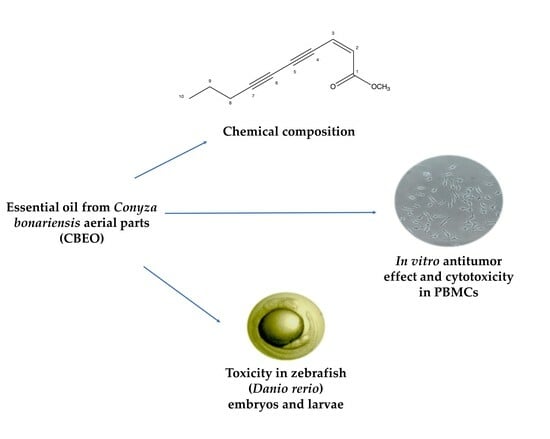
Chemical Composition, In Vitro Antitumor Effect, and Toxicity in Zebrafish of the Essential Oil from Conyza bonariensis (L.) Cronquist (Asteraceae)

by
Rafael Carlos Ferreira
1,
Yuri Mangueira do Nascimento
1,
Paulo Bruno de Araújo Loureiro
1,
Rafael Xavier Martins
2,
Maria Eduarda de Souza Maia
2,
Davi Felipe Farias
2,
Josean Fechine Tavares
1,
Juan Carlos Ramos Gonçalves
1,
Marcelo Sobral da Silva
1 and
Marianna Vieira Sobral
1,*
1
Postgraduate Program in Natural Products and Bioactive Synthetics, Federal University of Paraíba, João Pessoa 58051-970, PB, Brazil
2
Laboratory of Risk Assessment for Novel Technologies (LabRisk), Department of Molecular Biology, Federal University of Paraíba, João Pessoa 58051-970, PB, Brazil
*
Author to whom correspondence should be addressed.
Biomolecules 2023, 13(10), 1439; https://doi.org/10.3390/biom13101439
Submission received: 26 August 2023 / Revised: 13 September 2023 / Accepted: 17 September 2023 / Published: 24 September 2023

Abstract

:
The essential oil from Conyza bonariensis (Asteraceae) aerial parts (CBEO) was extracted by hydrodistillation in a Clevenger-type apparatus and was characterized by gas chromatography–mass spectrometry. The antitumor potential was evaluated against human tumor cell lines (melanoma, cervical, colorectal, and leukemias), as well as non-tumor keratinocyte lines using the MTT assay. The effect of CBEO on the production of Reactive Oxygen Species (ROS) was evaluated by DCFH-DA assay, and a protection assay using the antioxidant N-acetyl-L-cysteine (NAC) was also performed. Moreover, the CBEO toxicity in the zebrafish model was assessed. The majority of the CBEO compound was (Z)-2-lachnophyllum ester (57.24%). The CBEO exhibited selectivity towards SK-MEL-28 melanoma cells (half maximal inhibitory concentration, IC50 = 18.65 ± 1.16 µg/mL), and induced a significant increase in ROS production. In addition, the CBEO’s cytotoxicity against SK-MEL-28 cells was reduced after pretreatment with NAC. Furthermore, after 96 h of exposure, 1.5 µg/mL CBEO induced death of all zebrafish embryos. Non-lethal effects were observed after exposure to 0.50–1.25 µg/mL CBEO. Additionally, significant alterations in the activity of enzymes associated with oxidative stress in zebrafish larvae were observed. These results provide evidence that CBEO has a significant in vitro antimelanoma effect by increasing ROS production and moderate embryotoxicity in zebrafish.

Graphical Abstract

1. Introduction

Cancer is a relevant public health problem worldwide [1]. In 2020, approximately 19 million people were diagnosed with cancer and about 10 million deaths were recorded [2].
Currently, cancer treatment mainly includes surgery, radiotherapy, chemotherapy, and targeted therapy [2]. Nevertheless, limitations regarding therapeutic success have been observed, including patients relapsing or without an adequate response to therapy [3], in addition to significant adverse effects and chemoresistance [4]. Therefore, the search for new therapeutic opportunities for the treatment of this disease continues to grow globally [5].
Natural products or their derivatives represent more than 60% of the molecules used for cancer treatment [6]. In this context, essential oils (EOs) have been widely investigated for their pharmacological effects, including antimicrobial [7,8], anti-inflammatory [9], and antitumor effects [10,11,12,13,14,15]. EOs are aromatic oily liquids obtained from many parts of plants such as flowers, seeds, leaves, twigs, and roots. EO is composed of numerous volatile constituents such as sesquiterpenes, monoterpenes, aldehydes, alcohols, esters, and ketones [16]. The specific advantage of EOs appears to be in the synergistic effects of their components, as compared to the individual effects of these molecules [17].
Genus Conyza (Asteraceae) comprises about 50 species. The secondary metabolites in Conyza plants include alkaloids, terpenoids, steroids, phenolic compounds, flavonoids, and tannins. In most Conyza species EOs, the major chemical compounds found are limonene and β-farnesene [18]. Conyza bonariensis (L.) Cronquist is an invasive plant, native to South America [19]. Several studies show the biological potential of the essential oil from C. bonariensis aerial parts as anti-aging [20], antibacterial [21], and antitumor against HeLa (cervical carcinoma), MCF-7 (breast adenocarcinoma), A549 (lung adenocarcinoma), and HepG2 (hepatocellular carcinoma) human tumor cell lines. Additionally, few reports regarding its toxicity were found. Nevertheless, a significant difference in the chemical composition of these EOs was recorded [20,21].
Literature data show differences in the chemical composition of EOs even within the same species [22]. Studies on EOs from C. bonariensis aerial parts carried out in various countries have revealed different major components, such as (E)-β-farnesene [23,24], germacrene D [25], caryophyllene oxide [26], and methyl ester of matricaria [27,28].
Here, we present the chemical characterization of the EO from C. bonariensis aerial parts (CBEO) collected in João Pessoa, Paraíba, Brazil. Additionally, we evaluate its antitumor effect in human cell lines (SK-MEL-28, HeLa, HCT-116, HL-60, and K562), and its toxicity on human peripheral blood mononuclear cells (PBMCs) and zebrafish.

2. Materials and Methods

2.1. Drugs and Reagents

Dulbecco’s Modified Eagle’s Medium (DMEM) (Sigma-Aldrich®; St. Louis, MO, USA), Roswell Park Memorial Institute 1640 (RPMI) medium (Sigma-Aldrich®; St. Louis, MO, USA), Histopaque®-1077 (Sigma-Aldrich®; St. Louis, MO, USA), Buffered phosphate solution (PBS) (Sigma-Aldrich®; St. Louis, MO, USA), doxorubicin (DXR) (Sigma-Aldrich®; St. Louis, MO, USA), penicillin–streptomycin (Sigma-Aldrich®; St. Louis, MO, USA), 2,7-dichlorodihydrofluorescein diacetate (DCFH-DA) (Sigma-Aldrich®; St. Louis, MO, USA), N-acetylcysteine (NAC) (Sigma-Aldrich®; St. Louis, MO, USA), hydrogen peroxide (H2O2) (Sigma-Aldrich®; St. Louis, MO, USA), 3-(4,5-dimethylthiazol-2-yl)-2,5-diphenyltetrazolium bromide (MTT) (Sigma-Aldrich®; St. Louis, MO, USA), sodium chloride (NaCl) (Sigma-Aldrich®; St. Louis, MO, USA), potassium chloride (KCl) (Sigma-Aldrich®; St. Louis, MO, USA), calcium chloride (CaCl2) (Sigma-Aldrich®; St. Louis, MO, USA), magnesium sulfate (MgSO4) (Sigma-Aldrich®; St. Louis, MO, USA), sodium sulfate (Na2SO4) Sigma-Aldrich®; St. Louis, MO, USA), methylene blue Sigma-Aldrich®; St. Louis, MO, USA), dimethylsulfoxide (DMSO) (Dinâmica®, Indaiatuba, SP, Brazil), Sodium Dodecyl Sulfate (SDS) (Êxodo Científica®, Sumaré, SP, Brazil), phytohaemagglutinin (GIBCO®, Grand Island, NY, USA), trypsin 0.25% with ethylenediaminetetraacetic acid (EDTA) (GIBCO®, Grand Island, NY, USA), Fetal Bovine Serum (FBS) (GIBCO®, Grand Island, NY, USA), fish food (Tetra®, Melle, Germany), hexane (Biograde®, Anápolis, GO, Brazil).
The drugs and reagent solutions were prepared immediately before use.

2.2. Human Tumor Cell Lines

The SK-MEL-28 (human melanoma), HeLa (human cervical cancer), HCT-116 (human colon carcinoma), HL-60 (human promyelocytic leukemia), K562 (chronic myeloid leukemia), and HaCat (human immortalized keratinocytes) cell lines were obtained from Rio de Janeiro Cell Bank (BCRJ), Brazil, and cultured in Dulbecco’s Modified Eagle’s Medium (DMEM) (SK-MEL-28, HeLa and HaCaT) or Roswell Park Memorial Institute 1640 (RPMI) (HCT-116, HL-60 and K562) medium supplemented with 10% Fetal Bovine Serum and 1% penicillin–streptomycin at 37 °C with 5% CO2.

2.3. Human Peripheral Blood Mononuclear Cells (PBMC)

Blood samples were collected from healthy donors in tubes containing EDTA. Human peripheral blood mononuclear cells (PBMCs) were isolated after centrifugation (400× g, 20 °C, for 30 min) of the blood samples in tubes containing Histopaque®-1077. Subsequently, the interface containing the mononuclear cells was collected and washed with PBS (400× g, 10 min, 20 °C). Twenty-four hours before CBEO or DXR treatment, PBMCs were resuspended in RPMI-1640 medium supplemented with 10% FBS, 1% penicillin-streptomycin, and 2% phytohemagglutinin, and cultured in 96-well plates (1 × 10⁶ cells/mL) at 37 °C with 5% CO2 [29].

2.4. Zebrafish Embryos

Zebrafish embryos (AB wild-type strain) with approximately 1 h post-fertilization (hpf) were provided by the Production Unit for Alternative Model Organisms (UniPOM), Federal University of Paraíba (UFPB), João Pessoa, Brazil. The parents were maintained in a recirculation system with regular monitoring of water quality parameters (pH, ammonia, and nitrite levels). The room temperature (26 ± 1 °C) and photoperiod (14:10 light/dark cycle) were controlled. Fish were fed daily with commercial food and freeze-dried spirulina (Fazenda Tamanduá, Patos, PB, Brazil), and were also monitored for abnormal behavior or disease development.
The day before the experiment, zebrafish adults (male-to-female ratio of 2:1) were transferred to a 7 L spawning tank with a bottom mesh and a quick-opening valve for embryo collection. Embryos were collected on the day of the experiment and cultured in adapted embryonic medium E3 (5.0 mM NaCl, 0.17 mM KCl, 0.33 mM CaCl2, and 0.33 mM MgSO4) containing 0.005% methylene blue. Only spawning with a fertilization rate of ≥90% was used. Viable embryos (showing a normal cleavage pattern and without morphological changes) were selected under an inverted light microscope (Televal 31, Zeiss®, Oberkochen, Germany) at 50× magnification.

2.5. Botanical Material and Essential Oil Extraction

Conyza bonariensis (L.) Cronquist branches and leaves (1 kg) were collected from the Medicinal Plant Garden, Institute of Research in Drugs and Medicines of Federal University of Paraíba (UFPB), João Pessoa, Paraíba, Brazil (7°08′30.0″ S–34°50′46.7″ W) in September of 2019. An exsiccate of C. bonariensis was identified by Prof. Dr. Maria de Fátima Agra and deposited at Herbarium Lauro Pires Xavier-JPB of UFPB, under the number JPB 26391 (registry number SISGEN ABB39C8).
Essential oil extraction was performed by hydrodistillation in a Clevenger-type apparatus. The samples were crushed and subjected to distillation for 2 h [30]. After extraction, the essential oil was dried with anhydrous sodium sulfate (Na2SO4), with a yielding (w/w) of 1.3%.

2.6. Essential Oil Analysis

Gas chromatography–mass spectrometry (GC-MS) analysis was performed using a Shimadzu QP-2010 Ultra Quadrupole MS system, operating at 70 eV ionization energy. A capillary column RTX-5MS (30 m × 0.25 mm i.d., 0.25 μM film thickness) was used with Helium as a carrier gas at a flow rate of 3 mL/min with a 1:100 split. The injector and detector temperatures were set at 220 °C and 280 °C, respectively. The column temperature was programmed from 40 °C (isothermal for 1 min) to 220 °C at a rate of 10 °C/min (remaining isothermal for 2 min at 220 °C). Subsequently, the temperature was increased from 220 °C to 280 °C at a rate of 20 °C/min and held isothermally for 5 min at 280 °C. The ions were scanned in scan mode, ranging from m/z 50 to 500. The sample solution was prepared in hexane at a dilution of 999:1 (v/v) and 1 μL was injected into the chromatograph at a flow rate (split) of 1:200.
To calculate the retention indices of compounds, under the same operating conditions, a series of hydrocarbons (C10 to C40) (Sigma-Aldrich®) was injected. The retention index for each compound was determined based on a similarity index above 89% estimated by the libraries (Nist. 08 and Wiley 9) used for compound identification. The retention index was calculated using the chromatogram obtained through the Van Den Dool and Kratz equation [31].

2.7. Cytotoxicity Assessment of CBEO in Human Cells

The MTT (3-(4,5-dimethylthiazol-2-yl)-2,5-diphenyltetrazolium bromide) assay was performed to evaluate the CBEO cytotoxicity. MTT is a yellow tetrazolium salt that can permeate cell membranes. In viable cells, MTT is converted to insoluble purple formazan crystals, which can be measured spectrophotometrically. The optical density value is proportional to the number of viable cells [32]. The human cell lines SK-MEL-28, HeLa, HCT-116, HL-60, K562, and HaCaT were cultured in DMEM or RPMI medium supplemented with 10% FBS, 100 U/mL penicillin, and 100 μg/mL streptomycin at 37 °C in a humidified atmosphere with 5% CO2. The cell suspension was added to 96-well plates (100 µL/well) at a density of 3 × 10⁵ cells/mL (for SK-MEL-28, HeLa, HCT-116, and HaCaT), 5 × 10⁵ cells/mL (for HL-60 and K562), or 1 × 10⁶ cells/mL (PBMC). After culturing for 24 h, cells were incubated with 100 µL of CBEO (HaCaT and human tumor cell lines: 2.34–300 µg/mL; PBMC: 0.15–20 µg/mL) dissolved in DMSO. Doxorubicin (DXR) (molecular weight—MW: 543.52 g/mol) was used as a standard drug. After 72 h, 110 µL of the supernatant was discarded and 10 µL of the 3-(4,5-dimethylthiazol-2-yl)-2,5-diphenyltetrazolium bromide (MTT) solution (5 mg/mL) was added and incubated for another 4 h. The deposited formazan was dissolved with Sodium Dodecyl Sulfate (SDS) (100 mL/well) [33] and the optical densities were measured using a microplate reader (Synergy HT, BioTek®, Winooski, VT, USA) at λ = 570 nm and used to calculate the IC50 (half-maximal inhibitory concentration). Three independent experiments were performed in triplicate.
The Selectivity Index (SI) was determined from the ratio between the IC50 of the non-tumor cell line (HaCaT) and the IC50 of the tumor cell line.

2.8. Quantification of Reactive Oxygen Species in Human Tumor Cells

Reactive Oxygen Species (ROS) were quantified by the 2,7-dichlorodihydrofluorescein diacetate (DCFH-DA) reagent oxidation method [34]. DCFH-DA is a non-polar and non-fluorescent probe, that can freely cross cell membranes. Intracellular esterases cleave DCFH-DA to DCFH, which is oxidized by ROS to DCF, a highly fluorescent molecule. Therefore, the number of fluorescent cells is proportional to the amount of intracellular ROS [35]. For this experiment, SK-MEL-28 cells were seeded in 24-well plates at a concentration of 2 × 10⁵ cells/mL. After 24 h, cells were exposed to CBEO (20 or 40 µg/mL), DXR (4 µM), or hydrogen peroxide (H2O2) (500 µM) in the presence of DCFH-DA (10 µM), and incubated for 30 min, 1 h, or 3 h. After the incubation periods, the cells were trypsinized, washed, and resuspended in PBS. The percentage of fluorescent cells was obtained by flow cytometry from 10,000 events acquired at 530 nm fluorescence and 485 nm excitation wavelengths. Three independent experiments were performed in duplicate.

2.9. Evaluation of CBEO Cytotoxicity in the Presence or Absence of N-acetylcysteine (NAC)

To assess the involvement of ROS in CBEO cytotoxicity, SK-MEL-28 cells were added to 96-well plates (100 µL/well) at a density of 3 × 10⁵ cells/mL and incubated for 24 h (37 °C, CO2 5%). After this incubation period, cells were incubated for another 3 h (37 °C, CO2 5%) in the presence or absence of 5 µM of N-acetylcysteine (NAC). Subsequently, cells were treated with CBEO (20 or 40 µg/mL, 100 µL per well) or DXR (4 µM, 100 µL per well) and incubated for 72 h in an atmosphere of 5% CO2 and 37 °C. Then, the plates were centrifuged, and 110 µL of the supernatant was removed. Then, 10 µL of the MTT solution (5 mg/mL) was added, followed by incubation for 4 h at 37 °C, CO2 5%. The formazan was dissolved with 100 μL of SDS [32] and the optical densities were measured using a microplate reader (Synergy HT, BioTek®) at λ = 570 nm. Three independent experiments were performed in triplicate.

2.10. CBEO Toxicity in Zebrafish Model

2.10.1. Acute Toxicity Test Using Zebrafish Embryos

CBEO acute toxicity was determined by Fish Embryo Acute Toxicity (FET) assay. The FET test was conducted independently with CBEO according to OECD’s guideline number 236 [36] with slight modifications.
Zebrafish embryos with up to 3 hpf of age were exposed to five increasing concentrations (0.5, 0.75, 1.0, 1.25, and 1.5 mg/L) of CBEO. For each concentration tested, a 96-well plate was prepared, containing 20 fertilized eggs (1 embryo per well) exposed to the test sample, and 4 embryos were exposed only to E3 medium (internal controls). Two additional plates containing embryos exposed to E3 medium (negative control) and 0.1% DMSO (solvent control) were also assayed. The volume of liquid in each well at the beginning of the exposure was 0.3 mL. The plates were protected from evaporation by using their own lid and incubating them in a humidified chamber with controlled temperature (26 ± 1 °C) and humidity (70%).
The exposure was performed for 96 h, and the embryos were analyzed daily for lethality endpoints: egg coagulation; lack of somite formation; lack of detachment of the tail-bud from the yolk sac; and lack of heartbeat. In the presence of these endpoints, the embryo/larva was considered dead.
The number of deaths was used to calculate the survival rate (survival % = number of alive/total organisms × 100). Additionally, non-lethal effects (eye malformation, otolith malformation, mouth malformation, spine malformation, body pigmentation, hatching delay, yolk sac edema, yolk sac deformation, pericardial edema, head edema, blood clotting, and undersize) were also recorded every 24 h. The exposures were under static conditions (without renovation of the test sample or negative and solvent controls). Observations were using a stereomicroscope (50× magnification) and documented with photographs. After 96 h, surviving larvae were euthanized with eugenol and appropriately disposed of.
The number of deaths and prevalent non-lethal effects (presence in at least three concentrations) was used to calculate the LC50 (median lethal concentration) and EC50 (median effective concentration) through probit analysis [37]. These values were also used to determine the NOAEL (No Observed Adverse Effect Level) and LOAEL (Lowest Observable Adverse Effect Level).

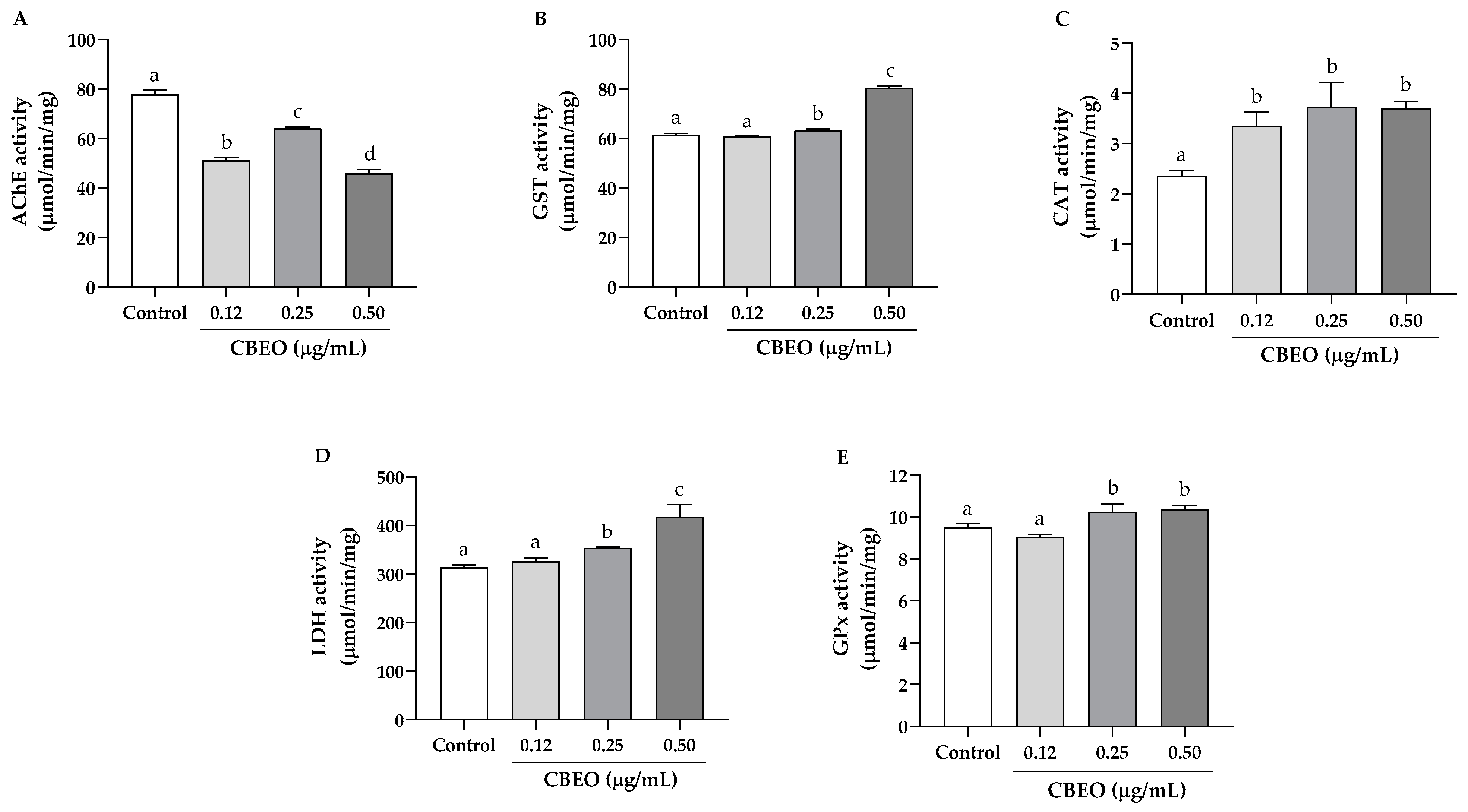
2.10.2. Oxidative Stress Biomarker Enzymes in Zebrafish Larvae

The FET test was repeated for CBEO under the same conditions described in item 2.8.1., but at this time the embryos were independently exposed to three sublethal concentrations of CBEO (0.12, 0.25, and 0.50 µg/mL) [38]. After 96 h of exposure, the larvae were quickly frozen in 0.1 M sodium phosphate buffer, pH 7.4.
Subsequently, the larvae were macerated using cold NaCl 0.9% 1:9 (w/v) solution. Homogenates were centrifuged at 10,000× g for 10 min at 4 °C, and the resulting supernatants were used for measurement of soluble protein content and enzymatic activity. The activities of lactate dehydrogenase (LDH), glutathione transferase (GST), acetylcholinesterase (AChE), glutathione peroxidase (GPx), and catalase (CAT) enzymes were measured according to Domingues et al. (2010) [39]. Tests were performed in quadruplicate for each enzyme.

2.11. Statistical Analysis

Statistical analysis was performed using GraphPad Prism 8.0.2 (Graphpad Software Inc., San Diego, CA, USA). Results are expressed as the mean ± standard error of the mean (SEM). Data statistical analysis was performed using Analysis of Variance (ANOVA), followed by Tukey’s test (p < 0.05). The half-maximal inhibitory concentrations (IC50) and their 95% confidence intervals (CI 95%) were obtained by non-linear regression analysis. For embryotoxicity assay, the median lethal concentration (LC50) values and the median effect concentration (EC50) values were calculated by probit regression analysis.

3. Results

3.1. (Z)-2-lachnophyllum Ester Was the Major Compound in the Chemical Characterization of CBEO

The analysis of the chemical profile of CBEO led to the identification of 96.95% of its components. The major compound was (Z)-2-lachnophyllum ester (57.24%) (MW: 176.21 g/mol), and the remaining components were monoterpenes and sesquiterpenes (39.71%), as shown in Table 1.

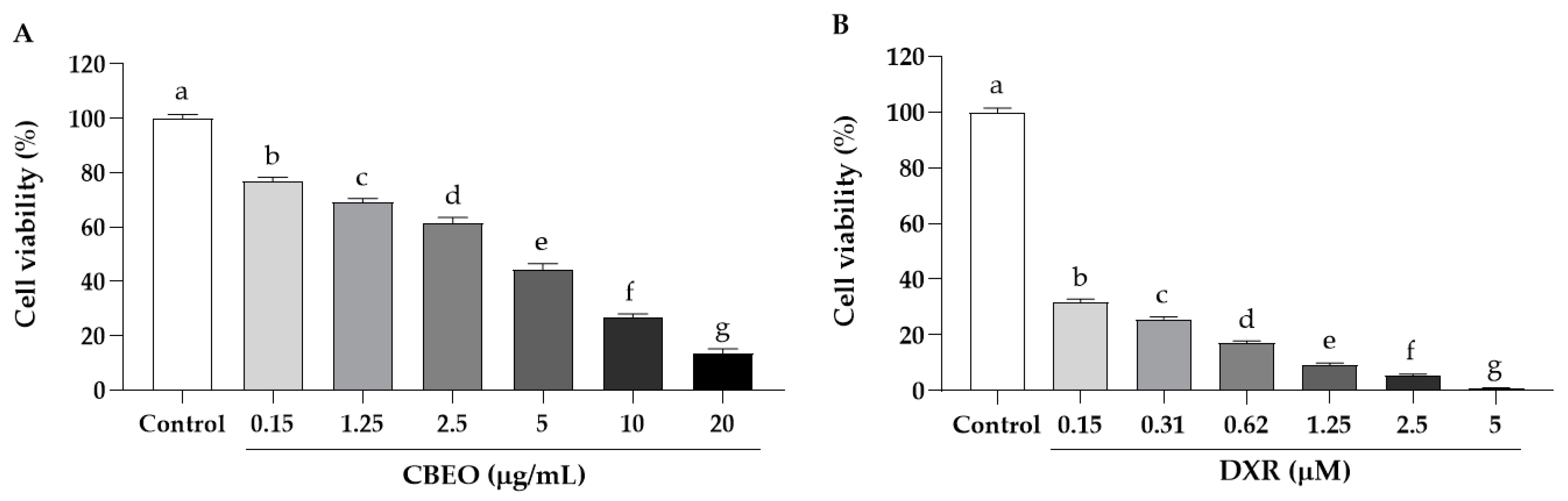
3.2. CBEO Induces Cytotoxicity in Human Cell Lines

CBEO induced the least cytotoxicity on the acute promyelocytic leukemia cell line (HL-60), IC50 of 32.20 ± 1.10 µg/mL, while the human melanoma cell line (SK-MEL-28) was the most sensitive to the treatment, IC50 of 18.65 ± 1.16 µg/mL. Regarding the non-tumor human keratinocyte cell line (HaCaT), CBEO presented IC50 of 56.49 ± 1.03, and the standard drug, doxorubicin (DXR), showed high cytotoxicity (IC50: 0.28 ± 0.001 µM) after 72 h of treatment. Then, the Selectivity Indices (SI) of CBEO and DXR were determined using the HaCaT healthy skin cell line as a non-tumor cell model. The CBEO showed the highest SI for SK-MEL-28 cells (3.03), as shown in Table 2.

3.3. CBEO Induces Less Cytotoxicity in PBMC Cells Than Doxorubicin

CBEO cytotoxicity was also evaluated in human peripheral blood mononuclear cells (PBMC). CBEO induced a concentration-dependent cytotoxicity. The IC50 values were 2.68 ± 1.29 (Figure 1A) and 0.06 ± 1.19 µM (Figure 1B) for CBEO and DXR, respectively, after 72 h of treatment.

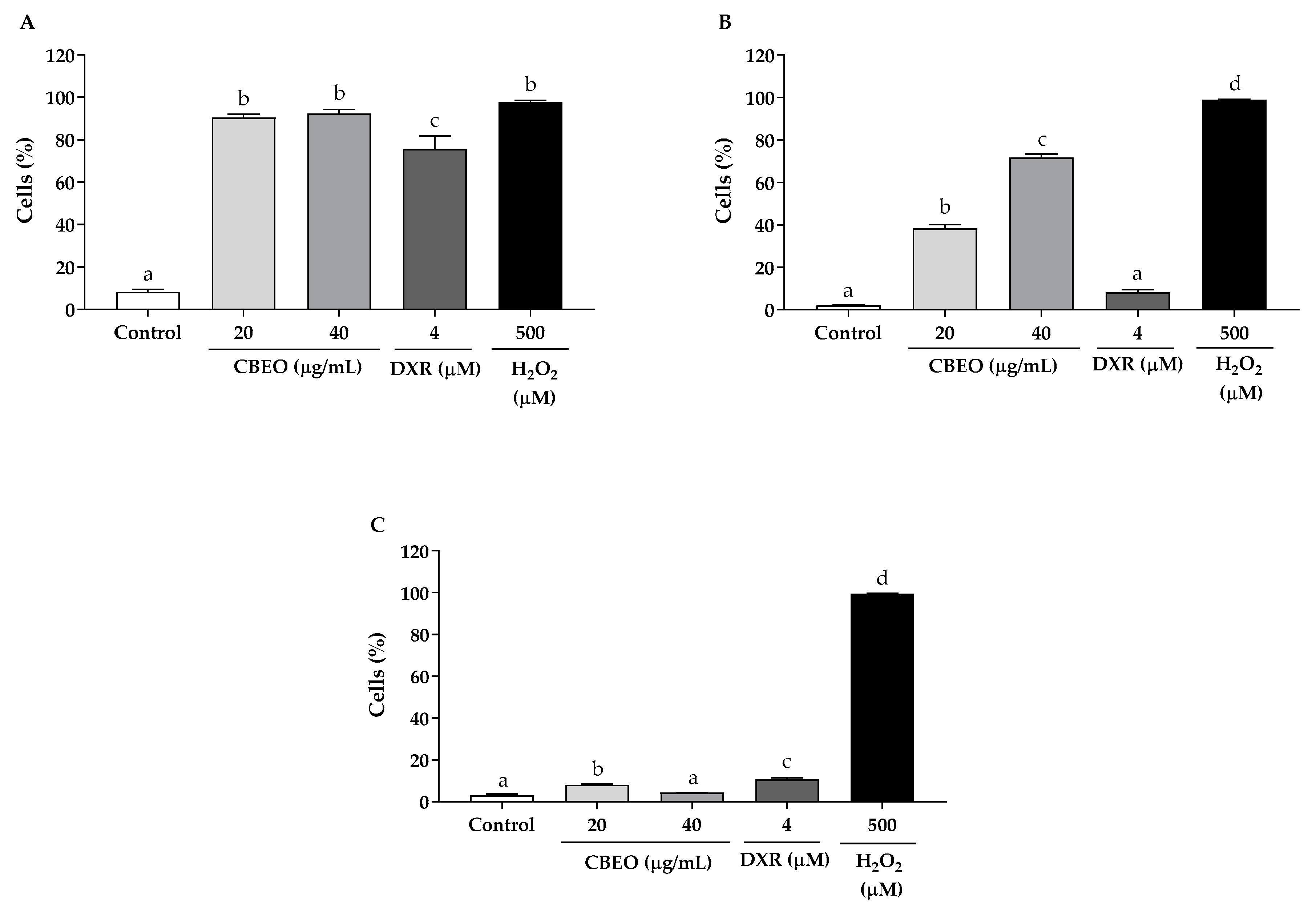
3.4. CBEO Induces Oxidative Stress in SK-MEL-28 Cells

In the dichlorodihydrofluorescein 2′7-diacetate (DCFH-DA) assay, CBEO treatment induced a significant increase in the percentage of fluorescent cells after 30 min (20 µg/mL: 90.32 ± 1.64%; 40 µg/mL: 92.33 ± 1.96%, p < 0.05 for both), 1 h (20 µg/mL: 38.36 ± 1.82%; 40 µg/mL: 71.60 ± 1.78%, p < 0.05 for both), and 3 h (20 µg/mL: 8.05 ± 0.36%, p < 0.05) of treatment, compared to the control (30 min: 8.22 ± 1.26%; 1 h: 2.18 ± 0.16%; and 3 h: 3.15 ± 0.63%). DXR, which was used as a standard drug, induced a significant increase in the percentage of fluorescent cells after 30 min (75.64 ± 6.02% p < 0.05) and 3 h (10.64 ± 0.92%, p < 0.05) of treatment, compared to the control. As expected, in the group exposed to hydrogen peroxide (H2O2), there was a significant increase in the percentage of fluorescent cells, compared to the control (30 min: 97.65 ± 0.86%; 1 h: 98.87 ± 0.17%; and 3 h: 99.45 ± 0.23%, p < 0.05 for all) (Figure 2).

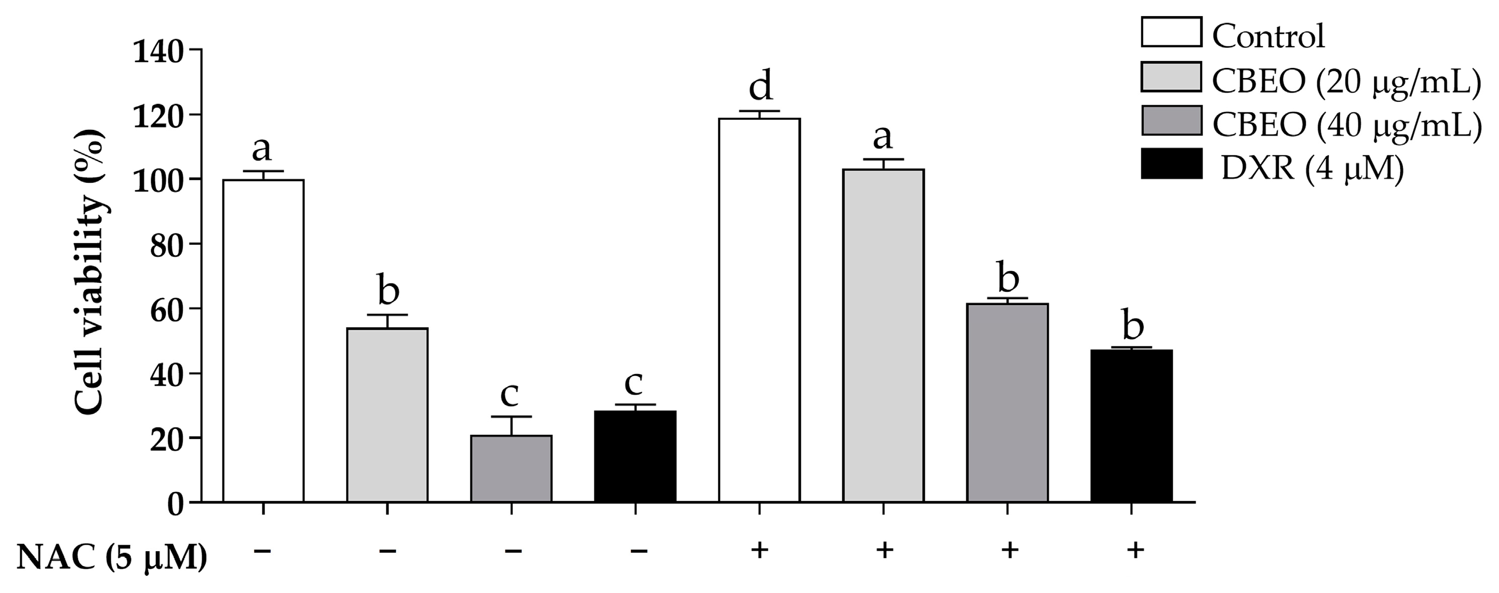
3.5. CBEO Cytotoxicity in SK-MEL-28 Cells Is ROS-Dependent

After 72 h of CBEO treatment, in the absence of N-acetylcysteine (NAC), a significant reduction in cell viability was observed (20 µg/mL: 54.06 ± 3.96%; 40 µg/mL: 20.97 ± 5.60%, p < 0.05 for both) compared to the control (100.00 ± 2.47%). NAC pretreatment significantly reduced the CBEO cytotoxic effect, compared to the groups treated only with CBEO at the respective tested concentrations (20 µg/mL in the presence of NAC: 103.3 ± 2.82%; 40 µg/mL in the presence of NAC: 61.63 ± 1.52%, p < 0.05 for both) (Figure 3).
As expected, NAC pretreatment significantly reduced the DXR cytotoxicity (28.45 ± 1.96%, p < 0.05) compared to the group treated with DXR in the absence of NAC (47.23 ± 0.81%) (Figure 3).

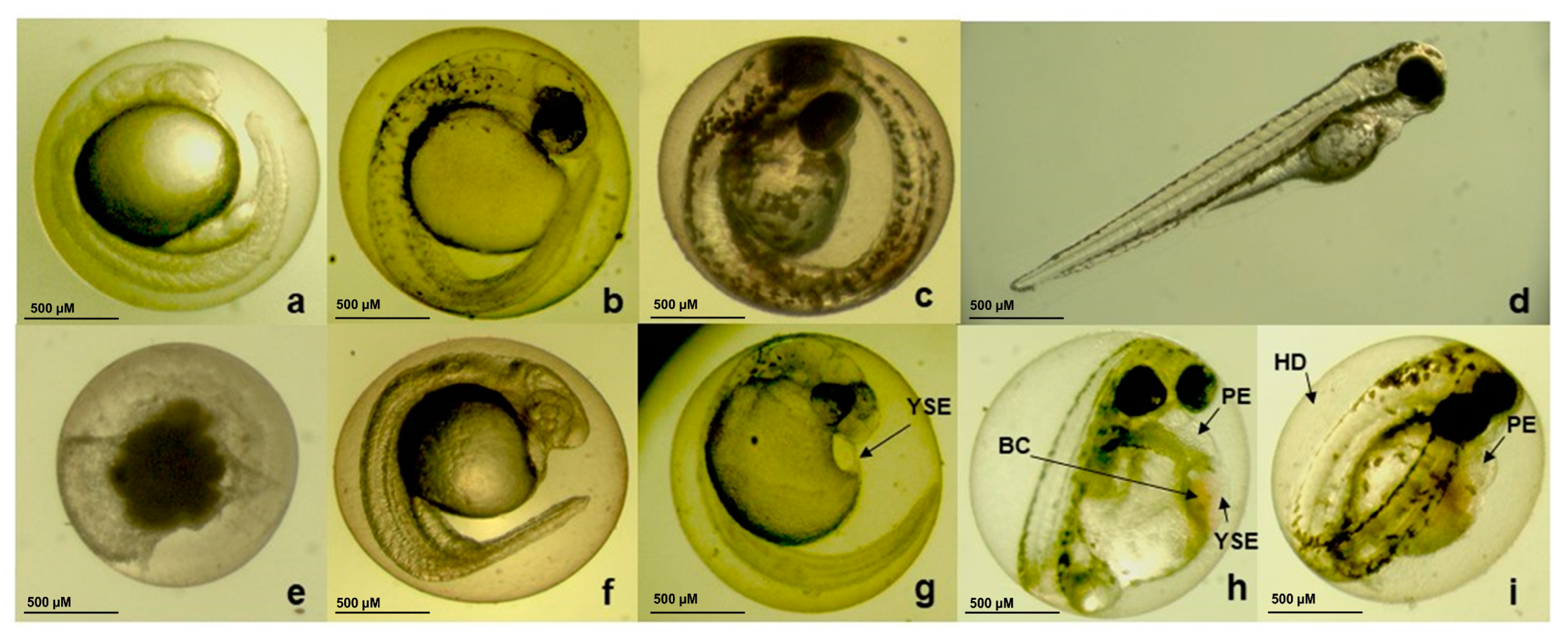
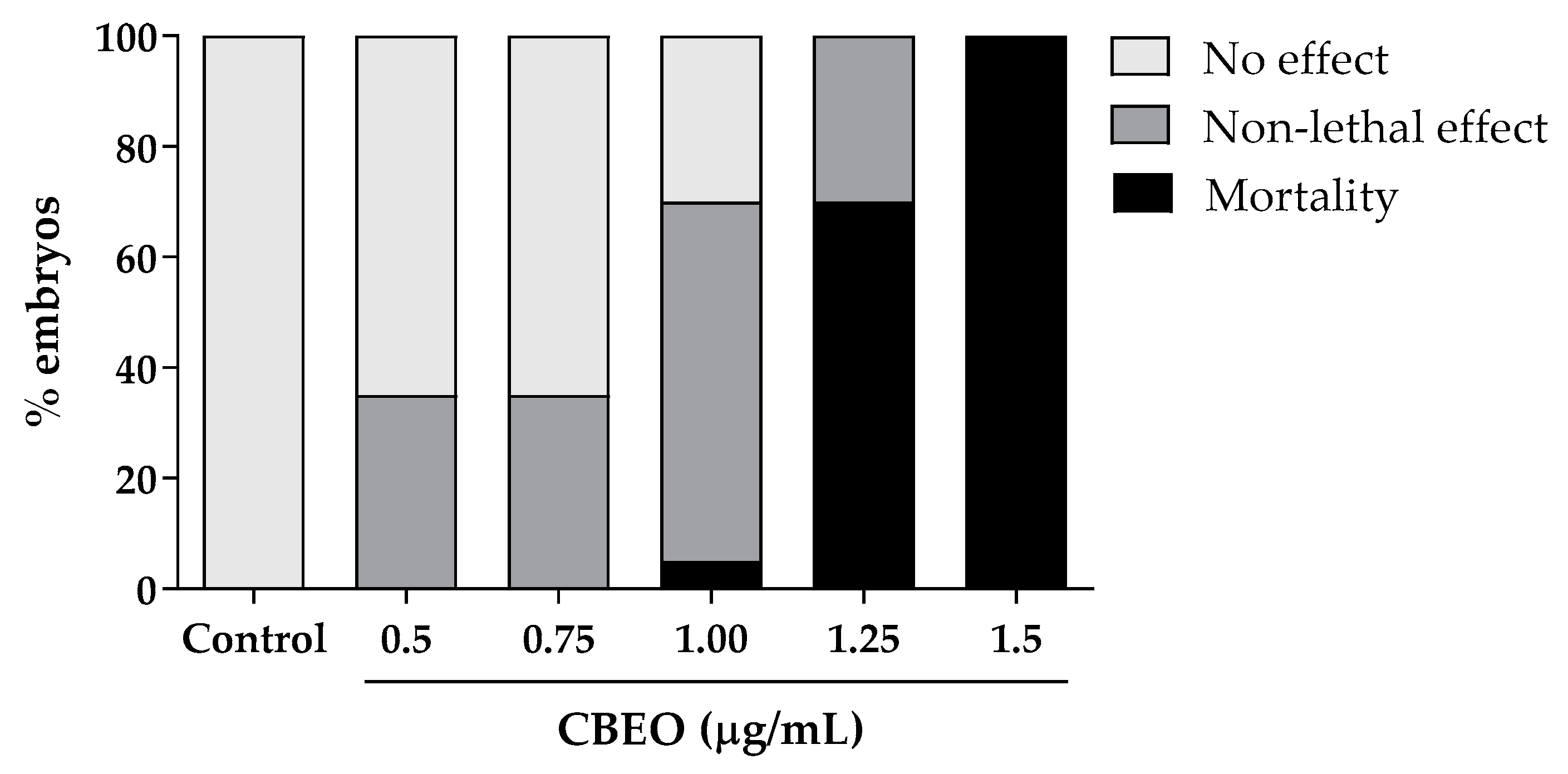
3.6. Embryotoxicity Induced by CBEO in Zebrafish Model

The embryotoxicity assay was performed using CBEO concentrations ranging from 0.5 to 1.5 µg/mL. After 96 h of exposure, all embryos died at the highest concentration tested. However, the two lowest concentrations (0.5 and 0.75 µg/mL) did not induce any mortality (Figure 4).
The non-lethal effects observed included pericardial edema, yolk sac edema, delayed egg hatching, and egg and blood clotting. Additionally, coagulation and absence of heartbeats were the only lethality outcomes observed in embryos and larvae after CBEO treatments (Figure 5).
As observed in Table 3, CBEO presented a median lethal concentration (LC50) of 1.20 µg/mL. The Lowest Observed Adverse Effect Level (LOAEL) values were 0.5 µg/mL for delayed hatching and 1.0 µg/mL for yolk sac edema, pericardial edema, and blood clotting. The No Observed Adverse Effect Level (NOAEL) value was 0.75 µg/mL for yolk sac edema, pericardial edema, and blood coagulation. The median effective concentration (EC50) for the non-lethal effects found were 0.99 (delayed hatching) and 1.36 (pericardial edema).
Subsequently, the activity of enzymes related to oxidative stress in zebrafish larvae exposed to low concentrations of CBEO (0.12 to 0.50 µg/mL) was studied. The rationale for choosing these concentrations lies in the fact that when conducting tests with sublethal concentrations, we typically observe the endogenous mechanisms of detoxification and neutralization of Reactive Oxygen Species (ROS) in action, rather than observing the final adverse effects such as cell death and necrosis, which are less informative from a mechanistic standpoint.
As shown in Figure 6, there was a significant reduction in acetylcholinesterase (AChE) activity in zebrafish larvae after treatment with CBEO at all concentrations (0.12 µg/mL: 51.27 ± 0.70 µmol/min/mg; 0.25 µg/mL: 64.12 ± 0.27 µmol/min/mg; and 0.50 µg/mL: 46.01 ± 0.87 µmol/min/mg; p < 0.05 for all), compared to the control (77.87 ± 1.12 µmol/min/mg). Furthermore, a significant increase in glutathione transferase (GST) activity was observed after treatment with CBEO (0.25 µg/mL: 63.30 ± 0.36 µmol/min/mg; and 0.50 µg/mL: 80.43 ± 0.50 µmol/min/mg; p < 0.05 for both) compared to the control (61.56 ± 0.34 µmol/min/mg). For catalase (CAT) activity, we observed a significant increase in the activity of this enzyme in larvae treated with CBEO (0.12 µg/mL: 3.36 ± 0.16 µmol/min/mg; 0.25 µg/mL: 3.73 ± 0.28 µmol/min/mg; and 0.50 µg/mL: 3.71 ± 0.08 µmol/min/mg, p < 0.05 for all) compared to the control (2.36 ± 0.06 µmol/min/mg). In addition, treatment of larvae with 0.25 and 0.50 µg/mL of CBEO induced a significant increase in lactate dehydrogenase (LDH) activity (353.6 ± 0.96 µmol/min/mg and 417.4 ± 14.57 µmol/min/mg, respectively; p < 0.05 for both) compared to the control (314.00 ± 2.65 µmol/min/mg). For glutathione peroxidase (GPx) activity, we observed a significant increase in the activity of this enzyme in larvae treated with 0.5 µg/mL CBEO (10.70 ± 0.37 µmol/min/mg, p < 0.05) compared to the control (9.52 ± 0.10 µmol/min/mg).

4. Discussion

Nature represents a significant source of bioactive products. Thus, research around the world has sought to discover and investigate the biological effects of natural products against diseases such as cancer [40]. The present study focuses on elucidating the chemical composition, in vitro antitumor activity, and the embryotoxicity in zebrafish model of the essential oil extracted from Conyza bonariensis (L.) aerial parts (CBEO).
The biological properties of essential oils (EOs) are due to their chemical composition [22]. Geographic origin [41] and environmental conditions such as temperature, precipitation, relative humidity, day length, and light intensity [42] influence the biosynthesis and accumulation of natural products [43]. Consequently, variations in the chemical profiles of EOs can occur from plant to plant, even within the same species [22].
Chemical analysis of the CBEO revealed the (Z)-2-lachnophyllum ester, an acetylenic compound, as the major compound (57.24%). However, it is worth noting that the chemical profile of the EO from Conyza bonariensis (L.) aerial parts can vary depending on the country and regions where the plant grows. Until now, (E)-β-farnesene [23], germacrene D [25], caryophyllene oxide [26], limonene [44], 1H-indene-3-carboxaldehyde,2,6,7,7a-tetrahydro-1,5-dimethyl [45], and allo-aromadendrene [46] have been identified as the major components of the essential oil from Conyza bonariensis (L.) aerial parts. Lundgren et al. (2021) [47] showed the characterization of the EO of C. bonariensis (L.) cultivated in the Medicinal Plant Garden of the Institute of Research in Drugs and Medicines of the Federal University of Paraíba (UFPB), João Pessoa, Paraíba, Brazil. These authors obtained a different profile chemical from that obtained in our work, with sesquicineole as the major compound. This shows the influence of conditions such as time of harvest (seasonality), mechanical or chemical injuries, genetic factors and evolution, storage, irrigation, herbivory, attack of fungal pathogens, and activity of the pollinators in the production and composition of EOs, as already described in the literature [48,49]. In addition, we have highlighted the presence of other acetylenic compounds in C. bonariensis such as methyl ester of matricaria. Specifically, acetylenic compounds constitute a group of molecules common in species of the Asteraceae family and the Conyza genus [50,51,52]. Barbosa et al. (2004) [27] obtained different major constituents in the essential oils from distinct parts of this species, such as the methyl ester of matricaria in the roots (74.4%). Furthermore, Mabrouk et al. (2011) [28] observed distinct chemical profiles of the essential oils from C. bonariensis cultivated in Tunisia in different seasons. However, the methyl ester of matricaria remained the predominant compound in all of them (63.5–76.4%).
Finally, the (Z)-2-lachnophyllum ester has also been described as the major component (21.2%) of Conyza bonariensis (L.) cultivated at the University of Athens, Athens, Greece [53]. Thus, our study presents an EO from Conyza bonariensis (L.) aerial parts with unprecedented chemical profile.
Currently, cancer represents a major public health problem worldwide [54], and many efforts are being made in the search for new therapies. The constituents of CBEO include α-pinene, β-pinene, p-cymene, limonene, terpinen-4-ol, caryophyllene oxide, β-sesquiphelandrene, α-humulene, carvacrol, and thymol [55,56,57,58,59,60,61,62,63,64,65,66,67,68,69]. The major compound of CBEO, (Z)-2-lachnophyllum ester, exhibited a significant in vitro antitumor effect on the tumor cell lines MDA-MB-231 (human breast carcinoma), MCF-7 (human breast carcinoma), and 5637 (human bladder carcinoma), with half-maximal inhibitory concentration (IC50) values ranging from 7.2 to 53.1 µg/mL [70]. Additionally, (Z)-2-lachnophyllum ester also possesses antifungal, antioxidant [71], insecticide [72], and nematicidal [73] properties. Considering the advantage of the synergistic effect of EO compounds compared to the biological effects of these constituents individually [17], the in vitro antitumor activity of CBEO was investigated against different human malignant cell lines. Doxorubicin (DXR), a drug widely used in antineoplastic chemotherapy [74] and in several in vitro studies, including the investigation of EO anticancer effects [75,76,77,78,79], was used as a standard drug. Our data show that CBEO induced the greatest cytotoxicity against SK-MEL-28 human melanoma cells. Still, considering the Selectivity Index (SI), we can suggest that this cytotoxic effect was more selective for SK-MEL-28 cells when compared to the effect observed in healthy human skin cells, HaCaT. Cutaneous melanoma is the most relevant malignant tumor among skin cancers, as it is responsible for the majority of deaths [80]. In 2020, approximately 325,000 patients were diagnosed with skin melanoma and approximately 57,000 died from this disease [81]. Thus, novel therapeutic alternatives are needed, and several EOs have been investigated for the treatment of this disease [82] showing effects such as induction of apoptosis by upregulation of Bax and downregulation of Bcl-2 genes [83], cell cycle arrest, and increase in Reactive Oxygen Species (ROS) production [15].
We also analyzed the effect of CBEO against human peripheral blood mononuclear cells (PBMCs). PBMCs, including monocytes and lymphocytes, are frequently isolated for use in preclinical research [84] as well as in studies of new candidates in anticancer therapy [85,86]. The assessment of cytotoxicity in PBMCs is a relevant indicator of human systemic toxicity of natural products [87]. EO effects have been investigated in PBMCs [15,88,89], demonstrating that this experimental model is useful in the investigation of toxicity in human healthy cells. Here, we show that CBEO induced cytotoxicity against PBMCs in a concentration-dependent manner. However, this toxic effect was lower than that observed for the standard drug DXR. Conventional chemotherapy is not selective [90], and thus, damage can also occur in healthy cells, such as blood cells [91,92,93,94]. EOs composed mostly of monoterpenes and sesquiterpenes show cytotoxicity against PBMCs, such as EO from Duguetia pycnastera leaves (IC50: 21.28 µg/mL) [95], EO from Satureja khuzistanica aerial parts (IC50: 28.21 µg/mL) [96], and EO from Xylopia laevigata leaves (IC50: 35.30 µg/mL) [97]. Thus, the greater cytotoxic effect of CBEO on PBMC (IC50: 2.68 µg/mL) may be related to the major compound, (Z)-2-lachnophyllum ester, which has no report in the literature.
We investigated the effect of CBEO on the redox state of SK-MEL-28 cells. In the 2,7-dichlorodihydrofluorescein diacetate (DCFH-DA) assay, elevated levels of ROS were obtained after CBEO treatment. In addition, pretreatment with N-acetylcysteine (NAC), an antioxidant molecule [98], significantly reduced the CBEO cytotoxicity, corroborating the involvement of ROS in the antitumor effect induced by this EO. It has been reported that increased ROS production is a common event in EO-induced tumor cell death [99]. In fact, oxidative stress induction has been observed in tumor cells after EO treatment [99,100,101]. Tumor cells are known to have high levels of ROS [102] involved in processes such as the induction of cell proliferation and metastasis, inhibition of apoptosis [103], and angiogenesis stimulation [104]. However, excessive intracellular ROS concentration leads to irreparable damage and death of tumor cells [105]. Antineoplastic agents such as doxorubicin, methotrexate, cisplatin, and topotecan induce apoptosis of tumor cells by inducing oxidative stress [106,107,108,109]. Therefore, the increase in ROS production induced by CBEO treatment is a mechanism involved in the antitumor effect of this EO in SK-MEL-28 cells.
The assessment of drug toxicity is a critical process in the development of new drug candidates [110]. In this context, the zebrafish (Danio rerio) genome shares 70% similarity with the human genome [111,112]. The zebrafish embryo test is a highly sensitive toxicity test [113], making this experimental model relevant in human health risk assessments [114,115]. Our study provides an assessment of CBEO toxicity in zebrafish embryos and larvae. No mortality of embryos or larvae was observed for the lowest CBEO concentrations (0.50 and 0.75 µg/mL) after 96 h of exposure. However, increasing CBEO concentrations were related to mortality in a concentration-dependent manner, as noted for other EOs [87,116]. In addition, the median lethal concentration (LC50) was considered low (1.20 µg/mL); however, similar results related to high toxicity in the zebrafish model were recorded for other EOs from different species, including Zingiber ottensii (LC50: 1.00 µg/mL) composed mainly of monoterpenes and sesquiterpenes [117], and Cupressus sempervirens (LC50: 6.60 µg/mL), whose major compound is α-pinene, a monoterpene [111]. To our knowledge, no zebrafish embryotoxicity results were found regarding the (Z)-2-lachnophyllum ester, the major compound of CBEO. In addition, this is the first toxicity report of essential oil from Conyza species in this experimental model.
Furthermore, delayed hatching, yolk sac, pericardial edema, and blood clotting were observed in embryos exposed to concentrations of 0.5–1.25 µg/mL. Similarly, the EO from Leonurus japonicus aerial parts, rich in phytol and (−)-caryophyllene oxide, induced effects such as yolk sac edema, curved spine, scattered hemorrhages in the edematous yolk sac, incomplete cardiac development, and pericardial edema [118]. Piasecki et al. (2021) [22] observed shortened tails after exposure of embryos to Cymbopogon nardus essential oil. In addition, embryos treated with Cymbopogon winterianus essential oil showed slightly shortened tails and mild cardiac changes. In addition, embryos treated with the EO from Cymbopogon citratus and Cymbopogon martini showed slightly slowed development and shortened tails, respectively. All these EOs have monoterpenes as major compounds, such as geraniol and citronellol. At lower concentrations, they all contain limonene (0.7–10%) [22]. Therefore, the significant sensitivity of these organisms to the action of chemical substances is evident, facilitating the search for information on the toxicity of EOs, which contributes to the development of potential medicines.
We also investigated the effect of CBEO on the activity of lactate dehydrogenase (LDH), glutathione transferase (GST), acetylcholinesterase (AChE), glutathione peroxidase (GPx), and catalase (CAT) enzymes in zebrafish larvae. AChE is responsible for hydrolyzing acetylcholine (ACh) [119], a neurotransmitter molecule that plays an important role in the central and peripheral nervous systems [120]. CBEO reduced AChE activity, indicating probable cholinergic system toxicity. Decreased AChE activity is reported with an increase in oxidative stress [121]. Cells need energy to counteract and repair oxidative stress. LDH involves an anaerobic pathway that helps meet energy demand under such conditions. Hence, increased LDH activity can serve as a biomarker for oxidative stress [120]. CBEO treatment led to increased LDH activity, indicating a higher energy requirement in zebrafish larvae exposed to the treatment [29]. CAT is an antioxidant enzyme that converts hydrogen peroxide (H2O2) into water and oxygen [122]. CAT is a ROS-scavenger that reduces the intracellular concentration of H2O2 [123]. CBEO induced an increase in CAT activity, suggesting a protective mechanism against a possible toxic oxidative effect [124]. GPx and GST enzymes belong to the family of glutathione-related enzymes present in the body’s antioxidant defense system [120]. Therefore, the increase in GPx and GST activity observed after exposure to CBEO may be associated with increased antioxidant capacity [29]. Taken together, these results indicate a redox imbalance with modulation of the activity of enzymes related to oxidative stress, possibly in an attempt to combat a probable increase in ROS production induced by CBEO.
Our results provide the characterization of an unprecedented EO from Conyza bonariensis (L.) that demonstrates significant in vitro antimelanoma activity through the induction of oxidative stress. Regarding the toxicity of CBEO, significant cytotoxicity towards PBMCs was observed. Additionally, an oxidative imbalance appears to be involved in the moderate toxicity observed in the zebrafish model. Therefore, further assays should be conducted to better understand the pharmacological and toxicological effects of this EO. Furthermore, these findings stimulate the isolation of (Z)-2-lachnophyllum ester and subsequent in vitro and/or in vivo tests to understand whether the oil’s activity is related to the phytocomplex or the isolated major component.

Author Contributions

R.C.F., M.V.S., J.C.R.G. and D.F.F. conceived and designed the experiments; R.C.F., R.X.M., P.B.d.A.L. and M.E.d.S.M. performed the experiments and analyzed the data; Y.M.d.N., M.S.d.S. and J.F.T. provided and characterized the CBEO; R.C.F., M.V.S. and D.F.F. wrote the paper. All authors have read and agreed to the published version of the manuscript.

Funding

This study was funded by the public call CNPq/INCT/RENNOFITO n° 465536/2014-0.

Institutional Review Board Statement

The experiments conducted with zebrafish in this study were approved by the Ethics Committee on the Use of Animals at the Federal University of Paraíba (UFPB), with authorization documented by protocol number 5391250222. The experiments conducted with PBMC were approved by the Ethics and Research Committee of the University Hospital Lauro Wanderley at UFPB, with authorization documented by protocol number 3.715.689.

Informed Consent Statement

Informed consent was obtained from all subjects involved in the study.

Data Availability Statement

The data presented in this study are available in this article.

Acknowledgments

We are grateful to Brazilian agencies CAPES (Coordenação de Aperfeiçoamento de Pessoal de Nível Superior, Finance Code 001) and CNPq (Conselho Nacional de Desenvolvimento Científico e Tecnológico).

Conflicts of Interest

The authors declare no conflict of interest.

References

  1. Cruz, A.; Barbosa, J.; Antunes, P.; Bonifácio, V.D.; Pinto, S.N. A Glimpse into dendrimers integration in cancer imaging and theranostics. Int. J. Mol. Sci. 2023, 24, 5430. [Google Scholar] [CrossRef] [PubMed]
  2. Debela, D.T.; Muzazu, S.G.; Heraro, K.D.; Ndalama, M.T.; Mesele, B.W.; Haile, D.C.; Kitui, S.K.; Manyazewal, T. New approaches and procedures for cancer treatment: Current perspectives. SAGE Open Med. 2021, 9, 20503121211034366. [Google Scholar] [CrossRef] [PubMed]
  3. Lopes, J.; Rodrigues, C.M.; Gaspar, M.M.; Reis, C.P. Melanoma management: From epidemiology to treatment and latest advances. Cancers 2022, 14, 4652. [Google Scholar] [CrossRef]
  4. Wang, L.; Li, P.; Feng, K. EGCG adjuvant chemotherapy: Current status and future perspectives. Eur. J. Med. Chem. 2023, 250, 115197. [Google Scholar] [CrossRef]
  5. Malyarenko, O.S.; Malyarenko, T.V.; Usoltseva, R.V.; Kicha, A.A.; Ivanchina, N.V.; Ermakova, S.P. Combined radiomodifying effect of fucoidan from the brown alga Saccharina cichorioides and pacificusoside D from the starfish Solaster pacificus in the model of 3D melanoma cells. Biomolecules 2023, 13, 419. [Google Scholar] [CrossRef]
  6. Rayan, A.; Raiyn, J.; Falah, M. Nature is the best source of anticancer drugs: Indexing natural products for their anticancer bioactivity. PLoS ONE 2017, 12, e0187925. [Google Scholar] [CrossRef]
  7. Felgueiras, H.P.; Homem, N.C.; Teixeira, M.A.; Ribeiro, A.R.M.; Antunes, J.C.; Amorim, M.T.P. Physical, thermal, and antibacterial effects of active essential oils with potential for biomedical applications loaded onto cellulose acetate/polycaprolactone wet-spun microfibers. Biomolecules 2020, 10, 1129. [Google Scholar] [CrossRef] [PubMed]
  8. Rojas, J.; Ntoutoume, G.M.-A.N.; Martin, P.; Morillo, M. Antibacterial activity and reversal of multidrug resistance of tumor cells by essential oils from fresh leaves, flowers, and stems of Montanoa quadrangularis Schultz Bipontinus (Asteraceae) collected in Mérida—Venezuela. Biomolecules 2021, 11, 605. [Google Scholar] [CrossRef]
  9. Yeh, T.-H.; Lin, J.-Y. Acorus gramineusand and Euodia ruticarpa steam distilled essential oils exert anti-inflammatory effects through decreasing Th1/Th2 and pro-/anti-inflammatory cytokine secretion ratios in vitro. Biomolecules 2020, 10, 338. [Google Scholar] [CrossRef]
  10. Pavithra, P.S.; Mehta, A.; Verma, R.S. Essential oils: From prevention to treatment of skin cancer. Drug Discov. Today 2019, 24, 644–655. [Google Scholar] [CrossRef]
  11. Hakkim, F.L.; Bakshi, H.A.; Khan, S.; Nasef, M.; Farzand, R.; Sam, S.; Rashan, L.; Al-Baloshi, M.S.; Anwar, S.S.; Hasson, A.; et al. Frankincense essential oil suppresses melanoma cancer through down regulation of Bcl-2/Bax cascade signaling and ameliorates heptotoxicity via phase I and II drug metabolizing enzymes. Oncotarget 2019, 10, 3472–3490. [Google Scholar] [CrossRef]
  12. Marques, M.P.; Neves, B.G.; Varela, C.; Zuzarte, M.; Gonçalves, A.C.; Dias, M.I.; Amaral, J.S.; Barros, L.; Magalhães, M.; Cabral, C. Essential oils from Côa Valley Lamiaceae species: Cytotoxicity and antiproliferative effect on glioblastoma cells. Pharmaceutics 2023, 15, 341. [Google Scholar] [CrossRef]
  13. Ghavam, M. Heracleum persicum Desf. ex Fisch., CA Mey. & Avé-Lall. fruit essential oil: Content, antimicrobial activity and cytotoxicity against ovarian cancer cell line. BMC Complement. Med. Ther. 2023, 23, 87. [Google Scholar]
  14. An, Z.; Feng, X.; Sun, M.; Wang, Y.; Wang, H.; Gong, Y. Chamomile essential oil: Chemical constituents and antitumor activity in MDA-MB-231 cells through PI3K/Akt/mTOR signaling pathway. Chem. Biodivers. 2023, 20, e202200523. [Google Scholar] [CrossRef]
  15. Cappelli, G.; Giovannini, D.; Vilardo, L.; Basso, A.; Iannetti, I.; Massa, M.; Ruberto, G.; Muir, R.; Pastore, C.; D’Agnano, I.; et al. Cinnamomum zeylanicum blume essential oil inhibits metastatic melanoma cell proliferation by triggering an incomplete tumour cell stress response. Int. J. Mol. Sci. 2023, 24, 5698. [Google Scholar] [CrossRef]
  16. Tohidi, B.; Rahimmalek, M.; Trindade, H. Review on essential oil, extracts composition, molecular and phytochemical properties of Thymus species in Iran. Ind. Crops Prod. 2019, 134, 89–99. [Google Scholar] [CrossRef]
  17. Baptista-Silva, S.; Borges, S.; Ramos, O.L.; Pintado, M.; Sarmento, B. The progress of essential oils as potential therapeutic agents: A review. J. Essent. Oil Res. 2020, 32, 279–295. [Google Scholar] [CrossRef]
  18. Opiyo, S.A.; Njoroge, P.W.; Kuria, K.M. Chemical composition and biological activity of extracts from Conyza species. J. Appl. Chem. 2023, 16, 61–71. [Google Scholar]
  19. Adande, K.; Eloh, K.; Simalou, O.; Bakaï, M.F.; Caboni, P. Chemical composition of different extracts of Conyza bonariensis: Insecticidal and nematicidal activities. Am. J. Anal. Chem. 2023, 14, 95–120. [Google Scholar] [CrossRef]
  20. Elgamal, A.M.; Ahmed, R.F.; Abd-ElGawad, A.M.; El Gendy, A.E.N.G.; Elshamy, A.I.; Nassar, M.I. Chemical profiles, anticancer, and anti-aging activities of essential oils of Pluchea dioscoridis (L.) DC. and Erigeron bonariensis L. Plants 2021, 10, 667. [Google Scholar] [CrossRef] [PubMed]
  21. Araujo, L.; Moujir, L.M.; Rojas, J.; Rojas, L.; Carmona, J.; Rondón, M. Chemical Composition and Biological Activity of Conyza bonariensis Essential Oil Collected in Mérida, Venezuela. Nat. Prod. Commun. 2013, 8, 1175–1178. [Google Scholar] [CrossRef]
  22. Piasecki, B.; Biernasiuk, A.; Skiba, A.; Skalicka-Woźniak, K.; Ludwiczuk, A. Composition, anti-MRSA activity and toxicity of essential oils from Cymbopogon species. Molecules 2021, 26, 7542. [Google Scholar] [CrossRef] [PubMed]
  23. Maia, J.G.S.; Silva, M.H.L.; Zoghbi, M.D.G.B.; Andrade, E.H.A. Composition of the essential oils of Conyza bonariensis (L.) Cronquist. J. Essent. Oil Res. 2020, 14, 325–326. [Google Scholar] [CrossRef]
  24. Santana, P.M.; Miranda, M.; Gutiérrez, Y.; García, G.; Orellana, T.; Orellana-Manzano, A. Anti-inflammatory and antimitotic effect of the alcoholic extract and chemical composition of the oil from Conyza bonariensis (L.) Cronquist (deer shinbone) leaves. Rev. Cuba. Plantas Med. 2011, 16, 13–23. [Google Scholar]
  25. Urdampilleta, J.D.; Amat, A.G.; Bidau, C.J.; Koslobsky, N.K. Biosystematic and chemosystematic studies in five South American species of Conyza (Asteraceae). Bol. Soc. Argent. Bot. 2005, 40, 2. [Google Scholar]
  26. Benzarti, A.; Hammami, S.; Piras, A.; Falconieri, D.; Elmokni, R.; M’henni, M.F.; Mighri, Z. Effects of different ecological conditions and extraction techniques on the quality of volatile oils from flaxleaf fleabane (Erigeron bonariensis L.). J. Med. Plant Res. 2013, 7, 3059–3065. [Google Scholar]
  27. Barbosa, L.C.; Paula, V.F.; Azevedo, A.S.; Silva, E.A.; Nascimento, E.A. Essential oil composition from some plant parts of Conyza bonariensis (L.) Cronquist. Flavour Fragr. J. 2005, 20, 39–41. [Google Scholar] [CrossRef]
  28. Mabrouk, S.; Elaissi, A.; Jannet, H.B.; Harzallah-Skhiri, F. Chemical composition of essential oils from leaves, stems, flower heads and roots of Conyza bonariensis L. from Tunisia. Nat. Prod. Res. 2011, 25, 77–84. [Google Scholar] [CrossRef]
  29. Lisboa, T.; Silva, D.; Duarte, S.; Ferreira, R.; Andrade, C.; Lopes, A.L.; Ribeiro, J.; Farias, D.; Moura, R.; Reis, M.; et al. Toxicity and antitumor activity of a thiophene–acridine hybrid. Molecules 2020, 25, 64. [Google Scholar] [CrossRef]
  30. National Health Surveillance Agency (ANVISA). Farmacopeia Brasileira, 6th ed.; National Health Surveillance Agency: Brasília, Brazil, 2019.
  31. Van Den Dool, H.; Kratz, P.D. A generalization of the retention index system including linear temperature programmed gas-liquid partition chromatography. J. Chromatogr. 1963, 11, 463–471. [Google Scholar] [CrossRef]
  32. Duarte, S.S.; Silva, D.K.F.; Lisboa, T.M.H.; Gouveia, R.G.; Andrade, C.C.N.; Sousa, V.M.; Ferreira, R.C.; Moura, R.O.; Gomes, J.N.S.; Silva, P.M.; et al. Apoptotic and antioxidant effects in HCT-116 colorectal carcinoma cells by a spiro-acridine compound, AMTAC-06. Pharmacol. Rep. 2022, 74, 545–554. [Google Scholar] [CrossRef] [PubMed]
  33. Mosmann, T. Rapid colorimetric assay for cellular growth and survival: Application to proliferation and cytotoxicity assays. J. Immunol. Methods 1983, 65, 55–63. [Google Scholar] [CrossRef] [PubMed]
  34. Hasui, M.; Hirabayashi, Y.; Kobayashi, Y. Simultaneous measurement by flow cytometry of phagocytosis and hydrogen peroxide production of neutrophils in whole blood. J. Immunol. Methods 1989, 117, 53–58. [Google Scholar] [CrossRef] [PubMed]
  35. Jávega, B.; Herrera, G.; Martínez-Romero, A.; O’Connor, J.E. Flow cytometry of oxygen and oxygen-related cellular stress. Oxygen 2023, 3, 222–255. [Google Scholar] [CrossRef]
  36. Organisation for Economic Co-operation and Development (OECD). Guidelines for the Testing of Chemicals: Fish Embryo Acute Toxicity (FET); Test No. 236; OECD: Paris, France, 2013; pp. 1–22. [Google Scholar]
  37. Finney, D.J. Probit Analysis, 3rd ed.; Cambridge University Press: Cambridge, UK, 1971. [Google Scholar]
  38. Martins, R.X.; Vieira, L.; Souza, J.A.C.R.; Silva, M.G.F.; Muniz, M.S.; Souza, T.; Queiroga, F.R.; Machado, M.R.F.; Silva, P.M.; Farias, D. Exposure to 2,4-D herbicide induces hepatotoxicity in zebrafish larvae. Comp. Biochem. Physiol. Part C Toxicol. Pharmacol. 2021, 248, 109110. [Google Scholar] [CrossRef]
  39. Domingues, I.; Oliveira, R.; Lourenço, J.; Grisolia, C.K.; Mendo, S.; Soares, A.M.V.M. Biomarkers as a tool to assess effects of chromium (VI): Comparison of responses in zebrafish early life stages and adults. Comp. Biochem. 2010, 152, 338–345. [Google Scholar] [CrossRef]
  40. Sairazi, N.S.M.; Sirajudeen, K.N.S. Natural products and their bioactive compounds: Neuroprotective potentials against neurodegenerative diseases. Evid. Based Complement. Altern. Med. 2020, 2020, 6565396. [Google Scholar]
  41. Marino, S.; Ahmad, U.; Ferreira, M.I.; Alvino, A. Evaluation of the effect of irrigation on biometric growth, physiological response, and essential oil of Mentha spicata (L.). Water 2019, 11, 2264. [Google Scholar] [CrossRef]
  42. Stefanakis, M.K.; Papaioannou, C.; Lianopoulou, V.; Philotheou-Panou, E.; Giannakoula, A.E.; Lazari, D.M. Seasonal variation of aromatic plants under cultivation conditions. Plants 2022, 11, 2083. [Google Scholar] [CrossRef]
  43. Li, Y.; Zidorn, C. Seasonal variations of natural products in european herbs. J. Food Process Eng. 2022, 21, 1549–1575. [Google Scholar] [CrossRef]
  44. Amaral, W.; Deschamps, C.; Biasi, L.A.; Bizzo, H.R.; Machado, M.P.; Silva, L.E. Yield and chemical composition of the essential oil of species of the Asteraceae family from Atlantic Forest, South of Brazil. J. Essent. Oil Res. 2018, 30, 278–284. [Google Scholar] [CrossRef]
  45. Musembei, R.; Kiplimo, J.J. Chemical composition and antibacterial activity of essential oil from Kenyan Conyza bonariensis (L.) Cronquist. Sci. Lett. 2017, 5, 180–185. [Google Scholar]
  46. Hoi, T.M.; Huong, L.T.; Chinh, H.V.; Hau, D.V.; Satyal, P.; Tai, T.A.; Dai, D.N.; Hung, N.H.; Hien, V.T.; Setzer, W.N. Essential oil compositions of three invasive Conyza species collected in Vietnam and their larvicidal activities against Aedes aegypti, Aedes albopictus, and Culex quinquefasciatus. Molecules 2020, 25, 4576. [Google Scholar] [CrossRef] [PubMed]
  47. Lundgren, G.A.; Braga, S.D.P.; Albuquerque, T.M.R.; Tavares, J.F.; Vieira, W.A.D.S.; Câmara, M.P.S.; Souza, E.L. Antifungal effects of Conyza bonariensis (L.) Cronquist essential oil against pathogenic Colletotrichum musae and its incorporation in gum Arabic coating to reduce anthracnose development in banana during storage. J. Appl. Microbiol. 2021, 132, 547–561. [Google Scholar] [CrossRef]
  48. Figueiredo, A.C.; Barroso, J.G.; Pedro, L.G.; Scheffer, J.J. Factors affecting secondary metabolite production in plants: Volatile components and essential oils. Flavour Fragr. J. 2008, 23, 213–226. [Google Scholar] [CrossRef]
  49. Prado, V.M.J.; Jesus, R.A.; Oliveira, J.M.A.; Blank, A.F.; Bezerra, D.P.; Soares, M.B.P.; Silva, V.R.; Santos, L.S.; Cardoso, C.L.; Vilela, A.F.L.; et al. Biological studies and chromatograms aided by chemometric analysis in evaluation of seasonality and extraction method of Croton grewioides extracts. Rev. Bras. Bot. 2022, 45, 607–618. [Google Scholar] [CrossRef]
  50. Lis, A.; Mielczarek, J.; Kalemba, D.; Nazaruk, J. Chemical composition of the essential oil from the herb of Erigeron annuus (L.). Pers. J. Essent. Oil Res. 2008, 20, 229–232. [Google Scholar] [CrossRef]
  51. Nazaruk, J.; Kalemba, D. Chemical composition of the essential oils from the roots of Erigeron acris L. and Erigeron annuus (L.) Pers. Molecules 2009, 14, 2458–2465. [Google Scholar] [CrossRef]
  52. Minteguiaga, M.; Umpierrez, N.; González, A.; Dellacassa, E.; Catalán, C.A. New C9-polyacetylenes from the essential oil of the highly endangered species Baccharis palustris Heering (Asteraceae). Phytochem. Lett. 2022, 48, 106–113. [Google Scholar] [CrossRef]
  53. Tzakou, O.; Vagias, C.; Gani, A.; Yannitsaros, A. Volatile constituents of essential oils isolated at different growth stages from three Conyza species growing in Greece. Flavour Fragr. J. 2005, 20, 425–428. [Google Scholar] [CrossRef]
  54. Sharaf, S.; Ziaee, A.; Dahmardeh, H. What are the outcomes of hospice care for cancer patients? A systematic review. Support. Cancer Ther. 2023, 31, 64. [Google Scholar] [CrossRef]
  55. Hou, J.; Zhang, Y.; Zhu, Y.; Zhou, B.; Ren, C.; Liang, S.; Guo, Y. α-Pinene induces apoptotic cell death via caspase activation in human ovarian cancer cells. Med. Sci. Monit. 2019, 25, 6631. [Google Scholar] [CrossRef]
  56. Machado, T.Q.; Felisberto, J.R.S.; Guimarães, E.F.; Queiroz, G.A.D.; Fonseca, A.C.C.D.; Ramos, Y.J.; Robbs, B.K. Apoptotic effect of β-pinene on oral squamous cell carcinoma as one of the major compounds from essential oil of medicinal plant Piper rivinoides Kunth. Nat. Prod. Res. 2022, 36, 1636–1640. [Google Scholar] [CrossRef]
  57. Wang, S.; Wang, X.; Wang, Y.U.; Leng, Q.; Sun, Y.U.; Hoffman, R.M.; Jin, H. The anti-oxidant monoterpene p-cymene reduced the occurrence of colorectal cancer in a hyperlipidemia rat model by reducing oxidative stress and expression of inflammatory cytokines. Anticancer Res. 2021, 41, 1213–1218. [Google Scholar] [CrossRef]
  58. Ye, Z.; Liang, Z.; Mi, Q.; Guo, Y. Limonene terpenoid obstructs human bladder cancer cell (T24 cell line) growth by inducing cellular apoptosis, caspase activation, G2/M phase cell cycle arrest and stops cancer metastasis. J. BUON 2020, 25, 280–285. [Google Scholar]
  59. Mandal, D.; Patel, P.; Verma, S.K.; Sahu, B.R.; Parija, T. Proximal discrepancy in intrinsic atomic interaction arrests G2/M phase by inhibiting Cyclin B1/CDK1 to infer molecular and cellular biocompatibility of d-limonene. Sci. Rep. 2022, 12, 18184. [Google Scholar] [CrossRef]
  60. Cao, W.; Tian, R.; Pan, R.; Sun, B.; Xiao, C.; Chen, Y.; Lei, S. Terpinen-4-ol inhibits the proliferation and mobility of pancreatic cancer cells by downregulating Rho-associated coiled-coil containing protein kinase 2. Bioengineered 2022, 13, 8643–8656. [Google Scholar] [CrossRef] [PubMed]
  61. Nakayama, K.; Murata, S.; Ito, H.; Iwasaki, K.; Villareal, M.O.; Zheng, Y.W.; Ohkohchi, N. Terpinen-4-ol inhibits colorectal cancer growth via reactive oxygen species. Oncol. Lett. 2017, 14, 2015–2024. [Google Scholar] [CrossRef] [PubMed]
  62. Li, Y.; Wen, J.M.; Du, C.J.; Hu, S.M.; Chen, J.X.; Zhang, S.G.; Ding, K.F. Thymol inhibits bladder cancer cell proliferation via inducing cell cycle arrest and apoptosis. Biochem. Biophys. Res. Commun. 2017, 491, 530–536. [Google Scholar] [CrossRef]
  63. Elbe, H.; Yigitturk, G.; Cavusoglu, T.; Uyanikgil, Y.; Ozturk, F. Apoptotic effects of thymol, a novel monoterpene phenol, on different types of cancer. Bratisl. Lek. Listy 2020, 121, 122–128. [Google Scholar] [CrossRef] [PubMed]
  64. Moradipour, A.; Dariushnejad, H.; Ahmadizadeh, C.; Lashgarian, H.E. Dietary flavonoid carvacrol triggers the apoptosis of human breast cancer MCF-7 cells via the p53/Bax/Bcl-2 axis. Med. Oncol. 2022, 40, 46. [Google Scholar] [CrossRef] [PubMed]
  65. Chen, H.; Yuan, J.; Hao, J.; Wen, Y.; Lv, Y.; Chen, L.; Yang, X. α-Humulene inhibits hepatocellular carcinoma cell proliferation and induces apoptosis through the inhibition of Akt signaling. Food Chem. Toxicol. 2019, 134, 110830. [Google Scholar] [CrossRef]
  66. Tyagi, A.K.; Prasad, S.; Yuan, W.; Li, S.; Aggarwal, B.B. Identification of a novel compound (β-sesquiphellandrene) from turmeric (Curcuma longa) with anticancer potential: Comparison with curcumin. Investig. New Drugs 2015, 33, 1175–1186. [Google Scholar] [CrossRef] [PubMed]
  67. Xiu, Z.; Zhu, Y.; Han, J.; Li, Y.; Yang, X.; Yang, G.; Jin, N. Caryophyllene oxide induces ferritinophagy by regulating the NCOA4/FTH1/LC3 pathway in hepatocellular carcinoma. Front. Pharmacol. 2022, 13, 930958. [Google Scholar] [CrossRef]
  68. Jo, H.W.; Kim, M.M. β-Caryophyllene oxide inhibits metastasis by downregulating MMP-2, p-p38 and p-ERK in human fibrosarcoma cells. J. Food Biochem. 2022, 46, e14468. [Google Scholar] [CrossRef]
  69. Delgado, C.; Mendez-Callejas, G.; Celis, C. Caryophyllene oxide, the active compound isolated from leaves of Hymenaea courbaril L. (Fabaceae) with antiproliferative and apoptotic effects on PC-3 androgen-independent prostate cancer cell line. Molecules 2021, 26, 6142. [Google Scholar] [CrossRef]
  70. Satyal, P.; Chhetri, B.K.; Dosoky, N.S.; Shrestha, S.; Poudel, A.; Setzer, W.N. Chemical composition of Blumea lacera essential oil from Nepal. Biological activities of the essential oil and (Z)-lachnophyllum ester. Nat. Prod. Commun. 2015, 10, 1934578X1501001028. [Google Scholar] [CrossRef]
  71. Sobrinho, A.C.N.; Fontenelle, R.O.D.S.; Souza, E.B.D.; Morais, S.M.D. Antifungal and antioxidant effect of the lachnophyllum ester, isolated from the essential oil of Baccharis trinervis (Lam.) Pers., against dermatophytes fungi. Rev. Bras. Saúde Prod. Anim. 2022, 22, 1–11. [Google Scholar] [CrossRef]
  72. Kumara, V.; Mathelaa, C.S.; Tewaria, G.; Pandeyb, A.K. Biopesticide potential of (7R)-trans, trans-nepetalactone and cis-lachnophyllum ester in control of mustard aphid, Lipaphis erysimi (Kalt.). J. Teknol. 2015, 77, 19–24. [Google Scholar] [CrossRef]
  73. Kimura, Y.; Mori, M.; Suzuki, A.; Kobayashi, A. Isolation and identification of two nematicidal substances from roots of Erigeron philadelphicus L. and nematicidal activities of their related compounds. Agric. Biol. Chem. 1981, 45, 2915–2917. [Google Scholar] [CrossRef]
  74. Van der Zanden, S.; Qiao, X.; Neefjes, J. New insights into the activities and toxicities of the old anticancer drug doxorubicin. FEBS J. 2021, 288, 6095–6111. [Google Scholar] [CrossRef] [PubMed]
  75. Ihsan, S.A.; Wang, M.; Zaki, A.A.; Khan, S.I.; Khan, I.A. Chemical analysis and biological activities of Salvia lavandulifolia Vahl. essential oil. Chem. Anal. 2017, 7, 71–78. [Google Scholar]
  76. Aranha, E.S.P.; Azevedo, S.G.; Reis, G.G.; Lima, E.S.; Machado, M.B.; Vasconcellos, M.C. Essential oils from Eugenia spp.: In vitro antiproliferative potential with inhibitory action of metalloproteinases. Ind. Crops Prod. 2019, 141, 111736. [Google Scholar] [CrossRef]
  77. Sousa, M.H.; Morgan, J.M.; Cesca, K.; Flach, A.; Moura, N.F. Cytotoxic activity of Cunila angustifolia essential oil. Chem. Biodivers. 2020, 17, e1900656. [Google Scholar] [CrossRef] [PubMed]
  78. Tabanca, N.; Nalbantsoy, A.; Kendra, P.E.; Demirci, F.; Demirci, B. Chemical characterization and biological activity of the mastic gum essential oils of Pistacia lentiscus var. chia from Turkey. Molecules 2020, 25, 2136. [Google Scholar] [CrossRef]
  79. Salama, Y.; Jaradat, N.; Hattori, K.; Heissig, B. Aloysia citrodora essential oil inhibits melanoma cell growth and migration by targeting HB-EGF-EGFR signaling. Int. J. Mol. Sci. 2021, 22, 8151. [Google Scholar] [CrossRef]
  80. Abarca, J.M.H.; Chávez, A.J.P. Malignant nail melanoma in a case report. J. Pharm. Negat. 2023, 14, 67–72. [Google Scholar]
  81. Arnold, M.; Singh, D.; Laversanne, M.; Vignat, J.; Vaccarella, S.; Meheus, F.; Cust, A.E.; Vries, E.; Whiteman, D.C.; Bray, F. Global burden of cutaneous melanoma in 2020 and projections to 2040. JAMA Dermatol. 2022, 158, 495–503. [Google Scholar] [CrossRef]
  82. Sangthong, S.; Promputtha, I.; Pintathong, P.; Chaiwut, P. Chemical constituents, antioxidant, anti-tyrosinase, cytotoxicity, and anti-melanogenesis activities of Etlingera elatior (Jack) leaf essential oils. Molecules 2022, 27, 3469. [Google Scholar] [CrossRef]
  83. Azadi, S.; Osanloo, M.; Zarenezhad, E.; Farjam, M.; Jalali, A.; Ghanbariasad, A. Nano-scaled emulsion and nanogel containing Mentha pulegium essential oil: Cytotoxicity on human melanoma cells and effects on apoptosis regulator genes. BMC Complement. Med. Ther. 2023, 23, 6. [Google Scholar] [CrossRef]
  84. Betsou, F.; Gaignaux, A.; Ammerlaan, W.; Norris, P.J.; Stone, M. Biospecimen science of blood for peripheral blood mononuclear cell (PBMC) functional applications. Curr. Pathobiol. Rep. 2019, 7, 17–27. [Google Scholar] [CrossRef]
  85. Hołota, M.; Magiera, J.; Michlewska, S.; Kubczak, M.; Olmo, N.S.; García-Gallego, S.; Ortega, P.; Mata, F.J.; Ionov, M.; Bryszewska, M. In vitro anticancer properties of copper metallodendrimers. Biomolecules 2019, 9, 155. [Google Scholar] [CrossRef]
  86. Ciftci, H.; Sever, B.; Bayrak, N.; Yıldız, M.; Yıldırım, H.; Tateishi, H.; Otsuka, M.; Fujita, M.; TuYuN, A.F. In vitro cytotoxicity evaluation of plastoquinone analogues against colorectal and breast cancers along with in silico insights. Pharmaceuticals 2022, 15, 1266. [Google Scholar] [CrossRef] [PubMed]
  87. Mektrirat, R.; Yano, T.; Okonogi, S.; Katip, W.; Pikulkaew, S. Phytochemical and safety evaluations of volatile terpenoids from Zingiber cassumunar Roxb. on mature carp peripheral blood mononuclear cells and embryonic zebrafish. Molecules 2020, 25, 613. [Google Scholar] [CrossRef] [PubMed]
  88. Silva, B.O.; Orlando, J.B.; Pires, C.L.; Hiruma-Lima, C.A.; Gaivão, I.M.; Perazzo, F.F.; Maistro, E.L. Genotoxicity induced by nerol, an essential oil present in citric plants using human peripheral blood mononuclear cells (PBMC) and HepG2/C3A cells as a model. J. Toxicol. Environ. Health 2021, 84, 518–528. [Google Scholar] [CrossRef] [PubMed]
  89. Yazdanian, M.; Rostamzadeh, P.; Alam, M.; Abbasi, K.; Tahmasebi, E.; Tebyaniyan, H.; Ranjbar, R.; Seifalian, A.; Moghaddam, M.M.; Kahnamoei, M.B. Evaluation of antimicrobial and cytotoxic effects of Echinacea and Arctium extracts and Zataria essential oil. AMB Express 2022, 12, 75. [Google Scholar] [CrossRef] [PubMed]
  90. Muhamad, N.; Plengsuriyakarn, T.; Na-Bangchang, K. Application of active targeting nanoparticle delivery system for chemotherapeutic drugs and traditional/herbal medicines in cancer therapy: A systematic review. Int. J. Nanomed. 2018, 13, 3921–3935. [Google Scholar] [CrossRef]
  91. Hadiman, J.; Susanah, S.; Sugianli, A.K. Prevalence of hematotoxic effect of intravenous chemotherapy among retinoblastoma population in tertiary hospital in Bandung, Indonesia. Int. J. Integr. Health Sci. 2019, 7, 34–38. [Google Scholar]
  92. Stepanov, I.A.; Shameeva, M.A.; Kruchinin, D.B.; Byvaltsev, V.A.; Shagdurova, I.A. Hematotoxic adverse drug reactions associated with vascular endothelial growth factor inhibitors and cytotoxic drugs in the treatment of glioblastoma: A systematic review. Sib. J. Oncol. 2020, 5, 121–130. [Google Scholar] [CrossRef]
  93. Cao, Y.; Zhang, Q.; Han, Y.; Huang, Z.; Chai, Z.; Liu, J.; Wang, J.; Sun, Z.; Zhao, T.; Wang, G.; et al. VHL expression level in the pathological tissue is significantly associated with hematotoxicity of platinum-based chemotherapy in non-small cell lung cancer patients. Res. Sq. 2021, 2021, 1–14. [Google Scholar]
  94. Gasmi, S.; Benaicha, B.; Rouabhi, R.; Kebieche, M. Hematotoxicity resulting from chemotherapy in patients with breast cancer in eastern Algeria. Ann. Rom. Soc. Cell Biol. 2021, 25, 20308–20319. [Google Scholar]
  95. Costa, E.V.; Souza, C.A.S.; Galvão, A.F.C.; Silva, V.R.; Santos, L.S.; Dias, R.B.; Rocha, C.A.G.; Soares, M.B.P.; Silva, F.M.A.; Koolen, H.H.F.; et al. Duguetia pycnastera Sandwith (Annonaceae) leaf essential oil inhibits HepG2 cell growth in vitro and in vivo. Molecules 2022, 27, 5664. [Google Scholar] [CrossRef]
  96. Khakzad, S.; Rahmani, F.; Hojjati, M.; Tabandeh, M.R. Anti-carcinogenic effects of Satureja khuzistanica and Zataria multiflora essential oils on K562 cell line proliferation. J. Food Process Eng. 2019, 2, 127–132. [Google Scholar]
  97. Quintans, J.S.S.; Soares, B.M.; Ferraz, R.P.C.; Oliveira, A.C.A.; Silva, T.B.; Menezes, L.R.A.; Sampaio, M.F.C.; Prata, A.P.N.; Moraes, M.O.; Pessoa, C.; et al. Chemical constituents and anticancer effects of the essential oil from leaves of Xylopia laevigata. Planta Med. 2013, 79, 123–130. [Google Scholar] [CrossRef]
  98. Sahasrabudhe, S.A.; Terluk, M.R.; Kartha, R.V. N-acetylcysteine pharmacology and applications in rare diseases—Repurposing an old antioxidant. Antioxidants 2023, 12, 1316. [Google Scholar] [CrossRef]
  99. Thalappil, M.A.; Butturini, E.; Prati, A.C.; Bettin, I.; Antonini, L.; Sapienza, F.U.; Garzoli, S.; Ragno, R.; Mariotto, S. Pinus mugo essential oil impairs STAT3 activation through oxidative stress and induces apoptosis in prostate cancer cells. Molecules 2022, 27, 4834. [Google Scholar] [CrossRef] [PubMed]
  100. Catalani, S.; Palma, F.; Battistelli, S.; Benedetti, S. Oxidative stress and apoptosis induction in human thyroid carcinoma cells exposed to the essential oil from Pistacia lentiscus aerial parts. PLoS ONE 2017, 12, e0172138. [Google Scholar] [CrossRef]
  101. Poma, P.; Labbozzetta, M.; Notarbartolo, M.; Bruno, M.; Maggio, A.; Rosselli, S.; Sajeva, M.; Zito, P. Chemical composition, in vitro antitumor and pro-oxidant activities of Glandora rosmarinifolia (Boraginaceae) essential oil. PLoS ONE 2018, 13, e0196947. [Google Scholar] [CrossRef]
  102. Yang, B.; Chen, Y.; Shi, J. Reactive oxygen species (ROS)-based nanomedicine. Chem. Rev. 2019, 119, 4881–4985. [Google Scholar] [CrossRef]
  103. Sajadimajd, S.; Khazaei, M. Oxidative stress and cancer: The role of Nrf2. Curr. Cancer Drug Targets 2018, 18, 538–557. [Google Scholar] [CrossRef]
  104. Huang, Y.-J.; Nan, G.-X. Oxidative stress-induced angiogenesis. J. Clin. Neurosci. 2019, 63, 13–16. [Google Scholar] [CrossRef]
  105. Kirtonia, A.; Sethi, G.; Garg, M. The multifaceted role of reactive oxygen species in tumorigenesis. Cell. Mol. Life Sci. 2020, 77, 4459–4483. [Google Scholar] [CrossRef] [PubMed]
  106. Davalli, P.; Marverti, G.; Lauriola, A.; D’arca, D. Targeting oxidatively induced DNA damage response in cancer: Opportunities for novel cancer therapies. Oxid. Med. Cell. Longev. 2018, 2018, 2389523. [Google Scholar] [CrossRef]
  107. Albasher, G.; Alkahtane, A.A.; Saud Alarifi, D.A.; Alessia, M.S.; Almeer, R.S.; Abdel-Daim, M.M.; Al-Sultan, N.K.; Al-Qahtani, A.A.; Ali, H.; Alkahtani, S. Methotrexate-induced apoptosis in human ovarian adenocarcinoma SKOV-3 cells via ROS-mediated bax/bcl-2-cyt-c release cascading. OncoTargets Ther. 2019, 12, 21. [Google Scholar] [CrossRef] [PubMed]
  108. Yu, W.; Chen, Y.; Putluri, N.; Coarfa, C.; Robertson, M.J.; Putluri, V.; Stossi, F.; Dubrulle, J.; Mancini, M.A.; Pang, J.C.; et al. Acquisition of cisplatin resistance shifts head and neck squamous cell carcinoma metabolism toward neutralization of oxidative stress. Cancers 2020, 12, 1670. [Google Scholar] [CrossRef]
  109. Sinha, B.K.; Tokar, E.J.; Bushel, P.R. Elucidation of mechanisms of topotecan-induced cell death in human breast MCF-7 cancer cells by gene expression analysis. Front. Genet. 2020, 11, 775. [Google Scholar] [CrossRef]
  110. Vo, A.H.; Van Vleet, T.R.; Gupta, R.R.; Liguori, M.J.; Rao, M.S. An overview of machine learning and big data for drug toxicity evaluation. Chem. Res. Toxicol. 2019, 33, 20–37. [Google Scholar] [CrossRef]
  111. Howe, K.; Clark, M.D.; Torroja, C.F.; Torrance, J.; Berthelot, C.; Muffato, M.; Collins, J.E.; Humphray, S.; McLaren, K.; Matthews, L.; et al. The zebrafish reference genome sequence and its relationship to the human genome. Nature 2013, 496, 498–503. [Google Scholar] [CrossRef]
  112. Patton, E.E.; Zon, L.I.; Langenau, D.M. Zebrafish disease models in drug discovery: From preclinical modelling to clinical trials. Nat. Rev. Drug Discov. 2021, 20, 611–628. [Google Scholar] [CrossRef] [PubMed]
  113. Dubey, S.; Maity, S.; Singh, M.; Saraf, S.A.; Saha, S. Phytochemistry, pharmacology and toxicology of Spilanthes acmella: A review. Adv. Pharmacol. Sci. 2013, 2013, 423750. [Google Scholar]
  114. Nishimura, Y.; Inoue, A.; Sasagawa, S.; Koiwa, J.; Kawaguchi, K.; Kawase, R.; Maruyama, T.; Kim, S.; Tanaka, T. Using zebrafish in systems toxicology for developmental toxicity testing. Congenit. Anom. 2016, 56, 18–27. [Google Scholar] [CrossRef] [PubMed]
  115. Lin, F.J.; Li, H.; Wu, D.T.; Zhuang, Q.G.; Li, H.B.; Geng, F.; Gan, R.Y. Recent development in zebrafish model for bioactivity and safety evaluation of natural products. Crit. Rev. Food Sci. Nutr. 2022, 62, 8646–8674. [Google Scholar] [CrossRef] [PubMed]
  116. Akermi, S.; Smaoui, S.; Elhadef, K.; Fourati, M.; Louhichi, N.; Chaari, M.; Mtibaa, A.C.; Baanannou, A.; Masmoudi, S.; Mellouli, L. Cupressus sempervirens Essential Oil: Exploring the Antibacterial Multitarget Mechanisms, Chemcomputational Toxicity Prediction, and Safety Assessment in Zebrafish Embryos. Molecules 2022, 27, 2630. [Google Scholar] [CrossRef]
  117. Thitinarongwate, W.; Mektrirat, R.; Nimlamool, W.; Khonsung, P.; Pikulkaew, S.; Okonogi, S.; Kunanusorn, P. Phytochemical and safety evaluations of Zingiber ottensii Valeton essential oil in zebrafish embryos and rats. Toxics 2021, 9, 102. [Google Scholar] [CrossRef] [PubMed]
  118. He, Y.L.; Shi, J.Y.; Peng, C.; Hu, L.J.; Liu, J.; Zhou, Q.M.; Guo, L.; Xiong, L. Angiogenic effect of motherwort (Leonurus japonicus) alkaloids and toxicity of motherwort essential oil on zebrafish embryos. Fitoterapia 2018, 128, 36–42. [Google Scholar] [CrossRef]
  119. Asghar, A.; Yousuf, M.; Fareed, G.; Nazir, R.; Hassan, A.; Maalik, A.; Noor, T.; Iqbal, N.; Rasheed, L. Synthesis, acetylcholinesterase (AChE) and butyrylcholinesterase (BuChE) activities, and molecular docking studies of a novel compound based on combination of flurbiprofen and isoniazide. RSC Adv. 2020, 10, 19346–19352. [Google Scholar] [CrossRef]
  120. Haridevamuthu, B.; Guru, A.; Murugan, R.; Sudhakaran, G.; Pachaiappan, R.; Almutairi, M.H.; Juliet, A.; Arockiaraj, J. Neuroprotective effect of biochanin a against bisphenol A-induced prenatal neurotoxicity in zebrafish by modulating oxidative stress and locomotory defects. Neurosci. Lett. 2022, 790, 136889. [Google Scholar] [CrossRef]
  121. Pullaguri, N.; Nema, S.; Bhargava, Y.; Bhargava, A. Triclosan alters adult zebrafish behavior and targets acetylcholinesterase activity and expression. Environ. Toxicol. Pharmacol. 2019, 75, 103311. [Google Scholar] [CrossRef]
  122. Loughland, I.; Lau, G.Y.; Jolly, J.; Seebacher, F. Rates of warming impact oxidative stress in zebrafish (Danio rerio). J. Exp. Biol. 2022, 225, jeb243740. [Google Scholar] [CrossRef]
  123. Félix, L.M.; Luzio, A.; Antunes, L.; Coimbra, A.M.; Valentim, A.M. Malformations and mortality in zebrafish early stages associated with elevated caspase activity after 24 h exposure to MS-222. Toxicol. Appl. Pharmacol. 2021, 412, 115385. [Google Scholar] [CrossRef]
  124. Félix, L.; Carreira, P.; Peixoto, F. Effects of chronic exposure of naturally weathered microplastics on oxidative stress level, behaviour, and mitochondrial function of adult zebrafish (Danio rerio). Chemosphere 2023, 310, 136895. [Google Scholar] [CrossRef] [PubMed]
Figure 1. Cytotoxicity of (A) essential oil from Conyza bonariensis (L.) aerial parts (CBEO) or (B) doxorubicin (DXR) against human peripheral blood mononuclear cells (PBMC) after 72 h of treatment. Data obtained from three independent experiments carried out in triplicate and expressed as mean ± standard error of the mean (SEM) analyzed by analysis of variance (ANOVA) followed by Tukey’s test. Different letters denote significant differences among conditions; p < 0.05.
Biomolecules 13 01439 g001
Figure 2. Percentage of 2,7-dichlorofluorescein (DCF) fluorescence cells after (A) 30 min, (B) 1 h, and (C) 3 h of treatment with essential oil from Conyza bonariensis (L.) aerial parts (CBEO) (20 or 40 µg/mL) or doxorubicin (DXR) (4 µM). Data obtained from three independent experiments carried out in triplicate were analyzed by analysis of variance (ANOVA) followed by Tukey’s test. Different letters denote significant differences among conditions; H2O2: hydrogen peroxide; p < 0.05.
Biomolecules 13 01439 g002
Figure 3. Cytotoxicity of essential oil from Conyza bonariensis (L.) aerial parts (CBEO) (20 or 40 µg/mL) or doxorubicin (DXR) (4 µM) in the presence or absence of N-acetylcysteine (NAC) (5 µM) against melanoma cells (SK-MEL-28) after 72 h. Data obtained from three independent experiments carried out in triplicate and expressed as a percentage of cell viability (%) were analyzed by analysis of variance (ANOVA) followed by Tukey’s test. Different letters denote significant differences among conditions; p < 0.05.
Biomolecules 13 01439 g003
Figure 4. Effects of exposure of zebrafish embryos and larvae to the essential oil from Conyza bonariensis (L.) aerial parts (CBEO) after 96 h (n = 20 embryos/concentration). No effect: morphological characteristics comparable to control organisms; non-lethal effect: presence of non-lethal endpoints (eye malformation; otolith malformation, mouth malformation, spine malformation, body pigmentation, hatching delay, yolk sac edema, yolk sac deformation, pericardial edema, head edema, blood clotting, and undersize); mortality: presence of lethality outcomes (egg coagulation; lack of somite formation; lack of detachment of the tail-bud from the yolk sac and lack of heartbeat).
Biomolecules 13 01439 g004
Figure 5. Representative images of lethal and non-lethal effects observed zebrafish embryos and larvae exposed to different concentrations of the essential oil from Conyza bonariensis (L.) aerial parts (CBEO) in the embryotoxicity test. In (ad), control organisms with normal development after 24, 48, 72 and 96 h, respectively, exposed only to E3 medium; (e) embryo after 24 h of exposure to CBEO (1.5 µg/mL) showed egg coagulation; (f) embryo after 24 h of exposure to CBEO (0.75 µg/mL) without lethal or non-lethal effects; (g) embryo after 48 h of exposure to CBEO (1.0 µg/mL) showed yolk sac edema (YSE); (h) embryo after 72 h exposure to CBEO (1.25 µg/mL) showed yolk sac edema (YSE), pericardial edema (PE) and blood clotting (BC); and (i) embryo after 96 h of exposure to CBEO (1.25 µg/mL) showed delayed hatching (HD) and pericardial edema (PE). 50× magnification.
Biomolecules 13 01439 g005
Figure 6. Activity of oxidative stress marker enzymes in zebrafish larvae after 96 h of exposure to different concentrations of essential oil from Conyza bonariensis (L.) aerial parts (CBEO). (A): acetylcholinesterase (AChE); (B): glutathione transferase (GST); (C): catalase activity (CAT); (D): lactate dehydrogenase (LDH); and (E): glutathione peroxidase (GPx) activity. Data are presented as mean ± standard error of the mean (SEM) and were analyzed using one-way ANOVA followed by Tukey’s test. Different letters denote significant differences among conditions; p < 0.05.
Biomolecules 13 01439 g006
Table 1. Identification and quantification of secondary metabolites present in the essential oil from Conyza bonariensis (L.) Cronquist aerial parts (CBEO).
Compound Area
(%)
Rt a
(min)
RI b
Kovats
(Calculated)
RI b
Kovats
(Literature)
α-thujene0.036.320928.792925.0
(-)-α-pinene0.666.458937.300937.0
Sabinene0.747.131978.792972.0
β-pinene1.707.209983.600976.0
Mircene0.517.376993.896993.0
p-cymene0.087.9901032.1981024.0
Limonene14.268.0621036.6991038.0
(E)-β-ocimene0.048.1601042.8261047.0
(Z)-β-ocimene1.328.3331053.6421036.0
Terpinen-4-ol0.1110.5001189.1221177.0
(E,E)-2,6-dimethyl-3,5,7-octatriene-2-ol0.1610.8731214.0391209.2
Thymol0.2412.1341302.9981297.0
Carvacrol0.6112.2821302.1141300.0
(E)-caryophyllene4.1914.0461442.8401433.0
(E)-α-bergamotene0.6414.1441450.6581434.0
(E)-β-farnesene0.7514.3161464.3801446.0
(+)-β-funebrene0.2014.3881470.1241415.0
α-humulene0.4114.4951478.6601459.0
1-(1,5-dimethyl-4-hexenyl)-4-methylbenzene0.3814.7301497.4071484.0
Germacrene-D0.7814.8351505.7841519.0
(Z)-2-lachnophyllum ester57.2415.1051527.3231512.0
β-sesquiphellandrene7.0415.2651540.0881525.0
(E)-nerolidol0.6815.6781573.0361565.0
Germacrene-B0.3415.8271584.9221566.0
Spathulenol1.6716.0651604.3631605.0
Caryophyllene oxide1.2216.1581612.6451613.0
Isospathulenol0.4116.7581666.0731666.0
Cadin-4-en-10-ol0.3316.9371682.0121673.0
Neophytadiene0.2118.6981839.4211849.0
Total 96.95%
a Rt: Retention time; b RI: Retention index.
Table 2. Cytotoxicity of the essential oil from Conyza bonariensis (L.) aerial parts (CBEO), and doxorubicin (DXR) against human tumor and non-tumor cell lines after 72 h of treatment.
Cell Lines aIC50 bSI c
CBEO (µg/mL)DXR (µM)CBEODXR
SK-MEL-2818.65 ± 1.163.55 ± 1.673.030.08
HeLa30.34 ± 1.083.80 ± 1.101.860.07
HCT-11631.28 ± 1.162.57 ± 0.0011.810.11
HL-6032.20 ± 1.100.22 ± 0.0011.751.27
K56232.13 ± 1.090.71 ± 1.131.760.39
HaCaT56.49 ± 1.030.28 ± 0.001--
Data obtained from three independent experiments carried out in triplicate and presented as IC50 values obtained by nonlinear regression with a 95% confidence interval and expressed as mean ± standard error of the mean (SEM); a SK-MEL-28: human melanoma cell line; HeLa: human cervical cancer cell line; HCT-116: human colon carcinoma cell line; HL-60: human promyelocytic leukemia cell line; K562: chronic myeloid leukemia cell line; HaCat: human immortalized keratinocytes cell line; b CI50: mean inhibitory concentration; c SI: selectivity index (IC50 non-tumor cell line/IC50 tumor cell line).
Table 3. Effects of exposure to essential oil from Conyza bonariensis (L.) aerial parts (CBEO) on developmental parameters of early stages of zebrafish after 96 h.
Embryotoxicological EndpointsNOAEL aLOAEL bEC50 c
Eye malformationn.e. n.e. n.e.
Otolith malformationn.e. n.e. n.e.
Mouth malformationn.e. n.e. n.e.
Spine malformationn.e. n.e. n.e.
Body pigmentationn.e. n.e. n.e.
Hatching delay0.5n.e. 0.99 (0.69–1.42) *
Yolk sac edema1.00.75n.e.
Pericardial edema1.00.751.36 (1.10–1.70) *
Head edeman.e. n.e. n.e.
Blood clotting1.00.75n.e.
Undersizen.e. n.e. n.e.
Mortality (LC50) d--1.20 (1.12–1.3) *
a NOAEL: No Observed Adverse Effect Level; b LOAEL: Lowest Observed Adverse Effect Level; c EC50: median effective concentration; d LC50: median lethal concentration; n.e: no effect or less than 20% of embryos affected in the analyzed parameter; * LC50 and EC50 values are expressed in µg/mL followed by 95% confidence interval (CI) in parentheses.
Disclaimer/Publisher’s Note: The statements, opinions and data contained in all publications are solely those of the individual author(s) and contributor(s) and not of MDPI and/or the editor(s). MDPI and/or the editor(s) disclaim responsibility for any injury to people or property resulting from any ideas, methods, instructions or products referred to in the content.

