Article

Campylobacter jejuni Serine Protease HtrA Induces Paracellular Transmigration of Microbiota across Polarized Intestinal Epithelial Cells

by
Irshad Sharafutdinov
1,†,
Nicole Tegtmeyer
1,†,
Mathias Müsken
2 and
Steffen Backert
1,*
1
Department of Biology, Division of Microbiology, Friedrich-Alexander-Universität Erlangen-Nürnberg, Staudtstr. 5, D-91058 Erlangen, Germany
2
Central Facility for Microscopy, Helmholtz Centre for Infection Research, Inhoffenstraße 7, D-38124 Braunschweig, Germany
*
Author to whom correspondence should be addressed.
These authors contributed equally to this work.
Biomolecules 2022, 12(4), 521; https://doi.org/10.3390/biom12040521
Submission received: 4 March 2022 / Revised: 24 March 2022 / Accepted: 28 March 2022 / Published: 30 March 2022

Abstract

:
Campylobacter jejuni represents an eminent zoonotic germ responsible for foodborne infections causing campylobacteriosis. In addition, infections with C. jejuni constitute a risk factor for the occurrence of inflammatory bowel disease (IBD). In the latter case, patients show inflammatory reactions not only against C. jejuni, but also against the non-infectious microbiota. However, the involved mechanisms and molecular basis are still largely unclear. We recently reported that C. jejuni breaches the intestinal epithelial barrier by secretion of serine protease HtrA (high temperature requirement A), which cleaves several major tight and adherens junction proteins. In the present study, we aimed to study if HtrA-expressing C. jejuni may also trigger the transepithelial migration of non-pathogenic gastrointestinal microbiota. Using confocal immunofluorescence and scanning electron microscopy, we demonstrate that C. jejuni wild-type (wt) as well as the isogenic ∆htrA mutant bind to the surface of polarized intestinal Caco-2 epithelial cells, but do not invade them at the apical side. Instead, C. jejuni wt, but not ∆htrA mutant, disrupt the cellular junctions and transmigrate using the paracellular route between neighboring cells. Using transwell filter systems, we then co-incubated the cells with C. jejuni and non-invasive microbiota strains, either Escherichia coli or Lactococcus lactis. Interestingly, C. jejuni wt, but not ∆htrA mutant, induced the efficient transmigration of these microbiota bacteria into the basal compartment. Thus, infection of the intestinal epithelium with C. jejuni causes local opening of cellular junctions and paracellular translocation in an HtrA-dependent manner, which paves the way for transmigration of microbiota that is otherwise non-invasive. Taken together, these findings may have impacts on various Campylobacter-associated diseases such as IBD, which are discussed here.

1. Introduction

Campylobacter (C.) jejuni is a leading zoonotic disease-causing agent and the primary cause of bacterial gastroenteritis transmitted through food worldwide, with more than 96 million cases every year [1]. Commonly, these bacteria asymptomatically inhabit the lower intestinal tract of many animals. However, C. jejuni can result in serious disease conditions when infecting humans [2,3]. The primary transmission route of the bacterium to humans occurs during the consumption of improperly cooked poultry meats. A recent meta-analysis study reported the mean occurrence rate of C. jejuni in broiler meat to be 33.7% worldwide [4]. Defeathering and exenteration have been detected as the most critical points that can seriously influence the microbial security of the foodstuff [5]. Typically, C. jejuni infections can last for up to two weeks and can be self-limited by healthy individuals, while complications mostly appear in children, elderly or immunocompromised persons. As a possible long-term sequelae, campylobacteriosis may rarely result in autoimmune diseases, including Miller Fisher or Guillain–Barré syndromes [6], reactive arthritis [7] and inflammatory bowel disease (IBD) [8]. IBD describes a chronic inflammatory disease of the gastrointestinal system, probably resulting from aberrant host immune reactions against the microflora, but its molecular basis is still not fully understood [9].
Usually found in the intestine, C. jejuni have been also isolated from other organs including spleen, mesenteric lymph nodes [10], liver [11] and blood [12]. Therefore, C. jejuni obviously has capabilities to infect the gastrointestinal tract and to travel into other organs of the host. However, the exact mechanisms allowing this plasticity in migration still remain widely unclear. Numerous virulence-associated determinants provide C. jejuni pathogenicity-associated survival, motility, attachment, invasion, cellular transmigration and access to deeper tissue [13]. There is evidence that the bacterial virulence factor HtrA (high temperature requirement A) has a major function in the transmigration of C. jejuni. HtrA family proteins are widely distributed in prokaryotes and eukaryotes and perform either proteolytic or chaperone activities [14,15]. Bacterial HtrA belonging to the serine protease family exhibit a signal peptide at the n-terminus, a trypsin-like serine protease domain as well as one or two c-terminal postsynaptic density 95/discs large/zonula occludens-1 (PDZ)-domains [16,17]. Importantly, HtrA family proteins play a crucial part in disease development of many Gram-negative and Gram-positive bacterial pathogens [15]. In particular, C. jejuni is able to secrete HtrA into the extracellular environment. On average, various strains obtained comparable high secretion values of HtrA, with strain 81–176 secreting 5483 ± 1246 and strain 11168 secreting 4314 ± 949 molecules per bacterium after 2 h in liquid growth medium [18]. Secreted HtrA can then cleave the host cell junction components claudin-8, occludin and E-cadherin [19,20,21,22]. Interestingly, an occludin gene knockout produced by clustered regularly interspaced short palindromic repeats/Cas9 (CRISPR/Cas9) in intestinal Caco-2 cells disturbed the cell polarity and reduced the transepithelial electrical resistance (TER) compared to wild-type control cells [22]. In addition, occludin deletion permitted higher numbers of C. jejuni to travel through the tight junctions. Therefore, disrupted junctions open up a transmigration route between neighboring cells that allows C. jejuni to easily reach basal layers of the intestinal epithelium. Moreover, a crucial role of HtrA in pathogenesis of C. jejuni was also verified in various murine models [23,24,25,26]. In addition, C. jejuni appears to have a great impact on the crosstalk between host and gut microbiota, likely by affecting the integrity of intestinal epithelia. Interestingly, mice seriously affected by intestinal inflammation as well as C. jejuni-susceptible infant mice have shown specific microbiota shifts with elevated amounts of commensal Escherichia coli [27].
The microbiota, composed of different microorganisms, including commensal bacteria, colonizes the human gut and was shown to greatly influence digestive functions, but also the host immune system [28]. Consequently, it has been shown that intestinal dysbiosis may result in overrepresentation of some putative pathogenic bacteria and promote chronic inflammation [29]. In particular, an increase in the population of some proteobacterial members has strong associations with the progression of Crohn’s disease and ulcerative colitis—two forms of IBD [30]. Two major proteobacteria, E. coli and C. jejuni, have been shown to promote IBD, however, the molecular mechanism remained widely unclear [8,31,32]. It was suggested that C. jejuni possibly facilitate transmigration of non-invasive E. coli through a transcellular pathway [33] or a paracellular pathway by disrupting tight junctional claudin-4 [34]. Nevertheless, the bacterial factor responsible for the disruption of tight junction components in order to facilitate the transmigration of other bacteria is still unknown. In this work, we studied if HtrA-expressing C. jejuni may trigger the transepithelial migration of non-pathogenic gastrointestinal microbiota strains such as E. coli or Lactococcus lactis. Here, we explicitly show that C. jejuni infection facilitates the effective paracellular transmigration of microbiota strains, and this scenario strongly depends on the protease HtrA.

2. Materials and Methods

2.1. Cultivation of Bacterial Strains

The C. jejuni wild-type (wt) isolate 81–176 and its isogenic chloramphenicol-resistant htrA-deficient mutant (ΔhtrA) were utilized in this work [19,35]. For the co-infection experiments with C. jejuni wt and E. coli or L. lactis, we used a streptomycin-resistant variant of 81–176 for selection, kindly provided by David Hendrixson (University of Texas Southwestern, Dallas, TX, USA). For C. jejuni cultivation, plates with Campylobacter blood-free selective agar base and CCDA selective supplement (Oxoid, Wesel, Germany) were used [36]. In the case of antibiotic-resistant strains, we added 20 μg/mL chloramphenicol or 10 μg/mL streptomycin (Sigma-Aldrich, St. Louis, MO, USA). The bacteria were incubated in jars for two days at 37 °C under microaerobic conditions produced by CampyGenTM pouches (Oxoid, Wesel, Germany) [37]. E. coli strain HB-101 (Promega GmbH, Walldorf, Germany) was grown on standard Luria broth (LB) agar plates for 16 h, and L. lactis (NCTC 6681) was cultivated for 48 h at 37 °C on MRS (de Man, Rogosa and Sharpe; Carl Roth, Karlsruhe, Germany) agar plates in a conventional incubator. For collection and resuspension of all plate-grown bacterial strains, sterile cotton swabs and liquid LB broth were used. To quantify the number of bacterial cells, we applied an Eppendorf spectrophotometer (Eppendorf, Hamburg, Germany) adjusted to a wavelength of 600 nm for measuring the optical density (OD).

2.2. Cultivation of Caco-2 Cells Used for Infection Assays

Human intestinal Caco-2 cells (American Type Culture Collection HTB-37™) were cultivated in T75 tissue culture flasks and, for infection, the cells were subcultured in 12-well plates using Dulbecco’s Modified Eagle Medium (DMEM) with high glucose and pyruvate supplemented with 10% FCS (both from Invitrogen, Waltham, MA, USA). Caco-2 cells were seeded in confluent single cell layers and further grown for 2 weeks to enable appropriate polarization of the cells, followed by host cell infection. For this purpose, we added the bacteria to the cells using multiplicities of infection (MOI), as indicated in the figure legends, followed by co-incubation for different time points. Afterwards, pre-warmed PBS buffer (Sigma-Aldrich, St. Louis, MO, USA) was used to wash the infected cells two times, followed by preparation for microscopy (Leica Microsystems, Wetzlar, Germany and Zeiss, Oberkochen, Germany respectively) as described below.

2.3. Transwell System and Measurement of Transepithelial Electrical Resistance (TER)

Caco-2 cells were cultivated in cell culture inserts (0.33 cm2) with 3 μm pore size (Merck Millipore, Darmstadt, Germany) for 14 days as described above. TER measurement was performed using the Electrical Resistance System (ERS) (Merck Millipore, Burlington, MA, USA). To calculate the TER values (given in Ohms x cm2), the fluid resistance was subtracted, and the monolayer surface area was multiplied. The obtained maximum TER values revealed the integrity of the cellular barriers and achievement of maximal cell polarity. Infection of the Caco-2 cells with the bacteria was carried out as described above in a time course up to 8 h. The number of colony-forming units (CFU) in the bottom chamber was counted by bacterial growth on Campylobacter agar, MRS or LB plates. To avoid cross-contamination during CFU determination, we confirmed that C. jejuni cannot grow on MRS or LB plates under aerobic conditions in the incubator, and that E. coli and L. lactis cannot grow on the streptomycin- or chloramphenicol-containing Campylobacter agar plates.

2.4. Scanning Electron Microscopy

Caco-2 cells were grown on glass cover slips (12 mm diameter) in a similar fashion as mentioned above. Uninfected Caco-2 cells or cells infected for 6 h by C. jejuni were fixed for one hour at 4 °C in HEPES buffer (0.1 M HEPES, 0.09 M sucrose, 0.01 M CaCl2, 0.01 M MgCl2, pH 6.9) including 5% formaldehyde and 2% glutaraldehyde [38], followed by two washing steps with TE buffer (TRIS 10 mM, EDTA 2 mM, all reagents from Sigma-Aldrich, St. Louis, MO, USA). Afterwards, dehydration of the samples was performed using a graded series of acetone (10%, 30%, 50%, 70% and 90%, Carl Roth, Karlsruhe, Germany) for 10 min at each step, followed by incubating the samples in 100% acetone twice at room temperature. The automated CPD300 dryer (Leica Microsystems, Wetzlar, Germany) was used to carry out critical point drying. Afterwards, cover slips were mounted on 12 mm aluminum stubs with Leit adhesive carbon tabs and sputter-coated with gold/palladium in the SCD 500 (Bal-Tec, Balzers, Lichtenstein). To analyze the samples, a Merlin field emission scanning electron microscope (Zeiss, Oberkochen, Germany) was applied at an acceleration voltage of 5 kV, an inlens-Se detector/Everhart-Thornley SE detector ratio of 75:25 and optimized contrast and brightness settings.

2.5. Confocal Immunofluorescence Staining

Immunofluorescence staining was performed as reported earlier [39]. Briefly, the infected cells of interest were washed two times with pre-warmed PBS buffer (Sigma-Aldrich, St. Louis, MO, USA) to eliminate unattached bacteria followed by fixation for 30 min in 4% paraformaldehyde (PFA, Sigma-Aldrich, St. Louis, MO, USA) at room temperature. Subsequently, 0.25% Triton-X100 was used to permeabilize the fixed cells for 10 min followed by blocking for 1 h in PBS buffer supplemented with 3% BSA (all reagents from Carl Roth, Karlsruhe, Germany). Immunostaining of the tight junctions was performed using either rabbit α-occludin (#42-2400) or FITC-conjugated mouse α-occludin (#331588, both from Invitrogen, Waltham, MA, USA) antibodies. Staining of C. jejuni bacteria was carried out by using a rabbit antibody against Campylobacter (Dako, Glostrup, Denmark), or the bacteria were transformed with GFP-expressing plasmid pWM1007 [40]. For co-infection, we used the E. coli strain HB-101 expressing either the yellow fluorescent protein (YFP) from plasmid pWM1008 or the cyan fluorescent protein (CFP) from plasmid pWM1009 [40]. AlexaFluor-633-conjugated α-rabbit (#A-21070, Thermo Fisher Scientific, Waltham, MA, USA) secondary antibodies were employed to detect either α-occludin or α-C. jejuni. The nuclei and actin cytoskeleton were visualized using DAPI (4′-6-diamidino-2-phenylindole dihydrochloride) and rhodamine-phalloidin (R415, both from Thermo Fisher Scientific, Waltham, MA, USA), respectively. All samples were examined by confocal laser fluorescence microscopy using a Leica Stellaris 8 (Leica Microsystems, Wetzlar, Germany), and LAS AF computer software (Leica Microsystems Wetzlar, Germany) was used to visualize the obtained data at the Optical Imaging Centre Erlangen (OICE, Erlangen, Germany). To analyze the integrity of occludin patterns, the corresponding areas of tight junctions were segmented into the regions of interest (ROI) followed by the quantification of their fluorescence intensity using the Fiji platform, as described previously [21]. The relative fluorescence intensities were expressed as mean ± standard deviation (SD).

2.6. SDS-PAGE and Immunoblot Analysis

Protein samples derived from infected or uninfected cells were boiled in 1x SDS-PAGE buffer and loaded onto 10% SDS-PAGE gels, followed by blotting on PVDF Western blot membranes, as outlined previously [41]. Then, the membranes were blocked for 1 h using 5% skim milk in TBST buffer (0.1% Tween-20, 140 mM NaCl, 25 mM Tris–HCl pH 7.4, Carl Roth, Karlsruhe, Germany) at room temperature [42]. Mouse α-GAPDH antibodies were applied at 4 °C overnight according to the data sheet (Santa Cruz Biotechnology, Dallas, TX, USA). Horseradish peroxidase-conjugated α-mouse immunoglobulins were used as secondary antibodies (Thermo Fisher Scientific, Waltham, MA, USA). Finally, the ECL Prime chemiluminescence Western blot detection reagent (GE Healthcare, Chicago, IL, USA) was used to identify bound antibodies, as stated before [43].

2.7. Statistics

Each experiment was performed in triplicate. Evaluation of the data from immunofluorescence staining was performed by two-tailed Mann–Whitney tests, while the transmigration assays were examined by using one-way ANOVA followed by Tukey’s test with GraphPad Prism software (Version 8.0, GraphPad Software Inc., San Diego, CA, USA,). Statistical significance was defined by the obtained p-value (p ≤ 0.0001 (****); n.s.—non significant).

3. Results and Discussion

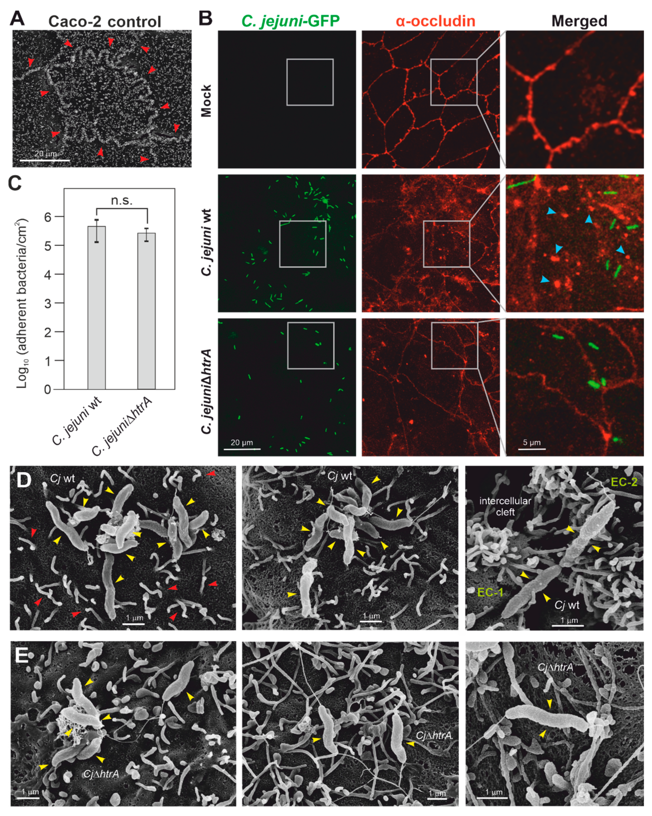
3.1. C. jejuni Colonization of Apical Caco-2 Cell Surfaces

To generate accurately polarized Caco-2 monolayers for our studies, it was necessary to let the cells differentiate for at least 14 days. During culturing, the functionality of the tight junctions was monitored every two days by phase contrast microscopy and by measuring the transepithelial electrical resistance (TER) in a transwell system. The overall TER values increased during this period until reaching maximal values between 350 and 400 Ohm × cm2, as described earlier [22]. In parallel, the cells were grown under identical conditions on glass slides placed in 12-well plates followed by analysis through scanning electron microscopy (SEM). The SEM technique identified the presence of characteristic cell-to-cell junctions and microvilli as apical markers (Figure 1A). The microvilli are the white-colored structures at the cell surface, which were enriched near the tight junctions (Figure 1A, red arrowheads, also exemplarily marked in image one of panel D). We then infected the Caco-2 cells with GFP-expressing C. jejuni wt and an available ∆htrA knockout mutant for 6 h, and investigated the bacteria bound apically to the Caco-2 cell surface by fluorescence microscopy (Figure 1B, green). The results show that the bacteria bound readily to the apical surface. Quantification revealed that C. jejuni wt and the ∆htrA mutant attached to the cells with similar numbers (Figure 1C). The cells were counter-stained using α-occludin antibodies to visualize proper tight junctions in the non-infected control cells (Figure 1B, red). In accordance with our former investigations [19,22], we found that C. jejuni wt infection induced the local disruption of cell-to-cell junctions visible as occludin aggregates formed at the apical cell surface (Figure 1B, blue arrowheads), while this phenotype was widely diminished during infection with the ∆htrA mutant (Figure 1B, bottom). Tight bacterial attachment to the apical surface was then confirmed by SEM, as observed for both wt (Figure 1D) and ∆htrA mutant C. jejuni (Figure 1E). However, signs of apical bacterial invasion into the host cell cytoplasm were not evident. In some cases, surface-bound wt bacteria were found near the cell–cell junctions, which formed local “clefts” between neighboring epithelial cells (Figure 1D, right panel). In contrast, the ∆htrA mutant bound non-specifically to the apical cell surface without obviously affecting the cell-to-cell junctions (Figure 1E, yellow arrowheads).

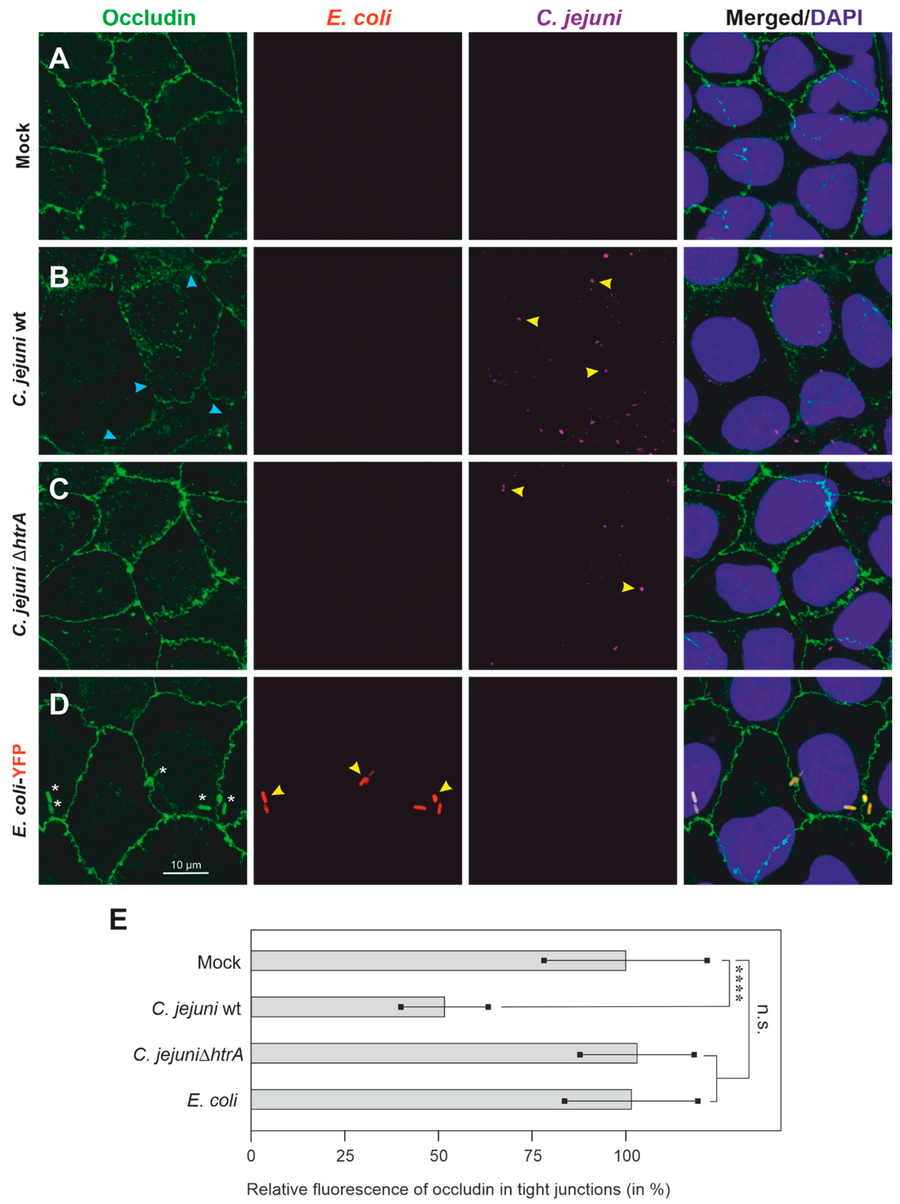
3.2. Colonization of Caco-2 Cells by C. jejuni and E. coli, and Impact on Tight Junctions

We have previously demonstrated that C. jejuni can colonize polarized epithelial cell monolayers and affect junctional proteins by HtrA-dependent cleavage, thus disrupting the epithelial barrier [19,21,22]. Next, we wanted to investigate whether such properties may impact further microorganisms, such as non-invasive E. coli. In a first experiment, we explored if polarized Caco-2 cells are suitable to address this question. Therefore, Caco-2 epithelial cells were cultured as described above, then infected with the two C. jejuni strains or YFP-expressing E. coli and subjected to immunofluorescence (Figure 2). Examples of bacteria bound to the apical Caco-2 cell surface are marked with yellow arrowheads. The immunofluorescence pictures show that C. jejuni wt adhered to the cell surface and induced the disruption of the cellular tight junction component occludin, as expected (Figure 2B, green, blue arrowheads). Infection with C. jejuni ΔhtrA mutant or E. coli-YFP showed that these bacteria also adhered to the cell surface, but the occludin-containing cellular tight junctions were not disrupted (Figure 2C,D, green). The corresponding quantification data are displayed in Figure 2E.

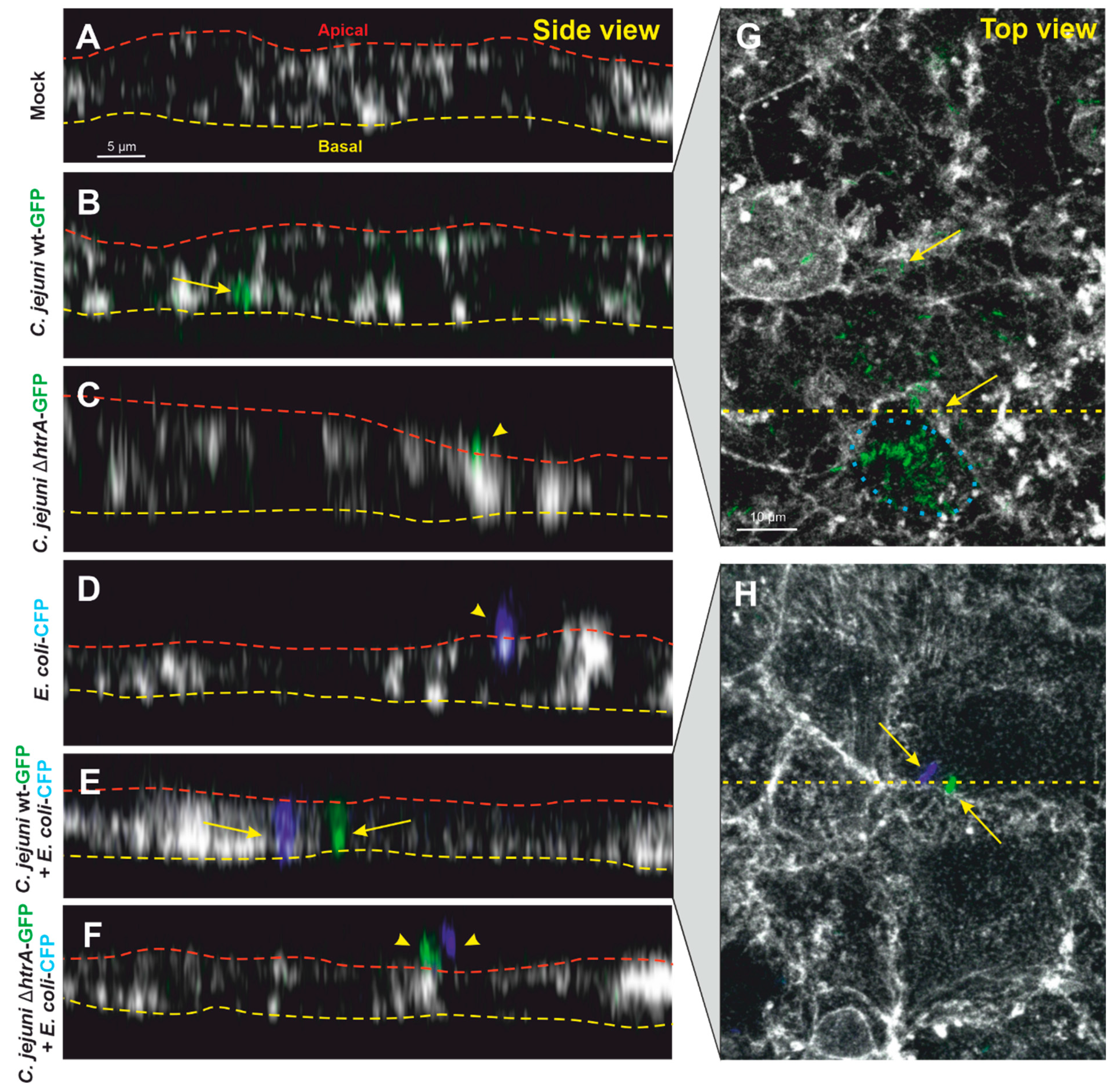
3.3. Confocal Microscopy of Co-Transmigrating C. jejuni and E. coli across Polarized Cells

We have previously shown that transmigration of C. jejuni across polarized epithelial cell monolayers is enabled by cleaving occludin and other junctional proteins in an HtrA-dependent manner [19,21,22]. Next, we wanted to examine if commensal bacteria such as E. coli can travel together with C. jejuni across polarized Caco-2 epithelial cells. For this purpose, we monitored the transmigration of C. jejuni and E. coli (either alone or together) by confocal microscopy (Figure 3). Indeed, wt C. jejuni-GFP alone could be detected between two neighboring cells or at the basal side of the infected Caco-2 monolayer (Figure 3B), while C. jejuni ΔhtrA-GFP or E. coli-CFP alone were only found on the apical cell surface (Figure 3C,D). However, when Caco-2 cells were co-infected with wt E. coli-CFP and C. jejuni-GFP, both bacterial species could be observed basolaterally between neighboring epithelial cells in close proximity to each other (Figure 3E). In contrast, when Caco-2 cells were co-infected with E. coli-CFP and C. jejuni ΔhtrA-GFP, both bacterial species were unable to transmigrate and could be found only on the apical monolayer surface (Figure 3F). In both infections, C. jejuni wt alone or co-infection with E. coli, bacterial cells were detected in close proximity to the tight junctions (enlarged sections in Figure 3G,H). Together, these findings underline the importance of HtrA in paracellular transmigration of C. jejuni, which at the same time can trigger the transmigration of non-invasive E. coli. In addition, we sporadically detected some intracellular C. jejuni wt (Figure 3G, blue dotted circle) that probably invaded the cells from the bottom via interaction of CadF with the basal fibronectin-integrin complex [13].

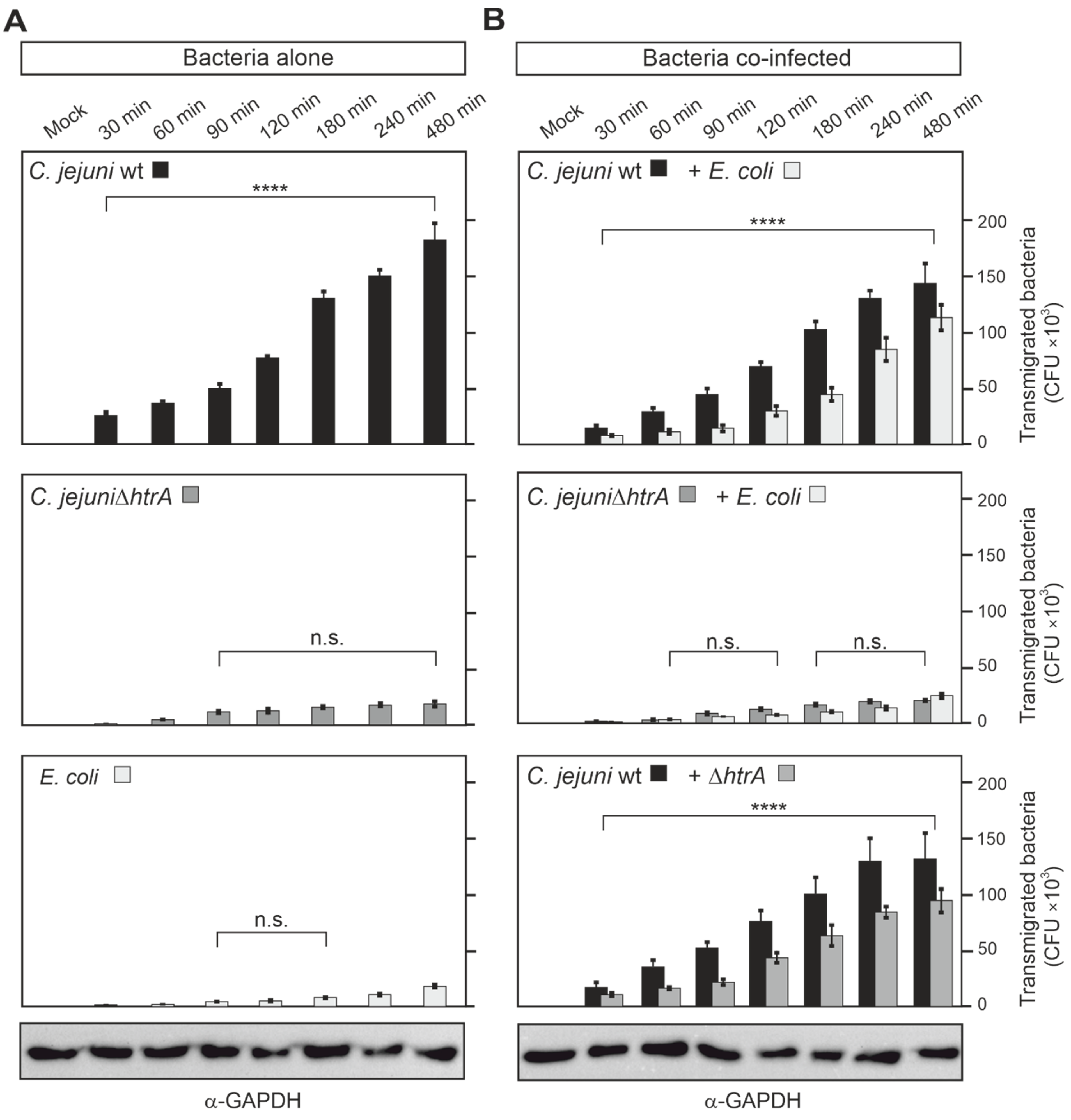
3.4. Quantification of Co-Transmigrating C. jejuni and E. coli across Polarized Caco-2 Cells

Next, our aim was to quantify the transmigration rates of C. jejuni and E. coli. For this reason, Caco-2 cells were cultivated on transwell inserts and differentiated as described above. Then, infection of the cells was performed by adding the bacteria to the apical chamber, and the numbers of transmigrating bacterial CFUs in the bottom chamber were quantified in a time course up to 8 h (Figure 4). In agreement with the findings we acquired from confocal microscopy, we found that C. jejuni wt transmigrated fast and effectively. The first transmigrated bacteria were even seen after 30 min, and up to 200,000 transmigrated bacteria at the 8 h time-point (Figure 4A). In contrast, both the ΔhtrA deletion mutant and E. coli alone revealed a robust deficiency in transmigration in comparison to C. jejuni wt (Figure 4A). When Caco-2 cells were co-infected with C. jejuni wt and E. coli, but not C. jejuni ΔhtrA with E. coli, both bacterial species transmigrated effectively (Figure 4B, top). After 8 h of infection, the number of transmigrated E. coli significantly increased (adjusted p-value less than 0.0001) from about 18,000 in Caco-2 cells infected alone to 113,000 in cells co-infected with C. jejuni wt and E. coli. Interestingly, when Caco-2 cells were co-infected with C. jejuni wt and C. jejuni ΔhtrA, the transmigration deficiency of the mutant was rescued (Figure 4B, bottom). The number of transmigrated ΔhtrA mutants significantly increased (adjusted p-value less than 0.0001) from about 19,000 when added to Caco-2 cells alone to 94,000 when added to the cells concurrently with C. jejuni wt. Together, these findings underline the importance of HtrA in paracellular transmigration of C. jejuni, which at the same time can trigger the transmigration of non-invasive E. coli or HtrA-defective C. jejuni.

3.5. Epithelial Transmigration of Other Microbiota by HtrA-Expressing C. jejuni

Finally, we wanted to explore if other microbiota bacteria can transmigrate across the intestinal epithelium in the presence of C. jejuni. We therefore tested Lactococcus lactis. This commensal bacterium alone showed weak transmigration capabilities, similar to non-pathogenic E. coli (Figure 5A). However, when Caco-2 cells were co-infected with L. lactis and C. jejuni wt, but not C. jejuni ΔhtrA and L. lactis, both bacterial species transmigrated effectively (Figure 5B). After 8 h of infection, the transmigration rate of L. lactis significantly increased (adjusted p-value less than 0.0001) from about 16,000 bacteria in Caco-2 cells when infected alone to 106,000 L. lactis bacteria when co-infected with C. jejuni wt. These observations confirm that secreted HtrA of C. jejuni has a crucial function in crossing the epithelial barrier by this pathogen, as well as commensal bacteria of the microbiota such as E. coli or L. lactis.

4. Conclusions

The epithelium in the digestive tract of humans represents an effective barrier, protecting the host against intruding microorganisms such as microbiota. Various gastrointestinal microbial pathogens, including Helicobacter, Shigella, Salmonella, Listeria and others, exhibit cell-invasive capabilities and damage the epithelial barrier, followed by transcellular or paracellular migration [15,44,45]. Earlier reports have shown that C. jejuni is able to transmigrate across polarized MKN-28, Caco-2 and T84 epithelial cell monolayers in vitro via the transcellular route [8,33] or paracellular route [34], but a final consensus is still not found in the community. However, Campylobacter infection represents the most common risk factor for developing IBD during the first year after campylobacteriosis. Patients with IBD possess pronounced inflammatory reactions to their intestinal microbiota by mechanisms that are not fully understood [29]. In the healthy intestine, the microbiota is successfully segregated to the lumen by the intestinal epithelium. Nevertheless, defects of the intestinal epithelial barrier by dysregulated cytokines/chemokines and disrupted tight junctions, known as the “leaky gut” phenotype, may contribute to IBD development, because bacteria can now translocate across the epithelium to the lamina propria. In this way, they can expose their antigens to submucosal immune cells and provoke an inflammatory reaction against the normal microbiota [28]. This scenario can trigger or aggravate inflammatory reactions in IBD patients.
C. jejuni infection has been known to disturb the intestinal barrier integrity [2,45,46]. Our recent data showed that C. jejuni serine protease HtrA can be secreted in the extracellular space, where HtrA cleaves the junctional proteins E-cadherin, occludin and claudin-8 [18,19,20,21,22,35]. Here, we demonstrate that C. jejuni bind to the apical surface of polarized Caco-2 cells, but do not invade into the cytoplasm from the apical side. Instead, they loosen the cell-to-cell junctions, and rapidly cross the epithelial monolayer between neighboring cells in an HtrA-dependent fashion. Moreover, we could demonstrate that in this scenario, microbiota strains such as non-invasive E. coli or L. lactis also translocate by the same route into the basal compartment in a time-dependent fashion. Remarkably, C. jejuni wt, but not ∆htrA mutant, induced the efficient transmigration of these microbiota bacteria. We therefore suggest that the microbiota strains, which all encode htrA genes, may express HtrA proteins that are different from C. jejuni and cannot cleave host junctional proteins. Taken together, C. jejuni infection of the intestinal epithelium results in local opening of cellular junctions and paracellular translocation in an HtrA-dependent manner, associated with co-transmigration of microbiota. This mechanism may explain how microbiota strains can traverse the gut epithelium with the help of C. jejuni. In this way, microbiota can establish contact with host immune cells and provoke undesired immune responses. The exact translocation procedure, however, is not yet clear and should be studied in future experiments. Nevertheless, these findings may have an important impact on our understanding of the molecular mechanism behind various Campylobacter-associated diseases such as IBD. Thus, C. jejuni HtrA could be a promising new anti-microbial target for therapy in IBD patients.

Author Contributions

I.S. and N.T. performed the infection studies, I.S. accomplished the immunofluorescence experiments, and M.M. conducted the SEM analyses. N.T. performed the statistics. S.B. conceptualized the study, analyzed the data and wrote the paper, together with I.S. All authors have read and agreed to the published version of the manuscript.

Funding

This work was supported by the German Federal Ministries of Education and Research (BMBF) in frame of the zoonoses research consortium PAC-Campylobacter to SBA (IP9/ 01KI2007E).

Institutional Review Board Statement

Not applicable.

Informed Consent Statement

Not applicable.

Data Availability Statement

The data that support the findings of this study are available from the corresponding author upon request.

Acknowledgments

We thank Ina Schleicher (HZI Braunschweig, Germany) and Nina Rottmann (FAU Erlangen, Germany) for outstanding technical support and Philipp Tripal (OICE Erlangen, Germany) for help with immunofluorescence microscopy. The pWM1007-1009 plasmids were kindly provided by William G. Miller (US Department of Agriculture, Albany, CA, United States).

Conflicts of Interest

The authors declare that they have no competing interests.

References

  1. Kirk, M.D.; Pires, S.M.; Black, R.E.; Caipo, M.; Crump, J.A.; Devleesschauwer, B.; Dopfer, D.; Fazil, A.; Fischer-Walker, C.L.; Hald, T.; et al. World Health Organization Estimates of the Global and Regional Disease Burden of 22 Foodborne Bacterial, Protozoal, and Viral Diseases, 2010: A Data Synthesis. PLoS Med. 2015, 12, e1001921. [Google Scholar] [CrossRef] [Green Version]
  2. Burnham, P.M.; Hendrixson, D.R. Campylobacter jejuni: Collective components promoting a successful enteric lifestyle. Nat. Rev. Microbiol. 2018, 16, 551–565. [Google Scholar] [CrossRef] [PubMed]
  3. Heimesaat, M.M.; Backert, S.; Alter, T.; Bereswill, S. Human Campylobacteriosis-A Serious Infectious Threat in a One Health Perspective. Curr. Top. Microbiol. Immunol. 2021, 431, 1–23. [Google Scholar] [CrossRef] [PubMed]
  4. Zbrun, M.V.; Rossler, E.; Romero-Scharpen, A.; Soto, L.P.; Berisvil, A.; Zimmermann, J.A.; Fusari, M.L.; Signorini, M.L.; Frizzo, L.S. Worldwide meta-analysis of the prevalence of Campylobacter in animal food products. Res. Vet. Sci. 2020, 132, 69–77. [Google Scholar] [CrossRef] [PubMed]
  5. Dogan, O.B.; Aditya, A.; Ortuzar, J.; Clarke, J.; Wang, B. A systematic review and meta-analysis of the efficacy of processing stages and interventions for controlling Campylobacter contamination during broiler chicken processing. Compr. Rev. Food Sci. Food Saf. 2021, 21, 227–271. [Google Scholar] [CrossRef]
  6. Ang, C.W.; Laman, J.D.; Willison, H.J.; Wagner, E.R.; Endtz, H.P.; De Klerk, M.A.; Tio-Gillen, A.P.; Van den Braak, N.; Jacobs, B.C.; Van Doorn, P.A. Structure of Campylobacter jejuni lipopolysaccharides determines antiganglioside specificity and clinical features of Guillain-Barre, and Miller Fisher patients. Infect. Immun. 2002, 70, 1202–1208. [Google Scholar] [CrossRef] [Green Version]
  7. Smith, J.L. Campylobacter jejuni infection during pregnancy: Long-term consequences of associated bacteremia, Guillain-Barre syndrome, and reactive arthritis. J. Food Prot. 2002, 65, 696–708. [Google Scholar] [CrossRef]
  8. Kalischuk, L.D.; Buret, A.G. A role for Campylobacter jejuni-induced enteritis in inflammatory bowel disease? Am. J. Physiol.-Gastrointest. Liver Physiol. 2010, 298, G1–G9. [Google Scholar] [CrossRef] [Green Version]
  9. Guan, Q.D. A Comprehensive Review and Update on the Pathogenesis of Inflammatory Bowel Disease. J. Immunol. Res. 2019, 2019, 7247238. [Google Scholar] [CrossRef] [Green Version]
  10. O’Loughlin, J.L.; Samuelson, D.R.; Braundmeier-Fleming, A.G.; White, B.A.; Haldorson, G.J.; Stone, J.B.; Lessmann, J.J.; Eucker, T.P.; Konkel, M.E. The Intestinal Microbiota Influences Campylobacter jejuni Colonization and Extraintestinal Dissemination in Mice. Appl. Environ. Microbiol. 2015, 81, 4642–4650. [Google Scholar] [CrossRef] [Green Version]
  11. Sylte, M.J.; Shippy, D.C.; Bearson, B.L.; Bearson, S.M.D. Detection of Campylobacter jejuni liver dissemination in experimentally colonized turkey poults. Poult. Sci. 2020, 99, 4028–4033. [Google Scholar] [CrossRef] [PubMed]
  12. Martora, F.; Pagliuca, C.; Della Pepa, M.E.; Della Rocca, M.T.; Curto, S.; Iovene, M.R.; Vitiello, M. Campylobacter jejuni bacteremia in Italian pediatric patients with acute lymphoblastic leukemia: Report of two cases. New Microbiol. 2020, 43, 96–98. [Google Scholar] [PubMed]
  13. Tegtmeyer, N.; Sharafutdinov, I.; Harrer, A.; Esmaeili, D.S.; Linz, B.; Backert, S. Campylobacter Virulence Factors and Molecular Host-Pathogen Interactions. Curr. Top. Microbiol. Immunol. 2021, 431, 169–202. [Google Scholar] [CrossRef] [PubMed]
  14. Clausen, T.; Kaiser, M.; Huber, R.; Ehrmann, M. HTRA proteases: Regulated proteolysis in protein quality control. Nat. Rev. Mol. Cell Biol. 2011, 12, 152–162. [Google Scholar] [CrossRef]
  15. Backert, S.; Bernegger, S.; Skorko-Glonek, J.; Wessler, S. Extracellular HtrA serine proteases: An emerging new strategy in bacterial pathogenesis. Cell. Microbiol. 2018, 20, e12845. [Google Scholar] [CrossRef] [Green Version]
  16. Kim, D.Y.; Kim, K.K. Structure and function of HtrA family proteins, the key players in protein quality control. J. Bioch. Mol. Biol. 2005, 38, 266–274. [Google Scholar] [CrossRef] [Green Version]
  17. Skorko-Glonek, J.; Zurawa-Janicka, D.; Koper, T.; Jarzab, M.; Figaj, D.; Glaza, P.; Lipinska, B. HtrA Protease Family as Therapeutic Targets. Curr. Pharm. Des. 2013, 19, 977–1009. [Google Scholar] [CrossRef]
  18. Neddermann, M.; Backert, S. Quantification of serine protease HtrA molecules secreted by the foodborne pathogen Campylobacter Jejuni. Gut Pathog. 2019, 11, 14. [Google Scholar] [CrossRef]
  19. Boehm, M.; Hoy, B.; Rohde, M.; Tegtmeyer, N.; Baek, K.T.; Oyarzabal, O.A.; Brondsted, L.; Wessler, S.; Backert, S. Rapid paracellular transmigration of Campylobacter jejuni across polarized epithelial cells without affecting TER: Role of proteolytic-active HtrA cleaving E-cadherin but not fibronectin. Gut Pathog. 2012, 4, 3. [Google Scholar] [CrossRef] [Green Version]
  20. Hoy, B.; Geppert, T.; Boehm, M.; Reisen, F.; Plattner, P.; Gadermaier, G.; Sewald, N.; Ferreira, F.; Briza, P.; Schneider, G.; et al. Distinct roles of secreted HtrA proteases from gram-negative pathogens in cleaving the junctional protein and tumor suppressor E-cadherin. J. Biol. Chem. 2012, 287, 10115–10120. [Google Scholar] [CrossRef] [Green Version]
  21. Sharafutdinov, I.; Esmaeili, D.S.; Harrer, A.; Tegtmeyer, N.; Sticht, H.; Backert, S. Campylobacter jejuni Serine Protease HtrA Cleaves the Tight Junction Component Claudin-8. Front. Cell. Infect. Microbiol. 2020, 10. [Google Scholar] [CrossRef] [PubMed]
  22. Harrer, A.; Bucker, R.; Boehm, M.; Zarzecka, U.; Tegtmeyer, N.; Sticht, H.; Schulzke, J.D.; Backert, S. Campylobacter jejuni enters gut epithelial cells and impairs intestinal barrier function through cleavage of occludin by serine protease HtrA. Gut Pathog. 2019, 11, 4. [Google Scholar] [CrossRef] [PubMed]
  23. Heimesaat, M.M.; Fischer, A.; Alutis, M.; Grundmann, U.; Boehm, M.; Tegtmeyer, N.; Gobel, U.B.; Kuhl, A.A.; Bereswill, S.; Backert, S. The impact of serine protease HtrA in apoptosis, intestinal immune responses and extra-intestinal histopathology during Campylobacter jejuni infection of infant mice. Gut Pathog. 2014, 6, 16. [Google Scholar] [CrossRef] [PubMed] [Green Version]
  24. Heimesaat, M.M.; Alutis, M.; Grundmann, U.; Fischer, A.; Tegtmeyer, N.; Bohm, M.; Kuhl, A.A.; Gobel, U.B.; Backert, S.; Bereswill, S. The role of serine protease HtrA in acute ulcerative enterocolitis and extra-intestinal immune responses during Campylobacter jejuni infection of gnotobiotic IL-10 deficient mice. Front. Cell. Infect. Microbiol. 2014, 4, 77. [Google Scholar] [CrossRef] [Green Version]
  25. Boehm, M.; Simson, D.; Escher, U.; Schmidt, A.-M.; Bereswill, S.; Tegtmeyer, N.; Backert, S.; Heimesaat, M.M. Function of Serine Protease HtrA in the Lifecycle of the Foodborne Pathogen Campylobacter jejuni. Eur. J. Microbiol. Immunol. 2018, 8, 70–77. [Google Scholar] [CrossRef]
  26. Schmidt, A.M.; Escher, U.; Mousavi, S.; Boehm, M.; Backert, S.; Bereswill, S.; Heimesaat, M.M. Protease Activity of Campylobacter jejuni HtrA Modulates Distinct Intestinal and Systemic Immune Responses in Infected Secondary Abiotic IL-10 Deficient Mice. Front. Cell. Infect. Microbiol. 2019, 9, 79. [Google Scholar] [CrossRef]
  27. Haag, L.M.; Fischer, A.; Otto, B.; Plickert, R.; Kuhl, A.A.; Gobel, U.B.; Bereswill, S.; Heimesaat, M.M. Intestinal Microbiota Shifts towards Elevated Commensal Escherichia coli Loads Abrogate Colonization Resistance against Campylobacter jejuni in Mice. PLoS ONE 2012, 7, e35988. [Google Scholar] [CrossRef]
  28. Aldars-Garcia, L.; Marin, A.C.; Chaparro, M.; Gisbert, J.P. The Interplay between Immune System and Microbiota in Inflammatory Bowel Disease: A Narrative Review. Int. J. Mol. Sci. 2021, 22, 3076. [Google Scholar] [CrossRef]
  29. Ahlawat, S.; Kumar, P.; Mohan, H.; Goyal, S.; Sharma, K.K. Inflammatory bowel disease: Tri-directional relationship between microbiota, immune system and intestinal epithelium. Crit. Rev. Microbiol. 2021, 47, 254–273. [Google Scholar] [CrossRef]
  30. Mukhopadhya, I.; Hansen, R.; El-Omar, E.M.; Hold, G.L. IBD-what role do Proteobacteria play? Nat. Rev. Gastroenterol. Hepatol. 2012, 9, 219–230. [Google Scholar] [CrossRef]
  31. Nielsen, H.L.; Dalager-Pedersen, M.; Nielsen, H. Risk of inflammatory bowel disease after Campylobacter jejuni and Campylobacter concisus infection: A population-based cohort study. Scand. J. Gastroenterol. 2019, 54, 265–272. [Google Scholar] [CrossRef] [PubMed]
  32. Mirsepasi-Lauridsen, H.C.; Vallance, B.A.; Krogfelt, K.A.; Petersen, A.M. Escherichia coli Pathobionts Associated with Inflammatory Bowel Disease. Clin. Microbiol. Rev. 2019, 32, e00060-18. [Google Scholar] [CrossRef] [PubMed] [Green Version]
  33. Kalischuk, L.D.; Inglis, G.D.; Buret, A.G. Campylobacter jejuni induces transcellular translocation of commensal bacteria via lipid rafts. Gut Pathog. 2009, 1, 2. [Google Scholar] [CrossRef] [PubMed] [Green Version]
  34. Lamb-Rosteski, J.M.; Kalischuk, L.D.; Inglis, G.D.; Buret, A.G. Epidermal growth factor inhibits Campylobacter jejuni-induced claudin-4 disruption, loss of epithelial barrier function, and Escherichia coli translocation. Infect. Immun. 2008, 76, 3390–3398. [Google Scholar] [CrossRef] [Green Version]
  35. Boehm, M.; Lind, J.; Backert, S.; Tegtmeyer, N. Campylobacter jejuni serine protease HtrA plays an important role in heat tolerance, oxygen resistance, host cell adhesion, invasion, and transmigration. Eur. J. Microbiol. Immunol. 2015, 5, 68–80. [Google Scholar] [CrossRef] [Green Version]
  36. Oyarzabal, O.A.; Backert, S.; Nagaraj, M.; Miller, R.S.; Hussain, S.K.; Oyarzabal, E.A. Efficacy of supplemented buffered peptone water for the isolation of Campylobacter jejuni and C. coli from broiler retail products. J. Microbiol. Methods 2007, 69, 129–136. [Google Scholar] [CrossRef]
  37. Backert, S.; Hofreuter, D. Molecular methods to investigate adhesion, transmigration, invasion and intracellular survival of the foodborne pathogen Campylobacter Jejuni. J. Microbiol. Methods 2013, 95, 8–23. [Google Scholar] [CrossRef] [Green Version]
  38. Krause-Gruszczynska, M.; Boehm, M.; Rohde, M.; Tegtmeyer, N.; Takahashi, S.; Buday, L.; Oyarzabal, O.A.; Backert, S. The signaling pathway of Campylobacter jejuni-induced Cdc42 activation: Role of fibronectin, integrin beta1, tyrosine kinases and guanine exchange factor Vav2. Cell Commun. Signal. 2011, 9, 1–18. [Google Scholar] [CrossRef] [Green Version]
  39. Krause-Gruszczynska, M.; Rohde, M.; Hartig, R.; Genth, H.; Schmidt, G.; Keo, T.; König, W.; Miller, W.G.; Konkel, M.E.; Backert, S. Role of the small Rho GTPases Rac1 and Cdc42 in host cell invasion of Campylobacter jejuni. Cell. Microbiol. 2007, 9, 2431–2444. [Google Scholar] [CrossRef]
  40. Miller, W.G.; Bates, A.H.; Horn, S.T.; Brandl, M.T.; Wachtel, M.R.; Mandrell, R.E. Detection on surfaces and in Caco-2 cells of Campylobacter jejuni cells transformed with new gfp, yfp, and cfp marker plasmids. Appl. Environ. Microbiol. 2000, 66, 5426–5436. [Google Scholar] [CrossRef] [Green Version]
  41. Hartung, M.L.; Gruber, D.C.; Koch, K.N.; Gruter, L.; Rehrauer, H.; Tegtmeyer, N.; Backert, S.; Muller, A.H. pylori-Induced DNA Strand Breaks Are Introduced by Nucleotide Excision Repair Endonucleases and Promote NF-kappaB Target Gene Expression. Cell Rep. 2015, 13, 70–79. [Google Scholar] [CrossRef] [PubMed] [Green Version]
  42. Krause-Gruszczynska, M.; van Alphen, L.B.; Oyarzabal, O.A.; Alter, T.; Hanel, I.; Schliephake, A.; Konig, W.; van Putten, J.P.; Konkel, M.E.; Backert, S. Expression patterns and role of the CadF protein in Campylobacter jejuni and Campylobacter coli. FEMS Microbiol. Lett. 2007, 274, 9–16. [Google Scholar] [CrossRef] [PubMed] [Green Version]
  43. Moese, S.; Selbach, M.; Zimny-Arndt, U.; Jungblut, P.R.; Meyer, T.F.; Backert, S. Identification of a tyrosine-phosphorylated 35 kDa carboxy-terminal fragment (p35CagA) of the Helicobacter pylori CagA protein in phagocytic cells: Processing or breakage? Proteomics 2001, 1, 618–629. [Google Scholar] [CrossRef]
  44. Vogelmann, R.; Amieva, M.R.; Falkow, S.; Nelson, W.J. Breaking into the epithelial apical-junctional complex--news from pathogen hackers. Curr. Opin. Cell. Biol. 2004, 16, 86–93. [Google Scholar] [CrossRef] [Green Version]
  45. Backert, S.; Boehm, M.; Wessler, S.; Tegtmeyer, N. Transmigration route of Campylobacter jejuni across polarized intestinal epithelial cells: Paracellular, transcellular or both? Cell. Commun. Signal. 2013, 11, 72. [Google Scholar] [CrossRef] [Green Version]
  46. Cróinín, T.Ó.; Backert, S. Host epithelial cell invasion by Campylobacter jejuni: Trigger or zipper mechanism? Front. Cell Infect. Microbiol. 2012, 2, 25. [Google Scholar] [CrossRef] [Green Version]
Figure 1. Scanning electron and fluorescence microscopy of Caco-2 cells before and after C. jejuni infection. (A) Top view of the apical surface of polarized Caco-2 cell monolayers without infection. Red arrowheads mark microvilli structures that are strongly expressed near the cell-to-cell junctions. (B) Fluorescence microscopy of Caco-2 cells counterstained with α-occludin (red), either as uninfected control (top), or infected with GFP-expressing wt bacteria (middle) or GFP-expressing ∆htrA mutant (bottom). The cells were infected with an MOI of 10. Images with the merged C. jejuni and occludin staining (right) correspond to enlarged areas marked in the gray boxes. Blue arrowheads indicate examples of disrupted patches in the tight junctions. (C) Quantification of apical bound bacteria revealed no significant (n.s.) differences among the two strains. Scanning electron microscopy of Caco-2 cells after infection with C. jejuni wt (D) or ∆htrA mutant (E). Attached C. jejuni bacteria are marked with yellow arrowheads and examples of microvilli with red arrowheads. Sporadically, infection with C. jejuni wt was seen with the local opening of cell-to-cell junctions visible as intercellular clefts formed at the apical cell surface between two neighboring epithelial cells (EC-1 and EC-2), as indicated.
Biomolecules 12 00521 g001
Figure 2. Top view of infected Caco-2 cells by immunofluorescence microscopy. Epithelial cell monolayers were left uninfected (Mock, A) or were infected with either C. jejuni wt (B), C. jejuni ΔhtrA (C) or E. coli-YFP (D). After infection with an MOI of 10, the cells were immunostained using α-occludin and α-C. jejuni-specific antibodies as well as DAPI. Yellow arrowheads indicate bacterial cells interacting with Caco-2 cells at their apical surface and blue arrowheads show disrupted tight junctions. The white stars (panel D, left) indicate non-specific fluorescence signals from E. coli-YFP. (E) Relative fluorescence intensities of occludin in the tight junction areas were quantified and presented as the mean ± SD. The mean relative fluorescence of uninfected mock cells was set as 100%. Significant differences shown in the graphs correspond to p ≤ 0.0001 (****) or non-significant (n.s.).
Biomolecules 12 00521 g002
Figure 3. Side and top view of infected Caco-2 monolayers by confocal fluorescence microscopy. The cells were infected with the indicated C. jejuni and/or E. coli bacteria and counterstained with rhodamine-phalloidin to visualize the actin cytoskeleton (gray). Lateral sections (Z-orientation) are given for showing bacterial transmigration to basolateral sides of epithelial cells. Caco-2 monolayers were left uninfected (A) or infected with either C. jejuni-GFP wt (B), C. jejuni ΔhtrA-GFP (C) or E. coli-CFP (D). Dual co-infections were performed using wt C. jejuni-GFP and E. coli-CFP (E) or C. jejuni ΔhtrA-GFP and E. coli-CFP (F). The red and yellow dashed lines mark apical and basal sides of the cell monolayers, respectively. The yellow arrowheads indicate attached bacterial cells, and yellow arrows mark transmigrating bacteria from apical to basal sides. (G,H) Top view of enlarged sections showing the localization of transmigrating bacteria that are between neighboring cells at the basal side (yellow arrows). The images also show some intracellular C. jejuni. Examples are marked with a blue dotted circle. The yellow dashed lines indicate the corresponding cross sections. The cells were infected with an MOI of 1 to detect single bacterial cells.
Biomolecules 12 00521 g003
Figure 4. Time course of transmigration properties of C. jejuni and E. coli bacteria across polarized Caco-2 cells using a transwell filter system. Caco-2 cells were cultivated for 14 days to form polarized monolayers on transwell filters and then infected with the indicated single bacterial strains (A) or mixed bacterial strains (B) at an MOI of 25 each. Transmigrated bacteria were collected from the bottom chambers, and C. jejuni were cultivated on Campylobacter agar plates and E. coli on LB agar plates, respectively. Finally, the CFUs were quantified in triplicate. On the bottom are two representative GAPDH blots as loading controls. Significant differences shown in the graphs correspond to p ≤ 0.0001 (****) or non-significant (n.s.).
Biomolecules 12 00521 g004
Figure 5. Time course of transmigration properties of C. jejuni and L. lactis bacteria across polarized Caco-2 cells using a transwell filter system. Caco-2 cells were cultivated for 14 days to form polarized monolayers on transwell filters and then infected with the indicated single L. lactis strain (A) or mixed bacterial strains (B) at an MOI of 25 each. Transmigrated bacteria were collected from the bottom chambers. C. jejuni were grown on Campylobacter agar plates and L. lactis on MRS agar plates. Finally, the CFUs were quantified in triplicate. On the bottom is a representative GAPDH blot as a loading control. Significant differences shown in the graphs correspond to p ≤ 0.0001 (****) or non-significant (n.s.).
Biomolecules 12 00521 g005
Publisher’s Note: MDPI stays neutral with regard to jurisdictional claims in published maps and institutional affiliations.

