
Cytotoxic Alkaloids Derived from Marine Sponges: A Comprehensive Review

 , ,
, ,  and
and
Abstract

1. Introduction
2. Alkaloid Classification
2.1. Acridine Alkaloids
2.2. β-Carboline Alkaloid
2.3. Bromotyrosine Alkaloids
2.4. Dibrominated and Brominated Alkaloids
2.5. Aaptamine Alkaloids
2.6. Guanidine Alkaloids
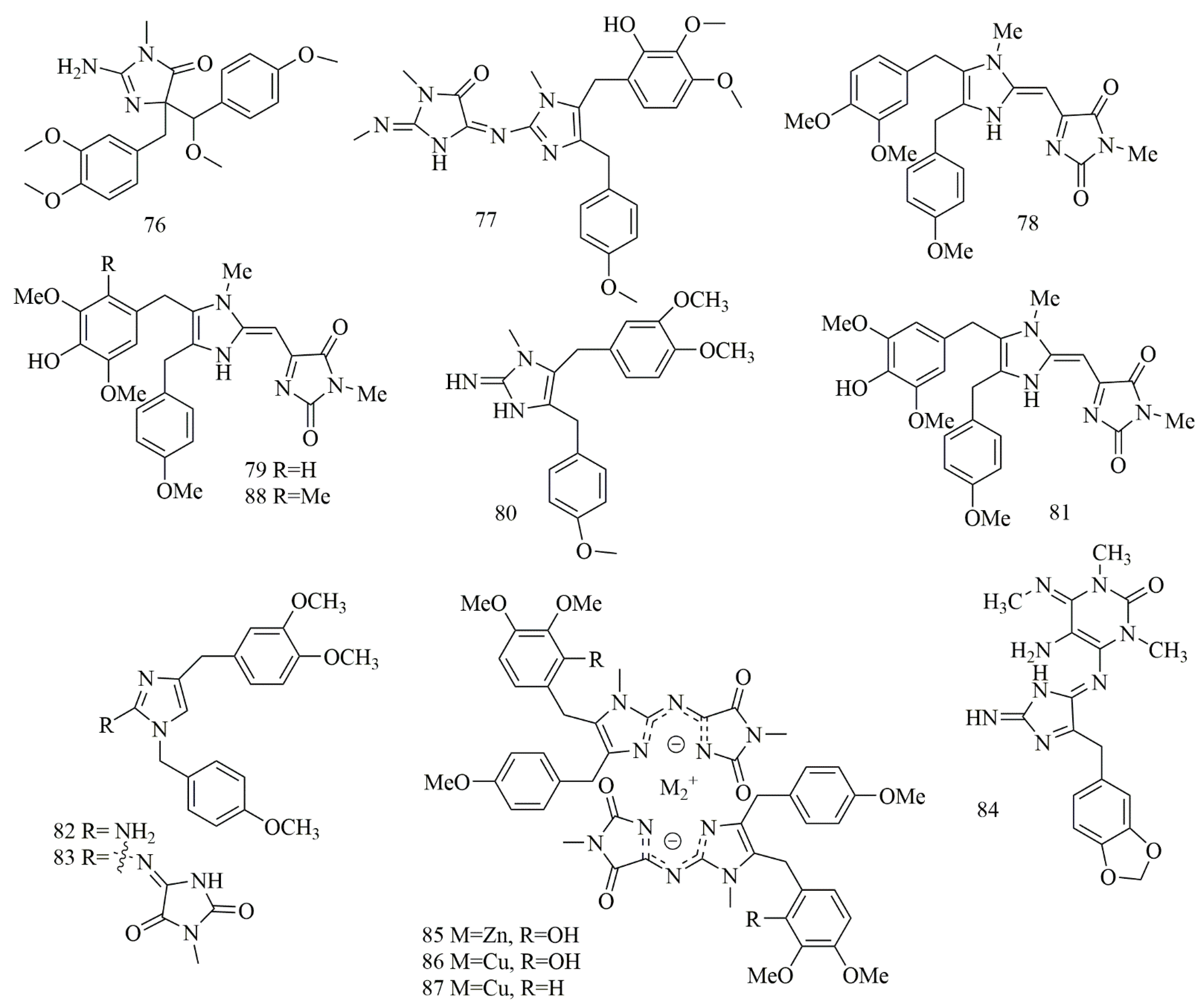
2.7. Imidazole Alkaloid
2.8. Indole, Bisindole, and Trisindole Alkaloids
2.9. Peptide Alkaloid
2.10. Piperidine Alkaloids
2.11. Pyrimidine Alkaloids
2.12. Pyridine Alkaloids
2.13. Pyrrole and Bromopyrrole Alkaloids
2.14. Pyrroloiminoquinone Alkaloids
2.15. Quinoline and Quinolizinde Alkaloids
2.16. Tetrahydroisoqouinoline Alkaloids
2.17. Steroidal Alkaloid
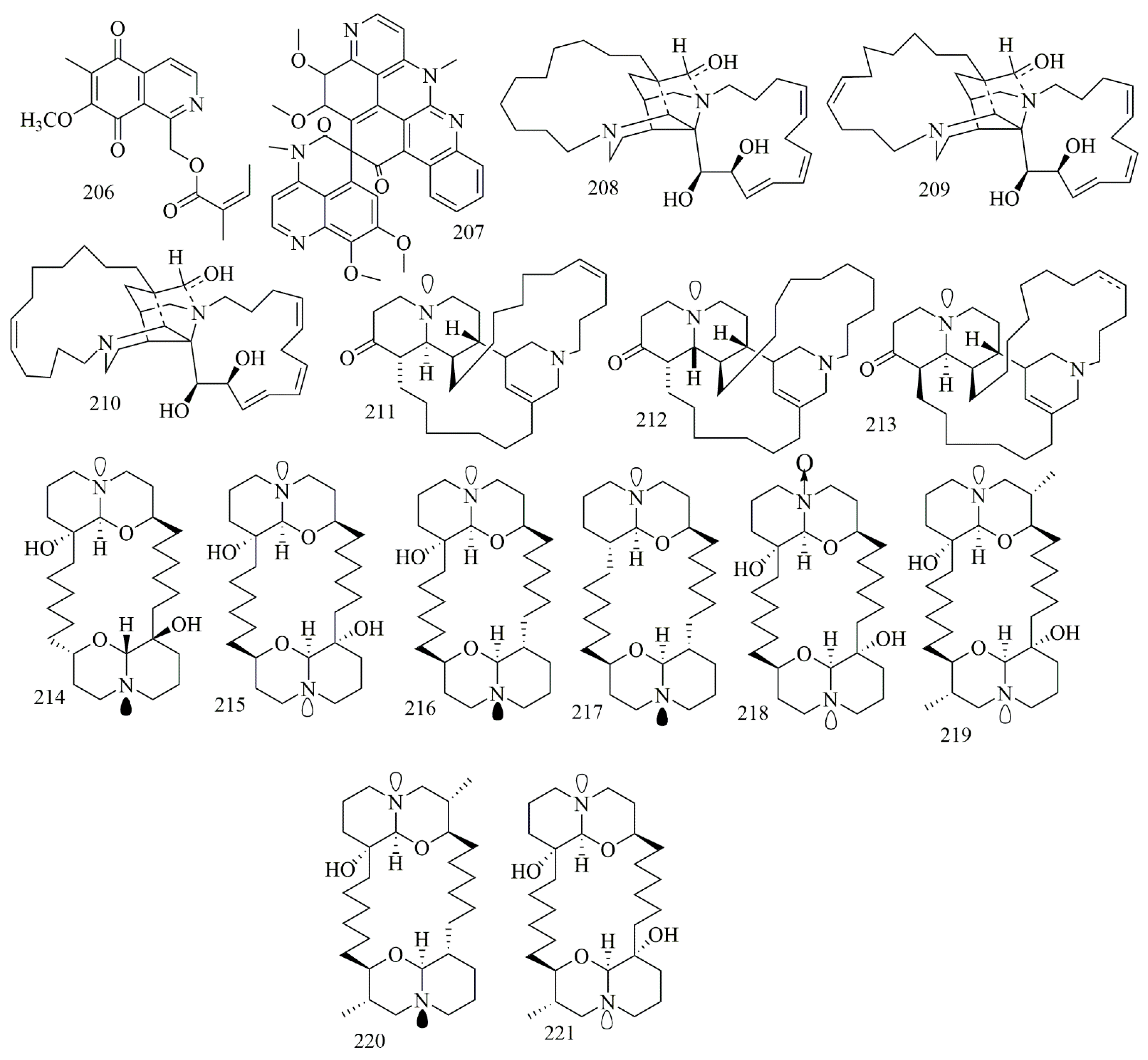
2.18. Manzamine Alkaloids
2.19. Diterpene Alkaloid
2.20. Sesquiterpene Quinones/Hydroquinones Alkaloid
3. Conclusions
Author Contributions
Funding
Conflicts of Interest
References
- Gul, W.; Hamann, M.T. Indole alkaloid marine natural products: An established source of cancer drug leads with considerable promise for the control of parasitic, neurological and other diseases. Life Sci. 2005, 78, 442–453. [Google Scholar] [CrossRef]
- Fattorusso, E.; Taglialatela-Scafati, O. Modern Alkaloids: Structure, Isolation, Synthesis, And Biology; John Wiley & Sons: Hoboken, NJ, USA, 2008. [Google Scholar]
- Kochanowska-Karamyan, A.J.; Hamann, M.T. Marine Indole Alkaloids: Potential New Drug Leads for the Control of Depression and Anxiety. Chem. Rev. 2010, 110, 4489–4497. [Google Scholar] [CrossRef] [PubMed]
- Carroll, A.R.; Copp, B.R.; Davis, R.A.; Keyzers, R.A.; Prinsep, M.R. Marine natural products. Nat. Prod. Rep. 2020, 37, 175–223. [Google Scholar] [CrossRef] [PubMed]
- Casertano, M.; Menna, M.; Imperatore, C. The Ascidian-Derived Metabolites with Antimicrobial Properties. Antibiotics 2020, 9, 510. [Google Scholar] [CrossRef] [PubMed]
- Matsunaga, S.; Yamashita, T.; Tsukamoto, S.; Fusetani, N. Three New Antibacterial Alkaloids from a Marine SpongeStellettaSpecies. J. Nat. Prod. 1999, 62, 1202–1204. [Google Scholar] [CrossRef]
- Baker, B.J.; Scheuer, P.J.; Shoolery, J.N. Papuamine, an antifungal pentacyclic alkaloid from a marine sponge, Haliclona sp. J. Am. Chem. Soc. 1988, 110, 965–966. [Google Scholar] [CrossRef]
- Perry, N.B.; Ettouati, L.; Litaudon, M.; Blunt, J.W.; Munro, M.H.; Parkin, S.; Hope, H. Alkaloids from the antarctic sponge Kirkpatrickia varialosa.: Part 1: Variolin b, a new antitumour and antiviral compound. Tetrahedron 1994, 50, 3987–3992. [Google Scholar] [CrossRef]
- Johnson, T.A.; Sohn, J.; Vaske, Y.M.; White, K.N.; Cohen, T.L.; Vervoort, H.C.; Tenney, K.; Valeriote, F.A.; Bjeldanes, L.F.; Crews, P. Myxobacteria versus sponge-derived alkaloids: The bengamide family identified as potent immune modulating agents by scrutiny of LC–MS/ELSD libraries. Bioorganic Med. Chem. 2012, 20, 4348–4355. [Google Scholar] [CrossRef] [PubMed]
- Agrawal, S.; Saraswati, S.; Mathur, R.; Pandey, M. Brucine, a plant derived alkaloid inhibits inflammatory angiogenesis in a murine sponge model. Biomed. Prev. Nutr. 2011, 1, 180–185. [Google Scholar] [CrossRef]
- Xu, M.; Andrews, K.T.; Birrell, G.W.; Tran, T.L.; Camp, D.; Davis, R.A.; Quinn, R.J. Psammaplysin H, a new antimalarial bromotyrosine alkaloid from a marine sponge of the genus Pseudoceratina. Bioorganic Med. Chem. Lett. 2011, 21, 846–848. [Google Scholar] [CrossRef]
- Ravichandran, S.; Kathiresan, K.; Balaram, H. Anti-malarials from marine sponges. Biotechnol. Mol. Biol. Rev. 2007, 2, 33–38. [Google Scholar]
- Munro, M.H.G.; Blunt, J.W.; Dumdei, E.J.; Hickford, S.J.; Lill, R.E.; Li, S.; Battershill, C.N.; Duckworth, A.R. The discovery and development of marine compounds with pharmaceutical potential. J. Biotechnol. 1999, 70, 15–25. [Google Scholar] [CrossRef]
- Blunt, J.W.; Copp, B.R.; Keyzers, R.A.; Munro, M.H.G.; Prinsep, M.R. Marine natural products. Nat. Prod. Rep. 2015, 32, 116–211. [Google Scholar] [CrossRef]
- Dembitsky, V.M.; Rezanka, T.; Srebnik, M. Lipid compounds of freshwater sponges: Family Spongillidae, class Demospongiae. Chem. Phys. Lipids 2003, 123, 117–155. [Google Scholar] [CrossRef]
- Vacelet, J. Diversity and evolution of deep-sea carnivorous sponges. Porifera research: Biodiversity, innovation and sustainability. Série Livros 2007, 28, 107–115. [Google Scholar]
- Mioso, R.; Marante, F.J.T.; Bezerra, R.S.; Borges, F.V.P.; Santos, B.V.D.O.; De Laguna, I.H.B. Cytotoxic Compounds Derived from Marine Sponges. A Review (2010–2012). Molecules 2017, 22, 208. [Google Scholar] [CrossRef] [PubMed]
- Fan, H.; Peng, J.; Hamann, M.T.; Hu, J.-F. Lamellarins and Related Pyrrole-Derived Alkaloids from Marine Organisms. Chem. Rev. 2007, 108, 264–287. [Google Scholar] [CrossRef] [PubMed]
- Joseph, B.; Sujatha, S. Pharmacologically important natural products from marine sponges. J. Nat. Prod. 2011, 4, 5–12. [Google Scholar]
- Kim, S.K.; Kalimuthu, S. Introduction to anticancer drugs from marine origin. In Handbook of Anticancer Drugs from Marine Origin; Springer: Cham, Switzerland, 2015; pp. 1–13. [Google Scholar]
- Han, B.N.; Hong, L.L.; Gu, B.B.; Sun, Y.T.; Wang, J.; Liu, J.T.; Lin, H.W. Natural Products from Sponges. In Symbiotic Microbiomes of Coral Reefs Sponges and Corals; Springer: Dordrecht, The Netherlands, 2019; pp. 329–463. [Google Scholar]
- Hanan, M.A.; Musarat, A. Marine sponge alkaloids: A source of novel anticancer agents. In Phytochemistry: Volume 3: Marine Sources, Industrial Applications, and Recent Advances; Egbuna, C., Chinenye Ifemeje, J., Kumar, S., Sharif, N., Eds.; Apple Academic Press Inc.: Palm Bay, FL, USA, 2018; pp. 35–64. [Google Scholar]
- Burres, N.S.; Sazesh, S.; Gunawardana, G.P.; Clement, J.J. Antitumor activity and nucleic acid binding properties of dercitin, a new acridine alkaloid isolated from a marine Dercitus species sponge. Cancer Res. 1989, 49, 5267–5274. [Google Scholar]
- Thale, Z.; Johnson, T.; Tenney, K.; Wenzel, P.J.; Lobkovsky, E.; Clardy, J.; Media, J.; Pietraszkiewicz, H.; Valeriote, F.A.; Crews, P. Structures and Cytotoxic Properties of Sponge-Derived Bisannulated Acridines. J. Org. Chem. 2002, 67, 9384–9391. [Google Scholar] [CrossRef]
- Ibrahim, S.R.; Mohamed, G.A. Ingenine E, a new cytotoxic β-carboline alkaloid from the Indonesian sponge Acanthostrongylophora ingens. J. Asian Nat. Prod. Res. 2017, 19, 504–509. [Google Scholar] [CrossRef] [PubMed]
- Göthel, Q.; Sirirak, T.; Köck, M. Bromotyrosine-derived alkaloids from the Caribbean sponge Aplysina lacunosa. Beilstein J. Org. Chem. 2015, 11, 2334–2342. [Google Scholar] [CrossRef] [PubMed]
- Tarazona, G.; Santamaría, G.; Cruz, P.G.; Fernández, R.; Pérez, M.; Martínez-Leal, J.F.; Rodríguez, J.; Jiménez, C.; Cuevas, C. Cytotoxic Anomoian B and Aplyzanzine B, New Bromotyrosine Alkaloids from Indonesian Sponges. ACS Omega 2017, 2, 3494–3501. [Google Scholar] [CrossRef]
- Kurimoto, S.I.; Seino, S.; Fromont, J.; Kobayashi, J.I.; Kubota, T. Ma’edamines C and D, New Bromotyrosine Alkaloids Pos-sessing a Unique Tetrasubstituted Pyridinium Moiety from an Okinawan Marine Sponge Suberea sp. Org. Lett. 2019, 21, 8824–8826. [Google Scholar] [CrossRef]
- Buchanan, M.S.; Carroll, A.R.; Addepalli, R.; Avery, V.M.; Hooper, J.N.; Quinn, R.J. Psammaplysenes C and D, cytotoxic alkaloids from Psammoclemma sp. J. Nat. Prod. 2007, 70, 1827–1829. [Google Scholar] [CrossRef] [PubMed]
- Tabudravu, J.N.; Jaspars, M. Purealidin S and purpuramine J, bromotyrosine alkaloids from the Fijian marine sponge Druinella sp. J. Nat. Prod. 2002, 65, 1798–1801. [Google Scholar] [CrossRef]
- Tsuda, M.; Sakuma, Y.; Kobayashi, J. Suberedamines A and B, New Bromotyrosine Alkaloids from a SpongeSubereaSpecies. J. Nat. Prod. 2001, 64, 980–982. [Google Scholar] [CrossRef]
- Tilvi, S.; Moriou, C.; Martin, M.T.; Gallard, J.F.; Sorres, J.; Patel, K.; Petek, S.; Debitus, C.; Ermolenko, L.; Al-Mourabit, A. Agelastatin E, agelastatin F, and benzosceptrin C from the marine sponge Agelas dendromorpha. J. Nat. Prod. 2010, 73, 720–723. [Google Scholar] [CrossRef]
- Shaala, L.A.; Youssef, D.T.A.; Badr, J.M.; Sulaiman, M.; Khedr, A. Bioactive Secondary Metabolites from the Red Sea Marine Verongid Sponge Suberea Species. Mar. Drugs 2015, 13, 1621–1631. [Google Scholar] [CrossRef]
- Rubnov, S.; Chevallier, C.; Thoison, O.; Debitus, C.; Laprévote, O.; Guénard, D.; Sévenet, T. Echinosulfonic acid D: An ESI MS n evaluation of a new cytotoxic alkaloid from the New-Caledonian sponge Psammoclemma sp. Nat. Prod. Res. 2005, 19, 75–79. [Google Scholar] [CrossRef]
- Tsukamoto, S.; Yamanokuchi, R.; Yoshitomi, M.; Sato, K.; Ikeda, T.; Rotinsulu, H.; Mangindaan, R.E.P.; De Voogd, N.J.; Van Soest, R.W.M.; Yokosawa, H. Aaptamine, an alkaloid from the sponge Aaptos suberitoides, functions as a proteasome inhibitor. Bioorganic Med. Chem. Lett. 2010, 20, 3341–3343. [Google Scholar] [CrossRef]
- Liu, C.; Tang, X.; Li, P.-L.; Li, G.-Q. Suberitine A–D, Four New Cytotoxic Dimeric Aaptamine Alkaloids from the Marine Sponge Aaptos suberitoides. Org. Lett. 2012, 14, 1994–1997. [Google Scholar] [CrossRef]
- Shubina, L.K.; Makarieva, T.N.; Guzii, A.G.; Denisenko, V.A.; Popov, R.S.; Dmitrenok, P.S.; Stonik, V.A. Absolute configuration of the cytotoxic marine alkaloid monanchocidin A. J. Nat. Prod. 2018, 81, 1113–1115. [Google Scholar] [CrossRef]
- Guzii, A.G.; Makarieva, T.N.; Denisenko, V.A.; Dmitrenok, P.S.; Kuzmich, A.S.; Dyshlovoy, S.A.; Krasokhin, V.B.; Stonik, V.A. Monanchocidin: A New Apoptosis-Inducing Polycyclic Guanidine Alkaloid from the Marine Sponge Monanchora pulchra. Org. Lett. 2010, 12, 4292–4295. [Google Scholar] [CrossRef] [PubMed]
- Makarieva, T.N.; Tabakmaher, K.M.; Guzii, A.G.; Denisenko, V.A.; Dmitrenok, P.S.; Shubina, L.K.; Kuzmich, A.S.; Lee, H.S.; Stonik, V.A. Monanchocidins B–E: Polycyclic guanidine alkaloids with potent antileukemic activities from the sponge Monanchora pulchra. J. Nat. Prod. 2011, 74, 1952–1958. [Google Scholar] [CrossRef] [PubMed]
- El-Demerdash, A.; Moriou, C.; Martin, M.-T.; Rodrigues-Stien, A.D.S.; Petek, S.; Demoy-Schneider, M.; Hall, K.; Hooper, J.N.A.; Debitus, C.; Al-Mourabit, A. Cytotoxic Guanidine Alkaloids from a French Polynesian Monanchora n. sp. Sponge. J. Nat. Prod. 2016, 79, 1929–1937. [Google Scholar] [CrossRef] [PubMed]
- Rubiolo, J.A.; López-Alonso, H.; Roel, M.; Vieytes, M.R.; Thomas, O.; Ternon, E.; Vega, F.V.; Botana, L.M. Mechanism of cytotoxic action of crambescidin-816 on human liver-derived tumour cells. Br. J. Pharmacol. 2014, 171, 1655–1667. [Google Scholar] [CrossRef]
- Mendez, A.G.; Juncal, A.B.; Silva, S.B.L.; Thomas, O.P.; Vázquez, V.M.; Alfonso, A.; Vieytes, M.R.; Vale, C.; Botana, L.M. The Marine Guanidine Alkaloid Crambescidin 816 Induces Calcium Influx and Cytotoxicity in Primary Cultures of Cortical Neurons through Glutamate Receptors. ACS Chem. Neurosci. 2017, 8, 1609–1617. [Google Scholar] [CrossRef]
- Kasmiati, K.; Yoshioka, Y.; Okamoto, T.; Ojika, M. New Crambescidin-Type Alkaloids from the Indonesian Marine Sponge Clathria bulbotoxa. Mar. Drugs 2018, 16, 84. [Google Scholar] [CrossRef]
- Tabakmakher, K.M.; Makarieva, T.N.; Denisenko, V.A.; Guzii, A.G.; Dmitrenok, P.S.; Kuzmich, A.S.; Stonik, V.A. Normonanchocidins A, B and D, New Pentacyclic Guanidine Alkaloids from the Far-Eastern Marine Sponge Monanchora pulchra. Nat. Prod. Commun. 2015, 10, 913–916. [Google Scholar] [CrossRef]
- Tabakmakher, K.M.; Denisenko, V.A.; Guzii, A.G.; Dmitrenok, P.S.; Dyshlovoy, S.A.; Lee, H.-S.; Makarieva, T.N. Monanchomycalin C, a New Pentacyclic Guanidine Alkaloid from the Far-Eastern Marine Sponge Monanchora Pulchra. Nat. Prod. Commun. 2013, 8, 1399–1402. [Google Scholar] [CrossRef]
- Shubina, L.K.; Makarieva, T.N.; Von Amsberg, G.; Denisenko, V.A.; Popov, R.S.; Dyshlovoy, S.A. Monanchoxymycalin C with anticancer properties, new analogue of crambescidin 800 from the marine sponge Monanchora pulchra. Nat. Prod. Res. 2019, 33, 1415–1422. [Google Scholar] [CrossRef]
- Gros, E.; Al-Mourabit, A.; Martin, M.T.; Sorres, J.; Vacelet, J.; Frederich, M.; Aknin, M.; Kashman, Y.; Gauvin-Bialecki, A. Netamines H–N, Tricyclic Alkaloids from the Marine Sponge Biemna laboutei and Their Antimalarial Activity. J. Nat. Prod. 2014, 77, 818–823. [Google Scholar] [CrossRef]
- Gros, E.; Martin, M.-T.; Sorres, J.; Moriou, C.; Vacelet, J.; Frederich, M.; Aknin, M.; Kashman, Y.; Gauvin-Bialecki, A.; Al-Mourabit, A. Netamines O-S, Five New Tricyclic Guanidine Alkaloids from the Madagascar Sponge Biemna laboutei, and Their Antimalarial Activities. Chem. Biodivers. 2015, 12, 1725–1733. [Google Scholar] [CrossRef]
- Bouaicha, N.; Amade, P.; Puel, D.; Roussakis, C. Zarzissine, a New Cytotoxic Guanidine Alkaloid from the Mediterranean Sponge Anchinoe paupertas. J. Nat. Prod. 1994, 57, 1455–1457. [Google Scholar] [CrossRef]
- El-Demerdash, A.; Moriou, C.; Martin, M.-T.; Petek, S.; Debitus, C.; Al-Mourabit, A. Unguiculins A-C: Cytotoxic bis-guanidine alkaloids from the French Polynesian sponge, Monanchora n. sp. Nat. Prod. Res. 2018, 32, 1512–1517. [Google Scholar] [CrossRef]
- Tang, W.Z.; Yang, Z.Z.; Sun, F.; Wang, S.P.; Yang, F.; Jiao, W.H.; Lin, H.W. (−)-Calcaridine B, a new chiral aminoimidazole-containing alkaloid from the marine sponge Leucetta chagosensis. J. Asian Nat. Prod. Res. 2019, 21, 1123–1128. [Google Scholar] [CrossRef] [PubMed]
- Wei, X.; Hu, X.; Yu, R.; Wan, S.-B.; Jiang, T. Efficient Total Synthesis of Lissodendrin B, 2-Aminoimidazole Marine Alkaloids Isolated from Lissodendoryx (Acanthodoryx) fibrosa. Mar. Drugs 2020, 18, 36. [Google Scholar] [CrossRef] [PubMed]
- Tang, W.-Z.; Yang, Z.-Z.; Sun, F.; Wang, S.-P.; Yang, F.; Lin, H.W. Leucanone A and naamine J, glycerol ether lipid and imidazole alkaloid from the marine sponge Leucandra sp. J. Asian Nat. Prod. Res. 2016, 19, 691–696. [Google Scholar] [CrossRef] [PubMed]
- Tsukamoto, S.; Kawabata, T.; Kato, H.; Ohta, T.; Rotinsulu, H.; Mangindaan, R.E.P.; Van Soest, R.W.M.; Ukai, K.; Kobayashi, H.; Namikoshi, M. Naamidines H and I, Cytotoxic Imidazole Alkaloids from the Indonesian Marine Sponge Leucetta chagosensis. J. Nat. Prod. 2007, 70, 1658–1660. [Google Scholar] [CrossRef] [PubMed]
- Gross, H.; Kehraus, S.; König, G.M.; Wörheide, G.; Wright, A.D. New and Biologically Active Imidazole Alkaloids from Two Sponges of the Genus Leucetta. J. Nat. Prod. 2002, 65, 1190–1193. [Google Scholar] [CrossRef] [PubMed]
- Ralifo, P.; Tenney, K.; Valeriote, F.A.; Crews, P. A Distinctive Structural Twist in the Aminoimidazole Alkaloids from a Calcareous Marine Sponge: Isolation and Characterization of Leucosolenamines A and B. J. Nat. Prod. 2007, 70, 33–38. [Google Scholar] [CrossRef] [PubMed]
- An, B.; Yin, F.; De Voogd, N.J.; Chen, X.; Cheng, W.; Lin, W. Chagosendines A—C, New Metal Complexes of Imidazole Alkaloids from the Calcareous Sponge Leucetta chagosensis. Chem. Biodivers. 2018, 15, e1700481. [Google Scholar] [CrossRef] [PubMed]
- Charan, R.D.; Mckee, T.C.; Boyd, M.R. Cytotoxic alkaloids from the marine sponge Thorectandra sp. Nat. Prod. Res. 2004, 18, 225–229. [Google Scholar] [CrossRef]
- Hitora, Y.; Takada, K.; Ise, Y.; Okada, S.; Matsunaga, S. Dragmacidins G and H, Bisindole Alkaloids Tethered by a Guanidino Ethylthiopyrazine Moiety, from a Lipastrotethya sp. Marine Sponge. J. Nat. Prod. 2016, 79, 2973–2976. [Google Scholar] [CrossRef]
- Endo, T.; Tsuda, M.; Fromont, J.; Kobayashi, J. Hyrtinadine A, a Bis-indole Alkaloid from a Marine Sponge. J. Nat. Prod. 2007, 70, 423–424. [Google Scholar] [CrossRef]
- Youssef, D.T. Hyrtioerectines A−C, Cytotoxic Alkaloids from the Red Sea Sponge Hyrtios erectus. J. Nat. Prod. 2005, 68, 1416–1419. [Google Scholar] [CrossRef]
- El-Hawary, S.S.; Sayed, A.M.; Mohammed, R.; Hassan, H.M.; Rateb, M.E.; Amin, E.; Mohammed, T.A.; El-Mesery, M.; Bin Muhsinah, A.; Alsayari, A.; et al. Bioactive brominated oxindole alkaloids from the Red Sea sponge Callyspongia siphonella. Mar. Drugs 2019, 17, 465. [Google Scholar] [CrossRef]
- Tasdemir, D.; Bugni, T.S.; Mangalindan, G.C.; Concepción, G.P.; Harper, M.K.; Ireland, C.M. Cytotoxic bromoindole derivatives and terpenes from the Philippine marine sponge Smenospongia sp. Z. Nat. C J. Biosci. 2002, 57, 914–922. [Google Scholar] [CrossRef]
- Tran, T.D.; Cartner, L.K.; Bokesch, H.R.; Henrich, C.J.; Wang, X.W.; Mahidol, C.; Ruchirawat, S.; Kittakoop, P.; O’Keefe, B.R.; Gustafson, K.R. NMR characterization of rearranged staurosporine aglycone analogues from the marine sponge Damiria sp. Magn. Reson. Chem. 2019. [Google Scholar] [CrossRef]
- Kim, G.D.; Cheong, O.J.; Bae, S.Y.; Shin, J.; Lee, S.K. 6″-Debromohamacanthin A, a Bis (Indole) Alkaloid, Inhibits Angiogenesis by Targeting the VEGFR2-Mediated PI3K/AKT/mTOR Signaling Pathways. Mar. Drugs 2013, 11, 1087–1103. [Google Scholar] [CrossRef]
- Bao, B.; Sun, Q.; Yao, X.; Hong, J.; Lee, C.O.; Sim, C.J.; Im, K.S.; Jung, J.H. Cytotoxic bisindole alkaloids from a marine sponge Spongosorites sp. J. Nat. Prod. 2005, 68, 711–715. [Google Scholar] [CrossRef] [PubMed]
- Schmidt, E.W.; Raventos-Suarez, C.; Bifano, M.; Menendez, A.T.; Fairchild, C.R.; Faulkner, D.J. Scleritodermin A, a Cytotoxic Cyclic Peptide from the Lithistid Sponge Scleritoderma nodosum. J. Nat. Prod. 2004, 67, 475–478. [Google Scholar] [CrossRef] [PubMed]
- Torres, Y.R.; Berlinck, R.G.; Magalhaes, A.; Schefer, A.B.; Ferreira, A.G.; Hajdu, E.; Muricy, G. Arenosclerins A−C and Haliclonacyclamine E, New Tetracyclic Alkaloids from a Brazilian Endemic Haplosclerid Sponge Arenosclera brasiliensis. J. Nat. Prod. 2000, 63, 1098–1105. [Google Scholar] [CrossRef] [PubMed]
- De Oliveira, J.H.; Nascimento, A.M.; Kossuga, M.H.; Cavalcanti, B.C.; Pessoa, C.O.; Moraes, M.O.; Macedo, M.L.; Ferreira, A.G.; Hajdu, E.; Pinheiro, U.S.; et al. Cytotoxic alkylpiperidine alkaloids from the Brazilian marine sponge Pachychalina alcaloidifera. J. Nat. Prod. 2007, 70, 538–543. [Google Scholar] [CrossRef]
- Wei, X.; Nieves, K.; Rodriguez, A.D. Neopetrosiamine A, biologically active bis-piperidine alkaloid from the Caribbean sea sponge Neopetrosia proxima. Bioorganic Med. Chem. Lett. 2010, 20, 5905–5908. [Google Scholar] [CrossRef]
- Coello, L.; Martín, M.J.; Reyes, F. 1,5-Diazacyclohenicosane, a New Cytotoxic Metabolite from the Marine Sponge Mycale sp. Mar. Drugs 2009, 7, 445–450. [Google Scholar] [CrossRef]
- De Oliveira, J.H.; Grube, A.; Köck, M.; Berlinck, R.G.; Macedo, M.L.; Ferreira, A.G.; Hajdu, E. Ingenamine G and Cyclostellettamines G−I, K, and L from the New Brazilian Species of Marine Sponge Pachychalina sp. J. Nat. Prod. 2004, 67, 1685–1689. [Google Scholar] [CrossRef]
- Liang, Z.; Sulzmaier, F.J.; Yoshida, W.Y.; Kelly, M.; Ramos, J.W.; Williams, P.G. Neopetrocyclamines A and B, polycyclic diamine alkaloids from the sponge Neopetrosia cf exigua. J. Nat. Prod. 2015, 78, 543–547. [Google Scholar] [CrossRef]
- Kanno, S.I.; Yomogida, S.; Tomizawa, A.; Yamazaki, H.; Ukai, K.; Mangindaan, R.E.; Namikoshi, M.; Ishikawa, M. Papuamine causes autophagy following the reduction of cell survival through mitochondrial damage and JNK activation in MCF-7 human breast cancer cells. Int. J. Oncol. 2013, 43, 1413–1419. [Google Scholar] [CrossRef] [PubMed]
- Rodríguez, J.; Jiménez, C.; Blanco, M.; Tarazona, G.; Fernández, R.; Cuevas, C. Lanesoic Acid: A Cytotoxic Zwitterion from Theonella sp. Org. Lett. 2016, 18, 5832–5835. [Google Scholar] [CrossRef]
- Fresneda, P.M.; Delgado, S.; Francesch, A.; Manzanares, I.; Cuevas, C.; Molina, P. Synthesis and Cytotoxic Evaluation of New Derivatives of the Marine Alkaloid Variolin B. J. Med. Chem. 2006, 49, 1217–1221. [Google Scholar] [CrossRef]
- Zhang, H.; Loveridge, S.T.; Tenney, K.; Crews, P. A new 3-alkylpyridine alkaloid from the marine sponge Haliclona sp. and its cytotoxic activity. Nat. Prod. Res. 2016, 30, 1262–1265. [Google Scholar] [CrossRef]
- Takekawa, Y.; Matsunaga, S.; van Soest, R.W.; Fusetani, N. Amphimedosides, 3-alkylpyridine glycosides from a marine sponge Amphimedon sp. J. Nat. Prod. 2006, 69, 1503–1505. [Google Scholar] [CrossRef]
- Tsukamoto, S.; Takahashi, M.; Matsunaga, S.; Fusetani, N.; Van Soest, R.W. Hachijodines A−G: Seven new cytotoxic 3-alkylpyridine alkaloids from two marine sponges of the Genera Xestospongia and Amphimedon. J. Nat. Prod. 2000, 63, 682–684. [Google Scholar] [CrossRef]
- Arai, M.; Kamiya, K.; Shin, D.; Matsumoto, H.; Hisa, T.; Setiawan, A.; Kotoku, N.; Kobayashi, M. N-Methylniphatyne A, a New 3-Alkylpyridine Alkaloid as an Inhibitor of the Cancer Cells Adapted to Nutrient Starvation, from an Indonesian Marine Sponge of Xestospongia sp. Chem. Pharm. Bull. 2016, 64, 766–771. [Google Scholar] [CrossRef] [PubMed]
- Hirano, K.; Kubota, T.; Tsuda, M.; Mikami, Y.; Kobayashi, J. Pyrinodemins B-D, Potent Cytotoxic bis-Pyridine Alkaloids from Marine Sponge Amphimedon sp. Chem. Pharm. Bull. 2000, 48, 974–977. [Google Scholar] [CrossRef] [PubMed]
- Kariya, Y.; Kubota, T.; Fromont, J.; Kobayashi, J. Pyrinadines B–G, new bis-pyridine alkaloids with an azoxy moiety from sponge Cribrochalina sp. Bioorganic Med. Chem. 2006, 14, 8415–8419. [Google Scholar] [CrossRef] [PubMed]
- Hamed, A.N.E.; Schmitz, R.; Bergermann, A.; Totzke, F.; Kubbutat, M.; Müller, W.E.; Youssef, D.T.; Bishr, M.M.; Kamel, M.; Edrada-Ebel, R.; et al. Bioactive pyrrole alkaloids isolated from the Red Sea: Marine sponge Stylissa carteri. Z. Nat. C 2018, 73, 199–210. [Google Scholar] [CrossRef] [PubMed]
- Dyson, L.; Wright, A.D.; Young, K.A.; Sakoff, J.A.; McCluskey, A. Synthesis and anticancer activity of focused compound libraries from the natural product lead, oroidin. Bioorganic Med. Chem. 2014, 22, 1690–1699. [Google Scholar] [CrossRef] [PubMed]
- Antunes, E.M.; Beukes, D.R.; Kelly, M.; Samaai, T.; Barrows, L.R.; Marshall, K.M.; Sincich, C.; Davies-Coleman, M.T. Cytotoxic Pyrroloiminoquinones from Four New Species of South African Latrunculid Sponges. J. Nat. Prod. 2004, 67, 1268–1276. [Google Scholar] [CrossRef] [PubMed]
- Reyes, F.; Martín, R.; Rueda, A.; Fernández, R.; Montalvo, D.; Gómez, C.; Sánchez-Puelles, J.M. Discorhabdins I and L, Cytotoxic Alkaloids from the Sponge Latrunculia b revis. J. Nat. Prod. 2004, 67, 463–465. [Google Scholar] [CrossRef] [PubMed]
- El-Naggar, M.; Capon, R.J. Discorhabdins Revisited: Cytotoxic Alkaloids from Southern Australian Marine Sponges of the Genera Higginsia and Spongosorites. J. Nat. Prod. 2009, 72, 1368. [Google Scholar] [CrossRef]
- Casapullo, A.; Cutignano, A.; Bruno, I.; Bifulco, G.; Debitus, C.; Gomez-Paloma, L.; Riccio, R. Makaluvamine P, a new cytotoxic pyrroloiminoquinone from Zyzzya cf. fuliginosa. J. Nat. Prod. 2001, 64, 1354–1356. [Google Scholar] [CrossRef] [PubMed]
- Guzmán, E.A.; Johnson, J.D.; Carrier, M.K.; Meyer, C.I.; Pitts, T.P.; Gunasekera, S.P.; Wright, A.E. Selective cytotoxic activity of the marine-derived batzelline compounds against pancreatic cancer cell lines. Anti-Cancer Drugs 2009, 20, 149–155. [Google Scholar] [CrossRef] [PubMed]
- McKee, T.C.; Ireland, C.M. Cytotoxic and Antimicrobial Alkaloids from the Fijian Sponge Xestospongia caycedoi. J. Nat. Prod. 1987, 50, 754–756. [Google Scholar] [CrossRef] [PubMed]
- Bowden, B.F.; McCool, B.J.; Willis, R.H. Lihouidine, a novel spiro polycyclic aromatic alkaloid from the marine sponge Suberea sp. (Aplysinellidae, Verongida). J. Org. Chem 2004, 69, 7791–7793. [Google Scholar] [CrossRef] [PubMed]
- Caprioll, V.; Cimino, G.; De Giulio, A.; Madaio, A.; Scognamiglio, G.; Trivellone, E. Selected biological activities of saraines. Comp. Biochem. Physiol. Part B Comp. Biochem. 1992, 103, 293–296. [Google Scholar] [CrossRef]
- Dung, D.T.; Hang, D.T.T.; Yen, P.H.; Quang, T.H.; Nhiem, N.X.; Tai, B.H.; Minh, C.V.; Kim, Y.C.; Kim, D.C.; Oh, H.; et al. Macrocyclic bis-quinolizidine alkaloids from Xestospongia muta. Nat. Prod. Res. 2019, 33, 400–406. [Google Scholar] [CrossRef]
- Sirimangkalakitti, N.; Chamni, S.; Charupant, K.; Chanvorachote, P.; Mori, N.; Saito, N.; Suwanborirux, K. Chemistry of Renieramycins. 15. Synthesis of 22-O-Ester Derivatives of Jorunnamycin A and Their Cytotoxicity against Non-Small-Cell Lung Cancer Cells. J. Nat. Prod. 2016, 79, 2089–2093. [Google Scholar] [CrossRef]
- Oku, N.; Matsunaga, S.; van Soest, R.W.; Fusetani, N. Renieramycin J, a highly cytotoxic tetrahydroisoquinoline alkaloid, from a marine sponge Neopetrosia sp. J. Nat. Prod. 2003, 66, 1136–1139. [Google Scholar] [CrossRef] [PubMed]
- Ridley, C.P.; Faulkner, D.J. New Cytotoxic Steroidal Alkaloids from the Philippine Sponge Corticium niger. J. Nat. Prod. 2003, 66, 1536–1539. [Google Scholar] [CrossRef]
- Sunassee, S.N.; Ransom, T.; Henrich, C.J.; Beutler, J.A.; Covell, D.G.; McMahon, J.B.; Gustafson, K.R. Steroidal Alkaloids from the Marine Sponge Corticium niger That Inhibit Growth of Human Colon Carcinoma Cells. J. Nat. Prod. 2014, 77, 2475–2480. [Google Scholar] [CrossRef] [PubMed]
- Samoylenko, V.; Khan, S.I.; Jacob, M.R.; Tekwani, B.L.; Walker, L.A.; Hufford, C.D.; Muhammad, I. Bioactive (+)-Manzamine A and (+)-8-Hydroxymanzamine A Tertiary Bases and Salts from Acanthostrongylophora Ingens and Their Preparations. Nat. Prod. Commun. 2009, 4, 185–192. [Google Scholar] [CrossRef]
- Stout, E.P.; Yu, L.C.; Molinski, T.F. Antifungal diterpene alkaloids from the Caribbean sponge Agelas citrina: Unified configurational assignments of agelasidines and agelasines. Eur. J. Org. Chem. 2012, 2012, 5131–5135. [Google Scholar] [CrossRef] [PubMed]
- Yao, G.; Kondratyuk, T.P.; Tan, G.T.; Pezzuto, J.M.; Chang, L.C. Bioactive sulfated sesterterpene alkaloids and sesterterpene sulfates from the marine sponge Fasciospongia sp. J. Nat. Prod. 2009, 72, 319–323. [Google Scholar] [CrossRef]
- Chu, M.-J.; Tang, X.-L.; Qin, G.-F.; Sun, Y.-T.; Li, L.; De Voogd, N.J.; Li, P.-L.; Li, G.-Q. Pyrrole Derivatives and Diterpene Alkaloids from the South China Sea Sponge Agelas nakamurai. Chem. Biodivers. 2017, 14, e1600446. [Google Scholar] [CrossRef] [PubMed]
- Imperatore, C.; Gimmelli, R.; Persico, M.; Casertano, M.; Guidi, A.; Saccoccia, F.; Ruberti, G.; Luciano, P.; Aiello, A.; Parapini, S.; et al. Investigating the Antiparasitic Potential of the Marine Sesquiterpene Avarone, Its Reduced Form Avarol, and the Novel Semisynthetic Thiazinoquinone Analogue Thiazoavarone. Mar. Drugs 2020, 18, 112. [Google Scholar] [CrossRef]
- Hamed, A.N.E.-S.; Wätjen, W.; Schmitz, R.; Chovolou, Y.; Edrada-Ebel, R.; Youssef, D.T.A.; Kamel, M.S.; Proksch, P. A New Bioactive Sesquiterpenoid Quinone from the Mediterranean Sea Marine Sponge Dysidea avara. Nat. Prod. Commun. 2013, 8, 289–292. [Google Scholar] [CrossRef]


| Compound | Cell Line (IC50 values µM) | Source | Place of Collection | Ref. | |
|---|---|---|---|---|---|
| 1 | dercitin | P388 = 0.081 A-549 = 0.075 HT-29 = 0.063 HL-60 = 0.150 HL-60/AR = 0.240 | Dercitus sp. | Bahamas | [23] |
| 2 | neoamphimedine | L1210 = 7.6 C38 = 7.6 H116 = 7.6 H125 = 7.6 CEM = 7.6 CFU-GM = 7.6 | Xestospongia sp. | Indonesia and Papua | [24] |
| 3 | 5-methoxyneoamphimedine | L1210 = 72.8 C38 = 72.8 H116 = 72.8 H125 = 72.8 CEM = 72.8 CFU-GM = 72.8 | |||
| 4 | amphimedine | L1210 = 11.9 C38 = 11.9 CFU-GM = 11.9 | |||
| 5 | neoamphimedine Z | - | |||
| 6 | alpkinidine | L1210 = 362 C38 = 362 CFU-GM = 362 |
| Compound | Cell Line (IC50 µM) | Source | Place of Collection | Ref. | |
|---|---|---|---|---|---|
| 7 | 1,2,3,4-tetrahydronorharman-1-one | MCF7 = 44.4 HCT116 = 40.0 A549 = 54.3 | Acanthostrongylophora ingens | Sulawesi Island in Indonesia | [25] |
| 8 | acanthomine A2 | MCF7 = 10.6 HCT116 = 2.2 A549 = 7.3 | |||
| 9 | annomontine1 | MCF7 = 4.6 HCT116 = 1.5 A549 = 4.1 | |||
| 10 | ingenine E | MCF7 = 13.1 HCT116 = 2.5 A549 = 8.0 |
| Compound | Cell Line (IC50 µM) | Source | Place of Collection | Ref. | |
|---|---|---|---|---|---|
| 11 | 14-debromo-11-deoxyfistularin-3 | KB-31 = 68.8 | Aplysina lacunosa | Bahamas | [26] |
| 12 | aplysinin A | KB-31 = 25.8 MCF-7 = 77.5 FS4-LTM = 32.2 | |||
| 13 | aplysinin B | - | |||
| 14 | aplyzanzine B | A549 = 6.1 HT-29 = 1.6 MDA-MB-231 = 7.8 | Jaspis sp. and Bubaris sp. | Indonesia | [27] |
| 15 | anomoian B | A549 = 5.1 HT-29 = 3.2 MDA-MB-231 = 5.3 | Hexadella sp. | Indonesia | |
| 16 | ma’edamines C | L1210 = 12.3 | Suberea sp. | Okinawa | [28] |
| 17 | ma’edaminesD | L1210 = 5.0 | |||
| 18 | psammaplysene C | THP-1 = 7 | Psammoclemma sp. | Australia | [29] |
| 19 | psammaplysene D | THP-1 = 7 | |||
| 20 | purealidin Q | A2780 = 3.4 K562 = 2 | Druinella sp. | Fiji Islands | [30] |
| 21 | purealidin S | A2780 = 10.2 K562 = 8.02 | |||
| 22 | aplysamine 2 | A2780 = 4.3 K562 = 2.1 | |||
| 23 | purpureamine I | A2780 = 2.6 K562 = 1.9 | |||
| 24 | purpureamine J | A2780 = 10.2 K562 = 9.0 | |||
| 25 | aerophobin 2 | K562 = 13.7 | |||
| 26 | aerophobin 1 | A2780 = 45.5 K562 = 50.9 | |||
| 27 | purealidin J | A2780 > 20.4 K562 > 20.4 | |||
| 28 | araplysillin 1 | A2780 = 26.0 K562 = 39.2 | |||
| 29 | araplysillin 2 | A2780 = 15.7 K562 = 45.5 | |||
| 30 | suberedamines A | L1210 = 12.6 KB = 14.2 | Suberea sp. | Okinawa | [32] |
| 31 | suberedamines B | L1210 = 13.2 |
| Compound | Cell Line (IC50 µM) | Source | Place of Collection | Ref. | |
|---|---|---|---|---|---|
| 32 | agelastatin A | KB = 3 | Agelas dendromorpha | New Caledonia | [32] |
| 33 | aerothionin | MDA-MB-231 > 50 Hella = 29 | Suberea sp. | Saudi Red Sea | [33] |
| 34 | echinosulfonic Acid D | KB = 3.7 | Psammoclemma sp. | New Caledonia | [34] |
| 35 | echinosulfonic Acid B | KB = 3.5 |
| Compound | Cell Line (IC50 µM) | Source | Place of Collection | Ref. | |
|---|---|---|---|---|---|
| 36 | aaptamine | Hela = 65.7 | Aaptos suberitoides | Indonesia | [35] |
| 37 | isoaaptamine | Hela = 13.5 | |||
| 38 | demethylaaptamine | Hela = 6.5 | |||
| 39 | suberitine A | P388 < 50% at 10 HeLa < 50% at 10 K562 < 50% at 10 | Aaptos suberitoides | South China Sea | [36] |
| 40 | suberitine B | P388 = 1.8 HeLa < 50% at 10 K562 < 50% at 10 | |||
| 41 | suberitine C | P388 < 50% at 10 HeLa < 50% at 10 K562 < 50% at 10 | |||
| 42 | suberitine D | P388 = 3.5 HeLa < 50% at 10 K562 < 50% at 10 |
| Compound | Cell Line (IC50 µM) | Source | Place of Collection | Ref. | |
|---|---|---|---|---|---|
| 43 | monanchocidin A | THP-1 = 5.1 HeLa = 11.8 JB6 Cl41 = 12.3 HL60 = 0.540 | Monanchora pulchra | Indonesia | [38,39] |
| 44 | monanchocidin B | HL60 = 0.200 | Monanchora pulchra | Indonesia | [39] |
| 45 | monanchocidin C | HL60 = 0.110 | |||
| 46 | monanchocidin D | HL60 = 0.830 | |||
| 47 | monanchocidin E | HL60 = 0.650 | |||
| 48 | monanchoradin A | HCT-116 = 9.9/9.9 MDA-435 = 11/9.3 HL-60 = 3.8/7.1 | Monanchora pulchra | Indonesia | [40] |
| 49 | dehydrocrambescin A2 418 | KB = 0.1/0.1 HCT-116 = 3.4/3.5 HL-60 = 3.6/5.4 MRC-5 = 3.4/3.9 | |||
| 50 | crambescidin 786 | KB = 0.3/0.3 HCT-116 = 3.1/3.4 HL-60 = 5.0/5.4 MRC-5 = 3.2/3.4 | |||
| 51 | (−)-crambescidin 814 | KB = 0.005/0.02 HCT-116 = 0.02/0.05 MDA-435 = 0.04/0.07 HL-60 = 0.01/0.03 B16-F10 = 0.20 | |||
| 52 | monalidin A | HCT-116 = 0.84/0.74 MDA-435 = 0.32/0.86 HL-60 = 1.3/1.3 MRC-5 = 0.55/0.60 | |||
| 53 | crambescidin 406 | HCT-116 = 3.4/4.2 HL-60 = 8.0/9.1 MRC-5 = 4.1/3.6 | |||
| 54 | crambescidin 800 | HCT-116 = 0.007 MDA-435 = 0.009/0.015 HL-60 = 0.004/0.006 B16-F10 = 0.2 A431 = 3.1 | |||
| 55 | crambescidin 826 | B16-F10 = 0.8 | |||
| 56 | 20-norcrambescidic acid | KB = 0.5/0.6 | |||
| 57 | crambescidin 816 | HepG2 = 0.150 | Crambe crambe | Indonesia | [41] |
| 58 | crambescidins 345 | A431 = 0.012 | Clathria bulbotoxa | Samalona Island, South Sulawesi, Indonesia | [43] |
| 59 | crambescidins 361 | A431 = 0.048 | |||
| 60 | crambescidins 373 | A431 = 0.007 | |||
| 61 | crambescidins 359 | A431 = 0.0025 | |||
| 62 | crambescidins 657 | A431 = 0.094 | |||
| 63 | normonanchocidins A | THP-1 = 2.1 HeLa = 3.7 | Monanchora pulchra | Urup Island | [44] |
| 64 | normonanchocidins B | THP-1 = 3.8 HeLa = 6.8 | |||
| 65 | normonanchocidins D | THP-1 = 3.8 HeLa = 6.8 | |||
| 66 | monanchomycalin C | MDA-MB-231 = 8.2 | Monanchora pulchra | Kunashir Island | [45] |
| 67 | ptilomycalin A | MDA-MB-231 = 4.3 | Monanchora pulchra, Ptilocaulis spiculifer, Hemimycale sp., M. arbuscula, M. unguifera | Kunashir Island | |
| 68 | monanchoxymycalin C | Hella = 3.5 | Monanchora pulchra | Chirpoi Island | [46] |
| 69 | netamines M | KB = 1 | Biemna laboutei | Salary Bay, Madagascar | [48] |
| 70 | netamines O | KB = 10 | |||
| 71 | netamines Q | KB = 10 | |||
| 72 | zanissine | P-388 = 88.8 KB = 37.0 NSCLC-N6 = 74.0 | Anchinoe pauperta | Zarzis, Tunisia | [49] |
| 73 | unguiculin A | KB = 0.2 | Monanchora sp. | Hiva Oa Island (French Polynesia) | [50] |
| 74 | unguiculin B | KB = 0.08 HCT-116 = 3.6 HL-60 = 10 MRC-5 = 11.4 | |||
| 75 | unguiculin C | KB = 0.03 |
| Compound | Cell Line (IC50 µM) | Source | Place of Collection | Ref. | |
|---|---|---|---|---|---|
| 76 | (−)-calcaridine | MCF-7 = 25.3 | Leucetta chagosensis | South China Sea | [51] |
| 77 | (2E, 9E)-pyronaamidine-9-(N-methylimine) | MCF-7 = 24.2 | |||
| 78 | naamidine J | K562 = 11.3 | Pericharax heteroraphis | South China Sea | [52,54,57] |
| 79 | naamidine H | K562 = 9.4 HeLa = 21.4 A549 = 22.4 Hella = 11.3 | |||
| 80 | naamine J | MCF-7 = 20.1 A549 = 23.7 HeLa = 28.2 PC9 = 45.3 | Leucandra sp. | Woody (Yongxing) Islands in the South China Sea | [53] |
| 81 | naamidine I | Hella = 29.6 | Leucetta chagosensis | North Sulawesi, Indonesia | [54] |
| 82 | isonaamine C | HM02 = 15.0 HepG2 = 6.2 Huh7 = 5.9 | Leucetta chagosensis | Bougainville Reef, Australia | [55] |
| 83 | isonaamidine E | HM02 = 15.1 HepG2 = 15.1 Huh7 = 2.8 | |||
| 84 | leucosolenamine B | C-38 = 19.6 | Leucosolenia sp. | Milne Bay in Papua New Guinea | [56] |
| 85 | chagosendine A | K562 > 10 HepG2 > 10 Hella > 10 | Leucetta chagosensis | South China Sea | [57] |
| 86 | chagosendine B | K562 = 0.62 HepG2 = 1.19 Hella = 0.58 | |||
| 87 | chagosendine C | K562 = 0.62 HepG2 = 0.31 Hella = 4.43 | |||
| 88 | pyronaamidine | K562 = 6.57 Hella = 5.62 K562 = 6.87 Hella = 5.62 |
| Compound | Cell Line (IC50 µM) | Source | Place of Collection | Ref. | |
|---|---|---|---|---|---|
| 89 | demethoxyfascaplysin | MCF-7 = 20.4 | Thorectandra sp. | Palau | [58] |
| 90 | 1-deoxysecofascaplysin A | MCF-7 = 4.9 OVCAR-3 = 7.2 A549 = 43.2 | |||
| 91 | fascaplysin | 0.11 ˂ MCF-7 ˂ 1.4 0.11 ˂ OVCAR-3 ˂ 1.4 0.11 ˂ MALME- 3M ˂ 1.4 0.11 ˂ A549 ˂ 1.4 | |||
| 92 | dragmacidin G | Hella = 4.2 | Lipastrotethya sp. | Japan | [59] |
| 93 | dragmacidin H | Hella = 4.6 | |||
| 94 | topsentin B1 | Hella = 4.4 | |||
| 95 | topsentin B2 | Hella = 1.7 | |||
| 96 | hyrtinadine A | L1210 = 2.9 KB = 8.7 | Hyrtios sp. | Japan | [60] |
| 97 | hyrtioerectine A | Hela = 25.8 | Hyrtios erectus | Red Sea | [61] |
| 98 | hyrtioerectine B | Hela = 20.3 | |||
| 99 | hyrtioerectine C | Hela = 20.4 | |||
| 100 | 5-bromotrisindoline | HT-29 = 8 OVCAR-3 = 7 MM.1S = 9 | Callyspongia siphonella | Red sea | [62] |
| 101 | 6-bromotrisindoline | HT-29 = 12.5 OVCAR-3 = 9 MM.1S = 11 | |||
| 102 | 5-bromo-l-tryptophan | p53+/+ > 177 p53−/− > 177 | Smenospongia sp. | Batanes, Philippines. | [63] |
| 103 | 5-bromoabrine | p53+/+ > 168 p53−/− > 168 | |||
| 104 | 5,6-dibromoabrine | p53+/+ > 133 p53−/− > 133 | |||
| 105 | 5-bromoindole-3-acetic acid | p53+/+ > 197 p53−/− > 197 | |||
| 106 | damirine A | MALME-3M = 1.9 Sw620 = 3.3 HCC-2998 = 2.3 MOLT-4 = 1.9 k562 = 2.2 | Damiria sp. | Thailand | [64] |
| 107 | 6″debromohamacanthin A | eMs = 28.5 | Spongosorites sp. | Korea | [65] |
| 108 | (R)-6′′-debromohamacanthin A | A549 = 13.7 SK-OV-3 = 10.2 SK-MEL-2 = 11.5 XF498 = 10.0 HCT15 = 8.7 | Spongosorites sp. | Korea | [66] |
| 109 | (R)-6′-debromohamacanthin A | A549 > 73.7 SK-OV-3 > 73.7 SK-MEL-2 > 73.7 XF498 > 73.7 HCT15 = 65.7 | |||
| 110 | (S)-6′′-debromohamacanthin B | A549 > 73.7 SK-OV-3 > 73.7 SK-MEL-2 > 73.7 XF498 > 73.7 HCT15 > 73.7 | |||
| 111 | dibromodeoxytopsentin | A549 > 61.8 SK-OV-3 > 61.8 SK-MEL-2 > 61.8 XF498 > 61.8 HCT15 > 61.8 | |||
| 112 | trans-3,4-dihydrohamacanthin A | A549 = 16.9 SK-OV-3 = 16.4 SK-MEL-2 = 18.7 XF498 = 14.0 HCT15 = 10.9 | |||
| 113 | cis-3,4-dihydrohamacanthin B | A549 = 7.0 SK-OV-3 = 7.4 SK-MEL-2 = 7.9 XF498 = 6.6 HCT15 = 5.8 | |||
| 114 | topsentin | A549 > 87.4 SK-OV-3 > 87.4 SK-MEL-2 > 87.4 XF498 > 87.4 HCT15 = 38.8 P388 = 5.8 | Spongosorites sp. | Jeju Island, Korea | [66] |
| 115 | bromotopsentin | A549 => 30.0 SK-OV-3 = 28.14 SK-MEL-2 = 7.02 XF498 = 14.99 HCT15 > 30.0 P388 = 7.0 | |||
| 116 | deoxytopsentin | A549 > 70.9 SK-OV-3 > 70.9 SK-MEL-2 > 70.9 XF498 > 70.9 HCT15 = 61.8 | |||
| 117 | bromodeoxytopsentin | A549 > 30.0 SK-OV-3 > 30.0 SK-MEL-2 > 30.0 XF498 > 30.0 HCT15 = 11.48 K562 = 0.6 | |||
| 118 | isobromodeoxytopsentin | A549 = 30.2 SK-OV-3 = 21.3 SK-MEL-2 = 11.1 XF498 = 13.5 HCT15 = 15.6 K562 = 5.1 |
| Compound | Cell Line (IC50 µM) | Source | Place of Collection | Ref. | |
|---|---|---|---|---|---|
| 119 | scleritodermin A | HCT116 = 1.9 A2780 = 0.940 SKBR3 = 0.670 | Scleritoderma nodosum | Philippines | [67] |
| Compound | Cell Line (IC50 µM) | Source | Place of Collection | Ref. | |
| 120 | arenosclerins A | HL-60 = 8.9 B16 = 3.6 L929 = 4.8 U138 = 7.9 | Arenosclera brasiliensis | Brazil | [68] |
| 121 | arenosclerins B | HL-60 = 8.4 B16 = 3.6 L929 = 4.6 U138 = 7.5 | |||
| 122 | arenosclerins C | HL-60 = 7.5 B16 = 3.5 L929 = 4.5 U138 = 7.4 | |||
| 123 | haliclonacyclamine E | HL-60 = 9.0 B16 = 3.9 L929 = 8.3 U138 = 13.0 | |||
| 124 | madangamine F | SF295 = 41.4 MDA-MB435 = 33.8 HCT8 > 52.3 HL60 = 34.9 | Pachychalina alcaloidifera | [69] | |
| 125 | haliclonacyclamine F | SF295 = 9.6 MDA-MB435 = 2.1 HCT8 = 18.4 HL60 = 4.7 | |||
| 126 | arenosclerins D | SF295 = 12.2 MDA-MB435 = 2.4 HCT8 = 12.8 HL60 = 4.3 | |||
| 127 | arenosclerins E | SF295 = 18.0 MDA-MB435 = 6.4 HCT8 > 51.8 HL60 = 14.3 | |||
| 128 | neopetrosiamine A | MALME-3M = 1.5 CCRF-CEM = 2.0 MCF-7 = 3.5 | Neopetrosia proxima | Puerto Rico | [70] |
| 129 | 1,5-diazacyclohenicosane | A549 = 5.41 HT29 = 5.07 MDA-MB-231 = 5.74 | Mycale sp. | Kenya | [71] |
| 130 | ingenamine G | HCT-8 = 17.9 B16 = 20.5 MCF-7 = 23.6 | Pachychalina sp. | Rio de Janeiro | [72] |
| 131 | papuamine | UO-31 = 3.0 A498 = 2.9 SF-295 = 0.8 and MCF-7 = 20 μM concentration at 3, 6, 12 and 24 h the cell survival is significantly reduced 40.0 ± 15.3%, 11.3 ± 10.4%, 5.2 ± 2.7% and 1.3 ± 1.1% | Neopetrosia cf exigua and Haliclona sp. | Indonesia | [73,74] |
| 132 | haliclonadiamine | UO-31 = 8.0 A498 = 5.9 SF-295 = 6.3 | Haliclona sp. | Indonesia | [73] |
| Compound | Cell Line (IC50 µM) | Source | Place of Collection | Ref. | |
|---|---|---|---|---|---|
| 133 | lanesoic acid | PSN1 = 28.2 | Theonella swinhoei | Indonesia | [75] |
| 134 | variolin B | DU-145 = 0.89 LN-caP = 0.05 SKOV-3 = 1.21 IGROV = 1.14 IGROV-ET = 1.28 SK-BR-3 = 0.85 MEL-28 = 1.20 H-MEC-1 = 0.27 A-549 = 0.98 K-562 = 1.55 PANC-1 = 1.68 HT-29 = 2.85 LOVO = 0.80 LOVO-DOX = 1.02 | Kirkpatrickia variolosa | [76] |
| Compound | Cell Line (IC50 µM) | Source | Place of Collection | Ref. | |
|---|---|---|---|---|---|
| 135 | 3-dodecyl pyridine along with terminal cyano entity | A549 = 41.8 MCF-7 = 48.4 Hela = 33.2 | Haliclona sp. | Indonesia | [77] |
| 136 | amphimedoside A | P388 = 21.7 | Amphimedon sp | Japan | [78] |
| 137 | amphimedoside B | P388 = 23.0 | |||
| 138 | amphimedoside C | P388 = 10.4 | |||
| 139 | amphimedoside D | P388 = 0.9 | |||
| 140 | amphimedoside E | P388 = 4.5 | |||
| 141 | hachijodine A | P388 = 7.5 | Xestospongia sp. | Japan | [79] |
| 142 | hachijodine B | P388 = 7.1 | |||
| 143 | hachijodine C | P388 = 7.1 | |||
| 144 | hachijodine D | P388 = 7.1 | |||
| 145 | hachijodine E | P388 = 7.4 | Amphimedon sp. | ||
| 146 | hachijodine F | P388 = 3.1 | |||
| 147 | hachijodine G | P388 = 2.9 | |||
| 148 | N-methylniphatyne A | PANC-1 = 16 | Xestospongia sp. | Indonesia | [80] |
| 149 | niphatyne A | P388 = 2 | |||
| 150 | pyrinodemin A | L1210, KB > 17.4 | Amphimedon sp. | Okinawa | [81] |
| 151 | pyrinodemin B | L1210 = 0.12 KB = 0.89 | |||
| 152 | pyrinodemin C | L1210 = 0.1 KB = 0.89 | |||
| 153 | pyrinodemin D | L1210 = 0.14 KB = 0.91 | |||
| 154 | pyrinadine B | L1210 = 23.0 KB > 34.9 | Cribrochalina sp. | Okinawa | [82] |
| 155 | pyrinadine C | L1210 = 18.1 KB > 36.3 | |||
| 156 | pyrinadines D | L1210 = 17.3 KB > 34.7 | |||
| 157 | pyrinadines E | L1210 = 16.0 KB > 35.5 | |||
| 158 | pyrinadines F | L1210 = 11.9 KB > 34.0 | |||
| 159 | pyrinadines G | L1210 = 11.9 KB > 34.0 |
| Compound | Cell Line (IC50 µM) | Source | Place of Collection | Ref. | |
| 160 | (−)-clathramide C | L5178Y = 25.3% | Stylissa carteri | Red Sea, Hurghada, Egypt | [83] |
| 161 | (+)-dibromophakelline | L5178Y = 57% | |||
| 162 | (Z)-spongiacidin D | L5178Y = 36.7% | |||
| 163 | (Z)-hymenialdisine | L5178Y = 37% HCT116 = 25 | |||
| 164 | (Z)- 3-bromohymenialdisine | L5178Y = 60.5% HCT116 = 25 | |||
| 165 | 3,4-dibromo-1H-pyrrole-2-carbamide | L5178Y = 38.4% | |||
| 166 | oroidin | * HT29 = 31 SW480 = 8 MCF-7 = 27 A2780 = 51 H460 = 7 A431 = 6 Du145 = 23 BE2-C = 6 MIA = 21 SMA = 15 U87 = 3 | Agelas oroides | [84] |
| Compound | Cell Line (IC50 µM) | Source | Place of Collection | Ref. | |
|---|---|---|---|---|---|
| 167 | 14-bromodiscorhabdin | HCT-116 = 0.077 | Tsitsikamm apedunculaa | South Africa | [85] |
| 168 | 14-bromo-3-dihydrodiscorhabdin C | HCT-116 = 0.645 | |||
| 169 | 3-dihydrodiscorhabdin C | HCT-116 = 0.323 | |||
| 170 | 3-dihydro-7,8-dehydrodiscorhabdin C | HCT-116 = 0.197 | |||
| 171 | 14-bromo-3-dihydro-7,8-dehydrodiscorhabdin C | HCT-116 = 0.222 | |||
| 172 | discorhabdin V | HCT-116 = 1.266 | |||
| 173 | 14-bromo-1-hydroxydiscorhabdin V | HCT-116 = 12.496 | |||
| 174 | tsitsikammamine A | HCT-116 = 1.414 | Tsitsikamma favus | South Africa | |
| 175 | tsitsikammamine B | HCT-116 = 2.382 | |||
| 176 | tsitsikammamine A N-18 oxime | HCT-116 = 128.213 | |||
| 177 | tsitsikammamine B N-18 oxime | HCT-116 = 16.541 | |||
| 178 | 1-methoxydiscorhabdin D | HCT-116 = 0.232 | Latrunculia bellae | South Africa. | |
| 179 | 1-aminodiscorhabdin D | HCT-116 = 0.119 | |||
| 180 | damirone B | HCT-116 = 3.102 | |||
| 181 | makaluvic acid A | HCT-116 = 28.399 | |||
| 182 | makaluvamine C | HCT-116 = 1.089 | |||
| 183 | discorhabdin G | HCT-116 = 0.327 | |||
| 184 | discorhabdin N | HCT-116 = 2.249 | |||
| 185 | discorhabdin A | HCT-116 = 0.007 | Strongylodesma algoaensis | South Africa | |
| 186 | discorhabdin D | HCT-116 = 0.595 | |||
| 187 | discorhabdin H | - | |||
| 188 | discorhabdins L | * HT-29 = 0.12 | Latrunculia brevis | Argentina | [86] |
| 189 | discorhabdins I | * HT-29 = 0.35 | |||
| 190 | dihydrodiscorhabdin A | - | Higginsia sp. | Deal Island | [87] |
| 191 | (+)-debromodiscorhabdin A | - | |||
| 192 | (+)-discorhabdin X | - | Spongosorites sp. | Port Campbell | |
| 193 | makaluvamine J | - | |||
| 194 | damirone | - | |||
| 195 | (+)-dihydrodiscorhabdin L | - | |||
| 196 | makaluvamine O | p53+/+ = 71 p53−/− = 79 p21+/+ = 94 p21−/− = 8.6 | Smenospongia sp. | Philippine | [63] |
| 197 | makaluvamine P | KB = 1.4 | Zyzzya cf. fuliginosa | Vanuatu Islands | [88] |
| 198 | batzelline A | Panc-1 > 17.683 AsPC1 > 17.683 BxPC3 > 17.683 MIA-PaCa2 > 17.683 | Batzella sp. | Madagascar | [89] |
| 199 | batzelline B | Panc-1 > 18.607 AsPC-1 > 18.607 BxPC-3 > 18.607 MIA-PaCa2 > 18.607 | |||
| 200 | isobatzelline A | Panc-1 = 9.37 ± 0.536 AsPC1 = 1.736 ± 0.415 BxPC3 = 2.392 ± 0.218 MIA-PaCa2 = 4.342 ± 0.22 | |||
| 201 | isobatzelline C | Panc-1 = 9.978 ± 0.384 AsPC-1 = 1.723 ± 0.168 BxPC-3 = 1.311 ± 0.185 MIA-PaCa2 = 2.343 ± 0.977 | |||
| 202 | isobatzelline D | Panc-1 = 5.723 ± 0.253 AsPC-1 = 1.477 ± 0.18 BxPC-3 = 1.48 ± 0.18 MIA-PaCa2 = 2.672 ± 0.29 | |||
| 203 | isobatzelline E | Panc-1 > 21.459 AsPC-1 > 21.459 BxPC-3 > 21.459 MIA-PaCa2 > 21.459 | |||
| 204 | secobatzelline A | Panc-1 = 10.389 ± 1.15 AsPC-1 = 3.623 ± 0.80 BxPC-3 = 4.095 ± 0.14 MIA-PaCa2 = 5.626 ± 0.739 | |||
| 205 | secobatzelline B | Panc-1 = 17.372 ± 0.281 AsPC-1 > 19.531 BxPC-3 > 19.531 MIA-PaCa2 > 19.531 |
| Compound | Cell Line (IC50 µM) | Source | Place of Collection | Ref. | |
|---|---|---|---|---|---|
| 206 | renierol | L1210 = 9.5 | Xestospongia | Fiji | [90] |
| 207 | lihouidine | P388D = 4.8 | Suberea sp. | Lihou reef | [91] |
| 208 | saraine A | * Artemia salina = 9.15 | Reniera sarai | Naples Gulf | [92] |
| 209 | saraine B | * Artemia salina = 13.0 | |||
| 210 | saraine C | * Artemia salina = 8.3 | |||
| 211 | saraine 1 | * Artemia salina = 7.1 | |||
| 212 | saraine 2 | * Artemia salina = 14.1 | |||
| 213 | saraine 3 | * Artemia salina = 5.2 | |||
| 214 | meso-araguspongine C | HepG-2 = 0.75 HL-60 = 0.88 LU-1 = 0.96 MCF-7 = 0.79 SK-Mel-2 = 1.02 | Xestospongia muta | Vietnam | [93] |
| 215 | araguspongine A | HepG-2 = 0.43 HL-60 = 0.62 LU-1 = 0.76 MCF-7 = 0.44 SK-Mel-2 = 0.77 | |||
| 216 | araguspongine C | HepG-2 = 6.58 HL-60 = 7.84 LU-1 = 9.20 MCF-7 = 7.36 SK-Mel-2 = 11.23 | |||
| 217 | araguspongine E | HepG-2 = 5.06 HL-60 = 5.65 LU-1 = 5.63 MCF-7 = 5.32 SK-Mel-2 = 5.45 | |||
| 218 | araguspongine L | HepG-2 = 5.55 HL-60 = 6.58 LU-1 = 5.84 MCF-7 = 5.68 SK-Mel-2 = 6.24 | |||
| 219 | araguspongine N | HepG-2 = 6.85 HL-60 = 9.19 LU-1 = 9.88 MCF-7 = 7.82 SK-Mel-2 = 7.51 | |||
| 220 | araguspongine O | HepG-2 = 30.35 HL-60 = 22.95 LU-1 = 32.59 MCF-7 = 24.8 SK-Mel-2 = 35.92 | |||
| 221 | araguspongine P | HepG-2 = 19.52 HL-60 = 16.79 LU-1 = 22.25 MCF-7 = 24.85 SK-Mel-2 = 23.04 |
| Compound | Cell Line (IC50 nM) | Source | Place of Collection | Ref. | |
|---|---|---|---|---|---|
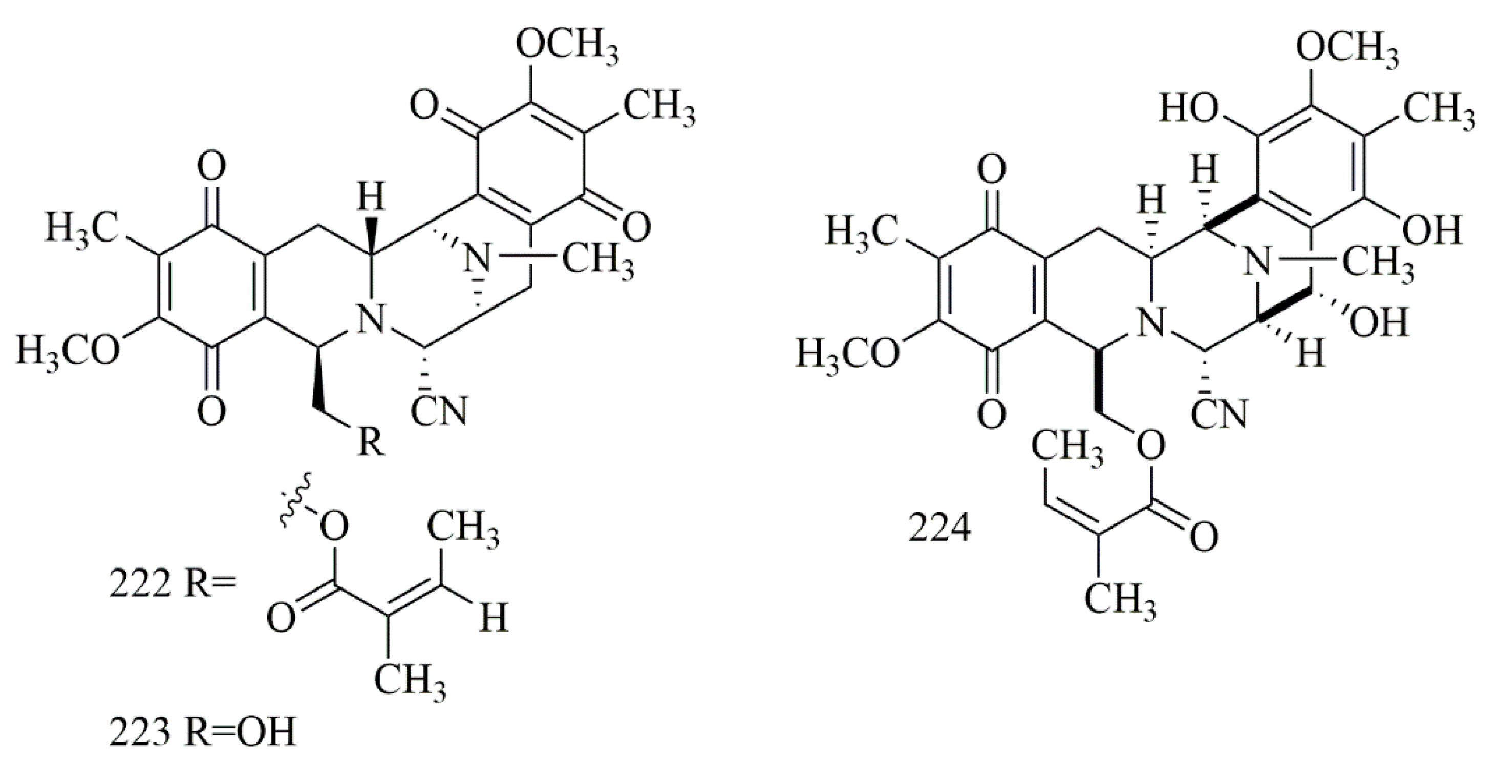
| 222 | renieramycin M | H292 = 23 H460 = 8.3 NSCLC = 24 | Xestospongia sp. | Thailand | [94] |
| 223 | jorunnamycin A | H292 = 220 H460 = 160 | |||
| 224 | renieramycin J | 3Y1 = 5.3 HeLa = 12.3 P388 = 0.53 | Neopetrosia sp. | Kuchinoerabu-jima Island | [95] |
| Compound | Cell Line (IC50 µM) | Source | Place of Collection | Ref. | |
|---|---|---|---|---|---|
| 225 | plakinamine I | HCT-116 = 10.6 | Corticium niger | Philippines | [96,97] |
| 226 | plakinamine J | HCT-116 = 6.1 NCI-60 screen = 1.4 | |||
| 227 | plakinamine K | HCT-116 = 1.4 | |||
| 228 | dihydroplakinamine K | HCT-116 = 1.4 | |||
| 229 | plakinamine N | NCI-60 screen = 11.5 | |||
| 230 | plakinamine O | NCI-60 screen = 2.4 |
| Compound | Cell Line (IC50 µM) | Source | Place of Collection | Ref. | |
| 231 | (+)-8-hydroxymanzamine A | SK-MEL = 0.83 KB = 1.38 BT-549 = 1.32 HepG2 = 0.90 LLC-PK11 = 2.21 | Acanthostrongylophora inges | Papua New Guinea | [98] |
| 232 | (+)-8-manzamine A | SK-MEL = 1.82 KB = 1.82 BT-549 = 1.82 HepG2 = 8.02 LLC-PK11 = 3.92 | |||
| 231 a | (+)-8-hydroxymanzamine A hydrochloride | SK-MEL = 1.30 KB = 1.15 BT-549 = 1.75 HepG2 = 2.5 LLC-PK11 = 3.08 | |||
| 232 a | (+)-8 manzamine A hydrochloride | SK-MEL = 0.97 KB = 0.56 BT-549 = 1.50 HepG2 = 2.65 LLC-PK11 = 1.18 |
| Compound | Cell Line (IC50 µM) | Source | Place of Collection | Ref. | |
|---|---|---|---|---|---|
| 233 | agelasine E | CLL = 16 | Agelas citrine and Agelas nakamurai | Caribbean Sea | [99,101] |
| 234 | 19-oxofasciospongine A | LNCaP = 21.8 LU = 5 MCF-7 = 13.4 | Fasciospongia sp. | Palau | [100] |
| 235 | iso-agelasine C | HL-60 = 25.3 K562 = 28.9 A549 > 50 HCT-116 = 38.8 | Agelas nakamurai | [101] | |
| 236 | agelasine J | HL-60 = 12.4 K562 = 16 A549 > 50 HCT-116 = 19.8 | |||
| 237 | nemoechine G | HL-60 = 18.4 K562 = 25.1 A549 > 50 HCT-116 = 33.9 |
| Compound | Cell Line (IC50 µM) | Source | Place of Collection | Ref. | |
|---|---|---|---|---|---|
| 238 | (−)-4’-methylaminoavarone | HCT116 = 9 H4IIE = 40 | Dysidea avara | Turkey | [102,103] |
| 239 | (−)-N-methylmelemeleone-A | HCT116 > 50 H4IIE > 50 | |||
| 240 | (−)-3’-methylaminoavarone | HCT116 = 45 H4IIE = 25 |
Publisher’s Note: MDPI stays neutral with regard to jurisdictional claims in published maps and institutional affiliations. |
© 2021 by the authors. Licensee MDPI, Basel, Switzerland. This article is an open access article distributed under the terms and conditions of the Creative Commons Attribution (CC BY) license (http://creativecommons.org/licenses/by/4.0/).
Share and Cite
Elissawy, A.M.; Soleiman Dehkordi, E.; Mehdinezhad, N.; Ashour, M.L.; Mohammadi Pour, P. Cytotoxic Alkaloids Derived from Marine Sponges: A Comprehensive Review. Biomolecules 2021, 11, 258. https://doi.org/10.3390/biom11020258
Elissawy AM, Soleiman Dehkordi E, Mehdinezhad N, Ashour ML, Mohammadi Pour P. Cytotoxic Alkaloids Derived from Marine Sponges: A Comprehensive Review. Biomolecules. 2021; 11(2):258. https://doi.org/10.3390/biom11020258
Chicago/Turabian StyleElissawy, Ahmed M., Ebrahim Soleiman Dehkordi, Negin Mehdinezhad, Mohamed L. Ashour, and Pardis Mohammadi Pour. 2021. "Cytotoxic Alkaloids Derived from Marine Sponges: A Comprehensive Review" Biomolecules 11, no. 2: 258. https://doi.org/10.3390/biom11020258
APA StyleElissawy, A. M., Soleiman Dehkordi, E., Mehdinezhad, N., Ashour, M. L., & Mohammadi Pour, P. (2021). Cytotoxic Alkaloids Derived from Marine Sponges: A Comprehensive Review. Biomolecules, 11(2), 258. https://doi.org/10.3390/biom11020258