Expression, Regulation, and Functions of the Galectin-16 Gene in Human Cells and Tissues


Abstract
1. Introduction
2. Materials and Methods
2.1. Bioinformatics Data and Tools
2.2. Cell Cultures
2.3. Gene Expression Analysis
2.4. Statistical Analysis
3. Results and Discussion
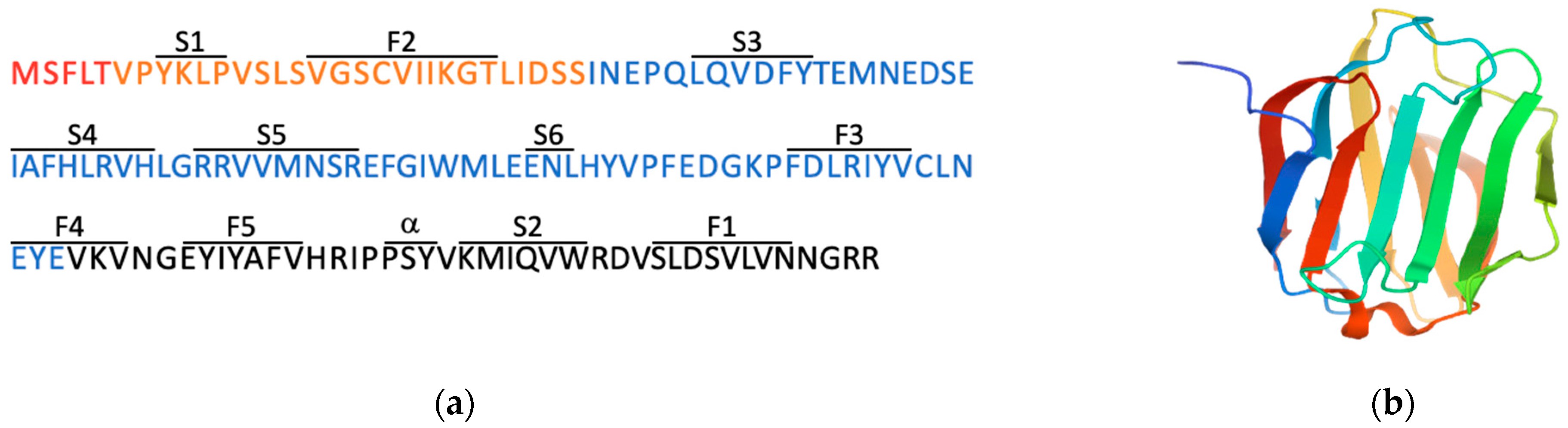
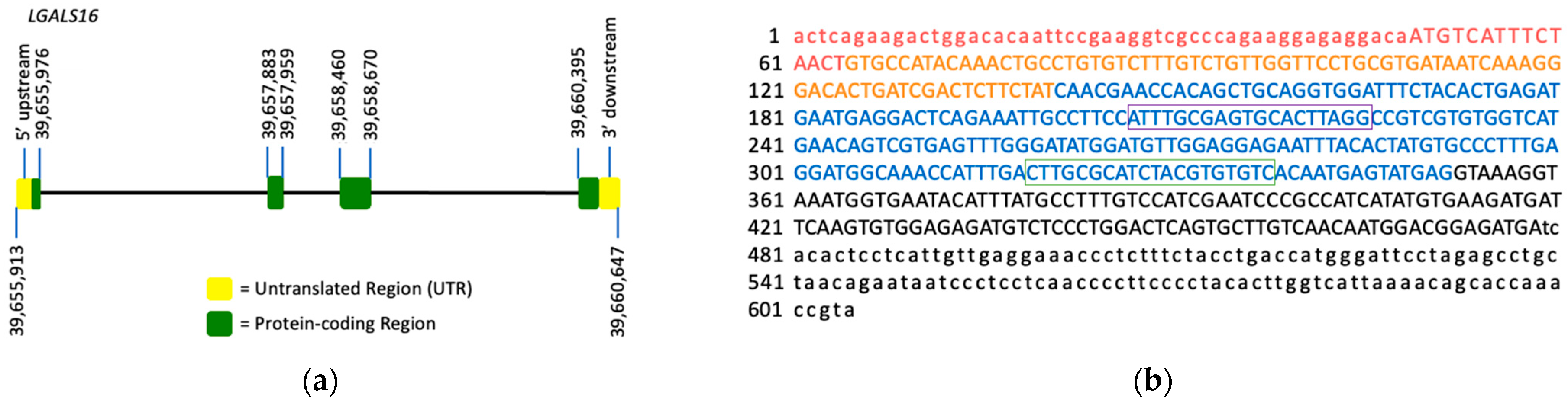
3.1. Molecular Characteristics of Galectin-16 Gene and Recombinant Protein
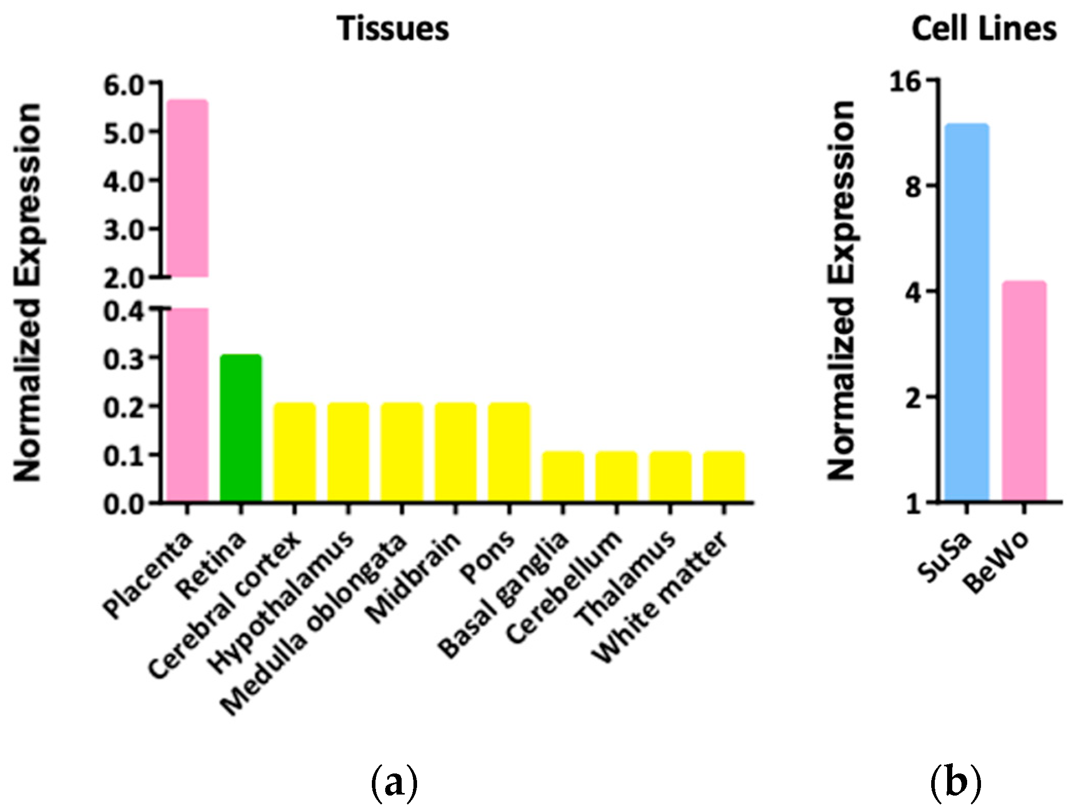
3.2. Expression Patterns and Functions of LGALS16 in Cells and Tissues
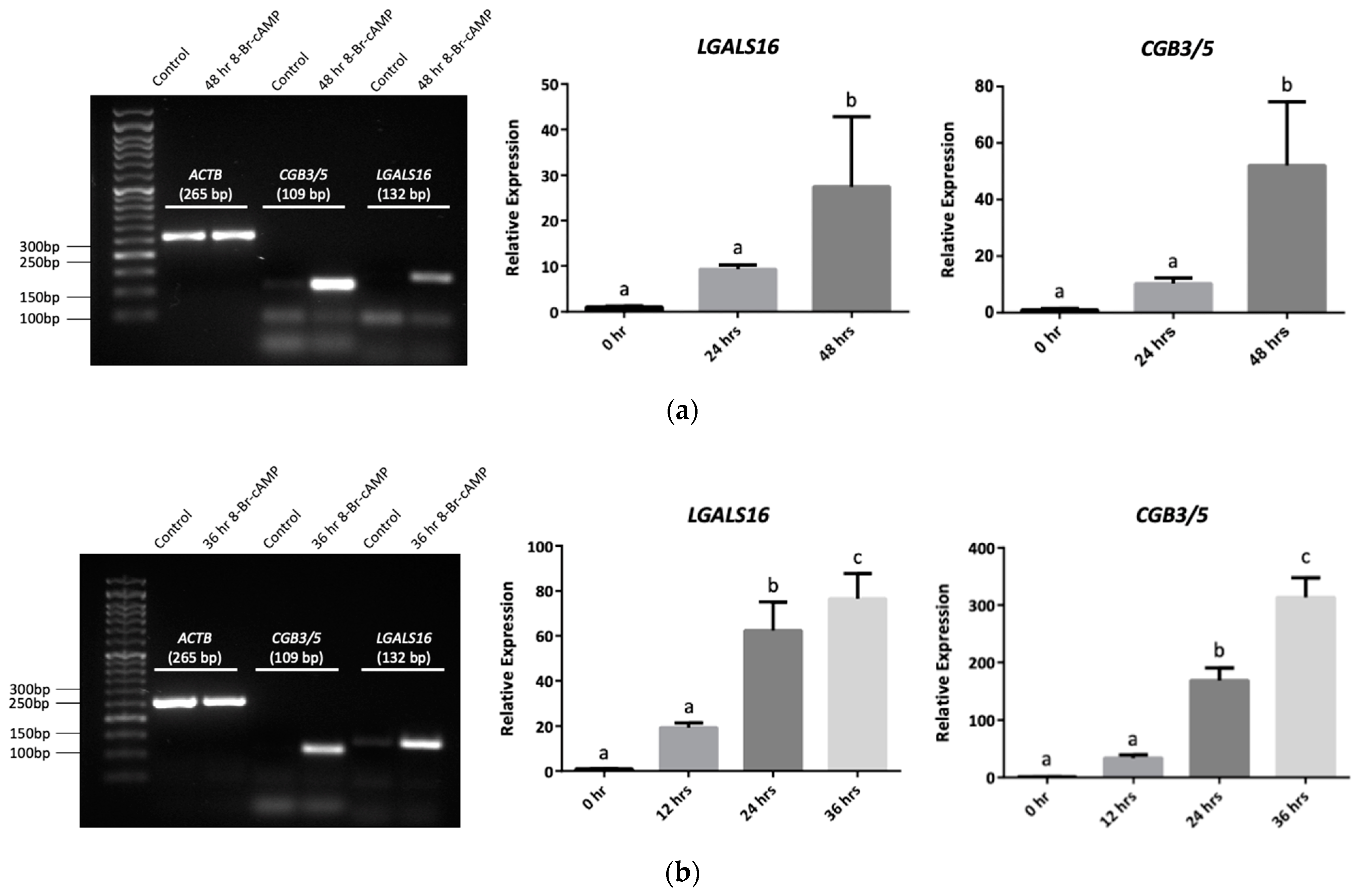
3.3. Transcriptional and Post-Transcriptional Regulation of LGALS16
3.3.1. Transcription Factors
3.3.2. miRNAs
3.4. LGALS16 and Human Diseases
4. Conclusions
Author Contributions
Funding
Data Availability Statement
Acknowledgments
Conflicts of Interest
References
- Timoshenko, A.V. Towards molecular mechanisms regulating the expression of galectins in cancer cells. Cell. Mol. Life Sci. 2015, 72, 4327–4340. [Google Scholar] [CrossRef] [Scilit] [PubMed]
- Allo, V.C.M.; Toscano, M.A.; Pinto, N.; Rabinovich, G.A. Galectins: Key players at the frontiers of innate and adaptive immunity. Trends Glycosci. Glycotechnol. 2018, 30, SE97–SE107. [Google Scholar] [CrossRef] [Scilit]
- Johannes, L.; Jacob, R.; Leffler, H. Galectins at a glance. J. Cell Sci. 2018, 131, jcs208884. [Google Scholar] [CrossRef] [Scilit] [PubMed]
- Tazhitdinova, R.; Timoshenko, A.V. The emerging role of galectins and O-GlcNAc homeostasis in processes of cellular differentiation. Cells 2020, 9, 8. [Google Scholar] [CrossRef] [Scilit] [PubMed]
- Vladoiu, M.C.; Labrie, M.; St-Pierre, Y. Intracellular galectins in cancer cells: Potential new targets for therapy (Review). Int. J. Oncol. 2014, 44, 1001–1014. [Google Scholar] [CrossRef] [Scilit]
- Patterson, R.J.; Haudek, K.C.; Voss, P.G.; Wang, J.L. Examination of the role of galectins in pre-mRNA splicing. Methods Mol. Biol. 2015, 1207, 431–449. [Google Scholar]
- Popa, S.J.; Stewart, S.E.; Moreau, K. Unconventional secretion of annexins and galectins. Semin. Cell Dev. Biol. 2018, 83, 42–50. [Google Scholar] [CrossRef] [Scilit] [PubMed]
- He, J.; Baum, L.G. Galectin interactions with extracellular matrix and effects on cellular function. Methods Enzymol. 2006, 417, 247–256. [Google Scholar]
- Nabi, I.R.; Shankar, J.; Dennis, J.W. The galectin lattice at a glance. J. Cell Sci. 2015, 128, 2213–2219. [Google Scholar] [CrossRef] [Scilit]
- Than, N.G.; Romero, R.; Kim, C.J.; McGowen, M.R.; Papp, Z.; Wildman, D.E. Galectins: Guardians of eutherian pregnancy at the maternal-fetal interface. Trends Endocrinol. Metab. 2012, 23, 23–31. [Google Scholar] [CrossRef] [Scilit] [PubMed]
- Than, N.G.; Romero, R.; Goodman, M.; Weckle, A.; Xing, J.; Dong, Z.; Xu, Y.; Tarquini, F.; Szilagyi, A.; Gal, P.; et al. A primate subfamily of galectins expressed at the maternal-fetal interface that promote immune cell death. Proc. Natl. Acad. Sci. USA 2009, 106, 9731–9736. [Google Scholar] [CrossRef] [Scilit]
- Than, N.G.; Romero, R.; Xu, Y.; Erez, O.; Xu, Z.; Bhatti, G.; Leavitt, R.; Chung, T.H.; El-Azzamy, H.; LaJeunesse, C.; et al. Evolutionary origins of the placental expression of chromosome 19 cluster galectins and their complex dysregulation in preeclampsia. Placenta 2014, 35, 855–865. [Google Scholar] [CrossRef] [Scilit]
- Pollheimer, J.; Vondra, S.; Baltayeva, J.; Beristain, A.G.; Knöfler, M. Regulation of placental extravillous trophoblasts by the maternal uterine environment. Front. Immunol. 2018, 9, 2597. [Google Scholar] [CrossRef] [Scilit]
- Blois, S.M.; Dveksler, G.; Vasta, G.R.; Freitag, N.; Blanchard, V.; Barrientos, G. Pregnancy galectinology: Insights into a complex network of glycan binding proteins. Front. Immunol. 2019, 10, 1166. [Google Scholar] [CrossRef] [Scilit] [PubMed]
- Messeguer, X.; Escudero, R.; Farré, D.; Núñez, O.; Martínez, J.; Albà, M.M. PROMO: Detection of known transcription regulatory elements using species-tailored searches. Bioinformatics 2002, 18, 333–334. [Google Scholar] [CrossRef] [Scilit] [PubMed]
- Farré, D.; Roset, R.; Huerta, M.; Adsuara, J.E.; Roselló, L.; Albà, M.M.; Messeguer, X. Identification of patterns in biological sequences at the ALGGEN server: PROMO and MALGEN. Nucleic Acids Res. 2003, 31, 3651–3653. [Google Scholar] [CrossRef] [Scilit] [PubMed]
- Paraskevopoulou, M.D.; Georgakilas, G.; Kostoulas, N.; Vlachos, I.S.; Vergoulis, T.; Reczko, M.; Filippidis, C.; Dalamagas, T.; Hatzigeorgiou, A.G. DIANA-microT web server v5.0: Service integration into miRNA functional analysis workflows. Nucleic Acids Res. 2013, 41, W169–W173. [Google Scholar] [CrossRef] [Scilit] [PubMed]
- Quillet, A.; Saad, C.; Ferry, G.; Anouar, Y.; Vergne, N.; Lecroq, T.; Dubessy, C. Improving bioinformatics prediction of microRNA targets by ranks aggregation. Front. Genet. 2020, 10, 1330. [Google Scholar] [CrossRef] [Scilit] [PubMed]
- Chen, Y.; Wang, X. miRDB: An online database for prediction of functional microRNA targets. Nucleic Acids Res. 2020, 48, D127–D131. [Google Scholar] [CrossRef] [Scilit] [PubMed]
- Agarwal, V.; Bell, G.W.; Nam, J.W.; Bartel, D.P. Predicting effective microRNA target sites in mammalian mRNAs. eLife 2015, 4, e05005. [Google Scholar] [CrossRef] [Scilit]
- Uhlén, M.; Fagerberg, L.; Hallström, B.M.; Lindskog, C.; Oksvold, P.; Mardinoglu, A.; Sivertsson, A.; Kampf, C.; Sjöstedt, E.; Asplund, A. Tissue-based map of the human proteome. Science 2015, 347, 1260419. [Google Scholar] [CrossRef] [Scilit]
- Clark, K.; Karsch-Mizrachi, I.; Lipman, D.J.; Ostell, J.; Sayers, E.W. GenBank. Nucleic Acids Res. 2016, 44, D67–D72. [Google Scholar] [CrossRef] [Scilit] [PubMed]
- Berman, H.M.; Westbrook, J.; Feng, Z.; Gilliland, G.; Bhat, T.N.; Weissig, H.; Shindyalov, I.N.; Bourne, P.E. The protein data bank. Nucleic Acids Res. 2000, 28, 235–242. [Google Scholar] [CrossRef] [Scilit] [PubMed]
- Ye, J.; Coulouris, G.; Zaretskaya, I.; Cutcutache, I.; Rozen, S.; Madden, T.L. Primer-BLAST: A tool to design target-specific primers for polymerase chain reaction. BMC Bioinform. 2012, 13, 134. [Google Scholar] [CrossRef] [Scilit] [PubMed]
- Timoshenko, A.V. Chitin hydrolysate stimulates VEGF-C synthesis by MDA-MB-231 breast cancer cells. Cell Biol. Int. 2011, 35, 281–286. [Google Scholar] [CrossRef] [Scilit]
- Renaud, S.J.; Chakraborty, D.; Mason, C.W.; Rumi, M.A.; Vivian, J.L.; Soares, M.J. OVO-like 1 regulates progenitor cell fate in human trophoblast development. Proc. Natl. Acad. Sci. USA 2015, 112, E6175–E6184. [Google Scholar] [CrossRef] [Scilit] [PubMed]
- Timoshenko, A.V.; Lanteigne, J.; Kozak, K. Extracellular stress stimuli alter galectin expression profiles and adhesion characteristics of HL-60 cells. Mol. Cell Biochem. 2016, 413, 137–143. [Google Scholar] [CrossRef] [Scilit]
- Sherazi, A.A.; Jariwala, K.A.; Cybulski, A.N.; Lewis, J.W.; Karagiannis, J.; Cumming, R.C.; Timoshenko, A.V. Effects of global O-GlcNAcylation on galectin gene-expression profiles in human cancer cell lines. Anticancer Res. 2018, 38, 6691–6697. [Google Scholar] [CrossRef] [Scilit] [PubMed]
- Ely, A.Z.; Moon, J.M.; Sliwoski, G.R.; Sangha, A.K.; Shen, X.-X.; Labella, A.L.; Meiler, J.; Capra, J.A.; Rokas, A. The impact of natural selection on the evolution and function of placentally expressed galectins. Genome Biol. Evol. 2019, 11, 2574–2592. [Google Scholar] [CrossRef] [Scilit]
- Singer, M.F. SINEs and LINEs: Highly repeated short and long interspersed sequences in mammalian genomes. Cell 1982, 28, 433–434. [Google Scholar] [CrossRef] [Scilit]
- Weckselblatt, B.; Rudd, M.K. Human structural variation: Mechanisms of chromosome rearrangements. Trends Genet. 2015, 31, 587–599. [Google Scholar] [CrossRef] [Scilit] [PubMed]
- Si, Y.; Yao, Y.; Ayala, G.J.; Li, X.; Han, Q.; Zhang, W.; Xu, X.; Tai, G.; Mayo, K.H.; Zhou, Y.; et al. Human galectin-16 has a pseudo ligand binding site and plays a role in regulating c-Rel mediated lymphocyte activity. Biochim. Biophys. Acta Gen. Subj. 2021, 1865, 129755. [Google Scholar] [CrossRef] [Scilit] [PubMed]
- Barrett, T.; Wilhite, S.E.; Ledoux, P.; Evangelista, C.; Kim, I.F.; Tomashevsky, M.; Marshall, K.A.; Phillippy, K.H.; Sherman, P.M.; Holko, M.; et al. NCBI GEO: Archive for functional genomics data sets—Update. Nucleic Acids Res. 2013, 41, D991–D995. [Google Scholar] [CrossRef] [Scilit]
- Rosenfeld, C.S. The placenta-brain-axis. J. Neurosci. Res. 2021, 99, 271–283. [Google Scholar] [CrossRef] [Scilit]
- Inamochi, Y.; Mochizuki, K.; Goda, T. Histone code of genes induced by co-treatment with a glucocorticoid hormone agonist and a p44/42 MAPK inhibitor in human small intestinal Caco-2 cells. Biochim. Biophys. Acta 2014, 1840, 693–700. [Google Scholar] [CrossRef] [Scilit] [PubMed]
- Park, M.H.; Hong, J.T. Roles of NF-κB in cancer and inflammatory diseases and their therapeutic approaches. Cells 2016, 5, 15. [Google Scholar] [CrossRef] [Scilit]
- Hayden, M.S.; Ghosh, S. NF-κB in immunobiology. Cell Res. 2011, 21, 223–244. [Google Scholar] [CrossRef] [Scilit]
- Balsa, E.; Perry, E.A.; Bennett, C.F.; Jedrychowski, M.; Gygi, S.P.; Doench, J.G.; Puigserver, P. Defective NADPH production in mitochondrial disease complex I causes inflammation and cell death. Nat. Commun. 2020, 11, 2714. [Google Scholar] [CrossRef] [Scilit]
- Nuzzo, A.M.; Giuffrida, D.; Zenerino, C.; Piazzese, A.; Olearo, E.; Todros, T.; Rolfo, A. JunB/Cyclin-D1 imbalance in placental mesenchymal stromal cells derived from preeclamptic pregnancies with fetal-placental compromise. Placenta 2014, 35, 483–490. [Google Scholar] [CrossRef] [Scilit]
- Han, Y.M.; Romero, R.; Kim, J.S.; Tarca, A.L.; Kim, S.K.; Draghici, S.; Kusanovic, J.P.; Gotsch, F.; Mittal, P.; Hassan, S.S.; et al. Region-specific gene expression profiling: Novel evidence for biological heterogeneity of the human amnion. Biol. Reprod. 2008, 79, 954–961. [Google Scholar] [CrossRef] [Scilit]
- Knyazev, E.N.; Zakharova, G.S.; Astakhova, L.A.; Tsypina, I.M.; Tonevitsky, A.G.; Sukhikh, G.T. Metabolic reprogramming of trophoblast cells in response to hypoxia. Bull. Exp. Biol. Med. 2019, 166, 321–325. [Google Scholar] [CrossRef] [Scilit] [PubMed]
- Walcott, B.P.; Winkler, E.A.; Zhou, S.; Birk, H.; Guo, D.; Koch, M.J.; Stapleton, C.J.; Spiegelman, D.; Dionne-Laporte, A.; Dion, P.A.; et al. Identification of a rare BMP pathway mutation in a non-syndromic human brain arteriovenous malformation via exome sequencing. Hum. Genome Var. 2018, 5, 18001. [Google Scholar] [CrossRef] [Scilit] [PubMed]
- Ma, S.; Pang, C.; Song, L.; Guo, F.; Sun, H. Activating transcription factor 3 is overexpressed in human glioma and its knockdown in glioblastoma cells causes growth inhibition both in vitro and in vivo. Int. J. Mol. Med. 2015, 35, 1561–1573. [Google Scholar] [CrossRef] [Scilit]
- Garces de Los Favos Alonso, I.; Liang, H.C.; Turner, S.D.; Lagger, S.; Merkel, O.; Kenner, L. The role of activator protein-1 (AP-1) family members in CD30-positive lymphomas. Cancers 2018, 10, 93. [Google Scholar] [CrossRef] [Scilit]
- Shankar, K.; Kang, P.; Zhong, Y.; Borengasser, S.J.; Wingfield, C.; Saben, J.; Gomez-Acevedo, H.; Thakali, K.M. Transcriptomic and epigenomic landscapes during cell fusion in BeWo trophoblast cells. Placenta 2015, 36, 1342–1351. [Google Scholar] [CrossRef] [Scilit]
- Cheng, Y.-H.; Richardson, B.D.; Hubert, M.A.; Handwerger, S. Isolation and characterization of the human syncytin gene promoter. Biol. Reprod. 2004, 70, 694–701. [Google Scholar] [CrossRef] [Scilit] [PubMed]
- Wang, Z.; Liu, Y.; Liu, J.; Kong, N.; Jiang, Y.; Jiang, R.; Zhen, X.; Zhou, J.; Li, C.; Sun, H.; et al. ATF3 deficiency impairs the proliferative-secretory phase transition and decidualization in RIF patients. Cell Death Dis. 2021, 12, 387. [Google Scholar] [CrossRef] [Scilit]
- Jadhav, K.; Zhang, Y. Activating transcription factor 3 in immune response and metabolic regulation. Liver Res. 2017, 1, 96–102. [Google Scholar] [CrossRef] [Scilit]
- Moslehi, R.; Mills, J.L.; Signore, C.; Kumar, A.; Ambroggio, X.; Dzutsev, A. Integrative transcriptome analysis reveals dysregulation of canonical cancer molecular pathways in placenta leading to preeclampsia. Sci. Rep. 2013, 3, 2407. [Google Scholar] [CrossRef] [Scilit]
- Tsukamoto, S.; Mizuta, T.; Fujimoto, M.; Ohte, S.; Osawa, K.; Miyamoto, A.; Yoneyama, K.; Murata, E.; Machiya, A.; Jimi, E.; et al. Smad9 is a new type of transcriptional regulator in bone morphogenetic protein signaling. Sci. Rep. 2014, 4, 7596. [Google Scholar] [CrossRef] [Scilit]
- Heo, K.S.; Fujiwara, K.; Abe, J. Disturbed-flow-mediated vascular reactive oxygen species induce endothelial dysfunction. Circ. J. 2011, 75, 2722–2730. [Google Scholar] [CrossRef] [Scilit] [PubMed]
- Fabian, M.R.; Sonenberg, N.; Filipowicz, W. Regulation of mRNA translation and stability by microRNAs. Annu. Rev. Biochem. 2010, 79, 351–379. [Google Scholar] [CrossRef] [Scilit] [PubMed]
- Menon, R.; Debnath, C.; Lai, A.; Guanzon, D.; Bhatnagar, S.; Kshetrapal, P.K.; Sheller-Miller, S.; Salomon, C.; Garbhini Study Team. Circulating exosomal miRNA profile during term and preterm birth pregnancies: A longitudinal study. Endocrinology 2019, 160, 249–275. [Google Scholar] [CrossRef] [Scilit]
- Yoshino, Y.; Roy, B.; Dwivedi, Y. Altered miRNA landscape of the anterior cingulate cortex is associated with potential loss of key neuronal functions in depressed brain. Eur. Neuropsychopharmacol. 2020, 40, 70–84. [Google Scholar] [CrossRef] [Scilit]
- Yan, S.; Zhang, H.; Xie, W.; Meng, F.; Zhang, K.; Jiang, Y.; Zhang, X.; Zhang, J. Altered microRNA profiles in plasma exosomes from mesial temporal lobe epilepsy with hippocampal sclerosis. Oncotarget 2017, 8, 4136–4146. [Google Scholar] [CrossRef] [Scilit] [PubMed]
- Liao, B.; Zhou, M.X.; Zhou, F.K.; Luo, X.M.; Zhong, S.X.; Zhou, Y.F.; Qin, Y.S.; Li, P.P.; Qin, C. Exosome-derived miRNAs as biomarkers of the development and progression of intracranial aneurysms. J. Atheroscler. Thromb. 2020, 27, 545–610. [Google Scholar] [CrossRef] [Scilit]
- Ludwig, N.; Leidinger, P.; Becker, K.; Backes, C.; Fehlmann, T.; Pallasch, C.; Rheinheimer, S.; Meder, B.; Stähler, C.; Meese, E.; et al. Distribution of miRNA expression across human tissues. Nucleic Acids Res. 2016, 44, 3865–3877. [Google Scholar] [CrossRef] [Scilit]
- Gong, S.; Gaccioli, F.; Dopierala, J.; Sovio, U.; Cook, E.; Volders, P.J.; Martens, L.; Kirk, P.D.W.; Richardson, S.; Smith, G.C.S.; et al. The RNA landscape of the human placenta in health and disease. Nat. Commun. 2021, 12, 2639. [Google Scholar] [CrossRef] [Scilit] [PubMed]
- Braun, A.E.; Muench, K.L.; Robinson, B.G.; Wang, A.; Palmer, T.D.; Winn, V.D. Examining Sex Differences in the Human Placental Transcriptome During the First Fetal Androgen Peak. Reprod Sci. 2021, 28, 801–818. [Google Scholar] [CrossRef] [Scilit]
- Vastrad, B.; Vastrad, C. Bioinformatics analyses of significant genes, related pathways and candidate prognostic biomarkers in Alzheimer’s disease. BioRxiv 2021. [Google Scholar] [CrossRef] [Scilit]
- Zhao, B.; Shan, Y.; Yang, Y.; Zhaolong, Y.; Li, T.; Wang, X.; Luo, T.; Zhu, Z.; Sullivan, P.; Zhao, H.; et al. Transcriptome-wide association analysis of brain structures yields insights into pleiotropy with complex neuropsychiatric traits. Nat. Commun. 2021, 12, 2878. [Google Scholar] [CrossRef] [Scilit] [PubMed]
- Čokić, V.P.; Mojsilović, S.; Jauković, A.; Kraguljac-Kurtović, N.; Mojsilović, S.; Šefer, D.; Mitrović Ajtić, O.; Milošević, V.; Bogdanović, A.; Đikić, D.; et al. Gene expression profile of circulating CD34(+) cells and granulocytes in chronic myeloid leukemia. Blood Cells Mol. Dis. 2015, 55, 373–381. [Google Scholar] [CrossRef] [Scilit] [PubMed]
- Rodrigues-Peres, R.M.; de Carvalho, B.S.; Anurag, M.; Lei, J.T.; Conz, L.; Gonçalves, R.; Cardoso Filho, C.; Ramalho, S.; de Paiva, G.R.; Derchain, S.; et al. Copy number alterations associated with clinical features in an underrepresented population with breast cancer. Mol. Genet. Genomic. Med. 2019, 7, e00750. [Google Scholar] [CrossRef] [Scilit] [PubMed]
- Santos, J.X.; Rasga, C.; Marques, A.R.; Martiniano, H.F.M.C.; Asif, M.; Vilela, J.; Oliveira, G.; Vicente, A.M. A role for gene-environment interactions in Autism Spectrum Disorder is suggested by variants in genes regulating exposure to environmental factors. BioRxiv 2019. [Google Scholar] [CrossRef] [Scilit]
- Arthur, S.E.; Jiang, A.; Grande, B.M.; Alcaide, M.; Cojocaru, R.; Rushton, C.K.; Mottok, A.; Hilton, L.K.; Kumar Lat, P.; Zhao, E.Y. Genome-wide discovery of somatic regulatory variants in diffuse large B-cell lymphoma. Nat. Commun. 2018, 9, 4001. [Google Scholar] [CrossRef] [Scilit] [PubMed]
- Keaton, J.M.; Hellwege, J.N.; Ng, M.C.; Palmer, N.D.; Pankow, J.S.; Fornage, M.; Wilson, J.G.; Correa, A.; Rasmussen-Torvik, L.J.; Rotter, J.I.; et al. Genome-wide interaction with insulin secretion loci reveals novel loci for type 2 diabetes in African Americans. PLoS ONE 2016, 11, e0159977. [Google Scholar] [CrossRef] [Scilit]
- Cheng, P.; Feng, F.; Yang, H.; Jin, S.; Lai, C.; Wang, Y.; Bi, J. Detection and significance of exosomal mRNA expression profiles in the cerebrospinal fluid of patients with meningeal carcinomatosis. J. Mol. Neurosci. 2021, 71, 790–803. [Google Scholar] [CrossRef] [Scilit]

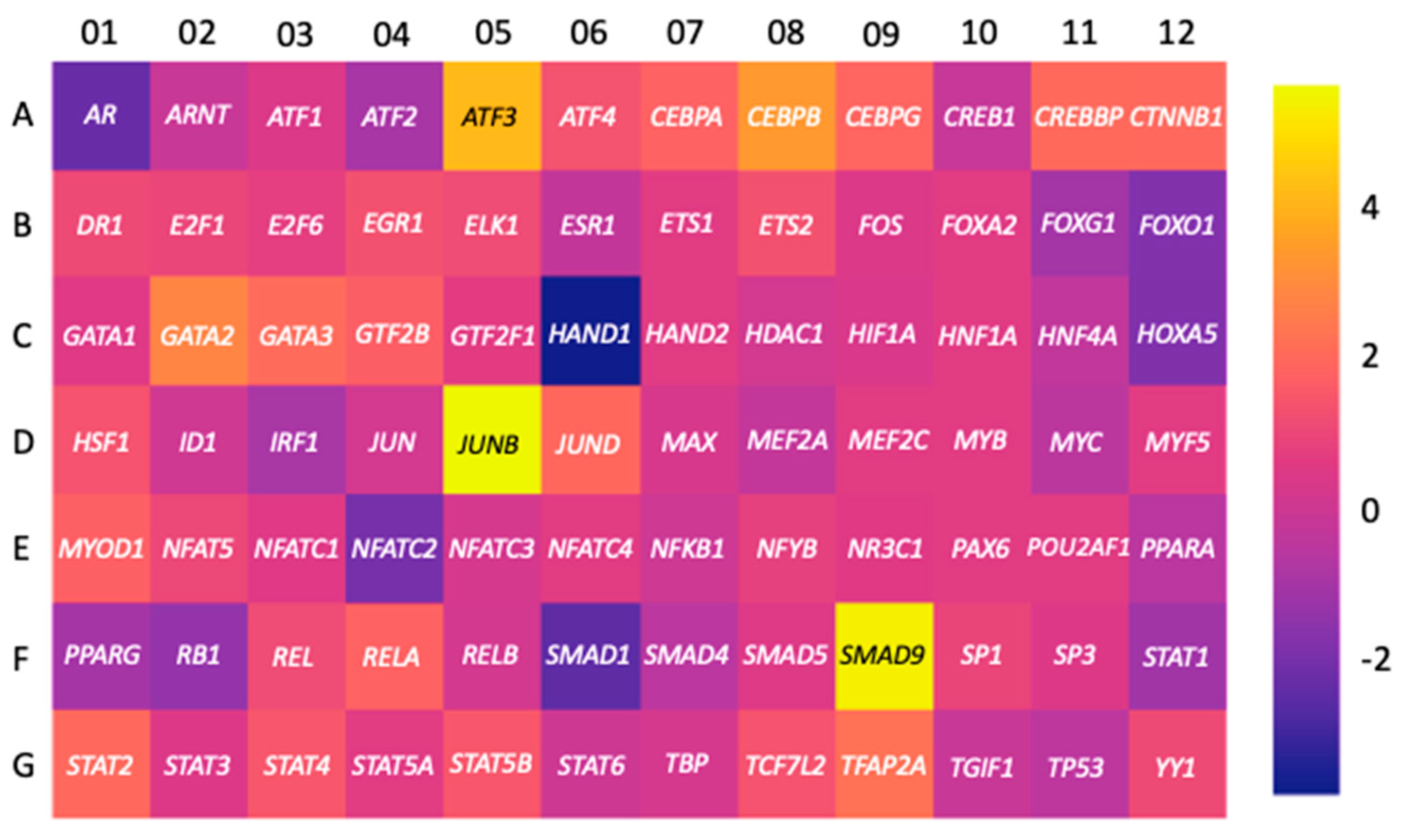
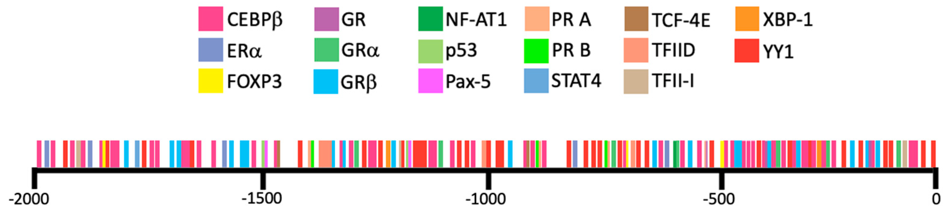
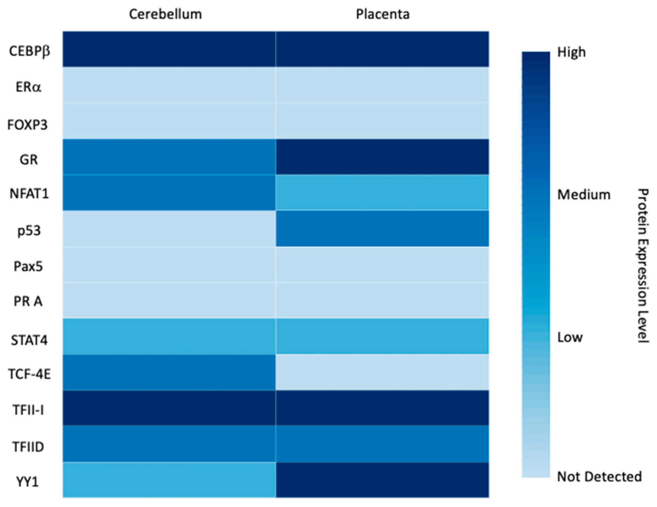
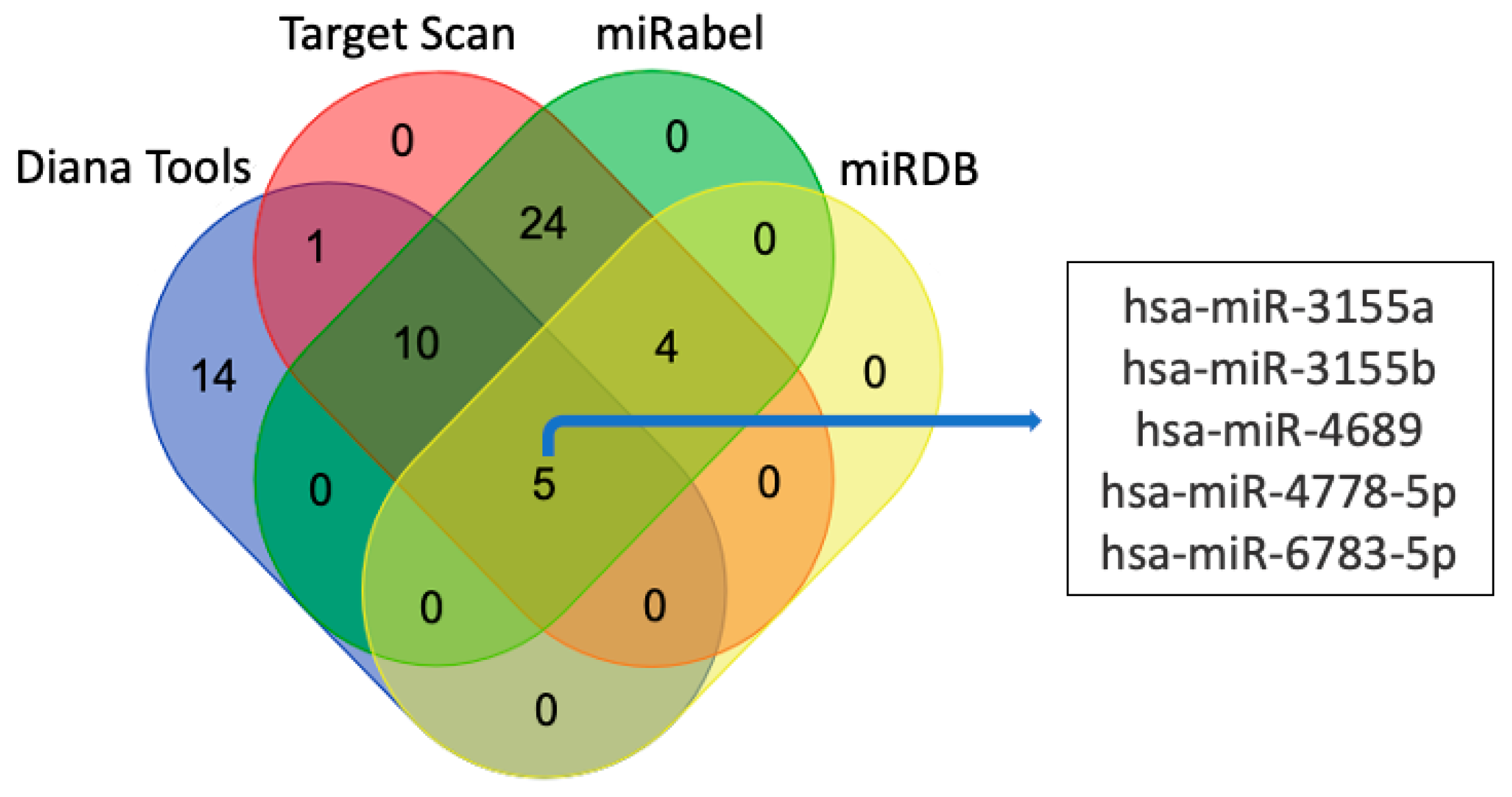

| Names of Cells or Tissues | GEO Accession Number | ACTB | LGALS1 | LGALS16 | Sample Size |
|---|---|---|---|---|---|
| Acetabular labrum cells | GDS5427 a | 12.682 ± 0.150 | 12.057 ± 0.107 | 2.949 ± 0.0093 | 3 |
| Acute lymphoblastic leukemia cell line RS4;11 | GDS4043 b | 13.861 ± 0.017 | 11.487 ± 0.033 | 0.4023 ± 0.607 | 2 |
| Acute myeloblastic leukemia cell line Kasumi-1 | GDS5600 a | 11.965 ± 0.025 | 6.455 ± 0.299 | 2.918 ± 0.036 | 3 |
| Acute promyelocytic leukemia cell line NB4 | GDS4180 a | 13.130 ± 0.035 | 10.823 ± 0.031 | 3.650 ± 0.108 | 3 |
| Adipocyte progenitor cells (subcutaneous) | GDS5171 a | 13.523 ± 0.038 | 13.397 ± 0.112 | 4.597 ± 0.251 | 6 |
| Adipocyte progenitors from deep neck | GDS5171 a | 13.469 ± 0.057 | 13.208 ± 0.177 | 4.505 ± 0.094 | 6 |
| Bone marrow CD34+ cells (chronic myeloid leukemia) | GDS4756 a | 13.524 | 11.137 | 3.050 | 1 |
| Bone marrow plasma cells | GDS4968 a | 11.990 ± 0.226 | 8.714 ± 0.515 | 3.052 ± 0.257 | 5 |
| Brain frontal cortex | GDS4758 a | 13.402 ± 0.125 | 96.333 ± 0.840 | 4.632 ± 0.249 | 18 |
| Brain hippocampus | GDS4758 a | 13.477 ± 0.130 | 11.133 ± 0.375 | 4.659 ± 0.300 | 10 |
| Brain hippocampus | GDS4879 a | 12.113 ± 0.409 | 9.076 ± 0.232 | 3.177 ± 0.177 | 19 |
| Brain temporal cortex | GDS4758 a | 13.560 ± 0.131 | 11.189 ± 0.280 | 4.749 ± 0.193 | 19 |
| Breast cancer cell line MCF-7 | GDS2759 b | 15.884 ± 0.030 | 13.752 ± 0.153 | 6.053 ± 0.237 | 2 |
| Breast cancer cell line MCF-7 | GDS4972 a | 13.029 ± 0.038 | 12.439 ± 0.083 | 3.892 ± 0.066 | 3 |
| Breast cancer cell line MCF-7 | GDS4090 a | 13.087 ± 0.019 | 9.566 ± 0.100 | 2.827 ± 0.405 | 3 |
| Breast cancer cell line MDA-MB-231 | GDS4800 a | 13.875 ± 0.007 | 13.565 ± 0.042 | 5.189 ± 0.085 | 3 |
| Bronchial smooth muscle primary cells | GDS4803 a | 11.629 ± 0.175 | 11.533 ± 0.041 | 3.181 ± 0.095 | 3 |
| Bronchopulmonary neuroendocrine cell line NCI-H727 | GDS4330 a | 11.978 | 5.715 | 3.808 | 1 |
| Burkitt lymphoma cell line Namalwa | GDS4978 a | 13.468 ± 0.187 | 8.005 ± 0.073 | 3.916 ± 0.297 | 3 |
| Burkitt lymphoma cell line Raji | GDS4978 a | 13.367 ± 0.093 | 8.052 ± 0.141 | 3.962 ± 0.019 | 3 |
| Colorectal adenocarcinoma cell line SW620 | GDS5416 e | 16.400 ± 0.362 | 17.280 ± 0.043 | 2.766 ± 0.554 | 2 |
| Embryonic kidney cell line HEK-293 | GDS4233 a | 10.330 ± 0.050 | 7.109 ± 0.098 | 3.757 ± 0.328 | 4 |
| Endothelial progenitor cells | GDS3656 c | 15.397 ± 0.174 | 13.845 ± 0.457 | 8.018 ± 0.103 | 11 |
| Esophagus biopsies | GDS4350 a | 12.617 ± 0.230 | 8.062 ± 0.507 | 3.255 ± 0.208 | 8 |
| Gastrointestinal neuroendocrine cell line KRJ-1 | GDS4330 a | 12.135 | 9.592 | 2.859 | 1 |
| Germinal center B cells | GDS4977 a | 9.793 ± 0.373 | 8.438 ± 0.225 | 6.723 ± 0.538 | 5 |
| Gingival fibroblasts | GDS5811 a | 13.628 ± 0.101 | 13.770 ± 0.174 | 3.674 ± 0.140 | 2 |
| Heart (left ventricle) | GDS4772 a | 11.293 ± 0.361 | 10.672 ± 0.377 | 2.941 ± 0.030 | 5 |
| Heart (left ventricle) | GDS4314 a | 12.142 ± 0.365 | 11.052 ± 0.223 | 3.344 ± 0.154 | 5 |
| Heart (right ventricular) | GDS5610 a | 11.930 ± 0.255 | 10.934 ± 0.044 | 3.637 ± 0.181 | 2 |
| Hepatocellular carcinoma cell line HepG2 | GDS5340 a | 13.259 ± 0.039 | 11.256 ± 0.054 | 4.281 ± 0.327 | 3 |
| Microglia cell line HMO6 | GDS4151 a | 13.545 | 12.231 | 2.979 | 1 |
| Keratinocytes | GDS4426 a | 12.679 ± 0.056 | 11.147 ± 0.236 | 3.804 ± 0.138 | 6 |
| Lung carcinoma cell line A549 | GDS4997 a | 10.970 ± 0.044 | 12.187 ± 0.049 | 2.418 ± 0.072 | 3 |
| Lung carcinoma cell line H460 | GDS5247 a | 12.504 ± 0.043 | 11.111 ± 0.063 | 3.439 ± 0.117 | 3 |
| Lung microvascular endothelial cell line CC-2527 | GDS2987 b | 32,061 ± 7366 | 15,158 ± 2227 | 8.100 ± 9.051 | 2 |
| Lymphoblastoid cell line TK6 | GDS4915 a | 13.365 ± 0.061 | 11.161 ± 0.323 | 4.005 ± 0.327 | 2 |
| Lymphoblastoid cell line TK6 | GDS4916 a | 13.940 ± 0.058 | 12.023 ± 0.130 | 4.061 ± 0.357 | 2 |
| Medulloblastoma tumor tissue | GDS4469 a | 13.099 ± 0.302 | 9.490 ± 0.801 | 4.005 ± 0.839 | 15 |
| Melanoma cell line A-375 | GDS5085 a | 13.888 ± 0.011 | 13.474 ± 0.101 | 4.618 ± 0.045 | 3 |
| Melanoma cell line FEMX-I | GDS3489 d | 16.04 ± 0.354 | 16.04 ± 0.354 | 0.550 ± 1.061 | 2 |
| Melanoma cell line Hs294T | GDS5670 a | 11.353 ± 0.245 | 10.349 ± 0.097 | 2.149 ± 0.585 | 2 |
| Microglia cell line HMO6 | GDS4151 a | 13.545 | 12.231 | 2.979 | 1 |
| Myotubes from musculus obliquus internus | GDS5378 a | 13.224 ± 0.099 | 12.925 ± 0.114 | 2.840 ± 0.057 | 4 |
| Pancreatic neuroendocrine cell line QGP-1 | GDS4330 a | 12.057 | 5.749 | 3.031 | 1 |
| Peripheral blood CD34+ cells (chronic myeloid leukemia) | GDS4756 a | 13.414 ± 0.049 | 11.144 ± 0.578 | 2.974 ± 0.140 | 2 |
| Peripheral blood CD4+ T cells | GDS5544 a | 13.598 ± 0.053 | 9.707 ± 0.247 | 4.584 ± 0.126 | 4 |
| Peripheral blood cells | GDS4240 a | 11.825 ± 0.084 | 7.307 ± 0.154 | 1.506 ± 0.112 | 7 |
| Renal adenocarcinoma cell line 786-O | GDS5810 a | 12.902 ± 0.030 | 12.809 ± 0.015 | 5.753 ± 0.031 | 2 |
| Retinal pigment epithelia primary cells | GDS4224 a | 13.407 ± 0.110 | 11.842 ± 0.449 | 3.468 ± 0.367 | 4 |
| Retinal pigmented epithelium cell line ARPE-19 | GDS4224 a | 13.288 | 11.946 | 3.646 | 1 |
| Skeletal muscle (vastus lateralis) primary cells | GDS4920 a | 13.649 ± 0.084 | 13.385 ± 0.114 | 4.609 ± 0.136 | 12 |
| Skeletal muscle tissue | GDS4841 a | 9.400 ± 0.190 | 11.486 ± 0.247 | 2.786 ± 0.355 | 5 |
| Skin cancer cell line RT3Sb | GDS5381 a | 13.409 ± 0.062 | 8.775 ± 0.114 | 3.539 ± 0.252 | 4 |
| Skin epidermis | GDS3806 c | 15.139 ± 0.141 | 9.534 ± 0.370 | 7.909 ± 0.469 | 7 |
| Visceral adipose tissue (omentum) | GDS4857 a | 11.875 ± 0.352 | 11.488 ± 0.416 | 4.666 ± 0.754 | 8 |
Publisher’s Note: MDPI stays neutral with regard to jurisdictional claims in published maps and institutional affiliations. |
© 2021 by the authors. Licensee MDPI, Basel, Switzerland. This article is an open access article distributed under the terms and conditions of the Creative Commons Attribution (CC BY) license (https://creativecommons.org/licenses/by/4.0/).
Share and Cite
Kaminker, J.D.; Timoshenko, A.V. Expression, Regulation, and Functions of the Galectin-16 Gene in Human Cells and Tissues. Biomolecules 2021, 11, 1909. https://doi.org/10.3390/biom11121909
Kaminker JD, Timoshenko AV. Expression, Regulation, and Functions of the Galectin-16 Gene in Human Cells and Tissues. Biomolecules. 2021; 11(12):1909. https://doi.org/10.3390/biom11121909
Chicago/Turabian StyleKaminker, Jennifer D., and Alexander V. Timoshenko. 2021. "Expression, Regulation, and Functions of the Galectin-16 Gene in Human Cells and Tissues" Biomolecules 11, no. 12: 1909. https://doi.org/10.3390/biom11121909
APA StyleKaminker, J. D., & Timoshenko, A. V. (2021). Expression, Regulation, and Functions of the Galectin-16 Gene in Human Cells and Tissues. Biomolecules, 11(12), 1909. https://doi.org/10.3390/biom11121909