Review

L-Type Ca2+ Channel Regulation by Calmodulin and CaBP1

Department of Chemistry, University of California, Davis, CA 95616, USA
Biomolecules 2021, 11(12), 1811; https://doi.org/10.3390/biom11121811
Submission received: 1 November 2021 / Revised: 30 November 2021 / Accepted: 30 November 2021 / Published: 2 December 2021
(This article belongs to the Collection Advances in Metal Binding Proteins)

Abstract

:
L-type voltage-gated Ca2+ channels (CaV1.2 and CaV1.3, called CaV) interact with the Ca2+ sensor proteins, calmodulin (CaM) and Ca2+ binding Protein 1 (CaBP1), that oppositely control Ca2+-dependent channel activity. CaM and CaBP1 can each bind to the IQ-motif within the C-terminal cytosolic domain of CaV, which promotes increased channel open probability under basal conditions. At elevated cytosolic Ca2+ levels (caused by CaV channel opening), Ca2+-bound CaM binding to CaV is essential for promoting rapid Ca2+-dependent channel inactivation (CDI). By contrast, CaV binding to CaBP1 prevents CDI and promotes Ca2+-induced channel opening (called CDF). In this review, I provide an overview of the known structures of CaM and CaBP1 and their structural interactions with the IQ-motif to help understand how CaM promotes CDI, whereas CaBP1 prevents CDI and instead promotes CDF. Previous electrophysiology studies suggest that Ca2+-free forms of CaM and CaBP1 may pre-associate with CaV under basal conditions. However, previous Ca2+ binding data suggest that CaM and CaBP1 are both calculated to bind to Ca2+ with an apparent dissociation constant of ~100 nM when CaM or CaBP1 is bound to the IQ-motif. Since the neuronal basal cytosolic Ca2+ concentration is ~100 nM, nearly half of the neuronal CaV channels are suggested to be bound to Ca2+-bound forms of either CaM or CaBP1 under basal conditions. The pre-association of CaV with calcified forms of CaM or CaBP1 are predicted here to have functional implications. The Ca2+-bound form of CaBP1 is proposed to bind to CaV under basal conditions to block CaV binding to CaM, which could explain how CaBP1 might prevent CDI.

1. Introduction

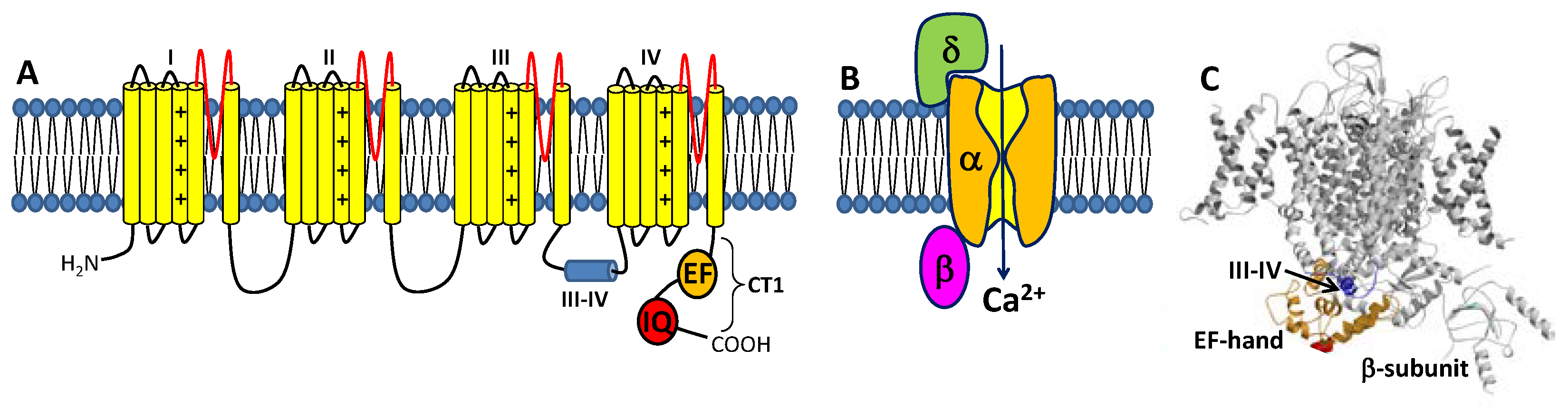
1.1. Voltage-Gated L-Type Ca2+ Channel Structure and Function

Synaptic transmission and neuronal excitability are regulated by the L-type voltage-gated Ca2+ channels (CaV1.2 and CaV1.3, called CaV) expressed in the brain and heart [1,2,3,4]. CaVs display slow voltage-dependent gating characteristics (L-type) and are sensitive to a number of different dihydropyridine (DHP) antagonists and agonists [5]. Under resting basal conditions, intracellular Ca2+ concentration is kept low (100 nM) due to the powerful action of Ca2+ pumps and exchangers [1,6] and Ca2+ sequestration into stores [1,7]. The opening of CaV channels causes intracellular Ca2+ levels to increase into the micromolar range [8]. This Ca2+ influx triggers a wide range of Ca2+-dependent processes including gene transcription [9], neurotransmitter release [10], neurite outgrowth [11], and the activation of Ca2+-dependent enzymes [12]. Prolonged elevation of intracellular Ca2+ levels is cytotoxic [13], and CaV channels are negatively regulated by a process known as Ca2+-dependent inactivation (CDI) [14,15,16]. Dysregulation of CaVs are linked to various types of neurological disorders, including epilepsy, migraine, and chronic pain [17].
The CaVs are a heteromultimeric protein complex formed by a pore-forming α-subunit and regulatory β and δ subunits (Figure 1). The α-subunit contains four major transmembrane domains (Figure 1A), each with six membrane-spanning helices (termed S1–S6) and a positively charged S4 segment that controls voltage-dependent activation [18]. The transmembrane domains are connected by long cytoplasmic linkers (III-IV inactivation gate [19]), bracketed by cytoplasmic N-terminal and C-terminal domains [20]. The C-terminal domain (residues 1508-1665, called CT1) is important for Ca2+-dependent regulation of channel function and contains important sites (EF-hand and IQ motifs) for protein–protein interactions [21,22,23]. A three-dimensional structure of the skeletal muscle CaV (called CaV1.1) in the absence of CaM was solved by cryo-EM (Figure 1C) [24,25]. The CaV1.1 structure reveals long-range contacts between the inactivation gate (III-IV linker) and the channel EF-hand domain (orange in Figure 1C), which may undergo Ca2+-induced conformational changes during CDI (see Section 3 below).
CaV channels inactivate rapidly by a process known as CDI (Figure 2) that depends critically on CaM [16,26] and CaBP1 [27,28]. Ca2+-free CaM is believed to be pre-associated with the CT1 domain such that the C-lobe of CaM interacts with the ‘‘IQ’’ domain and the N-lobe may interact with the EF-hand in order to increase the channel open probability under basal conditions [29,30,31]. Membrane depolarization causes CaV channel opening, which promotes a rise in intracellular Ca2+ that causes a conformational change in the CaV/CaM complex and gives rise to rapid channel inactivation called CDI [29,32,33,34]. CaBP1 competes with CaM for binding to CT1 [2,35], which prevents channel pre-association of CaM and abolishes CDI (Figure 2B).

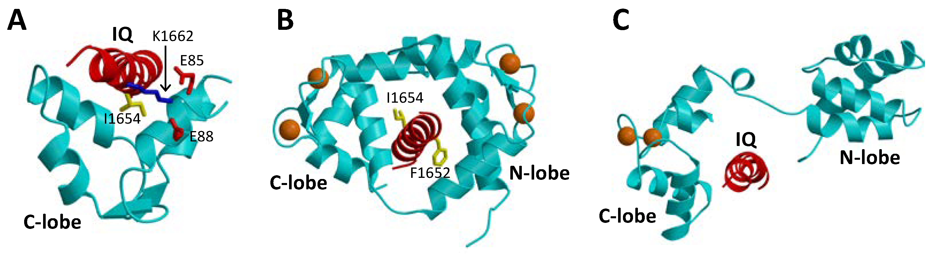
1.2. CaM Is a Ca2+ Sensor for CaVs

CaM is a 16.7 kDa Ca2+ sensor protein that belongs to the EF-hand superfamily [36]. CaM contains four EF-hand motifs (EF1, EF2, EF3, and EF4) that are grouped into two domains that are separately folded (EF1 and EF2 form the CaM N-lobe, while EF3 and EF4 form the CaM C-lobe) [37]. The CaM C-lobe and N-lobe each bind to Ca2+ with a dissociation constant of ~1 μM and 10 μM, respectively [38]. Thus, Ca2+ binding to CaM is an ordered process in which two Ca2+ bind to the C-lobe first before binding to the N-lobe. The Ca2+-bound form of CaM is known to bind to hundreds of different target proteins, including dozens of enzymes, receptors, ion channels, and other Ca2+ transporters [39]. The Ca2+-induced binding of CaM to its various target proteins usually serves to augment the biological activity of the target protein.
The binding of CaM to CaVs is critically important for promoting CDI [16,26]. In particular, CaM has been shown to bind to the IQ-motif (residues 1640–1665, highlighted red in Figure 1A) within the C-terminal cytosolic domain of CaVs [40], because deletion of the IQ-motif prevents CaV binding to CaM [26]. The NMR structure of Ca2+-free CaM (apoCaM) bound to the IQ-motif reveals that the IQ peptide forms an α-helix that interacts solely with the CaM C-lobe, while the IQ helix does not interact with the apoCaM N-lobe (Figure 3A). The most prominent intermolecular contacts involve IQ residues I1654 and K1662, and the mutations I1654E and K1662E each weaken apoCaM binding by nearly 10-fold [41]. The crystal structure of Ca2+-bound CaM bound to the IQ-motif reveals that both CaM lobes bind to opposite sides of the IQ helix (Figure 3B). The CaM C-lobe forms hydrophobic contacts with IQ residues I1654 and Q1655 that are essential for binding [42], hence the name IQ-motif. The CaM N-lobe forms hydrophobic contacts with aromatic IQ residues (Y1649 and F1652) that are essential for N-lobe binding. CaV mutations in the IQ-motif (I1654E and I1654M) that weaken CaM binding abolish CDI [43]. Much is known about how CaM interacts with the IQ-motif, but less is known about how the CaM-IQ interaction leads to channel inactivation. In this review, I present the possible molecular mechanisms of CDI to suggest how conformational changes in CaM and CaV might lead to CDI.

1.3. CaBP1 Promotes Activation of CaVs

Neuronal Ca2+-binding proteins (CaBP1-5 [46]) represent a sub-branch of the CaM superfamily [39] that regulate various Ca2+ channel targets. Multiple splice-variants and isoforms of CaBPs are localized in different neuronal cell types [47,48,49] and perform specialized roles in signal transduction. CaBP1, also termed caldendrin [50], has been shown to modulate the Ca2+-sensitive activity of L-type channels [51], and the transient receptor potential channel, TRPC5 [52]. CaBP1 contains four EF-hands, similar in sequence to those found in CaM [39]. By analogy to CaM [37], the four EF-hands are grouped into two domains connected by a central linker that is four residues longer in CaBP1 than in CaM. In contrast to CaM, the first and second EF-hands of CaBP1 lack critical residues required for high affinity Ca2+ binding [46]. CaBP1 binds Ca2+ only at EF3 and EF4, whereas it binds Mg2+ at EF1 that may serve a functional role [53]. In addition to binding Ca2+, CaBP1 also binds tightly to the CaV IQ-motif [35]. A crystal structure is known for CaBP1 with Ca2+ bound to EF3 and EF4 (Figure 3C) [45]. A structural model of CaBP1 bound to the IQ-motif (Figure 3C) was generated here by homology modeling that was calculated based on the crystal structure of the CaM-IQ complex [44]. In this model, the Ca2+-bound CaBP1 C-lobe makes hydrophobic intermolecular contacts with IQ residues I1654 and Y1657, whereas the CaBP1 N-lobe does not make any intermolecular contacts. Future structural and mutagenesis studies of CaBP1 bound to the IQ-motif are needed to test the validity of the structural model in Figure 3C.
The binding of CaBP1 to CaV has been shown to increase the channel open probability and to abolish or prevent CDI. Unlike CaM, CaBP1 appears to cause CaV channel activation at high cytosolic Ca2+ levels, which gives rise to CaV channel CDF [45]. CaBP1 has been suggested to bind to multiple sites within CaV [54]; however, CaBP1 binding to the IQ-motif is believed to cause CDF [55]. The CaBP1 binding to the IQ-motif under basal conditions [35] may serve to block CaM binding to CaV, which may explain how CaBP1 prevents CDI. Schematic mechanisms are presented below to speculate how CaBP1 binding to CaV might activate channel open probability and prevent CDI.

2. CaV Channel Function Regulated by CaM and CaBP1

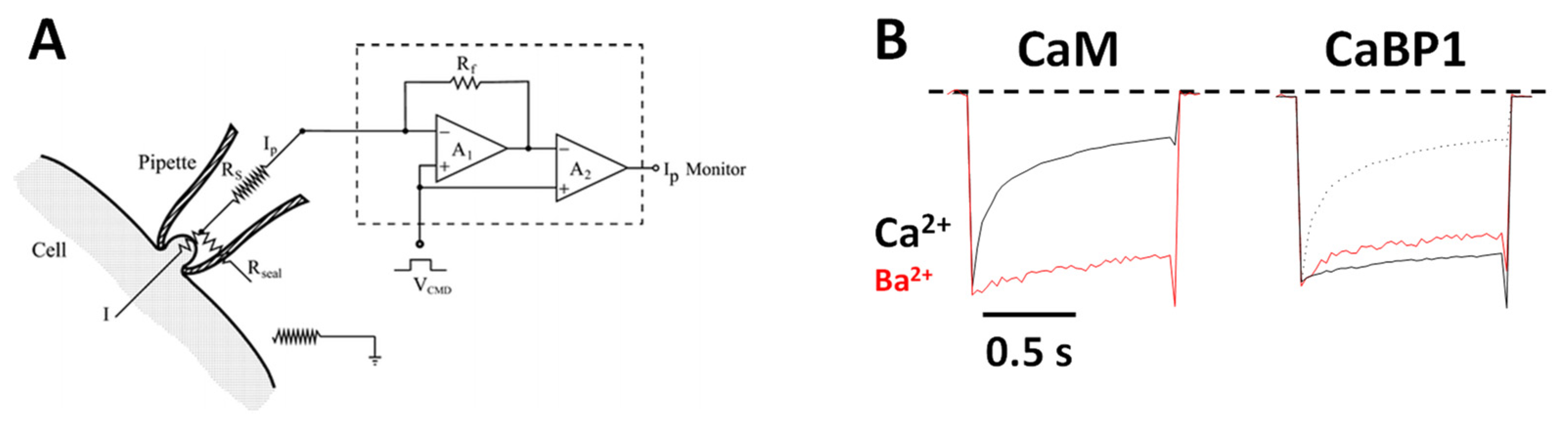
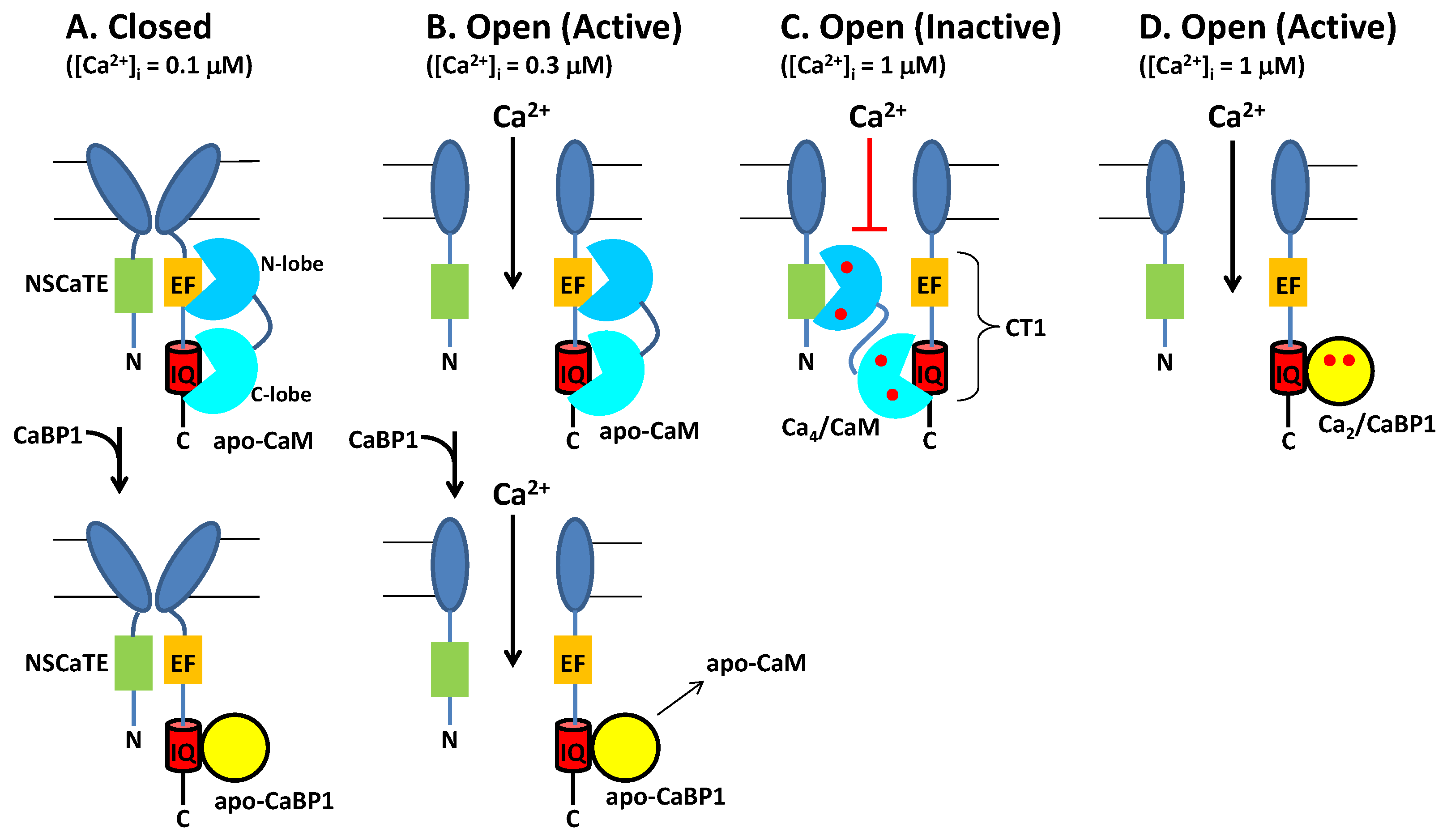
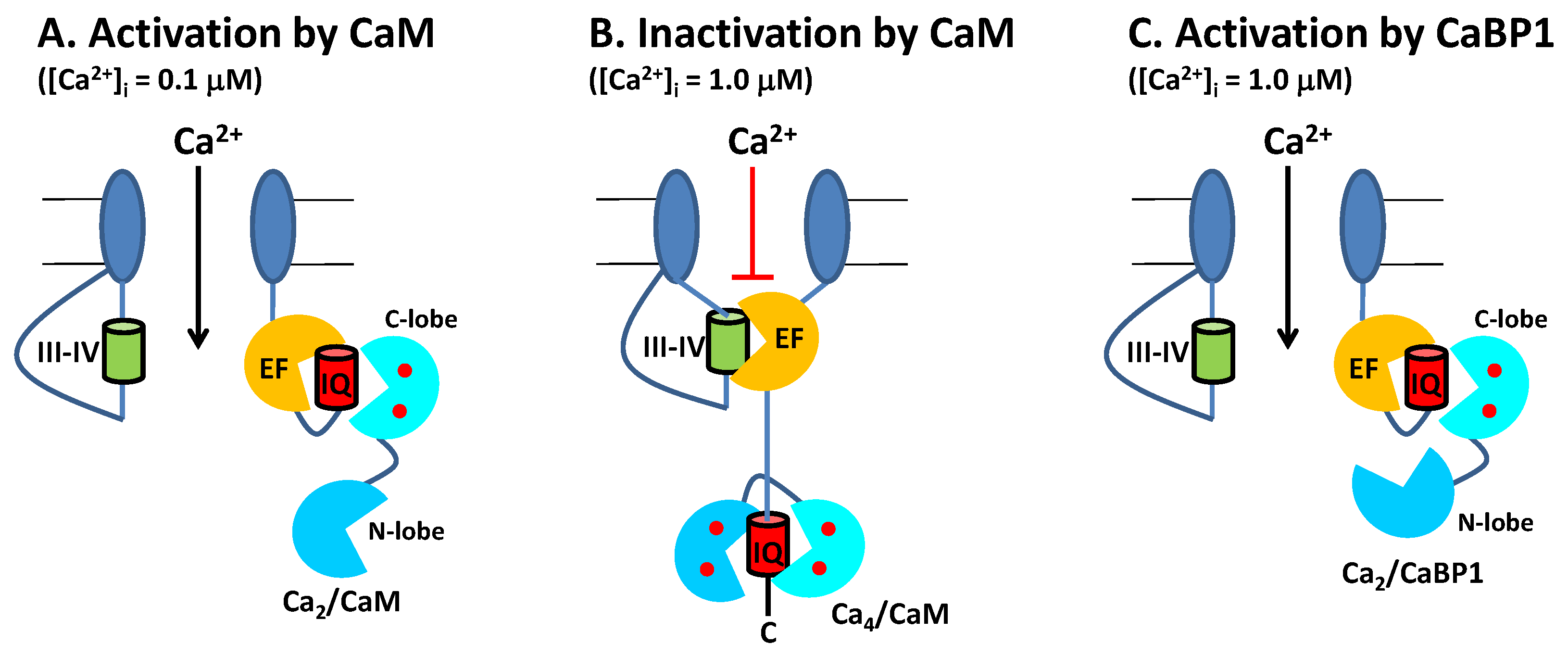
2.1. CaM Is Both an Accelerator and a Brake for CaV Channel Activity

Neuronal excitability is modulated in part by the Ca2+-dependent activity of CaV channels localized at the synaptic membrane. CaM binding to CaV serves to increase channel activity at low cytosolic Ca2+ levels under basal conditions ([Ca2+]i = 100 nM). Conversely, CaM decreases CaV channel activity at higher cytosolic Ca2+ levels (([Ca2+]i = 1.0 μM) caused by neuronal stimulation. Thus, CaM acts as both an accelerator and a brake to control CaV channel opening [16]. Ca2+ influx through CaV channels causes elevated intracellular Ca2+ levels that in turn promote a rapid negative feedback channel inactivation (called Ca2+-dependent inactivation or CDI [16]), mediated by CaM (Figure 4). Rapid CDI requires CaM to be pre-associated with CaV under basal conditions [29,33]. The channel has been suggested to be pre-associated with apoCaM under basal conditions (Figure 4A) [35], and apoCaM binding to CaV may increase Ca2+ currents (ICa) and channel open probability (Po) [56], whereas ICa is dramatically decreased at elevated Ca2+ levels, because Ca2+-bound CaM inactivates the channel [15,16]. As a result, apoCaM binding to CaV in which the CaM C-lobe is bound to the IQ motif (red box in Figure 4) and CaM N-lobe is bound to the channel EF-hand (orange box in Figure 4) is believed to stabilize the channel in the open state at low Ca2+ levels under basal conditions (Figure 4B). At elevated Ca2+ levels (caused by neuronal stimulation), Ca2+-saturated CaM has been suggested to bind to the full-length CaV at two different sites: The N-lobe binds to the NSCaTE domain [15,57] and the CaM C-lobe binds to the IQ motif [44], which is hypothesized to stabilize the channel in the inactive state (Figure 4C). Atomic-level structures are known for Ca2+/CaM bound to IQ [44] and NSCaTE [57] domains. However, structures are not yet known for apoCaM and Ca2+/CaM each bound to the entire C-terminal cytosolic domain of CaV comprised of the channel EF-hand and IQ-motif (called CT1 domain, Figure 4C). Future studies are needed to elucidate the structural interaction of apoCaM and Ca2+/CaM each bound to the full-length channel to further test the model in Figure 4.

2.2. CaBP1 Binding to CaV Prevents CDI and Activates Channel Opening

The upregulated expression of excess CaBP1 in particular neuronal cell types is known to abolish CDI of CaV [2,28,35,51] (Figure 4B, lower panel). The Ca2+-bound CaBP1 also increases CaV channel activity (Figure 4D) during Timothy Syndrome [58] and CaBP1 binding to CaVs could be targeted by therapeutics for the disease. CaBP1 was shown to compete with CaM for binding to the IQ-motif [35]. Thus, excess CaBP1 binds to the IQ motif and displaces apoCaM by mass action at low basal Ca2+ levels to prevent CaM-mediated CDI (Figure 4B, bottom panel). Previous studies have suggested that CaBP1 may bind to additional sites within CaV [54]. However, CaBP1 binding to the IQ-motif alone (as depicted in Figure 4) is believed to cause increased NPo under basal conditions and suppress CDI [28,55]. Future studies are needed to elucidate the atomic-level structural interactions between CaBP1 and CaV to further test the model in Figure 4.

3. Functional Role of Ca2+-Bound Forms of CaM and CaBP1 under Basal Conditions

Previous Ca2+ binding studies reveal that Ca2+/CaM binds to the IQ-motif with a dissociation constant (Kd = 10−12 M [35,40]) that is a million times smaller than the Kd for apoCaM binding to the IQ [40]. The huge stabilization of Ca2+/CaM caused by IQ binding implies that CaM-IQ should bind to Ca2+ with much higher affinity than CaM alone. Indeed, on the basis of previous binding data [38,40], the apparent Ca2+ binding dissociation constant of the CaM C-lobe in the CaM-IQ complex (KDapp) can be calculated to be ~100 nM ( K D a p p = 1 K 1 K 2 K 3 ), where K1 = 1012 M−2 for the binding of two Ca2+ to the CaM C-lobe alone [38], K2 = 107 M−1 for Ca2+/CaM C-lobe binding to the IQ-motif [40], and K3 = 10−5 M for the apoCaM C-lobe dissociation from the IQ peptide [40,41]. The predicted 100 nM binding of Ca2+ to the C-lobe of CaM-IQ implies that ~50% of the CaM-IQ complex should have Ca2+ bound to the CaM C-lobe under basal conditions ([Ca2+]i = 100 nM, K D a p p = 100   nM , and Y = ( [ C a 2 + ] i K D a p p + ( [ C a 2 + ] i = 0.5 ). By contrast, the CaM N-lobe in the CaM-IQ complex is estimated to bind to Ca2+ with an apparent dissociation constant ( K D a p p ) equal to ~1 μM ( K D a p p = 1 K 1 K 2 K 3 ), where K1 = 1010 M−2 for the binding of two Ca2+ to the CaM N-lobe alone [38], K2 = 106 M−1 for Ca2+/CaM N-lobe binding to the IQ [40], and K3 = 10−4 M for apoCaM N-lobe dissociation from the IQ [40]. This relatively low affinity Ca2+ binding to the CaM N-lobe predicts that the CaM N-lobe in the CaM-IQ complex should be devoid of Ca2+ under basal conditions. Therefore, I propose that a half saturated state of CaM with two Ca2+ bound to the C-lobe may exist under basal conditions ([Ca2+]i = 100 nM) (see Figure 5A). This calcified CaM species would allow its Ca2+-bound C-lobe to be anchored tightly to CaV under basal conditions, which would enable tight CaM pre-association that is needed for rapid CDI. By contrast, the CaM N-lobe is predicted to be in the Ca2+-free state at basal Ca2+ levels that can switch to the Ca2+-bound state upon Ca2+ influx and serve as a Ca2+ sensor during CDI (Figure 5B). Future experiments are needed to measure the apparent Ca2+ binding affinity of CaM-IQ and CaM-CaV to experimentally verify whether the CaM C-lobe in these complexes can bind to Ca2+ with a K D a p p near 100 nM as predicted above. Future studies are also needed to test whether Ca2+ binding to CaM is required to cause increased CaV channel open probability under basal conditions. In particular, the model in Figure 5 predicts that EF-hand mutations in CaM (that disable Ca2+ binding to the third and fourth EF-hands in the C-lobe) should prevent CaM pre-association, abolish CDI, and prevent the increased channel open probability observed under basal conditions [56]. Future electrophysiology and in vivo functional studies on CaV should be carried out in the presence of these CaM mutants to test the predictions in Figure 5.
A similar analysis performed here using the previous Ca2+ binding data for CaBP1 suggests that the binding of the IQ-motif to CaBP1 should allow the CaBP1-IQ complex to bind to Ca2+ in the nanomolar range, in contrast to the micromolar Ca2+ binding observed for CaBP1 alone [35,53]. The apparent Ca2+ binding dissociation constant of CaBP1 in the CaBP1-IQ complex (KDapp) can be calculated to be ~100 nM ( K D a p p = 1 K 1 K 2 K 3 ), where K1 = 1012 M−2 for the binding of two Ca2+ to CaBP1 alone [53], K2 = 109 M−1 for Ca2+/CaBP1 binding to the IQ [35], and K3 = 10−7 M for apoCaBP1 dissociation from the IQ [35]. Future Ca2+ binding experiments are needed to verify whether the CaBP1-IQ complex (and CaBP1-CaV) can bind to Ca2+ in the nanomolar range as predicted here. The predicted 100 nM binding of Ca2+ to CaBP1-IQ implies that ~50% of the CaBP1-IQ complex should have two Ca2+ bound to CaBP1 under basal conditions ([Ca2+]i = 100 nM, K D a p p = 100   n M ,   and Y = [ C a 2 + ] i K D a p p + ( [ C a 2 + ] i = 0.50 ). Therefore, I propose that upregulated expression of CaBP1 in neurons will generate a Ca2+-bound form of CaBP1 that can exist at low Ca2+ levels under basal conditions as well as at high Ca2+ levels following Ca2+ influx (Figure 5C). Thus, Ca2+-bound CaBP1 is proposed here to constitutively activate CaV channels, which can both prevent CDI and promote CDF. Future experiments are needed to test whether Ca2+ binding to CaBP1 is required to cause increased CaV channel open probability. In particular, the model in Figure 5C predicts that EF-hand mutations in CaBP1 (that disable Ca2+ binding to the third and fourth EF-hands) should weaken CaBP1 binding to CaV, re-enable CDI, and prevent CDF. Future electrophysiology and in vivo functional studies on CaV should be carried out in the presence of these CaBP1 mutants to test the predictions in Figure 5C.

4. Concluding Remarks

CaV channels are oppositely regulated by CaM and CaBP1: CaM binding to CaV is essential for channel CDI, whereas CaBP1 binding prevents CDI and promotes CDF. A careful analysis of available Ca2+ binding data suggests that CaV binding to CaM (or CaBP1) causes a more than 10-fold increase in the apparent Ca2+ binding affinity of CaM (or CaBP1). Thus, a significant fraction of CaV channels are predicted to be bound to calcified forms of CaM (or CaBP1) under basal conditions ([Ca2+]i = 100 nM vs KDapp = 100 nM), which may have functional implications (Figure 5). Future studies on EF-hand mutants (that specifically abolish Ca2+ binding to either CaM or CaBP1) are needed to test whether Ca2+ binding to the C-lobe of CaM (or CaBP1) is essential for the increased CaV channel open probability caused by CaM (or CaBP1) under basal conditions.

Funding

This research was funded by a grant from the National Institutes of Health, grant number R01 GM130925.

Institutional Review Board Statement

Not applicable.

Informed Consent Statement

Not applicable.

Acknowledgments

This work was supported by a grant to JBA from the National Institutes of Health (R01 GM130925).

Conflicts of Interest

The author declares no conflict of interest.

References

  1. Clapham, D.E. Calcium signaling. Cell 2007, 6, 1047–1058. [Google Scholar] [CrossRef] [Green Version]
  2. Hardie, J.; Lee, A. Decalmodulation of Cav1 channels by CaBPs. Channels 2015, 10, 33–37. [Google Scholar] [CrossRef] [Green Version]
  3. Minor, D.L.; Findeisen, F., Jr. Progress in the structural understanding of voltage-gated calcium channel (CaV) function and modulation. Channels (Austin) 2010, 4, 459–474. [Google Scholar] [PubMed] [Green Version]
  4. Simms, B.A.; Zamponi, G.W. Neuronal Voltage-Gated Calcium Channels: Structure, Function, and Dysfunction. Neuron 2014, 82, 24–45. [Google Scholar] [CrossRef] [Green Version]
  5. Randall, A.; Tsien, R.W. Pharmacological dissection of multiple types of Ca2+ channel currents in rat cerebellar granule neurons. J. Neurosci. 1995, 15, 2995–3012. [Google Scholar] [CrossRef]
  6. Berridge, M.J.; Bootman, M.; Roderick, H. Calcium signalling: Dynamics, homeostasis and remodelling. Nat. Rev. Mol. Cell Biol. 2003, 4, 517–529. [Google Scholar] [CrossRef] [Green Version]
  7. Berridge, M.J. The endoplasmic reticulum: A multifunctional signaling organelle. Cell Calcium 2002, 32, 235–249. [Google Scholar] [CrossRef] [PubMed]
  8. Wadel, K.; Neher, E.; Sakaba, T. The Coupling between Synaptic Vesicles and Ca2+ Channels Determines Fast Neurotransmitter Release. Neuron 2007, 53, 563–575. [Google Scholar] [CrossRef] [PubMed] [Green Version]
  9. Wheeler, D.G.; Groth, R.D.; Ma, H.; Barrett, C.F.; Owen, S.F.; Safa, P.; Tsien, R.W. CaV1 and CaV2 Channels Engage Distinct Modes of Ca2+ Signaling to Control CREB-Dependent Gene Expression. Cell 2012, 149, 1112–1124. [Google Scholar] [CrossRef] [PubMed] [Green Version]
  10. Wheeler, D.B.; Randall, A.; Tsien, R.W. Roles of N-Type and Q-Type Ca2+ Channels in Supporting Hippocampal Synaptic Transmission. Science 1994, 264, 107–111. [Google Scholar] [CrossRef] [PubMed] [Green Version]
  11. Lewis, B.B.; Wester, M.R.; Miller, L.E.; Nagarkar, M.D.; Johnson, M.B.; Saha, M.S. Cloning and characterization of voltage-gated calcium channel alpha1 subunits in Xenopus laevis during development. Dev. Dyn. 2009, 238, 2891–2902. [Google Scholar] [CrossRef]
  12. Simms, B.A.; Souza, I.A.; Rehak, R.; Zamponi, G.W. The Cav1.2 N terminus contains a CaM kinase site that modulates channel trafficking and function. Pflüg. Arch. Eur. J. Physiol. 2014, 467, 677–686. [Google Scholar] [CrossRef]
  13. Stanika, R.; Villanueva, I.; Kazanina, G.; Andrews, S.B.; Pivovarova, N.B. Comparative Impact of Voltage-Gated Calcium Channels and NMDA Receptors on Mitochondria-Mediated Neuronal Injury. J. Neurosci. 2012, 32, 6642–6650. [Google Scholar] [CrossRef] [PubMed] [Green Version]
  14. Christel, C.; Lee, A. Ca2+-dependent modulation of voltage-gated Ca2+ channels. Biochim. et Biophys. Acta (BBA) Gen. Subj. 2011, 1820, 1243–1252. [Google Scholar] [CrossRef] [PubMed] [Green Version]
  15. Dick, I.E.; Tadross, M.R.; Liang, H.; Tay, L.H.; Yang, W.; Yue, D.T. A modular switch for spatial Ca2+ selectivity in the calmodulin regulation of CaV channels. Nature 2008, 451, 830–834. [Google Scholar] [CrossRef] [PubMed]
  16. Zühlke, R.D.; Pitt, G.S.; Deisseroth, K.; Tsien, R.W.; Reuter, H. Calmodulin supports both inactivation and facilitation of L-type calcium channels. Nature 1999, 399, 159–162. [Google Scholar] [CrossRef] [PubMed]
  17. Cain, S.M.; Snutch, T.P. Voltage-gated calcium channels and disease. BioFactors 2011, 37, 197–205. [Google Scholar] [CrossRef]
  18. Catterall, W.A. Ion Channel Voltage Sensors: Structure, Function, and Pathophysiology. Neuron 2010, 67, 915–928. [Google Scholar] [CrossRef] [Green Version]
  19. Patton, D.E.; West, J.W.; Catterall, W.A.; Goldin, A.L. A peptide segment critical for sodium channel inactivation functions as an inactivation gate in a potassium channel. Neuron 1993, 11, 967–974. [Google Scholar] [CrossRef]
  20. Catterall, W.A.; Perez-Reyes, E.; Snutch, T.P.; Striessnig, J. International Union of Pharmacology. XLVIII. Nomenclature and Structure-Function Relationships of Voltage-Gated Calcium Channels. Pharmacol. Rev. 2005, 57, 411–425. [Google Scholar] [CrossRef]
  21. Dai, S.; Hall, D.D.; Hell, J.W. Supramolecular Assemblies and Localized Regulation of Voltage-Gated Ion Channels. Physiol. Rev. 2009, 89, 411–452. [Google Scholar] [CrossRef] [Green Version]
  22. Hall, D.D.; Dai, S.; Tseng, P.-Y.; Malik, Z.; Nguyen, M.; Matt, L.; Schnizler, K.; Shepherd, A.; Mohapatra, D.P.; Tsuruta, F.; et al. Competition between α-actinin and Ca2+-Calmodulin Controls Surface Retention of the L-type Ca2+ Channel CaV1.2. Neuron 2013, 78, 483–497. [Google Scholar] [CrossRef] [Green Version]
  23. Zamponi, G.; Bourinet, E.; Nelson, D.; Nargeot, J.; Snutch, T.P. Crosstalk between G proteins and protein kinase C mediated by the calcium channel α1 subunit. Nature 1997, 385, 442–446. [Google Scholar] [CrossRef]
  24. Wu, J.; Yan, Z.; Li, Z.Q.; Qian, X.Y.; Lu, S.; Dong, M.Q.; Zhou, Q.; Yan, N. Structure of the voltage-gated calcium channel Ca(v)1.1 at 3.6 A resolution. Nature. 2016, 537, 191–196. [Google Scholar] [CrossRef]
  25. Wu, J.; Yan, Z.; Li, Z.Q.; Yan, C.; Lu, S.; Dong, M.Q.; Yan, N. Structure of the voltage-gated calcium channel Cav1.1 complex. Science 2015, 350. [Google Scholar] [CrossRef]
  26. Peterson, B.Z.; DeMaria, C.D.; Yue, D.T. Calmodulin Is the Ca2+ Sensor for Ca2+-Dependent Inactivation of L-Type Calcium Channels. Neuron 1999, 22, 549–558. [Google Scholar] [CrossRef] [Green Version]
  27. Tippens, A.L.; Lee, A. Caldendrin, a Neuron-specific Modulator of Cav/1.2 (L-type) Ca2+ Channels. J. Biol. Chem. 2007, 282, 8464–8473. [Google Scholar] [CrossRef] [Green Version]
  28. Zhou, H.; Kim, S.-A.; Kirk, E.A.; Tippens, A.L.; Sun, H.; Haeseleer, F.; Lee, A. Ca2+-Binding Protein-1 Facilitates and Forms a Postsynaptic Complex with Cav1.2 (L-Type) Ca2+ Channels. J. Neurosci. 2004, 24, 4698–4708. [Google Scholar] [CrossRef] [PubMed] [Green Version]
  29. Erickson, M.G.; Liang, H.; Mori, M.X.; Yue, D.T. FRET Two-Hybrid Mapping Reveals Function and Location of L-Type Ca2+ Channel CaM Preassociation. Neuron 2003, 39, 97–107. [Google Scholar] [CrossRef] [Green Version]
  30. Lian, L.-Y.; Myatt, D.; Kitmitto, A. Apo calmodulin binding to the L-type voltage-gated calcium channel Cav1.2 IQ peptide. Biochem. Biophys. Res. Commun. 2007, 353, 565–570. [Google Scholar] [CrossRef]
  31. Xiong, L.; Kleerekoper, Q.K.; He, R.; Putkey, J.A.; Hamilton, S.L. Sites on Calmodulin That Interact with the C-terminal Tail of Cav1.2 Channel. J. Biol. Chem. 2005, 280, 7070–7079. [Google Scholar] [CrossRef] [PubMed] [Green Version]
  32. Johny, M.; Yang, P.S.; Bazzazi, H.; Yue, D.T. Dynamic switching of calmodulin interactions underlies Ca2+ regulation of CaV1.3 channels. Nat. Commun. 2013, 4, 1717. [Google Scholar] [CrossRef] [Green Version]
  33. Erickson, M.G.; Alseikhan, B.A.; Peterson, B.Z.; Yue, D.T. Preassociation of Calmodulin with Voltage-Gated Ca2+ Channels Revealed by FRET in Single Living Cells. Neuron 2001, 31, 973–985. [Google Scholar] [CrossRef] [Green Version]
  34. Peterson, B.Z.; Lee, J.S.; Mulle, J.G.; Wang, Y.; de Leon, M.; Yue, D.T. Critical Determinants of Ca2+-Dependent Inactivation within an EF-Hand Motif of L-Type Ca2+ Channels. Biophys. J. 2000, 78, 1906–1920. [Google Scholar] [CrossRef] [Green Version]
  35. Findeisen, F.; Rumpf, C.H.; Minor, D.L. Apo States of Calmodulin and CaBP1 Control CaV1 Voltage-Gated Calcium Channel Function through Direct Competition for the IQ Domain. J. Mol. Biol. 2013, 425, 3217–3234. [Google Scholar] [CrossRef] [PubMed] [Green Version]
  36. Moncrief, N.; Kretsinger, R.H.; Goodman, M. Evolution of EF-hand calcium-modulated proteins. I. Relationships based on amino acid sequences. J. Mol. Evol. 1990, 30, 522–562. [Google Scholar] [CrossRef]
  37. Babu, Y.; Bugg, C.E.; Cook, W.J. Structure of calmodulin refined at 2.2 Å resolution. J. Mol. Biol. 1988, 204, 191–204. [Google Scholar] [CrossRef]
  38. Gilli, R.; Lafitte, D.; Lopez, C.; Kilhoffer, M.-C.; Makarov, A.; Briand, A.C.; Haiech, J. Thermodynamic Analysis of Calcium and Magnesium Binding to Calmodulin. Biochemistry 1998, 37, 5450–5456. [Google Scholar] [CrossRef]
  39. Ikura, M. Calcium binding and conformational response in EF-hand proteins. Trends Biochem. Sci. 1996, 21, 14–17. [Google Scholar] [CrossRef]
  40. Evans, T.I.A.; Hell, J.W.; Shea, M.A. Thermodynamic linkage between calmodulin domains binding calcium and contiguous sites in the C-terminal tail of CaV1.2. Biophys. Chem. 2011, 159, 172–187. [Google Scholar] [CrossRef] [Green Version]
  41. Turner, M.; Anderson, D.E.; Bartels, P.; Nieves, C.; Andrea, M.C.; Henderson, P.B.; Man, K.N.M.; Tseng, P.-Y.; Yarov-Yarovoy, V.; Bers, D.M.; et al. α-Actinin-1 promotes gating of the L-type Ca2+ Channel CaV1.2. EMBO J. 2020, 39, e102622. [Google Scholar] [CrossRef] [PubMed]
  42. Zühlke, R.D.; Pitt, G.S.; Tsien, R.W.; Reuter, H. Ca2+-sensitive Inactivation and Facilitation of L-type Ca2+ Channels Both Depend on Specific Amino Acid Residues in a Consensus Calmodulin-binding Motif in theα1C subunit. J. Biol. Chem. 2000, 275, 21121–21129. [Google Scholar] [CrossRef] [PubMed] [Green Version]
  43. Kim, J.; Ghosh, S.; A Nunziato, D.; Pitt, G.S. Identification of the Components Controlling Inactivation of Voltage-Gated Ca2+ Channels. Neuron 2004, 41, 745–754. [Google Scholar] [CrossRef] [Green Version]
  44. Van Petegem, F.; Chatelain, F.C.; Minor, J.D.L. Insights into voltage-gated calcium channel regulation from the structure of the CaV1.2 IQ domain–Ca2+/calmodulin complex. Nat. Struct. Mol. Biol. 2005, 12, 1108–1115. [Google Scholar] [CrossRef]
  45. Findeisen, F.; Minor, D.L. Structural Basis for the Differential Effects of CaBP1 and Calmodulin on CaV1.2 Calcium-Dependent Inactivation. Structure 2010, 18, 1617–1631. [Google Scholar] [CrossRef] [PubMed] [Green Version]
  46. Haeseleer, F.; Sokal, I.; Verlinde, C.; Erdjument-Bromage, H.; Tempst, P.; Pronin, A.N.; Benovic, J.L.; Fariss, R.; Palczewski, K. Five Members of a Novel Ca2+-binding Protein (CABP) Subfamily with Similarity to Calmodulin. J. Biol. Chem. 2000, 275, 1247–1260. [Google Scholar] [CrossRef] [Green Version]
  47. Haynes, L.P.; Tepikin, A.; Burgoyne, R.D. Calcium-binding Protein 1 Is an Inhibitor of Agonist-evoked, Inositol 1,4,5-Trisphosphate-mediated Calcium Signaling. J. Biol. Chem. 2004, 279, 547–555. [Google Scholar] [CrossRef] [Green Version]
  48. Menger, N.; Seidenbecher, C.I.; Gundelfinger, E.D.; Kreutz, M. The cytoskeleton-associated neuronal calcium-binding protein caldendrin is expressed in a subset of amacrine, bipolar and ganglion cells of the rat retina. Cell Tissue Res. 1999, 298, 21–32. [Google Scholar] [CrossRef] [PubMed]
  49. Seidenbecher, C.I.; Reissner, C.; Kreutz, M.R. Caldendrins in the Inner Retina. Photorecept. Calcium 2002, 514, 451–463. [Google Scholar] [CrossRef]
  50. Laube, G.; Seidenbecher, C.; Richter, K.; Dieterich, D.; Hoffmann, B.; Landwehr, M.; Smalla, K.; Winter, C.; Böckersbc, T.M.; Wolf, G.; et al. The Neuron-Specific Ca2+-Binding Protein Caldendrin: Gene Structure, Splice Isoforms, and Expression in the Rat Central Nervous System. Mol. Cell. Neurosci. 2002, 19, 459–475. [Google Scholar] [CrossRef] [PubMed]
  51. Zhou, H.; Yu, K.; McCoy, K.L.; Lee, A. Molecular mechanism for divergent regulation of Cav1.2 Ca2+ channels by calmodulin and Ca2+-binding protein 1. J. Biol. Chem. 2005, 280, 29612–29619. [Google Scholar] [CrossRef] [PubMed] [Green Version]
  52. Kinoshita-Kawada, M.; Tang, J.; Xiao, R.; Kaneko, S.; Foskett, J.K.; Zhu, M.X. Inhibition of TRPC5 channels by Ca2+-binding protein 1 in Xenopus oocytes. Pflug. Arch. 2005, 450, 345–354. [Google Scholar] [CrossRef]
  53. Wingard, J.N.; Chan, J.; Bosanac, I.; Haeseleer, F.; Palczewski, K.; Ikura, M.; Ames, J.B. Structural Analysis of Mg2+ and Ca2+ Binding to CaBP1, a Neuron-specific Regulator of Calcium Channels. J. Biol. Chem. 2005, 280, 37461–37470. [Google Scholar] [CrossRef] [PubMed] [Green Version]
  54. Oz, S.; Tsemakhovich, V.; Christel, C.J.; Lee, A.; Nathan, D. CaBP1 regulates voltage-dependent inactivation and activation of Ca(V)1.2 (L-type) calcium channels. J. Biol. Chem. 2011, 286, 13945–13953. [Google Scholar] [CrossRef] [PubMed] [Green Version]
  55. Oz, S.; Benmocha, A.; Sasson, Y.; Sachyani, D.; Almagor, L.; Lee, A.; Hirsch, J.A.; Dascal, N. Competitive and Non-competitive Regulation of Calcium-dependent Inactivation in CaV1.2 L-type Ca2+ Channels by Calmodulin and Ca2+-binding Protein 1. J. Biol. Chem. 2013, 288, 12680–12691. [Google Scholar] [CrossRef] [Green Version]
  56. Adams, P.J.; Johny, M.; Dick, I.; Inoue, T.; Yue, D.T. Apocalmodulin Itself Promotes Ion Channel Opening and Ca2+ Regulation. Cell 2014, 159, 608–622. [Google Scholar] [CrossRef] [Green Version]
  57. Liu, Z.; Vogel, H.J. Structural basis for the regulation of L-type voltage-gated calcium channels: Interactions between the N-terminal cytoplasmic domain and Ca2+-calmodulin. Front. Mol. Neurosci. 2012, 5, 38. [Google Scholar] [CrossRef] [Green Version]
  58. Dixon, R.; Yuan, C.; Cheng, E.P.; Navedo, M.F.; Santana, L.F. Ca2+ signaling amplification by oligomerization of L-type Cav1.2 channels. Proc. Natl. Acad. Sci. USA 2012, 109, 1749–1754. [Google Scholar] [CrossRef] [Green Version]
Figure 1. Structure of CaVs. (A) The α-subunit consists of 4 transmembrane domains (I-IV) that contain 6 helices (yellow) and pore loop (red). The III-IV linker is the inactivation gate. The cytosolic C-terminal domain (CT1) is comprised of an EF-hand domain (orange) and IQ-motif (red). (B) CaV is composed of pore-forming α-subunit attached to β- and δ-subunits. (C) Cryo-EM structure of CaV1.1 (PDB ID: 5GJW) showing the inactivation gate (III-IV linker in blue) connected to the EF-hand domain (orange). The IQ-motif is structurally disordered and missing in the cryo-EM structure of CaV1.1.
Biomolecules 11 01811 g001
Figure 2. Ca2+-dependent Inactivation (CDI) of CaV. (A) Schematic representation of the electrophysiology experiment used to record CDI. (B) Normalized Ca2+ and Ba2+ currents evoked by 1 s pulse (−80 to +10 mV). Adapted from [2]. Fast decay of Ca2+ current due to CaM (black trace in left panel, CDI). The decay of the Ca2+ current is much slower in the presence of CaBP1 (black solid trace in the right panel, CDI abolished). Dotted line is the Ca2+ current in the absence of CaBP1, caused by endogenous CaM. Red traces are Ba2+ currents that lack fast inactivation because Ba2+ does not bind to CaM.
Biomolecules 11 01811 g002
Figure 3. Atomic-level structures of CaM and CaBP1. (A) NMR structure of Ca2+-free CaM C-lobe (cyan) bound to the CaV1.2 IQ-motif in red (PDB ID: 6CTB) [41]. (B) Crystal structure of Ca2+-bound CaM (cyan) bound to the CaV1.2 IQ-motif in red (PDB ID: 2BE6) [44]. (C) Structural model of the crystal structure of CaBP1 (PDB ID: 3OX6) [45] bound to the CaV1.2 IQ-motif (red). Bound Ca2+ are indicated by orange spheres.
Biomolecules 11 01811 g003
Figure 4. Conventional Model of CDI from CaV regulated by CaM and CaBP1. (A) Under resting conditions ([Ca2+]i = 100 nM), CaV (dark blue) is in the closed channel state, which has been suggested to be pre-associated with Ca2+-free forms of CaM (cyan) or CaBP1 (yellow). (B) Membrane depolarization causes channel opening, which causes Ca2+ influx. Initially at low cytosolic Ca2+ levels (([Ca2+]i < 300 nM), CaV is bound to Ca2+-free forms of CaM or CaBP1, which stabilize the active open state. (C) After sufficient Ca2+ influx, the cytosolic Ca2+ level increases to above 1 micromolar, which causes Ca2+ binding to CaM and the Ca2+-bound CaM promotes channel inactivation (CDI). Alternatively, CaV binding to CaBP1 (yellow) displaces CaM and prevents CDI (bottom panel). (D) The binding of Ca2+-bound CaBP1 to CaV promotes channel opening at elevated Ca2+ levels (called CDF). Bound Ca2+ are indicated by red circles.
Biomolecules 11 01811 g004
Figure 5. IQ-switch Model for CaV Regulation by Ca2+-bound forms of CaM and CaBP1. The channel EF-hand (orange) is proposed here to undergo a Ca2+-induced conformational change in which the channel EF-hand switches contact between the IQ-motif (red) and the III-IV linker (called IQ-switch). (A) Voltage-gated channel opening under resting conditions ([Ca2+]i = 100 nM) generates CaV (dark blue) in the open channel state, which is pre-associated with CaM that contains a Ca2+ bound C-lobe (cyan) and Ca2+-free N-lobe (blue). In this activated open state of the channel, the IQ-motif is hypothesized here to be sandwiched between the channel EF-hand (orange notched circle) on one side and the Ca2+-bound CaM C-lobe on the other. (B) After sufficient Ca2+ influx, the cytosolic Ca2+ level increases to above 1 micromolar, which causes Ca2+ binding to both lobes of CaM that enables each CaM lobe to bind to opposite sides of the IQ helix (red) and promote channel inactivation (CDI). In the inactivated channel state, the channel EF-hand interacts with the III-IV linker (green helix called the inactivation gate) in place of the structurally related IQ-motif. A triple mutation in the channel EF-hand (T1591A/L1592L/F1593A) that disrupts interaction with the III-IV linker also abolishes CDI [43]. In essence, the channel EF-hand bound to the III-IV linker is proposed here to serve as a channel plug that blocks the channel entrance in the inactivated state (red bar in panel B). Channel opening is proposed to occur at low Ca2+ levels when the channel EF-hand engages the helical IQ-motif (stabilized by its binding to the CaM C-lobe), which disconnects the EF-hand from the III-IV linker to unblock the channel entrance (arrow in panel A). Therefore, Ca2+ binding to the N-lobe of CaM is hypothesized here to switch the channel EF-hand from interacting with the IQ-motif at low Ca2+ levels (active state in panel A) to interacting with the III-IV linker at high Ca2+ levels (inactive state in panel B) in order to promote CDI. (C) CaV binding to Ca2+-bound CaBP1 prevents CDI and promotes channel opening at elevated Ca2+ levels (called CDF). Ca2+-bound CaBP1 in panel C resembles CaM with two Ca2+ bound in panel A. Bound Ca2+ are indicated by red circles.
Biomolecules 11 01811 g005
Publisher’s Note: MDPI stays neutral with regard to jurisdictional claims in published maps and institutional affiliations.

