Review

Decoding Stem Cells: An Overview on Planarian Stem Cell Heterogeneity and Lineage Progression

by
M. Dolores Molina
1,2,* and
Francesc Cebrià
1,2,*
1
Department of Genetics, Microbiology and Statistics, Faculty of Biology, University of Barcelona, 08028 Barcelona, Spain
2
Institute of Biomedicine of the University of Barcelona (IBUB), 08028 Barcelona, Spain
*
Authors to whom correspondence should be addressed.
Biomolecules 2021, 11(10), 1532; https://doi.org/10.3390/biom11101532
Submission received: 15 September 2021 / Revised: 11 October 2021 / Accepted: 12 October 2021 / Published: 17 October 2021
(This article belongs to the Special Issue New Insights into Stem Cell Regulation)

Abstract

:
Planarians are flatworms capable of whole-body regeneration, able to regrow any missing body part after injury or amputation. The extraordinary regenerative capacity of planarians is based upon the presence in the adult of a large population of somatic pluripotent stem cells. These cells, called neoblasts, offer a unique system to study the process of stem cell specification and differentiation in vivo. In recent years, FACS-based isolation of neoblasts, RNAi functional analyses as well as high-throughput approaches such as single-cell sequencing have allowed a rapid progress in our understanding of many different aspects of neoblast biology. Here, we summarize our current knowledge on the molecular signatures that define planarian neoblasts heterogeneity, which includes a percentage of truly pluripotent stem cells, and guide the commitment of pluripotent neoblasts into lineage-specific progenitor cells, as well as their differentiation into specific planarian cell types.

1. Introduction

Some of the main challenges in regenerative medicine are related to the capacity to control stem cell self-renewal and differentiation, as well as to try to induce and/or improve our limited regenerative abilities as mammals. Planarians are one of the few animal models that combine two amazing features: They can regenerate the whole body from a tiny piece of them, and they do it because they maintain a population of adult pluripotent stem cells throughout their lives, referred to as neoblasts [1,2]. Neoblasts are not only needed during regeneration, but they also ensure a continuous cell turnover to replace aged specialized cells and maintain tissue functionality. Therefore, planarians become a good model in which to study in vivo the responses of pluripotent stem cells to a huge variety of external insults such as small injuries, large amputations, presence of toxic substances, long-term starvation and sub-lethal irradiations, among others. Over the last 20 years, significant advances have been made in the planarian field mainly through the use of a large number of different techniques, molecular tools and high-throughput approaches. Consequently, a significant knowledge on how planarians respond to an insult, how regeneration is triggered and how polarity and patterning are achieved during this process has been gathered [3]. Focusing on neoblasts, these stem cells can be easily isolated through FACS [4], can be individually transplanted from one animal to another [5] or even cultured in vitro although with certain current limitations [6]. In addition, neoblasts can be labeled with BrdU and EdU [7,8], which allow for some lineage tracing that can be complemented with cell-type atlases originated from single-cell sequencing experiments [9,10,11,12,13,14,15,16]. From several transcriptomes, dozens of neoblast-specific markers have been identified [12,13,14,16,17,18]. Remarkably, neoblast transcriptomes show several conserved features compared to mammalian stem cells at the level of the specific markers being expressed [19,20].
It is now clear that neoblasts constitute a heterogeneous cell population that includes real pluripotent stem cells as well as a large number of lineage-committed progenitors [21]. By identifying specific markers of all these populations several studies have characterized important genes and pathways required for the differentiation of the distinct mature cell types [12,13,22,23,24,25,26,27,28]. Interestingly, these lineage-committed progenitors appear to maintain a certain plasticity that allows them to switch to a different cell fate depending on the context [29]. Despite all these advances many questions are still under extensive research. For example: What is the role of asymmetric cell division in neoblast self-renewal? Which are the signals that trigger the neoblast response to amputation? How do neoblasts re-adjust to new positional cues? How does metabolic status regulate neoblasts? How and when is a transition between a regenerative and homeostatic context achieved? What is the role of apoptosis in neoblast proliferation and differentiation? Are the gut or the extracellular matrix (ECM) a niche for neoblasts?
In this review, we summarize our current knowledge on some of these questions, as well as on the molecular signatures that define planarian neoblasts’ heterogeneity and guide the commitment of pluripotent neoblasts into lineage-specific progenitors first and mature cells later.

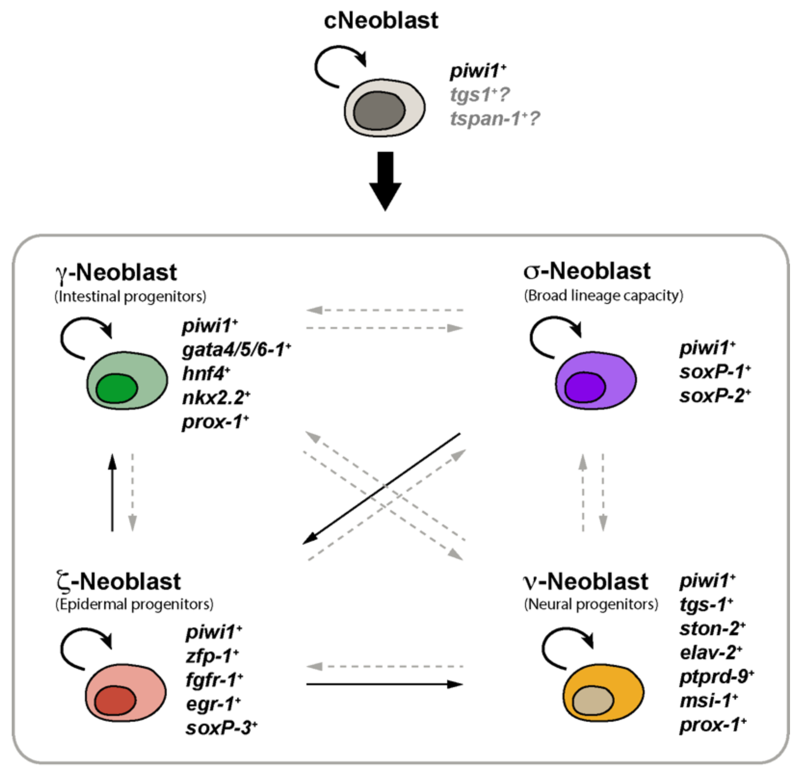
2. Planarian Neoblasts Are a Heterogeneous Population of Stem Cells

For many years, planarian stem cells were considered a rather homogeneous cell population. These neoblasts were defined as the only mitotic cells in the asexual strain and were morphologically characterized as small cells that present chromatoid bodies, an elevated nucleus/cytoplasm ratio and that locate in the mesenchymal tissue that fills the space between the planarian organs and the epithelia, among others features [2,30]. Planarian neoblasts are nowadays molecularly defined by the expression of piwi-1 [31]. In the last decade, however, several studies have revealed that planarian piwi-1-expressing cells constitute indeed a heterogeneous population that includes both pluripotent stem cells capable of reconstituting the entire animal [5], as well as specialized progenitors stem cells that co-express tissue-specific transcription factors and contribute to the maintenance and regeneration of specific mature tissues (Figure 1) [21]. Elegant experiments involving single-cell transplantation have demonstrated that a small percentage of planarian neoblasts can be considered truly pluripotent stem cells [5]. These cells, named cNeoblasts (clonogenic neoblasts), are capable of clonal expansion when transplanted individually into planarians that have been irradiated and therefore completely depleted of neoblasts, repopulating the host and differentiating into all of the planarian cell types [5].
Our knowledge regarding neoblasts’ heterogeneity has greatly progressed during the last years [32]. Four main neoblast classes were initially identified and named σ-, γ-, ζ- and ν-neoblasts (Figure 1) [12,13]. σ-neoblasts (sigma-neoblasts) were associated with pluripotency, as they proliferate in response to injury, possess a broad lineage capacity and can regenerate other classes of neoblasts, such as the ζ-neoblasts (zeta-neoblasts) subtype. These neoblasts were characterized by the elevated expression of genes such as soxP-1, soxP-2, soxB-1, smad6/7, inx-13, pbx-1, fgfr-4 and nlk-1 [12]. A second type of stem cells, the ζ-Neoblasts, were considered specialized cells involved in epidermal maintenance and regeneration and express high levels of genes such as zfp-1, fgfr-1, p53, soxP-3, egr-1 and g6pd [12]. A third type, the γ-neoblasts (gamma-neoblasts), were considered a subclass of the σ-neoblasts that express gata4/5/6, nkx2.2, hnf4 and prox-1 and likely identify the population of intestinal lineage-committed progenitor stem cells [12]. Finally, ν-neoblasts (nu-neoblasts) comprise a neural-committed stem cell population that express low levels of piwi-1 and neural genes such as ston-2+ [13]. Several studies have validated the existence of these epidermal, neural and intestinal progenitor stem cells, as well as identified specialized stem cells for other planarian tissues [9,10,16], such as the pharynx [23], eyes [26,27], pigment [28], excretory [22], parenchymal [9,10] or muscle cell types [24,25].
Because of the observation that different levels of piwi-1 transcript occur in neoblasts and their progeny [13], Zeng and collaborators performed single-cell RNA sequencing and identified at least 12 discrete subpopulations of neoblasts expressing different levels of piwi-1 [16]. Several of these subpopulations relate to the previously defined subtypes of γ, ζ and ν-neoblasts [12,13]. The previously characterized class of σ-neoblasts associated with pluripotency appeared related to several clusters, suggesting that σ-neoblasts constitute indeed a heterogeneous population. Interestingly, the authors found that a neoblast class marked by the expression of tgs-1 can be enriched by FACS using an antibody against the cell-surface protein Tetraspanin-1 and suggested that these TSPAN-1+ cells are the true pluripotent stem cells that behave as clonogenic neoblasts that self-renew as well as give rise to progenitor cells of the six major cell lineages: epidermal, neural, protonephridia, muscle, pharynx and gut progenitors [16]. It is important to mention, however, that Tspan-1 does not seem to be exclusively expressed in cNeoblasts, and a notable number of postmitotic cells also express this gene [9,16,33]. In addition, it has been recently proposed that tgs-1 indeed marks neural-specialized neoblasts as well as postmitotic cells associated with neural fates [29]. Thus, a unique marker of cNeoblasts remains to be characterized, and it has been proposed that these cells may be instead defined by the absence of the expression of any tissue-specific marker [9]. On the other hand, importantly, novel findings suggest that no known neoblast class is uniquely pluripotent, and neoblasts expressing tissue-specific transcription factors from multiple lineages can be clonogenic both after single cell transplantation or under challenging conditions in which stem cells are massively depleted and the few remaining repopulate the entire body [29]. Thus, at least some specialized neoblasts show some degree of plasticity, retain pluripotency and, thereby, should be considered functionally pluripotent and potentially clonogenic (Figure 1) [29]. Therefore, a unique molecular marker of pluripotent neoblasts may not exist, and if it does, it should still be identified.
Although neoblast heterogeneity has been mainly addressed from a lineage potential view, Molinaro and co-workers have recently complemented this analysis and approached the question from a regulatory perspective [33]. In their work, the authors analyzed neoblasts proliferation and identified a population of slow cycling neoblasts that present low transcriptional activity under homeostatic conditions but that enter the cell cycle and mediate regeneration upon injury. The authors propose that these RNAlow neoblasts may represent a population of regeneration-reserved neoblasts that relate to both tspan1+ cNeoblasts and other molecularly defined subclasses of neoblasts [33]. In addition to RNA, mitochondrial content has also been associated with neoblast potency [34]. By combining nuclear and mitochondrial staining, Haroon and collaborators conclude that neoblasts with low mitochondrial mass and activity are the true pluripotent stem cells, whereas high mitochondrial mass associates to differentiated cells [34]. Interestingly, it is known that high mitochondrial activity leads to an increased ROS production [35], and ROS production has been shown to be important for the differentiation of stem cells in planarians [36]. In agreement with these observations, experimental inhibition of mitochondrial activity results in increased stemness when assayed by transplantation experiments [34]. Future work will further clarify how RNA and mitochondrial content contribute to neoblast potency and to regeneration.
In summary, advances in single-cell transcriptomics, the isolation of clonogenic TSPAN1+ neoblasts, the identification of specific molecular signatures as well as the use of more classical candidate functional approaches have revealed the enormous complexity of the neoblast compartment and its hierarchical organization in these amazing animals (Figure 1).

3. From Pluripotent to Lineage-Specialized Stem Cells: Stem Cell Specification

The expression in neoblasts of tissue-associated transcription factors involved in specifying progenitors has been attributed to neoblast specification during regeneration [21]. As differentiation proceeds, these progenitors downregulate piwi-1 expression, and the expression of tissue-associated transcription factors increases [9,16,29]. As mentioned in the previous section, the gene expression profiles of multiple stem cell populations have been described in recent years [12,13,14,15,16,21,37,38,39]. In addition, recent single-cell transcriptomic analyses have provided a more complete cell type atlas of the animal and greatly improved our knowledge on the cellular dynamics and putative transition states between stem and differentiated cells in planarians [9,10,11,12,13,14,15,16], allowing the reconstruction of the first lineage tree that connects all planarian cell types to the stem cell compartment [10]. Altogether, these studies have revealed dozens of markers of most planarian cell types. However, the exact cues that maintain the pluripotency of the neoblasts and cause the activation of the genetic programs that specify them into epidermal, gut or neural progenitors, among other cell types, remain poorly understood (Figure 2).

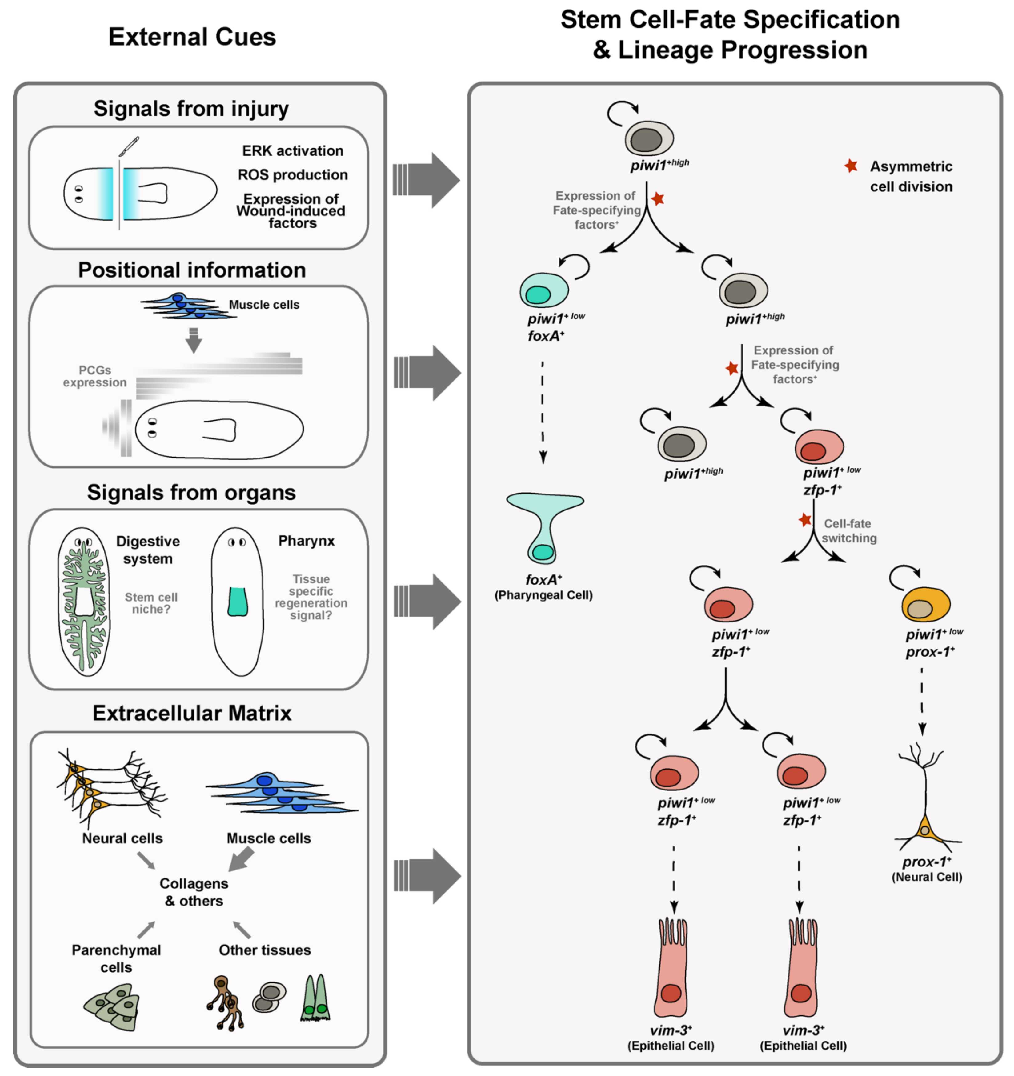
3.1. Distinct Responses of Stem Cells to Amputation and Loss of Specific Organs Occur in Planarians

The ability to replace exactly the body regions and tissues that have been damaged or removed by injury is one of the greatest interests in the field of study of planarian regeneration. Several studies have started to investigate how regeneration signals initiate exit from pluripotency and restrict cells to specific differentiation paths to produce the appropriate number and type of specialized neoblasts in response to different regeneration requirements (Figure 2). The rapid activation of ERK after any type of injury has been hypothesized to be one of the earliest requirements to trigger planarian regeneration [40]. ROS production might play a role in this initial ERK activation [41]. Independently of whether injury implies loss of tissue, neoblasts respond by increasing their proliferative rate, and the expression of wound-induced genes is initiated [14,42]. In the case of tissue loss, this initial response is followed by a second proliferative response that will give rise to the regenerative blastema in which the missing tissues will differentiate [43,44]. Cell fate specification during planarian regeneration appears to occur in the stem cells before entering the blastema, suggesting that planarian blastemas are a mosaic of fate-specified progenitors [45]. The intestine might play a niche-like role in modulating neoblast dynamics [7,46,47]. Some neoblast subtypes such as tgs-1-expressing neoblasts are located in the parenchyma in close proximity to the gut [16] and knockdown of intestine-enriched transcription factors such as nkx2.2 or gata4/5/6-1 or the E3 ligase wwp1 causes reduced blastema formation and decreased neoblast proliferation [47,48,49]. In addition, the disruption of the integration of new intestinal cells into gut branches by silencing the egfr-1/ngr-1 ligand receptor pair does alter the number of several progenitor subtypes in addition to increasing the number of gut progenitor cells [50].
Recent studies have started to investigate how stem cells respond to damage or loss of particular organs. The resection of discrete organs such as the eyes or the pharynx combined with the tracking of the specific progenitor cells that target those organs has revealed that the loss of different tissues stimulates distinct behaviors in planarian stem cells. Eye resection, for instance, does not cause eye progenitor amplification, and constant eye progenitor cell production occurs independently of the presence or absence of differentiated eyes [51]. Interestingly, eye progenitor cells are found exclusively in the head region of the planarian, from the eyes to near the pharynx [26,27], suggesting that unidentified extrinsic cues regulate their specification from pluripotent neoblasts distributed uniformly throughout the body. During homeostasis, differentiated eye cells attract these eye progenitor cells for normal cell turnover through an unknown mechanism, while de novo eye formation after resection or amputation depends on the combination of progenitor production with migratory targeting cues and positional information that defines the site of organ regeneration [51,52,53,54]. Similar mechanisms seem to govern de novo pharynx formation and homeostasis [23,53], although in contrast to eye resection, specific removal of the pharynx does trigger a higher proliferation of pharynx progenitors compared to other progenitor types, suggesting that neoblasts sense the identity and the absence of this tissue to launch targeted regeneration [23,55]. Interestingly, these distinct responses of eye and pharynx progenitor pools to the specific loss of the eye and pharynx organs, respectively, have been correlated with the differential activation of ERK signaling. Thus, only pharynx but not eye resection stimulates a proliferative wound response that requires ERK activation and selective progenitor production by stem cell division [55], suggesting that the loss of different organs can activate distinct regenerative mechanisms and stem cell responses in planarians (Figure 2).

3.2. The Body-Wall Musculature Serves as a Source of Positional Information in Planarians

The importance of the body-wall musculature for proper regeneration has been established after experiments in which the lack of particular subsets of muscle fibers yield aberrant phenotypes. Thus, phred-1 is required for proper muscle regeneration which subsequently affects gut patterning [56]. On the other hand, the lack of longitudinal fibers impairs regeneration initiation, whereas the loss of circular fibers generates bifurcated AP axis [25]. In addition, dorsoventral muscles appear to be required for proper medial-lateral patterning [24]. These results point out that different types of body-wall muscle fibers play different roles in wound signaling and patterning to enable regeneration. To our knowledge, no differences between the different fibers of the body-wall musculature depending on their orientation has been described at the structural level. In fact, for example, all of them express the same myosin heavy chain gene [57]. However, at the molecular level, different conserved transcription factors are required for the differentiation of distinct sets of body-wall muscle. Although a planarian myoD homolog is required for the differentiation of the longitudinal fibers, the differentiation of circular fibers depends on nkx1-1 [25]. Non-body wall muscle as intestinal and pharynx muscle express a different myosin heavy chain gene [58,59]. Recently, different transcription factors required for the differentiation of non-body wall muscle have also been described. Thus, foxF-1 is required for the differentiation of both the dorsoventral and the intestinal muscle, whereas the transcription factors nk4 and gata4/5/6-2 specify lateral and medial dorsoventral fibers, respectively [24]. Finally, a different gata4/5/6 homolog is required for the intestinal muscle [24,60].
Planarians constitutively and regionally express dozens of genes that drive instructions for the maintenance and regeneration of the body plan and whose silencing gives rise to defects in polarity and patterning [2]. These genes, known as positional control genes (PCGs), are expressed along the anterior–posterior, dorsal–ventral and medial–lateral axes of the planarian and include members of the Wnt/β-catenin and BMP signaling pathways, as well as transcription factors of the Forkhead and Pbx families, among others [2]. Interestingly, Witchley and co-workers described that PCGs expression occurs in practically all muscle cells from different regions of the planarian, such as the body-wall, gut and pharynx musculature [61,62]. The expression of PCGs in muscle cells is dynamically regulated during regeneration, so muscle cells readjust the expression of these PCGs according to the new position they acquire after amputation [61]. Therefore, the musculature acts not only as a supportive skeletal-like tissue but also as a source of positional information that drives cell fate in planarians (Figure 2) [63]. For example, expression by muscle cells of the tip of the head of wntA, ndk or of the fz5/8-4 receptor is important to restrict differentiation of the brain and eyes to the most anterior region of the planarian body [62,64,65,66,67]. Similarly, BMP secreted by dorsal muscle cells regulate the fate of epidermal progenitors by inhibiting the expression of genes associated with ventral epidermal identity [68]. PCGs expressed by muscle cells can also help to guide the regeneration of specific organs, such as the prototypical neural projection pattern of the planarian visual system [69]. In addition to expressing PCGs, muscle cells are also a source of secreted extracellular factors such as the EGF-like ligands EGF-6 and NRG-1 that influence the differentiation of nearby neoblasts into protonephridia and digestive cells, respectively [50,70].

3.3. Planarian Muscle Cells Are the Primary Source of Extracellular Matrix

In addition to positional cues, planarian muscle cells are the primary source of extracellular matrix (ECM) and function as connective tissue [71]. The ECM provides the molecular and physical environment for the cell maintenance, self-renewal and differentiation of neoblasts and is required for the proper localization of the cells in the parenchyma (Figure 2) [71,72]. Impaired neoblast migration, proliferation and general tissue growth is observed when the activity of ECM-degrading enzymes is reduced [73,74,75,76]. More recent studies have also revealed the importance of some ECM components such as the EGF repeat-containing genes megf6 and hemicentin to maintain the structure of the basal lamina, restrict the stem cell compartment and retain the parenchymal cell localization of neoblasts and differentiated cells [71,72]. Although some ECM genes are expressed in other tissue types, including intestine, parenchymal cells, neoblasts, neurons and pigment cells, among others [77], muscle cells are the main source of ECM and express core components such as collagen genes [71,77]. Interestingly, these basal membrane collagen and fibrillar collagen genes have been recently shown to play differential roles regulating proliferation during neoblast repopulation, as well as controlling lineage progression. In particular, the basal membrane Type IV collagen col4-1, expressed by both neoblasts and muscle cells, is needed for proper planarian tissue maintenance and regeneration, as well as to restrict neoblast number and promote progenitor progression and cell differentiation. These effects are in part mediated by the EGF signaling pathway, suggesting a role of these ECM components in the control of cell fate specification and symmetric versus asymmetric cell division, as mentioned below [77].

3.4. Neoblasts Cell Fate Specification Occurs through the Cell Cycle: Asymmetric vs. Symmetric Cell Divisions

A recent study has proposed that planarian cell fate establishment occurs through cell divisions [29]. Neoblasts seem to become specialized around the onset of DNA replication [12,29,78]. The expression of fate-specifying factors correlates with cell-cycle progression, and G1 neoblasts show less expression of the lineage-restricted transcription factors that are associated with cell fate specification and thereby are less specialized than neoblasts in S/G2/M cell-cycles phases or G0 postmitotic cells [29]. In other words, most S/G2/M neoblasts are specialized and express fate-specific transcription factors [29]. This seminal recent study revealed that neoblast specialization is dynamic and not only cell fate establishment but also cell fate switching can occur through cell divisions. Thus, when a specialized neoblast divides, it can give rise to a neoblast that can specialize into the mother fate, into a different fate or even generate an unspecialized daughter cell (Figure 2) [29]. Even though a few pathways of neoblast progressive determination have been documented, especially for the eye and the epidermal lineage [12,26,27,39,54,79,80], these late results challenge the classical view of a categorized stem cell population with specific transcriptional profiles and established differentiation potential and reveal that some of the previously considered lineage-committed neoblasts are instead specialized neoblasts that retain pluripotency [29]. Thus, every subclass of neoblast/progenitor cell is a cloud of likelihood, and instead of a discrete tree-like hierarchy, planarian stem cells can acquire multiple direction lineage biases [81]. As the authors state [29], an important future direction will be to assess the frequency with which fate switching through cell division occurs across multiple specialized neoblasts states.
Neoblast specialization decisions are often not passed on to their neoblast daughters through symmetric, amplifying divisions. Cell fate switching through asymmetric cell divisions frequently occurs in at least one of the daughter cells [29]. During neoblast repopulation, approximately 50% of neoblast divisions are asymmetric and give rise to a neoblast daughter and a post-mitotic daughter cell with high and low expression levels of the neoblast marker piwi-1, respectively (Figure 2) [29,82]. Understanding how stem cells control the balance between self-renewal and the production of post-mitotic cells or identifying which signals regulate the asymmetric segregation of cell fate components center some of the current efforts in the study of neoblast biology. Post-transcriptional regulation has been suggested to play an important role in controlling neoblast cell fate decisions and lineage progression [83]. For instance, the influence of alternative splicing for neoblast biology has been evidenced by functional studies of CELF and MBNL RNA binding factors. Planarian neoblasts have a characteristic set of specific mRNA isoforms [84]. Interestingly, Solana and collaborators found that the silencing of mbln genes results in the expression of neoblast-specific mRNA isoforms by differentiated cells, while the silencing of celf genes causes opposing effects and mRNA isoforms specific of differentiated cells are detected in neoblasts, suggesting a role of alternative splicing on controlling neoblast self-renewal and differentiation [84]. Similarly, based on the increased rate of UsnRNA (Uridylate-rich small nuclear RNA) 3′-processing observed in stem cells compared to differentiated cells, it has been proposed that the cell-type-specific modulation of UsnRNA composition and maturation might contribute to neoblasts’ self-renewal and cell fate choices in planarians [85]. In addition, the RNA-binding translational repressor mex-3 has been related with stem cell lineage progression and is suggested to act as a repressor of stem cell identity and self-renewal genes in postmitotic progenitors to promote their differentiation. Although the symmetric/asymmetric expression of piwi-1 transcripts in neoblast doublets has not been analyzed in the context of mex-3 silencing, MEX3 was proposed as a candidate mediator of asymmetric cell fate in planarians [8]. Interestingly, recent studies have started to identify some of the molecular mechanisms that control the balance of symmetric versus asymmetric cell division of neoblasts. The interaction between the ECM component type IV collagen, the discoidin domain receptor (DDR) and the EGF ligand Neuregulin-7 (NRG-7), via the NRG-7/EGFR pathway, have been found to be important in the process [77]. On the one hand, supporting neurons interact with COL-IV from the extracellular matrix via the DDR1 receptor and regulate the expression of nrg-7 in neurons [77]. On the other hand, the binding of NRG-7 to its receptor EGFR-3 on neoblasts regulates asymmetric cell divisions and cell fate decision during neoblast repopulation [82]. The EGFR-3 receptor localizes asymmetrically within the cell membrane of neoblasts, which tend to divide symmetrically and show defects in their proliferation and differentiation in egfr-3 RNAi-silenced planarians [82]. Further investigations would be required to clarify how NRG-7/EGFR-3 signaling regulates neoblast asymmetric division and cell fate choice and whether RNA-binding proteins or mRNA processing play a role in this process.

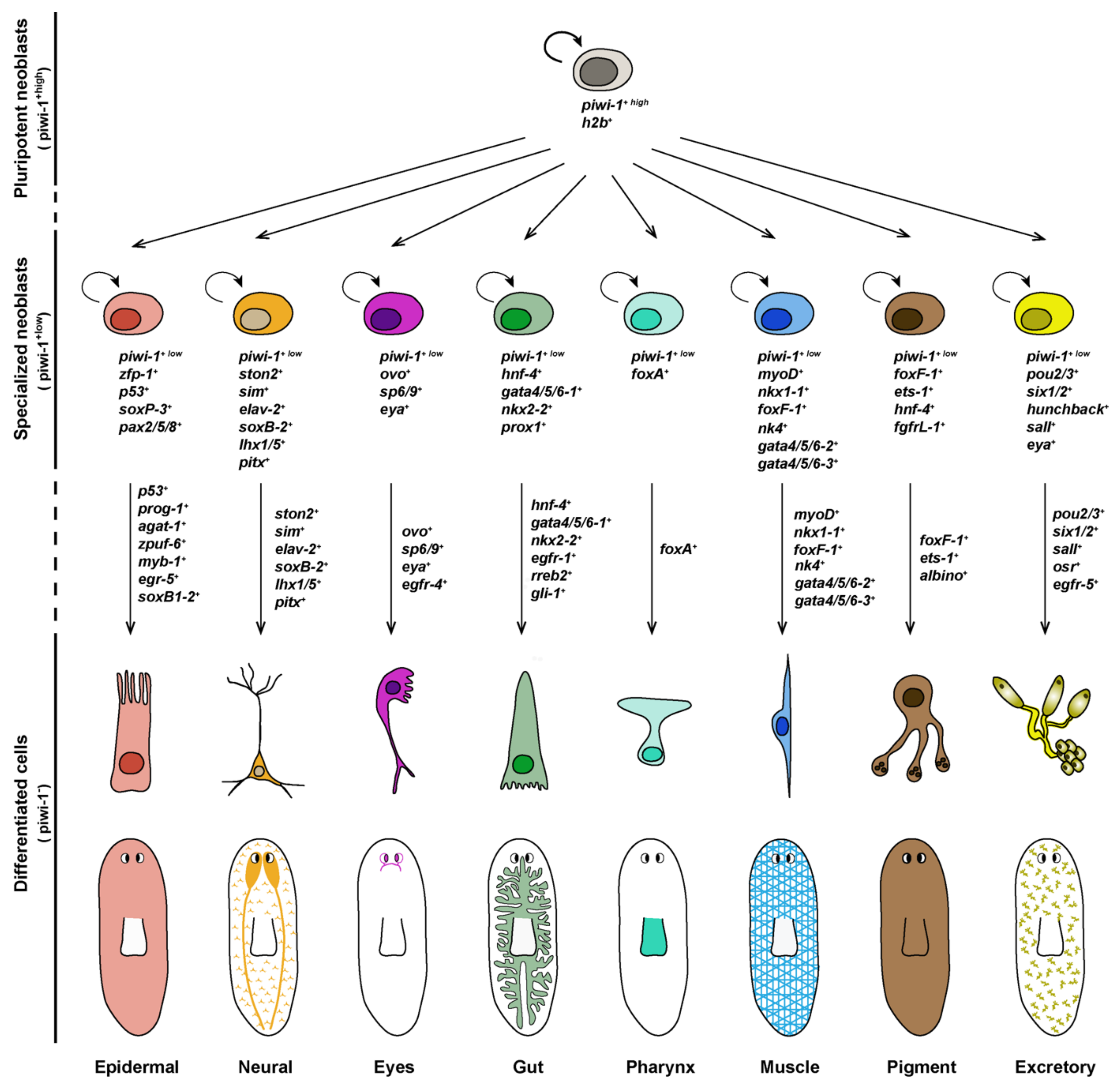
4. Differentiation of Lineage-Committed Stem Cells into Tissue Specific Cell Types

Once individual progenitor populations have been specified, each is characterized by the expression of specific transcription factors often required for their final differentiation into distinct cell fates and organs. For instance, individual progenitors express genes such as ovo, foxA, myoD, gata4/5/6, six-1/2 or pax6a, which are also expressed in mature organs such as the eye, pharynx, muscle, gut, excretory system and nervous system, respectively (Figure 3) [21,22,23,25,26,48]. However, researchers have only recently begun to identify the genes and signaling pathways that are activated downstream of these transcription factors during the final stages of differentiation of these progenitors [8,50]. An exhaustive revision of the most important factors required for specification and differentiation of the diverse planarian tissues has been recently reported elsewhere [3,54,86]. Here, we will comment on the latest findings of some key regulators of lineage progression, paying special attention to those important for general differentiation. In addition, a more extensive list of the transcription factors required for differentiation of specific planarian cell types and organs is detailed in Table 1.
The epidermal lineage is among the best characterized in planarians. The epidermis is a monostratified layer of non-ciliated and ciliated epithelial cells that intercalate mucus-secreting cells. The current model of epidermal lineage progression comprises several intermediate cellular stages between epidermal progenitors (the ζ-neoblasts) and mature epidermal cell types: Epidermal progenitors express zfp-1 and give rise to prog-1 expressing cells (early epidermal progeny, formerly known as nb21.11e) through the activity of zfp-1; these prog-1 cells in turn give rise to agat-1-expressing cells (late epidermal progeny), from which cells expressing zpuf-6 emerge and differentiate into distinct mature epidermal cell types. These different stages of epidermal lineage progression are spatially segregated, with ζ-neoblasts located the deepest in the mesenchyme, and late epidermal progeny cells are closer to the external epithelia [12,39,79]. Epidermal maintenance and regeneration depend on the expression of the transcription factor zfp-1 by the epidermal progenitors [12], as well as on the activity of p53, sox and pax, which cooperate to regulate genes associated with early epidermal precursor cell differentiation [80]. Zhu and co-workers have recently reported that the specification of the first postmitotic epidermal progenitors depends on myb-1, as the silencing of this transcription factor causes a spatiotemporal shift that accelerates epidermal maturation due to the direct specification of stem cell descendants into late epidermal progenitors [88]. The early growth response gene egr-5, which co-expresses with agat-1 in late epidermal progenitors, is required for further progression of the epidermal lineage to mature epidermal cells [79], while the differentiation of subsets of ciliated epidermal cells needs soxB1-2 (Figure 3) [89].
The planarian digestive system consists of one anterior and two posterior highly ramified gut branches that occupy almost the entire body to ensure that nutrients reach all planarian cells. The intestine is a monostratified epithelium composed of only three cell types: phagocytes, secretory goblet cells and basal cells [9,100], but regionalized gene expression along the mediolateral axis, especially among goblet cells, has been recently described [100]. Gut regeneration is accomplished by the differentiation of stem cells and the remodeling of the pre-existing gastrodermis [104]. Transcriptomic analysis and RNAi screenings have identified a set of genes expressed by intestinal progenitors and their progeny required for processes such as branching, the differentiation of functional phagocytes or neoblast proliferation [5,9,12,16,20,47,100]. Gut progenitor cells have been defined by the expression of the transcription factors hnf-4 and gata4/5/6 in piwi-1-expressing cells [5,12,21], although the silencing of gata4/5/6 but not hnf4 causes defects in gut regeneration and maintenance [48,60]. So far, only the function of the EGFR-1/NRG-1 ligand receptor pair has been shown definitively required for differentiation but not specification of gut progenitor cells into mature phagocytes and goblet cells [50]. Other studies have identified the requirement of the hedgehog signaling effector gli-1 and the ras-responsive element binding protein 2 (rreb2) for the regeneration and maintenance of goblet cells, respectively [100], while the differentiation of functional phagocytes requires the activity of the ceramide synthase gene (Figure 3) [47].
Our knowledge on the molecular regulators required for pigment cell lineage development in planarians has progressed rapidly in recent years [105]. The brown body color of freshwater planarians depends on the presence of mature pigment-producing cells that are located in the mesenchyme just underneath the epithelium. The ets-1 and foxF-1 transcription factors are found expressed in mature pigment cells [9,28], and its co-expression in some piwi-1-positive cells identify pigment stem cell progenitors [9]. Proper pigment lineage progression and dynamics during both homeostasis and regeneration requires the activity of these transcription factors, as well as another Forkhead family member, named albino, and the FGF receptor (FGFR)-like molecule fgfrL-1 [28,101]. ets-1, foxF-1 and fgfrL-1 play an important role in early pigment progenitor specification, while albino may act at later stages of the pigment cell lineage (Figure 3) [28,101,105]. The silencing of any of these factors strongly reduces planarian pigmentation [28]. Although the intermediate stages between pigment progenitor cells and mature pigment-producing cells remain to be characterized, two classes of pigment cell types, dendritic and punctated, have been recently described based on the expression pattern of several pigment markers [28]. Both cell types may represent fully differentiated pigment cell types, but the fact that during regeneration dendritic marker expression precedes punctate marker expression suggests that the former may constitute the progenitor cell type of the latter [28]. Interestingly, the knockdown of ets-1 during homeostasis primarily affects the punctate cell population, raising the possibility that this factor regulates the shift between dendritic and punctate cell populations [28].
Stem cells should exit the cell cycle to undergo differentiation [106]. The silencing of genes involved in general lineage progression and cell differentiation are predicted to reduce or deplete the number of postmitotic differentiating cells but do not affect the stem cell population. Examples of such factors include the already mentioned MEX3 RNA-binding protein, since the silencing of its expression results in the expansion of the stem cell compartment in parallel with a decrease in the number of lineage-restricted progenitors committed towards eye, brain, pharynx and protonephridia fates [8]. In contrast, the silencing of apolipoprotein b triggers stem cell progeny accumulation without affecting neoblast maintenance or proliferation, suggesting that intestine-derived lipids may serve as a source of metabolites needed for neoblast differentiation [107]. The accumulation of key stem cell markers have been shown to occur in neoblasts that fail to differentiate following the silencing of not1, a central scaffolding protein of the CCR4-NOT complex involved in mRNA degradation through deadenylation [108]. Similarly, the mRNA base-modificator m6A negatively regulates the transcription of histone variants, and its silencing results in the accumulation of undifferentiated cells [109]. PIWI-piRNA complexes are also needed for proper gene expression during neoblast self-renewal and differentiation [110,111,112], and a PIWI homologue inherited from pluripotent neoblasts has been reported to be necessary for transposon silencing and the normal differentiation of descendent cells [113,114]. Other factors such as ROS production [36], the EGF signaling pathway [115], correct lipid metabolism [116] or the activity of genes such as the tumor suppressor p53 [37,87], the chromatin-remodeling protein CHD4 [37,117] or the transcription factors zfp1 and vasa [37] also play a general role in planarian cell differentiation, although some of them are also important for the proliferative cell expansion of neoblasts [37]. Finally, several studies have highlighted the importance of epigenetic regulation in the regulation of stem cell differentiation [118,119], as we will further discuss in the following section.

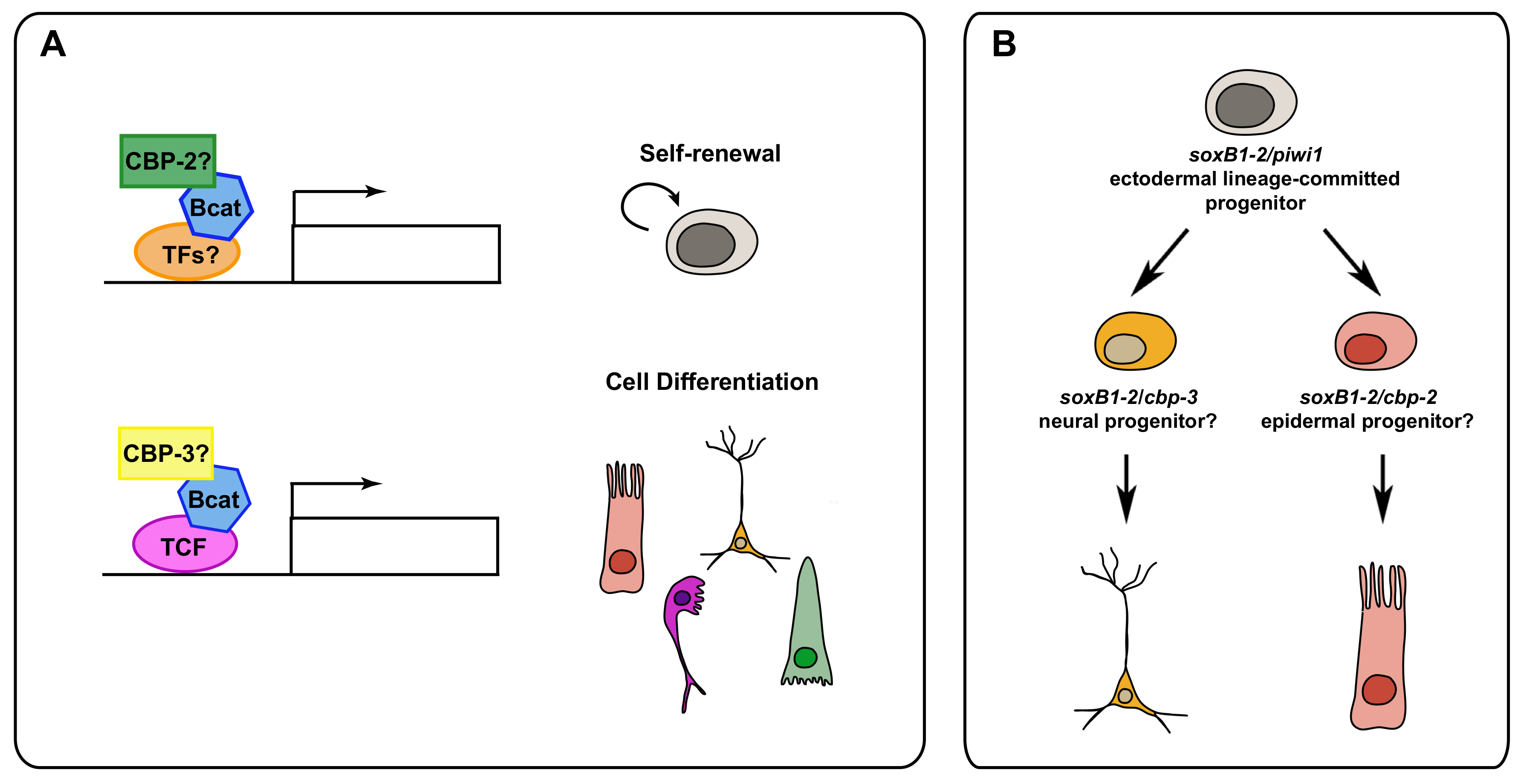
5. Post-Translational Modifications and Epigenetic Regulation in Neoblasts

Protein function is often tightly regulated by post-translational modifications (PTMs) that diversify their function through the reversible or irreversible alteration of their structure and properties through biochemical reactions including phosphorylation, glycosylation, ubiquitylation, methylation and acetylation [120]. PTMs, together with other epigenetic modifications, play pivotal roles to regulate stem cell renewal and differentiation [120,121,122]. Those regulatory mechanisms include histone, DNA and chromatin modifications.
Chromatin is important because in addition to compacting DNA it regulates how and when specific molecules such as repair, replication and transcription factors access to the DNA to undertake their function. Thus, gene expression will be mainly regulated through the capacity of these activating and repressing factors to bind their specific DNA targets. Histones undergo many distinct PTMs, including methylation and acetylation [123]. Histone acetylation mainly on lysine side chains promotes the decompaction of chromatin and therefore is associated with the activation of gene expression. Histone acetylation will depend on the balance between two families of enzymes with opposite functions: histone acetyltransferases (HATs) and histone deacetylases (HDACs). For histone methylation the residues involved are lysines and arginines. Whereas lysines can be mono-, di- or trimethylated, arginines can be mono- or dimethylated. Depending on the specific residues that are methylated the effects on gene expression will be opposed. Thus, lysine methylations at H3K4, H3K6 and H3K79 are associated with active genes. whereas those at H3K9, H3K27 and H4K20 are associated with transcriptional repression [123]. It has been shown that in different types of stem cells, developmental genes are marked with both silencing H3K27me3 and active H3K4me3 marks. This bivalent state keeps a poised-transcriptional context that can be rapidly activated upon differentiation [124,125].
In recent years significant progress has been made in order to understand the role of PTMs and epigenetic modifications on planarian regeneration [119,126]. Strand and collaborators have reviewed extensively how post-translational modifications regulate planarian regeneration, mainly phosphorylation, ubiquitination and chromatin modifications [118]. Thus, for example, MAPK (Mitogen-Activated protein kinases) such as JNK and ERK are required for normal regeneration. JNK appears to trigger apoptotic cell-death required for coordinating neoblast proliferation and differentiation, as well as tissue remodeling [127]. On the other hand, ERK has been described as one of the early signals to induce regeneration [40,128] probably through the activation of ROS [41]. Similarly, other kinases involved in neoblast proliferation and differentiation include those from signaling pathways such as PI3K-AKT-TOR [129,130,131], Hippo [132] and the EGFR that has been shown to be required for the differentiation of several cell types [50,115] as well as for asymmetric division [82], as discussed above.
Ubiquitylation and SUMOylation have been shown also to be important for planarian regeneration [118]. Thus, for example, the silencing of the E3 ligase huwe1 results in an increase in apoptosis and regeneration inhibition despite also inducing an increase in cell proliferation [49]. Similarly, SUMOylation has been reported to regulate cell death and neoblast proliferation probably through the Hedgehog pathway [133].
In addition, several factors involved in chromatin formation and remodeling have been shown to play a role in neoblast function. Thus, a homologue to heterochromatin protein 1 (HP-1) seems to be required to maintain neoblast self-renewal and promote proliferation probably by inducing the expression of mcm5 [134]. Other important factors in chromatin remodeling belong to the highly conserved SWI/SNF family, which has been also characterized in planarians [135]. The silencing of some components of the two main SWI/SNF complexes BAF and PBAF has revealed their role in neoblast proliferation as well as in the differentiation of the epidermal and muscular lineages [135]. The MLL3/4 histone methyltransferases also participate in stem cell proliferation and the differentiation of the epidermis and neurons [136,137,138]. Moreover, a recent study has found that genes that are mainly silenced in neoblasts but that are activated in their post-mitotic progeny show bivalent H3K4me3 and H3K27me3 marks [139].
Recently, two different studies have uncovered the function of the planarian CREB-binding protein (CBP)/p300 family in neoblast biology [126,140]. These transcriptional co-activators play different roles either by acetylating both histone and non-histone proteins (i.e., transcription factors) or serving as protein scaffolds [141,142,143]. On one hand, cbp-2 appears to be required for stem cell maintenance and planarian survival, as after its silencing, the animals show a significant reduction in the number of neoblasts and proliferative cells and die after a few days of treatment [126,140]. On the other side, cbp-3 appears to be mainly involved in neoblast differentiation, although some differences are reported in both studies that might be explained by the different RNAi experimental set up as well as the long-term effects of its silencing. Most invertebrates have just one CBP homologue, and a duplication at the base of the vertebrate lineage seems to be the origin of the p300 gene in that phylum [144]. Remarkably, S. mediterranea possess five CBP homologues as a consequence of possible duplication events specific to the phylum Platyhelminthes [126,140]. Even though vertebrates CBP and p300 interact with mainly the same proteins, their functions are not completely redundant [144]. Interestingly, it has been shown that CBP and p300 could regulate embryonic stem cell self-renewal versus differentiation, respectively. These differential roles seem to be mediated by the interaction of CBP and p300 with the Wnt/β-Catenin pathway [145,146]. However, it is still not clear if the function of the β-catenin/CBP complex on stem cell differentiation depends or not on TCF enhancers [147]. Remarkably, cbp-2 and cbp-3 appear to play quite complementary functions. Thus, the silencing of cbp-2 leads to the absence of the first mitotic peak associated with the wound response together with a significant decrease in the number of piwi-1+ cells. In contrast, cbp-3 RNAi results in a significant increase in the first mitotic peak, as well as of the piwi-1+ population [140]. In addition, for example, cbp-2(RNAi) animals show a decrease in the number of gut progenitors, whereas an increase is observed after silencing cbp-3 [140]. Therefore, it is tempting to speculate that planarian CBP proteins may have diverged functionally in a similar way as vertebrate CBP and p300 appear to regulate stem cell self-renewal and differentiation (Figure 4A).
Another process in which cbp-2 and cbp-3 could have complementary functions is that concerning the neuroectodermal lineage. It has been shown recently that a SoxB1 homologue is expressed in a common neuroectodermal lineage that will give rise to either epidermal or neural cells [89]. Remarkably, a small percentage of SoxB1-positive cells co-express either cbp-2 or cbp-3 [140], which suggests that planarian CBPs could have a role in specifying the final fates within the neuroectodermal lineage (Figure 4B). In agreement with this putative role of planarian CBPs on neural specification, the inhibition of CBP/p300 in Xenopus leads to an increase in neuronal tissues throughout the embryo at the expense of non-neural tissues [148].

6. Conclusions and Future Perspectives

The fine molecular characterization of the planarian neoblasts has uncovered their high level of heterogeneity and a complex hierarchical organization. Through the combination of FACS-based neoblast isolation, single neoblast transplantations and single-cell sequencing, the planarian stem cell compartment has been shown to be constituted by truly pluripotent stem cells that become specialized into multiple lineage progenitors. These distinct cell lineages have been characterized by the expression of specific transcription factors and other genes required for their final differentiation. Importantly, recent studies have started to characterize the onset of neoblast differentiation in relationship to the cell-cycle and their symmetric or asymmetric division. Thus, for example, specialized lineage progenitors show a certain degree of plasticity that allows them to step back towards a pluripotent state and switch cell fate in particularly challenging contexts. Although advances have been made in terms of characterizing the early signals that trigger regeneration (i.e., ERK activation and ROS signaling), how these signals regulate neoblasts behavior remains to be fully understood. Similarly, the muscle fibers provide multiple signals required to regulate the proper patterning during regeneration. How some of these signals act on neoblasts to regulate their proliferation and differentiation is something that requires further studies. In this same line, the possible role of the ECM and the gut as putative niches for neoblasts deserves further investigation. Recent efforts have been made to establish consistent neoblast culture conditions which might be pivotal for the future gene editing of neoblasts as well as to implement transgenesis in these animals [6,149,150]. In addition, some studies have already shown the role that the epigenetic regulation and chromatin remodeling has on neoblasts maintenance and differentiation and will become another important field of study in the near future. Finally, the development of novel tools such as ACME maceration [151] that will allow for a better characterization of planarian cell types and lineages will definitely help to advance in our knowledge on how neoblasts drive regeneration in these amazing animals.

Author Contributions

Conceptualization, M.D.M. and F.C.; writing—original draft preparation, M.D.M. and F.C.; writing—review and editing, M.D.M. and F.C.; funding acquisition, M.D.M. and F.C. All authors have read and agreed to the published version of the manuscript.

Funding

M.D.M. has received funding from the postdoctoral fellowship programme Beatriu de Pinós, funded by the Secretary of Universities and Research (Government of Catalonia) and by the European Union Horizon 2020 research and innovation programme under Marie Sklodowska-Curie grant agreement No. 801370. F.C. was supported by grant PGC2018-100747-B-100 from the Ministerio de Ciencia, Innovación y Universidades, Spain.

Institutional Review Board Statement

Not applicable.

Informed Consent Statement

Not applicable.

Acknowledgments

We are grateful to members of the Planarian Lab Barcelona, especially Susanna Fraguas for helpful discussions. We would like to apologize to those authors whose work was neglected owing to space constraints or lack of awareness.

Conflicts of Interest

The authors declare no conflict of interest.

References

  1. Ivankovic, M.; Haneckova, R.; Thommen, A.; Grohme, M.A.; Vila-Farré, M.; Werner, S.; Rink, J.C. Model Systems for Regeneration: Planarians. Development 2019, 146, dev167684. [Google Scholar] [CrossRef] [PubMed] [Green Version]
  2. Reddien, P.W. The Cellular and Molecular Basis for Planarian Regeneration. Cell 2018, 175, 327–345. [Google Scholar] [CrossRef] [PubMed] [Green Version]
  3. Cebrià, F.; Adell, T.; Saló, E. Rebuilding a Planarian: From Early Signaling to Final Shape. Int. J. Dev. Biol. 2018, 62, 537–550. [Google Scholar] [CrossRef] [PubMed] [Green Version]
  4. Hayashi, T.; Asami, M.; Higuchi, S.; Shibata, N.; Agata, K. Isolation of Planarian X-Ray-Sensitive Stem Cells by Fluorescence-Activated Cell Sorting. Dev. Growth Differ. 2006, 48, 371–380. [Google Scholar] [CrossRef] [PubMed]
  5. Wagner, D.E.; Wang, I.E.; Reddien, P.W. Clonogenic Neoblasts Are Pluripotent Adult Stem Cells That Underlie Planarian Regeneration. Science 2011, 332. [Google Scholar] [CrossRef] [PubMed]
  6. Lei, K.; McKinney, S.A.; Ross, E.J.; Lee, H.C.; Alvarado, A.S. Cultured Pluripotent Planarian Stem Cells Retain Potency and Express Proteins from Exogenously Introduced MRNAs. bioRxiv 2019. [Google Scholar] [CrossRef]
  7. Newmark, P.A.; Sánchez Alvarado, A. Bromodeoxyuridine Specifically Labels the Regenerative Stem Cells of Planarians. Dev. Biol. 2000, 220, 142–153. [Google Scholar] [CrossRef] [Green Version]
  8. Zhu, S.J.; Hallows, S.E.; Currie, K.W.; Xu, C.; Pearson, B.J. A Mex3 Homolog Is Required for Differentiation during Planarian Stem Cell Lineage Development. eLife 2015, 4, e07025. [Google Scholar] [CrossRef]
  9. Fincher, C.T.; Wurtzel, O.; de Hoog, T.; Kravarik, K.M.; Reddien, P.W. Cell Type Transcriptome Atlas for the Planarian Schmidtea Mediterranea. Science 2018, 360, 1–12. [Google Scholar] [CrossRef] [Green Version]
  10. Plass, M.; Solana, J.; Alexander Wolf, F.; Ayoub, S.; Misios, A.; Glažar, P.; Obermayer, B.; Theis, F.J.; Kocks, C.; Rajewsky, N. Cell Type Atlas and Lineage Tree of a Whole Complex Animal by Single-Cell Transcriptomics. Science 2018, 360, 1–10. [Google Scholar] [CrossRef] [Green Version]
  11. Swapna, L.S.; Molinaro, A.M.; Lindsay-Mosher, N.; Pearson, B.J.; Parkinson, J. Comparative Transcriptomic Analyses and Single-Cell RNA Sequencing of the Freshwater Planarian Schmidtea Mediterranea Identify Major Cell Types and Pathway Conservation. Genome Biol. 2018, 19, 1–22. [Google Scholar] [CrossRef] [PubMed]
  12. van Wolfswinkel, J.C.; Wagner, D.E.; Reddien, P.W. Single-Cell Analysis Reveals Functionally Distinct Classes within the Planarian Stem Cell Compartment. Cell Stem Cell 2014, 15, 326–339. [Google Scholar] [CrossRef] [PubMed] [Green Version]
  13. Molinaro, A.M.; Pearson, B.J. In Silico Lineage Tracing through Single Cell Transcriptomics Identifies a Neural Stem Cell Population in Planarians. Genome Biol. 2016, 17, 1–17. [Google Scholar] [CrossRef] [PubMed] [Green Version]
  14. Wurtzel, O.; Cote, L.E.; Poirier, A.; Satija, R.; Regev, A.; Reddien, P.W. A Generic and Cell-Type-Specific Wound Response Precedes Regeneration in Planarians. Dev. Cell 2015, 35, 632–645. [Google Scholar] [CrossRef] [Green Version]
  15. Benham-Pyle, B.W.; Brewster, C.E.; Kent, A.M.; Mann, F.G.; Chen, S.; Scott, A.R.; Box, A.C.; Sánchez Alvarado, A. Identification of Rare, Transient Post-Mitotic Cell States That Are Induced by Injury and Required for Whole-Body Regeneration in Schmidtea Mediterranea. Nat. Cell Biol. 2021, 23, 939–952. [Google Scholar] [CrossRef] [PubMed]
  16. Zeng, A.; Li, H.; Guo, L.; Gao, X.; McKinney, S.; Wang, Y.; Yu, Z.; Park, J.; Semerad, C.; Ross, E.; et al. Prospectively Isolated Tetraspanin+ Neoblasts Are Adult Pluripotent Stem Cells Underlying Planaria Regeneration. Cell 2018, 173, 1593–1608.e20. [Google Scholar] [CrossRef] [Green Version]
  17. Resch, A.M.; Palakodeti, D.; Lu, Y.C.; Horowitz, M.; Graveley, B.R. Transcriptome Analysis Reveals Strain-Specific and Conserved Stemness Genes in Schmidtea Mediterranea. PLoS ONE 2012, 7, e34447. [Google Scholar] [CrossRef]
  18. Solana, J.; Kao, D.; Mihaylova, Y.; Jaber-Hijazi, F.; Malla, S.; Wilson, R.; Aboobaker, A. Defining the Molecular Profile of Planarian Pluripotent Stem Cells Using a Combinatorial RNAseq, RNA Interference and Irradiation Approach. Genome Biol. 2012, 13, 1–23. [Google Scholar] [CrossRef] [PubMed] [Green Version]
  19. Önal, P.; Grün, D.; Adamidi, C.; Rybak, A.; Solana, J.; Mastrobuoni, G.; Wang, Y.; Rahn, H.P.; Chen, W.; Kempa, S.; et al. Gene Expression of Pluripotency Determinants Is Conserved between Mammalian and Planarian Stem Cells. EMBO J. 2012, 31, 2755–2769. [Google Scholar] [CrossRef] [PubMed] [Green Version]
  20. Labbé, R.M.; Irimia, M.; Currie, K.W.; Lin, A.; Zhu, S.J.; Brown, D.D.R.; Ross, E.J.; Voisin, V.; Bader, G.D.; Blencowe, B.J.; et al. A Comparative Transcriptomic Analysis Reveals Conserved Features of Stem Cell Pluripotency in Planarians and Mammals. Stem Cells 2012, 30, 1734–1745. [Google Scholar] [CrossRef] [PubMed] [Green Version]
  21. Scimone, M.L.; Kravarik, K.M.; Lapan, S.W.; Reddien, P.W. Neoblast Specialization in Regeneration of the Planarian Schmidtea Mediterranea. Stem Cell Rep. 2014, 3, 339–352. [Google Scholar] [CrossRef] [PubMed] [Green Version]
  22. Scimone, M.L.; Srivastava, M.; Bell, G.W.; Reddien, P.W. A Regulatory Program for Excretory System Regeneration in Planarians. Development 2011, 138, 4387–4398. [Google Scholar] [CrossRef] [PubMed] [Green Version]
  23. Adler, C.E.; Seidel, C.W.; McKinney, S.A.; Sánchez Alvarado, A. Selective Amputation of the Pharynx Identifies a FoxA-Dependent Regeneration Program in Planaria. eLife 2014, 3, e02238. [Google Scholar] [CrossRef]
  24. Scimone, M.L.; Wurtzel, O.; Malecek, K.; Fincher, C.T.; Oderberg, I.M.; Kravarik, K.M.; Reddien, P.W. FoxF-1 Controls Specification of Non-Body Wall Muscle and Phagocytic Cells in Planarians. Curr. Biol. 2018, 28, 3787–3801.e6. [Google Scholar] [CrossRef] [Green Version]
  25. Scimone, L.M.; Cote, L.E.; Reddien, P.W. Orthogonal Muscle Fibres Have Different Instructive Roles in Planarian Regeneration. Nature 2017, 551, 623–628. [Google Scholar] [CrossRef]
  26. Lapan, S.W.; Reddien, P.W. Transcriptome Analysis of the Planarian Eye Identifies Ovo as a Specific Regulator of Eye Regeneration. Cell Rep. 2012, 2, 294–307. [Google Scholar] [CrossRef] [PubMed] [Green Version]
  27. Lapan, S.W.; Reddien, P.W. Dlx and Sp6-9 Control Optic Cup Regeneration in a Prototypic Eye. PLoS Genet. 2011, 7, e1002226. [Google Scholar] [CrossRef] [Green Version]
  28. He, X.; Lindsay-Mosher, N.; Li, Y.; Molinaro, A.M.; Pellettieri, J.; Pearson, B.J. FOX and ETS Family Transcription Factors Regulate the Pigment Cell Lineage in Planarians. Development 2017, 144, 4550–4551. [Google Scholar] [CrossRef] [Green Version]
  29. Raz, A.A.; Wurtzel, O.; Reddien, P.W. Planarian Stem Cells Specify Fate yet Retain Potency during the Cell Cycle. Cell Stem Cell 2021, 28, 1307–1322.e5. [Google Scholar] [CrossRef]
  30. Baguñà, J. The Planarian Neoblast: The Rambling History of Its Origin and Some Current Black Boxes. Int. J. Dev. Biol. 2012, 56, 19–37. [Google Scholar] [CrossRef] [Green Version]
  31. Reddien, P.W.; Oviedo, N.J.; Jennings, J.R.; Jenkin, J.C.; Sánchez Alvarado, A. SMEDWI-2 Is a PIWI-like Protein That Regulates Planarian Stem Cells. Science 2005, 310, 1327–1330. [Google Scholar] [CrossRef] [Green Version]
  32. Salvetti, A.; Rossi, L. Planarian Stem Cell Heterogeneity. In Advances in Experimental Medicine and Biology; Springer: Berlin/Heidelberg, Germany, 2019; Volume 1123. [Google Scholar]
  33. Molinaro, A.M.; Lindsay-Mosher, N.; Pearson, B.J. Identification of TOR-responsive Slow-cycling Neoblasts in Planarians. EMBO Rep. 2021, 22, e50292. [Google Scholar] [CrossRef]
  34. Mohamed Haroon, M.; Lakshmanan, V.; Sarkar, S.R.; Lei, K.; Vemula, P.K.; Palakodeti, D. Mitochondrial State Determines Functionally Divergent Stem Cell Population in Planaria. Stem Cell Rep. 2021, 16, 1302–1316. [Google Scholar] [CrossRef]
  35. Murphy, M.P. How Mitochondria Produce Reactive Oxygen Species. Biochem. J. 2009, 417, 1–13. [Google Scholar] [CrossRef] [Green Version]
  36. Pirotte, N.; Stevens, A.S.; Fraguas, S.; Plusquin, M.; van Roten, A.; van Belleghem, F.; Paesen, R.; Ameloot, M.; Cebrià, F.; Artois, T.; et al. Reactive Oxygen Species in Planarian Regeneration: An Upstream Necessity for Correct Patterning and Brain Formation. Oxidative Med. Cell. Longev. 2015, 2015, 1–19. [Google Scholar] [CrossRef]
  37. Wagner, D.E.; Ho, J.J.; Reddien, P.W. Genetic Regulators of a Pluripotent Adult Stem Cell System in Planarians Identified by RNAi and Clonal Analysis. Cell Stem Cell 2012, 10, 299–311. [Google Scholar] [CrossRef] [PubMed] [Green Version]
  38. Rodríguez-Esteban, G.; González-Sastre, A.; Rojo-Laguna, J.I.; Saló, E.; Abril, J.F. Digital Gene Expression Approach over Multiple RNA-Seq Data Sets to Detect Neoblast Transcriptional Changes in Schmidtea Mediterranea. BMC Genom. 2015, 16, 361. [Google Scholar] [CrossRef] [Green Version]
  39. Eisenhoffer, G.T.; Kang, H.; Alvarado, A.S. Molecular Analysis of Stem Cells and Their Descendants during Cell Turnover and Regeneration in the Planarian Schmidtea Mediterranea. Cell Stem Cell 2008, 3, 327–339. [Google Scholar] [CrossRef] [PubMed] [Green Version]
  40. Owlarn, S.; Klenner, F.; Schmidt, D.; Rabert, F.; Tomasso, A.; Reuter, H.; Mulaw, M.A.; Moritz, S.; Gentile, L.; Weidinger, G.; et al. Generic Wound Signals Initiate Regeneration in Missing-Tissue Contexts. Nat. Commun. 2017, 8, 1–13. [Google Scholar] [CrossRef] [PubMed]
  41. Jaenen, V.; Fraguas, S.; Bijnens, K.; Heleven, M.; Artois, T.; Romero, R.; Smeets, K.; Cebrià, F. Reactive Oxygen Species Rescue Regeneration after Silencing the MAPK–ERK Signaling Pathway in Schmidtea Mediterranea. Sci. Rep. 2021, 11, 1–16. [Google Scholar] [CrossRef] [PubMed]
  42. Wenemoser, D.; Lapan, S.W.; Wilkinson, A.W.; Bell, G.W.; Reddien, P.W. A Molecular Wound Response Program Associated with Regeneration Initiation in Planarians. Genes Dev. 2012, 26, 988–1002. [Google Scholar] [CrossRef] [Green Version]
  43. Wenemoser, D.; Reddien, P.W. Planarian Regeneration Involves Distinct Stem Cell Responses to Wounds and Tissue Absence. Dev. Biol. 2010, 344, 979–991. [Google Scholar] [CrossRef] [PubMed] [Green Version]
  44. Saló, E.; Baguñà, J. Regeneration and Pattern Formation in Planarians. I. The Pattern of Mitosis in Anterior and Posterior Regeneration in Dugesia (G) Tigrina, and a New Proposal for Blastema Formation. J. Embryol. Exp. Morph. 1984, 83, 63–80. [Google Scholar] [PubMed]
  45. Reddien, P.W. Specialized Progenitors and Regeneration. Developement 2013, 140, 951–957. [Google Scholar] [CrossRef] [Green Version]
  46. Rossi, L.; Salvetti, A. Planarian Stem Cell Niche, the Challenge for Understanding Tissue Regeneration. Semin. Cell Dev. Biol. 2019, 87, 30–36. [Google Scholar] [CrossRef] [PubMed]
  47. Forsthoefel, D.J.; James, N.P.; Escobar, D.J.; Stary, J.M.; Vieira, A.P.; Waters, F.A.; Newmark, P.A. An RNAi Screen Reveals Intestinal Regulators of Branching Morphogenesis, Differentiation, and Stem Cell Proliferation in Planarians. Dev. Cell 2012, 23, 691–704. [Google Scholar] [CrossRef] [PubMed] [Green Version]
  48. Flores, N.M.; Oviedo, N.J.; Sage, J. Essential Role for the Planarian Intestinal GATA Transcription Factor in Stem Cells and Regeneration. Dev. Biol. 2016, 418, 179–188. [Google Scholar] [CrossRef] [PubMed] [Green Version]
  49. Henderson, J.M.; Nisperos, S.V.; Weeks, J.; Ghulam, M.; Marín, I.; Zayas, R.M. Identification of HECT E3 Ubiquitin Ligase Family Genes Involved in Stem Cell Regulation and Regeneration in Planarians. Dev. Biol. 2015, 404, 21–34. [Google Scholar] [CrossRef] [PubMed] [Green Version]
  50. Barberán, S.; Fraguas, S.; Cebrià, F. The EGFR Signaling Pathway Controls Gut Progenitor Differentiation during Planarian Regeneration and Homeostasis. Developement 2016, 143, 2089–2102. [Google Scholar] [CrossRef] [PubMed] [Green Version]
  51. LoCascio, S.A.; Lapan, S.W.; Reddien, P.W. Eye Absence Does Not Regulate Planarian Stem Cells during Eye Regeneration. Dev. Cell 2017, 40, 381–391.e3. [Google Scholar] [CrossRef] [Green Version]
  52. Atabay, K.D.; LoCascio, S.A.; de Hoog, T.; Reddien, P.W. Self-Organization and Progenitor Targeting Generate Stable Patterns in Planarian Regeneration. Science 2018, 360, 404–409. [Google Scholar] [CrossRef] [PubMed] [Green Version]
  53. Hill, E.M.; Petersen, C.P. Positional Information Specifies the Site of Organ Regeneration and Not Tissue Maintenance in Planarians. eLife 2018, 7, e33680. [Google Scholar] [CrossRef]
  54. Reddien, P.W. Principles of Regeneration Revealed by the Planarian Eye. Curr. Opin. Cell Biol. 2021, 73, 19–25. [Google Scholar] [CrossRef]
  55. Bohr, T.E.; Shiroor, D.A.; Adler, C.E. Planarian Stem Cells Sense the Identity of the Missing Pharynx to Launch Its Targeted Regeneration. eLife 2021, 10, e68830. [Google Scholar] [CrossRef]
  56. Adler, C.E.; Sánchez Alvarado, A. PHRED-1 Is a Divergent Neurexin-1 Homolog That Organizes Muscle Fibers and Patterns Organs during Regeneration. Dev. Biol. 2017, 427, 165–175. [Google Scholar] [CrossRef] [PubMed]
  57. Cebrià, F.; Vispo, M.; Newmark, P.; Bueno, D.; Romero, R. Myocyte Differentiation and Body Wall Muscle Regeneration in the Planarian Giardia Tigrina. Dev. Genes Evol. 1997, 207, 306–316. [Google Scholar] [PubMed]
  58. Cebrià, F.; Bueno, D.; Reigada, S.; Romero, R. Intercalary Muscle Cell Renewal in Planarian Pharynx. Dev Genes Evol. 1999, 209, 249–253. [Google Scholar] [CrossRef] [PubMed]
  59. Kobayashi, C.; Watanabe, K.; Agata, K. The Process of Pharynx Regeneration in Planarians. Dev. Biol. 1999, 211, 27–38. [Google Scholar] [CrossRef] [PubMed] [Green Version]
  60. González-Sastre, A.; de Sousa, N.; Adell, T.; Saló, E. The Pioneer Factor Smed-Gata456-1 Is Required for Gut Cell Differentiation and Maintenance in Planarians. Int. J. Dev. Biol. 2017, 61, 53–63. [Google Scholar] [CrossRef] [PubMed] [Green Version]
  61. Witchley, J.N.; Mayer, M.; Wagner, D.E.; Owen, J.H.; Reddien, P.W. Muscle Cells Provide Instructions for Planarian Regeneration. Cell Rep. 2013, 4, 633–641. [Google Scholar] [CrossRef] [Green Version]
  62. Scimone, L.M.; Cote, L.E.; Rogers, T.; Reddien, P.W. Two FGFRL-Wnt Circuits Organize the Planarian Anteroposterior Axis. eLife 2016, 5, e12845. [Google Scholar] [CrossRef] [Green Version]
  63. Cebrià, F. Planarian Body-Wall Muscle: Regeneration and Function beyond a Simple Skeletal Support. Front. Cell Dev. Biol. 2016, 4, 1–10. [Google Scholar] [CrossRef] [Green Version]
  64. Hill, E.M.; Petersen, C.P. Wnt/Notum Spatial Feedback Inhibition Controls Neoblast Differentiation to Regulate Reversible Growth of the Planarian Brain. Developement 2015, 142, 4217–4229. [Google Scholar] [CrossRef] [PubMed] [Green Version]
  65. Adell, T.; Salò, E.; Boutos, M.; Bartscherer, K. Smed-Evi/Wntless Is Required for β-Catenin-Dependent and -Independent Processes during Planarian Regeneration. Development 2009, 136, 905–910. [Google Scholar] [CrossRef] [PubMed] [Green Version]
  66. Cebrià, F.; Kobayashi, C.; Umesono, Y.; Nakazawa, M.; Mineta, K.; Ikeo, K.; Gojobori, T.; Itohk, M.; Tairak, M.; Sánchez Alvarado, A.; et al. FGFR-Related Gene Nou-Darake Restricts Brain Tissues to the Head Region of Planarians. Nature 2002, 419, 620–624. [Google Scholar] [CrossRef]
  67. Kobayashi, C.; Saito, Y.; Ogawa, K.; Agata, K. Wnt Signaling Is Required for Antero-Posterior Patterning of the Planarian Brain. Dev. Biol. 2007, 306, 714–724. [Google Scholar] [CrossRef] [PubMed] [Green Version]
  68. Wurtzel, O.; Oderberg, I.M.; Reddien, P.W. Planarian Epidermal Stem Cells Respond to Positional Cues to Promote Cell-Type Diversity. Dev. Cell 2017, 40, 491–504.e5. [Google Scholar] [CrossRef] [PubMed] [Green Version]
  69. Scimone, M.L.; Atabay, K.D.; Fincher, C.T.; Bonneau, A.R.; Li, D.J.; Reddien, P.W. Muscle and Neuronal Guidepost-like Cells Facilitate Planarian Visual System Regeneration. Science 2020, 368, 1–14. [Google Scholar] [CrossRef] [PubMed]
  70. Barberán, S.; Martín-Durán, J.M.; Cebrià, F. Evolution of the EGFR Pathway in Metazoa and Its Diversification in the Planarian Schmidtea Mediterranea. Sci. Rep. 2016, 6, 1–14. [Google Scholar] [CrossRef] [Green Version]
  71. Cote, L.E.; Simental, E.; Reddien, P.W. Muscle Functions as a Connective Tissue and Source of Extracellular Matrix in Planarians. Nat. Commun. 2019, 10, 1–13. [Google Scholar] [CrossRef] [PubMed]
  72. Lindsay-Mosher, N.; Chan, A.; Pearson, B.J. Planarian EGF Repeat-Containing Genes Megf6 and Hemicentin Are Required to Restrict the Stem Cell Compartment. PLoS Genet. 2020, 16, e1008613. [Google Scholar] [CrossRef] [Green Version]
  73. Dingwall, C.B.; King, R.S. Muscle-Derived Matrix Metalloproteinase Regulates Stem Cell Proliferation in Planarians. Dev. Dyn. 2016, 245, 963–970. [Google Scholar] [CrossRef] [PubMed] [Green Version]
  74. Abnave, P.; Aboukhatwa, E.; Kosaka, N.; Thompson, J.; Hill, M.A.; Aboobaker, A.A. Epithelial-Mesenchymal Transition Transcription Factors Control Pluripotent Adult Stem Cell Migration in Vivo in Planarians. Developement 2017, 144, 3440–3453. [Google Scholar] [CrossRef] [Green Version]
  75. Isolani, M.E.; Abril, J.F.; Saló, E.; Deri, P.; Bianucci, A.M.; Batistoni, R. Planarians as a Model to Assess In Vivo the Role of Matrix Metalloproteinase Genes during Homeostasis and Regeneration. PLoS ONE 2013, 8, e55649. [Google Scholar] [CrossRef] [Green Version]
  76. Van Roten, A.; Barakat, A.Z.; Wouters, A.; Tran, T.A.; Mouton, S.; Noben, J.P.; Gentile, L.; Smeets, K. A Carcinogenic Trigger to Study the Function of Tumor Suppressor Genes in Schmidtea Mediterranea. DMM Dis. Models Mech. 2018, 11, dmm032573. [Google Scholar] [CrossRef] [Green Version]
  77. Chan, A.; Ma, S.; Pearson, B.J.; Chan, D. Collagen IV Differentially Regulates Planarian Stem Cell Potency and Lineage Progression. Proc. Natl. Acad. Sci. USA 2021, 118, e2021251118. [Google Scholar] [CrossRef] [PubMed]
  78. Hayashi, T.; Shibata, N.; Okumura, R.; Kudome, T.; Nishimura, O.; Tarui, H.; Agata, K. Single-Cell Gene Profiling of Planarian Stem Cells Using Fluorescent Activated Cell Sorting and Its “Index Sorting” Function for Stem Cell Research. Dev. Growth Differ. 2010, 52, 131–144. [Google Scholar] [CrossRef] [PubMed]
  79. Tu, K.C.; Cheng, L.-C.; Tk Vu, H.; Lange, J.J.; Mckinney, S.A.; Seidel, C.W.; Sá Nchez Alvarado, A. Egr-5 Is a Post-Mitotic Regulator of Planarian Epidermal Differentiation. eLife 2015, 4, e10501. [Google Scholar] [CrossRef] [PubMed] [Green Version]
  80. Cheng, L.C.; Tu, K.C.; Seidel, C.W.; Robb, S.M.C.; Guo, F.; Sánchez Alvarado, A. Cellular, Ultrastructural and Molecular Analyses of Epidermal Cell Development in the Planarian Schmidtea Mediterranea. Dev. Biol. 2018, 433, 357–373. [Google Scholar] [CrossRef]
  81. Tran, T.A.; Gentile, L. A Lineage CLOUD for Neoblasts. Semin. Cell Dev. Biol. 2019, 87, 22–29. [Google Scholar] [CrossRef]
  82. Lei, K.; Thi-Kim Vu, H.; Mohan, R.D.; McKinney, S.A.; Seidel, C.W.; Alexander, R.; Gotting, K.; Workman, J.L.; Sánchez Alvarado, A. Egf Signaling Directs Neoblast Repopulation by Regulating Asymmetric Cell Division in Planarians. Dev. Cell 2016, 38, 413–429. [Google Scholar] [CrossRef] [Green Version]
  83. Krishna, S.; Palakodeti, D.; Solana, J. Post-Transcriptional Regulation in Planarian Stem Cells. Semin. Cell Dev. Biol. 2019, 87, 69–78. [Google Scholar] [CrossRef]
  84. Solana, J.; Irimia, M.; Ayoub, S.; Orejuela, M.R.; Zywitza, V.; Jens, M.; Tapial, J.; Ray, D.; Morris, Q.; Hughes, T.R.; et al. Conserved Functional Antagonism of CELF and MBNL Proteins Controls Stem Cell-Specific Alternative Splicing in Planarians. eLife 2016, 5, e16797. [Google Scholar] [CrossRef] [Green Version]
  85. Schmidt, D.; Reuter, H.; Hüttner, K.; Ruhe, L.; Rabert, F.; Seebeck, F.; Irimia, M.; Solana, J.; Bartscherer, K. The Integrator Complex Regulates Differential SnRNA Processing and Fate of Adult Stem Cells in the Highly Regenerative Planarian Schmidtea Mediterranea. PLoS Genet. 2018, 14, e1007828. [Google Scholar] [CrossRef] [PubMed]
  86. Ross, K.G.; Currie, K.W.; Pearson, B.J.; Zayas, R.M. Nervous System Development and Regeneration in Freshwater Planarians. Wiley Interdiscip. Rev. Dev. Biol. 2017, 6, e266. [Google Scholar] [CrossRef] [PubMed]
  87. Pearson, B.J.; Alvarado, A.S. A Planarian P53 Homolog Regulates Proliferation and Self-Renewal in Adult Stem Cell Lineages. Development 2010, 137, 213–221. [Google Scholar] [CrossRef] [PubMed] [Green Version]
  88. Zhu, S.J.; Pearson, B.J. Smed-Myb-1 Specifies Early Temporal Identity during Planarian Epidermal Differentiation. Cell Rep. 2018, 25, 38–46.e3. [Google Scholar] [CrossRef] [Green Version]
  89. Ross, K.G.; Molinaro, A.M.; Romero, C.; Dockter, B.; Cable, K.L.; Gonzalez, K.; Zhang, S.; Collins, E.M.S.; Pearson, B.J.; Zayas, R.M. SoxB1 Activity Regulates Sensory Neuron Regeneration, Maintenance, and Function in Planarians. Dev. Cell 2018, 47, 331–347.e5. [Google Scholar] [CrossRef] [Green Version]
  90. Cowles, M.W.; Brown, D.D.R.; Nisperos, S.V.; Stanley, B.N.; Pearson, B.J.; Zayas, R.M. Genome-Wide Analysis of the BHLH Gene Family in Planarians Identifies Factors Required for Adult Neurogenesis and Neuronal Regeneration. Developement 2013, 140, 4691–4702. [Google Scholar] [CrossRef] [PubMed] [Green Version]
  91. Roberts-Galbraith, R.H.; Brubacher, J.L.; Newmark, P.A. A Functional Genomics Screen in Planarians Reveals Regulators of Whole-Brain Regeneration. eLife 2016, 5, e17002. [Google Scholar] [CrossRef] [PubMed]
  92. Currie, K.W.; Molinaro, A.M.; Pearson, B.J. Neuronal Sources of Hedgehog Modulate Neurogenesis in the Adult Planarian Brain. eLife 2016, 5, e19735. [Google Scholar] [CrossRef] [PubMed] [Green Version]
  93. März, M.; Seebeck, F.; Bartscherer, K. A Pitx Transcription Factor Controls the Establishment and Maintenance of the Serotonergic Lineage in Planarians. Developement 2013, 140, 4499–4509. [Google Scholar] [CrossRef] [Green Version]
  94. Currie, K.W.; Pearson, B.J. Transcription Factors Lhx1/5-1 and Pitx Are Required for the Maintenance and Regeneration of Serotonergic Neurons in Planarians. Developement 2013, 140, 3577–3588. [Google Scholar] [CrossRef] [PubMed] [Green Version]
  95. Cowles, M.W.; Omuro, K.C.; Stanley, B.N.; Quintanilla, C.G.; Zayas, R.M. COE Loss-of-Function Analysis Reveals a Genetic Program Underlying Maintenance and Regeneration of the Nervous System in Planarians. PLoS Genet. 2014, 10, e1004746. [Google Scholar] [CrossRef] [Green Version]
  96. Pineda, D.; Gonzalez, J.; Callaerts, P.; Ikeo, K.; Gehring, W.J.; Salo, E. Searching for the Prototypic Eye Genetic Network: Sine Oculis Is Essential for Eye Regeneration in Planarians. Proc. Natl. Acad. Sci. USA 2000, 97, 4525–4529. [Google Scholar] [CrossRef] [PubMed] [Green Version]
  97. Mannini, L.; Rossi, L.; Deri, P.; Gremigni, V.; Salvetti, A.; Saló, E.; Batistoni, R. Djeyes Absent (Djeya) Controls Prototypic Planarian Eye Regeneration by Cooperating with the Transcription Factor Djsix-1. Dev. Biol. 2004, 269, 346–359. [Google Scholar] [CrossRef]
  98. Fraguas, S.; Barberán, S.; Cebrià, F. EGFR Signaling Regulates Cell Proliferation, Differentiation and Morphogenesis during Planarian Regeneration and Homeostasis. Dev. Biol. 2011, 354, 87–101. [Google Scholar] [CrossRef]
  99. Emili, E.; Pallarès, M.E.; Romero, R.; Cebrià, F. Smed-Egfr-4 Is Required for Planarian Eye Regeneration. Int. J. Dev. Biol. 2019, 63, 9–15. [Google Scholar] [CrossRef] [PubMed] [Green Version]
  100. Forsthoefel, D.J.; Cejda, N.I.; Khan, U.W.; Newmark, P.A. Cell-Type Diversity and Regionalized Gene Expression in the Planarian Intestine. eLife 2020, 9, e52613. [Google Scholar] [CrossRef] [PubMed]
  101. Wang, C.; Han, X.S.; Li, F.F.; Huang, S.; Qin, Y.W.; Zhao, X.X.; Jing, Q. Forkhead Containing Transcription Factor Albino Controls Tetrapyrrole-Based Body Pigmentation in Planarian. Cell Discov. 2016, 2, 1–17. [Google Scholar] [CrossRef]
  102. Thi-Kim Vu, H.; Rink, J.C.; McKinney, S.A.; McClain, M.; Lakshmanaperumal, N.; Alexander, R.; Sanchez Alvarado, A. Stem Cells and Fluid Flow Drive Cyst Formation in an Invertebrate Excretory Organ. eLife 2015, 4, e07405. [Google Scholar] [CrossRef] [PubMed]
  103. Rink, J.C.; Vu, H.T.K.; Alvarado, A.S. The Maintenance and Regeneration of the Planarian Excretory System Are Regulated by EGFR Signaling. Development 2011, 138, 3769–3780. [Google Scholar] [CrossRef] [PubMed] [Green Version]
  104. Forsthoefel, D.J.; Park, A.E.; Newmark, P.A. Stem Cell-Based Growth, Regeneration, and Remodeling of the Planarian Intestine. Dev. Biol. 2011, 356, 445–459. [Google Scholar] [CrossRef] [Green Version]
  105. Lindsay-Mosher, N.; Pearson, B.J. The True Colours of the Flatworm: Mechanisms of Pigment Biosynthesis and Pigment Cell Lineage Development in Planarians. Semin. Cell Dev. Biol. 2019, 87, 37–44. [Google Scholar] [CrossRef] [PubMed]
  106. Sato, Y.; Umesono, Y.; Kuroki, Y.; Agata, K.; Hashimoto, C. Proliferation Maintains the Undifferentiated Status of Stem Cells: The Role of the Planarian Cell Cycle Regulator Cdh1. bioRxiv 2021. [Google Scholar] [CrossRef]
  107. Wong, L.L.; Bruxvoort, C.G.; Cejda, N.I.; Otero, J.R.; Forsthoefel, D.J. Intestine-Enriched Apolipoprotein b Orthologs Regulate Stem Cell 1 Differentiation and Regeneration in Planarians 2 3. bioRxiv 2021. [Google Scholar] [CrossRef]
  108. Solana, J.; Gamberi, C.; Mihaylova, Y.; Grosswendt, S.; Chen, C.; Lasko, P.; Rajewsky, N.; Aboobaker, A.A. The CCR4-NOT Complex Mediates Deadenylation and Degradation of Stem Cell MRNAs and Promotes Planarian Stem Cell Differentiation. PLoS Genet. 2013, 9, e1004003. [Google Scholar] [CrossRef]
  109. Dagan, Y.; Yesharim, Y.; Bonneau, A.R.; Schwartz, S.; Wurtzel, O. M6A Is Required for Resolving Progenitor Identity during 1 Planarian Stem Cell Differentiation. bioRxiv 2021. [Google Scholar] [CrossRef]
  110. Kim, I.V.; Duncan, E.M.; Ross, E.J.; Gorbovytska, V.; Nowotarski, S.H.; Elliott, S.A.; Alvarado, A.S.; Kuhn, C.-D. Planarians Recruit PiRNAs for MRNA Turnover in Adult Stem Cells. Genes Dev. 2019, 33, 1575–1590. [Google Scholar] [CrossRef]
  111. Rouhana, L.; Weiss, J.A.; King, R.S.; Newmark, P.A. PIWI Homologs Mediate Histone H4 MRNA Localization to Planarian Chromatoid Bodies. Developement 2014, 141, 2592–2601. [Google Scholar] [CrossRef] [Green Version]
  112. Kashima, M.; Agata, K.; Shibata, N. Searching for Non-Transposable Targets of Planarian Nuclear PIWI in Pluripotent Stem Cells and Differentiated Cells. Dev. Growth Differ. 2018, 60, 260–277. [Google Scholar] [CrossRef] [PubMed] [Green Version]
  113. Shibata, N.; Kashima, M.; Ishiko, T.; Nishimura, O.; Rouhana, L.; Misaki, K.; Yonemura, S.; Saito, K.; Siomi, H.; Siomi, M.C.; et al. Inheritance of a Nuclear PIWI from Pluripotent Stem Cells by Somatic Descendants Ensures Differentiation by Silencing Transposons in Planarian. Dev. Cell 2016, 37, 226–237. [Google Scholar] [CrossRef] [PubMed] [Green Version]
  114. Kashima, M.; Agata, K.; Shibata, N. What Is the Role of PIWI Family Proteins in Adult Pluripotent Stem Cells? Insights from Asexually Reproducing Animals, Planarians. Dev. Growth Differ. 2020, 62, 407–422. [Google Scholar] [CrossRef] [PubMed]
  115. Barberán, S.; Cebrià, F. The Role of the EGFR Signaling Pathway in Stem Cell Differentiation during Planarian Regeneration and Homeostasis. Semin. Cell Dev. Biol. 2019, 87, 45–57. [Google Scholar] [CrossRef]
  116. Deb, S.; Felix, D.A.; Koch, P.; Deb, M.K.; Szafranski, K.; Buder, K.; Sannai, M.; Groth, M.; Kirkpatrick, J.; Pietsch, S.; et al. Tnfaip2/Exoc3-driven Lipid Metabolism Is Essential for Stem Cell Differentiation and Organ Homeostasis. EMBO Rep. 2021, 22, e49328. [Google Scholar] [CrossRef] [PubMed]
  117. Scimone, M.L.; Meisel, J.; Reddien, P.W. The Mi-2-like Smed-CHD4 Gene Is Required for Stem Cell Differentiation in the Planarian Schmidtea Mediterranea. Development 2010, 137, 1231–1241. [Google Scholar] [CrossRef] [Green Version]
  118. Strand, N.S.; Allen, J.M.; Zayas, R.M. Post-Translational Regulation of Planarian Regeneration. Semin. Cell Dev. Biol. 2019, 87, 58–68. [Google Scholar] [CrossRef] [PubMed]
  119. Dattani, A.; Sridhar, D.; Aziz Aboobaker, A. Planarian Flatworms as a New Model System for Understanding the Epigenetic Regulation of Stem Cell Pluripotency and Differentiation. Semin. Cell Dev. Biol. 2019, 87, 79–94. [Google Scholar] [CrossRef]
  120. Wang, Y.C.; Peterson, S.E.; Loring, J.F. Protein Post-Translational Modifications and Regulation of Pluripotency in Human Stem Cells. Cell Res. 2014, 24, 143–160. [Google Scholar] [CrossRef] [Green Version]
  121. Avgustinova, A.; Benitah, S.A. Epigenetic Control of Adult Stem Cell Function. Nat. Rev. Mol. Cell Biol. 2016, 17, 643–658. [Google Scholar] [CrossRef] [PubMed]
  122. Godini, R.; Lafta, H.Y.; Fallahi, H. Epigenetic Modifications in the Embryonic and Induced Pluripotent Stem Cells. Gene Expr. Patterns 2018, 29, 1–9. [Google Scholar] [CrossRef]
  123. Bannister, A.J.; Falcão, A.M.; Castelo-Branco, G. Histone Modifications and Histone Variants in Pluripotency and Differentiation. In Chromatin Regulation and Dynamics; Academic Press: Cambridge, MA, USA, 2017. [Google Scholar]
  124. Bernstein, B.E.; Mikkelsen, T.S.; Xie, X.; Kamal, M.; Huebert, D.J.; Cuff, J.; Fry, B.; Meissner, A.; Wernig, M.; Plath, K.; et al. A Bivalent Chromatin Structure Marks Key Developmental Genes in Embryonic Stem Cells. Cell 2006, 125, 315–326. [Google Scholar] [CrossRef] [PubMed] [Green Version]
  125. Harikumar, A.; Meshorer, E. Chromatin Remodeling and Bivalent Histone Modifications in Embryonic Stem Cells. EMBO Rep. 2015, 16, 1609–1619. [Google Scholar] [CrossRef] [Green Version]
  126. Stelman, C.R.; Smith, B.M.; Chandra, B.; Roberts-Galbraith, R.H. CBP/P300 Homologs CBP2 and CBP3 Play Distinct Roles in Planarian Stem Cell Function. Dev. Biol. 2021, 473, 130–143. [Google Scholar] [CrossRef]
  127. Almuedo-Castillo, M.; Crespo, X.; Seebeck, F.; Bartscherer, K.; Salò, E.; Adell, T. JNK Controls the Onset of Mitosis in Planarian Stem Cells and Triggers Apoptotic Cell Death Required for Regeneration and Remodeling. PLoS Genet. 2014, 10, e1004400. [Google Scholar] [CrossRef] [Green Version]
  128. Tasaki, J.; Shibata, N.; Nishimura, O.; Itomi, K.; Tabata, Y.; Son, F.; Suzuki, N.; Araki, R.; Abe, M.; Agata, K.; et al. ERK Signaling Controls Blastema Cell Differentiation during Planarian Regeneration. Development 2011, 138, 2417–2427. [Google Scholar] [CrossRef] [Green Version]
  129. Peiris, T.H.; Ramirez, D.; Barghouth, P.G.; Oviedo, N.J. The Akt Signaling Pathway Is Required for Tissue Maintenance and Regeneration in Planarians. BMC Dev. Biol. 2016, 16, 7. [Google Scholar] [CrossRef] [PubMed] [Green Version]
  130. Peiris, T.H.; Weckerle, F.; Ozamoto, E.; Ramirez, D.; Davidian, D.; García-Ojeda, M.E.; Oviedo, N.J. TOR Signaling Regulates Planarian Stem Cells and Controls Localized and Organismal Growth. J. Cell Sci. 2012, 125, 1657–1665. [Google Scholar] [CrossRef] [Green Version]
  131. González-Estévez, C.; Felix, D.A.; Smith, M.D.; Paps, J.; Morley, S.J.; James, V.; Sharp, T.V.; Aboobaker, A.A. SMG-1 and MTORC1 Act Antagonistically to Regulate Response to Injury and Growth in Planarians. PLoS Genet. 2012, 8, e1002619. [Google Scholar] [CrossRef] [PubMed] [Green Version]
  132. De Sousa, N.; Rodríguez-Esteban, G.; Rojo-Laguna, J.I.; Saló, E.; Adell, T. Hippo Signaling Controls Cell Cycle and Restricts Cell Plasticity in Planarians. PLoS Biol. 2018, 16, e2002399. [Google Scholar] [CrossRef] [PubMed]
  133. Thiruvalluvan, M.; Barghouth, P.G.; Tsur, A.; Broday, L.; Oviedo, N.J. SUMOylation Controls Stem Cell Proliferation and Regional Cell Death through Hedgehog Signaling in Planarians. Cell. Mol. Life Sci. 2018, 75, 1285–1301. [Google Scholar] [CrossRef] [PubMed] [Green Version]
  134. Zeng, A.; Li, Y.Q.; Wang, C.; Han, X.S.; Li, G.; Wang, J.Y.; Li, D.S.; Qin, Y.W.; Shi, Y.; Brewer, G.; et al. Heterochromatin Protein 1 Promotes Self-Renewal and Triggers Regenerative Proliferation in Adult Stem Cells. J. Cell Biol. 2013, 201, 409–425. [Google Scholar] [CrossRef]
  135. Trost, T.; Haines, J.; Dillon, A.; Mersman, B.; Robbins, M.; Thomas, P.; Hubert, A. Characterizing the Role of SWI/SNF-Related Chromatin Remodeling Complexes in Planarian Regeneration and Stem Cell Function. Stem Cell Res. 2018, 32, 91–103. [Google Scholar] [CrossRef]
  136. Mihaylova, Y.; Abnave, P.; Kao, D.; Hughes, S.; Lai, A.; Jaber-Hijazi, F.; Kosaka, N.; Aboobaker, A.A. Conservation of Epigenetic Regulation by the MLL3/4 Tumour Suppressor in Planarian Pluripotent Stem Cells. Nat. Commun. 2018, 9, 1–17. [Google Scholar] [CrossRef] [Green Version]
  137. Duncan, E.M.; Chitsazan, A.D.; Seidel, C.W.; Alvarado, A.S. Set1 and MLL1/2 Target Distinct Sets of Functionally Different Genomic Loci in Vivo. Cell Rep. 2015, 13, 2741–2755. [Google Scholar] [CrossRef] [PubMed] [Green Version]
  138. Hubert, A.; Henderson, J.M.; Ross, K.G.; Cowles, M.W.; Torres, J.; Zayas, R.M. Epigenetic Regulation of Planarian Stem Cells by the SET1/MLL Family of Histone Methyltransferases. Epigenetics 2013, 8, 79–91. [Google Scholar] [CrossRef] [PubMed] [Green Version]
  139. Dattani, A.; Kao, D.; Mihaylova, Y.; Abnave, P.; Hughes, S.; Lai, A.; Sahu, S.; Aboobaker, A.A. Epigenetic Analyses of Planarian Stem Cells Demonstrate Conservation of Bivalent Histone Modifications in Animal Stem Cells. Genome Res. 2018, 28, 1543–1554. [Google Scholar] [CrossRef] [PubMed] [Green Version]
  140. Fraguas, S.; Cárcel, S.; Vivancos, C.; Molina, M.D.; Ginés, J.; Mazariegos, J.; Sekaran, T.; Bartscherer, K.; Romero, R.; Cebrià, F. CREB-Binding Protein (CBP) Gene Family Regulates Planarian Survival and Stem Cell Differentiation. Dev. Biol. 2021, 476, 53–67. [Google Scholar] [CrossRef]
  141. Voss, A.K.; Thomas, T. Histone Lysine and Genomic Targets of Histone Acetyltransferases in Mammals. BioEssays 2018, 40. [Google Scholar] [CrossRef]
  142. Dutto, I.; Scalera, C.; Prosperi, E. CREBBP and P300 Lysine Acetyl Transferases in the DNA Damage Response. Cell. Mol. Life Sci. 2018, 75, 1325–1338. [Google Scholar] [CrossRef]
  143. Yuan, L.W.; Giordano, A. Acetyltransferase Machinery Conserved in P300/CBP-Family Proteins. Oncogene 2002, 21, 2253–2260. [Google Scholar] [CrossRef] [PubMed] [Green Version]
  144. Thomas, P.D.; Kahn, M. Kat3 Coactivators in Somatic Stem Cells and Cancer Stem Cells: Biological Roles, Evolution, and Pharmacologic Manipulation. Cell Biol. Toxicol. 2016, 32, 61–81. [Google Scholar] [CrossRef] [PubMed]
  145. Teo, J.-L.; Ma, H.; Nguyen, C.; Lam, C.; Kahn, M. Specific Inhibition of CBP-Catenin Interaction Rescues Defects in Neuronal Differentiation Caused by a Presenilin-1 Mutation. Proc. Natl. Acad. Sci. USA 2005, 102, 12171–12176. [Google Scholar] [CrossRef] [Green Version]
  146. Rebel, V.I.; Kung, A.L.; Tanner, E.A.; Yang, H.; Bronson, R.T.; Livingston, D.M. Distinct Roles for CREB-Binding Protein and P300 in Hematopoietic Stem Cell Self-Renewal. Proc. Natl. Acad. Sci. USA 2002, 99, 14789–14794. [Google Scholar] [CrossRef] [PubMed] [Green Version]
  147. Kahn, M. Wnt Signaling in Stem Cells and Cancer Stem Cells: A Tale of Two Coactivators. In Progress in Molecular Biology and Translational Science; Elsevier B.V.: Amsterdam, The Netherlands, 2018; Volume 153, pp. 209–244. [Google Scholar]
  148. Kato, Y.; Shi, Y.; He, X. Neuralization of the Xenopus Embryo by Inhibition of P300/ CREB-Binding Protein Function. J. Neurosci. 1999, 19, 9364–9373. [Google Scholar] [CrossRef] [Green Version]
  149. Niu, K.; Xu, H.; Xiong, Y.Z.; Zhao, Y.; Gao, C.; Seidel, C.W.; Pan, X.; Ying, Y.; Lei, K. Canonical and Early Lineage-Specific Stem Cell Types Identified in Planarian SirNeoblasts. Cell Regen. 2021, 10, 1–11. [Google Scholar] [CrossRef]
  150. Hall, R.N.; Weill, U.; Khariton, M.; Leal-Ortiz, S.; Drees, L.; Chai, C.; Xue, Y.; Rosental, B.; Quake, S.R.; Alvarado, A.S.; et al. Heterologous Reporter Expression in the Planarian Schmidtea Mediterranea through Somatic MRNA Transfection. bioRxiv 2021. [Google Scholar] [CrossRef]
  151. García-Castro, H.; Kenny, N.J.; Iglesias, M.; Álvarez-Campos, P.; Mason, V.; Elek, A.; Schönauer, A.; Sleight, V.A.; Neiro, J.; Aboobaker, A.; et al. ACME Dissociation: A Versatile Cell Fixation-Dissociation Method for Single-Cell Transcriptomics. Genome Biol. 2021, 22, 1–34. [Google Scholar] [CrossRef] [PubMed]
Figure 1. Planarian neoblasts’ heterogeneity. cNeoblasts are pluripotent and can give rise to different subtypes of specialized neoblasts [5] that express a specific subset of transcription factors and will eventually differentiate into distinct planarian tissues such as the gut (γ-neoblasts), epidermis (ζ-neoblasts) or nervous system (ν-neoblasts) [5,12,13]. Some of these specialized neoblasts retain pluripotency and can produce specialized neoblasts for other subtypes [12,29]. Grey discontinued arrows represent untested cell relationships. + symbols indicate expression of the genes. Question marks indicate that the genes cannot be considered exclusive markers of cNeoblasts as are also expressed by other cellular populations.
Biomolecules 11 01532 g001
Figure 2. External cues guide planarian stem cell behavior. The balance between cell fate commitment and self-renewal is influenced by external cues such as signals from injury and missing organs and from the extracellular matrix. As differentiation proceeds, cell-fate-specialized progenitors downregulate piwi-1 expression while the expression of tissue-associated transcription factors increases [9,16,29]. Some specialized neoblasts retain pluripotency and cell fate switching can occur to produce specialized neoblasts for other subtypes [12,29]. Asymmetric cell divisions are marked by a red star.
Biomolecules 11 01532 g002
Figure 3. Schematic of planarian stem cell lineages. Markers for each progenitor lineage are indicated. The expression of a subset of these factors is probably further restricted to different cell-specific progenitors within each lineage.
Biomolecules 11 01532 g003
Figure 4. Putative models for the different roles of cbp-2 and cbp-3. Planarian CBP proteins may have diverged functionally to regulate stem cell maintenance and differentiation (A) and neuroectodermal lineage progression (B).
Biomolecules 11 01532 g004
Table 1. Transcription factors and signaling molecules required for specification and/or differentiation of planarian cell types.
LineageGene
Name
In Situ Expressionpiwi-1+
or PIWI-1+
Coexpression
RNAi
Phenotype
Reference
Epidermalzfp-1ProgenitorsyesDepletion of epidermal progenitors[12,37]
soxP-3Progenitorsn.d.Reduced early progeny markers[12,80]
egr-1Progenitorsyesn.d.[12,37]
p53Progenitors and early progenyyesReduced epidermal progeny[12,80,87]
prog-1Early progenyyesn.d.[8,39]
myb-1Early and late progenyyesAbsent early progeny fate[88]
agat-1Late progenynon.d.[39]
Pax2/5/8Late progeny and mature epidermisn.d.Reduced early progeny markers[80]
zpuf-6Late progeny and mature epidermisn.d.n.d.[79]
egr-5Late progenynoImpaired epidermal differentiation[79]
SoxB1-2Neuroectodermal progenitorsyesAbnormal behavior and movement[89]
Neuralston-2Progenitors and different neuronsyesn.d.[13]
elav-2Progenitors and differentiated neuronsyesn.d.[13]
klfProgenitors and differentiated neuronsyesAbsent cintillo sensory neurons[21]
pax3/7Progenitors and differentiated neuronsyesAbsent dbh+ neurons[21]
neuroD-1Progenitors and differentiated neuronsyesn.d.[21,90]
soxB-2Progenitors and differentiated neuronsyesReduced neural progenitor expression[21,91]
arrowheadDifferentiated neuronsn.d.Defects at the brain commissure[91]
mblkProgeny and different neuronsn.d.Small brain regeneration[91]
tcf/lef-1Progenitors and differentiated neuronsyesReduced dorsolateral GABA neurons[21]
nkx2.2Progenitors and differentiated neuronsyesReduced ventromedial neurons[21]
arxDifferentiated neuronsyesReduced ventromedial neurons[92]
PitxSerotonergic and other neuronsyesAbsence of serotonergic neurons[93,94]
lhx1/5Serotonergic and other neuronsyesAbsence of serotonergic neurons[21,93,94]
hesl-3Progenitors and differentiated neuronsyesDefects in CNS pattern and organization[90]
simProgenitors and differentiated neuronsyesDefects in CNS regeneration[21,90]
coeProgenitors and differentiated neuronsyesDefects in CNS size and organization[90,95]
ap-2Progenitors and differentiated neuronsyesReduced TrpA-expressing neurons[21,42]
runt-1Neoblasts at woundsyesPerturbed ap-2+; sp6-9+ expression
and neural differentiation
[42]
EyesovoProgenitor and differentiated eye cellsyesLack of eye cells[26]
six1/2Progenitor and differentiated eye cellsn.d.Lack of eye cells[27,42,96,97]
eyaProgenitor and differentiated eye cellsyesLack of eye cells[26,27,42,97]
sp6/9Progenitor and differentiated PC cellsyesLack of PC cells[27,42]
dlxProgenitor and differentiated PC cellsyesLack of PC cells[27,42]
otxAProgenitor and differentiated PH cellsyesLack of PH cells[21,26,27]
meisProgenitor and differentiated PH cellsyesDisorganized eye regeneration[21,26]
klfProgenitor and differentiated PH cellsyesDisorganized eye regeneration[21,26]
foxQ2Progenitor and differentiated PH cellsyesDisorganized eye regeneration[21,26]
soxBProgenitor and differentiated PH cellsyesSmall eyes and lack anterior PH cells[21,26]
egfr-1Differentiated PC cellsn.d.Reduced number PC cells[98]
egr-4Differentiated PH cellsn.d.Less differentiated and more eye progenitors[99]
Intestinalgata4/5/6-1Progenitors and differentiated gut cellsyesImpaired differentiated gut progenitors[5,12,48,60]
nkx2-2Progenitors and differentiated gut cellsn.d.Impaired gut regeneration and lysis[12,47]
hnf4Progenitors and differentiated gut cellsyesReduced expression gut markers[5,12,21]
egfr1/nrg-1Gut cellsyesImpaired gut progenitor differentiation[50]
rreb2Several gut cell typesn.d.Impaired goblet cell regeneration[100]
gli-1Several gut cell typesn.d.Impaired goblet cell regeneration[100]
Pharyngealegfr-1Pharynx and othersn.d.Aberrant pharynx regeneration[98]
FoxAProgenit. and differentiated pharynx cellsyesImpaired pharynx regeneration[21,23]
Muscularnkx1-1Progenitors and BWMyesLack of circular fibers and bifurcated AP axis and fused heads[24,25]
myoDProgenitors and BWMyesLack of longitudinal fibers and defects in regeneration initiation[24,25]
foxF-1Progenitors and non-BWM(DVM, DVL, IM, PM)yesDepigmentation, lack of non-BWM and ML defects[24]
nk4Progenitors and DVL cellsyesReduced DVL markers and ML defects[24]
gata4/5/6-2Progenitors and DVM cellsyesReduced DVM number and ML defects[24]
gata4/5/6-3Progenitors and IM and PM cellsyesReduced number of IM cells[24]
Pigmentets-1Progenitors and differentiated cellsyesDepigmentation and reduce markers [9,28]
foxF-1Progenitors and differentiated cellsyesDepigmentation and lack of markers [9,28]
fgfrL-1Differentiated pigment cellsn.d.Depigmentation and reduced punctated marker[28]
albinoProgenitors and differentiated cellsyesDepigmentation and lack markers[28,101]
ExcretorySix1/2Progenitors and tubule cellsyesImpaired protonephridia regeneration[22]
Pou2/3Progenitors and tubule-related cellsyesImpaired protonephridia regeneration [22,102]
hunchbackn.d.n.d.Impaired protonephridia regeneration [22]
eyaProgenitors and differentiated cellsyesImpaired protonephridia regeneration[22]
osrTubule cellsyesn.d.[22]
SalIProgenitors and tubule cellsyesEdemas and decreased tubule cell number[22]
Egfr-5Flame cellsn.dAbsence of flame cells and protonephridia disorganization[103]
n.d., not determined; CNS, central nervous system; PC, pigment cup cells; PH, photoreceptor cells; BWM, Body wall muscle; DVM, Dorsoventral medial muscle; DVL, Dorsoventral lateral muscle; IM, intestinal muscle; PM, Pharyngeal muscle; AP, anteroposterior; ML, mediolateral.
Publisher’s Note: MDPI stays neutral with regard to jurisdictional claims in published maps and institutional affiliations.

