Review

Alternative Pre-mRNA Splicing of the Mu Opioid Receptor Gene, OPRM1: Insight into Complex Mu Opioid Actions

1
Department of Anesthesiology, Rutgers New Jersey Medical School, Newark, NJ 07103, USA
2
Department of Chemical Biology, Ernest Mario School of Pharmacy Rutgers University, Piscataway, NJ 08854, USA
3
Center for Clinical Pharmacology, University of Health Sciences & Pharmacy and Washington University School of Medicine, St. Louis, MO 63110, USA
*
Author to whom correspondence should be addressed.
These authors contributed equally to this work.
Biomolecules 2021, 11(10), 1525; https://doi.org/10.3390/biom11101525
Submission received: 30 August 2021 / Revised: 11 October 2021 / Accepted: 11 October 2021 / Published: 15 October 2021
(This article belongs to the Special Issue GPCRs: Structure, Biology and Potential Applications)

Abstract

:
Most opioid analgesics used clinically, including morphine and fentanyl, as well as the recreational drug heroin, act primarily through the mu opioid receptor, a class A Rhodopsin-like G protein-coupled receptor (GPCR). The single-copy mu opioid receptor gene, OPRM1, undergoes extensive alternative splicing, creating multiple splice variants or isoforms via a variety of alternative splicing events. These OPRM1 splice variants can be categorized into three major types based on the receptor structure: (1) full-length 7 transmembrane (TM) C-terminal variants; (2) truncated 6TM variants; and (3) single TM variants. Increasing evidence suggests that these OPRM1 splice variants are pharmacologically important in mediating the distinct actions of various mu opioids. More importantly, the OPRM1 variants can be targeted for development of novel opioid analgesics that are potent against multiple types of pain, but devoid of many side-effects associated with traditional opiates. In this review, we provide an overview of OPRM1 alternative splicing and its functional relevance in opioid pharmacology.

1. Introduction

The actions of most clinically used opioid analgesics, such as morphine and fentanyl, are complex. Although they provide potent analgesia, they produce many undesirable side-effects such as respiratory depression, constipation, pruritus, physical dependence, and addiction. Misusing these opioids often leads to development of opioid use disorder, contributing significantly to the worldwide opioid epidemic. These opioid drugs act primarily on the mu opioid receptor (MOR) that was initially proposed by Martin in 1967 [1], established by pharmacological studies in 1973 [2,3,4], and verified by molecular cloning in 1993 [5,6,7]. Clinically, patients often showed divergent responses to mu opioids not only in analgesia but also in side-effects, and incomplete cross-tolerance among mu opioids led to the clinical practice of opioid rotation. These observations suggest the presence of multiple mu opioid receptors, a concept that was originally proposed by early pharmacological studies [8,9,10] and evolved by recent molecular studies of the mu opioid receptor gene, OPRM1 [11,12].
Only a single-copy OPRM1 gene has been identified, raising the hypothesis that alternative pre-mRNA splicing of the OPRM1 gene generates multiple mu opioid receptors, contributing to the diversity and complexity of opioid responses seen in humans and animals. Driven by this hypothesis, many efforts have been made to isolate alternatively spliced OPRM1 variants. We now know that the single-copy OPRM1 gene undergoes extensive alternative splicing, creating an array of splice variants that are far more complicated than those defined by the early pharmacological studies. Conservation of OPRM1 alternative splicing from rodents to humans suggests evolutionary and functional importance of the OPRM1 gene. These OPRM1 splice variants can be categorized mainly into three structurally distinct types: (1) full-length 7 transmembrane domain (TM) C-terminal variants; (2) truncated 6TM variants; and (3) truncated single TM variants, all of which are conserved from rodents to humans. Accumulated evidence indicates that these OPRM1 splice variants play important roles in mu opioid pharmacology. This review outlines the progress regarding OPRM1 alternative splicing and its pharmacological functions.

2. Mechanisms and Functions of Alternative Pre-mRNA Splicing

RNA splicing or precursor mRNA (pre-mRNA) splicing discovered in 1977 [13,14] involves removing introns and connecting exons to produce mature mRNA, a post-transcriptional event in metazoan organisms essential for many biological and pathological processes. RNA splicing is mediated through the spliceosome, a large complex including five small nuclear ribonucleoprotein particles (snRNPs, U1, U2, U4/U6, and U5) and more than 150 proteins. Alternative splicing is a major source of generating proteomic diversity. It has been estimated that ~95% of human multiple-exon genes undergo alternative splicing [15,16,17]. There are several types of alternative splicing, including exon skipping, alterative 5′ or 3′ splicing, intron retention, mutually exclusive exons, and alternative promoters and poly(A) sites [15]. Alternative splicing is differentially regulated in various tissues and cell types, as well as in different developmental stages, providing its diverse functions in a spatial and temporal manner. The magnitude of these effects varies from very subtle modifications to a complete loss or gain of function. Alternative splicing also participates in RNA processing itself, from pre- to post-transcriptional events [18].
Extensive studies of alternative splicing in a number of genes, including the c-src [19,20,21], Troponin T [22,23,24], and FGFR2 [25,26,27], elucidated mechanisms of alternative splicing. Our understanding of alternative splicing mechanisms was further extended by the identification of RNA binding proteins that modulate alternative splicing, such as the serine/arginine-rich (SR) family proteins [28,29,30,31], hnRNPs [32,33,34], the CELF protein family [35,36,37], and the neuro-oncological ventral antigen (NOVA) family [38,39,40]. In general, alternative splicing is a process of coordinating specific cis-acting elements in pre-mRNA and certain trans-acting factors with the basal spliceosome.
Aberrant alternative splicing has been associated with a number of human diseases [41,42], such as amyotrophic lateral sclerosis [43,44], paraneoplastic neurological disorders [38,45], myotonic dystrophy [46], spinal muscular atrophy [47,48], and cancer [27,49,50,51,52]. Antisense technologies have been used to correct aberrant alternative splicing for treating several human diseases. Spinraza (nusinersen), an antisense oligonucleotide targeting exon 7 of survival of moter neuron 2 (SMN2) gene, was the first FDA-approved drug to treat spinal muscular atrophy.

3. Alternative Splicing in GPCRs

There are about 800 different G protein-coupled receptors (GPCRs), representing the largest group of transmembrane receptors encoded by the human genome. GPCRs are targets for ~35% of FDA approved drugs [53,54]. Structurally, canonical GPCRs are characterized by an extracellular N-terminus, followed by seven transmembrane (TM) α-helices (TM-1 to TM-7) connected by three intracellular and three extracellular loops, and finally an intracellular C-terminus. GPCRs mediate diverse physiological responses, such as sense of vision, smell, taste, and nociception, by transducing extracellular stimuli to intracellular signaling molecules, such as heterotrimeric G proteins and β-arrestins.
Approximately 50% of GPCRs are subject to alternative pre-mRNA splicing [55], producing structurally diverse GPCR isoforms or variants. These splice variants can have alternative N- or C-termini, variable extracellular or intracellular loops, and truncated transmembrane domains, as well as alternative 5′ or 3′ untranslated regions (UTR). For example, genomic data search of non-olfactory receptor GPCR genes predicts ~12% of human GPCR genes have one or more alternatively spliced C-terminal variants [56], while over 9 and 7% of human GPCR genes contain truncated 6TM and single TM variants, respectively [57,58]. Alternative splicing of GPCR genes adds additional layers of complexity to the regulation and function of GPCR pharmacology. Table 1 lists selected human GPCRs with known splice variants along with their tissue distribution, and agonist/antagonist. However, the function of a majority of GPCR splice variants remains unknown.

4. Alternative Splicing of the Mu Opioid Receptor Gene, OPRM1

Most opioid analgesics used clinically, including morphine and fentanyl, as well as the recreational drug heroin, act primarily through mu opioid receptors. The mu opioid receptor was first proposed as the M (morphine, µ) receptor, together with the N (nalorphine, probably corresponding to the kappa1 receptor) receptor, by Martin in 1967, based on the pharmacological studies of nalorphine [1]. Establishment of opioid receptor binding assays [2,3,4] and identification of endogenous opioid peptides [86,87,88,89,90] facilitated the pharmacological defining of opioid receptor subtypes as mu, delta, and kappa receptors. Molecular cloning of the delta opioid receptor (DOR-1) [91,92] quickly led to isolation of the mu opioid receptor (MOR-1) [5,6,7] and kappa1 opioid (KOR-1) [93,94], as well as KOR-3 or opioid receptor-like receptor (ORL-1) [95,96,97,98,99]. All these opioid receptors belong to the GPCR Class A rhodopsin family.

4.1. Concept of Multiple Mu Opioid Receptors

The concept of multiple mu opioid receptors was originally suggested based on clinical observations that different patients displayed divergent responses to mu opioids not only in analgesia, but also in side-effects and that incomplete cross-tolerance among mu opioids led to the clinical practice of opioid rotation. Similar observations were seen in animal models. Different inbred mouse strains showed various sensitivities to mu opioids. For example, the 129/P3 inbred mouse strain is resistant to produce morphine tolerance and naloxone-precipitated withdrawal, sharply contrasting with the robust morphine tolerance and physical dependence seen in C56BL/6J and SWR/J inbred mouse strains [100,101]. Multiple mu binding sites were first demonstrated with opioid receptor binding assays using [3H]-labeled opioids in 1981 [9], subsequently validated by computer modeling approaches [102,103] and visualized by autoradiography [104,105,106]. Further pharmacological studies using two antagonists, naloxazone and naloxonazine, defined two mu subtypes, mu1 and mu2 [10]. A third mu subtype, morphine-6β-glucuronide (M6G) receptor, was proposed based on differences in analgesia potency, binding affinity of morphine, morphine analgesia in CXBK mice, and sensitivity toward this.
3-O-methylnaltrexone, a mu antagonist, had high affinity toward M6G and blocked analgesia of M6G, but not morphine [10,107,108,109]. Subsequent antisense oligonucleotide and Oprm1 knockout studies provided molecular insights into the M6G site (see below) [107,110,111,112]. The identification of the MOR-1 sequence and its gene, OPRM1, allows us to explore multiple mu opioid receptors at the molecular level. The concept of multiple mu opioid receptors has been evolved by isolation and characterization of an array of alternatively spliced variants or isoforms from the single-copy OPRM1 gene. The correlation between mu subtypes such as mu1 and mu2 defined by the early pharmacological studies and the OPRM1 splice variants remains unknown, although several hypotheses such as heterodimerization were speculated.

4.2. Evolution of OPRM1 Gene

All four opioid receptor genes, including mu receptor (OPRM1), delta receptor (OPRD1), kappa receptor (OPRK1), and opioid-receptor like (OPRL1), are found only in vertebrates. As suggested by phylogenetic studies, the opioid receptor genes were generated through a two-round genomic duplication (paleoploidization) from a single ancestral opioid receptor gene (unireceptor) [113,114,115]. The first-round duplication produced OPRD1/OPRM1 and OPRL1/OPRK1 genes, which were subsequently segregated into individual OPRD1, OPRM1, OPRL1, and OPRK1 [116,117,118]. Like the evolutionary tree of life, analyzing MOR-1 protein sequences predicted from 27 species define four major clades as follows: (1) fish, (2) amphibians, (3) birds, and (4) mammals [119]. Sequence alignments of MOR-1 proteins from different species reveal that the high-homology regions are located in the transmembrane domains and the three intracellular loops, suggesting the importance of these conserved structures in mu ligand binding and G protein coupling.
The genomic structure of the OPRM1 gene appears to have evolved along with the four polygenetic clades [12,120]. In fish (teleosts), the OPRM1 has five exons and each exon encodes regions with related TM domains: exon 1 (TM1), exon 2 (TM2–TM4), exon 3 (TM5), exon 4 (TM6), and exon 5 (TM7). In amphibians, the last three exons were merged into a single third exon encoding TM5–TM7. This structure with three exons encoding 7TMs has been maintained throughout birds and mammals. Starting in the chicken, exon 4 that encods 12 amino acids at the intracellular C-terminal tail evolved, which is maintained in all the mammals. Additional 5′ and/or 3′ exons such as exons 7 and 11 were integrated in mannals as well, providing a genomic structure for complex alternative splicing of the OPRM1 gene. The functional importance of exon 7- and exon 11-associated splice variants in opioid pharmacology is discussed below.

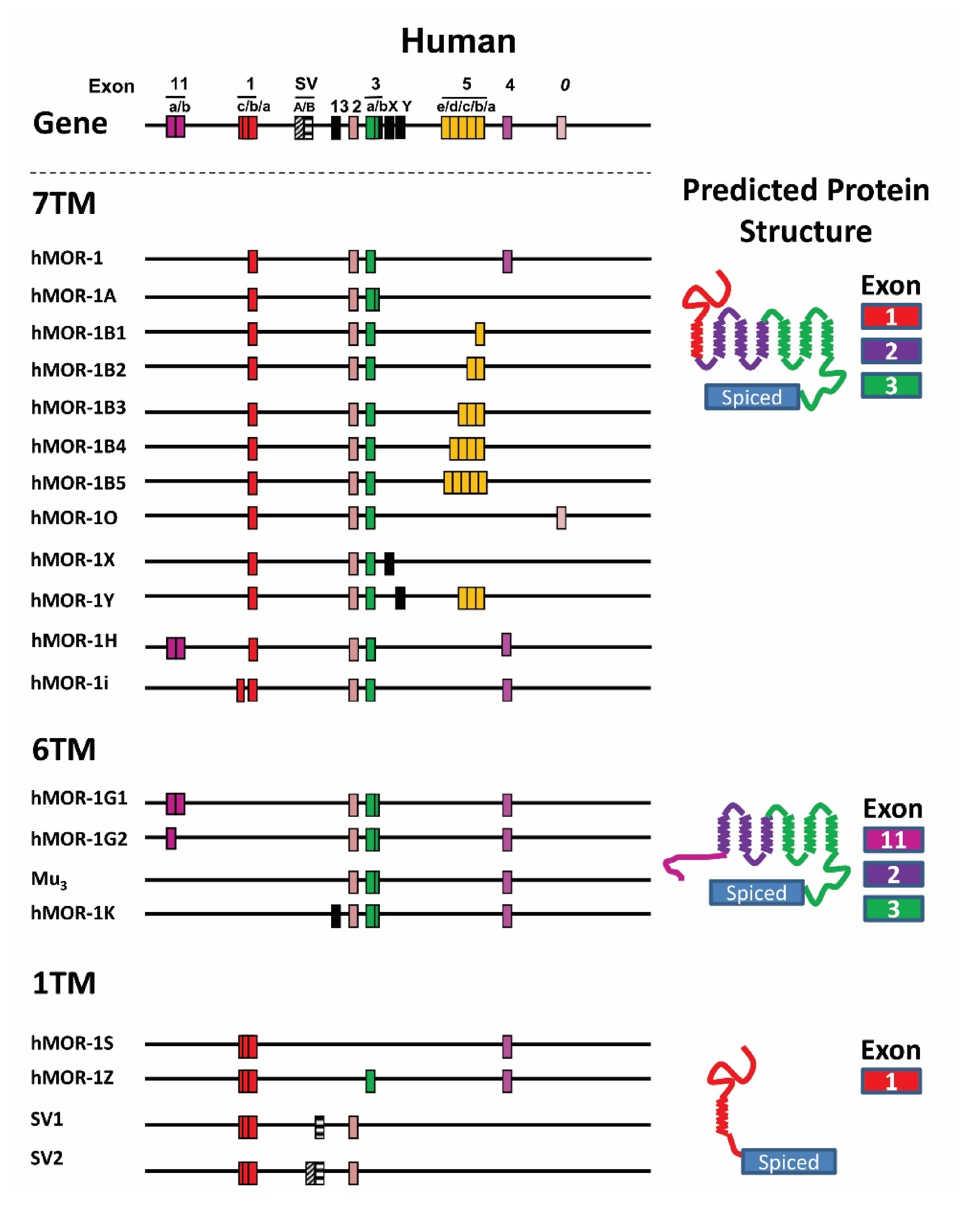
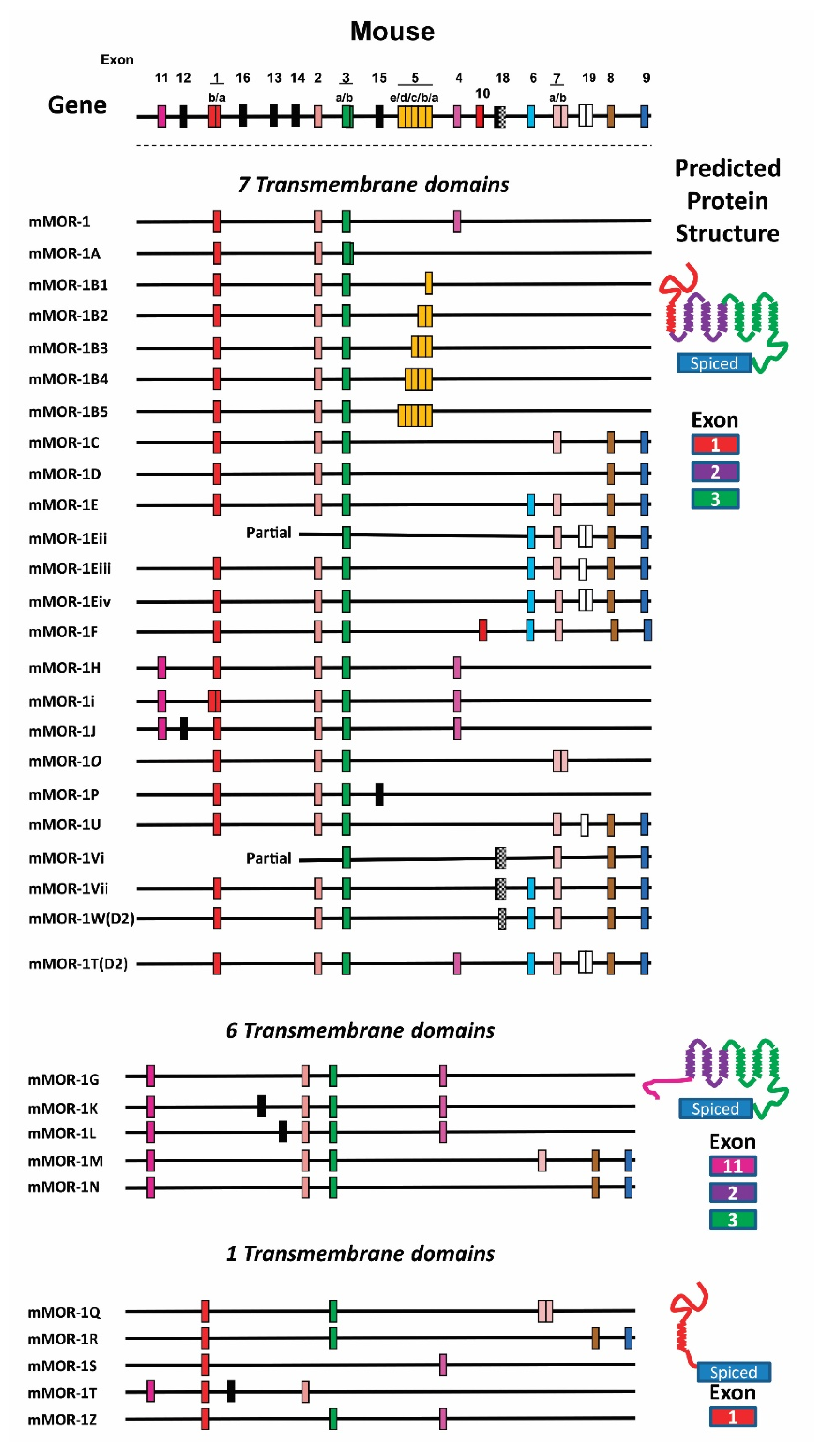
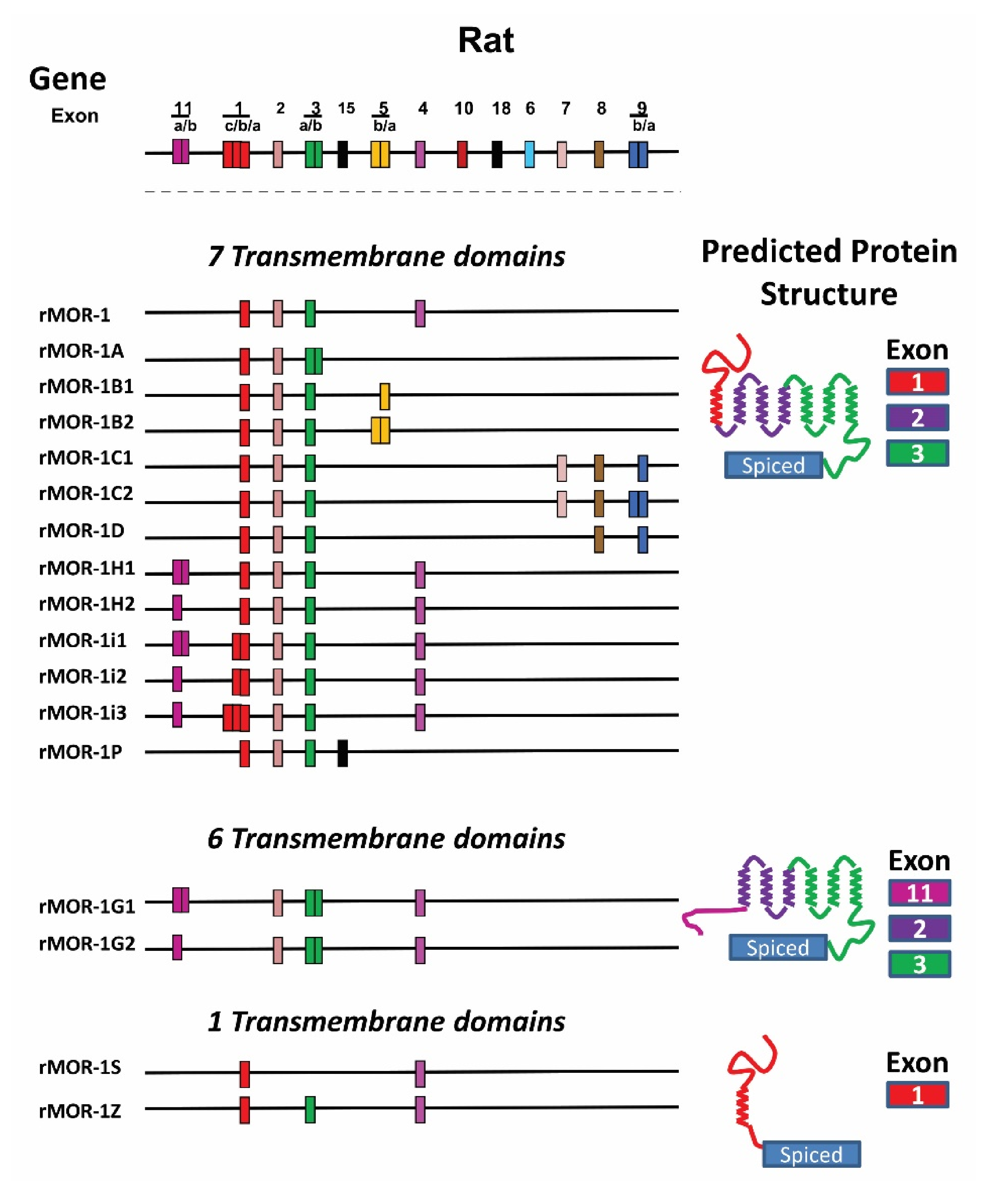
4.3. OPRM1 Gene Structure

The chromosonal location of the mouse and human OPRM1 gene was initially mapped to chromosomes 10 (10 cM from the centromere) and 6 (6q24-25), respectively, using traditional chromosomal mapping approaches [121,122,123,124], soon after MOR-1 cDNA was identified. Genomic sequencing initiated by the Human Genome Project confirmed the early OPRM1 mapping results, and revealed the precise chromosomal locations of the OPRM1 gene not only in mice and humans, but also in over 30 species. All the species contain a single copy of OPRM1 gene. Genomic sequencing also showed that genes adjacent to OPRM1 are conserved [11,120]. For example, the regulator of G protein signaling 17 (RGS17) gene is located at the upstream of the OPRM1 gene, while the interaction protein for cytohesin exchanges factors 1 (IPCEF1) gene and subunit 5 of the splicing factor 3b (SF3b5) gene are found downstream of the OPRM1 gene. Interestingly, a 70 bp sequence of exon 7 or exon O in the OPRM1 gene overlaps with the IPCEF1 gene in the reversed orientation, raising questions about their interaction and regulation at transcription and posttranscription levels [11].
The OPRM1 gene was initially identified as a structure with four exons and three introns using the original MOR-1 cDNA probe [125]. Mapping of additional exons at the 5′ and 3′ of the OPRM1 genes identified in alternatively spliced transcripts further extends the OPRM1 genes. Currently, the mouse, rat and human OPRM1 genes have approximate 270, 264, and 210 kb, respectively [11,12].
The OPRM1 gene has two independent promoters, exon 1 (E1) and exon 11 (E11) promoters, which are conserved from rodents to humans. E1 promoter was identified as a dual-promoter model that contains a proximal promoter and distal promoter [126,127,128]. E1 promoter lacks a TATA box but has several cis-acting elements such as GC-rich elements with multiple transcription start points, suggesting a housekeeping gene mode. E11 promoter, however, has a TATA box using a single transcription start point, favoring an eukaryote class II promoter mode. Each promoter controls expression of a unique set of splice variants. Differential expressions of the two promoters in brain regions such as hippocampus and substantia nigra in a transgenic mouse model suggest region-specific promoter activity [129].

4.4. Alternatively Spliced OPRM1 Variants

The OPRM1 splice variants can be categorized into three major types based on receptor structure: (1) full-length 7TM C-terminal variants; (2) truncated 6TM Variants; and (3) single TM variants [11,12] (Figure 1, Figure 2 and Figure 3).

4.4.1. Full-Length 7TM C-Terminal Splice Variants

Full-length C-terminal variants are generated through OPRM1 3′ splicing, which mainly refers to splicing from exon 3 to its downstream exons. All of the full-length 7TM C-terminal variants share the same exons 1/2/3 that encode the majority of the receptor, including the N-terminus, 7TM domains, three intracellular and three extracellular loops, and part of intracellular C-terminus, except for a distinct C-terminal tail encoded by an alternative exon(s) produced from the 3′ splicing.
The first two full-length 7TM C-terminal variants, hMOR-1A [130] and rMOR-1B [131], were identified in human and rat, respectively. Subsequent 3′RACE using anchor primers from exon 3 isolated many alternative 3′ exons and further PCR cloning generated full-length C-terminal variant sequences. To date, 22 full-length 7TM C-terminal variants in mice, 13 in rats, and 12 in humans have been isolated (Figure 1, Figure 2 and Figure 3) [132,133,134,135,136,137,138,139,140]. The predicted amino sequences from each of the 7TM C-terminal variants are unique and the lengths of the C-terminal tail sequences vary from the shortest with one amino acid in hMOR-1B4 [136] and rMOR-1D [137] to the longest with 88 amino acids in mMOR-1U [138] (Table 2). Moreover, several potential phosphorylation sites, such as protein kinase C, GRKs, cAMP- and cGMP-dependent protein kinase, and Casein kinase II, were identified in these C-terminal tails. The functional importance of these 7TM C-terminal variants in opioid pharmacology has been demonstrated using in vitro cell models showing differences in mu agonist-induced G protein coupling, phosphorylation, internalization and post-endocytic sorting and by differential region- and cell-specific expression, as well as suggested by findings using C-terminal truncation mouse models (see below).
Several C-terminal tail sequences are highly conserved from rodents to humans. For example, exon 3b in MOR-1A, an intron retention variant, encodes identical four amino acids (VCAF) in mouse MOR-1A (mMOR-1A) [142] and human MOR-1A (hMOR-1A) [130]. Exon 5a encodes the same five amino acids (KIDLF) in mMOR-1B1 and rMOR-1B1, which matches exactly to the first five amino acids encoded by human exon 5a in hMOR-1B1. However, hMOR-1B1 has an additional nine amino acids beyond the conserved five amino acids. Human exon O in hMOR-1O is a homolog of mouse exons 7a7b in mMOR-1O. Both exons predict an intracellular C-terminal tail with 30 amino acids share 63% of identity [137].

4.4.2. Truncated 6TM Variants

Searching alternative 5′ exons of the mouse Oprm1 gene using a modified 5′RACE approach led to identification of four new exons including exons 11, 12, 13, and 14 [143]. Both exons 11 and 12 were mapped to ~30 kb and 28 kb upstream of exon 1, respectively, while exons 13 and 14 were located between exons 1 and 2. Alternative splicing from exon 11 to its downstream exons generates nine exon 11-associated variants, five of which, mMOR-1G, mMOR-1K, mMOR-1L, mMOR-1M, and mMOR-1N, are truncated 6TM variants, in addition to three full-length 7TM variants, mMOR-1H, mMOR-1i, and mMOR-1J, and one single TM variant mMOR-1T (Figure 2). Splicing from exon 11 to exon 2 by skipping exon 1 generates three 6TM variants mMOR-1G, mMOR-1M, and mMOR-1N, which lack the first TM encoded by exon 1 [143]. Since the mouse exon 11 encodes 27 amino acids that do not predict a transmembrane domain, translation from exon 11 AUG in frame with exon 2 yields a receptor with 6TM (TM2–TM7) from exons 2 and 3. Splicing from exon 11 to exons 13 and 14 also by skipping exon 1 generates two 6TM variants, mMOR-1K and mMOR-1L. Although translation from exon 11 AUG predicts a short peptide beyond the 27 amino acids from exon 11, both mMOR-1K and mMOR-1L can produce a 6TM receptor by using an alternative AUG in exon 2.
Identification of the mouse exon 11-associated variants quickly led to isolation of the mouse homologs of exon 11-associated variants in rats and humans (Figure 1 and Figure 3) [144,145]. Based on the genomic databases, rat and human exon 11 are found at ~21 and ~28 kb upstream of their exon 1, respectively, a genomic location similar to that of the mouse exon 11 (~30 kb). PCR cloning using primers anchoring the respective exon 11 homologs and downstream exons identified four exon 11-associated variants in humans and seven in rats [144,145]. Like mMOR-1G, splicing from exon 11 to exon 2 generates two 6TM variants, rMOR-1G1, and rMOR-1G2 or hMOR-1G1 and hMOR-1G2, in both rats and humans. However, both rat and human exon 11 have an alternative 5′ splice site, which splits exon 11 as exons 11a and 11b. Splicing from exon 11a to exon 2 produces rMOR-1G2 or hMOR-1G2. The rat and human exon 11a predict 7 and 16 amino acids, respectively, which are in frame with exon 2. Thus, both rMOR-1G2 and hMOR-1G2 encode a 6TM receptor translated from the exon 11a AUG without the first TM. On the other hand, splicing from exon 11b to exon 2 yields rMOR-1G1 and hMOR-1G1. Similar to mMOR-1K and mMOR-1L, both rMOR-1G1 and hMOR-1G1 predict a 6TM receptor when the AUG in exon 2 is used, and produce a short peptide sequence if exon 11a AUG is used. Splicing from exon 11 to exon 1 produces an additional five variants in rats and two variants in humans, all of which encode a 7TM receptor when translated from exon 1 AUG. The functional relevance of the 6TM variants has been implicated in mediating the actions of a new type of opioid analgesics that are potent against a spectrum of pain models without many side-effects associated with traditional opiates [12] (see below).
An additional human 6TM variant hMOR-1K was isolated based on the finding of a mouse exon 13 homolog in the human OPRM1 gene [146]. Unlike mMOR-1K that is associated with exon 11, hMOR-1K did not have exon 11 and its expression appeared to be controlled by its own promoter located at upstream of the human exon 13. Another human 6TM variant was identified as mu3 receptor with an exon composition of 2/3/new exon [147]. Both hMOR-1K and mu3 variants predict a 6TM receptor when translated using the exon 2 AUG, a scenario similar to rMOR-1G1, hMOR-1G1, mMOR-1K, and mMOR-1L (Figure 1).

4.4.3. Single TM Variants

All of the single TM variants contain exon 1 that encodes the entire N-terminus and the first TM. Alternative splicing from exon 1 to downstream exons by exon skipping or insertion causes a reading-frame shift to terminate translation early, predicting a truncated receptor with a single TM. Currently, there are five single TM variants in mice, two in rats, and four in humans (Figure 1, Figure 2 and Figure 3). The first two single TM variants, hMOR-1S and hMOR-1Z, were isolated from the human OPRM1 gene [11,85,148]. Splicing from exon 1 to exon 4 by skipping exons 2 and 3 produces hMOR-1S, while hMOR-1Z has exons 1/3/4 by skipping exon 2. Both MOR-1S and MOR-1Z were subsequently identified in mice and rats with the same splicing mechanisms [85]. An additional two human single TM variants were isolated as SV1 and SV2 in which an exon A or B was inserted between exons 1 and 2, respectively [149]. Early translation termination in exon A or B results in a truncated single TM receptor.
The mouse Oprm1 gene has another three single TM variants, mMOR-1Q, mMOR-1R, and mMOR-1T [85]. Both mMOR-1Q and mMOR-1R have exon 2 skipping, but contain a different exon composition downstream of exon 3. In mMOR-1T, there is an exon 16 insertion between exons 1 and 2, predicting a single TM variant. Although the single TM variants cannot bind any opioids, their roles were shown in modulating the expression and function of the full-length 7-TM mu opioid receptor (MOR-1) (see below) [85].

5. Expression and Function of the OPRM1 Variants

5.1. Expression of the OPRM1 Variant mRNAs and Proteins

5.1.1. Region-Specific and Strain-Specific Expression of the OPRM1 Variant mRNAs

Northern blot analysis was initially employed to determine mRNA expression of OPRM1 variants in the brain using individual exon probes or combined exon probes. Multiple bands with different sizes and intensities were observed [6,123,132,134,135,136,137,143,150], suggesting OPRM1 alternative splicing. However, it is difficult to examine mRNA expression of individual splice variants using exon probes in Northern blot analyses because some exons are shared among several splice variants. Semi-quantitative RT-PCR assays were designed to investigate the expressions of the OPRM1 variant mRNAs in a number of dissected brain regions, including the cortex, thalamus, striatum, hypothalamus, hippocampus, brain stem, cerebellum, and spinal cord [132,133,143,144]. For example, high level of mMOR-1C mRNA but low levels of mMOR1D and mMOR-1E mRNAs were expressed in the mouse thalamus [132]. In human brain, hMOR-1G2 was highly expressed in the prefrontal cortex, piriform cortex, nucleus accumbens, and pons, but not in the temporal cortex and spinal cord [144].
Using real-time quantitative PCR (qPCR), a more accurate method measuring mRNA levels, the expression profiles of thirty Oprm1 splice variant mRNAs in ten different brain regions of four inbred mouse strains, C56BL/6J, 129P3/J, SJL/J, and SWR/J [151], and CD-1 mice [152] were examined. The results showed marked differences of the variant mRNA expression among the brain regions or among the mouse strains, suggesting region-specific or strain-specific alternative splicing of the OPRM1 gene. For example, 7TM C-terminal variant mMOR-1A was expressed highly in the hypothalamus and thalamus but lowly in the striatum and prefrontal cortex in SJL/J mice. In the same PAG region, mMOR-1G, a 6TM variant, was expressed much higher in C57Bl/6J mouse strain than in the other three mouse strains.
Among the three types of Oprm1 variants, the overall expressions of all 7TM variants were dominant throughout the brain regions and mouse strains, while the overall expressions of all 6TM and 1TM variants were lower than those of all 7TM variants (Table 3) [153]. However, all 6TM and 1TM variants were expressed at relatively high levels in certain regions or mouse strains. For example, in the PAG region of C57BL/6J mice, the expression level of all 6TM variants was 20.4% of all 7TM variants, which was the highest among the brain regions and among the mouse strains. The expression level of all 1TM variants in the prefrontal cortex of SJL/J mice was higher than those in the other three mouse strains. These results further demonstrated region-specific and strain-specific alternative splicing of the Oprm1 gene.
Altered expression of the OPRM1 splice variants were seen in multiple brain regions in a morphine tolerance mouse model [152], the human brain of HIV-infected individuals [153,154], and the medial prefrontal cortex of human heroin abusers and heroin self-administering rats [155]. Sex difference in expression of the Oprm1 variant mRNAs among the mouse brain regions was also observed [156].

5.1.2. Region-Specific and Cell-Specific Expression of the OPRM1 Variant Proteins

The distribution of OPRM1 variant proteins was investigated using several antisera generated against individual exon coding sequences [157,158,159,160]. These studies revealed dramatic differences in the regional distribution of the exon-containing OPRM1 variant proteins. For example, the distribution of exons 7/8-like immunoreactivity (LI) detected with an antisera against a peptide from mouse exons 7/8 clearly differed from that of exon 4-LI using an antisera against a peptide from exon 4 in several regions, such as the medial eminence, thalamic nuclei, and nucleus ambiguous [158]. The two antisera also labeled different cell populations, as demonstrated in the laminae layer I–II of the spinal cord [158]. An antisera raised against an exon 8 peptide labeled uniquely in the dentate gurus, the mossy fibers of the hippocampal formation and the nucleus of the solitary tract [157], while an antisera raised against exon 3/5a hybridized predominantly in the olfactory bulb [161]. The localization of variant proteins was also examined at the ultrastructural level [162]. For example, in the superficial laminae of the spinal cord, the exon 7/8-LI was predominantly distributed in presynaptic cells and co-localized with calcitonin gene-related peptide (CGPR), whose antibody labels unmyelinated primary afferents, in contrast to the exon 4-LI, which displayed an equal distribution between presynaptic and postsynaptic membranes and did not co-localize with CGPR [162].
It should be noted that several variant proteins encoded by the same exon(s) commonly share identical epitope(s), thus it is challenging to distinguish each individual variant at the protein level using exon-specific antisera by immunohistochemistry. For example, antisera against a peptide from mouse exon 4 would detect at least five exon 4-containing variants in mice including mMOR-1, mMOR-1H, mMOR-1I, mMOR-1J, and mMOR-1G. Similarly, exon 8-LI would correspond to mMOR-1D and mMOR-1N, whereas exon 7/8-LI would represent two variants, mMOR-1C and mMOR-1M. These studies also revealed the discrepancies between mRNA and protein levels for some of the variants. For example, in the mouse brain, the protein level of mMOR-1 detected by the exon 4-specific antisera was found to be comparable to that of mMOR-1C and mMOR-1M as determined by the exon 7/8-specific antisera [158], while the expression level of mMOR-1 mRNA was much higher than that of both mMOR-1C and mMOR-1M mRNA [132,143]. Although different antisera may differ in sensitivity, these studies reiterated the poor correlation between mRNA and protein levels in the brain and highlighted the importance of examining the expression of variants at both the mRNA and proteins expression levels.

5.2. The Function of Full-Length 7TM C-Terminal Variants

5.2.1. Opioid Receptor Binding Affinity

To determine the binding profiles of full-length 7TM C-terminal variants, opioid receptor binding assays using [3H] DAMGO, a full mu agonist, were performed with membranes from CHO cell lines that stably express each individual full-length C-terminal variant [132,133,134,135,136,137,142]. The results from saturation studies indicated that all the 7TM C-terminal variants had high affinity toward [3H] DAMGO, which was anticipated since they all share the same binding pocket. Competition studies showed mu selectivity with a number of mu opioids such as morphine, M6G, and naloxone for all the 7TM C-terminal variants. However, several endogenous opioid peptides revealed different binding affinities among the 7TM C-terminal variants. For example, both β-endorphin and dynorphin A were over four times more potent in mMOR-1D than in mMOR-1 [132]. These results raise intriguing questions of how different C-terminal tails impact on the binding pocket, specifically toward the endogenous opioid peptides.

5.2.2. Mu Agonist-Induced G Protein Coupling, Internalization, Phosphorylation, and Post-Endocytic Sorting

The intracellular C-termini of GPCRs play an important role in the signaling transduction system, especially the G-protein activation. Mu agonist-induced G protein activation in the OPRM1 7TM C-terminal variants were extensively studied using membranes from CHO cells stably expressing the individual variant in [35S] GTPγS binding assay, a commonly used method to assess the overall agonist-induced receptor-G-protein coupling. The results revealed marked differences in mu agonist-induced G-protein activation in both potency, measured by the EC50 values, and efficacy, determined by the maximal stimulation, among the 7TM C-terminal variants in mice, rats, and humans [135,136,137,163]. Several mu agonists displayed varied efficacies among various 7TM C-terminal variants. For example, morphine and M6G were more efficacious in hMOR-1A than hMOR-1B1, hMOR-1B3, and hMOR-1B5, while β-endorphin was a full agonist for hMOR-1B5 and a partial agonist for hMOR-1A [136]. There was lack of correlation between the binding affinity (Ki) and potency (EC50). β-endorphin bound to both rMOR-1 and rMOR-1C1 with similar affinity. However, β-endorphin was over 20 times more potent in rMOR-1 than in rMOR-1C1 in [35S] GTPγS binding. Also, there was little correlation between potency (EC50) and efficacy. Together, these results demonstrated that different intracellular C-terminal tails in the 7TM full-length variants have significant influence on mu agonist-induced G protein activation.
Mu agonist-induced MOR-1 phosphorylation involves receptor desensitization and internalization, contributing to the development of mu opioid tolerance and physical dependence [164,165,166,167]. Several mu agonist-induced phosphorylation sites in the intracellular C-terminus encoded by exon 3 in MOR-1, such as 354TSST357 and 375STANT379 motifs, were identified [168,169,170]. Involvement of these phosphorylation sites in mu opioid actions such as analgesia, tolerance, respiratory depression, and constipation was demonstrated using several phosphorylation-deficient mouse models [171]. However, these phosphorylation sites are shared by all the 7TM C-terminal variants. The potential phosphorylation sites for several kinases such as GRKs, protein kinase C, casein kinase, and tyrosine kinase predicted from alternatively spliced C-terminal tails raise questions regarding their responses to mu agonists (Table 2). Koch et al. showed that morphine-induced receptor phosphorylation in mMOR-1D and mMOR-1E was robust, sharply contrasting with the nonresponsiveness in mMOR-1 and mMOR-1C [172], suggesting additional morphine-induced phosphorylation sites in the C-terminal tails encoded by the respective C-terminal tails or modulation of overall receptor phosphorylation by the C-terminal tails. The differential phosphorylation profiles among these variants were correlated with morphine-induced internalization and resensitization [172]. Additionally, threonine 394 in the exon 4-encoded C-terminal tail of rMOR-1 was phosphorylated in response to DAMGO [173]. Interestingly, a mutant mouse with the single point mutation T394A showed loss of etorphine-induced MOR-1 internalization in the spinal dorsal horn neurons and diminished tolerance by morphine and etorphine, as well as increased vulnerability to heroin self-administration [174]. Furthermore, morphine induced robust internalization of exon 7-associated mMOR-1C, but not exon 4-associated mMOR-1, in the mouse lateral septum [175].
After being internalized by mu agonists, mMOR-1 is either migrated to the lysosome for degradation or recycled back to the plasma membrane. Exon 4-encoded C-terminal sequences contain a motif termed the MOR1-derived recycling sequence (MRS) that allows efficient post-endocytic sorting of mMOR-1 to the plasma membrane [176]. Absence of the exon 4-encoded MRS in mMOR-1B, mMOR-1D, and mMOR-1E led to rapid lysosomal degradation due to inefficient receptor recycling following DAMGO-induced internalization, indicating the importance of the C-terminal sequences in determining the post-endocytic sorting direction of GPCRs [177].

5.2.3. Mu Agonist-Induced Biased Signaling at 7TM C-Terminal Variants

Biased signaling of GPCRs is defined based on observations that different agonists can produce divergent signaling transduction pathways through a single receptor. Biased signaling through G protein and β-arrestin 2 has been extensively studied in the original MOR-1 [168,178,179,180,181,182]. Identification of multiple OPRM1 7TM C-terminal variants opened questions about their roles in mu agonist-induced biased signaling. The effect of different 7TM full-length C-terminal variants on mu agonist-induced signaling bias between G protein coupling and β-arrestin 2 recruitment was studied using [35S] GTPγS binding and PathHunter β-arrestin 2 recruitment assay in the same CHO cells stably expressing individual 7TM C-terminal variants [183]. The results showed marked differences in mu agonist-induced G protein activation and β-arrestin 2 recruitment among the 7TM variants. For example, exon 7-associated 7TM variants, particularly mMOR-1O, displayed greater β-arrestin 2 bias for most mu agonists than exon 4-associated 7TM variant mMOR-1, suggesting that exon 7-associated variants favor mu agonist-induced β-arrestin 2 binding. This hypothesis was supported by the identification of a consensus phosphorylation code, PxPxxE/D or PxxPxxE/D, in exon 7-encoded sequences for high affinity arrestin binding proposed by homology modeling with the crystal structures of several GPCRs (Table 2) [141]. When this predicted phosphorylation code was mutated in mMOR-1O, DAMGO-induced β-arrestin 2 recruitment was greatly attenuated (our unpublished data) [183]. These results also suggested a mechanism of the in vivo function of exon 7-associated variants in several morphine actions observed in an exon 7 C-terminal truncation mouse model (mE7M-B6), which showed similar phenotypes in a β-arrestin 2 knockout (KO) mouse (see below) [83]. The finding that a single mu agonist can produce differential signaling through multiple 7TM C-terminal variants from in vitro cell line studies offers new insights into our understanding of biased signaling in vivo where all the 7TM C-terminal variants are co-expressed.

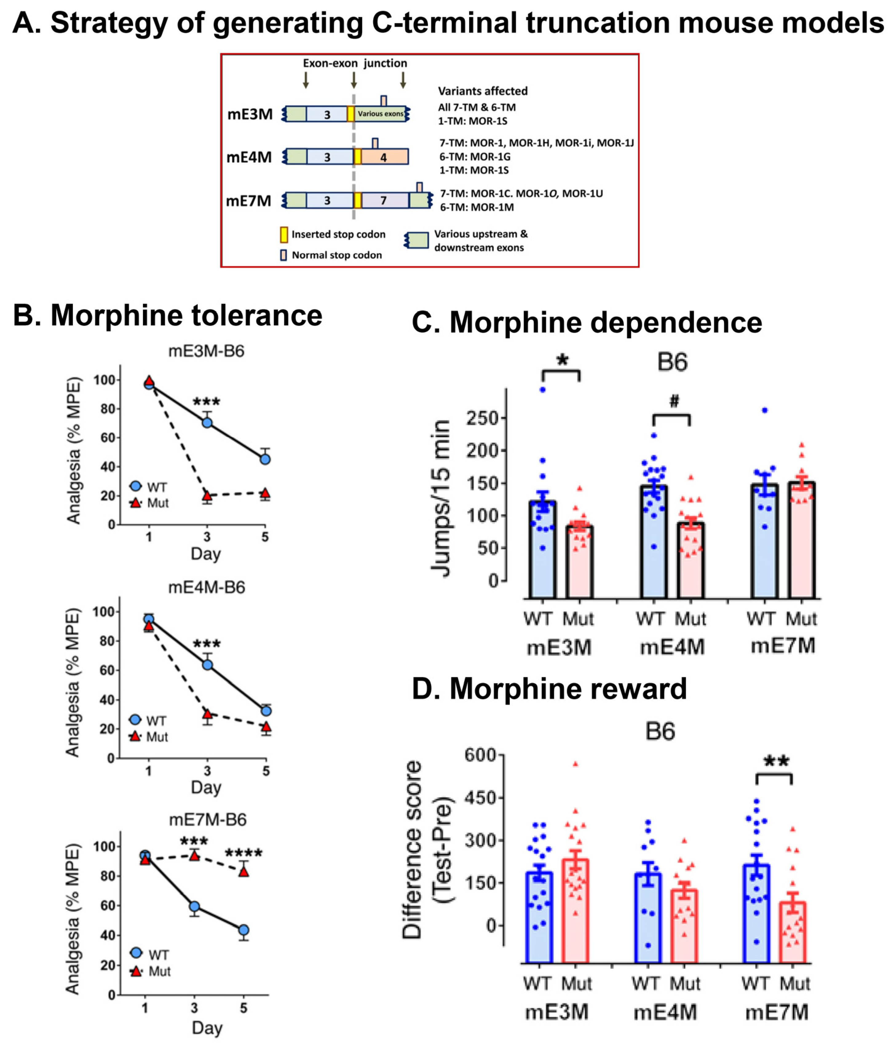
5.2.4. In Vivo Function of 7TM C-Terminal Variants

Morphine-Induced Itch (Pruritus)

Opioid-induced itch (pruritus) is one of the most prevalent acute side effects of the spinal or epidural use of opioids in patients [184,185,186], which has limited the clinical use of opioids. However, the molecular mechanisms underlying opioid-induced itch remained largely unknown. Liu et al. reported that an Oprm1 7TM C-terminal splice variant mMOR-1D was the major player in spinal morphine-induced scratching in mice [187]. Downregulating the spinal mMOR-1D with siRNAs reduced morphine-induced scratching without affecting morphine analgesia. They further demonstrated that morphine-induced scratching was mediated through heterodimerization of mMOR-1D and gastrin-releasing peptide receptor (GRPR) in the spinal cord. Disrupting mMOR-1D/GRPR heterodimerization attenuated morphine-induced scratching, but not morphine-induced analgesia [187]. The same group further found that similar to mMOR-1D, one human 7TM C-terminal variant hMOR-1Y can physically associate with GRPR to function as an itch receptor [188]. These studies further established the pharmacological relevance of OPRM1 7TM C-terminal variants and also provided a potential therapeutic strategy to specifically alleviate itch without attenuation of opioid analgesia.

Morphine Tolerance, Dependence, and Reward

To explore the in vivo functions of alternatively spliced C-terminal tails, three C-terminal truncation mouse models were established in two inbred mouse strains, C57BL/6J (B6) and 129/SvEv (129), using a stop codon targeting strategy that truncates a designated exon at the translational level with limited effects on overall Oprm1 transcription and alternative splicing (Figure 4) [83]. In the first model (mE3M), the C-terminal tails of all the variants are truncated at the end of E3 by a stop codon introduced at the end of E3. In the second (mE4M) and third models (mE7M), the E4- or E7-encoded C-terminal tails are truncated by inserting a stop codon at the beginning of E4 or E7, respectively [83]. Studies using these mice show divergent roles of the C-termini in various morphine-induced behaviors. For example, the E7-encoded C-terminal truncation in mE7M-B6 mice decreases morphine tolerance and reward without altering physical dependence, whereas the E4-encoded C-terminal truncation in mE4M-B6 mice accelerates morphine tolerance and reduces morphine dependence without affecting morphine reward (Figure 4) [83]. Moreover, in mE7M-B6 mutant mice, morphine-induced receptor desensitization in the hypothalamus and brain stem was lost, suggesting an E7-mediated mechanism underlying morphine tolerance [83].
The similarities found in several morphine-related behaviors and receptor desensitization between mE7M-B6 mutant and β-arrestin 2 KO mice suggest a physical and functional association of E7-encoded C-terminal tails with β-arrestin 2 [83,189,190,191,192]. This supposition was supported by our in vitro cell studies showing greater β-arrestin 2 bias for several mu opioids in the E7-associated variant mMOR-1O than in the E4-associated variant mMOR-1 [83,183]. However, several mu opioid–induced actions, including morphine analgesia, locomotion, and reward [193,194], and fentanyl and methadone tolerance (unpublished observation), observed in mE7M-B6 mice were unlike those detected in β-arrestin 2 KO mice, suggesting a β-arrestin 2-independent mechanism. Together, these results further highlight the functional relevance of the 7TM C-terminal variants, particularly exon 7-associated 7TM variants, in mu opioid pharmacology.

5.3. The Function of Truncated 6TM Variants

5.3.1. Involvement of 6TM Variants in Heroin and M6G Analgesia

Although the truncated 6TM variants did not bind any available radiolabeled opioids when expressed in cell lines, their functional relevance was mainly revealed using gene targeting mouse models (Table 4). The pharmacological function of 6TM variants was first shown to involve the analgesic actions of heroin and M6G, but not morphine and methadone, by using two Oprm1 KO mouse models. In an exon 1 knockout (E1 KO) mouse, morphine and methadone analgesia was completely lost [111]. However, heroin and M6G analgesia still was active despite a slightly reduced potency. Since knocking out E1 abolished all 7TM C-terminal variants and did not disrupt 6TM variants, these results suggested that 6TM variants involve heroin and M6G analgesia, while 7TM variants were responsible for morphine and methadone analgesia. This suggestion was further supported by the opposite phenotypes seen in E11 KO mice in which heroin and M6G analgesia were greatly decreased, while morphine and methadone analgesia were normal [112]. Furthermore, the analgesic actions of all mu opioids including morphine, M6G, and heroin were lost in a exon 2 deficient mouse in which both 7TM and 6TM variants were disrupted [111], consistent with the results from E1 KO and E11 KO.

5.3.2. Essential Role of 6TM Variants in a Novel Type of Opioid Analgesic Drugs

The truncated 6TM variants were further discovered to mediate the actions of a novel opioid analgesic drug, 3′-iodobenzoylnaltrexamide (IBNtxA), which was derived from naltrexone [195,196]. Although the N-cyclopropyl group on the morphinan template leads to an antagonist pharmacophore, an example being naltrexone, incorporating an aryl amido group at the 6-position of the morphinan template leads to agonism at 6TM sites in the case of IBNtxA [195]. Similarly, such an incorporation in MP1104 confers its characterization as a kappa agonist at KOR-1 site [197], while MP1207 and MP1208 with the incorporation become mixed mu/kappa agonists [182]. Therefore, the 6-position arylamido group on the morphinan template puts the receptor in an agonist conformation. IBNtxA analgesia was lost in E11 KO mice, suggesting the role of exon 11-associated 6TM variants in IBNtxA analgesia. On the other hand, IBNtxA analgesia was fully active in a triple KO mouse in which only Oprm1 6TM variants remained, while all other opioid receptors including MOR 7TM variants, kappa opioid receptor (KOR-1), and delta opioid receptor (DOR-1) were knocked out, indicating that IBNtxA analgesia was independent of 7TM MOR variants, DOR-1, and KOR-1 [195]. Furthermore, intrathecal administration of lentivirus expressing individual 6TM variants, including mMOR-1G, mMOR-1M, mMOR-1N, mMOR-1K, and mMOR-1L, restored IBNtxA analgesia in a complete Oprm1 E1/E11 KO mouse in which all 7TM, 6TM, and single TM variants were disrupted [84,198]. Together, these results demonstrate that 6TM variants are essential for IBNtxA analgesia. Additionally, the opioid receptor binding studies with [125I]BNtxA revealed a unique binding site in the triple KO mouse that was lost in both E11-KO and E2 KO mice [195,196,199,200]. Intriguingly, kappa-like drugs, such as ketocyclazocine and kappa3-like drug NalBzoH, competed for this site with high affinity, while many traditional mu, delta, and kappa opioids had very poor affinity for the site.
IBNtxA represents a novel class of opioid analgesics that are potent against multiple types of pain including thermal, inflammatory, and neuropathic pain, but do not produce many sides effects of traditional opioids, such as respiratory depression, physical dependence, and reward [195,201]. Therefore, the 6TM variants represent promising therapeutic targets for development of the novel analgesics with more favorable side-effect profiles.

5.3.3. Function of 6TM Variants in Delta/Kappa Opioids and Non-Opioids

Using E11 KO and E1/E11 KO mice in combination with lentivirus rescue and antisense studies, Marrone et al. revealed the involvement of truncated 6TM variants in the analgesic actions of delta and kappa opioids, as well as α2-adrenergic drug [202]. The analgesia of DPDPE, a delta agonist, U50,488H, a kappa agonist and clonidine, a α2-adrenergic agonist, was lost in the E11 KO mice, consistent with the results from the antisense studies using an antisense oligo targeting E11 [202]. Re-expression of mMOR-1G, a 6TM variant, through lentivirus in E11 KO mice rescued the analgesia of DPDPE, U50,488H and clonidine [202], further confirming that the analgesia of delta, kappa and α2-adrenergic agonists is dependent on the 6TM variants. However, 6TM variants did not involve delta agonist-induced seizure, kappa agonist-induced aversion, and α2 agonist-induced hypolocomotion since these activities were still active in E11 KO mice [202]. Additionally, DPDPE analgesia was also dependent on Oprm1 7TM variants based on the observation that lentivirus-expressing mMOR-1G can only restore DPDPE analgeia in E11 KO mice, but not in the E1/E11 KO mice that lacked both 7TM and 6TM variants [202]. The 6TM variants demonstrated the ability to heterodimerize with β2-adrenergic receptors, providing a molecular mechanism of the analgesic synergy between the 6TM variants and β2-adrenergic drugs [203]. These studies expended the known functions of the 6TM variants beyond mu opioid receptors.

5.3.4. A Chaperon-Like Function of 6TM Variants in Enhancing Expression of the 7TM MOR-1 at Protein Level

The function of the 6TM variants was also demonstrated in enhancing expression of the 7TM MOR-1 at the protein level through a chaperon-like function [204], a similar scenario seen in the single TM variants [85] (see below). When co-expressed with mMOR-1 in a Tet-Off inducible CHO cell line, mMOR-1G dose-dependently promoted the expression of the protein but not the mRNA of mMOR-1, as determined by opioid receptor binding and [35S]GTPγS binding. Using co-immunopreciptation and subcellular fractionation analysis, the study showed that the increased mMOR-1 receptor protein was mainly found in the plasma membrane, which was mediated through heterodimerization of mMOR-1G with mMOR-1 in the endoplasmic reticulum, suggesting a chaperone-like function [204].

5.3.5. Role of 6TM Variants in Morphine Hyperalgesia

Several studies indicated that the 6TM variants were involved in morphine hyperalgesia. In the E11-KO mouse without any 6TM variants, morphine analgesia was fully retained, but morphine-induced hyperalgesia was diminished [112,205]. However, the triple KO mouse, which retained only the truncated 6TM variants and disrupted KOR1, DOR-1, and all the E1-associated 7TM MOR variants, lost morphine analgesia but retained morphine hyperalgesia [206]. Downregulating the 6TM variant mMOR-1K with intrathecal administration of a siRNA in CXB7/ByJ mice diminished morphine-induced hyperalgesia [207], suggesting involvement of mMOR-1K in morphine hyperalgesia. Together, these findings suggest that morphine analgesia is independent of truncated 6TM variants, whereas morphine hyperalgesia is dependent on truncated 6TM variants. In addition, truncation of 6TM variants in E11-KO mice led to reduction of morphine-induced hyperlocomotion and tolerance with no effect on morphine reward and respiratory depression [205].

5.4. The Function of Single TM Variants

It is intriguing that all the single TM variants contain the first TM encoded by exon 1, which could complement the 6TM variants that lack the exon 1. Although our unpublished data showed that the single TM variants can physically associate with the 6TM variants when co-transfected into CHO cells, the functional relevance of this physical association remains unknown. Similar to the 6TM variants, the functions of the single TM variants were indicated to increase expression of 7TM MOR-1 at the protein level through a chaperone-like mechanism [85]. Using a Tet-Off CHO system, Xu et al. showed that the single TM variants can dimerize with the 7TM MOR-1 in the endoplasmic reticulum to facilitate the proper folding of MOR-1 and reduce MOR-1 degradation through endoplamic reticulum-associated degradation (ERAD), leading to increased expression of MOR-1 protein. In vivo antisense studies further suggested the role of the single TM variants in morphine analgesia through modulation of 7TM MOR-1 expression [85].
Interestingly, the first TM domain of MOR-1 was also implicated in heterodimerization with DOR-1 to disrupt MOR-1/DOR-1 dimer using an engineered protein, MORTM1-TAT (transactivator of transcription), resulting in enhanced morphine analgesia and reduced morphine tolerance [208]. This study raises questions on whether endogenously expressed single TM variants containing the first TM have a similar function.

6. Molecular Mechanisms Underlying OPRM1 Alternative Splicing

Identification of multiple OPRM1 splice variants raises questions regarding the molecular mechanisms underlying OPRM1 alternative splicing. For example, what are cis-acting elements and trans-acting factors that control the OPRM1 alternative splicing? Xu et al. initially identified an intronic single nucleotide polymorphism (SNP)(rs9479757) located near the 3′ of OPRM1 exon 2 that associated with heroin addiction severity in a cohort of Chinese male heroin addicts [209]. Further characterizing this SNP using electrophoretic mobility shift assay (EMSA), minigene constructs, and RNA affinity purification coupled with proteomics led to the identification of a SNP-containing cis-acting element as an intronic splicing enhancer (ISE) that can interact with a splicing factor hnRNPH to regulate the exon 2 exclusion (skipping) or inclusion, and modulate expression of the full-length 7TM MOR-1 and 1TM variants hMOR-1S and hMOR-1Z [209]. The G allele of rs9479757 enhanced the binding of hnRNPH to this ISE, facilitating exon 2 inclusion to increase the expression of the 7TM hMOR-1 and reduce the expression of the 1TM hMOR-1S and hMOR-1Z, while the A allele had opposite effects. These results not only revealed the molecular mechanisms underlying OPRM1 exon 2 exclusion/inclusion, but also demonstrated the function of rs9479757 in regulating OPRM1 alternative splicing and contributing to heroin addiction severity. OPRM1 alternative splicing is complex. Further deciphering the molecular mechanisms of OPRM1 alternative splicing will advance our understanding of OPRM1 gene regulation and function.

7. Conclusions

The early concept of multiple mu opioid receptors suggested by the pharmacological studies over 40 years ago has now been revolutionized by identifying an array of OPRM1 alternatively spliced variants. These splice variants are generated through various splicing mechanisms such as alternative 3′ or 5′ splicing, intron retention, alternative promoters, or combinations of two or three mechanisms in a region-specific, cell-specific, or strain-specific fashion and conserved from rodents to humans. Based on the predicted transmembrane domains, these splice variants can be categorized mainly into three types: (1) full-length 7TM C-terminal variants, (2) truncated 6TM variants; and (3) truncated single TM variants. The functional relevance of these variants demonstrated by both in vitro cell lines and in vivo gene targeting animal models provides new insights into our understanding of complex mu opioid actions. More importantly, these OPRM1 splice variants are potential therapeutic targets for developing novel analgesic drugs with more favorable side-effect profiles. Many GPCRs have predicted splice variants like those of the OPRM1 gene. However, the functions of these variants remain largely unknown. Revealing the distinct functions of various OPRM1 splice variants opens questions of whether alternative splicing may extend the pharmacological repertoire of GPCRs in general.

Author Contributions

Conceptualization, S.L., W.-J.K., L.C., S.M., A.A., L.C., S.M., A.B. and Y.-X.P.; methodology, S.L., W.-J.K., A.B., J.X., P.H. and Y.-X.P.; validation, S.L., W.-J.K., J.X. and Y.-X.P.; formal analysis, S.L., W.-J.K., A.B., J.X., L.C., S.M. and Y.-X.P.; investigation, S.L., W.-J.K., J.X., L.C., S.M., P.H. and Y.-X.P.; resources, J.X., S.M., A.B. and Y.-X.P.; data curation, S.L., W.-J.K., A.B., J.X., L.C., S.M. and Y.-X.P.; writing-original draft preparation, S.L., W.-J.K. and Y.-X.P.; writing-review and editing, S.L., W.-J.K., A.A., L.C., S.M., P.H., A.B. and Y.-X.P.; visualization, S.L., W.-J.K. and Y.-X.P.; supervision, A.B. and Y.-X.P.; project administration, A.B. and Y.-X.P.; funding acquisition, S.M. and Y.-X.P. All authors have read and agreed to the published version of the manuscript.

Funding

This research was supported, in part, by grants from the National institute on Drug Abuse of the National Institutes of Health; DA042888, DA048379 and DA07242 to YXP; DA045884, DA046487, and DA048379 to SM; the Mayday Foundation and the Peter F. McManus Charitable Trust to YXP.

Institutional Review Board Statement

Not applicable.

Informed Consent Statement

Not applicable.

Data Availability Statement

Not applicable.

Conflicts of Interest

Y.-X.P. and S.M. are co-scientific founders of Sparian Biosciences. All other authors declare that there are no conflicts of interest.

References

  1. Martin, W.R. Opioid antagonists. Pharmacol. Rev. 1967, 19, 463–521. [Google Scholar] [PubMed]
  2. Pert, C.B.; Pasternak, G.W.; Snyder, S.H. Opiate agonists and antagonists discriminated by receptor binding in brain. Science 1973, 182, 1359–1361. [Google Scholar] [CrossRef] [PubMed]
  3. Terenius, L. Stereospecific interaction between narcotic analgesics and a synaptic plasma membrane fraction of rat cerebral cortex. Acta Pharmacol. Toxicol. 1973, 32, 317–320. [Google Scholar] [CrossRef] [PubMed]
  4. Simon, E.J.; Hiller, J.M.; Edelman, I. Stereospecific binding of the potent narcotic analgesice [3H]Etorphine to rat-brain homogenate. Proc. Natl. Acad. Sci. USA 1973, 70, 1947–1949. [Google Scholar] [CrossRef] [Green Version]
  5. Chen, Y.; Mestek, A.; Liu, J.; Hurley, J.A.; Yu, L. Molecular cloning and functional expression of a μ-opioid receptor from rat brain. Mol. Pharmacol. 1993, 44, 8–12. [Google Scholar] [CrossRef]
  6. Thompson, R.C.; Mansour, A.; Akil, H.; Watson, S.J. Cloning and pharmacological characterization of a rat μ opioid receptor. Neuron 1993, 11, 903–913. [Google Scholar] [CrossRef] [Green Version]
  7. Wang, J.B.; Imai, Y.; Eppler, C.M.; Gregor, P.; Spivak, C.E.; Uhl, G.R. μ opiate receptor: cDNA cloning and expression. Proc. Natl. Acad. Sci. USA 1993, 90, 10230–10234. [Google Scholar] [CrossRef] [Green Version]
  8. Pasternak, G.W.; Childers, S.R.; Snyder, S.H. Opiate analgesia: Evidence for mediation by a subpopulation of opiate receptors. Science 1980, 208, 514–516. [Google Scholar] [CrossRef]
  9. Wolozin, B.L.; Pasternak, G.W. Classification of multiple morphine and enkephalin binding sites in the central nervous system. Proc. Natl. Acad. Sci. USA 1981, 78, 6181–6185. [Google Scholar] [CrossRef] [Green Version]
  10. Pasternak, G.W. The pharmacology of mu analgesics: From patients to genes. Neuroscientist 2001, 7, 220–231. [Google Scholar] [CrossRef]
  11. Pan, Y.X. Diversity and complexity of the mu opioid receptor gene: Alternative pre-mRNA splicing and promoters. DNA Cell Biol. 2005, 24, 736–750. [Google Scholar] [CrossRef]
  12. Pasternak, G.W.; Pan, Y.X. Mu opioids and their receptors: Evolution of a concept. Pharmacol. Rev. 2013, 65, 1257–1317. [Google Scholar] [CrossRef] [Green Version]
  13. Chow, L.T.; Gelinas, R.E.; Broker, T.R.; Roberts, R.J. An amazing sequence arrangement at the 5′ ends of adenovirus 2 messenger RNA. Cell 1977, 12, 1–8. [Google Scholar] [CrossRef]
  14. Berget, S.M.; Moore, C.; Sharp, P.A. Spliced segments at the 5’ terminus of adenovirus 2 late mRNA. Proc. Natl. Acad. Sci. USA 1977, 74, 3171–3175. [Google Scholar] [CrossRef] [Green Version]
  15. Black, D.L. Mechanisms of alternative pre-messenger RNA splicing. Annu. Rev. Biochem. 2003, 72, 291–336. [Google Scholar] [CrossRef] [Green Version]
  16. Wahl, M.C.; Will, C.L.; Luhrmann, R. The spliceosome: Design principles of a dynamic RNP machine. Cell 2009, 136, 701–718. [Google Scholar] [CrossRef] [Green Version]
  17. Lee, Y.; Rio, D.C. Mechanisms and regulation of alternative Pre-mRNA splicing. Annu. Rev. Biochem. 2015, 84, 291–323. [Google Scholar] [CrossRef] [Green Version]
  18. Wang, Y.; Liu, J.; Huang, B.O.; Xu, Y.M.; Li, J.; Huang, L.F.; Lin, J.; Zhang, J.; Min, Q.H.; Yang, W.M.; et al. Mechanism of alternative splicing and its regulation. Biomed. Rep. 2015, 3, 152–158. [Google Scholar] [CrossRef] [Green Version]
  19. Black, D.L. Activation of c-src neuron-specific splicing by an unusual RNA element in vivo and in vitro. Cell 1992, 69, 795–807. [Google Scholar] [CrossRef]
  20. Chan, R.C.; Black, D.L. The polypyrimidine tract binding protein binds upstream of neural cell-specific c-src exon N1 to repress the splicing of the intron downstream. Mol. Cell. Biol. 1997, 17, 4667–4676. [Google Scholar] [CrossRef] [Green Version]
  21. Chan, R.C.; Black, D.L. Conserved intron elements repress splicing of a neuron-specific c-src exon in vitro. Mol. Cell. Biol. 1997, 17, 2970. [Google Scholar] [CrossRef] [Green Version]
  22. Xu, R.; Teng, J.; Cooper, T.A. The cardiac troponin T alternative exon contains a novel purine-rich positive splicing element. Mol. Cell. Biol. 1993, 13, 3660–3674. [Google Scholar] [CrossRef] [Green Version]
  23. Ryan, K.J.; Cooper, T.A. Muscle-specific splicing enhancers regulate inclusion of the cardiac troponin T alternative exon in embryonic skeletal muscle. Mol. Cell. Biol. 1996, 16, 4014–4023. [Google Scholar] [CrossRef] [Green Version]
  24. Cooper, T.A. Muscle-specific splicing of a heterologous exon mediated by a single muscle-specific splicing enhancer from the cardiac troponin T gene. Mol. Cell. Biol. 1998, 18, 4519–4525. [Google Scholar] [CrossRef] [Green Version]
  25. Carstens, R.P.; Wagner, E.J.; Garcia-Blanco, M.A. An intronic splicing silencer causes skipping of the IIIb exon of fibroblast growth factor receptor 2 through involvement of polypyrimidine tract binding protein. Mol. Cell. Biol. 2000, 20, 7388–7400. [Google Scholar] [CrossRef] [Green Version]
  26. Carstens, R.P.; McKeehan, W.L.; Garcia-Blanco, M.A. An intronic sequence element mediates both activation and repression of rat fibroblast growth factor receptor 2 pre-mRNA splicing. Mol. Cell. Biol. 1998, 18, 2205–2217. [Google Scholar] [CrossRef] [Green Version]
  27. Carstens, R.P.; Eaton, J.V.; Krigman, H.R.; Walther, P.J.; Garcia-Blanco, M.A. Alternative splicing of fibroblast growth factor receptor 2 (FGF-R2) in human prostate cancer. Oncogene 1997, 15, 3059–3065. [Google Scholar] [CrossRef] [Green Version]
  28. Fu, X.D. The superfamily of arginine/serine-rich splicing factors. RNA 1995, 1, 663–680. [Google Scholar]
  29. Graveley, B.R. Sorting out the complexity of SR protein functions. RNA 2000, 6, 1197–1211. [Google Scholar] [CrossRef] [Green Version]
  30. Hastings, M.L.; Krainer, A.R. Pre-mRNA splicing in the new millennium. Curr. Opin. Cell Biol. 2001, 13, 302–309. [Google Scholar] [CrossRef]
  31. Caceres, J.F.; Stamm, S.; Helfman, D.M.; Krainer, A.R. Regulation of alternative splicing in vivo by overexpression of antagonistic splicing factors. Science 1994, 265, 1706–1709. [Google Scholar] [CrossRef] [PubMed] [Green Version]
  32. Mayeda, A.; Krainer, A.R. Regulation of alternative pre-mRNA splicing by hnRNP A1 and splicing factor SF2. Cell 1992, 68, 365–375. [Google Scholar] [CrossRef]
  33. Yang, X.; Bani, M.R.; Lu, S.J.; Rowan, S.; Ben-David, Y.; Chabot, B. The A1 and A1B proteins of heterogeneous nuclear ribonucleoparticles modulate 5’ splice site selection in vivo. Proc. Natl. Acad. Sci. USA 1994, 91, 6924–6928. [Google Scholar] [CrossRef] [PubMed] [Green Version]
  34. Fiset, S.; Chabot, B. hnRNP A1 may interact simultaneously with telomeric DNA and the human telomerase RNA in vitro. Nucleic Acids Res. 2001, 29, 2268–2275. [Google Scholar] [CrossRef] [PubMed] [Green Version]
  35. Ladd, A.N.; Charlet, N.; Cooper, T.A. The CELF family of RNA binding proteins is implicated in cell-specific and developmentally regulated alternative splicing. Mol. Cell. Biol. 2001, 21, 1285–1296. [Google Scholar] [CrossRef] [Green Version]
  36. Ladd, A.N.; Nguyen, N.H.; Malhotra, K.; Cooper, T.A. CELF6, a member of the CELF family of RNA-binding proteins, regulates muscle-specific splicing enhancer-dependent alternative splicing. J. Biol. Chem. 2004, 279, 17756–17764. [Google Scholar] [CrossRef] [Green Version]
  37. Ladd, A.N.; Taffet, G.; Hartley, C.; Kearney, D.L.; Cooper, T.A. Cardiac tissue-specific repression of CELF activity disrupts alternative splicing and causes cardiomyopathy. Mol. Cell. Biol. 2005, 25, 6267–6278. [Google Scholar] [CrossRef] [Green Version]
  38. Jensen, K.B.; Dredge, B.K.; Stefani, G.; Zhong, R.; Buckanovich, R.J.; Okano, H.J.; Yang, Y.Y.; Darnell, R.B. Nova-1 regulates neuron-specific alternative splicing and is essential for neuronal viability. Neuron 2000, 25, 359–371. [Google Scholar] [CrossRef] [Green Version]
  39. Ule, J.; Stefani, G.; Mele, A.; Ruggiu, M.; Wang, X.; Taneri, B.; Gaasterland, T.; Blencowe, B.J.; Darnell, R.B. An RNA map predicting Nova-dependent splicing regulation. Nature 2006, 444, 580–586. [Google Scholar] [CrossRef]
  40. Ule, J.; Ule, A.; Spencer, J.; Williams, A.; Hu, J.S.; Cline, M.; Wang, H.; Clark, T.; Fraser, C.; Ruggiu, M.; et al. Nova regulates brain-specific splicing to shape the synapse. Nat. Genet. 2005, 37, 844–852. [Google Scholar] [CrossRef]
  41. Cooper, T.A.; Mattox, W. The regulation of splice-site selection, and its role in human disease. Am. J. Hum. Genet. 1997, 61, 259–266. [Google Scholar] [CrossRef] [Green Version]
  42. Xiong, H.Y.; Alipanahi, B.; Lee, L.J.; Bretschneider, H.; Merico, D.; Yuen, R.K.; Hua, Y.; Gueroussov, S.; Najafabadi, H.S.; Hughes, T.R.; et al. RNA splicing. The human splicing code reveals new insights into the genetic determinants of disease. Science 2015, 347, 1254806. [Google Scholar] [CrossRef] [Green Version]
  43. Lin, C.L.; Bristol, L.A.; Jin, L.; Dykes-Hoberg, M.; Crawford, T.; Clawson, L.; Rothstein, J.D. Aberrant RNA processing in a neurodegenerative disease: The cause for absent EAAT2, a glutamate transporter, in amyotrophic lateral sclerosis. Neuron 1998, 20, 589–602. [Google Scholar] [CrossRef] [Green Version]
  44. Meyer, T.; Fromm, A.; Munch, C.; Schwalenstocker, B.; Fray, A.E.; Ince, P.G.; Stamm, S.; Gron, G.; Ludolph, A.C.; Shaw, P.J. The RNA of the glutamate transporter EAAT2 is variably spliced in amyotrophic lateral sclerosis and normal individuals. J. Neurol. Sci. 1999, 170, 45–50. [Google Scholar] [CrossRef]
  45. Jaeckle, K.A.; Graus, F.; Houghton, A.; Cardon-Cardo, C.; Nielsen, S.L.; Posner, J.B. Autoimmune response of patients with paraneoplastic cerebellar degeneration to a Purkinje cell cytoplasmic protein antigen. Ann. Neurol. 1985, 18, 592–600. [Google Scholar] [CrossRef]
  46. Philips, A.V.; Timchenko, L.T.; Cooper, T.A. Disruption of splicing regulated by a CUG-binding protein in myotonic dystrophy. Science 1998, 280, 737–741. [Google Scholar] [CrossRef]
  47. Lorson, C.L.; Rindt, H.; Shababi, M. Spinal muscular atrophy: Mechanisms and therapeutic strategies. Hum. Mol. Genet. 2010, 19, R111–R118. [Google Scholar] [CrossRef] [Green Version]
  48. Cartegni, L.; Krainer, A.R. Disruption of an SF2/ASF-dependent exonic splicing enhancer in SMN2 causes spinal muscular atrophy in the absence of SMN1. Nat. Genet. 2002, 30, 377–384. [Google Scholar] [CrossRef]
  49. Resnick, D.K.; Resnick, N.M.; Welch, W.C.; Cooper, D.L. Differential expressions of CD44 variants in tumors affecting the central nervous system. Mol. Diagn. 1999, 4, 219–232. [Google Scholar] [CrossRef]
  50. Ge, K.; DuHadaway, J.; Du, W.; Herlyn, M.; Rodeck, U.; Prendergast, G.C. Mechanism for elimination of a tumor suppressor: Aberrant splicing of a brain-specific exon causes loss of function of Bin1 in melanoma. Proc. Natl. Acad. Sci. USA 1999, 96, 9689–9694. [Google Scholar] [CrossRef] [Green Version]
  51. Yamaguchi, F.; Saya, H.; Bruner, J.M.; Morrison, R.S. Differential expression of two fibroblast growth factor-receptor genes is associated with malignant progression in human astrocytomas. Proc. Natl. Acad. Sci. USA 1994, 91, 484–488. [Google Scholar] [CrossRef] [Green Version]
  52. Spraggon, L.; Cartegni, L. Antisense modulation of RNA processing as a therapeutic approach in cancer therapy. Drug Discov. Today Ther. Strat. 2013, 10, e139–e148. [Google Scholar] [CrossRef] [Green Version]
  53. Insel, P.A.; Sriram, K.; Gorr, M.W.; Wiley, S.Z.; Michkov, A.; Salmeron, C.; Chinn, A.M. GPCRomics: An approach to discover GPCR drug targets. Trends Pharm. Sci. 2019, 40, 378–387. [Google Scholar] [CrossRef]
  54. Hauser, A.S.; Chavali, S.; Masuho, I.; Jahn, L.J.; Martemyanov, K.A.; Gloriam, D.E.; Babu, M.M. Pharmacogenomics of GPCR drug targets. Cell 2018, 172, 41–54. [Google Scholar] [CrossRef] [Green Version]
  55. Markovic, D.; Challiss, R.A. Alternative splicing of G protein-coupled receptors: Physiology and pathophysiology. Cell Mol. Life Sci. 2009, 66, 3337–3352. [Google Scholar] [CrossRef] [Green Version]
  56. Xu, J.; Lu, Z.; Narayan, A.; Le Rouzic, V.P.; Xu, M.; Hunkele, A.; Brown, T.G.; Hoefer, W.F.; Rossi, G.C.; Rice, R.C.; et al. Alternatively spliced mu opioid receptor C termini impact the diverse actions of morphine. J. Clin. Investig. 2017, 127, 1561–1573. [Google Scholar] [CrossRef]
  57. Lu, Z.; Xu, J.; Xu, M.; Rossi, G.C.; Majumdar, S.; Pasternak, G.W.; Pan, Y.X. Truncated mu-opioid receptors with 6 transmembrane domains are essential for opioid analgesia. Anesth. Analg. 2018, 126, 1050–1057. [Google Scholar] [CrossRef]
  58. Xu, J.; Xu, M.; Brown, T.; Rossi, G.C.; Hurd, Y.L.; Inturrisi, C.E.; Pasternak, G.W.; Pan, Y.X. Stabilization of the mu-opioid receptor by truncated single transmembrane splice variants through a chaperone-like action. J. Biol. Chem. 2013, 288, 21211–21227. [Google Scholar] [CrossRef] [Green Version]
  59. Hawrylyshyn, K.A.; Michelotti, G.A.; Coge, F.; Guenin, S.P.; Schwinn, D.A. Update on human alpha1-adrenoceptor subtype signaling and genomic organization. Trends Pharm. Sci. 2004, 25, 449–455. [Google Scholar] [CrossRef]
  60. Michelotti, G.A.; Price, D.T.; Schwinn, D.A. Alpha 1-adrenergic receptor regulation: Basic science and clinical implications. Pharm. Ther. 2000, 88, 281–309. [Google Scholar] [CrossRef]
  61. Chang, D.J.; Chang, T.K.; Yamanishi, S.S.; Salazar, F.H.; Kosaka, A.H.; Khare, R.; Bhakta, S.; Jasper, J.R.; Shieh, I.S.; Lesnick, J.D.; et al. Molecular cloning, genomic characterization and expression of novel human alpha1A-adrenoceptor isoforms. FEBS Lett. 1998, 422, 279–283. [Google Scholar] [CrossRef] [Green Version]
  62. Price, R.R.; Morris, D.P.; Biswas, G.; Smith, M.P.; Schwinn, D.A. Acute agonist-mediated desensitization of the human alpha 1a-adrenergic receptor is primarily independent of carboxyl terminus regulation: Implications for regulation of alpha 1aAR splice variants. J. Biol. Chem. 2002, 277, 9570–9579. [Google Scholar] [CrossRef] [PubMed] [Green Version]
  63. Coge, F.; Guenin, S.P.; Renouard-Try, A.; Rique, H.; Ouvry, C.; Fabry, N.; Beauverger, P.; Nicolas, J.P.; Galizzi, J.P.; Boutin, J.A.; et al. Truncated isoforms inhibit [3H]prazosin binding and cellular trafficking of native human alpha1A-adrenoceptors. Biochem. J. 1999, 343 Pt 1, 231–239. [Google Scholar] [CrossRef] [PubMed]
  64. Tseng-Crank, J.; Kost, T.; Goetz, A.; Hazum, S.; Roberson, K.M.; Haizlip, J.; Godinot, N.; Robertson, C.N.; Saussy, D. The alpha 1C-adrenoceptor in human prostate: Cloning, functional expression, and localization to specific prostatic cell types. Br. J. Pharm. 1995, 115, 1475–1485. [Google Scholar] [CrossRef]
  65. Galiegue, S.; Mary, S.; Marchand, J.; Dussossoy, D.; Carriere, D.; Carayon, P.; Bouaboula, M.; Shire, D.; Le Fur, G.; Casellas, P. Expression of central and peripheral cannabinoid receptors in human immune tissues and leukocyte subpopulations. Eur. J. Biochem. 1995, 232, 54–61. [Google Scholar] [CrossRef]
  66. Ryberg, E.; Vu, H.K.; Larsson, N.; Groblewski, T.; Hjorth, S.; Elebring, T.; Sjogren, S.; Greasley, P.J. Identification and characterisation of a novel splice variant of the human CB1 receptor. FEBS Lett. 2005, 579, 259–264. [Google Scholar] [CrossRef] [Green Version]
  67. Shire, D.; Carillon, C.; Kaghad, M.; Calandra, B.; Rinaldi-Carmona, M.; Le Fur, G.; Caput, D.; Ferrara, P. An amino-terminal variant of the central cannabinoid receptor resulting from alternative splicing. J. Biol. Chem. 1995, 270, 3726–3731. [Google Scholar] [CrossRef] [Green Version]
  68. Liu, Q.R.; Pan, C.H.; Hishimoto, A.; Li, C.Y.; Xi, Z.X.; Llorente-Berzal, A.; Viveros, M.P.; Ishiguro, H.; Arinami, T.; Onaivi, E.S.; et al. Species differences in cannabinoid receptor 2 (CNR2 gene): Identification of novel human and rodent CB2 isoforms, differential tissue expression and regulation by cannabinoid receptor ligands. Genes Brain Behav. 2009, 8, 519–530. [Google Scholar] [CrossRef]
  69. Hillhouse, E.W.; Grammatopoulos, D.K. The molecular mechanisms underlying the regulation of the biological activity of corticotropin-releasing hormone receptors: Implications for physiology and pathophysiology. Endocr. Rev. 2006, 27, 260–286. [Google Scholar] [CrossRef] [Green Version]
  70. Fong, T.M.; Anderson, S.A.; Yu, H.; Huang, R.R.; Strader, C.D. Differential activation of intracellular effector by two isoforms of human neurokinin-1 receptor. Mol. Pharmacol. 1992, 41, 24–30. [Google Scholar]
  71. Munoz, M.; Covenas, R. Involvement of substance P and the NK-1 receptor in human pathology. Amino Acids 2014, 46, 1727–1750. [Google Scholar] [CrossRef]
  72. Piltonen, M.; Krokhotin, A.; Parisien, M.; Berube, P.; Djambazian, H.; Sladek, R.; Dokholyan, N.V.; Shabalina, S.A.; Diatchenko, L. Alternative splicing of opioid receptor genes shows a conserved pattern for 6tm receptor variants. Cell. Mol. Neurobiol. 2021, 41, 1039–1055. [Google Scholar] [CrossRef]
  73. Pasternak, G.W.; Childers, S.R.; Pan, Y.X. Emerging Insights into mu opioid pharmacology. Handb. Exp. Pharmacol. 2020, 258, 89–125. [Google Scholar] [CrossRef]
  74. Sugimoto, Y.; Narumiya, S. Prostaglandin E receptors. J. Biol. Chem. 2007, 282, 11613–11617. [Google Scholar] [CrossRef] [Green Version]
  75. Hannon, J.; Hoyer, D. Molecular biology of 5-HT receptors. Behav. Brain Res. 2008, 195, 198–213. [Google Scholar] [CrossRef]
  76. Guest, P.C.; Salim, K.; Skynner, H.A.; George, S.E.; Bresnick, J.N.; McAllister, G. Identification and characterization of a truncated variant of the 5-hydroxytryptamine(2A) receptor produced by alternative splicing. Brain Res. 2000, 876, 238–244. [Google Scholar] [CrossRef]
  77. Canton, H.; Emeson, R.B.; Barker, E.L.; Backstrom, J.R.; Lu, J.T.; Chang, M.S.; Sanders-Bush, E. Identification, molecular cloning, and distribution of a short variant of the 5-hydroxytryptamine2C receptor produced by alternative splicing. Mol. Pharmacol. 1996, 50, 799–807. [Google Scholar]
  78. Wang, Q.; O’Brien, P.J.; Chen, C.X.; Cho, D.S.; Murray, J.M.; Nishikura, K. Altered G protein-coupling functions of RNA editing isoform and splicing variant serotonin2C receptors. J. Neurochem. 2000, 74, 1290–1300. [Google Scholar] [CrossRef]
  79. Medhurst, A.D.; Lezoualc’h, F.; Fischmeister, R.; Middlemiss, D.N.; Sanger, G.J. Quantitative mRNA analysis of five C-terminal splice variants of the human 5-HT4 receptor in the central nervous system by TaqMan real time RT-PCR. Brain Res. Mol. Brain Res. 2001, 90, 125–134. [Google Scholar] [CrossRef]
  80. Coupar, I.M.; Desmond, P.V.; Irving, H.R. Human 5-HT(4) and 5-HT(7) receptor splice variants: Are they important? Curr. Neuropharmacol. 2007, 5, 224–231. [Google Scholar] [CrossRef] [Green Version]
  81. Olsen, M.A.; Nawoschik, S.P.; Schurman, B.R.; Schmitt, H.L.; Burno, M.; Smith, D.L.; Schechter, L.E. Identification of a human 5-HT6 receptor variant produced by alternative splicing. Brain Res. Mol. Brain Res. 1999, 64, 255–263. [Google Scholar] [CrossRef]
  82. Mahe, C.; Bernhard, M.; Bobirnac, I.; Keser, C.; Loetscher, E.; Feuerbach, D.; Dev, K.K.; Schoeffter, P. Functional expression of the serotonin 5-HT7 receptor in human glioblastoma cell lines. Br. J. Pharm. 2004, 143, 404–410. [Google Scholar] [CrossRef] [Green Version]
  83. Krobert, K.A.; Bach, T.; Syversveen, T.; Kvingedal, A.M.; Levy, F.O. The cloned human 5-HT7 receptor splice variants: A comparative characterization of their pharmacology, function and distribution. Naunyn-Schmiedeberg’s Arch. Pharmacol. 2001, 363, 620–632. [Google Scholar] [CrossRef]
  84. Mahe, C.; Loetscher, E.; Dev, K.K.; Bobirnac, I.; Otten, U.; Schoeffter, P. Serotonin 5-HT7 receptors coupled to induction of interleukin-6 in human microglial MC-3 cells. Neuropharmacology 2005, 49, 40–47. [Google Scholar] [CrossRef]
  85. Jasper, J.R.; Kosaka, A.; To, Z.P.; Chang, D.J.; Eglen, R.M. Cloning, expression and pharmacology of a truncated splice variant of the human 5-HT7 receptor (h5-HT7b). Br. J. Pharmacol. 1997, 122, 126–132. [Google Scholar] [CrossRef] [Green Version]
  86. Hughes, J.; Smith, T.W.; Kosterlitz, H.W.; Fothergill, L.A.; Morgan, B.A.; Morris, H.R. Identification of two related pentapeptides from the brain with potent opiate agonist activity. Nature 1975, 258, 577–579. [Google Scholar] [CrossRef]
  87. Pasternak, G.W.; Goodman, R.; Snyder, S.H. An endogenous morphine like factor in mammalian brain. Life Sci. 1975, 16, 1765–1769. [Google Scholar] [CrossRef]
  88. Terenius, L.; Wahlstrom, A. Search for an endogenous ligand for the opiate receptor. Acta Physiol. Scand. 1975, 94, 74–81. [Google Scholar] [CrossRef]
  89. Goldstein, A. Opioid peptides (endorphins) in pituitary and brain. Science 1976, 193, 1081–1086. [Google Scholar] [CrossRef]
  90. Birdsall, N.J.M.; Hulme, E.C. C fragment of lipotropin has a high affinity for brain opiate receptors. Nature 1976, 260, 793–795. [Google Scholar]
  91. Evans, C.J.; Keith, D.J.; Morrison, H.M.; Edwards, R.H. Cloning of a cDNA encoding delta opioid receptor characteristics. Soc. Neurosci. 1992, 18, 21–21. [Google Scholar]
  92. Kieffer, B.L.; Befort, K.; Gaveriaux-Ruff, C.; Hirth, C.G. The δ-opioid receptor: Isolation of a cDNA by expression cloning and pharmacological characterization. Proc. Natl. Acad. Sci. USA 1992, 89, 12048–12052. [Google Scholar] [CrossRef] [PubMed] [Green Version]
  93. Chen, Y.; Mestek, A.; Liu, J.; Yu, L. Molecular cloning of a rat kappa opioid receptor reveals sequence similarities to the μ and δ opioid receptors. Biochem. J. 1993, 295, 625–628. [Google Scholar] [CrossRef] [PubMed]
  94. Meng, F.; Xie, G.-X.; Thompson, R.C.; Mansour, A.; Goldstein, A.; Watson, S.J.; Akil, H. Cloning and pharmacological characterization of a rat kappa opioid receptor. Proc. Natl. Acad. Sci. USA 1993, 90, 9954–9958. [Google Scholar] [CrossRef] [Green Version]
  95. Bunzow, J.R.; Saez, C.; Mortrud, M.; Bouvier, C.; Williams, J.T.; Low, M.; Grandy, D.K. Molecular cloning and tissue distribution of a putative member of the rat opioid receptor gene family that is not a μ, δ or kappa opioid receptor type. FEBS Lett. 1994, 347, 284–288. [Google Scholar] [CrossRef] [Green Version]
  96. Chen, Y.; Fan, Y.; Liu, J.; Mestek, A.; Tian, M.; Kozak, C.A.; Yu, L. Molecular cloning, tissue distribution and chromosomal localization of a novel member of the opioid receptor gene family. FEBS Lett. 1994, 347, 279–283. [Google Scholar] [CrossRef] [Green Version]
  97. Mollereau, C.; Parmentier, M.; Mailleux, P.; Butour, J.L.; Moisand, C.; Chalon, P.; Caput, D.; Vassart, G.; Meunier, J.C. ORL-1, a novel member of the opioid family: Cloning, functional expression and localization. FEBS Lett. 1994, 341, 33–38. [Google Scholar] [CrossRef]
  98. Pan, Y.X.; Cheng, J.; Xu, J.; Pasternak, G.W. Cloning, expression and classification of A Kappa(3)-related opioid receptor using antisense oligodeoxynucleotides. Regul. Pept. 1994, 54, 217–218. [Google Scholar] [CrossRef]
  99. Pan, Y.X.; Cheng, J.; Xu, J.; Rossi, G.; Jacobson, E.; RyanMoro, J.; Brooks, A.I.; Dean, G.E.; Standifer, K.M.; Pasternak, G.W. Cloning and functional-characterization through antisense mapping of A Kappa(3)-related opioid receptor. Mol. Pharmacol. 1995, 47, 1180–1188. [Google Scholar]
  100. Kest, B.; Hopkins, E.; Palmese, C.A.; Adler, M.; Mogil, J.S. Genetic variation in morphine analgesic tolerance: A survey of 11 inbred mouse strains. Pharm. Biochem. Behav. 2002, 73, 821–828. [Google Scholar] [CrossRef]
  101. Kest, B.; Palmese, C.A.; Hopkins, E.; Adler, M.; Juni, A.; Mogil, J.S. Naloxone-precipitated withdrawal jumping in 11 inbred mouse strains: Evidence for common genetic mechanisms in acute and chronic morphine physical dependence. Neuroscience 2002, 115, 463–469. [Google Scholar] [CrossRef]
  102. Lutz, R.A.; Cruciani, R.A.; Costa, T.; Munson, P.J.; Rodbard, D. A very high affinity opioid binding site in rat brain: Demonstration by computer modeling. Biochem. Biophys. Res. Commun. 1984, 122, 265–269. [Google Scholar] [CrossRef]
  103. Munson, P.J.; Cruciani, R.A.; Lutz, R.A.; Rodbard, D. New methods for characterization of complex receptor systems involving 3 or more biniding sites: Application to brain opiate receptors. J. Recept. Res. 1984, 4, 339–355. [Google Scholar] [CrossRef]
  104. Moskowitz, A.S.; Goodman, R.R. Autoradiographic distribution of μ1 and μ2 opioid binding in the mouse central nervous system. Brain Res. 1985, 360, 117–129. [Google Scholar] [CrossRef]
  105. Moskowitz, A.S.; Goodman, R.R. Autoradiographic analysis of mu1, mu2, and delta opioid binding in the central nervous of C57BL/6BY and CXBK (opioid receptor-deficient) mice. Brain Res. 1985, 360, 108–116. [Google Scholar] [CrossRef]
  106. Goodman, R.R.; Pasternak, G.W. Visualization of mu1 opiate receptors in rat brain using a computerized autoradiographic subtraction technique. Proc. Natl. Acad. Sci. USA 1985, 82, 6667–6671. [Google Scholar] [CrossRef] [Green Version]
  107. Rossi, G.C.; Brown, G.P.; Leventhal, L.; Yang, K.; Pasternak, G.W. Novel receptor mechanisms for heroin and morphine-6β -glucuronide analgesia. Neurosci. Lett. 1996, 216, 1–4. [Google Scholar] [CrossRef]
  108. Brown, G.P.; Yang, K.; Ouerfelli, O.; Standifer, K.M.; Byrd, D.; Pasternak, G.W. 3H-morphine-6β-glucuronide binding in brain membranes and an MOR-1-transfected cell line. J. Pharmacol. Exp. Ther. 1997, 282, 1291–1297. [Google Scholar]
  109. Brown, G.P.; Yang, K.; King, M.A.; Rossi, G.C.; Leventhal, L.; Chang, A.; Pasternak, G.W. 3-Methoxynaltrexone, a selective heroin/morphine-6β-glucuronide antagonist. FEBS Lett. 1997, 412, 35–38. [Google Scholar] [CrossRef] [Green Version]
  110. Rossi, G.C.; Pan, Y.-X.; Brown, G.P.; Pasternak, G.W. Antisense mapping the MOR-1 opioid receptor: Evidence for alternative splicing and a novel morphine-6β-glucuronide receptor. FEBS Lett. 1995, 369, 192–196. [Google Scholar] [CrossRef] [Green Version]
  111. Schuller, A.G.; King, M.A.; Zhang, J.; Bolan, E.; Pan, Y.X.; Morgan, D.J.; Chang, A.; Czick, M.E.; Unterwald, E.M.; Pasternak, G.W.; et al. Retention of heroin and morphine-6 beta-glucuronide analgesia in a new line of mice lacking exon 1 of MOR-1. Nat. Neurosci. 1999, 2, 151–156. [Google Scholar] [CrossRef]
  112. Pan, Y.X.; Xu, J.; Xu, M.; Rossi, G.C.; Matulonis, J.E.; Pasternak, G.W. Involvement of exon 11-associated variants of the mu opioid receptor MOR-1 in heroin, but not morphine, actions. Proc. Natl. Acad. Sci. USA 2009, 106, 4917–4922. [Google Scholar] [CrossRef] [Green Version]
  113. Ohno, S. Gene duplication and the uniqueness of vertebrate genomes circa 1970-1999. Semin. Cell Dev. Biol. 1999, 10, 517–522. [Google Scholar] [CrossRef]
  114. Escriva, H.; Manzon, L.; Youson, J.; Laudet, V. Analysis of lamprey and hagfish genes reveals a complex history of gene duplications during early vertebrate evolution. Mol. Biol. Evol. 2002, 19, 1440–1450. [Google Scholar] [CrossRef] [Green Version]
  115. Lundin, L.G.; Larhammar, D.; Hallbook, F. Numerous groups of chromosomal regional paralogies strongly indicate two genome doublings at the root of the vertebrates. J. Struct. Funct. Genom. 2003, 3, 53–63. [Google Scholar] [CrossRef]
  116. Dreborg, S.; Sundstrom, G.; Larsson, T.A.; Larhammar, D. Evolution of vertebrate opioid receptors. Proc. Natl. Acad. Sci. USA 2008, 105, 15487–15492. [Google Scholar] [CrossRef] [Green Version]
  117. Larhammar, D.; Dreborg, S.; Larsson, T.A.; Sundstrom, G. Early duplications of opioid receptor and Peptide genes in vertebrate evolution. Ann. N. Y. Acad. Sci. 2009, 1163, 451–453. [Google Scholar] [CrossRef]
  118. Stevens, C.W. The evolution of vertebrate opioid receptors. Front. Biosci. 2009, 14, 1247–1269. [Google Scholar] [CrossRef] [PubMed] [Green Version]
  119. Pan, Y.-X.; Pasternak, G.W. Molecular Biology of Mu Opioid Receptors. In Opiate, 2nd ed.; Pasternak, G.W., Ed.; Humana Press: Totowa, NJ, USA, 2010; pp. 121–160. [Google Scholar]
  120. Herrero-Turrion, M.J.; Rodríguez, R.E. Bioinformatic analysis of the origin, sequence and diversification of [mu] opioid receptors in vertebrates. Mol. Phylogenet. Evol. 2008, 49, 877–892. [Google Scholar] [CrossRef] [PubMed]
  121. Kozak, C.A.; Filie, J.; Adamson, M.C.; Chen, Y.; Yu, L. Murine chromosomal location of the μ and kappa opioid receptor genes. Genomics 1994, 21, 659–661. [Google Scholar] [CrossRef] [PubMed]
  122. Giros, B.; Pohl, M.; Rochelle, J.M.; Seldin, M.F. Chromosomal localization of opioid peptide and receptor genes in the mouse. Life Sci. 1995, 56, PL369–PL375. [Google Scholar] [CrossRef]
  123. Kaufman, D.L.; Keith, D.E., Jr.; Anton, B.; Tian, J.; Magendzo, K.; Newman, D.; Tran, T.H.; Lee, D.S.; Wen, C.; Xia, Y.-R.; et al. Characterization of the murine μ opioid receptor gene. J. Biol. Chem. 1995, 270, 15877–15883. [Google Scholar] [CrossRef] [Green Version]
  124. Wang, J.B.; Johnson, P.S.; Persico, A.M.; Hawkins, A.L.; Griffin, C.A.; Uhl, G.R. Human [mu] opiate receptor: cDNA and genomic clones, pharmacologic characterization and chromosomal assignment. FEBS Lett. 1994, 338, 217–222. [Google Scholar] [CrossRef] [Green Version]
  125. Min, B.H.; Augustin, L.B.; Felsheim, R.F.; Fuchs, J.A.; Loh, H.H. Genomic structure and analysis of promoter sequence of a mouse μ opioid receptor gene. Proc. Natl. Acad. Sci. USA 1994, 91, 9081–9085. [Google Scholar] [CrossRef] [Green Version]
  126. Ko, J.L.; Minnerath, S.R.; Loh, H.H. Murine chromosomal location of the μ and kappa opioid receptor genes. Biochem. Biophys. Res. Commun. 1997, 234, 351–357. [Google Scholar] [CrossRef]
  127. Liang, Y.; Carr, L.G. Transcription of the mouse mu-opioid receptor gene is regulated by two promoters. Brain Res. 1997, 769, 372–374. [Google Scholar] [CrossRef]
  128. Xu, Y.; Carr, L.G. Transcriptional regulation of the human mu opioid receptor (hMOR) gene: Evidence of positive and negative cis-acting elements in the proximal promoter and presence of a distal promoter. DNA Cell Biol. 2001, 20, 391–402. [Google Scholar] [CrossRef]
  129. Xu, J.; Xu, M.; Pan, Y.X. Characterizing exons 11 and 1 promoters of the mu opioid receptor (Oprm) gene in transgenic mice. BMC Mol. Biol. 2006, 7, 41. [Google Scholar] [CrossRef] [Green Version]
  130. Bare, L.A.; Mansson, E.; Yang, D. Expression of two variants of the human μ opioid receptor mRNA in SK-N-SH cells and human brain. FEBS Lett. 1994, 354, 213–216. [Google Scholar] [CrossRef] [Green Version]
  131. Zimprich, A.; Simon, T.; Hollt, V. Cloning and expression of an isoform of the rat μ opioid receptor (rMOR 1 B) which differs in agonist induced desensitization from rMOR1. FEBS Lett. 1995, 359, 142–146. [Google Scholar] [CrossRef] [Green Version]
  132. Pan, Y.X.; Xu, J.; Bolan, E.; Abbadie, C.; Chang, A.; Zuckerman, A.; Rossi, G.; Pasternak, G.W. Identification and characterization of three new alternatively spliced mu-opioid receptor isoforms. Mol. Pharmacol. 1999, 56, 396–403. [Google Scholar] [CrossRef]
  133. Pan, Y.X.; Xu, J.; Bolan, E.; Chang, A.; Mahurter, L.; Rossi, G.; Pasternak, G.W. Isolation and expression of a novel alternatively spliced mu opioid receptor isoform, MOR-1F. FEBS Lett. 2000, 466, 337–340. [Google Scholar] [CrossRef] [Green Version]
  134. Pan, Y.X.; Xu, J.; Mahurter, L.; Xu, M.; Gilbert, A.K.; Pasternak, G.W. Identification and characterization of two new human mu opioid receptor splice variants, hMOR-1O and hMOR-1X. Biochem. Biophys. Res. Commun. 2003, 301, 1057–1061. [Google Scholar] [CrossRef]
  135. Pan, Y.X.; Xu, J.; Bolan, E.; Moskowitz, H.S.; Xu, M.; Pasternak, G.W. Identification of four novel exon 5 splice variants of the mouse mu-opioid receptor gene: Functional consequences of C-terminal splicing. Mol. Pharmacol. 2005, 68, 866–875. [Google Scholar] [CrossRef] [Green Version]
  136. Pan, L.; Xu, J.; Yu, R.; Xu, M.M.; Pan, Y.X.; Pasternak, G.W. Identification and characterization of six new alternatively spliced variants of the human mu opioid receptor gene, Oprm. Neuroscience 2005, 133, 209–220. [Google Scholar] [CrossRef]
  137. Pasternak, D.A.; Pan, L.; Xu, J.; Yu, R.; Xu, M.M.; Pasternak, G.W.; Pan, Y.X. Identification of three new alternatively spliced variants of the rat mu opioid receptor gene: Dissociation of affinity and efficacy. J. Neurochem. 2004, 91, 881–890. [Google Scholar] [CrossRef] [PubMed]
  138. Doyle, G.A.; Sheng, X.R.; Lin, S.S.; Press, D.M.; Grice, D.E.; Buono, R.J.; Ferraro, T.N.; Berrettini, W.H. Identification of five mouse mu-opioid receptor (MOR) gene (Oprm1) splice variants containing a newly identified alternatively spliced exon. Gene 2007, 395, 98–107. [Google Scholar] [CrossRef] [Green Version]
  139. Doyle, G.A.; Sheng, X.R.; Lin, S.S.; Press, D.M.; Grice, D.E.; Buono, R.J.; Ferraro, T.N.; Berrettini, W.H. Identification of three mouse micro-opioid receptor (MOR) gene (Oprm1) splice variants containing a newly identified alternatively spliced exon. Gene 2007, 388, 135–147. [Google Scholar] [CrossRef]
  140. Kvam, T.M.; Baar, C.; Rakvag, T.T.; Kaasa, S.; Krokan, H.E.; Skorpen, F. Genetic analysis of the murine mu opioid receptor: Increased complexity of Oprm gene splicing. J. Mol. Med. 2004, 82, 250–255. [Google Scholar] [CrossRef]
  141. Zhou, X.E.; He, Y.; de Waal, P.W.; Gao, X.; Kang, Y.; Van Eps, N.; Yin, Y.; Pal, K.; Goswami, D.; White, T.A.; et al. Identification of phosphorylation codes for arrestin recruitment by G protein-coupled receptors. Cell 2017, 170, 457–469. [Google Scholar] [CrossRef] [Green Version]
  142. Xu, J.; Xu, M.; Bolan, E.; Gilbert, A.K.; Pasternak, G.W.; Pan, Y.X. Isolating and characterizing three alternatively spliced mu opioid receptor variants: mMOR-1A, mMOR-1O, and mMOR-1P. Synapse 2014, 68, 144–152. [Google Scholar] [CrossRef] [PubMed] [Green Version]
  143. Pan, Y.X.; Xu, J.; Mahurter, L.; Bolan, E.; Xu, M.; Pasternak, G.W. Generation of the mu opioid receptor (MOR-1) protein by three new splice variants of the Oprm gene. Proc. Natl. Acad. Sci. USA 2001, 98, 14084–14089. [Google Scholar] [CrossRef] [PubMed] [Green Version]
  144. Xu, J.; Xu, M.; Hurd, Y.L.; Pasternak, G.W.; Pan, Y.X. Isolation and characterization of new exon 11-associated N-terminal splice variants of the human mu opioid receptor gene. J. Neurochem. 2009, 108, 962–972. [Google Scholar] [CrossRef] [PubMed] [Green Version]
  145. Xu, J.; Xu, M.; Rossi, G.C.; Pasternak, G.W.; Pan, Y.X. Identification and characterization of seven new exon 11-associated splice variants of the rat mu opioid receptor gene, OPRM1. Mol. Pain 2011, 7, 9. [Google Scholar] [CrossRef] [Green Version]
  146. Shabalina, S.A.; Zaykin, D.V.; Gris, P.; Ogurtsov, A.Y.; Gauthier, J.; Shibata, K.; Tchivileva, I.E.; Belfer, I.; Mishra, B.; Kiselycznyk, C.; et al. Expansion of the human {micro}-opioid receptor gene architecture: Novel functional variants. Hum. Mol. Genet. 2009, 18, 1037–1051. [Google Scholar] [CrossRef]
  147. Stefano, G.B.; Hartman, A.; Bilfinger, T.V.; Magazine, H.I.; Liu, Y.; Casares, F.; Goligorsky, M.S. Presence of the mu3 opiate receptor in endothelial cells. Coupling to nitric oxide production and vasodilation. J. Biol. Chem. 1995, 270, 30290–30293. [Google Scholar] [CrossRef] [Green Version]
  148. Du, Y.-L.; Elliot, K.; Pan, Y.-X.; Pasternak, G.W.; Inturrisi, C.E. A splice variant of the mu opioid receptor is present in human SHSY-5Y cells. Soc. Neurosci. 1997, 23, 1206. [Google Scholar]
  149. Choi, H.S.; Kim, C.S.; Hwang, C.K.; Song, K.Y.; Wang, W.; Qiu, Y.; Law, P.Y.; Wei, L.N.; Loh, H.H. The opioid ligand binding of human mu-opioid receptor is modulated by novel splice variants of the receptor. Biochem. Biophys. Res. Commun. 2006, 343, 1132–1140. [Google Scholar] [CrossRef]
  150. Raynor, K.; Kong, H.; Mestek, A.; Bye, L.S.; Tian, M.; Liu, J.; Yu, L.; Reisine, T. Characterization of the cloned human mu opioid receptor. J. Pharmacol. Exp. Ther. 1995, 272, 423–428. [Google Scholar]
  151. Xu, J.; Lu, Z.; Xu, M.; Rossi, G.C.; Kest, B.; Waxman, A.R.; Pasternak, G.W.; Pan, Y.X. Differential expressions of the alternatively spliced variant mRNAs of the micro opioid receptor gene, OPRM1, in brain regions of four inbred mouse strains. PLoS ONE 2014, 9, e111267. [Google Scholar] [CrossRef] [Green Version]
  152. Xu, J.; Faskowitz, A.J.; Rossi, G.C.; Xu, M.; Lu, Z.; Pan, Y.X.; Pasternak, G.W. Stabilization of morphine tolerance with long-term dosing: Association with selective upregulation of mu-opioid receptor splice variant mRNAs. Proc. Natl. Acad. Sci. USA 2015, 112, 279–284. [Google Scholar] [CrossRef] [Green Version]
  153. Dever, S.M.; Xu, R.; Fitting, S.; Knapp, P.E.; Hauser, K.F. Differential expression and HIV-1 regulation of mu-opioid receptor splice variants across human central nervous system cell types. J. Neurovirol. 2012, 18, 181–190. [Google Scholar] [CrossRef]
  154. Dever, S.M.; Costin, B.N.; Xu, R.; El-Hage, N.; Balinang, J.; Samoshkin, A.; O’Brien, M.A.; McRae, M.; Diatchenko, L.; Knapp, P.E.; et al. Differential expression of the alternatively spliced OPRM1 isoform mu-opioid receptor-1K in HIV-infected individuals. AIDS 2014, 28, 19–30. [Google Scholar] [CrossRef] [Green Version]
  155. Brown, T.G.; Xu, J.; Hurd, Y.L.; Pan, Y.X. Dysregulated expression of the alternatively spliced variant mRNAs of the mu opioid receptor gene, OPRM1, in the medial prefrontal cortex of male human heroin abusers and heroin self-administering male rats. J. Neurosci. Res. 2020. [Google Scholar] [CrossRef]
  156. Liu, A.; Zhang, H.; Qin, F.; Wang, Q.; Sun, Q.; Xie, S.; Wang, Q.; Tang, Z.; Lu, Z. Sex associated differential expressions of the alternatively spliced variants mRNA of OPRM1 in brain regions of C57BL/6 mouse. Cell Physiol. Biochem. 2018, 50, 1441–1459. [Google Scholar] [CrossRef]
  157. Abbadie, C.; Pan, Y.X.; Drake, C.T.; Pasternak, G.W. Comparative immunohistochemical distributions of carboxy terminus epitopes from the mu-opioid receptor splice variants MOR-1D, MOR-1 and MOR-1C in the mouse and rat CNS. Neuroscience 2000, 100, 141–153. [Google Scholar] [CrossRef]
  158. Abbadie, C.; Pan, Y.X.; Pasternak, G.W. Differential distribution in rat brain of mu opioid receptor carboxy terminal splice variants MOR-1C-like and MOR-1-like immunoreactivity: Evidence for region-specific processing. J. Comp. Neurol. 2000, 419, 244–256. [Google Scholar] [CrossRef]
  159. Abbadie, C.; Pan, Y.X.; Pasternak, G.W. Immunohistochemical study of the expression of exon11-containing mu opioid receptor variants in mouse brain. Neuroscience 2004, 127, 419–430. [Google Scholar] [CrossRef]
  160. Zhang, Y.; Pan, Y.X.; Kolesnikov, Y.; Pasternak, G.W. Immunohistochemical labeling of the mu opioid receptor carboxy terminal splice variant mMOR-1B4 in the mouse central nervous system. Brain Res. 2006, 1099, 33–43. [Google Scholar] [CrossRef]
  161. Schulz, S.; Schreff, M.; Koch, T.; Zimprich, A.; Gramsch, C.; Elde, R.; Hollt, V. Immunolocalization of two mu-opioid receptor isoforms (MOR1 and MOR1B) in the rat central nervous system. Neuroscience 1998, 82, 613–622. [Google Scholar] [CrossRef]
  162. Abbadie, C.; Pasternak, G.W.; Aicher, S.A. Presynaptic localization of the carboxy-terminus epitopes of the [mu] opioid receptor splice variants MOR-1C and MOR-1D in the superficial laminae of the rat spinal cord. Neuroscience 2001, 106, 833–842. [Google Scholar] [CrossRef]
  163. Bolan, E.A.; Pan, Y.X.; Pasternak, G.W. Functional analysis of MOR-1 splice variants of the mouse mu opioid receptor gene Oprm. Synapse 2004, 51, 11–18. [Google Scholar] [CrossRef]
  164. Law, P.Y.; Wong, Y.H.; Loh, H.H. Molecular mechanisms and regulation of opioid receptor signaling. Annu. Rev. Pharmacol. Toxicol. 2000, 40, 389–430. [Google Scholar] [CrossRef] [PubMed]
  165. Koch, T.; Schulz, S.; Schroder, H.; Wolf, R.; Raulf, E.; Hollt, V. Carboxyl-terminal splicing of the rat mu opioid receptor modulates agonist-mediated internalization and receptor resensitization. J. Biol. Chem. 1998, 273, 13652–13657. [Google Scholar] [CrossRef] [PubMed] [Green Version]
  166. Waldhoer, M.; Bartlett, S.E.; Whistler, J.L. Opioid receptors. Annu. Rev. Biochem. 2004, 73, 953–990. [Google Scholar] [CrossRef] [PubMed] [Green Version]
  167. Von Zastrow, M.; Svingos, A.; Haberstock-Debic, H.; Evans, C. Regulated endocytosis of opioid receptors: Cellular mechanisms and proposed roles in physiological adaptation to opiate drugs. Curr. Opin. Neurobiol. 2003, 13, 348–353. [Google Scholar] [CrossRef]
  168. Williams, J.T.; Ingram, S.L.; Henderson, G.; Chavkin, C.; von Zastrow, M.; Schulz, S.; Koch, T.; Evans, C.J.; Christie, M.J. Regulation of mu-opioid receptors: Desensitization, phosphorylation, internalization, and tolerance. Pharmacol. Rev. 2013, 65, 223–254. [Google Scholar] [CrossRef] [Green Version]
  169. Lau, E.K.; Trester-Zedlitz, M.; Trinidad, J.C.; Kotowski, S.J.; Krutchinsky, A.N.; Burlingame, A.L.; von Zastrow, M. Quantitative encoding of the effect of a partial agonist on individual opioid receptors by multisite phosphorylation and threshold detection. Sci. Signal. 2011, 4, ra52. [Google Scholar] [CrossRef] [Green Version]
  170. Mann, A.; Illing, S.; Miess, E.; Schulz, S. Different mechanisms of homologous and heterologous mu-opioid receptor phosphorylation. Br. J. Pharmacol. 2015, 172, 311–316. [Google Scholar] [CrossRef] [Green Version]
  171. Kliewer, A.; Schmiedel, F.; Sianati, S.; Bailey, A.; Bateman, J.T.; Levitt, E.S.; Williams, J.T.; Christie, M.J.; Schulz, S. Phosphorylation-deficient G-protein-biased mu-opioid receptors improve analgesia and diminish tolerance but worsen opioid side effects. Nat. Commun. 2019, 10, 1–11. [Google Scholar] [CrossRef]
  172. Koch, T.; Schulz, S.; Pfeiffer, M.; Klutzny, M.; Schroder, H.; Kahl, E.; Hollt, V. C-terminal splice variants of the mouse mu-opioid receptor differ in morphine-induced internalization and receptor resensitization. J. Biol. Chem. 2001, 276, 31408–31414. [Google Scholar] [CrossRef] [Green Version]
  173. Deng, H.B.; Yu, Y.; Pak, Y.; O’Dowd, B.F.; George, S.R.; Surratt, C.K.; Uhl, G.R.; Wang, J.B. Role for the C-terminus in agonist-induced mu opioid receptor phosphorylation and desensitization. Biochemistry 2000, 39, 5492–5499. [Google Scholar] [CrossRef]
  174. Wang, X.F.; Barbier, E.; Chiu, Y.T.; He, Y.; Zhan, J.; Bi, G.H.; Zhang, H.Y.; Feng, B.; Liu-Chen, L.Y.; Wang, J.B.; et al. T394A Mutation at the mu Opioid receptor blocks opioid tolerance and increases vulnerability to heroin self-administration in mice. J. Neurosci. Off. J. Soc. Neurosci. 2016, 36, 10392–10403. [Google Scholar] [CrossRef] [Green Version]
  175. Abbadie, C.; Gultekin, S.H.; Pasternak, G.W. Immunohistochemical localization of the carboxy terminus of the novel mu opioid receptor splice variant MOR-1C within the human spinal cord. Neuroreport 2000, 11, 1953–1957. [Google Scholar] [CrossRef]
  176. Tanowitz, M.; von Zastrow, M. A novel endocytic recycling signal that distinguishes the membrane trafficking of naturally occurring opioid receptors. J. Biol. Chem. 2003, 278, 45978–45986. [Google Scholar] [CrossRef] [Green Version]
  177. Tanowitz, M.; Hislop, J.N.; von Zastrow, M. Alternative splicing determines the post-endocytic sorting fate of G-protein-coupled receptors. J. Biol. Chem. 2008, 283, 35614–35621. [Google Scholar] [CrossRef] [Green Version]
  178. Rankovic, Z.; Brust, T.F.; Bohn, L.M. Biased agonism: An emerging paradigm in GPCR drug discovery. Bioorganic Med. Chem. Lett. 2016, 26, 241–250. [Google Scholar] [CrossRef] [Green Version]
  179. Grim, T.W.; Acevedo-Canabal, A.; Bohn, L.M. Toward directing opioid receptor signaling to refine opioid therapeutics. Biol. Psychiatry 2020, 87, 15–21. [Google Scholar] [CrossRef] [Green Version]
  180. Faouzi, A.; Varga, B.R.; Majumdar, S. Biased opioid ligands. Molecules 2020, 25, 4257. [Google Scholar] [CrossRef]
  181. Varadi, A.; Marrone, G.F.; Palmer, T.C.; Narayan, A.; Szabo, M.R.; Le Rouzic, V.; Grinnell, S.G.; Subrath, J.J.; Warner, E.; Kalra, S.; et al. Mitragynine/corynantheidine pseudoindoxyls as opioid analgesics with mu agonism and delta antagonism, which do not recruit beta-arrestin-2. J. Med. Chem. 2016, 59, 8381–8397. [Google Scholar] [CrossRef] [Green Version]
  182. Uprety, R.; Che, T.; Zaidi, S.A.; Grinnell, S.G.; Varga, B.R.; Faouzi, A.; Slocum, S.T.; Allaoa, A.; Varadi, A.; Nelson, M.; et al. Controlling opioid receptor functional selectivity by targeting distinct subpockets of the orthosteric site. Elife 2021, 10, e56519. [Google Scholar] [CrossRef] [PubMed]
  183. Narayan, A.; Hunkele, A.; Xu, J.; Bassoni, D.L.; Pasternak, G.W.; Pan, Y.X. Mu opioids induce biased signaling at the full-length seven transmembrane c-terminal splice variants of the mu opioid receptor gene, Oprm1. Cell. Mol. Neurobiol. 2020, 41, 1059–1074. [Google Scholar] [CrossRef] [PubMed]
  184. Ballantyne, J.C.; Loach, A.B.; Carr, D.B. Itching after epidural and spinal opiates. Pain 1988, 33, 149–160. [Google Scholar] [CrossRef]
  185. Chaney, M.A. Side effects of intrathecal and epidural opioids. Can. J. Anaesth. 1995, 42, 891–903. [Google Scholar] [CrossRef] [Green Version]
  186. Hales, P. Pruritus after epidural morphine. Lancet 1980, 2, 204. [Google Scholar] [CrossRef]
  187. Liu, X.Y.; Liu, Z.C.; Sun, Y.G.; Ross, M.; Kim, S.; Tsai, F.F.; Li, Q.F.; Jeffry, J.; Kim, J.Y.; Loh, H.H.; et al. Unidirectional cross-activation of GRPR by MOR1D uncouples itch and analgesia induced by opioids. Cell 2011, 147, 447–458. [Google Scholar] [CrossRef] [Green Version]
  188. Liu, X.Y.; Ginosar, Y.; Yazdi, J.; Hincker, A.; Chen, Z.F. Cross-talk between human spinal cord mu-opioid receptor 1Y isoform and gastrin-releasing peptide receptor mediates opioid-induced scratching behavior. Anesthesiology 2019, 131, 381–391. [Google Scholar] [CrossRef]
  189. Bohn, L.M.; Gainetdinov, R.R.; Lin, F.T.; Lefkowitz, R.J.; Caron, M.G. Mu-opioid receptor desensitization by beta-arrestin-2 determines morphine tolerance but not dependence. Nature 2000, 408, 720–723. [Google Scholar] [CrossRef]
  190. Bohn, L.M.; Lefkowitz, R.J.; Caron, M.G. Differential mechanisms of morphine antinociceptive tolerance revealed in (beta)arrestin-2 knock-out mice. J. Neurosci. Off. J. Soc. Neurosci. 2002, 22, 10494–10500. [Google Scholar] [CrossRef] [Green Version]
  191. Raehal, K.M.; Bohn, L.M. The role of beta-arrestin2 in the severity of antinociceptive tolerance and physical dependence induced by different opioid pain therapeutics. Neuropharmacology 2011, 60, 58–65. [Google Scholar] [CrossRef] [Green Version]
  192. Raehal, K.M.; Walker, J.K.; Bohn, L.M. Morphine side effects in beta-arrestin 2 knockout mice. J. Pharmacol. Exp. Ther. 2005, 314, 1195–1201. [Google Scholar] [CrossRef] [Green Version]
  193. Bohn, L.M.; Gainetdinov, R.R.; Sotnikova, T.D.; Medvedev, I.O.; Lefkowitz, R.J.; Dykstra, L.A.; Caron, M.G. Enhanced rewarding properties of morphine, but not cocaine, in beta(arrestin)-2 knock-out mice. J. Neurosci. 2003, 23, 10265–10273. [Google Scholar] [CrossRef] [Green Version]
  194. Bohn, L.M.; Lefkowitz, R.J.; Gainetdinov, R.R.; Peppel, K.; Caron, M.G.; Lin, F.T. Enhanced morphine analgesia in mice lacking β-arrestin 2. Science 1999, 286, 2495–2498. [Google Scholar] [CrossRef]
  195. Majumdar, S.; Grinnell, S.; Le Rouzic, V.; Burgman, M.; Polikar, L.; Ansonoff, M.; Pintar, J.; Pan, Y.X.; Pasternak, G.W. Truncated G protein-coupled mu opioid receptor MOR-1 splice variants are targets for highly potent opioid analgesics lacking side effects. Proc. Natl. Acad. Sci. USA 2011, 108, 19778–19783. [Google Scholar] [CrossRef] [Green Version]
  196. Majumdar, S.; Subrath, J.; Le Rouzic, V.; Polikar, L.; Burgman, M.; Nagakura, K.; Ocampo, J.; Haselton, N.; Pasternak, A.R.; Grinnell, S.; et al. Synthesis and Evaluation of aryl-naloxamide opiate analgesics targeting truncated exon 11-associated mu opioid receptor (MOR-1) splice variants. J. Med. Chem. 2012, 55, 6352–6362. [Google Scholar] [CrossRef] [Green Version]
  197. Che, T.; Majumdar, S.; Zaidi, S.A.; Ondachi, P.; McCorvy, J.D.; Wang, S.; Mosier, P.D.; Uprety, R.; Vardy, E.; Krumm, B.E.; et al. Structure of the nanobody-stabilized active state of the kappa opioid receptor. Cell 2018, 172, 55–67. [Google Scholar] [CrossRef] [Green Version]
  198. Lu, Z.; Xu, J.; Rossi, G.C.; Majumdar, S.; Pasternak, G.W.; Pan, Y.X. Mediation of opioid analgesia by a truncated 6-transmembrane GPCR. J. Clin. Investig. 2015, 125, 2626–2630. [Google Scholar] [CrossRef] [Green Version]
  199. Grinnell, S.G.; Uprety, R.; Varadi, A.; Subrath, J.; Hunkele, A.; Pan, Y.X.; Pasternak, G.W.; Majumdar, S. Synthesis and characterization of azido aryl analogs of IBNtxA for Radio-photoaffinity labeling opioid receptors in cell lines and in mouse brain. Cell Mol. Neurobiol. 2021, 41, 977–993. [Google Scholar] [CrossRef]
  200. Pickett, J.E.; Varadi, A.; Palmer, T.C.; Grinnell, S.G.; Schrock, J.M.; Pasternak, G.W.; Karimov, R.R.; Majumdar, S. Mild, Pd-catalyzed stannylation of radioiodination targets. Bioorg. Med. Chem. Lett. 2015, 25, 1761–1764. [Google Scholar] [CrossRef] [Green Version]
  201. Wieskopf, J.S.; Pan, Y.X.; Marcovitz, J.; Tuttle, A.H.; Majumdar, S.; Pidakala, J.; Pasternak, G.W.; Mogil, J.S. Broad-spectrum analgesic efficacy of IBNtxA is mediated by exon 11-associated splice variants of the mu-opioid receptor gene. Pain 2014, 155, 2063–2070. [Google Scholar] [CrossRef] [Green Version]
  202. Marrone, G.F.; Grinnell, S.G.; Lu, Z.; Rossi, G.C.; Le Rouzic, V.; Xu, J.; Majumdar, S.; Pan, Y.X.; Pasternak, G.W. Truncated mu opioid GPCR variant involvement in opioid-dependent and opioid-independent pain modulatory systems within the CNS. Proc. Natl. Acad. Sci. USA 2016, 113, 3663–3668. [Google Scholar] [CrossRef] [PubMed] [Green Version]
  203. Samoshkin, A.; Convertino, M.; Viet, C.T.; Wieskopf, J.S.; Kambur, O.; Marcovitz, J.; Patel, P.; Stone, L.S.; Kalso, E.; Mogil, J.S.; et al. Structural and functional interactions between six-transmembrane mu-opioid receptors and beta2-adrenoreceptors modulate opioid signaling. Sci. Rep. 2015, 5, 18198. [Google Scholar] [CrossRef] [PubMed]
  204. Zhang, T.; Xu, J.; Pan, Y.X. A truncated six transmembrane splice variant MOR-1G enhances expression of the full-length seven transmembrane mu-opioid receptor through heterodimerization. Mol. Pharmacol. 2020, 98, 518–527. [Google Scholar] [CrossRef] [PubMed]
  205. Marrone, G.F.; Le Rouzic, V.; Varadi, A.; Xu, J.; Rajadhyaksha, A.M.; Majumdar, S.; Pan, Y.X.; Pasternak, G.W. Genetic dissociation of morphine analgesia from hyperalgesia in mice. Psychopharmacology 2017, 234, 1891–1900. [Google Scholar] [CrossRef]
  206. Juni, A.; Klein, G.; Pintar, J.E.; Kest, B. Nociception increases during opioid infusion in opioid receptor triple knock-out mice. Neuroscience 2007, 147, 439–444. [Google Scholar] [CrossRef]
  207. Oladosu, F.A.; Conrad, M.S.; O’Buckley, S.C.; Rashid, N.U.; Slade, G.D.; Nackley, A.G. Mu opioid splice variant MOR-1K contributes to the development of opioid-induced hyperalgesia. PLoS ONE 2015, 10, e0135711. [Google Scholar] [CrossRef] [Green Version]
  208. He, S.Q.; Zhang, Z.N.; Guan, J.S.; Liu, H.R.; Zhao, B.; Wang, H.B.; Li, Q.; Yang, H.; Luo, J.; Li, Z.Y.; et al. Facilitation of mu-opioid receptor activity by preventing delta-opioid receptor-mediated codegradation. Neuron 2011, 69, 120–131. [Google Scholar] [CrossRef] [Green Version]
  209. Xu, J.; Lu, Z.; Xu, M.; Pan, L.; Deng, Y.; Xie, X.; Liu, H.; Ding, S.; Hurd, Y.L.; Pasternak, G.W.; et al. A heroin addiction severity-associated intronic single nucleotide polymorphism modulates alternative pre-mRNA splicing of the mu opioid receptor gene OPRM1 via hnRNPH interactions. J. Neurosci. 2014, 34, 11048–11066. [Google Scholar] [CrossRef] [Green Version]
Figure 1. Schematic of the human OPRM1 gene structure and alternative splicing. The top panel is the OPRM1 gene structure. Alternatively spliced variants reported in the literature are grouped as 7TM, 6TM, and 1TM with predicted protein structure shown to the right and exon color coded to match the splicing schematic. From Pasternak and Pan 2013 [12].
Biomolecules 11 01525 g001
Figure 2. Schematic of the mouse Oprm1 gene structure and alternative splicing. The top panel is the Oprm1 gene structure. Alternatively spliced variants reported in the literature are grouped as 7TM, 6TM, and 1TM with predicted protein structure shown to the right and exon color coded to match the splicing schematic. From Pasternak and Pan 2013 [12].
Biomolecules 11 01525 g002
Figure 3. Schematic of the rat Oprm1 gene structure and alternative splicing. The top panel is the Oprm1 gene structure. Alternatively spliced variants reported in the literature are grouped as 7TM, 6TM, and 1TM with predicted protein structure shown to the right and exon color coded to match the splicing schematic. From Pasternak and Pan 2013 [12].
Biomolecules 11 01525 g003
Figure 4. Oprm1 C-terminal truncation mouse models adapted from Xu et al., 2017 [83]. (A). Schematic of the strategy generating C-terminal truncation mouse models. Three targeted mouse models, mE3M, mE4M, and mE7M, were generated by inserting a stop codon at an appropriate site within indicated exons shown by colored boxes. Inserted and original stop codons are indicated by yellow and pink bars, respectively. In mE3M, a stop codon was inserted at the end of exon 3 to eliminate every C-terminal tails of all 7TM and 6TM variants, as well as 1TM mMOR-1S. In mE4M and mE7M, a stop codon was created at the beginning of exon 4 or exon 7 to eliminate individual C-terminal tails encoded by exon 4 or exon 7 of indicated variants, respectively. (B). Morphine tolerance in the mutant mice on C57BL/6J (B6) background was induced by twice daily injections with morphine (10 mg/kg, s.c.) for 5 days. Morphine analgesia was determined 30 min after s.c. injection using a radiant-heat tail-flick assay. Results are shown as the percentage of maximum possible effect (% MPE). WT, wildtype mice; Mut, homozygous mice. ***: compared to WT, p < 0.001, 2-way ANOVA with Bonferroni’s post hoc test. (C). Morphine physical dependence was assessed on day 5 of chronic morphine treatment with naloxone (s.c.,1.0 mg/kg) injection 3 h after the last morphine treatment to precipitate withdrawal. The number of jumps within 15 min was used for the measurement of withdrawal. *: p < 0.05; #: p < 0.0001, compared with WT, 1-way ANOVA with Bonferroni’s post hoc test. (D). Morphine reward was assessed using a 6-day conditioned place preference protocol. **: p < 0.01, compared with WT, 1-way ANOVA with Bonferroni’s post hoc test. Figure (AD) were adapted from Figure S2, Figure 1 and Figure 2 of Xu et al. 2017 [83], respectively.
Biomolecules 11 01525 g004
Table 1. List of selected human GPCRs splice variants along with their agonist, antagonist, and tissue distribution.
GPCR
Receptors
Gene
Symbol
Receptor Name
(Number of Variants)
AgonistAntagonistTissue
Distribution
Reference
Adrenergic ReceptorsADRA1Aα1A-AR
C-term variant (4)
6-TM variant (11)
DabuzalgronAmitriptyline
Promethazine
Nortriptyline
Mainly liver, heart, and brain [59,60,61,62,63]
ADRA1Bα1B-AR:
6-TM variant (1)
PhenylephrineConopeptide
p-TIA
Brain[64]
Cannabinoid ReceptorsCNR1CB1
N-term variant (2)
THC
2-AG
ACEA
TM38837
VD60
Mainly brain, DRG, and kidney[65,66,67]
CNR2CB2
N-term variant (2)
THC
2-AG
Nabilone
SR144528
AM-630
Mainly testis and spleen[68]
Corticotropin-Releasing Hormone ReceptorCRHR1CRH-R1
IL variant (1)
C-term variant (1)
N-term variant (2)
Stressin I
Ovine CRH
AntalarminMainly brain[69]
CRHR2CRH-R2
N-term variant (3)
Urocortin II
Urocortin III
K41,498Mainly brain[69]
Tachykinin ReceptorTACR1NK-1R
C-term variant (2)
GR73632CP96345Mainly brain, GI tract, and lung[70,71]
Opioid ReceptorsOPRD1DOR-1
6TM variant (4)
1TM variant (3)
DPDPE
Enkephalin
Deltorphin
SNC-80
NaltrindoleMainly brain[72]
OPRK1KOR-1
N-term variant (5)
6TM variant (2)
1TM variant (1)
U50,488H
U69,593
Nalfurafine
Dynorphins
nor-BNI
DIPPA
JDTic
Mainly brain and prostate[72]
OPRL1ORL-1/KOR-3
N-term variant (18)
6TM variant (3)
1TM variant (1)
SR-8993
Ro65-6570
N/OFQ
J-113,397
JTC-801
Mainly brain and blood[72]
OPRM1MOR-1
C-term variant (12)
N-term variant (1)
6TM variant (4)
1TM variant (4)
DAMGO
Morphine
Fentanyl
Methadone
Β-Endorphin
β-FNA
Naloxone
Naltrexone
Cyprodime
CTAP
Mainly brain[11,12,73]
Prostaglandin E ReceptorsPTGER3EP3
C-term variant (6)
SC-46275
ONO-AE-248
DG-041Mainly kidney, uterus, and stomach[74]
Serotonin ReceptorsHTR2A5-HT2A
6-TM variant (1)
TCB-2VolinanserinMainly brain[75,76]
HTR2C5-HT2C
C-term variant (1)
6-TM variant (1)
LoreaserinAgomelatineMainly brain[77,78]
HTR45-HT4
C-term variant (9)
EL variant (1)
Mosapride
Prucalopride
Velusetrag
SDZ 205–557
SB-207266
GR113808
Mainly intestine and brain[79,80]
HTR65-HT6
6-TM variant (1)
WAY-181187
EMD-386088
E-6801
Ro 04-6790
SB271046
Mainly brain[81]
HTR75-HT7
C-term variant (3)
AS-19
LP-12
E-55888
SB-269970
SB-258719
JNJ-18038683
Mainly brain, heart, and GI tract[82,83,84,85]
Listed human GPCR splice variants were obtained from the indicated literature. C-Term: Full-length 7 transmembrane domains (TM) C-terminal splice variants; N-term: 5′ splice variant; 6TM variant: truncated 6TM variant; 1TM variant: single TM variant; IL variant: intracellular loop variant; EL variant: extracellular loop variant. THC: tetrahydrocannabinol; 2-AG: 2-arachidonylglycerol; ACEA: arachidonyl-2′-chloroethylamide; DPDPE: [D-Pen2,D-Pen5]Enkephalin; DAMGO: D-Ala2, N-MePhe4, Gly-ol]-enkephalin); N/OFQ: nociceptin/orphanin FQ; β-FNA: β-Funaltrexamine; TCB-2: (7R)-3-bromo-2, 5-dimethoxy-bicyclo[4.2.0]octa-1,3,5-trien-7-yl]methanamine; DIPPA: 2-(3,4-Dichlorophenyl)-N-methyl-N-[(1S)-1-(3-isothiocyanatophenyl)-2-(1-pyrrolidinyl)ethyl]acetamide hydrochloride; CTAP: D-Phe-Cys-Tyr-D-Trp-Arg-Thr-Pen-Thr-NH2; DG-041: (2E)-3-[1-[(2,4-Dichlorophenyl)methyl]-5-fluoro-3-methyl-1H-indol-7-yl]-N-[(4,5-dichloro-2-thienyl)sulfonyl]-2-propenamide; AS-19: (2S)-5-(1,3,5-Trimethylpyrazol-4-yl)-2-(dimethylamino)tetralin; LP-12: 4-(1,1′-Biphenyl)-2-yl-N-(1,2,3,4-tetrahydro-1-naphthalenyl)-1-piperazinehexanamide hydrochloride; VD60: 3,4,22-3-demethoxycarbonyl-3-hydroxylmethyl-4-deacetyl-vindoline 3,4-thionocarbonate; GI: gastrointestinal; DRG: dorsal root ganglion.
Table 2. Amino acid sequences of encoded by alternative exons downstream of exon 3 in 7TM C-terminal variants.
SpeciesVariantExonsAmino Acid Sequence Downstream of Exon3
MousemMOR-14LENLEAETAPLP
mMOR-1A3bVCAF
mMOR-1B15aKIDLF
mMOR-1B25abKLLMWRAMPTFKRHLAIMLSLDN
mMOR-1B35abcTSLTLQ
mMOR-1B45abcdAHQKPQECLKCRCLSLTILVICLHFQHQQFFIMIKKNVS
mMOR-1B55abcdeCV
mMOR-1C7/8/9PTLAVSVAQIFTGYPSPTHVEKPCKSCMDRGMRNLLPDDGPRQESGEGQLGR
mMOR-1D8/9RNEEPSS
mMOR-1E/Eii/Eiiii/Eiv6/7/8/9KKKLDSQRGCVQHPV
mMOR-1F10/6/7/8/9APCACVPGANRGQTKASDLLDLELETVGSHQADAETNPGPYEGSKCAEPLAISLVPLY
mMOR-1H/-1i/-1J4LENLEAETAPLP
mMOR-1O7abPTLAVSVAQIFTGYPSPTHVEKPCKSCMDR
mMOR-1P15IMKFEAIYPKLSFKSWALKYFTFIREKKRNTKAGALPPLPTCHAGSPSQAHRGVAAWLLPLRHMGPSYPS
mMOR-1V/-1Vii18/6/7/8/9KQEKTKTKSAWEIWEQKEHTLLLGETHLTIQHLS
mMOR-1U7/19/8/9PTLAVSVAQIFTGYPSPTHVEKPCKSCMDSVDCYNRKQQTGSLRKNKKKKKRRKNKQNILEAGISRGMRNLLPDDGPRQESGEGQLGR
mMOR-1W(D2)18/6/7/8/9LAFGCCNEHHDQR
mMOR-1T(D2)4LENLEAETAPLP
HumanhMOR-14LENLEAETAPLP
hMOR-1A3bVRSL
hMOR-1B15aKIDLFQKSSLLNCE
hMOR-1B25abRERRQKSDW
hMOR-1B35abcGPPAKFVADQLAG
hMOR-1B45abcdS
hMOR-1B55abcdeVELNLDCHCENAKPWPLSYNAG
hMOR-1OOPPLAVSMAQIFTRYSPPTHREKTCNDYMKR
hMOR-1XXCLPIPSLSCWALEHGCLVVYPGPLQGPLVRYDLPAILHSSCLRGNTAPSPSGGAFLLS
hMOR-1YY/5abcIRDPISNLPRVSVF
hMOR-1i4LENLEAETAPLP
RatrMOR-14LENLEAETAPLP
rMOR-1A3bVCAF
rMOR-1B15aKIDLF
rMOR-1B25abEPQSVET
rMOR-1C15abcPALAVSVAQIFTGYPSPTHGEKPCKSYRDRPRPCGRTWSLKSRAESNVENHFHCGAALIYNNVNFI
rMOR-1C25abcdPALAVSVAQIFTGYPSPTHGEKPCKSYRDRPRPCGRTWSLKSRAESNVENHFHCGAALIYNNELKIGPVSWLQMPAHVLVRPW
rMOR-1D5abcdeT
rMOR-1H1/H2/i1/i2/i34LENLEAETAPLP
rMOR-1P15GAEL
The amino acid sequences resulting from 3′ splicing are presented. S, T, and Y are predicted phosphorylation sites, labeled with red and italic letters, and highlighted with yellow. Underlined and yellow-highlighted sequences are predicted phosphorylation codes, PxPxxE/D or PxxPxxE/D, for β-arrestin binding based on crystal G protein coupled receptors (GPCR) structures [141]. Adapted from Pasternak and Pan 2013 [12].
Table 3. mRNA expression levels of all 7TM, 6TM, and 1TM splice variants in brain regions of four inbred mouse strains.
StrainBrain RegionAll 7TM Variants (mE1-2)All 6TM VariantsAll 1TM Variants
Expression (E-ΔC(t))% of all 7TM VariantsExpression (E-ΔC(t))% of All 7TM VariantsExpression (E-ΔC(t))% of All 7TM Variants
129P3Pfc0.012789100 0.0002843.00.0004064.2
Str0.0278711000.0011792.20.0021814.0
Tha0.0265751000.0012642.40.0018143.4
Hyp0.0361921000.0011702.60.0026706.0
Hip0.0218091000.0008733.20.0016546.1
PAG0.0209241000.0010902.40.0015103.3
BS0.0169481000.0012914.10.0016465.2
Cb0.0020931000.0000575.00.00017515.5
Spc0.0096371000.00197111.90.0012397.4
WB0.0087261000.00064713.50.00163434.1
C57Bl/6JPfc0.003384100 0.0002157.60.0002067.3
Str0.0088201000.0003273.20.0006035.9
Tha0.0298641000.0032118.00.0022395.5
Hyp0.0118621000.0009784.60.0010905.1
Hip0.0116381000.0004913.70.0007205.4
PAG0.0299101000.01306020.90.0033175.3
BS0.0129931000.0006703.90.0011456.7
Cb0.0034031000.0000786.20.00018714.8
Spc0.0095161000.0010877.50.0010877.5
WB0.0089821000.0004779.00.0004167.8
SJL/JPfc0.006287100 0.0001414.90.00037513.0
Str0.0115001000.0005686.70.0005186.1
Tha0.0220851000.0006452.40.0014725.4
Hyp0.0329381000.0025886.20.0021665.2
Hip0.0165931000.0007015.60.0010848.7
PAG0.0179551000.0007483.00.0008693.5
BS0.0290921000.0015234.40.0019175.5
Cb0.0034751000.00024417.70.00025318.3
Spc0.0302111000.0029085.90.0020114.1
WB0.0103181000.0003712.70.0012969.3
SWR/JPfc0.042361100 0.0003714.10.0006817.5
Str0.0128891000.0003842.20.0010836.3
Tha0.0109311000.0002801.50.0009625.0
Hyp0.0303921000.0016082.20.0031084.2
Hip0.0108291000.0002322.20.0006616.3
PAG0.0266771000.0008581.50.0017923.2
BS0.0206721000.0014504.00.0014374.0
Cb0.0145941000.0005989.80.0005839.6
Spc0.0319861000.0024582.80.0035014.0
WB0.0186811000.0005992.40.0010334.2
The expression level (E-ΔC(t)) was calculated from Table S2 of Xu et al., 2014 [151]. The level of all 7TM variants was determined by qPCRs using mE1-2 primers. The level of all 6TM variants was calculated by adding the E-ΔC(t values of individual five 6TM variants together, including mMOR-1G, mMOR-1K, mMOR-1L, mMOR-1M, and mMOR-1N. The level of all 1TM variants was calculated by adding the E-ΔC(t values of individual five 1TM variants together, including mMOR-1Q, mMOR-1R, mMOR-1S, mMOR-1T, and mMOR-1Z. Percentage (%) of all 7TM variants was calculated by the formula: E-ΔC(t value of 6TM or 1TM variants/ E-ΔC(t value of mE1-2 × 100, so all 7TM variants are always as 100%. Pfc, prefrontal cortex; Str, striatum; Tha, thalamus; Hyp, hypothalamus; Hip, hippocampus; PAG, periaqueductal gray; BS, brain stem; Cb, cerebelum; Spc, spinal cord; WB, whole brain.
Table 4. Oprm1 KO mouse models.
KO Mouse ModelVariant
7TM6TM1TM
E1 KOLostRetainedLost
E11 KORetainedLostRetained
E1/E11 KOLostLostLost
Triple KO
(E1+DOR-1+KOR-1)
LostRetainedLost
Publisher’s Note: MDPI stays neutral with regard to jurisdictional claims in published maps and institutional affiliations.

