Sertoli Cells Improve Myogenic Differentiation, Reduce Fibrogenic Markers, and Induce Utrophin Expression in Human DMD Myoblasts
Abstract
:1. Introduction
2. Materials and Methods
2.1. Sertoli Cell Isolation, Purification, and Characterization
2.2. Sertoli Cell-Conditioned Medium
2.3. Cell Culture
2.4. May–Grünwald/Giemsa Staining and Morphometric Evaluations
2.5. Immunofluorescence
2.6. Western Blotting
2.7. Real-Time PCR
2.8. Cytofluorimetric Analysis
2.9. DCFH-DA Assay
2.10. Statistical Analysis
3. Results
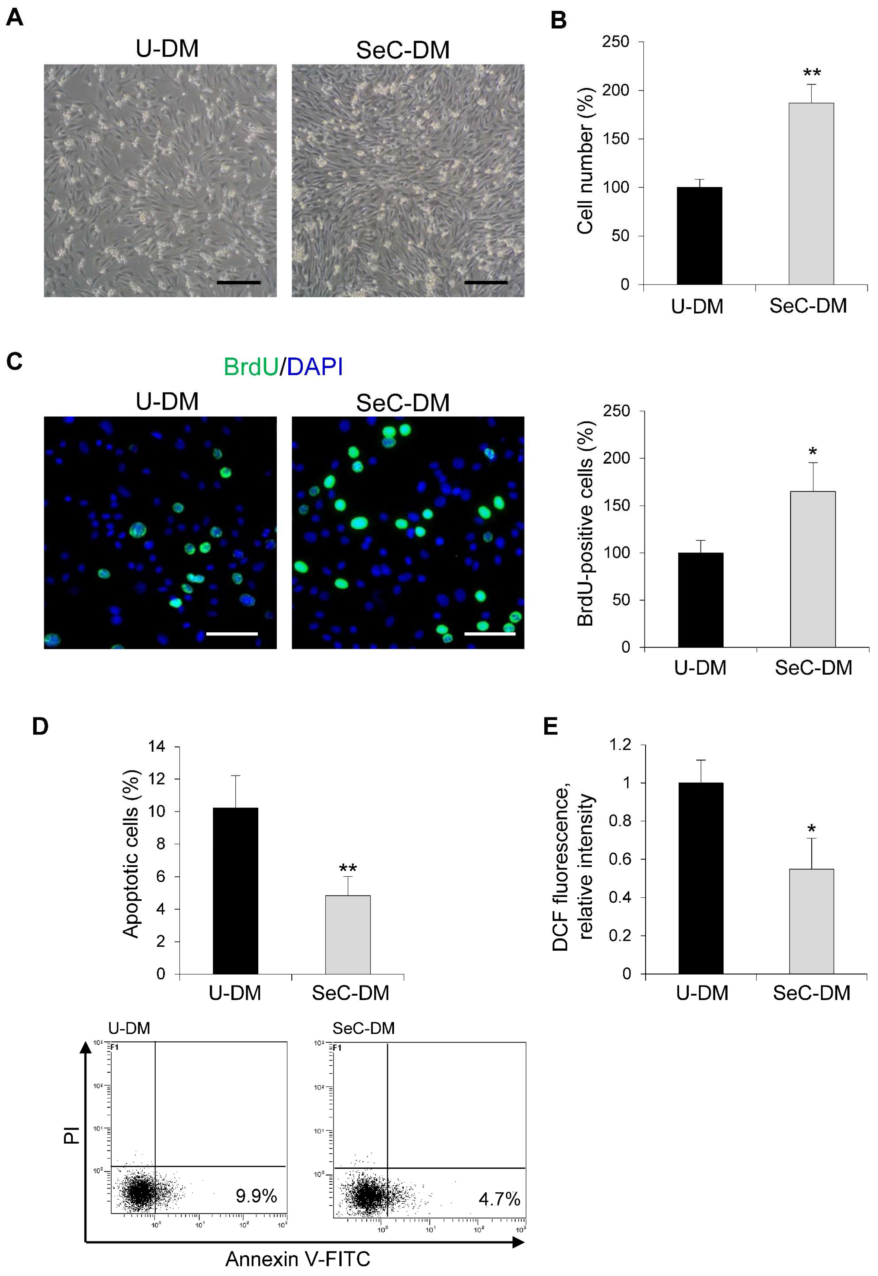
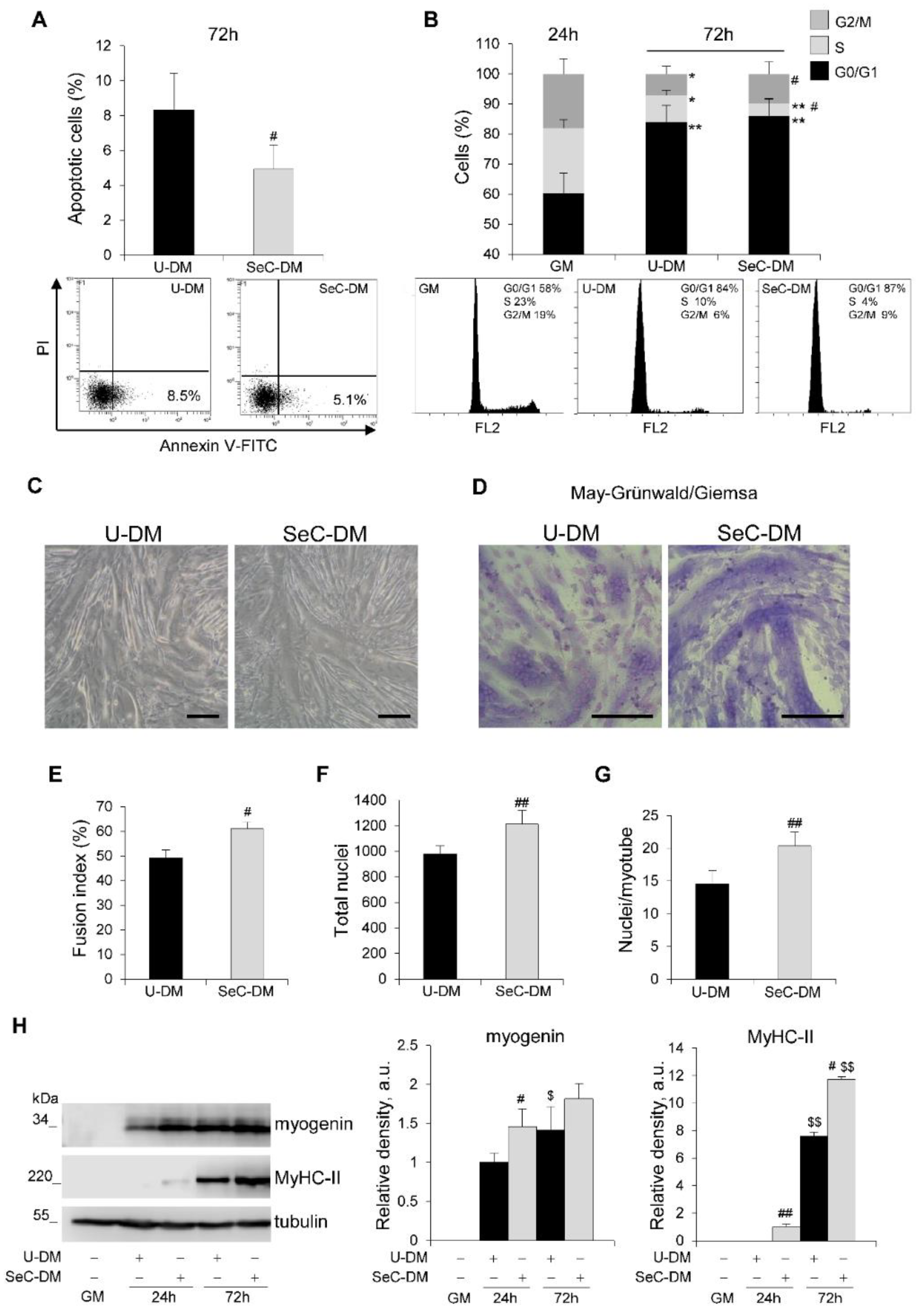
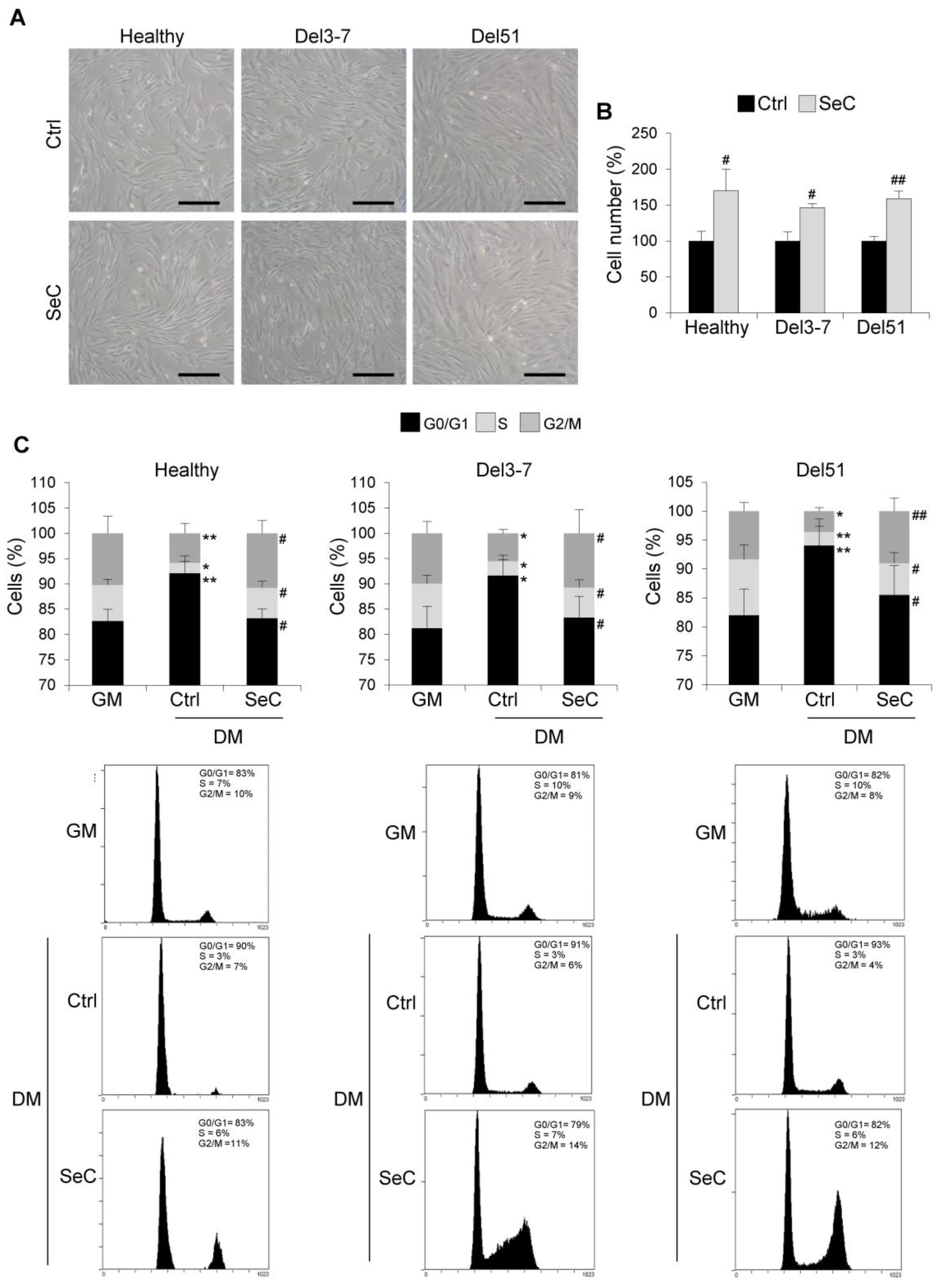
3.1. SeC Stimulate Myoblast Proliferation without Affecting the Myogenic Potential
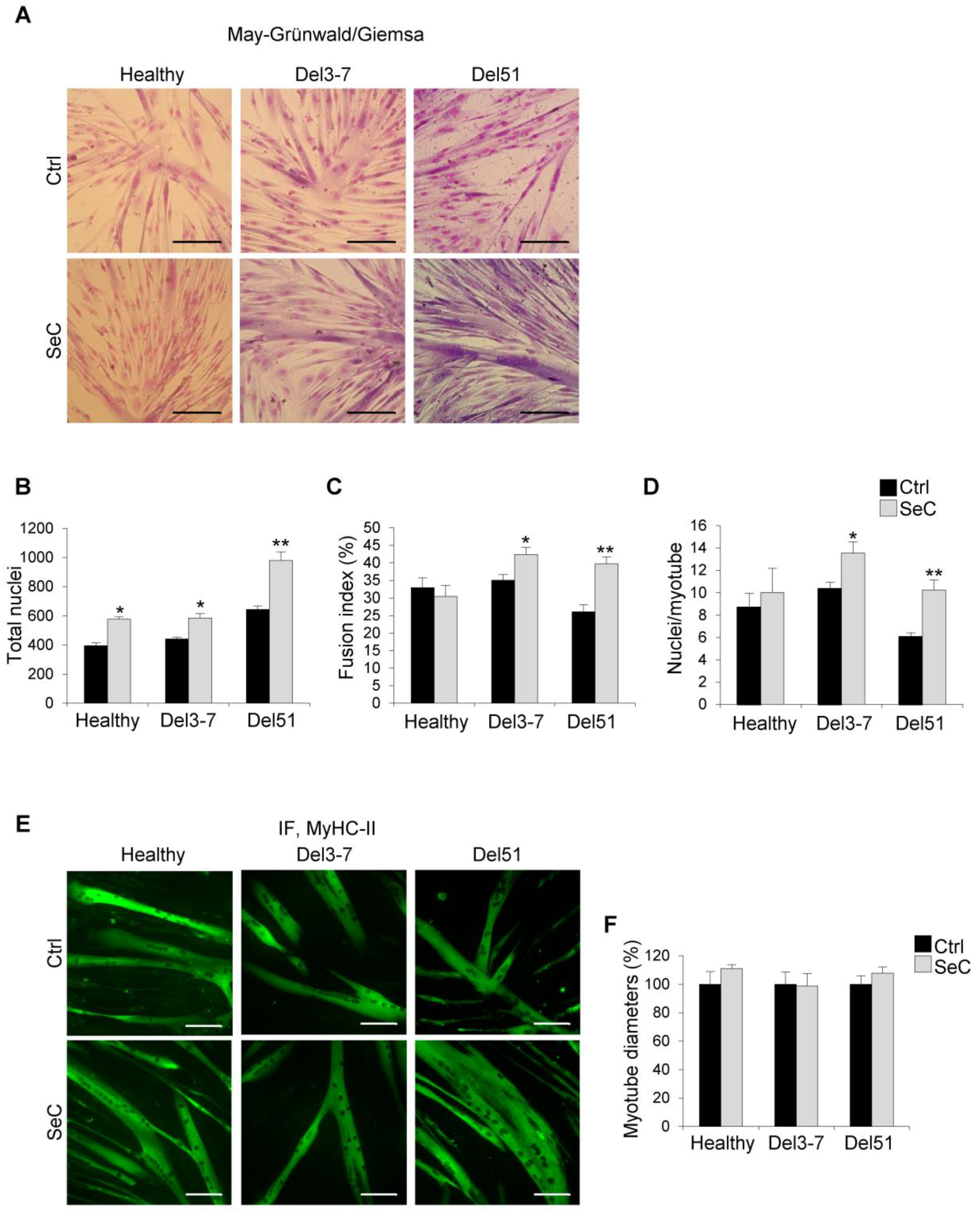
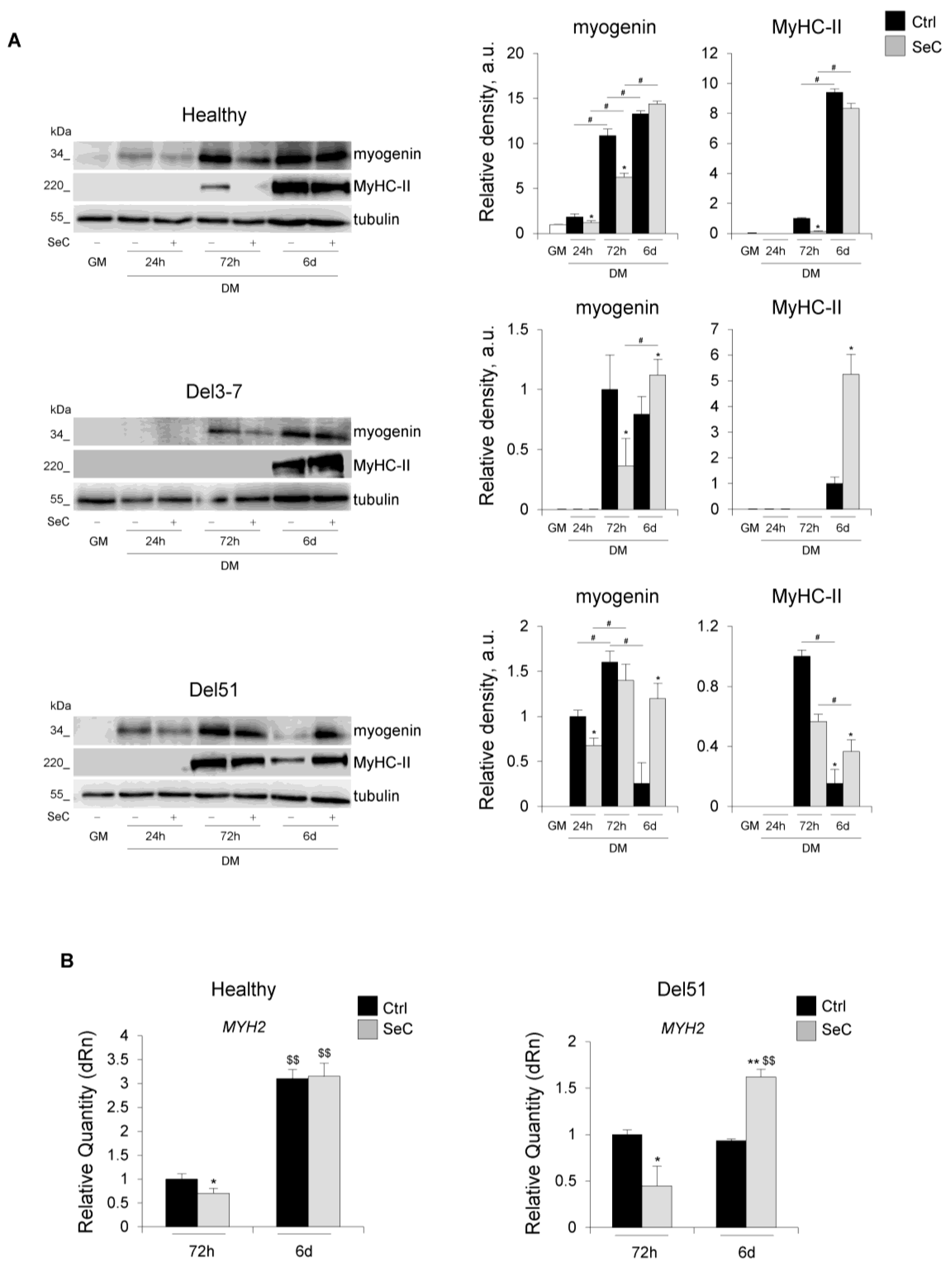
3.2. SeC Favor Myotube Elongation and Myosin Heavy Chain Expression in Differentiating DMD Myoblasts
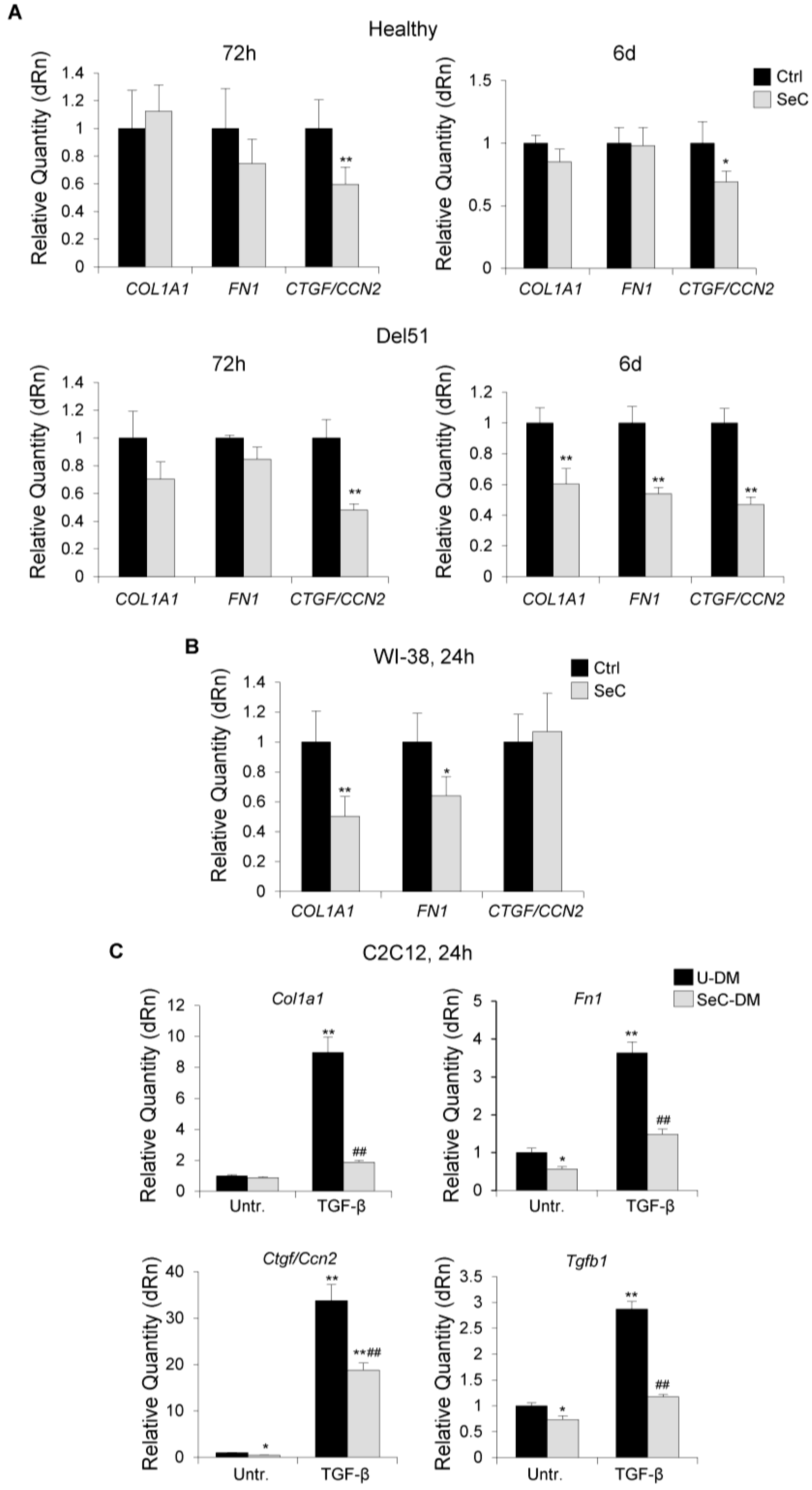
3.3. SeC Restrain the Fibrogenic Potential of Fibroblasts and Inhibit Myoblast–Myofibroblast Transdifferentiation
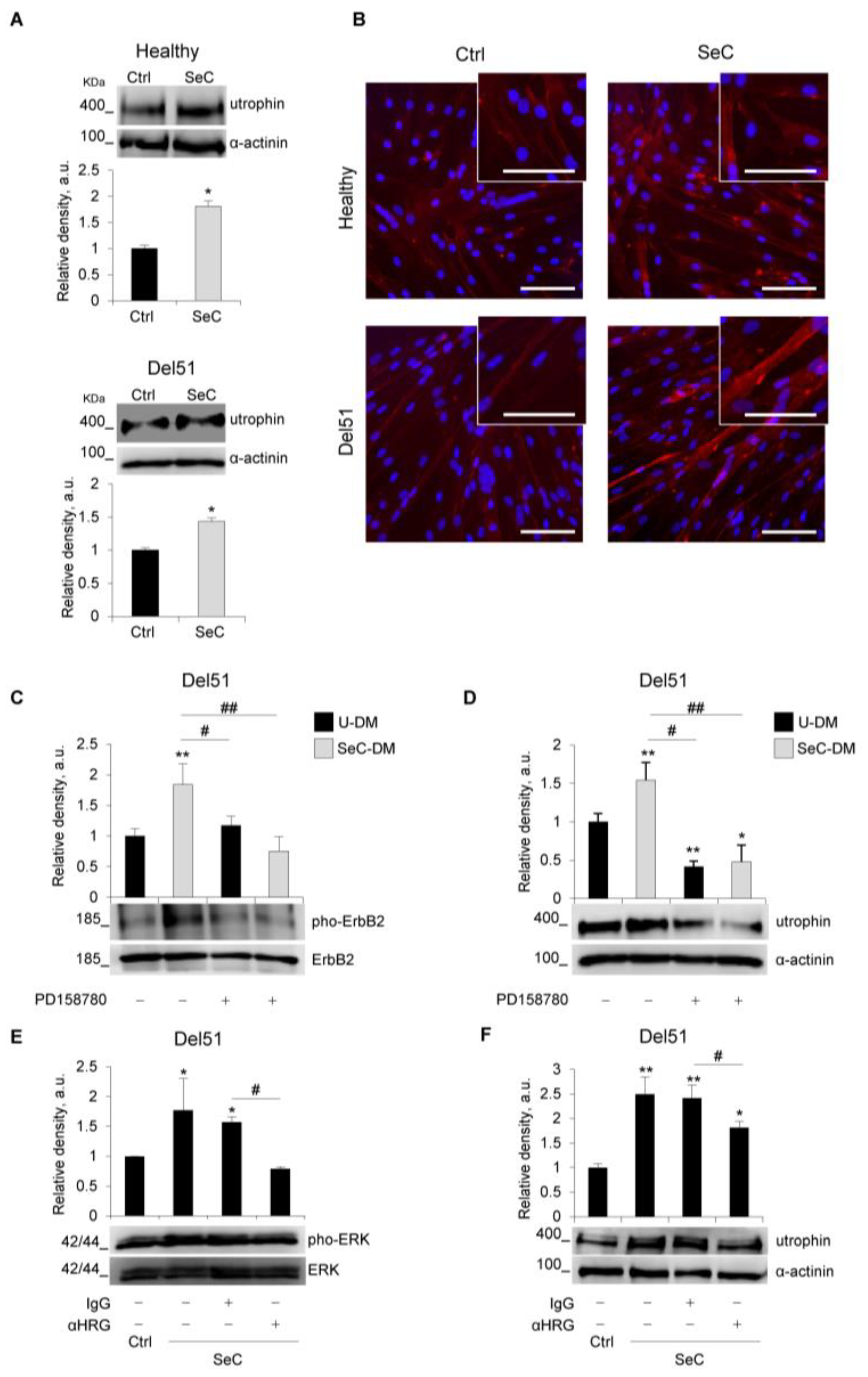
3.4. SeC Induce Utrophin Expression in DMD Myotubes Regardless of the Mutation
4. Discussion
Supplementary Materials
Author Contributions
Funding
Institutional Review Board Statement
Informed Consent Statement
Data Availability Statement
Acknowledgments
Conflicts of Interest
Appendix A
| Reagent or Resource | Source | Identifier |
|---|---|---|
| Antibodies | ||
| Donkey anti-Mouse IgG (H + L), Alexa Fluor 488 conjugated | Thermo Fisher Scientific | Cat#A-21202 |
| Donkey anti-Mouse IgG (H + L), Alexa Fluor 594 conjugated | Thermo Fisher Scientific | Cat#A-21203 |
| Goat anti-mouse IgG/IgM-HRP conjugated | Merck | Cat#AP130P |
| Goat anti-rabbit IgG-HRP conjugated | Sigma-Aldrich | Cat#A9169 |
| IgG from rabbit serum | Sigma-Aldrich | Cat#I5006 |
| Mouse monoclonal anti- α-Dystroglycan (IIH6) | Santa Cruz Biotechnology | Cat# sc-53987 |
| Mouse monoclonal anti-Myogenin (F5D) | Santa Cruz Biotechnology | Cat#sc-12735 |
| Mouse monoclonal anti-Utrophin (8A4) | Santa Cruz Biotechnology | Cat#sc-33700 |
| Mouse monoclonal anti-α-Actinin (H-2) | Santa Cruz Biotechnology | Cat#sc-17829 |
| Mouse monoclonal anti-α-Tubulin (DM1A) | Santa Cruz Biotechnology | Cat#sc-32293 |
| Rabbit polyclonal anti-HER2/ErbB2 | Cell Signaling Tech | Cat#2242 |
| Rabbit polyclonal anti-Heregulin-beta1 | ProSci | Cat#38-254 |
| Rabbit polyclonal anti-MAP Kinase (ERK-1, ERK-2) | Cell Signaling Tech | Cat#M5670 |
| Rabbit polyclonal anti-phospho-ErbB2 (HER-2) (Tyr877) | Invitrogen | Cat#PA5-77970 |
| Rabbit polyclonal anti-phospho-p44/42 MAPK (Erk1/2) (Thr202/Tyr204) | Cell Signaling Tech | Cat#9101S |
| Chemicals, Peptides, and Recombinant Proteins | ||
| Blotto, non-fat dried milk | Santa Cruz Biotechnology | Cat#sc-2325 |
| Bovine Serum Albumin (BSA) | Sigma-Aldrich | Cat#A7030 |
| DAPI dihydrochloride | Sigma-Aldrich | Cat#D9542 |
| DCFH-DA (2′,7′-Dichlorofluorescin diacetate) | Sigma-Aldrich | Cat#D6883 |
| DMSO (Dimethyl sulfoxide) | Sigma-Aldrich | Cat#67-68-5 |
| Gelatin solution | Sigma-Aldrich | Cat#G1393 |
| Giemsa stain, modified | Sigma-Aldrich | Cat#GS500 |
| May–Grünwald stain | Sigma-Aldrich | Cat#MG500 |
| PD 158780 tyrosine kinase inhibitor | Santa Cruz Biotechnology | Cat#CAS 171179-06-9 |
| Propidium iodide | Sigma-Aldrich | Cat#81845 |
| Recombinant human TGF-β | R&D System | Cat#240-B |
| TRIsure ™ | Bioline | Cat#BIO-38033 |
| Western Bright Quantum HRP substrate | Advansta | Cat#K-12042-D20 |
| Experimental Models: Cell Lines | ||
| C2C12 mouse myoblasts | ATCC, American Type Culture Collection | Cat#CRL-1772 |
| WI-38 human fibroblasts | ATCC, American Type Culture Collection | Cat#CCL-75 |
| Culture Medium | ||
| Apo-Transferrin human | Sigma-Aldrich | Cat#T1147 |
| Collagenase P | Sigma Aldrich | Cat#11213865001 |
| DNase I | Sigma Aldrich | Cat #SLCB2343 |
| Fetal bovine serum (FBS) | Gibco | Cat#10270-106 |
| Gentamicin 10 mg/mL sulphate | Euroclone | Cat#ECM0011B |
| HAM’S F12 | Euroclone | Cat#AL025A |
| High-glucose Dulbecco’s Modified Eagle’s Medium (HG-DMEM) | Gibco | Cat#41966-029 |
| Horse Serum (HS) | Gibco | Cat#16050-122 |
| Insulin solution from bovine pancreas | Sigma-Aldrich | Cat#I0516-5ML |
| Insulin-Transferrin-Selenium (ITS) + Premix | Corning | Cat#354352 |
| L-Glutamine 100× (200 mM) | Euroclone | Cat#ECB3000D |
| Penicillin/Streptomicin 100× | Euroclone | Cat#CB3001D |
| Retinoic acid | Sigma-Aldrich | Cat#R-2625100 |
| RPMI Medium 1640 | Gibco | Cat#21875-034 |
| Skeletal Muscle Cell Growth Medium (ready-to-use kit) | Promo Cell | Cat#39365 |
| Trypsin (2.5%) | Gibco | Cat#15090-046 |
| Oligonucleotides | ||
| Primers (see Appendix B) | Invitrogen | N/A |
| Software and Algorithms | ||
| IBM® SPSS® Statistics v18 software | SPSS (Chicago, IL, USA) | |
| Image Studio Digits v3.1.4 | LI-COR | |
| ImageJ v1.8.0_172 | Wayne Rasband (NIH, USA) | |
| LAS (Leica Application Suite) software v4.12.0 | LEICA | |
| MxPro-Mx3000P v4.10 | Agilent Technologies Stratagene | |
| SPOT Imaging v3.5.4 | Diagnostic Instruments | |
| Other | ||
| Annexin V-FITC apoptosis detection kit | BioVision Inc. | K101-100 |
| 5× HOT FIREPol EvaGreen qPCR Mix Plus (ROX) | Solis BioDyne | 08-24-0000 |
| PrimeScript ™ RT reagent Kit with gDNA Eraser | Takara Bio Europe | RR047B |
Appendix B
| Primary Antibody | Molecular Weight (kDa) | Dilution | Secondary Antibody | Dilution |
|---|---|---|---|---|
| Mouse monoclonal anti-MyHC-II (MF20) | 220 | 1:10,000 | Goat anti-mouse IgG/IgM-HRP conjugated | 1:10,000 |
| Mouse monoclonal anti-myogenin (F5D) | 34 | 1:1000 | Goat anti-mouse IgG/IgM-HRP conjugated | 1:1000 |
| Mouse monoclonal anti-Neu (ErbB2) (3B5) | 185 | 1:200 | Goat anti-mouse IgG/IgM-HRP conjugated | 1:1000 |
| Mouse monoclonal anti-utrophin (8A4) | 400 | 1:1000 | Goat anti-mouse IgG/IgM-HRP conjugated | 1:1000 |
| Mouse monoclonal anti-α-actinin (H-2) | 100 | 1:5000 | Goat anti-mouse IgG/IgM-HRP conjugated | 1:5000 |
| Mouse monoclonal anti-α-tubulin (DM1A) | 55 | 1:5000 | Goat anti-mouse IgG/IgM-HRP conjugated | 1:5000 |
| Rabbit polyclonal anti-MAP kinase (ERK1, ERK2) | 42–44 | 1:10,000 | Goat anti-rabbit IgG-HRP conjugated | 1:10,000 |
| Rabbit polyclonal anti-phospho-ErbB2 (HER-2) (Tyr877) | 185 | 1:500 | Goat anti-rabbit IgG-HRP conjugated | 1:2000 |
| Rabbit polyclonal anti-phospho-p44/42 MAPK (ERK1/2) (Thr202/Tyr204) | 42–44 | 1:1000 | Goat anti-rabbit IgG-HRP conjugated | 1:2000 |
Appendix C
| Gene | Forward Primer 5′-3′ | Reverse Primer 5′-3′ |
|---|---|---|
| COL1A1 | TCTGCGACAACGGCAAGGTG | GACGCCGGTGGTTTCTTGGT |
| CTGF/CCN2 | CTTGCGAAGCTGACCTGGAAGA | CCGTCGGTACATACTCCACAGA |
| FN1 | ACAACACCGAGGTGACTGAGAC | GGACACAACGATGCTTCCTGAG |
| GAPDH | AAGAAGGTGGTGAAGCAGG | GTCAAAGGTGGAGGAGTGG |
| MYH2 | CAGCTACTGCACACCCAGAA | CTTCACGGTCTGCTCCATGT |
| Col1a1 | TCATCGTGGCTTCTCTGGTC | GACCGTTGAGTCCGTCTTTG |
| Ctgf/Ccn2 | GCCTACCGACTGGAAGACAC | GTAACTCGGGTGGAGATGCC |
| Fn1 | GAAGACAGATGAGCTTCCCCA | GGTTGGTGATGAAGGGGGTC |
| Gapdh | GCCTTCCGTGTTCCTACCC | CAGTGGGCCCTCAGATGC |
| Tgfb1 | GCCTGAGTGGCTGTCTTTTGA | CACAAGAGCAGTGAGCGCTGAA |
References
- Emery, A.E. Population frequencies of inherited neuromuscular diseases—A world survey. Neuromuscul. Disord. 1991, 1, 19–29. [Google Scholar] [CrossRef]
- Mendell, J.R.; Shilling, C.; Leslie, N.D.; Flanigan, K.M.; al-Dahhak, R.; Gastier-Foster, J.; Kneile, K.; Dunn, D.M.; Duval, B.; Aoyagi, A.; et al. Evidence-based path to newborn screening for Duchenne muscular dystrophy. Ann. Neurol. 2012, 71, 304–313. [Google Scholar] [CrossRef]
- Duan, D.; Goemans, N.; Takeda, S.; Mercuri, E.; Aartsma-Rus, A. Duchenne muscular dystrophy. Nat. Rev. Dis. Prim. 2021, 7, 13. [Google Scholar] [CrossRef] [PubMed]
- McGreevy, J.W.; Hakim, C.H.; McIntosh, M.A.; Duan, D. Animal models of Duchenne muscular dystrophy: From basic mechanisms to gene therapy. Dis. Model Mech. 2015, 8, 195–213. [Google Scholar] [CrossRef] [PubMed] [Green Version]
- Tidball, J.G.; Welc, S.S.; Wehling-Henricks, M. Immunobiology of Inherited Muscular Dystrophies. Compr. Physiol. 2018, 8, 1313–1356. [Google Scholar] [CrossRef] [PubMed]
- Bello, L.; Pegoraro, E. The “Usual Suspects”: Genes for Inflammation, Fibrosis, Regeneration, and Muscle Strength Modify Duchenne Muscular Dystrophy. J. Clin. Med. 2019, 8, 649. [Google Scholar] [CrossRef] [Green Version]
- Ryder, S.; Leadley, R.M.; Armstrong, N.; Westwood, M.; de Kock, S.; Butt, T.; Jain, M.; Kleijnen, J. The burden, epidemiology, costs and treatment for Duchenne muscular dystrophy: An evidence review. Orphanet J. Rare Dis. 2017, 12, 79. [Google Scholar] [CrossRef] [Green Version]
- Chiappalupi, S.; Salvadori, L.; Luca, G.; Riuzzi, F.; Calafiore, R.; Donato, R.; Sorci, G. Do porcine Sertoli cells represent an opportunity for Duchenne muscular dystrophy? Cell Prolif. 2019, 52, e12599. [Google Scholar] [CrossRef] [PubMed] [Green Version]
- Verhaart, I.E.C.; Aartsma-Rus, A. Therapeutic developments for Duchenne muscular dystrophy. Nat. Rev. Neurol. 2019, 15, 373–386. [Google Scholar] [CrossRef]
- Werneck, L.C.; Lorenzoni, P.J.; Ducci, R.D.; Fustes, O.H.; Kay, C.S.K.; Scola, R.H. Duchenne muscular dystrophy: An historical treatment review. Arq. Neuro-Psiquiatria 2019, 77, 579–589. [Google Scholar] [CrossRef]
- Chiappalupi, S.; Luca, G.; Mancuso, F.; Madaro, L.; Fallarino, F.; Nicoletti, C.; Calvitti, M.; Arato, I.; Falabella, G.; Salvadori, L.; et al. Effects of intraperitoneal injection of microencapsulated Sertoli cells on chronic and presymptomatic dystrophic mice. Data Brief 2015, 5, 1015–1021. [Google Scholar] [CrossRef] [PubMed] [Green Version]
- Chiappalupi, S.; Luca, G.; Mancuso, F.; Madaro, L.; Fallarino, F.; Nicoletti, C.; Calvitti, M.; Arato, I.; Falabella, G.; Salvadori, L.; et al. Intraperitoneal injection of microencapsulated Sertoli cells restores muscle morphology and performance in dystrophic mice. Biomaterials 2016, 75, 313–326. [Google Scholar] [CrossRef] [PubMed]
- Griswold, M.D. 50 years of spermatogenesis: Sertoli cells and their interactions with germ cells. Biol. Reprod. 2018, 99, 87–100. [Google Scholar] [CrossRef]
- Mital, P.; Kaur, G.; Dufour, J.M. Immunoprotective sertoli cells: Making allogeneic and xenogeneic transplantation feasible. Reproduction 2010, 139, 495–504. [Google Scholar] [CrossRef] [Green Version]
- Kaur, G.; Thompson, L.A.; Dufour, J.M. Sertoli cells-immunological sentinels of spermatogenesis. Semin Cell Dev. Biol. 2014, 30, 36–44. [Google Scholar] [CrossRef] [PubMed] [Green Version]
- Luca, G.; Arato, I.; Sorci, G.; Cameron, D.F.; Hansen, B.C.; Baroni, T.; Donato, R.; White, D.G.J.; Calafiore, R. Sertoli cells for cell transplantation: Pre-clinical studies and future perspectives. Andrology 2018, 6, 385–395. [Google Scholar] [CrossRef]
- Chiappalupi, S.; Salvadori, L.; Mancuso, F.; Arato, I.; Calvitti, M.; Riuzzi, F.; Calafiore, R.; Luca, G.; Sorci, G. Microencapsulated Sertoli cells sustain myoblast proliferation without affecting the myogenic potential. In vitro data. Data Brief, submitted.
- Salvadori, L.; Mandrone, M.; Manenti, T.; Ercolani, C.; Cornioli, L.; Lianza, M.; Tomasi, P.; Chiappalupi, S.; Di Filippo, E.S.; Fulle, S.; et al. Identification of Withania somnifera-Silybum marianum-Trigonella foenum-graecum Formulation as a Nutritional Supplement to Contrast Muscle Atrophy and Sarcopenia. Nutrients 2020, 13, 49. [Google Scholar] [CrossRef]
- Chiappalupi, S.; Sorci, G.; Vukasinovic, A.; Salvadori, L.; Sagheddu, R.; Coletti, D.; Renga, G.; Romani, L.; Donato, R.; Riuzzi, F. Targeting RAGE prevents muscle wasting and prolongs survival in cancer cachexia. J. Cachexia Sarcopenia Muscle 2020, 11, 929–946. [Google Scholar] [CrossRef] [Green Version]
- Fidziańska, A.; Goebel, H.H. Human ontogenesis. 3. Cell death in fetal muscle. Acta Neuropathol. 1991, 81, 572–577. [Google Scholar] [CrossRef]
- Bier, A.; Berenstein, P.; Kronfeld, N.; Morgoulis, D.; Ziv-Av, A.; Goldstein, H.; Kazimirsky, G.; Cazacu, S.; Meir, R.; Popovtzer, R.; et al. Placenta-derived mesenchymal stromal cells and their exosomes exert therapeutic effects in Duchenne muscular dystrophy. Biomaterials 2018, 174, 67–78. [Google Scholar] [CrossRef]
- Li, Y.; Foster, W.; Deasy, B.M.; Chan, Y.; Prisk, V.; Tang, Y.; Cummins, J.; Huard, J. Transforming growth factor-beta1 induces the differentiation of myogenic cells into fibrotic cells in injured skeletal muscle: A key event in muscle fibrogenesis. Am. J. Pathol. 2004, 164, 1007–1019. [Google Scholar] [CrossRef]
- Bernasconi, P.; Di Blasi, C.; Mora, M.; Morandi, L.; Galbiati, S.; Confalonieri, P.; Cornelio, F.; Mantegazza, R. Transforming growth factor-beta1 and fibrosis in congenital muscular dystrophies. Neuromuscul. Disord. 1999, 9, 28–33. [Google Scholar] [CrossRef]
- Sliwkowski, M.X.; Schaefer, G.; Akita, R.W.; Lofgren, J.A.; Fitzpatrick, V.D.; Nuijens, A.; Fendly, B.M.; Cerione, R.A.; Vandlen, R.L.; Carraway, K.L., III. Coexpression of erbB2 and erbB3 proteins reconstitutes a high affinity receptor for heregulin. J. Biol. Chem. 1994, 269, 14661–14665. [Google Scholar] [CrossRef]
- Washburn, R.L.; Hibler, T.; Thompson, L.A.; Kaur, G.; Dufour, J.M. Therapeutic application of Sertoli cells for treatment of various diseases. Semin Cell Dev. Biol. 2021. [Google Scholar] [CrossRef]
- Chiappalupi, S.; Salvadori, L.; Luca, G.; Riuzzi, F.; Calafiore, R.; Donato, R.; Sorci, G. Employment of Microencapsulated Sertoli Cells as a New Tool to Treat Duchenne Muscular Dystrophy. J. Funct. Morphol. Kinesiol. 2017, 2, 47. [Google Scholar] [CrossRef] [Green Version]
- Fallarino, F.; Luca, G.; Calvitti, M.; Mancuso, F.; Nastruzzi, C.; Fioretti, M.C.; Grohmann, U.; Becchetti, E.; Burgevin, A.; Kratzer, R.; et al. Therapy of experimental type 1 diabetes by isolated Sertoli cell xenografts alone. J. Exp. Med. 2009, 206, 2511–2526. [Google Scholar] [CrossRef] [PubMed] [Green Version]
- Luca, G.; Calvitti, M.; Mancuso, F.; Falabella, G.; Arato, I.; Bellucci, C.; List, E.O.; Bellezza, E.; Angeli, G.; Lilli, C.; et al. Reversal of experimental Laron Syndrome by xenotransplantation of microencapsulated porcine Sertoli cells. J. Control Release 2013, 165, 75–81. [Google Scholar] [CrossRef] [PubMed]
- Luca, G.; Bellezza, I.; Arato, I.; Di Pardo, A.; Mancuso, F.; Calvitti, M.; Falabella, G.; Bartoli, S.; Maglione, V.; Amico, E.; et al. Terapeutic Potential of Microencapsulated Sertoli Cells in Huntington Disease. CNS Neurosci. Ther. 2016, 22, 686–690. [Google Scholar] [CrossRef] [PubMed]
- Luca, G.; Arato, I.; Mancuso, F.; Calvitti, M.; Falabella, G.; Murdolo, G.; Basta, G.; Cameron, D.F.; Hansen, B.C.; Fallarino, F.; et al. Xenograft of microencapsulated Sertoli cells restores glucose homeostasis in db/db mice with spontaneous diabetes mellitus. Xenotransplantation 2016, 23, 429–439. [Google Scholar] [CrossRef] [PubMed] [Green Version]
- Rosenberg, A.S.; Puig, M.; Nagaraju, K.; Hoffman, E.P.; Villalta, S.A.; Rao, V.A.; Wakefield, L.M.; Woodcock, J. Immune-mediated pathology in Duchenne muscular dystrophy. Sci. Transl. Med. 2015, 7, 299rv4. [Google Scholar] [CrossRef] [Green Version]
- Skuk, D.; Tremblay, J.P. Myoblast transplantation: The current status of a potential therapeutic tool for myopathies. J. Muscle Res. Cell. Motil. 2003, 24, 285–300. [Google Scholar] [CrossRef]
- Duan, D. Systemic AAV Micro-dystrophin Gene Therapy for Duchenne Muscular Dystrophy. Mol. Ther. 2018, 26, 2337–2356. [Google Scholar] [CrossRef] [Green Version]
- Sun, C.; Shen, L.; Zhang, Z.; Xie, X. Therapeutic Strategies for Duchenne Muscular Dystrophy: An Update. Genes 2020, 11, 837. [Google Scholar] [CrossRef]
- Yin, H.; Price, F.; Rudnicki, M.A. Satellite cells and the muscle stem cell niche. Physiol. Rev. 2013, 93, 23–67. [Google Scholar] [CrossRef] [Green Version]
- Cappellari, O.; Mantuano, P.; De Luca, A. “The Social Network” and Muscular Dystrophies: The Lesson Learnt about the Niche Environment as a Target for Therapeutic Strategies. Cells 2020, 9, 1659. [Google Scholar] [CrossRef]
- Chargé, S.B.; Rudnicki, M.A. Cellular and molecular regulation of muscle regeneration. Physiol. Rev. 2004, 84, 209–238. [Google Scholar] [CrossRef] [PubMed]
- Riuzzi, F.; Sorci, G.; Donato, R. S100B protein regulates myoblast proliferation and differentiation by activating FGFR1 in a bFGF-dependent manner. J. Cell. Sci. 2011, 124, 2389–2400. [Google Scholar] [CrossRef] [Green Version]
- Ono, Y.; Calhabeu, F.; Morgan, J.; Katagiri, T.; Amthor, H.; Zammit, P.S. BMP signalling permits population expansion by preventing premature myogenic differentiation in muscle satellite cells. Cell. Death Differ. 2011, 18, 222–234. [Google Scholar] [CrossRef] [PubMed] [Green Version]
- Florini, J.R.; Ewton, D.Z.; Coolican, S.A. Growth hormone and the insulin-like growth factor system in myogenesis. Endocr. Rev. 1996, 17, 481–517. [Google Scholar] [CrossRef] [PubMed] [Green Version]
- D’Andrea, P.; Sciancalepore, M.; Veltruska, K.; Lorenzon, P.; Bandiera, A. Epidermal Growth Factor-based adhesion substrates elicit myoblast scattering, proliferation, differentiation and promote satellite cell myogenic activation. Biochim. Biophys. Acta Mol. Cell. Res. 2019, 1866, 504–517. [Google Scholar] [CrossRef]
- Wang, Y.X.; Feige, P.; Brun, C.E.; Hekmatnejad, B.; Dumont, N.A.; Renaud, J.M.; Faulkes, S.; Guindon, D.E.; Rudnicki, M.A. EGFR-Aurka Signaling Rescues Polarity and Regeneration Defects in Dystrophin-Deficient Muscle Stem Cells by Increasing Asymmetric Divisions. Cell Stem Cell 2019, 24, 419–432.e6. [Google Scholar] [CrossRef] [PubMed] [Green Version]
- Rende, M.; Brizi, E.; Donato, R.; Provenzano, C.; Bruno, R.; Mizisin, A.P.; Garrett, R.S.; Calcutt, N.A.; Campana, W.M.; O’Brien, J.S. Prosaposin is immunolocalized to muscle and prosaptides promote myoblast fusion and attenuate loss of muscle mass after nerve injury. Muscle Nerve 2001, 24, 799–808. [Google Scholar] [CrossRef] [PubMed]
- Melendez, J.; Sieiro, D.; Salgado, D.; Morin, V.; Dejardin, M.J.; Zhou, C.; Mullen, A.C.; Marcelle, C. TGFβ signalling acts as a molecular brake of myoblast fusion. Nat. Commun. 2021, 12, 749. [Google Scholar] [CrossRef]
- Li, Y.; Huard, J. Differentiation of muscle-derived cells into myofibroblasts in injured skeletal muscle. Am. J. Pathol. 2002, 161, 895–907. [Google Scholar] [CrossRef] [Green Version]
- Vial, C.; Zúñiga, L.M.; Cabello-Verrugio, C.; Cañón, P.; Fadic, R.; Brandan, E. Skeletal muscle cells express the profibrotic cytokine connective tissue growth factor (CTGF/CCN2), which induces their dedifferentiation. J. Cell Physiol. 2008, 215, 410–421. [Google Scholar] [CrossRef] [PubMed]
- Hillege, M.M.G.; Galli Caro, R.A.; Offringa, C.; de Wit, G.M.J.; Jaspers, R.T.; Hoogaars, W.M.H. TGF-β Regulates Collagen Type I Expression in Myoblasts and Myotubes via Transient Ctgf and Fgf-2 Expression. Cells 2020, 9, 375. [Google Scholar] [CrossRef] [Green Version]
- EL Andaloussi, S.; Mäger, I.; Breakefield, X.O.; Wood, M.J. Extracellular vesicles: Biology and emerging therapeutic opportunities. Nat. Rev. Drug. Discov. 2013, 12, 347–357. [Google Scholar] [CrossRef]
- Bladen, C.L.; Salgado, D.; Monges, S.; Foncuberta, M.E.; Kekou, K.; Kosma, K.; Dawkins, H.; Lamont, L.; Roy, A.J.; Chamova, T.; et al. The TREAT-NMD DMD Global Database: Analysis of more than 7,000 Duchenne muscular dystrophy mutations. Hum. Mutat. 2015, 36, 395–402. [Google Scholar] [CrossRef]
- Guiraud, S.; Roblin, D.; Kay, D.E. The potential of utrophin modulators for the treatment of Duchenne muscular dystrophy. Expert Opin. Orphan Drugs 2018, 6, 179–192. [Google Scholar] [CrossRef] [Green Version]
- Andrechek, E.R.; Hardy, W.R.; Girgis-Gabardo, A.A.; Perry, R.L.; Butler, R.; Graham, F.L.; Kahn, R.C.; Rudnicki, M.A.; Muller, W.J. ErbB2 is required for muscle spindle and myoblast cell survival. Mol. Cell. Biol. 2002, 22, 4714–4722. [Google Scholar] [CrossRef] [Green Version]
Publisher’s Note: MDPI stays neutral with regard to jurisdictional claims in published maps and institutional affiliations. |
© 2021 by the authors. Licensee MDPI, Basel, Switzerland. This article is an open access article distributed under the terms and conditions of the Creative Commons Attribution (CC BY) license (https://creativecommons.org/licenses/by/4.0/).
Share and Cite
Salvadori, L.; Chiappalupi, S.; Arato, I.; Mancuso, F.; Calvitti, M.; Marchetti, M.C.; Riuzzi, F.; Calafiore, R.; Luca, G.; Sorci, G. Sertoli Cells Improve Myogenic Differentiation, Reduce Fibrogenic Markers, and Induce Utrophin Expression in Human DMD Myoblasts. Biomolecules 2021, 11, 1504. https://doi.org/10.3390/biom11101504
Salvadori L, Chiappalupi S, Arato I, Mancuso F, Calvitti M, Marchetti MC, Riuzzi F, Calafiore R, Luca G, Sorci G. Sertoli Cells Improve Myogenic Differentiation, Reduce Fibrogenic Markers, and Induce Utrophin Expression in Human DMD Myoblasts. Biomolecules. 2021; 11(10):1504. https://doi.org/10.3390/biom11101504
Chicago/Turabian StyleSalvadori, Laura, Sara Chiappalupi, Iva Arato, Francesca Mancuso, Mario Calvitti, Maria Cristina Marchetti, Francesca Riuzzi, Riccardo Calafiore, Giovanni Luca, and Guglielmo Sorci. 2021. "Sertoli Cells Improve Myogenic Differentiation, Reduce Fibrogenic Markers, and Induce Utrophin Expression in Human DMD Myoblasts" Biomolecules 11, no. 10: 1504. https://doi.org/10.3390/biom11101504
APA StyleSalvadori, L., Chiappalupi, S., Arato, I., Mancuso, F., Calvitti, M., Marchetti, M. C., Riuzzi, F., Calafiore, R., Luca, G., & Sorci, G. (2021). Sertoli Cells Improve Myogenic Differentiation, Reduce Fibrogenic Markers, and Induce Utrophin Expression in Human DMD Myoblasts. Biomolecules, 11(10), 1504. https://doi.org/10.3390/biom11101504


