Article

DNA Phosphorothioate Modifications Are Widely Distributed in the Human Microbiome

1
State Key Laboratory of Microbial Metabolism, Joint International Research Laboratory of Metabolic and Developmental Sciences, and School of Life Sciences & Biotechnology, Shanghai Jiao Tong University, Shanghai 200240, China
2
Department of Biological Engineering and Center for Environmental Health Science, Massachusetts Institute of Technology, Cambridge, MA 02139, USA
3
Antimicrobrial Resistance Interdisciplinary Research Group, Singapore-MIT Alliance for Research and Technology, 1 CREATE Way, Singapore 138602, Singapore
*
Authors to whom correspondence should be addressed.
Biomolecules 2020, 10(8), 1175; https://doi.org/10.3390/biom10081175
Submission received: 10 July 2020 / Revised: 7 August 2020 / Accepted: 10 August 2020 / Published: 12 August 2020
(This article belongs to the Section Molecular Biology)

Abstract

:
The DNA phosphorothioate (PT) modification existing in many prokaryotes, including bacterial pathogens and commensals, confers multiple characteristics, including restricting gene transfer, influencing the global transcriptional response, and reducing fitness during exposure to chemical mediators of inflammation. While PT-containing bacteria have been investigated in a variety of environments, they have not been studied in the human microbiome. Here, we investigated the distribution of PT-harboring strains and verified their existence in the human microbiome. We found over 2000 PT gene-containing strains distributed in different body sites, especially in the gastrointestinal tract. PT-modifying genes are preferentially distributed within several genera, including Pseudomonas, Clostridioides, and Escherichia, with phylogenic diversities. We also assessed the PT modification patterns and found six new PT-linked dinucleotides (CpsG, CpsT, ApsG, TpsG, GpsC, ApsT) in human fecal DNA. To further investigate the PT in the human gut microbiome, we analyzed the abundance of PT-modifying genes and quantified the PT-linked dinucleotides in the fecal DNA. These results confirmed that human microbiome is a rich reservoir for PT-containing microbes and contains a wide variety of PT modification patterns.

1. Introduction

Phosphorothioate (PT) DNA modifications, in which the nonbridging oxygen in the phosphate backbone is replaced by sulfur, are widespread among bacteria and archaea [1]. The PT-modifying gene cluster, dndA-E, has been found in more than 1300 sequenced genomes [2] and confers cells with 5′-GpsAAC-3′/5′-GpsTTC-3′ or 5′-GpsGCC-3′ consensus sequences [3]. Recently, another PT-modifying gene cluster has been reported, sspA-D, which confers cells with 5′-CpsCA-3′ on the single strand [4]. The DndA protein, which can be functionally substituted by an IscS (a cysteine desulfurase located elsewhere in the genome) [5,6], transfers sulfur into the Fe-S cluster of DndC [5,7]. SspA and SspD may share the same initial sulfur mobilization pathway with DndA and DndC, respectively [4]. Meanwhile, SspC shows ATPase activity and may provide energy in a manner similar to DndD [4]. Though PT modifications are usually introduced in a sequence-specific manner, only 12%–14% of target motifs are modified [3,8]. In some bacteria, PT modifications are involved in a restriction-modification (R-M) system with gene cassette dndF-H/sspE or an antiviral system with gene cassette pbeA-D [9]. However, nearly half of the PT-containing strains lack dndF-H/sspE and pbeA-D, indicating that the PT modifications possess other functions [4,10]. A recent transcriptomic and metabolomic analysis showed that PT modifications contribute to the cellular redox state [11], while, at the same time, PT modifications confer sensitivity to the HOCl produced by neutrophils [12]. To date, little is known about the presence of PT-containing bacteria in the human microbiome. Here, we performed an informatic analysis of sequenced microbiome-related genomes and a mass spectrometric analysis of fecal DNA to show the widespread presence of PT-modifying genes and PT-linked dinucleotides in the human microbiome.

2. Materials and Methods

2.1. Multiple Sequence Alignments

All publicly available genomes (including the complete and partial genomes) were downloaded from the online databases of NCBI (National Center for Biotechnology Information) (https://www.ncbi.nlm.nih.gov/genome/), EMBL (European Molecular Biology Laboratory) (https://www.ebi.ac.uk/), and HMP (Human Microbiome Project) (https://www.hmpdacc.org/). We used the MakeDB of multigeneblast (software version 1.1.13) [13] to convert the original databases into the GBK format. The dnd genes of Salmonella enterica serovar Cerro 87 (CP008925: 3477655...3481641) and the ssp genes of Vibrio cyclitrophicus FF75 (NZ_ATLT01000001: 2194844...2200061) were used as PT-modifying component queries [4]. The DndF-H cluster of S. enterica serovar Cerro 87 (CP008925: 3467758…3475796) was used as the restriction component query. The PbeA-D cluster of Haloterrigena jeotgali A29 (CP031303: 460843…466000) was used as the antiviral component query. The amino acid translation of each gene sequence within the query gene cluster is searched against the selected GBK (Genebank) database, yielding a dataset of BLAST (Basic Local Alignment Search Tool) hits [13]. The BLAST hits are then mapped to their parent nucleotide scaffolds, based on the information from the database [13]. The nucleotide scaffolds are then sorted according to their empirical similarity scores with the query gene cluster [13]. The number of blast hits per gene to be mapped is 250. The weight of synteny conversion in hit sorting is 0.5. The minimal sequence coverage of blast hits is 25. The minimal identity of blast hits is 30%. The maximum distance between genes in locus is 20 kb. These parameters above were set up for multiple sequence alignments in multigeneblast (software version 1.1.13) [13].

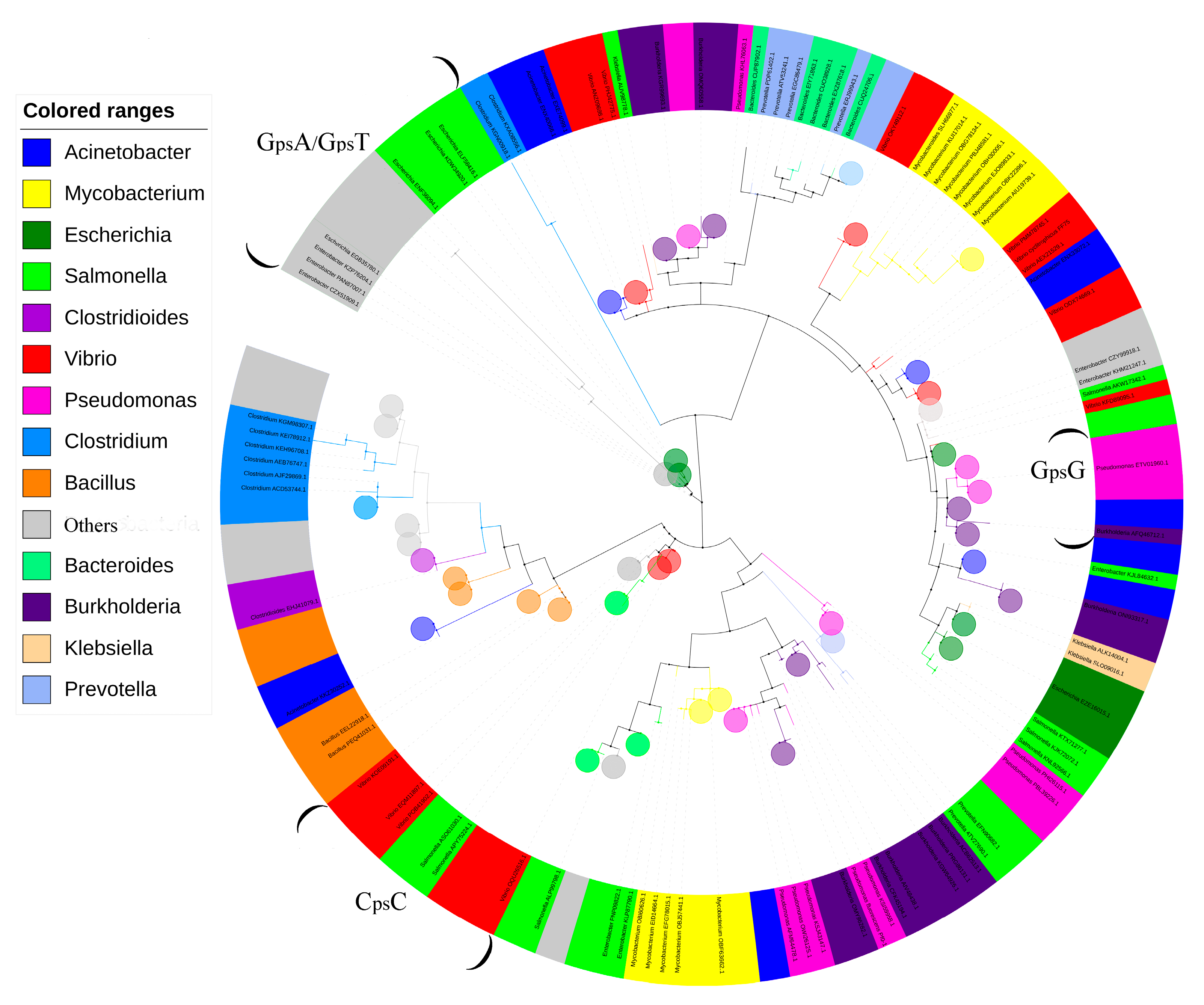
2.2. Phylogenic Analysis

The identification of protein sequences was based on the TIGR (The Institute for Genomic Research) database (http://www.tigr.org) annotation (e.g., TIGR03233, TIGR03183, TIGR03185, and TIGR03184 for DndA, B, C, D, and E, respectively) [14]. The amino acid sequences of DndC/SspD (accession numbers were shown in dataset) were downloaded from the online databases (e.g., https://www.ncbi.nlm.nih.gov/genome/). Then. we combined these amino acid sequences with the DndC/SspD of V. cyclitrophicus FF75 (CpsC) and Pseudomonas fluorescens Pf0-1 (GpsG) and aligned them by MEGA7 with the maximum likelihood method (500 bootstrap replications). The phylogenic tree was visualized using iTOL (http://itol.embl.de/).

2.3. Gene Abundance Calculation

A thenon-redundant human gut microbial gene catalog was constructed in our previous study [15]. In brief, the reads from the metagenomes were de novo assembled into contigs. Genes were predicted from the contigs and merged into non-redundant genes based on sequence similarity. The abundance of the genes was obtained by mapping the reads on the catalog, obtaining the gene-length normalized base counts, and adjusting the sequencing depth with a resampling procedure. All the genes were clustered into CAGs based on their abundance data using the canopy-based algorithm with default parameters. CAGs with more than 700 genes were regarded as bacterial CAGs for further analysis. The CAG abundance profiles were calculated as the sample-wise median gene abundance, essentially as described elsewhere [16].

2.4. Fecal DNA Preparation

All subjects gave their informed consent for inclusion before they participated in the study [15]. The study was conducted in accordance with the Declaration of Helsinki, and the protocol was approved by the Ethics Committee of the School of Life Sciences and Biotechnology, Shanghai Jiao Tong University (No. 2012-016). Feces samples were obtained and immediately frozen on dry ice upon collection and stored at −80 °C until further analysis [15]. DNA extraction from the human fecal samples was conducted as previously described [15] and purified by the QIAamp DNA mini kit (51304, QIAGEN, Germany).

2.5. Detection of PT-Linked Dinucleotides

All the fecal DNA samples were hydrolyzed with 4 units of nuclease P1 (Sigma, St. Louis, MO, USA) and subsequently dephosphorylated by 10 units of alkaline phosphatase (Fermentas), essentially as described elsewhere [17]. For qualitation, the digested DNA samples were pre-purified by reversed-phase high-performance liquid chromatography on a ThermoHypersil Gold Aq column (250 × 4.6; SN:1292941W) at a flow rate of 0.8 mL/min with the following parameters: solvent A: water with 8 mM NH4OAC; B: Acetonitrile; gradient: 3% B for 17 min; 40% B for 23 min; 100% B for 10 min; 3% B for 10 min. The pre-purified samples were dried and re-suspended in 50 μL of deionized water for analysis by the Agilent 6410 Triple Quad liquid chromatography mass spectrometer, as previously described [18].
For quantification, the digested DNA samples were purified by ultrafiltration, dried, and resuspended in 40 μL of deionized water. Then, the mixture containing PT-dinucleotides was resolved on an Agilent ZORBAX SB-C18 column (2.1 × 150 mm, 3.5 μm bead size) with a flow rate of 0.3 mL/min and the following parameters: column temperature: 25 °C; solvent A: 0.1% formic acid in H2O; solvent B: 0.1% formic acid in acetonitrile; gradient: 4% B for 5 min, 4% to 15% B over 15 min, 15 to 20% B for 5 min, and 20 to 100% B for 5 min. The high-performance liquid chromatography column was coupled to an Agilent G6470A Triple Quadrupole mass spectrometer with an electrospray ionization source in positive mode with the following parameters: gas flow, 10 L/min; nebulizer pressure, 30 psi; drying gas temperature, 325 °C; and capillary voltage, 3000 V. Multiple reaction monitoring modes were used for the detection of ions derived from the precursor ions, with all the instrument parameters optimized for maximal sensitivity (retention time in min, precursor ion m/z, fagmentor voltage, product ion m/z and collision energy for qualitation, product ion m/z and collision energy for quantification): d(ApsA), 10. 4, 581, 102 V, 348, 18 V, 136. 1, 38 V; d(ApsC), 11.3, 557, 102 V, 81.1, 42 V, 136, 30 V; d(ApsG), 11. 9, 597, 118 V, 81.1, 54 V, 152, 22 V;d(ApsT), 13.1, 572, 102 V, 81.1, 50 V, 136, 18 V; d(CpsA), 9. 5, 557, 102 V, 348.1, 18 V, 136, 30 V; d(CpsC), 8.5, 533, 86 V, 81.1, 42 V, 112 V, 22 V; d(CpsG), 9.3, 573, 118 V, 81.1, 74 V, 152.1, 30 V; d(CpsT), 12.8, 548, 86 V, 81, 54 V, 112, 14 V; d(GpsA), 15.8, 597.14, 118 V, 81.1, 12 V, 136, 32 V; d(GpsC), 11.9, 573, 86 V, 152.1, 26 V, 112, 34 V; d(GpsT), 18.2, 588.13, 102 V, 81.1, 24 V, 152, 75V; d(GpsG), 12.4, 613, 102 V, 462.1, 8 V, 152.1, 30 V;d(TpsA), 18. 0, 572, 118 V, 81.1, 74 V, 136, 18 V; d(TpsC), 16. 0, 548, 102 V, 81.1, 66 V, 112, 18 V; d(TpsG), 13. 0, 588, 70 V, 135, 70 V, 152.1, 18 V; d(TpsT), 15.2, 563, 110 V, 81.1, 56 V, 127, 37 V. The hydrolyzed mono nucleosides were quantified by high-performance liquid chromatography, as described [8].

3. Results

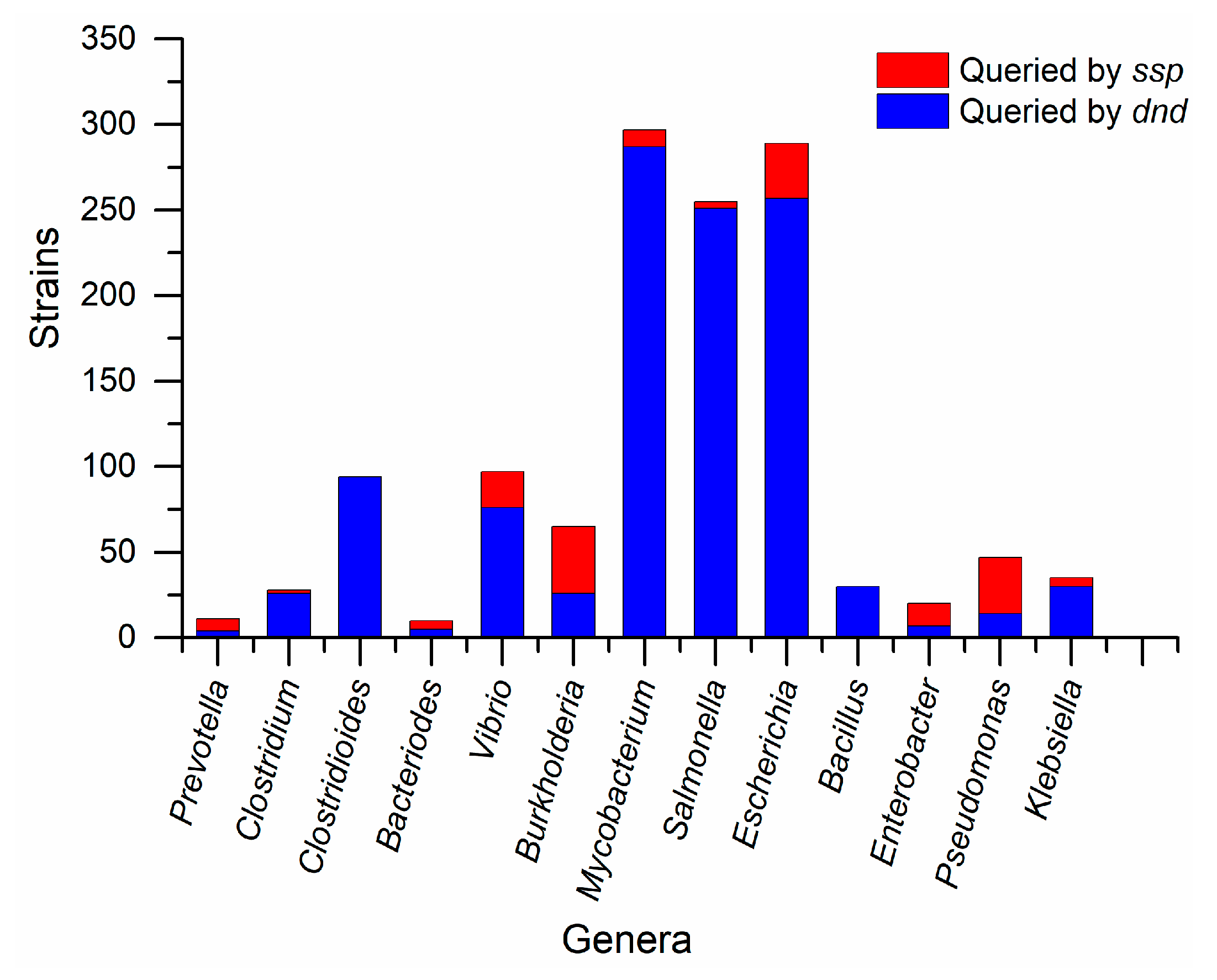
3.1. PT Genes Are Widely Distributed in the Human Microbiome

We searched for PT-modifying genes in over 40,000 bacterial genomes under the human microbiome category from websites (https://www.ncbi.nlm.nih.gov/genome/, https://www.ebi.ac.uk/, and https://www.hmpdacc.org/). Multiple sequence alignments were performed using the genomes for strains harboring PT-modifying genes. A total of 2621 strains harboring PT-modifying genes (dataset) were obtained from the results queried by the PT-modifying genes. The amino acid sequences of DndC/SspD were used to construct a phylogenetic tree (Figure 1). The strains in the same genus are clustered into different branches according to sequence similarity (Figure 1). The bubbles represent collapsed clades and each bubble clustered strains with sequence identities over 60%. These bubbles were used to show the distribution of homogeneous sequences due to the strong correlation between modifying types and amino acid sequences [17]. The PT-linked dinucleotides of GpsA/GpsT, GpsG, and CpsC were separated on relatively independent clades with sequence identities over 30%. The boundaries of these clades were defined by the brackets (Figure 1). It was reported that strains with the same PT-linked dinucleotides were presumed to be closer on the phylogenetic tree [11]. Intriguingly, over 60% of the strains were clustered on distinct branches far from the clades marked with PT-linked dinucleotides (Figure 1). Thus, we speculated that unknown modification patterns could exist in the human microbiome.
We speculated that PT-harboring species spread across the entire human body due to the distribution of PT genes in diverse species (Figure 1). Thus, we collected bacterial genomes directly from distinct anatomical sites on the human body (https://www.hmpdacc.org/) to perform multiple sequence alignments. Although the total number of genomes is limited, we found diverse species harboring PT in four body parts (Table 1). Notably, there are species differences in the distribution of dnd genes and ssp genes (Table 1). Intriguingly, the PT-modifying gene-harboring strains in the genus Prevotella were found only in the oral and urogenital systems (Table 1), despite the relatively abundant distribution of Prevotella species in the human intestinal tract [19]. All the data above (Figure 1, Table 1) confirm that strains harboring PT-modifying genes are widely distributed in the human body.

3.2. Species Difference of PT-Related Genes

It has been reported that PT gene clusters can be spread by horizontal transfer [17,20], and we find that the distribution of PT-modifying genes varied among genera. For instance, over 90% of the Mycobacterium strains harbor dnd genes rather than ssp genes (Figure 2). On the other hand, over 60% of the Pseudomonas strains harbor ssp genes rather than dnd genes (Figure 2). Notably, dnd genes are widely distributed in Mycobacterium, Salmonella, and Escherichia rather than the other genera (Figure 2). To further investigate the role of PT, we performed a multiple sequence alignment for dndF-H and PbeA-D. Notably, the dndF-H gene cassette was “missing” in 90% of strains of Mycobacterium, which means that most Mycobacterium strains lack the R component (Figure 3). On the contrary, the dndF-H gene cassette was widely distributed in Escherichia strains. Meanwhile, the pbeA-D gene cassette existed in 10% of the Mycobacterium and Vibrio, which suggests that most human microorganisms lack the antiviral component (Figure 3). We speculated that PT may play other roles in these species without both dndF-H and PbeA-D. It seems that there exist undiscovered PT-dependent genes in their genomes. On the contrary, over 60% of the Bacillus strains harbor the R component without any modifying genes (Figure 3), which means these strains harbor undiscovered modifying systems to protect their DNA against nickase [3].

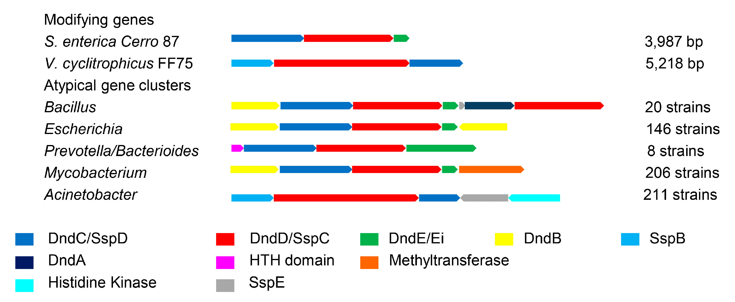
3.3. Atypical PT Gene Clusters of Different Genera

The organization of the PT-related genes varied among the genera (Figure 4). In 146 strains of E. coli and 20 strains of Bacillus cereus, the genes surrounding the PT-modifying genes encoded homologs of dndA (cysteine desulfurase) [5], dndB (negative transcriptional regulator) [18], and dndD (possibly related to DNA structure alteration) [21,22]. For instance, the identities are 23.98% between DndB and its homolog from E. coli Ecol_316 (NCBI: CP018957). Genomic islands may facilitate the horizontal transfer of PT systems and result in multiple copies of genes for E. coli and B. cereus [23]. Several strains from Bacteroidales (Prevotella and Bacteroides) showed a rare dndEi in PT-modifying genes that have an additional DNA helicase domain compared to canonical DndE [24]. In 211 Acinetobacter strains, the histidine kinase genes are adjacent to the SspE, which is the R component of the ssp system [4]. We speculate that the histidine kinase genes (Figure 4) involve in a two-component system to regulate the expression of SspE. Moreover, it was previously reported that there are complex interactions between DNA methylation and PT [25]. Intriguingly, 72% of PT-modifying genes in Mycobacterium strains lack both dndF-H and pbeA-D (Figure 3) and are adjacent to methyltransferase genes (Figure 4). This suggests that there is a shared mechanism linking the R system between DNA methylation and the PT [25] in Mycobacterium strains. These results indicate that PT plays various roles in different species.

3.4. Detection of PT-Linked Dinucleotides in Human Fecal DNA

For DNA methylation, restriction-modification systems are classified into four types depending on multiple factors, including subunits and target motifs [26]. In bacteria, the sequence specificity of DNA methylation depends on the target recognition domains (TRDs) of the modifying complex, and the variation in the TRDs impacts the sequence specificity upon modification [27]. For PT, although the TRDs have not been demonstrated, there is a strong correlation between the PT sequence contexts and modifying complex sequences [17]. Based on the variation in the dndC/sspD genes (Figure S1) and the phylogenic distribution of PT-linked dinucleotides (Figure 1), it remains a possibility that there are undiscovered modifying patterns, including different motifs and PT-linked dinucleotides. Exploiting the fact that most strains possessing PT-modifying genes were assigned to the gut microbiome (Table 1), we initiated a search for new PT consensus sequences by analyzing the diversity of PT-linked dinucleotides in human fecal DNA. Notably, six new PT-linked dinucleotides (CpsG, CpsT, ApsG, TpsG, GpsC, ApsT) were detected from human fecal DNA (Table 2, Figure S2). Additionally, two PT nucleotides (CpsA, TpsA) previously discovered in mutants were detected, which were previously discovered in E. coli DH10B expressing PT-modifying genes from S. enterica 87 [17].

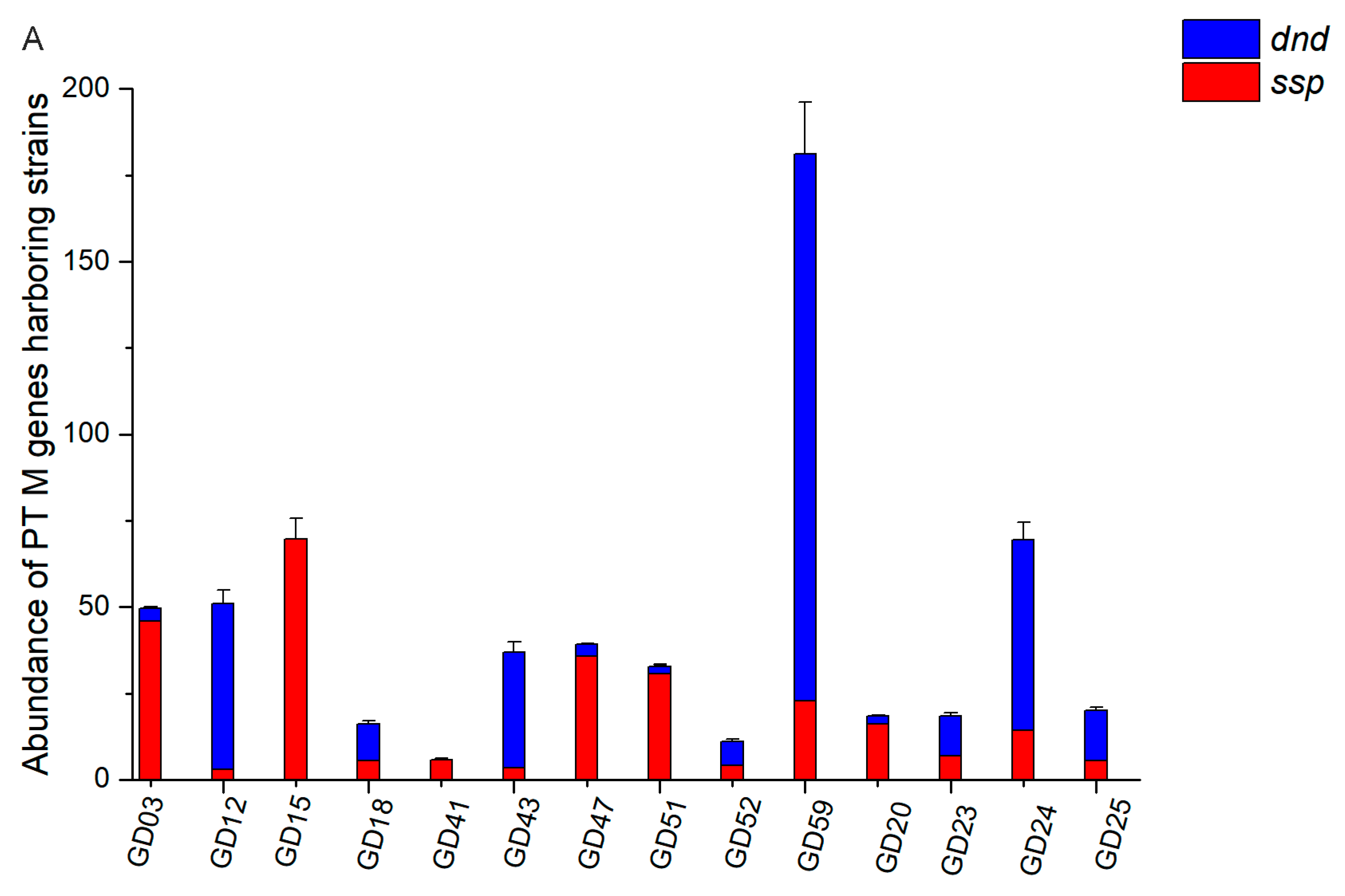
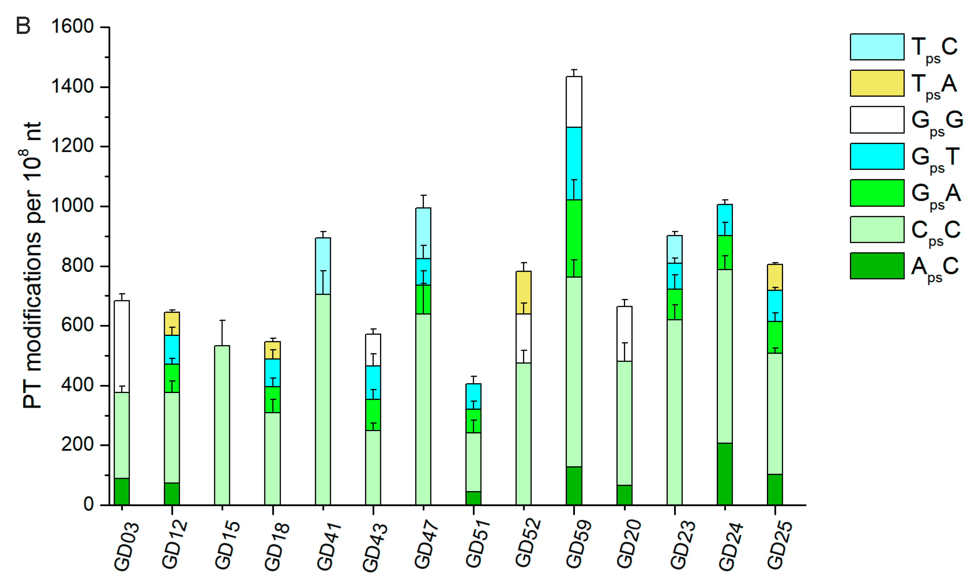
3.5. The Abundance of PT Modifications Varied Among Individuals

Although we could detect PT-linked dinucleotides in the fecal DNA, the abundance of PT genes and the modification types remain unknown in the gut microbiome without quantitative analysis. Meanwhile, we speculated that the distribution of the dnd/ssp genes and modification patterns varied among individuals. Thus, we performed a quantitative analysis of the PT genes and modifications in human fecal DNA. Based on the metagenomic sequencing performed on 109 human fecal DNA samples, a gene catalog with ~ 2 million non-redundant human gut microbial genes was constructed [15]. By BLAST alignment, 209 potential PT genes with at least 30% identity and 80% coverage compared with the PT references were identified. We were able to assemble 118 high-quality draft genomes of prevalent gut bacteria from these metagenomic data. Among these bacterial genomes, nine harbored PT genes: Bacteroides plebeius CAG00296, Bacteroidesplebeius CAG0079, Klebsiella sp. CAG00146, and Faecalibacterium prausnitzii CAG00158 were queried by dnd genes, while Bacteroides sp. CAG00020, Eubacterium ventriosu CAG00166, Clostridium bolteae CAG00012, Clostridiales bacterium CAG00239, and Clostridiales bacterium CAG00048 were queried by ssp genes. To further investigate the PT landscapes in the human gut microbiome, we analyzed the strains harboring PT-modifying genes (dataset) in the 14 individuals (Figure 5A). Then, we quantified the PT-linked dinucleotides in these fecal samples and found seven different PT-dinucleotides (Figure 5B).

4. Discussion

Over 80% of the strains harboring PT-modifying genes could be assigned to Actinobacteria, Proteobacteria, and Bacteroidetes [11]. Notably, our survey in the human microbiome shows that PT-modifying genes are distributed in these phyla (Figure 1). The phylogenic tree (Figure 1) was used to demonstrate the distribution of homogeneous sequences rather than the genera distribution, though it exhibits the horizontal transfer of PT-modifying genes, which is consistent with the previous conclusions [28]. Although we could not demonstrate DndC/SspD-containing TRDs, the DndC/SspD may play the role of site recognition according to their function of inserting sulfur into the sugar-phosphate backbone of DNA [4]. Thus, we built the tree by DndC/SspD and assumed that the human microbiome contained new PT-linked dinucleotides due to the phylogenic distribution of GpsA/GpsT, GpsG, and GpsC (Figure 1). Fortunately, we discovered new PTs in human fecal DNA. We speculated that 5 μg of fecal DNA was not enough to detect multiple PT-linked dinucleotides. Thus, we tested 30 μg of fecal DNA with a control group of 1493. However, we found no more new PT-linked dinucleotides compared to 5 μg of fecal DNA. Additionally, the variety of PT-linked dinucleotides varied among the fecal DNA samples, which suggested that the PT-containing strains carried by each person could be classified through metadata. As shown in Table 2, z1 and z3 both carried GpsT, assigned to species queried by dnd genes (Figure 1); z2 and z3 both carried CpsC and ApsC, assigned to species queried by ssp genes (Figure 1); z2 and z3 both carried ApsT, assigned to unknown species. The qualitation and quantification of PT-linked dinucleotides confirmed the existence of PT in the human gut microbiome with a strong correlation between the PT-modifying gene abundance and the modified dinucleotide yield. Thus, the abundance of PT modifications varied among individuals due to the different quantities of PT-harboring bacteria.
Intriguingly, our data showed that the PT-modifying genes seemed widely distributed in different pathogens or opportunistic pathogens, such as the strains that belonged to A. baumannii, P. aeruginosa, C. difficile, and M. abscessus (Dataset). The different bioinformatic approaches including this work confirmed that PT-modifying genes widely spread in human microbiome-associated strains, especially pathogenic candidates [11,20,23]. The previous study reported that nearly half of the US clinical isolates of M. abscessus exhibited the PT phenotype during pulsed field gel electrophoresis [29]. It was also reported that PT-modifying genes were frequently found in pathogenic E. coli [28]. One of the possible causes is that PT modifications were presumed to enhance the antioxidative ability of these bacteria against environmental stress [14,30]. In contrast, halogens could induce PT-linked DNA breaks, which implicated that PT reduced the fitness for bacterial pathogens during human infections [12]. Thus, it remains a possibility that the microenvironment or drugs may intervene in the abundance of bacteria with PT in the human microbiome, while there are few related reports. Additionally, there are few reports about how PT-modifying genes spread in human pathogens or opportunistic pathogens.

5. Conclusions

PT confers bacteria with multiple physiological functions, making them a specific microbial population sharing unusual characteristics [10,11,12]. However, our understanding of PT-containing bacteria tends to be restricted to isolated strains rather than a community in the ecosystem. This study, to our knowledge, is the first time to systematically investigate the wide distribution of PT-containing microbes in the human microbiome. Our results firstly reveal the landscape of PT modifications in the gut microbiome. This work will guide future research on the transmission of PT among microbial communities in the micro-ecosystem of the human body. With the rapid growth of the human microbiome database, more PT-containing bacteria could be found to analyze the routine of PT transmission and discover the unknown motive force behind this phenomenon with effects on human health.

Supplementary Materials

The following are available online at https://www.mdpi.com/2218-273X/10/8/1175/s1, Supplementary data and Datasets.

Author Contributions

Y.S. and G.W. performed the computational analysis. Y.S. and B.C. performed the detection of PT-linked dinucleotides. X.P. and Z.D. designed and supervised the collection of fecal samples from healthy individuals and patients. Y.S., G.W., C.Z., P.C.D., L.K., and D.Y. wrote the manuscript. All authors have read and agreed to the published version of the manuscript.

Funding

This work was supported by grants from the National Natural Science Foundation of China (31630002, 31700029, 31770038 and 21661140002); the National Key R & D Program of China (2018YFA0900400) from the Ministry of Science and Technology; Shanghai Pujiang Program from the Shanghai Municipal Council of Science and Technology (12PJD021); the National Science Foundation of the United States of America (CHE-1709364 to P. C. D.); and the National Science Foundation of Singapore under the Singapore-MIT Alliance for Research and Technology Programme (P.C.D.).

Acknowledgments

In memory of Liping Zhao from Shanghai Jiao Tong University, who provided experimental material and device.

Conflicts of Interest

The authors declare that they have no competing interests.

References

  1. Wang, L.; Chen, S.; Xu, T.; Taghizadeh, K.; Wishnok, J.S.; Zhou, X.; You, D.; Deng, Z.; Dedon, P.C. Phosphorothioation of DNA in bacteria by dnd genes. Nat. Chem. Biol. 2007, 3, 709–710. [Google Scholar] [CrossRef]
  2. Zhou, X.; He, X.; Liang, J.; Li, A.; Xu, T.; Kieser, T.; Helmann, J.D.; Deng, Z. A novel DNA modification by sulphur. Mol. Microbiol. 2005, 57, 1428–1438. [Google Scholar] [CrossRef] [PubMed]
  3. Cao, B.; Chen, C.; DeMott, M.S.; Cheng, Q.; Clark, T.A.; Xiong, X.; Zheng, X.; Butty, V.; Levine, S.S.; Yuan, G.; et al. Genomic mapping of phosphorothioates reveals partial modification of short consensus sequences. Nat. Commun. 2014, 5, 3951. [Google Scholar] [CrossRef] [PubMed] [Green Version]
  4. Xiong, X.; Wu, G.; Wei, Y.; Liu, L.; Zhang, Y.; Su, R.; Jiang, X.; Li, M.; Gao, H.; Tian, X.; et al. SspABCD-SspE is a phosphorothioation-sensing bacterial defence system with broad anti-phage activities. Nat. Microbiol. 2020, 5, 917–928. [Google Scholar] [CrossRef] [PubMed]
  5. You, D.; Wang, L.; Yao, F.; Zhou, X.; Deng, Z. A novel DNA modification by sulfur: DndA is a NifS-like cysteine desulfurase capable of assembling DndC as an iron-sulfur cluster protein in Streptomyces lividans. Biochemistry 2007, 46, 6126–6133. [Google Scholar] [CrossRef] [PubMed]
  6. An, X.; Xiong, W.; Yang, Y.; Li, F.; Zhou, X.; Wang, Z.; Deng, Z.; Liang, J. A novel target of IscS in Escherichia coli: Participating in DNA phosphorothioation. PLoS ONE 2012, 7, e51265. [Google Scholar] [CrossRef] [Green Version]
  7. Pu, T.; Mei, Z.; Zhang, W.; Liang, W.J.; Zhou, X.; Liang, J.; Deng, Z.; Wang, Z. An in vitro DNA phosphorothioate modification reaction. Mol. Microbiol. 2019, 113, 452–463. [Google Scholar] [CrossRef]
  8. Li, J.; Chen, Y.; Zheng, T.; Kong, L.; Zhu, S.; Sun, Y.; Deng, Z.; Yang, L.; You, D. Quantitative mapping of DNA phosphorothioatome reveals phosphorothioate heterogeneity of low modification frequency. PLoS Genet. 2019, 15, e1008026. [Google Scholar] [CrossRef]
  9. Xiong, L.; Liu, S.; Chen, S.; Xiao, Y.; Zhu, B.; Gao, Y.; Zhang, Y.; Chen, B.; Luo, J.; Deng, Z.; et al. A new type of DNA phosphorothioation-based antiviral system in archaea. Nat. Commun. 2019, 10, 1688. [Google Scholar] [CrossRef]
  10. Xu, T.; Yao, F.; Zhou, X.; Deng, Z.; You, D. A novel host-specific restriction system associated with DNA backbone S-modification in Salmonella. Nucleic Acids Res. 2010, 38, 7133–7141. [Google Scholar] [CrossRef] [Green Version]
  11. Tong, T.; Chen, S.; Wang, L.; Tang, Y.; Ryu, J.Y.; Jiang, S.; Wu, X.; Chen, C.; Luo, J.; Deng, Z.; et al. Occurrence, evolution, and functions of DNA phosphorothioate epigenetics in bacteria. Proc. Natl. Acad. Sci. USA 2018, 115, E2988–E2996. [Google Scholar] [CrossRef] [PubMed] [Green Version]
  12. Kellner, S.; DeMott, M.S.; Cheng, C.P.; Russell, B.S.; Cao, B.; You, D.; Dedon, P.C. Oxidation of phosphorothioate DNA modifications leads to lethal genomic instability. Nat. Chem. Biol. 2017, 13, 888–894. [Google Scholar] [CrossRef] [PubMed]
  13. Medema, M.H.; Takano, E.; Breitling, R. Detecting sequence homology at the gene cluster level with MultiGeneBlast. Mol. Biol. Evol. 2013, 30, 1218–1223. [Google Scholar] [CrossRef] [PubMed]
  14. Dawid, S.; Barenkamp, S.J.; St Geme, J.W. Variation in expression of the Haemophilus influenzae HMW adhesins: A prokaryotic system reminiscent of eukaryotes. Proc. Natl. Acad. Sci. USA 1999, 96, 1077–1082. [Google Scholar] [CrossRef] [PubMed] [Green Version]
  15. Zhang, C.; Yin, A.; Li, H.; Wang, R.; Wu, G.; Shen, J.; Zhang, M.; Wang, L.; Hou, Y.; Ouyang, H.; et al. Dietary Modulation of Gut Microbiota Contributes to Alleviation of Both Genetic and Simple Obesity in Children. EBioMedicine 2015, 2, 968–984. [Google Scholar] [CrossRef] [PubMed]
  16. Nielsen, H.B.; Almeida, M.; Juncker, A.S.; Rasmussen, S.; Li, J.; Sunagawa, S.; Plichta, D.R.; Gautier, L.; Pedersen, A.G.; Le Chatelier, E.; et al. Identification and assembly of genomes and genetic elements in complex metagenomic samples without using reference genomes. Nat. Biotechnol. 2014, 32, 822–828. [Google Scholar] [CrossRef]
  17. Wang, L.; Chen, S.; Vergin, K.L.; Giovannoni, S.J.; Chan, S.W.; DeMott, M.S.; Taghizadeh, K.; Cordero, O.X.; Cutler, M.; Timberlake, S.; et al. DNA phosphorothioation is widespread and quantized in bacterial genomes. Proc. Natl. Acad. Sci. USA 2011, 108, 2963–2968. [Google Scholar] [CrossRef] [Green Version]
  18. Cheng, Q.; Cao, B.; Yao, F.; Li, J.; Deng, Z.; You, D. Regulation of DNA phosphorothioate modifications by the transcriptional regulator DptB in Salmonella. Mol. Microbiol. 2015, 97, 1186–1194. [Google Scholar] [CrossRef]
  19. Aagaard, K.; Petrosino, J.; Keitel, W.; Watson, M.; Katancik, J.; Garcia, N.; Patel, S.; Cutting, M.; Madden, T.; Hamilton, H.; et al. The Human Microbiome Project strategy for comprehensive sampling of the human microbiome and why it matters. FASEB J. 2013, 27, 1012–1022. [Google Scholar] [CrossRef]
  20. He, X.; Ou, H.Y.; Yu, Q.; Zhou, X.; Wu, J.; Liang, J.; Zhang, W.; Rajakumar, K.; Deng, Z. Analysis of a genomic island housing genes for DNA S-modification system in Streptomyces lividans 66 and its counterparts in other distantly related bacteria. Mol. Microbiol. 2007, 65, 1034–1048. [Google Scholar] [CrossRef] [Green Version]
  21. Yao, F.; Xu, T.; Zhou, X.; Deng, Z.; You, D. Functional analysis of spfD gene involved in DNA phosphorothioation in Pseudomonas fluorescens Pf0-1. FEBS Lett. 2009, 583, 729–733. [Google Scholar] [CrossRef] [PubMed] [Green Version]
  22. Cao, B.; Zheng, X.; Cheng, Q.; Yao, F.; Zheng, T.; Ramesh Babu, I.; Zhou, H.; Dedon, P.; You, D. In vitro analysis of phosphorothioate modification of DNA reveals substrate recognition by a multiprotein complex. Sci. Rep. 2015, 5, 12513. [Google Scholar] [CrossRef] [PubMed] [Green Version]
  23. Ou, H.Y.; He, X.; Shao, Y.; Tai, C.; Rajakumar, K.; Deng, Z. dndDB: A database focused on phosphorothioation of the DNA backbone. PLoS ONE 2009, 4, e5132. [Google Scholar] [CrossRef] [PubMed] [Green Version]
  24. Zheng, T.; Jiang, P.; Cao, B.; Cheng, Q.; Kong, L.; Zheng, X.; Hu, Q.; You, D. DndEi Exhibits Helicase Activity Essential for DNA Phosphorothioate Modification and ATPase Activity Strongly Stimulated by DNA Substrate with a GAAC/GTTC Motif. J. Biol. Chem. 2016, 291, 1492–1500. [Google Scholar] [CrossRef] [Green Version]
  25. Chen, C.; Wang, L.; Chen, S.; Wu, X.; Gu, M.; Chen, X.; Jiang, S.; Wang, Y.; Deng, Z.; Dedon, P.C.; et al. Convergence of DNA methylation and phosphorothioation epigenetics in bacterial genomes. Proc. Natl. Acad. Sci. USA 2017, 114, 4501–4506. [Google Scholar] [CrossRef] [PubMed] [Green Version]
  26. Murray, N.E. 2001 Fred Griffith review lecture. Immigration control of DNA in bacteria: Self versus non-self. Microbiology 2002, 148, 3–20. [Google Scholar] [CrossRef] [Green Version]
  27. Murray, N.E. Type I restriction systems: Sophisticated molecular machines (a legacy of Bertani and Weigle). Microbiol. Mol. Biol. Rev. 2000, 64, 412–434. [Google Scholar] [CrossRef] [Green Version]
  28. Ho, W.S.; Ou, H.Y.; Yeo, C.C.; Thong, K.L. The dnd operon for DNA phosphorothioation modification system in Escherichia coli is located in diverse genomic islands. BMC Genom. 2015, 16, 199. [Google Scholar] [CrossRef] [Green Version]
  29. Zhang, Y.; Yakrus, M.A.; Graviss, E.A.; Williams-Bouyer, N.; Turenne, C.; Kabani, A.; Wallace, R.J., Jr. Pulsed-field gel electrophoresis study of Mycobacterium abscessus isolates previously affected by DNA degradation. J. Clin. Microbiol. 2004, 42, 5582–5587. [Google Scholar] [CrossRef] [Green Version]
  30. Xie, X.; Liang, J.; Pu, T.; Xu, F.; Yao, F.; Yang, Y.; Zhao, Y.L.; You, D.; Zhou, X.; Deng, Z.; et al. Phosphorothioate DNA as an antioxidant in bacteria. Nucleic Acids Res. 2012, 40, 9115–9124. [Google Scholar] [CrossRef] [Green Version]
Figure 1. Phylogenetic distribution of the strains containing PT-modifying genes. The DndC/SspD in 2623 strains were created by MEGA7 with the maximum likelihood method with 500 bootstrap replications and was visualized using iTOL. V. cyclitrophicus FF75 contains CpsC, and P. fluorescens Pf0-1 contains GpsG. E. coli B7A and S. enterica serovar Cerro 87 both contain GpsA/GpsT.
Biomolecules 10 01175 g001
Figure 2. Distribution of PT-modifying gene cluster in different genera. Query genes were from S. enterica serovar Cerro 87 (CP008925: 3477655...3481641) and V. cyclitrophicus FF75 (NZ_ATLT01000001: 2194844...2200061), respectively.
Biomolecules 10 01175 g002
Figure 3. Distribution of PT-related gene cassettes in different genera. “R” means strains harboring dndF-H without PT-modifying genes. “M” means strains harboring PT-modifying genes without dndF-H. “RM” means strains harboring dndF-H and PT-modifying genes. “MA” means strains harboring PT-modifying genes and pbeA-D. “RMA” means strains harboring PT-modifying genes, dndF-H and pbeA-D.
Biomolecules 10 01175 g003
Figure 4. Atypical PT gene clusters in different genera. Genes are indicated as arrows and are colored based on their predicted function. DndC/SspD: ATP pyrophosphatase. DndD/SspC: ATPase. DndE/Ei: helicase. DndB: regulatory protein. SspB: nickase. DndA: cysteine desulfurase. SspE: nickase.
Biomolecules 10 01175 g004
Figure 5. (A)The abundance of strains harboring PT-modifying genes in the fecal DNA from 14 individuals. (B) Quantification of PT-linked dinucleotides in fecal DNA from 14 individuals. Data are shown as mean ± SD. “dnd” means strains harboring dnd genes. “ssp” means strains harboring ssp genes.
Biomolecules 10 01175 g005aBiomolecules 10 01175 g005b
Table 1. Strains containing PT-modifying genes from the human body.
Body PartsQueried by dnd GenesQueried by ssp Genes
Gut systemEscherichia coli MS 45-1Dysgonomonas mossii DSM 22836
Escherichia coli MS 117-3Alistipes indistinctus YIT 12060
Enterobacter cloacae NCTC 9394Prevotella HGA0225
Escherichia coli SE11Bacteroides dorei 5_1_36/D4
Lachnospiraceae bacterium 6_1_37FAABacteroides 1_1_14
Desulfovibrio piger ATCC 29098Roseburia inulinivorans DSM 16841
Helicobacter bilis ATCC 43879Roseburia intestinalis M50/1
Paraprevotella xylaniphila YIT 11841Roseburia intestinalis L1-82
Lachnospiraceae bacterium 2_1_58FAAEubacterium ventriosum ATCC 27560
Clostridium asparagiforme DSM 15981Eubacterium siraeum V10Sc8a
Peptoclostridium difficile 70-100-2010Megamonas funiformis YIT 11815
Bacteroides 2_1_33BMitsuokella multacida DSM 20544
Bacteroides xylanisolvens XB1AButyrivibrio fibrisolvens 16/4
Clostridium citroniae WAL-17108
Faecalibacterium prausnitzii M21/2
Fusobacterium necrophorum
funduliforme 1_1_36S
Oral systemNeisseria 020Prevotella salivae F0493
Neisseria subflava NJ9703 NPrevotella F0055
Neisseria bacilliformis ATCC BAA-1200Prevotella tannerae ATCC 51259
Eikenella corrodens ATCC 23834
Lachnospiraceae bacterium F0431
Selenomonas CM52
Urogenital systemPrevotella amnii CRIS 21A-APrevotella oralis ATCC 33269
Mycobacterium parascrofulaceum ATCC BAA-614Prevotella denticola CRIS 18C-A
Prevotella bivia JCVIHMP010
Skin Acinetobacter baumannii 6014059
Table 2. PT-linked dinucleotides in the human fecal DNA.
PT-Linked DinucleotidesPrecursor IonProduct Ionz1z2z31488148914941493
d(CpsG)573152Y Y N
d(CpsC)533112 YY YY
d(GpsG)613152 YY YY
d(CpsA)557136 Y *Y * Y *Y *
d(CpsT)548112N **N **YN **NY
d(ApsG)597136Y Y
d(TpsG)588152Y YY Y
d(GpsA)597136 Y Y
d(GpsC)573112 NY NY
d(GpsT)588152YNYYNY
d(ApsA)581136
d(TpsA)572136 Y Y N
d(ApsC)557112 Y *Y * Y *Y *
d(TpsC)548112 N **Y NY
d(ApsT)572136 YY YYN
d(TpsT)563127
d(GpsA) Sp597136
Blank means not detected; “Y” means sufficient levels to be quantified; “N” means close to detection limit; “*” means retention time slightly shifted from expected values; “**” means retention time shift.

