Article

Genetic Factors Associated with Increased Host Defense Antimicrobial Peptide Resistance in Sequence Type 5 Healthcare-Associated MRSA Clinical Isolates

Department of Animal Science and Technology, School of Bioresources and Bioscience, Chung-Ang University, Anseong 17546, Korea
*
Author to whom correspondence should be addressed.
Biomolecules 2020, 10(10), 1415; https://doi.org/10.3390/biom10101415
Submission received: 1 August 2020 / Revised: 28 September 2020 / Accepted: 6 October 2020 / Published: 7 October 2020
(This article belongs to the Section Molecular Genetics)

Abstract

:
Sequence type (ST) 72 methicillin-resistant Staphylococcus aureus with staphylococcal cassette chromosome mec (SCCmec) type IV (ST72-MRSA-IV) and ST5-MRSA-II are the most significant lineages found in community-associated (CA) and healthcare-associated (HA) environments in Korea, respectively. ST5 HA-MRSA-II tend to display enhanced resistance to host defense-cationic antimicrobial peptides (HD-CAPs) compared to ST72 CA-MRSA-IV and ST72 livestock-associated (LA)-MRSA-IV due to mechanisms involving a higher surface positive charge. Thus, the present study explored the genetic factors contributing to the enhanced HD-CAP resistance phenotype in ST5 MRSA strains. The ST5 HA-MRSA-II strains displayed higher levels of mprF and dltABCD expression compared to the ST72 CA-/LA-MRSA-IV strains. The increase in expression of mprF and dltABCD in ST5 HA-MRSA-II strains was correlated with dysregulation of the upstream transcriptional regulator, graRS. However, single nucleotide polymorphisms (SNPs) within mprF and graRS ORFs were not involved in the enhanced surface positive charge or the altered expression of mprF/dltABCD.

1. Introduction

Methicillin-resistant Staphylococcus aureus (MRSA) is a serious nosocomial pathogen that can lead to septicemia and death [1]. Besides healthcare-associated (HA)-MRSA infections, the incidence of human infections with community-associated (CA)-MRSA and livestock-associated (LA)-MRSA has also been increasing in recent years [2,3,4,5]. Clonal lineages of HA-, CA-, and LA-MRSA isolates and their distributions vary according to geographical region [4,5,6]. Sequence type (ST) 5 MRSA with staphylococcal cassette chromosome mec (SCCmec) type II (ST5-MRSA-II) and ST72-MRSA-IV represent the most significant HA- and CA-MRSA clones in Korea, respectively [6,7,8]. However, the differences in virulence factors critical for the clinical outcomes of infections caused by ST5-MRSA-II and ST72-MRSA-IV are largely unknown.
Host defense cationic antimicrobial peptides (HD-CAPs) play a critical role in host innate immune defense against bacterial infections including MRSA [9]. In a recent report from our laboratory, we showed that ST5 HA-MRSA II strains tended to be more resistant to HD-CAPs of different origins compared to ST72 CA-/LA-MRSA-IV strains [10]. In particular, ST5 HA-MRSA strains displayed higher levels of resistance to LL-37 (human cathelicidin), BMAP-28 (bovine myeloid antimicrobial peptide), and polymyxin B (bacterial antimicrobial peptide) than ST72 CA-/LA-MRSA-IV strains via mechanisms involving enhanced cell surface positive charge [10]. Over the past several years, the enhanced surface positive charge in S. aureus has been linked to (i) enhanced transcription of mprF, dltABCD, or both [11,12,13], (ii) single nucleotide polymorphisms (SNPs) within the mprF ORF (the gain-in-function mutations) [14,15,16,17], or (iii) perturbed upstream transcriptional regulation of mprF and dltABCD by the two-component regulatory system (TCRS), graRS [18,19,20].
Based on these prior findings, the present study aimed to define genetic factors associated with the enhanced HD-CAP resistance in ST5 HA-MRSA-II strains. Using the same 26 MRSA strains as those used in our previous study [10], we examined the transcriptional profiles of mprF, dltABCD, and graRS during the exponential- and stationary-growth phases. Next, sequences of mprF ORFs were analyzed to identify SNPs linked to the enhanced surface positive charge in ST5 HA-MRSA strains. Moreover, correlations between SNPs within graRS ORFs and the dysregulation of mprF/dltABCD were determined.

2. Materials and Methods

2.1. S. aureus Strains and Culture

The MRSA strains used in this investigation are listed in Table 1. We used the 26 MRSA strains previously described by Kang et al.: 8 ST5 HA-MRSA-II strains [10], 11 ST72 CA-MRSA IV strains [21], and 7 ST72 LA-MRSA IV strains [22].
All 26 MRSA strains were cultured in either Mueller–Hinton broth (MHB; Difco Laboratories, Detroit, MI, USA) or Tryptic Soy broth (TSB; Difco Laboratories) for each experiment. All MRSA cultures were incubated in 500 mL Erlenmeyer flasks at 37 °C with shaking (200 rpm) in less than 15% of the flask’s volume for optimal aeration.

2.2. Sequencing and Cloning of mprF and graRS

Genomic DNA samples were prepared from the MRSA strains using a method described previously [23]. For sequence analyses, the mprF and graRS ORFs were amplified through PCR using mprF- or graRS-specific primer pairs as described previously [12,20]. The PCR-amplified mprF and graRS ORFs from 26 MRSA strains were sequenced at Cosmo Genetech, Seoul, Korea. SNPs within the mprF and graRS ORFs were identified using a multiple sequence alignment tool on the BoxShade server (embnet.vital-it.ch/software/BOX_form.html).
To assess the role of the SNPs identified within mprF ORFs in conferring a positive charge to the cell surface, mutated or nonmutated mprF genes were amplified from each MRSA strain (HA7 or CA7 strains) and then expressed in the S. aureus Newman ∆mprF mutant strain [24]. Complementation of the mprF genes in Newman ∆mprF was achieved using the previously described shuttle vector, pRB474 [16,25]. Similarly, the role of SNPs identified within graS ORFs in the regulation of mprF and dltABCD expression was evaluated by expressing the mutated or nonmutated graS genes in the S. aureus MW2 ∆graS mutant strain [20]. The graS genes from MRSA strains with or without graS SNPs were PCR amplified and then ligated into pRB474 using the BamHI and SpHI sites.
Plasmid DNA was prepared from Escherichia coli and S. aureus strains using the Wizard Plus SV Miniprep kit (Promega, Madison, WI, USA). Transformation of the plasmid constructs into E. coli DH5α or S. aureus strains was accomplished as described previously [26,27].

2.3. Quantification of Ttranscriptional Expression by RT-qPCR

To assess the expression levels of mprF, dltABCD, and graRS during different growth phases, RNA samples were prepared from broth cultures of the 26 MRSA strains in the exponential and stationary growth phases. For RNA isolation, overnight cultures of the MRSA strains were used to inoculate 50 mL of fresh TSB to an OD600 of 0.05. MRSA cell pellets were then collected at either the exponential (3 h) or stationary (12 h) growth phase of the cultures. Total cellular RNA samples were prepared using the FastPrep FP120 Cell Disrupter instrument (Bio101, Vista, CA, USA) and the RNeasy kit (Qiagen, Valencia, CA, USA) as described in earlier study [28].
For quantitative real-time PCR (RT-qPCR) assays, 3 µg of each RNA sample was reverse transcribed using the Superscript III first-strand synthesis system (Thermo Fisher Scientific), as recommended by the manufacturer. Quantification of mprF, dltA, and graRS cDNA levels was carried out according to the protocol described by the manufacturer of the Power SYBR green master mix kit in a LineGene 9600 Plus system (Bioer Technology, Hangzhou, China). The transcripts of mprF, dltABCD, graRS, and gyrB were amplified using their respective gene-specific primers, as explained previously [18,29,30]. Fold changes in the transcription levels of all genes were quantified in relation to the transcription level of gyrB. At least two independent experiments were carried out for each RNA sample

2.4. Net Cell Surface Charge in MRSA Strains

The binding of cytochrome c (Sigma-Aldrich, St. Louis, MO, USA) to the MRSA cell surface was measured to determine the relative surface positive charge using the spectrophotometric method as described in prior publications [16,31]. Three independent cytochrome c binding assays were carried out on separate days.

2.5. Statistical Analyses

The Kruskal–Wallis ANOVA test with the Tukey post hoc correlation was performed for multiple comparisons (IBM SPSS Statistics 23, Chicago, IL, USA). Statistical significance was considered at p < 0.05.

3. Results

3.1. SNPs within mprF and graRS ORFs

All 26 MRSA strains were subjected to sequencing analyses for mprF and graRS genes. As presented in Table 1, sequencing analyses of the mprF ORFs revealed that all the 18 ST72 MRSA-IV strains (7 ST72 LA-MRSA-IV and 11 ST72 CA-MRSA-IV strains) had two nonsynonymous mutations (ATT→ATG at position 1125; ATT→ACT at position 1382) within mprF ORFs distinct from those of the ST5 HA-MRSA-II strains, resulting in I375M and I461T substitutions, respectively (I375M, isoleucine to methionine at position 375; I461T, isoleucine to threonine at position 461). Similarly, all of the eight ST5 HA-MRSA-II strains exhibited a distinctive I224T amino acid substitution in the graS ORF compared to the 18 ST72 MRSA-IV strains. In addition to the two most common mprF mutations in ST72 MRSA strains (I375M and I461T), the amino acid substitutions F550C and T666I were identified in two ST72 MRSA-IV strains (LA2 and CA1 strains). Although a few MRSA strains had amino acid substitutions in graR ORF, such as K100N, E15K, and V87G, the mutations resulting in such substitutions were observed in only one strain each of ST5 HA-MRSA and ST72 CA-MRSA.

3.2. Effect of mprF SNPs on The Net Cell Surface Positive Charge

A number of previous publications have demonstrated that the presence of SNPs within the mprF ORF is often correlated with an increase in net cell surface positive charge as a result of either enhanced synthesis or outer membrane translocation of lysyl-phosphatidylglycerol (L-PG) in S. aureus [12,13,17,32,33,34,35].
To determine whether the two amino acid substitutions (I375M and I461T) observed in ST72 MRSA strains contribute to the cell surface positive charge phenotype, Newman ∆mprF strains with plasmids expressing one of two different forms of mprF genes (mprFHA7 or mprFCA7) were subjected to cytochrome c binding analysis. As shown in Figure 1, the two ∆mprF strains expressing mprF genes either with or without the I375M and I461T substitutions did not show any difference in bacterial cell surface charge, indicating that these two amino acid substitutions in the ST72 MRSA strains are not involved in surface charge regulation in S. aureus.

3.3. Expression of mprF, dltA, and graS in MRSA Strains

As shown in Figure 2a,b, RT-qPCR analyses revealed that ST5 HA-MRSA strains had significantly higher levels of mprF transcription than the ST72 CA-MRSA and ST72 LA-MRSA strains either in the exponential or stationary growth phase (p < 0.05). Moreover, since dltABCD genes also contribute to the net surface positive charge in S. aureus [12,29], the transcriptional profiles of dltA were determined in the 26 MRSA strains assessed herein. As presented in Figure 3a,b, ST5 HA-MRSA strains exhibited significantly enhanced dltA expression levels during the exponential growth phase compared to the ST72 LA-MRSA and ST72 CA-MRSA strains (p < 0.01). The difference in dltA expression between the ST5 HA-MRSA and ST72 CA-MRSA strains continued to the stationary growth phase (p < 0.01) (Table 2).
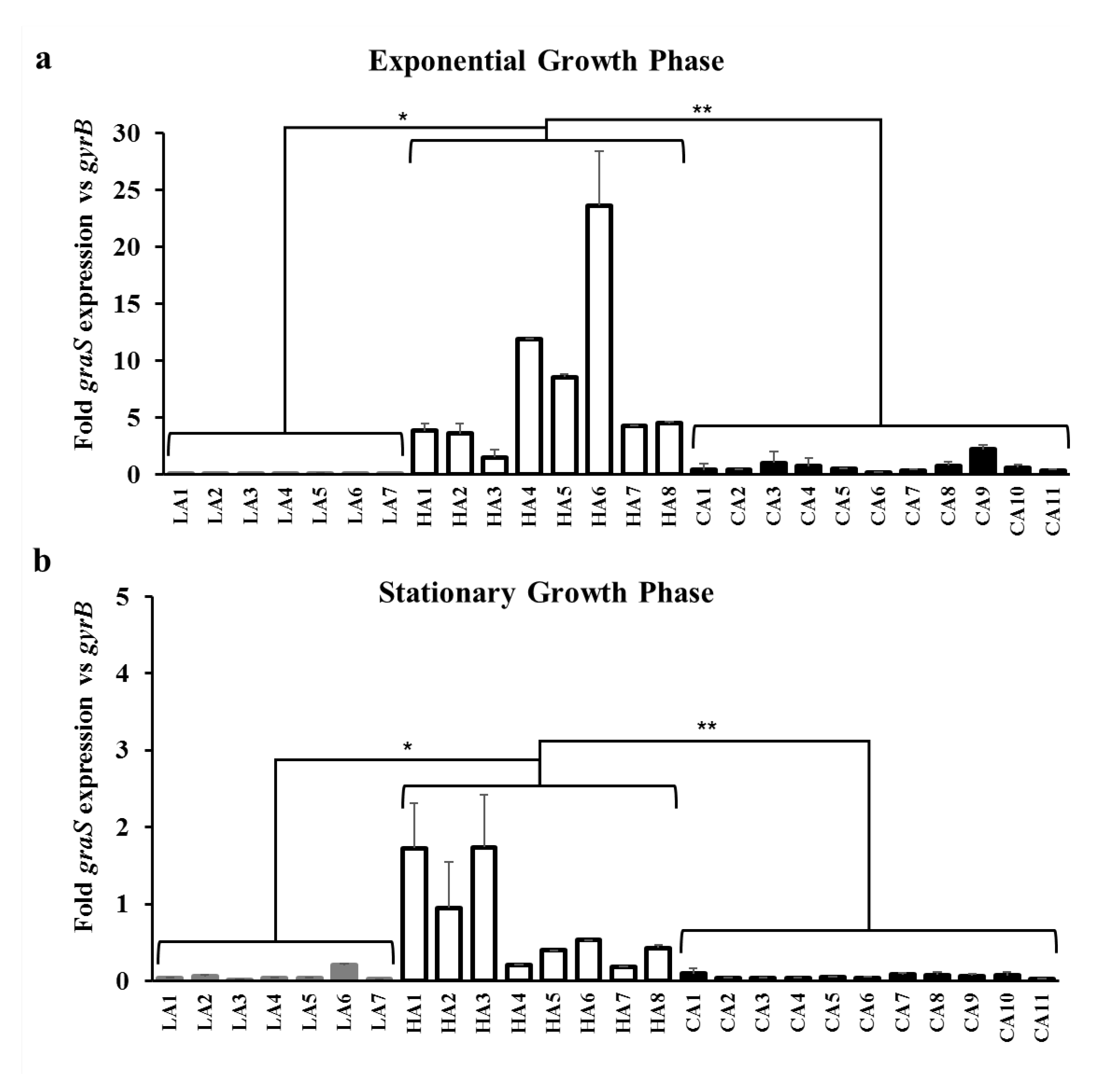
Since the ST5 HA-MRSA strains tended to transcribe higher levels of mprF and dltA than the ST72 LA-/CA-MRSA strains, the expression levels of graRS, the transcriptional regulator of mprF and dltABCD [19], were determined in the 26 MRSA strains. The ST5 HA-MRSA strains exhibited significantly higher expression levels of graS during both exponential and stationary growth phases compared to the two ST72 MRSA strain groups (Figure 4a,b).

3.4. Effect of GraS SNPs on the Regulation of mprF and dltABCD Transcription

The prototypical two-component regulatory system (TCRS) GraRS up-regulates the transcription of mprF and dltABCD, which encode protein products that modify the net surface charge in S. aureus [11,18,20,36,37]. To assess whether the amino acid substitution I224T within graS genes in ST5 HA-MRSA strains affects the expression of downstream target genes such as mprF and dltA, S. aureus MW2 ∆graS strains expressing either graSCA7 or graSHA7 in trans were subjected to RT-qPCR analysis. As presented in Figure 5, the two strains displayed almost identical levels of graS, mprF, and dltA transcription, suggesting that I224T substitution is not involved in the dysregulation of graS, mprF, and dltA in ST5 HA-MRSA strains.

4. Discussion

Previously, we have shown that ST5 HA-MRSA-II isolates, the most significant HA-MRSA in Korea, were more resistant to HD-CAPs of human, bovine, and prokaryotic origins compared with human- and animal-derived ST72 MRSA-IV isolates [10]. The observed enhancement in resistance to bactericidal action of HD-CAP in ST5 HA-MRSA was associated with an enhanced surface positive charge [10]. Thus, the current study was designed to explore the genetic factors responsible for the enhanced surface positive charge in ST5 HA-MRSA strains compared to those in ST72 CA-/LA-MRSA-IV strains.
Sequencing analyses of mprF ORFs in the 26 MRSA strains revealed that there are genotype specific SNPs within these gene loci (Table 1). Previous studies have demonstrated that SNPs within specific domains of MprF are often associated with increased synthesis or translocation of positively charged L-PG, and thus contributes to HD-CAP resistance via charge repulsion mechanisms [32,34,35,38]. The two mprF SNPs (I375M and I461T) identified in the ST72 CA-/LA-MRSA strains in this study were reported in a previous publication in a daptomycin-nonsusceptible (DAP-NS) ST72 MRSA strain that displayed an enhanced surface positive charge versus DAP-susceptible MRSA strains [39]. However, in a recent publication [10], the 18 ST72 MRSA-IV strains with the I375M and I461T mutations were shown to have reduced levels of surface positive charge compared to those of the ST5 HA-MRSA strains. Therefore, to determine whether the I375M and I461T mutations in the mprF ORF are directly linked to the enhanced surface positive charge, and thus the HD-CAP resistance phenotype of ST5 HA-MRSA strains, the Newman ∆mprF strains expressing the mprF genes from ST72 MRSA (mprFI375M+I461T) or ST5 MRSA (mprF) strains were subjected to cytochrome c binding assays. As presented in Figure 1, complementation of the Newman ∆mprF strain with mprFI375M+I461T did not result in an enhanced surface positive charge versus the Newman ∆mprF strain expressing a nonmutated form of mprF, suggesting that the two SNPs (I375M and I461T) are likely not involved in the previously observed increase in the surface positive charge of the ST5 HA-MRSA II strains [10,39]. Bayer et al. also reported frequent SNPs within mprF ORFs among clinical DAP-NS MRSA strains, but only a limited number of SNPs within the hot spot loci of mprF were shown to be linked to the gain-of-function phenotype that resulted in enhanced surface positive charge [40]. These results indicate that the heterogeneity within mprF ORFs is not associated with the higher levels of surface positive charge and enhanced resistance to HD-CAP in the ST5 HA-MRSA-II strains compared to those of the ST72 CA-/LA-MRSA-IV strains.
Besides the gain-of-function mutations in mprF ORFs [12,13,14,17,32,34,35], it has been previously reported that the increased transcription of mprF, dltABCD, or both, resulted in altered surface positive charge in DAP-NS and HD-CAP resistant S. aureus [11,36]. Along with MprF, dltABCD gene products contribute to the accumulation of net surface positive charge in S. aureus by increasing D-alanylation of teichoic acids in the peptidoglycan cell wall [12,41]. The graRS TCRS also contributes to surface positive charge regulation in staphylococci by inducing the transcription of target genes, such as mprF, dltABCD, and vraFG [11,18,36,37]. As shown in Figure 2 and Figure 3 and Table 2, ST5 HA-MRSA-II strains displayed significantly higher levels of mprF and dltA expression during exponential and stationary growth phases compared with those of ST72 CA-/LA-MRSA-II strains. In combination with previous results [10], data obtained in the present study suggest that the higher level of HD-CAP resistance in ST5 HA-MRSA-II compared to that in ST72 MRSA strains due to enhanced surface positive charge is correlated with the increased transcription of mprF and dltABCD. Since similar overall profiles of mprF and dltA transcription in the 26 MRSA strains were observed (Figure 2 and Figure 3), the co-regulation of these two genetic factors by the upstream transcriptional regulator graRS was determined. As presented in Figure 4 and Table 2, the ST5 HA-MRSA-II strains exhibited a significantly enhanced transcription of graS throughout the two growth phases, correlating well with the enhanced expression of mprF and dltABCD in ST5 HA-MRSA-II strains compared to that in the two groups of ST72 MRSA-IV strains. Furthermore, as shown in Table 1, sequencing analyses of graRS ORFs revealed that all ST5 HA-MRSA-II strains carried a SNP in the graS ORF, which caused an amino acid substitution of I224T distinct from those in ST72 MRSA strains. However, complementation of the MW2 ∆graS strain either with graS from the CA7 or HA7 strains failed to show any difference in mprF, dltA, and graS expression (Figure 5), suggesting that the I224T mutation in ST5 HA-MRSA strains was not involved in the dysregulation of mprF and dltABCD expression. Sequence analyses of graRS promoter regions in the HA1, HA6, LA1, and CA1 strains also revealed that all the four strains had identical promoter sequences (data not shown), demonstrating that the dysregulation of graS expression in ST5 HA-MRSA-II strains was not resulted from mutations in their promoter sequences.
It should also be noted that S. aureus generally depends on three major events to modulate its relatively more positive in charge surface: (i) D-alanylation of wall teichoic acids via the dltABCD genes, (ii) amidation of glutamine residues in the peptidoglycan cell wall via the murT and gatD genes (Munch et al., PLoS Pathogen, 2012; Figueiredo, PLoS Pathogen, 2112), and (iii) lysinylation of the PG to L-PG via the mprF in the cell membrane. In addition, a certain level of the N-acetylmuramic acid in the cell wall becomes O-acetylated on position C6 by the oatA gene product, which could affect hydrophobicity and surface charge in S. aureus.

5. Conclusions

To conclude, in combination with previously published data [10], which demonstrated that the ST5 HA-MRSA isolates were more resistant to HD-CAPs of various origins than ST72 CA-MRSA isolates via increased positive charge in cell envelopes, our current data suggest that (i) the enhanced HD-CAP resistance resulting from the enhanced net surface positive charge in ST5 HA-MRSA-II strains compared to that in the ST72 CA-/LA-MRSA-II strains was associated with the dysregulation of mprF and dltABCD expression, (ii) the altered expression of mprF and dltABCD in ST5 HA-MRSA-II strains was caused by the dysregulation of the upstream transcriptional regulator graRS TCRS, (iii) the two mprF SNPs (I375M and I461T) identified in ST72-MRSA strains did not affect net surface positive charge, and (iv) the I224T mutation in the graS ORF was not involved in the dysregulation of mprF and dltABCD by altered graRS expression in ST5 HA-MRSA-II strains.

Author Contributions

Conceptualization, S.-J.Y.; methodology, S.-J.Y.; formal analysis, S.-J.Y., G.Y.L., and K.-M.K.; investigation, G.Y.L. and K.-M.K.; writing—original draft preparation, S.-J.Y., G.Y.L., and K.-M.K.; writing—review and editing, S.-J.Y.; visualization, K.-M.K.; supervision, S.-J.Y.; project administration, K.-M.K. and S.-J.Y.; funding acquisition, S.-J.Y. All authors have read and agreed to the published version of the manuscript.

Funding

This research was funded by the National Research Foundation of Korea, NRF-2019R1F1A1058397 to S.J. Yang.

Conflicts of Interest

The authors declare no conflict of interest.

References

  1. Graffunder, E.M.; Venezia, R.A. Risk factors associated with nosocomial methicillin-resistant Staphylococcus aureus (MRSA) infection including previous use of antimicrobials. J. Antimicrob. Chemother. 2002, 49, 999–1005. [Google Scholar] [CrossRef] [PubMed]
  2. Soo Ko, K.; Kim, Y.S.; Song, J.H.; Yeom, J.S.; Lee, H.; Jung, S.I.; Jeong, D.R.; Kim, S.W.; Chang, H.H.; Ki, H.K.; et al. Genotypic diversity of methicillin-resistant Staphylococcus aureus isolates in Korean hospitals. Antimicrob. Agents Chemother. 2005, 49, 3583–3585. [Google Scholar] [CrossRef] [PubMed] [Green Version]
  3. Okuma, K.; Iwakawa, K.; Turnidge, J.D.; Grubb, W.B.; Bell, J.M.; O’Brien, F.G.; Coombs, G.W.; Pearman, J.W.; Tenover, F.C.; Kapi, M.; et al. Dissemination of new methicillin-resistant Staphylococcus aureus clones in the community. J. Clin. Microbiol. 2002, 40, 4289–4294. [Google Scholar] [CrossRef] [PubMed] [Green Version]
  4. Mediavilla, J.R.; Chen, L.; Mathema, B.; Kreiswirth, B.N. Global epidemiology of community-associated methicillin resistant Staphylococcus aureus (CA-MRSA). Curr. Opin. Microbiol. 2012, 15, 588–595. [Google Scholar] [CrossRef]
  5. Price, L.B.; Stegger, M.; Hasman, H.; Aziz, M.; Larsen, J.; Andersen, P.S.; Pearson, T.; Waters, A.E.; Foster, J.T.; Schupp, J.; et al. Staphylococcus aureus CC398: Host adaptation and emergence of methicillin resistance in livestock. mBio 2012, 3. [Google Scholar] [CrossRef] [Green Version]
  6. Song, J.-H.; Hsueh, P.-R.; Chung, D.R.; Ko, K.S.; Kang, C.-I.; Peck, K.R.; Yeom, J.-S.; Kim, S.-W.; Chang, H.-H.; Kim, Y.-S. Spread of methicillin-resistant Staphylococcus aureus between the community and the hospitals in Asian countries: An ANSORP study. J. Antimicrob. Chemother. 2011, 66, 1061–1069. [Google Scholar] [CrossRef] [Green Version]
  7. Kim, E.S.; Song, J.S.; Lee, H.J.; Choe, P.G.; Park, K.H.; Cho, J.H.; Park, W.B.; Kim, S.-H.; Bang, J.-H.; Kim, D.-M. A survey of community-associated methicillin-resistant Staphylococcus aureus in Korea. J. Antimicrob. Chemother. 2007, 60, 1108–1114. [Google Scholar] [CrossRef] [Green Version]
  8. Joo, E.-J.; Chung, D.; Ha, Y.; Park, S.; Kang, S.-J.; Kim, S.; Kang, C.-I.; Peck, K.; Lee, N.; Ko, K. Community-associated Panton–Valentine leukocidin-negative meticillin-resistant Staphylococcus aureus clone (ST72-MRSA-IV) causing healthcare-associated pneumonia and surgical site infection in Korea. J. Hosp. Infect. 2012, 81, 149–155. [Google Scholar] [CrossRef]
  9. Mishra, N.N.; Bayer, A.S.; Moise, P.A.; Yeaman, M.R.; Sakoulas, G. Reduced susceptibility to host-defense cationic peptides and daptomycin coemerge in methicillin-resistant Staphylococcus aureus from daptomycin-naive bacteremic patients. J. Infect. Dis. 2012, 206, 1160–1167. [Google Scholar] [CrossRef]
  10. Kang, K.M.; Park, J.H.; Kim, S.H.; Yang, S.J. Potential role of host defense antimicrobial peptide resistance in increased virulence of health care-associated MRSA strains of sequence type (ST) 5 versus livestock-associated and community-associated MRSA strains of ST72. Comp. Immunol. Microbiol. Infect. Dis. 2019, 62, 13–18. [Google Scholar] [CrossRef]
  11. Mishra, N.N.; Yang, S.-J.; Sawa, A.; Rubio, A.; Nast, C.C.; Yeaman, M.R.; Bayer, A.S. Analysis of cell membrane characteristics of in vitro-selected daptomycin-resistant strains of methicillin-resistant Staphylococcus aureus. Antimicrob. Agents Chemother. 2009, 53, 2312–2318. [Google Scholar] [CrossRef] [PubMed] [Green Version]
  12. Yang, S.J.; Kreiswirth, B.N.; Sakoulas, G.; Yeaman, M.R.; Xiong, Y.Q.; Sawa, A.; Bayer, A.S. Enhanced expression of dltABCD is associated with the development of daptomycin nonsusceptibility in a clinical endocarditis isolate of Staphylococcus aureus. J. Infect. Dis. 2009, 200, 1916–1920. [Google Scholar] [CrossRef] [PubMed] [Green Version]
  13. Yang, S.J.; Xiong, Y.Q.; Dunman, P.M.; Schrenzel, J.; Francois, P.; Peschel, A.; Bayer, A.S. Regulation of mprF in daptomycin-nonsusceptible Staphylococcus aureus strains. Antimicrob. Agents Chemother. 2009, 53, 2636–2637. [Google Scholar] [CrossRef] [PubMed] [Green Version]
  14. Mishra, N.N.; Yang, S.J.; Chen, L.; Muller, C.; Saleh-Mghir, A.; Kuhn, S.; Peschel, A.; Yeaman, M.R.; Nast, C.C.; Kreiswirth, B.N.; et al. Emergence of daptomycin resistance in daptomycin-naive rabbits with methicillin-resistant Staphylococcus aureus prosthetic joint infection is associated with resistance to host defense cationic peptides and mprF polymorphisms. PLoS ONE 2013, 8, e71151. [Google Scholar] [CrossRef] [PubMed]
  15. Yang, S.J.; Mishra, N.N.; Kang, K.M.; Lee, G.Y.; Park, J.H.; Bayer, A.S. Impact of multiple single-nucleotide polymorphisms within mprF on daptomycin resistance in Staphylococcus aureus. Microb. Drug Resist. 2018, 24, 1075–1081. [Google Scholar] [CrossRef]
  16. Yang, S.-J.; Mishra, N.N.; Rubio, A.; Bayer, A.S. Causal role of single nucleotide polymorphisms within the mprF gene of Staphylococcus aureus in daptomycin resistance. Antimicrob. Agents Chemother. 2013, 57, 5658–5664. [Google Scholar] [CrossRef] [Green Version]
  17. Bayer, A.S.; Mishra, N.N.; Chen, L.; Kreiswirth, B.N.; Rubio, A.; Yang, S.J. Frequency and distribution of single-nucleotide polymorphisms within mprF in methicillin-resistant Staphylococcus aureus clinical isolates and their role in cross-resistance to daptomycin and host defense antimicrobial peptides. Antimicrob. Agents Chemother. 2015, 59, 4930–4937. [Google Scholar] [CrossRef] [Green Version]
  18. Yang, S.J.; Bayer, A.S.; Mishra, N.N.; Meehl, M.; Ledala, N.; Yeaman, M.R.; Xiong, Y.Q.; Cheung, A.L. The Staphylococcus aureus two-component regulatory system, GraRS, senses and confers resistance to selected cationic antimicrobial peptides. Infect Immun. 2012, 80, 74–81. [Google Scholar] [CrossRef] [Green Version]
  19. Chaili, S.; Cheung, A.L.; Bayer, A.S.; Xiong, Y.Q.; Waring, A.J.; Memmi, G.; Donegan, N.; Yang, S.J.; Yeaman, M.R. The GraS sensor in Staphylococcus aureus mediates resistance to host defense peptides differing in mechanisms of action. Infect Immun. 2016, 84, 459–466. [Google Scholar] [CrossRef] [Green Version]
  20. Cheung, A.L.; Bayer, A.S.; Yeaman, M.R.; Xiong, Y.Q.; Waring, A.J.; Memmi, G.; Donegan, N.; Chaili, S.; Yang, S.J. Site-specific mutation of the sensor kinase GraS in Staphylococcus aureus alters the adaptive response to distinct cationic antimicrobial peptides. Infect Immun. 2014, 82, 5336–5345. [Google Scholar] [CrossRef] [Green Version]
  21. Kim, E.S.; Bae, I.-G.; Cho, J.E.; Choi, Y.J.; Kim, I.-H.; Kang, G.-S.; Sin, H.-y.; Song, K.-H.; Park, C.; Lee, D.-G. Clinical and molecular characterization of invasive heteroresistant vancomycin-intermediate Staphylococcus aureus infections in Korean hospitals. J. Clin. Microbiol. 2016, 54, 760–763. [Google Scholar] [CrossRef] [PubMed] [Green Version]
  22. Song, J.W.; Yang, S.J.; Shin, S.; Seo, K.S.; Park, Y.H.; Park, K.T. Genotypic and phenotypic characterization of methicillin-resistant Staphylococcus aureus isolated from bovine mastitic milk in Korea. J. Food Prot. 2016, 79, 1725–1732. [Google Scholar] [CrossRef] [PubMed]
  23. Dyer, D.W.; Iandolo, J.J. Rapid isolation of DNA from Staphylococcus aureus. Appl. Environ. Microbiol. 1983, 46, 283–285. [Google Scholar] [CrossRef] [PubMed] [Green Version]
  24. Peschel, A.; Jack, R.W.; Otto, M.; Collins, L.V.; Staubitz, P.; Nicholson, G.; Kalbacher, H.; Nieuwenhuizen, W.F.; Jung, G.; Tarkowski, A.; et al. Staphylococcus aureus resistance to human defensins and evasion of neutrophil killing via the novel virulence factor MprF is based on modification of membrane lipids with l-lysine. J. Exp. Med. 2001, 193, 1067–1076. [Google Scholar] [CrossRef] [Green Version]
  25. Bruckner, R. A series of shuttle vectors for Bacillus subtilis and Escherichia coli. Gene 1992, 122, 187–192. [Google Scholar] [CrossRef]
  26. Inoue, H.; Nojima, H.; Okayama, H. High efficiency transformation of Escherichia coli with plasmids. Gene 1990, 96, 23–28. [Google Scholar] [CrossRef]
  27. Schenk, S.; Laddaga, R.A. Improved method for electroporation of Staphylococcus aureus. FEMS Microbiol. Lett. 1992, 73, 133–138. [Google Scholar] [CrossRef]
  28. Lan, L.; Cheng, A.; Dunman, P.M.; Missiakas, D.; He, C. Golden pigment production and virulence gene expression are affected by metabolisms in Staphylococcus aureus. J. Bacteriol. 2010, 192, 3068–3077. [Google Scholar] [CrossRef] [Green Version]
  29. Bertsche, U.; Weidenmaier, C.; Kuehner, D.; Yang, S.J.; Baur, S.; Wanner, S.; Francois, P.; Schrenzel, J.; Yeaman, M.R.; Bayer, A.S. Correlation of daptomycin resistance in a clinical Staphylococcus aureus strain with increased cell wall teichoic acid production and D-alanylation. Antimicrob. Agents Chemother. 2011, 55, 3922–3928. [Google Scholar] [CrossRef] [Green Version]
  30. Bayer, A.S.; Mishra, N.N.; Cheung, A.L.; Rubio, A.; Yang, S.J. Dysregulation of mprF and dltABCD expression among daptomycin-non-susceptible MRSA clinical isolates. J. Antimicrob. Chemother. 2016, 71, 2100–2104. [Google Scholar] [CrossRef] [Green Version]
  31. Yang, S.J.; Nast, C.C.; Mishra, N.N.; Yeaman, M.R.; Fey, P.D.; Bayer, A.S. Cell wall thickening is not a universal accompaniment of the daptomycin nonsusceptibility phenotype in Staphylococcus aureus: Evidence for multiple resistance mechanisms. Antimicrob. Agents Chemother. 2010, 54, 3079–3085. [Google Scholar] [CrossRef] [PubMed] [Green Version]
  32. Jones, T.; Yeaman, M.R.; Sakoulas, G.; Yang, S.J.; Proctor, R.A.; Sahl, H.G.; Schrenzel, J.; Xiong, Y.Q.; Bayer, A.S. Failures in clinical treatment of Staphylococcus aureus infection with daptomycin are associated with alterations in surface charge, membrane phospholipid asymmetry, and drug binding. Antimicrob. Agents Chemother. 2008, 52, 269–278. [Google Scholar] [CrossRef] [PubMed] [Green Version]
  33. Murthy, M.H.; Olson, M.E.; Wickert, R.W.; Fey, P.D.; Jalali, Z. Daptomycin non-susceptible meticillin-resistant Staphylococcus aureus USA 300 isolate. J. Med. Microbiol. 2008, 57, 1036–1038. [Google Scholar] [CrossRef] [PubMed]
  34. Ernst, C.M.; Staubitz, P.; Mishra, N.N.; Yang, S.J.; Hornig, G.; Kalbacher, H.; Bayer, A.S.; Kraus, D.; Peschel, A. The bacterial defensin resistance protein MprF consists of separable domains for lipid lysinylation and antimicrobial peptide repulsion. PLoS Pathog. 2009, 5, e1000660. [Google Scholar] [CrossRef] [PubMed] [Green Version]
  35. Ernst, C.M.; Kuhn, S.; Slavetinsky, C.J.; Krismer, B.; Heilbronner, S.; Gekeler, C.; Kraus, D.; Wagner, S.; Peschel, A. The lipid-modifying multiple peptide resistance factor is an oligomer consisting of distinct interacting synthase and flippase subunits. mBio 2015, 6. [Google Scholar] [CrossRef] [PubMed] [Green Version]
  36. Peschel, A.; Otto, M.; Jack, R.W.; Kalbacher, H.; Jung, G.; Götz, F. Inactivation of the dlt operon in Staphylococcus aureus confers sensitivity to defensins, protegrins, and other antimicrobial peptides. J. Biol. Chem. 1999, 274, 8405–8410. [Google Scholar] [CrossRef] [PubMed] [Green Version]
  37. Meehl, M.; Herbert, S.; Gotz, F.; Cheung, A. Interaction of the GraRS two-component system with the VraFG ABC transporter to support vancomycin-intermediate resistance in Staphylococcus aureus. Antimicrob. Agents Chemother. 2007, 51, 2679–2689. [Google Scholar] [CrossRef] [Green Version]
  38. Mishra, N.N.; Bayer, A.S.; Tran, T.T.; Shamoo, Y.; Mileykovskaya, E.; Dowhan, W.; Guan, Z.; Arias, C.A. Daptomycin resistance in enterococci is associated with distinct alterations of cell membrane phospholipid content. PLoS ONE 2012, 7, e43958. [Google Scholar] [CrossRef]
  39. Nam, E.Y.; Yang, S.J.; Kim, E.S.; Cho, J.E.; Park, K.H.; Jung, S.I.; Yoon, N.; Kim, D.M.; Lee, C.S.; Jang, H.C.; et al. Emergence of daptomycin-nonsusceptible methicillin-resistant Staphylococcus aureus clinical isolates among daptomycin-naive patients in Korea. Microb. Drug Resist. 2018, 24, 534–541. [Google Scholar] [CrossRef] [Green Version]
  40. Bayer, A.S.; Mishra, N.N.; Sakoulas, G.; Nonejuie, P.; Nast, C.C.; Pogliano, J.; Chen, K.T.; Ellison, S.N.; Yeaman, M.R.; Yang, S.J. Heterogeneity of mprF sequences in methicillin-resistant Staphylococcus aureus clinical isolates: Role in cross-resistance between daptomycin and host defense antimicrobial peptides. Antimicrob. Agents Chemother. 2014, 58, 7462–7467. [Google Scholar] [CrossRef] [Green Version]
  41. Mishra, N.N.; Bayer, A.S.; Weidenmaier, C.; Grau, T.; Wanner, S.; Stefani, S.; Cafiso, V.; Bertuccio, T.; Yeaman, M.R.; Nast, C.C.; et al. Phenotypic and genotypic characterization of daptomycin-resistant methicillin-resistant Staphylococcus aureus strains: Relative roles of mprF and dlt operons. PLoS ONE 2014, 9, e107426. [Google Scholar] [CrossRef] [PubMed] [Green Version]
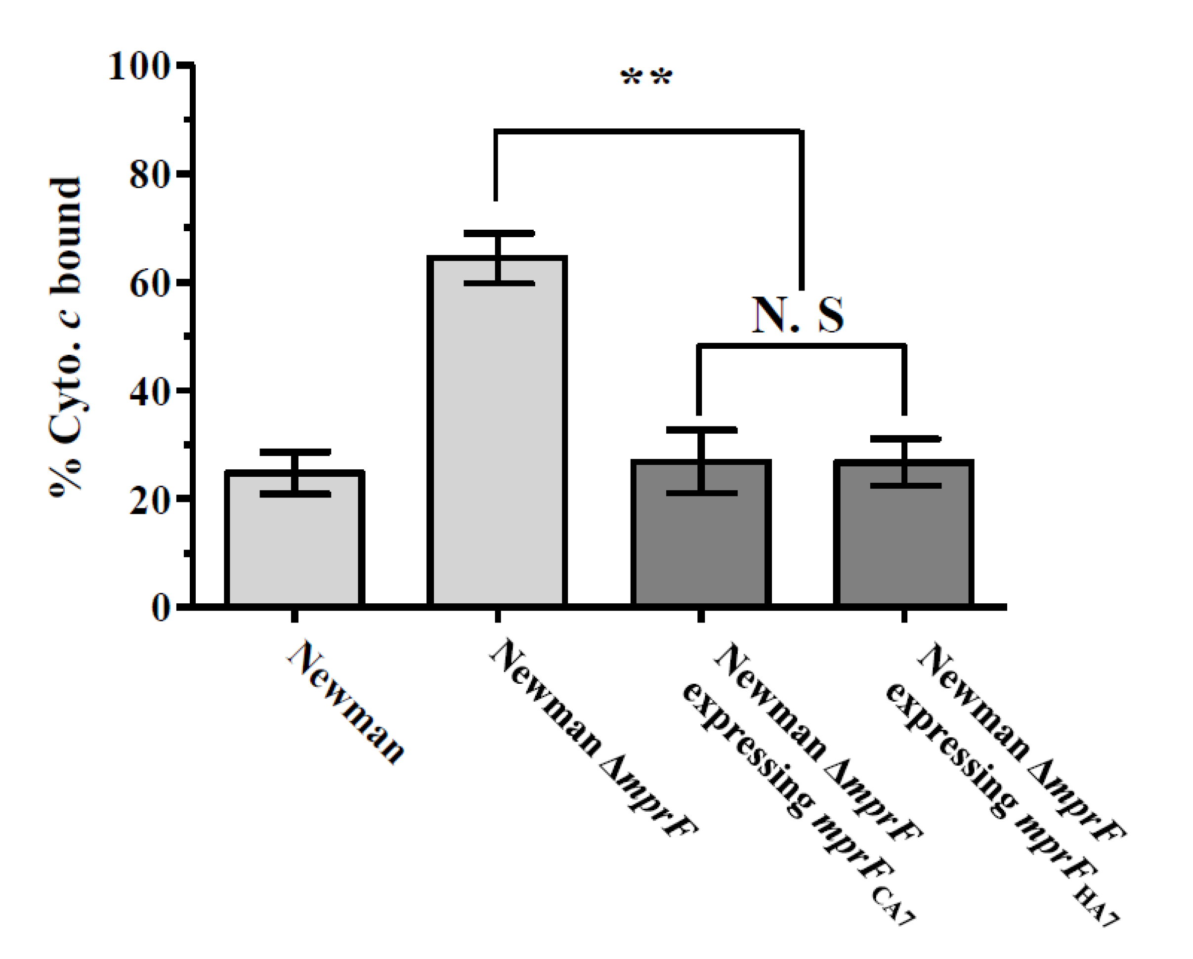
Figure 1. Effect of mprF SNPs (I375M and I461T) on the net surface positive charge in Staphylococcus aureus. The graph shows the percentage of positively-charged cytochrome c bound after 15 min of incubation with each S. aureus strain at room temperature. ** p < 0.01; N.S, not significant.
Biomolecules 10 01415 g001
Figure 2. Transcriptional expression of mprF in the MRSA strains during the exponential (a) and stationary (b) growth phases. RNA samples from the 26 MRSA strains cultured in Mueller–Hinton broth were isolated 3 h (exponential growth phase) and 12 h (stationary growth phase) after initial inoculation and used for RT-qPCR assays. The fold expression of mprF was quantified relative to the levels of gyrB expression. * p < 0.05.
Biomolecules 10 01415 g002
Figure 3. Transcriptional expression of dltA in the MRSA strains during the exponential (a) and stationary (b) growth phases. RNA samples from the 26 MRSA strains cultured in Mueller–Hinton broth were isolated 3 h (exponential growth phase) and 12 h (stationary growth phase) after initial inoculation and used for RT-qPCR assays. The fold expression of dltA was quantified relative to the levels of gyrB expression. ** p < 0.01.
Biomolecules 10 01415 g003
Figure 4. Transcriptional expression of graS in the MRSA strains during the exponential (a) and stationary (b) growth phases. RNA samples from the 26 MRSA strains cultured in Mueller–Hinton broth were isolated 3 h (exponential growth phase) and 12 h (stationary growth phase) after initial inoculation and used for RT-qPCR assays. The fold expression of graS was quantified relative to the levels of gyrB expression. * p < 0.05; ** p < 0.01.
Biomolecules 10 01415 g004
Figure 5. Effect of graS SNP (I224T) on the transcription of graS, mprF, and dltA in S. aureus during the exponential and stationary growth phases. RNA samples from the two MW2 ∆graS strains expressing graRS genes cloned either from CA7 or HA7 strains were isolated 3 h post-inoculation. Fold changes in the expression levels of graS, mprF, and dltA in MW2 ∆graSHA7 were normalized to the levels of the expression of their respective target genes in MW2 ∆graSCA7.
Biomolecules 10 01415 g005
Table 1. Genotypes and SNPs in mprF and graRS ORFs of 26 MRSA strains.
mprF SNPs agraS SNPs agraR SNPs a
StrainMLSTSCC
mec
Codon Changeb Nucleotide PositionAmino Acid ChangeCodon Changeb Nucleotide PositionAmino Acid ChangeCodon Changeb Nucleotide PositionAmino Acid Change
LA1ST72IVATT→ATG
ATT→ACT
1125
1382
I375M
I461T
No changec NANANo changeNANA
LA2ST72IVATT→ATG
ATT→ACT
TTC→TGC
1125
1382
1649
I375M
I461T
F550C
No changeNANANo changeNANA
LA3ST72IVATT→ATG
ATT→ACT
1125
1382
I375M
I461T
No changeNANANo changeNANA
LA4ST72IVATT→ATG
ATT→ACT
1125
1382
I375M
I461T
No changeNANANo changeNANA
LA5ST72IVATT→ATG
ATT→ACT
1125
1382
I375M
I461T
No changeNANANo changeNANA
LA6ST72IVATT→ATG
ATT→ACT
1125
1382
I375M
I461T
No changeNANANo changeNANA
LA7ST72IVATT→ATG
ATT→ACT
1125
1382
I375M
I461T
No changeNANANo changeNANA
HA1ST5IINo changeNANAATA→ACA671I224TAAA→AAT300K100N
HA2ST5IINo changeNANAATA→ACA671I224TNo changeNANA
HA3ST5IINo changeNANAATA→ACA671I224TNo changeNANA
HA4ST5IINo changeNANAATA→ACA671I224TNo changeNANA
HA5ST5IINo changeNANAATA→ACA671I224TNo changeNANA
HA6ST5IICAT→CCT
GAG→AAG
362
802
H121P
E268K
ATA→ACA671I224TNo changeNANA
HA7ST5IINo changeNANAATA→ACA671I224TNo changeNANA
HA8ST5IINo changeNANAATA→ACA671I224TNo changeNANA
CA1ST72IVATT→ATG
ATT→ACT
ACT→ATT
1125
1382
1997
I375M
I461T
T666I
No changeNANANo changeNANA
CA2ST72IVATT→ATG
ATT→ACT
1125
1382
I375M
I461T
No changeNANANo changeNANA
CA3ST72IVATT→ATG
ATT→ACT
1125
1382
I375M
I461T
No changeNANANo changeNANA
CA4ST72IVATT→ATG
ATT→ACT
1125
1382
I375M
I461T
GAT→AAT631D211NNo changeNANA
CA5ST72IVATT→ATG
ATT→ACT
1125
1382
I375M
I461T
No changeNANANo changeNANA
CA6ST72IVATT→ATG
ATT→ACT
1125
1382
I375M
I461T
No changeNANANo changeNANA
CA7ST72IVATT→ATG
ATT→ACT
1125
1382
I375M
I461T
No changeNANANo changeNANA
CA8ST72IVATT→ATG
ATT→ACT
1125
1382
I375M
I461T
No changeNANANo changeNANA
CA9ST72IVATT→ATG
ATT→ACT
1125
1382
I375M
I461T
No changeNANANo changeNANA
CA10ST72IVATT→ATG
ATT→ACT
1125
1382
I375M
I461T
No changeNANAGTG→GGG260V87G
CA11ST72IVATT→ATG
ATT→ACT
1125
1382
I375M
I461T
No changeNANANo changeNANA
a The mprF and graRS sequences from the S. aureus MW2 strain were used as the consensus reference sequences to identify single nucleotide polymorphisms (SNPs) among the study strains.; b Position of nucleotide change within the mprF or graRS ORF; c NA, not applicable.
Table 2. Group comparison for transcriptional expression of mprF, dltA, and graS among LA-MRSA, HA-MRSA, and CA-MRSA strains.
ParameterGroupsp Value for
ST72
LA-MRSA
ST5
HA-MRSA
ST72
CA-MRSA
ST72
LA-MRSA
Vs. ST5
HA-MRSA
ST72
CA-MRSA
Vs. ST5
HA-MRSA
Fold mprF expression vs. gyrB in:
Exponential growth phase2.12 ± 1.074.68 ± 1.924.21 ± 1.40<0.05a NS
Stationary growth phase3.54 ± 3.235.38 ± 4.641.63 ± 0.60a NS<0.05
Fold dltA expression vs. gyrB in:
Exponential growth phase1.24 ± 1.659.80 ± 7.022.88 ± 0.81<0.01<0.01
Stationary growth phase1.60 ± 1.639.65 ± 9.860.39 ± 0.16NS<0.01
Fold graS expression vs. gyrB in:
Exponential growth phase0.03 ± 0.047.71 ± 7.220.67 ± 0.55<0.05<0.01
Stationary growth phase0.06 ± 0.070.77 ± 0.640.05 ± 0.02<0.05<0.01
a NS, not significant.