Article

Enhancing Plant Resistance to Sri Lankan Cassava Mosaic Virus Using Salicylic Acid

by
Chonnipa Pattanavongsawat
1,
Srihunsa Malichan
1,
Nattachai Vannatim
1,
Somruthai Chaowongdee
1,
Nuannapa Hemniam
1,
Atchara Paemanee
2,3 and
Wanwisa Siriwan
1,*
1
Department of Plant Pathology, Faculty of Agriculture, Kasetsart University, Bangkok 10900, Thailand
2
National Omics Center (NOC), National Center for Genetic Engineering and Biotechnology (BIOTEC), National Science and Technology Development Agency (NSTDA), Pathum Thani 12120, Thailand
3
Food Biotechnology Research Team, Functional Ingredients and Food Innovation Research Group, National Center for Genetic Engineering and Biotechnology (BIOTEC), Thailand Science Park, Pathum Thani 12120, Thailand
*
Author to whom correspondence should be addressed.
Metabolites 2025, 15(4), 261; https://doi.org/10.3390/metabo15040261
Submission received: 27 March 2025 / Revised: 6 April 2025 / Accepted: 8 April 2025 / Published: 10 April 2025

Abstract

:
Background: Cassava mosaic disease (CMD), caused by the Sri Lankan cassava mosaic virus (SLCMV), significantly increases cassava yield losses in Thailand, with losses ranging from 30% to 80%, and is exacerbated by limited access to healthy planting materials. Methods: This study explored salicylic acid (SA) as a potential treatment for enhancing disease resistance in CMD infected cassava plants. SA was applied at 100 and 200 mg/mL, and its effects were evaluated using quantitative real-time polymerase chain reaction (qPCR) and reverse transcription qPCR (RT-qPCR) to measure viral loads and the expression levels of resistance genes. Results: Although SA treatment did not considerably affect disease severity, foliar CMD symptoms visibly decreased, particularly with 200 mg/mL SA, which also reduced SLCMV particle counts at 1- and 2-weeks post-treatment. SA upregulated the expression of pathogenesis-related proteins (PRs), including HSP90.9, WRKY59, SRS1, and PR9e. Additionally, SA enhanced the regulation of secondary metabolite pathways involving L-serine within the glycine, serine, and threonine metabolism, as well as the phenylpropanoid biosynthesis pathways. Conclusions: These findings collectively indicate that SA enhances resistance through the systemic acquired resistance (SAR) pathway and can serve as a potential strategy for the management of CMD, particularly in regions where healthy cassava planting materials are scarce. The study highlights the efficacy of SA in reducing viral particles, inducing the immune response, and providing a promising approach for controlling CMD.

1. Introduction

Cassava (Manihot esculenta Crantz) is a crucial root vegetable worldwide, known for its high carbohydrate content and role as a primary energy source [1]. In Thailand, cassava holds a major position in the agricultural sector, and the area of cassava plantation in Thailand is the second largest worldwide, after Nigeria. Cassava production in Thailand exceeds 31 million tons, which contributes considerably to the economy, and the annual yield and export value of cassava production exceeded 4.27 million tons and 2427 million dollars, respectively, in 2023. Despite its economic importance, the commercial varieties of cassava are susceptible to various diseases that can significantly reduce the yield. The cassava plantations in Thailand are vulnerable to various diseases, including cassava bacterial blight, brown leaf spot, blight leaf spot, stem rot, cassava mosaic disease (CMD), and witch’s broom disease [2], of which CMD poses a severe threat, substantially reducing cassava production by 30–80% [3].
CMD is caused by the cassava mosaic virus, which belongs to the Begomovirus genus under the Geminiviridae family [4]. To date, 11 species of CMV have been reported globally, with Sri Lankan cassava mosaic virus (SLCMV) being the predominant species in Thailand [5]. The first outbreak of CMD was identified in Thailand in August 2018 in a region bordering Cambodia [6], where previous CMD outbreaks had been reported [7]. CMD is transmitted by the tobacco whitefly (Bemisia tabaci) as well as through infected stem cuttings. Whitefly can efficiently spread the virus within crop field [8]. After feeding on CMD -infected plants for 4–6 h, the insect becomes capable of transmitting the virus. Although it is possible that the virus replicates within the insect vector, the exact mechanism remains unclear. Transmission to a healthy plant can occur within as little 15 min of feeding, with longer feeding duration increasing the likelihood of infection, thereby facilitating rapid and widespread disease dissemination [9,10]. Propagation through stem cutting is the common method used in cassava cultivation [11]. The Geminiviridae viruses present in the phloem of CMD- infected cuttings can contribute to the spread of the disease across fields and over long distances [12]. Typical symptoms include light green or yellow and dark green mosaic patterns on young shoots and new growth. Infected plants exhibit stunted trunks and growth retardation, leading to a reduction in cassava root production by over 70% [13].
Plants respond to diseases via various mechanisms such as induced resistance (IR), which is mediated by structural and biochemical defenses, including the synthesis of a wax or cutin coat and phytoalexins. Pathogenesis-related proteins (PRs) play a key role in the process of IR by retarding infections without harming the plant. Plants can also enhance disease resistance using biotic elicitors derived from pathogens [14,15]. Systemic acquired resistance (SAR) is another crucial mechanism of resistance in which plants respond to elicitors by producing proteins and chemicals that inhibit the growth of pathogens to confer sustained resistance [16]. Phytohormones are key signaling substances in plant resistance, of which salicylic acid (SA) plays a crucial role in inducing resistance to pathogens and signaling [17,18].
SA is a naturally occurring phytohormone that regulates various aspects of plant physiology. It influences plant growth, stomatal activity, nutrient absorption, and the synthesis of chlorophyll, phenolic compounds, amino acids, and antioxidants [19]. SA also acts as a signaling molecule, inducing the expression of genes that enhance resistance to pathogens [20]. In plants, SA is synthesized from the amino acid phenylalanine, primarily by the action of phenylalanine ammonia lyase and isochorismate pyruvate lyase [21]. During infection, SA production increases, leading to the activation of non-expressor of pathogen-related gene 1 (NPR1). NPR1, in turn, triggers the accumulation of several PRs, including PR-1, PR-2, PR-4, PR-5, PR-8, and PR-11. This cascade induces SAR and enhances resistance against various pathogens, including tobacco mosaic virus (TMV) and the fungus, Colletotrichum lagenarium [22,23,24].
The study by Király et al. [25] demonstrated that the application of sufficient quantities of sulfate to tobacco plants reduced the viral loads and increased the expression of PR1 and PR2, indicating an enhanced resistance to TMV. The application of sulfate induced the production of SA and triggered the defense mechanisms of tobacco plants. Wang et al. [26] investigated the effect of spraying SA, jasmonic acid (JA), and coronatine (COR) on potato plants infected with tomato yellow leaf curl virus (TYLCV). The findings revealed that treatment with 0.5 mM SA, 0.5 mM JA, and 0.1 µM COR significantly reduced the viral load compared to the control. The reduction was most significant following treatment with JA, followed by SA and COR. These compounds also upregulated the genes that were involved in pathogen response and reduced the elevated viral load.
Recent studies on plant metabolism provide insights into the differential expression of plant traits, including physiology, yield, and photosynthesis, across different cultivars. For instance, the study by Obata et al. [27] analyzed the metabolites in six commercial cultivars of cassava from Africa to investigate the cultivar-specific factors contributing to high yields. The findings indicated that traits such as carbon assimilation rate, K battery, root starch synthesis, and the accumulation of trehalose and chlorogenic acid were strongly associated with high yield. Chaowongdee et al. [28] additionally analyzed the metabolite profiles of different cultivars of SLCMV-infected and healthy cassava plants. The results demonstrated that flavonoid biosynthesis, as well as the phenylalanine, tyrosine, and tryptophan biosynthesis pathways, contribute to SAR.
The present study aimed to determine the concentration at which SA can induce resistance to CMD in infected cassava plants. Resistance to CMD can decrease the viral load and induce the expression of genes related to SAR and defense mechanisms. These findings can provide novel insights into the role of SA in inducing resistance in cassava plants and facilitating its application for the management of CMD.

2. Materials and Methods

2.1. Plant Materials, SA Treatment, and Collection of Leaf Samples

The KU50 landrace cultivar of cassava infected with CMD was vegetatively propagated. The stems were cut into sections with 3–4 buds per segment and planted in a greenhouse at the Department of Plant Pathology, Faculty of Agriculture, Kasetsart University, Thailand. The temperature of the greenhouse was maintained at 27–29 °C, under 14 h of light, and a relative humidity of 70–80%. After six weeks of planting, the leaves were sprayed with SA (Acros Organics, Shanghai, China) dissolved in 60% ethanol, and then diluted with sterile water to final concentrations of 100 and 200 mg/mL. The control plants were treated with 60% ethanol. Each treatment was replicated using five plants, and each plant was sprayed with 1 mL of SA solution. Leaf samples were collected at 0, 1, 2, 3, and 4 weeks post-treatment, and stored at −20 °C. The leaf samples were used to measure the viral particles counts and expression of resistant genes through quantitative real-time polymerase chain reaction (qPCR) and reverse transcription qPCR (RT-qPCR) analyses.

2.2. Disease Severity

The symptoms of CMD were assessed from the photographs captured at 0, 1, 2, 3, and 4 weeks post-treatment. The disease severity was evaluated according to the method described by Sseruwagi et al. [29], in which the symptoms are classified into five levels: 1 = no symptoms, 2 = mild chlorotic patterns, 3 = mosaic patterns on all the leaves and foliar distortion, 4 = mosaic pattern on all the leaves, foliar distortion, and a general reduction in leaf size, and 5 = misshapen and twisted leaves and stunting of the entire plant. The disease-severity percentage was calculated using the following formula by Olasanmi et al. [30]:
Disease severity = Sum of all disease ratings Total number of plants × maximum disease rating × 100

2.3. DNA Extraction

The samples of cassava leaves were used to extract DNA for quantifying the number of SLCMV particles in each of the treatment groups. DNA extraction was performed using the cetyl trimethylammonium bromide (CTAB) method of Doyle and Doyle (1987) Doyle and Doyle [31], with slight modifications. Cassava leaves (100 mg) were mixed with 700 µL CTAB buffer (1 M Tris-HCl (pH 8), 1.4 M NaCl, 0.5 M ethylenediaminetetraacetic acid (EDTA), 4.0 g CTAB, and 2% polyvinylpyrrolidone (PVP)) and crushed. The mixture was then incubated at 65 °C for 30 min, following which 700 µL chloroform/isoamyl (24:1) was added to the tubes. The mixture was then centrifuged at 12,000 rpm for 10 min, following which 400 µL of the supernatant was transferred to a clean 1.5 mL tube. The supernatant was mixed with 400 µL isopropanol and centrifuged at 12,000 rpm for 10 min. The supernatant was discarded, and the DNA pellet was washed twice with 400 µL of 70% ethanol, dried at room temperature for 1–2 h, and then resuspended in 30 µL of nuclease-free water (Thermo Fisher Scientific, Waltham, MA, USA). The extracted DNA was visualized by 1.5% agarose TAE gel electrophoresis (100 V for 30 min) using RedSafe Nucleic Acid Staining Solution (iNtRON Biotechnology, Sangdaewon, South Korea) in 0.5× TAE buffer (1 M Tris-HCl (pH 8), 0.5 M EDTA, and glacial acetic acid). The images of the gels were analyzed using the Syngene software v1.8.13. (Synoptics Ltd., Cambridge, UK). The DNA solutions were stored at −20 °C for further experiments.

2.4. Quantification of SLCMV Particles

Amplification and quantification were performed using the SYBR Green dye for measuring the viral particle using qPCR, which was performed on a CFX96 Connect RT-PCR System (Bio-Rad, Hercules, CA, USA). For qPCR, 0.5 µL of the 5′-TAAGAGGTTTTGCGTTAAGTCCG-3′ and 5′-TGTACAGCATCAATGCATTCTCG-3′ forward and reverse primers specific for the SLCMV coat protein, which is encoded by the AV1 gene, were used along with 3 µL of nuclease-free water, 1 µL of 100 ng/mL DNA template, and 5 µL of 2x qPCRBIO SyGreen Mix Lo-ROX (Copenhagen Biotech Supply, Bronshoj, Denmark). The conditions of qPCR amplification included pre-incubation at 95 °C for 2 min, followed by 40 cycles of denaturation at 95 °C for 5 s, annealing at 60 °C for 30 s, and extension at 65 °C for 5 s. Fluorescence was measured at the end of each cycle, and the melting curve was analyzed between 65 °C and 95 °C with a 10 s hold at increments of 1 °C. Each reaction was performed in triplicate, and the standard curve was prepared for each experiment by co-amplifying known quantities of the SLCMV coat protein. Seven consecutive dilutions (dilution factor 1:10) of the SLCMV coat protein, containing 106 to 101 copies per reaction, were prepared. The quantity of the SLCMV coat protein DNA in the sample was determined by plotting the threshold cycle (Ct) values onto the standard curve.

2.5. RNA Extraction and cDNA Synthesis

RNA was extracted from the samples of cassava leaves sprayed with SA and the control solution after 0, 1, 2, 3, and 4 weeks of treatment, according to the method described by Behnam et al. (2019) Behnam et al. [32]. To this end, 100 mg of the leaf samples was crushed to a powder using liquid nitrogen and transferred to a 2 mL tube, following which 700 µL of the pre-heat extraction buffer (100 mM Tris-HCl (pH 8), 100 mM NaCl, 25 mM EDTA, 1% sodium dodecyl sulfate (SDS), 2% PVP, and 2% mercaptoethanol) was added to the powdered sample. The mixture was vortexed and 700 µL of chloroform was added to the sample, followed by centrifugation at 15,000 rpm for 10 min. The supernatant was transferred to a clean 2 mL tube, to which 600 µL chloroform was added, and the mixture was centrifuged at 15,000 rpm for 10 min, and the process was repeated twice. The supernatant was transferred to a clean 1.5 mL tube, to which 0.33 volumes of 8 M lithium chloride were added, and the mixture was allowed to incubate overnight at 4 °C. The mixture was then centrifuged at 15,000 rpm for 10 min, and the supernatant was discarded. The pellet was dissolved in 500 µL diethyl pyrocarbonate (DEPC) and centrifuged at 15,000× g for 10 min, following which one volume of phenol: chloroform: isoamyl alcohol (25:24:1) and one volume of chloroform: isoamyl alcohol (24:1) was added to the mixture. The RNA pellet was then precipitated overnight with 500 µL of 5 M NaCl at −80 °C. The precipitated tube was centrifuged at 15,000 rpm for 10 min and the supernatant was discarded. The RNA pellet was washed with 70% ethanol, air-dried on ice for 10 min, and subsequently resuspended in 30 µL of nuclease-free water. The extracted RNA was stored at −80 °C, and its purity was evaluated based on the A260:A280 and A260:A230 absorbance ratios using a NanoDrop® ND-1000 spectrophotometer (Thermo Fisher Scientific, Waltham, MA, USA). The quality of the extracted RNA was assessed by 1.5% agarose TAE gel electrophoresis (100 V for 30 min) using RedSafe Nucleic Acid Staining Solution (iNtRON Biotechnology, Sangdaewon, Republic of Korea) in 0.5x TAE buffer (1 M Tris-HCl (pH 8), 0.5 M EDTA, and glacial acetic acid). A 1 kb DNA ladder (Thermo Fisher Scientific, USA) was used as reference. The images of the gels were subsequently analyzed using the Syngene software v1.8.13. (Synoptics Ltd., Cambridge, UK).
In this study, cDNA was synthesized using 1 µL of RevertAid reverse transcriptase (Thermo Fisher Scientific, Waltham, MA, USA), 1 µL of RiboLock RNase Inhibitor, 2 µL of 10 mM dNTP mix, 1 µL of 10 mM Oligo(dT), 4 µL of 5× reaction buffer, 10 µL water, and 1 µL of 100 ng/µL RNA template. The cDNA products were stored at −20 °C for further experiments.

2.6. RT-qPCR

The PDF2, PR1, PR7f5, PR9e, SYP121, WRKY89, Hsp90.9, Hsf8, and SRS1 genes were amplified to evaluate the relative expression levels of the genes that were involved in the induction of plant defenses by SA. The UBQ10 gene served as the reference for the RT-qPCR assays. The nucleotide sequences of the primers are provided in Table S1. RT-qPCR amplification was performed using a CFX96 Connect RT-PCR System (Bio-Red, Hercules, CA, USA). The 10 µL reaction mixtures comprised 5 µL of 2x qPCRBIO SyGreen Mix Lo-ROX (PCR Biosystems, London, UK), 0.5 µL of each of the forward and reverse primers, 1 µL of 100 ng/µL cDNA template, and 3 µL of nuclease-free water The conditions for thermocycling were as follows: initial denaturation at 95 °C for 2 min, followed by 40 cycles of denaturation at 95 °C for 5 s, annealing at 60 °C for 30 s, and extension at 65 °C for 5 s. The fluorescence signals were recorded at the end of each cycle. The melting curve was analyzed from 65 °C to 95 °C, with a 10 s hold at increments of 1 °C for verifying the specificity of the amplification. Each reaction was performed in triplicate to ensure reproducibility. The relative mRNA abundance of the target genes was normalized to that of UBQ10, and the fold change (log2) in gene expression was calculated using the 2−∆∆CT method [33] (Rao et al., 2013).

2.7. Metabolomic Analysis

The metabolites were extracted according to the method described by Chaowongdee et al. (2023) [28], and UHPLC-HRMS/MS was performed for analyzing the metabolites. A 200 ppm cassava metabolite extract (2 µL) was injected onto a Hypersil GOLD™ Vanquish C18 column (2.1  ×  100 mm, 1.9 µm, Thermo Fisher Scientific, USA) equipped with a guard column, which was maintained at 40 °C and operated at a flow rate of 0.4 mL/min. Mobile phase A consisted of 0.1% FA in water, and mobile phase B consisted of 0.1% FA in acetonitrile. The gradient was initiated with 5% B for 4 min, increased to 90% B over 10 min, held at 90% B for 4 min, returned to 5% B over 1 min, and finally equilibrated for 25 min. Mass spectrometry was performed using a Q-Exactive HF-X Orbitrap system (Thermo Fisher Scientific, USA) equipped with a heated electrospray ionization source, and the positive and negative ions were acquired in the full-scan MS1/data-dependent MS2 (dd-MS2) mode. The following settings were used for mass spectrometry: spray voltage of 3.5 kV (positive) and 2.5 kV (negative); sheath gas, 45 AU; auxiliary gas, 10 AU; sweep gas, 2 AU; capillary temperature, 250 °C; MS1 resolution, 120,000; dd-MS2 resolution, 30,000; scan range, 100–1500 m/z; AGC target, 3e6; max injection time, 100 ms; and stepped NCE of 20, 30, and 40 eV. The data were processed with Compound Discoverer, whereas compound annotation was performed using the mzCloud, mzVault, and ChemSpider databases.
The metabolite data were analyzed using MetaboAnalyst 6.0 (https://www.metaboanalyst.ca/, accessed on 31 October 2024) The significant differences between the mean values of the different groups were determined using Fisher’s least significant difference (LSD) test, and p ≤ 0.05 considered statistically significant [34]. The functions and pathways associated with the differential compounds were identified through Kyoto Encyclopedia of Genes and Genomes (KEGG) pathway analysis (https://www.genome.jp/kegg/, accessed on 31 October 2024) [35].

2.8. Statistical Analyses

The data obtained from five biological replicates and three technical replicates were analyzed for viral particle quantification, disease severity, relative gene expression levels, and regression analysis using one-way analysis of variance (ANOVA) in the RStudio program v2024.12.1+563. Significant differences among the means of different groups were determined using the Least Significant Difference (LSD) test, with p ≤ 0.05 considered statistically significant.

3. Results and Discussion

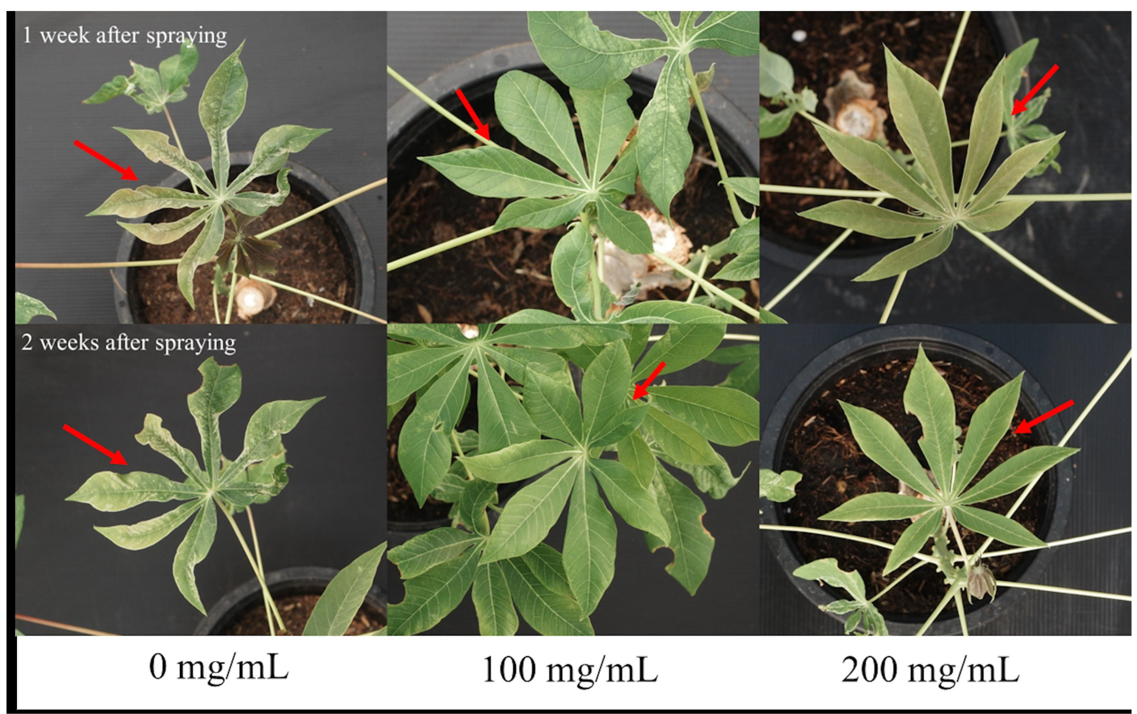
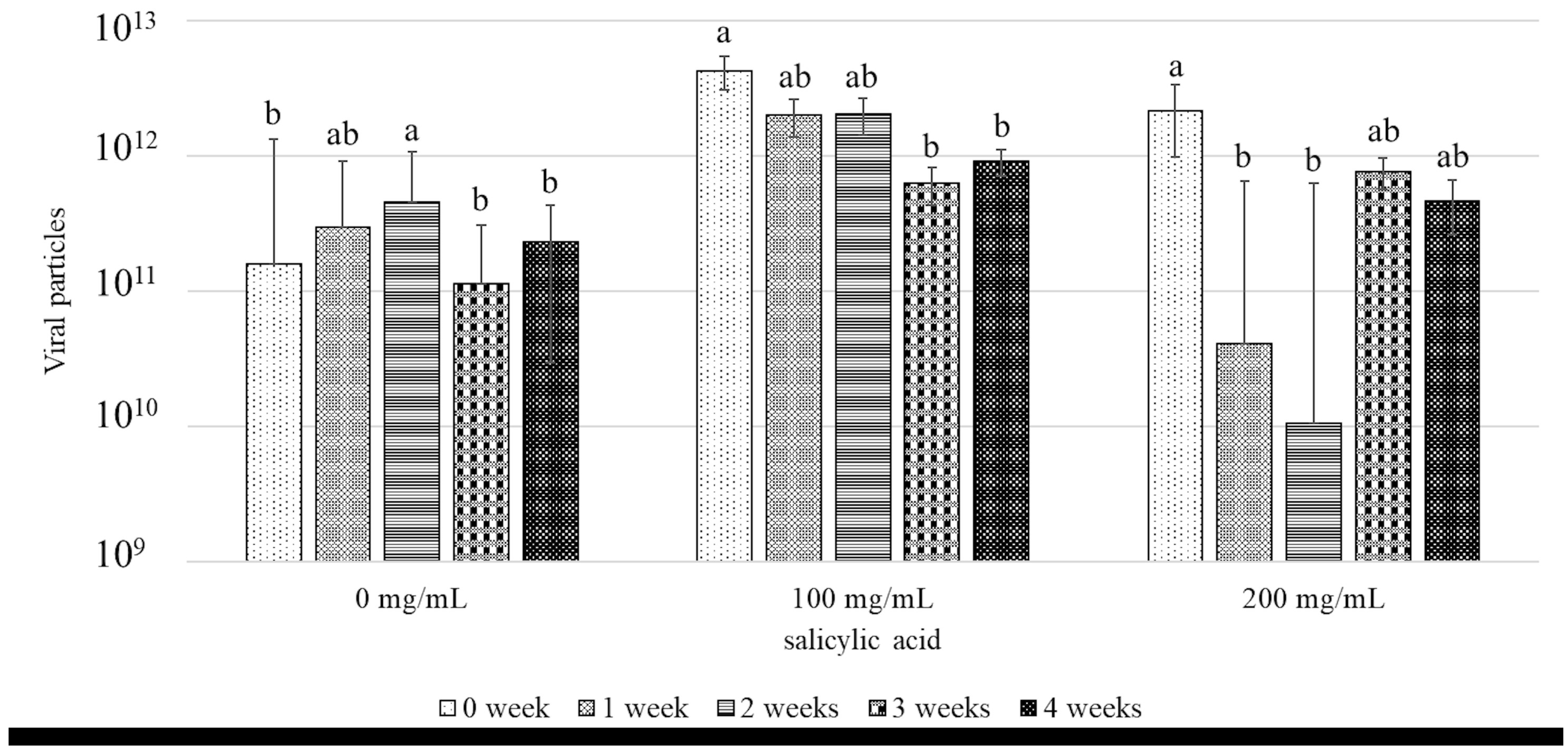
3.1. Effects of SA Against SLCMV

The effects of SA on the percentage of disease severity in CMD-infected plants were evaluated at SA concentrations of 0, 100, and 200 mg/mL. The infected cassava plants were sprayed with different concentrations of SA at 0, 1, 2, 3, and 4 weeks post-treatment. The results of statistical analysis indicated that there were no significant differences in the percentage of disease severity among the different treatment groups across all the time periods tested herein. The results are provided in Figure 1. Interestingly, disease severity decreased after 1 and 2 weeks of spraying SA. As depicted in Figure 2, the reduction in disease severity visually correlated with the absence of the foliar symptoms of CMD. These findings align with the results of the study by Sofy et al. [36], which reported that the disease severity in eggplants infected with Alfalfa mosaic virus [37] decreased by 68.34% after 3 weeks of treatment with SA. Similarly, Basit et al. [38] observed that the disease severity in tobacco plants infected with TMV reduced following the application of 60% SA. These results demonstrated that SA effectively reduced the severity of CMD, and that the symptoms improved after 1 and 2 weeks of treatment. This indicated that, although the plants were infected with SLCMV, which had already triggered SAR before treatment, the application of SA further enhanced SAR in infected cassava plants. The application of SA further triggered SAR in the infected plants, which induced a more rapid defense response against SLCMV.
Regarding the effect of SA on endogenous SA content, some studies have indicated that the application of exogenous SA can increase endogenous SA levels, thereby enhancing the plant’s defense mechanisms. However, higher concentrations of SA can sometimes induce a feedback mechanism that reduces endogenous SA production. The impact of exogenous SA may also vary based on the plant’s developmental stage and its ability to regulate hormonal levels. While literature specific to the effect of exogenous SA treatment on endogenous SA in cassava is limited, similar findings have been reported in other crops, such as tomato and tobacco [38]. Further research is needed to better understand the dynamics of SA regulation in cassava during virus-induced stress and hormonal treatment.
The viral count in cassava plants infected with CMD following treatment with SA at concentrations of 0, 100, and 200 mg/mL was measured by qPCR. The results indicated that treatment with 200 mg/mL SA significantly reduced the number of SLCMV particles at 1 and 2 weeks post-treatment compared to that before treatment. In contrast, the viral particle counts gradually decreased at 1, 2, and 3 weeks post-treatment with 100 mg/mL SA. Meanwhile, the number of viral particles in the control group increased steadily during the same period (Figure 3). The trend in the number of SLCMV particles following the application of SA was examined by regression analysis, and the results indicated that the viral counts decreased following treatment with SA, and assumed the equation b = −8.07 × 10¹¹ ± 3.12 × 10¹¹ after treatment with 100 mg/mL SA and b = −2.68 × 10¹¹ ± 1.77 × 10¹¹ after treatment with 200 mg/mL SA, where b denotes the slope of viral load versus time (Supplementary S1). These results suggest that the application of 200 mg/mL SA would be effective for inducing disease resistance in cassava plants infected with CMD, which aligns with the findings of other studies. For instance, Li et al. [39] observed that the viral load in tomato leaves inoculated with TYLCV decreased from days 2 to 10 after spraying SA, but increased on day 14. The study also revealed that SA IR to yellow leaf curl disease in tomatoes after approximately 10 days of treatment. The study by Sofy et al. (2021) [36], additionally revealed that the treatment of tomato plants with SA before inoculating with TYLCV reduced by the viral count in SA-treated plants by 74.6% compared to that of the control on day 7 post-treatment, indicating that SA can effectively reduce the viral load in infected plants. The reduction in disease severity and viral loads could be attributed to the activation of the hypersensitive response in plants by SA, which induces the death of infected tissues to eliminate viruses [40,41]. SA can also inhibit viruses by inducing the generation of reactive oxygen species (ROS), a key defense mechanism in plants. This in turn triggers stomatal closure, induces programmed cell death, and activates various genes to trigger pathogen resistance [42].
These findings suggested that treatment with SA, especially at a concentration of 200 mg/mL, enhances SAR in cassava plants even after infection with SLCMV. The observed reduction in viral load and disease severity highlights the potential of SA as a viable strategy for managing CMD in cassava cultivation. However, the long-term effects of SA treatment and its efficacy across different varieties under varying environmental conditions require further investigation. In this study, the strategic application of SA to combat CMD is directly linked to the enhancement of SAR, as SA is widely recognized as an inducer of this defense mechanism [43].
SA functions as a mobile signaling molecule, transported to uninfected areas via apoplastic or simplistic pathways to activate systemic immunity [44,45,46]. Recently, Kim and Lim [47] reported that the pH gradient and deprotonation may lead to the apoplastic accumulation of SA before its transportation into the cytosol following pathogen infection. Additionally, SA functions in association with other phytohormones such as JA, ethylene (ET), and abscisic acid, and interacts with genes, proteins, and transcription factors (TFs), including PRs and phytoalexins, to trigger SAR and induced systemic resistance [48].
SAR provides sustained protection, and the continued reduction in viral load and disease severity suggests that SAR was sustained throughout the treatment period, which aided in the effective management of the viral infection over time. This finding supports the notion that treatment with SA can enhance plant immunity, which makes it a valuable tool in integrated pest management strategies [31].
The production of higher quantities of SA in plants induces the generation of various substances that inhibit pathogens, including ROS and enzymatic antioxidants, and reduces the pathogen count. Sofy et al. (2021) [36] observed that the production of ROS and the expression of enzymatic antioxidants related to disease resistance increased in AMV-infected eggplants after 14 days of treatment with SA.

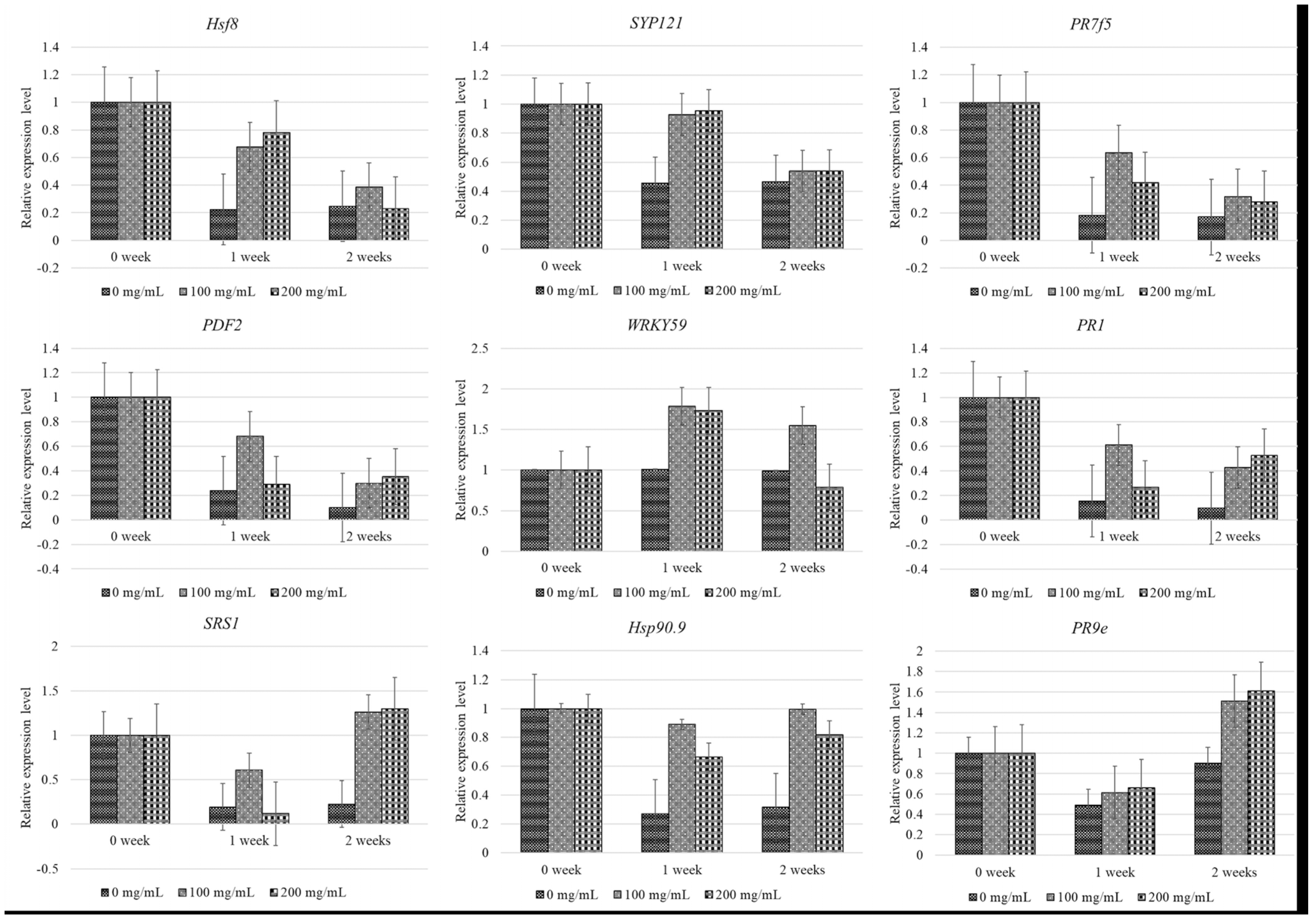
3.2. Relative Gene Expression Levels of Resistance Genes Following SA Treatment

In this study, the expression levels of nine genes, namely, PR1, PDF2, WRKY59, PR9e, PR7f5, Hsp90.9, Hsf8, SPS1, and SYP121 were analyzed due to their association with SAR, especially after treatment with SA. Gene expression was analyzed following the foliar application of SA at varying concentrations and time intervals and compared to that of the control plants. The expression levels of Hsf8, SYP121, PR7f5, PDF2, and WRKY59 gradually downregulated after 1 week of treatment with 100 or 200 mg/mL SA, and were comparable to those of the control group (Figure 4). In contrast, the expression levels of SRS1, Hsp90.9, and PR9e were significantly upregulated at 1 week post-treatment, and increased continually for up to 2 weeks post-treatment. Notably, the upregulation in the expression levels of these genes was sustained beyond 2 weeks, and the gene expression patterns were consistent between the groups treated with 100 and 200 mg/mL SA. Interestingly, analysis of the gene expression dynamics of PR1, especially after treatment with 200 mg/mL SA, revealed that its expression was reduced markedly at first, from 0 to 1 week post-treatment, but increased after 1 week, as depicted in Figure 4. In this study, the collection of leaf samples following was performed concurrently during the measurement of viral particles described in the previous section.
Regression analysis of the expression levels of the nine aforementioned genes revealed that the gene expression patterns of Hsf8, SYP121, PR7f5, PDF2, and PR1 in the plants treated with SA did not differ significantly from those of the control group, and were downregulated in CMD-infected cassava plants (Supplementary S2 and S3). However, the expression levels of PDF2 and PR1 were upregulated following treatment with SA, compared to those of the control plants. We additionally observed that the gene expression levels of Hsp90.9, WRKY59, SRS1, and PR9e, which are involved in plant immunity, tended to increase after treatment with SA. A concentration of 100 mg/mL was found to be most effective in inducing these genes (Supplementary S4).
Malichan et al. [49] demonstrated that the expression levels of SRS1, Hsp90.9, and PR9e in the KU50 cultivar of cassava, which is tolerant to SLCMV, were downregulated seven days after inoculation with SLCMV via whiteflies. Our findings revealed that treatment with SA further upregulated the expression of SRS1, Hsp90.9, and PR9e at one week post-treatment, indicating an enhanced response in cassava plants. Recently, Wei et al. [50] identified that the expression of MeHsp90.9 and the plant immune response is enhanced by the interaction of MeHsp90.9 with MeSRS1 (SHI-related sequence 1) and MeWRKY20, which promotes the transcriptional activation of other genes via SA. Consistent with this finding, the results of RT-PCR demonstrated that SRS1 and Hsp90.9 were upregulated in a coordinated manner, suggesting that SA can enhance gene expression at increasing concentrations and fortify disease resistance.
The PR1 gene serves as a marker for monitoring the activation of the SA and JA pathways in Arabidopsis during pest/pathogen interactions [51]. In this study, we observed that PR1 was downregulated 1 week post-treatment with 100 or 200 mg/mL SA. However, the expression of PR1 increased continually after 1 week of treatment with 200 mg/mL SA. Interestingly, Irigoyen et al. [52] observed that 10 PR genes, except PR1, of the COL2246 variety of cassava, were upregulated at 0, 0.5, 1, 2, 4, 8, 12, and 24 h post-treatment with SA. We propose that the genotype of cassava, concentration of SA, and time of sample collection influence the gene expression levels.
WRKY TFs play a crucial role in plant responses to various environmental stresses. In particular, the WRKY59 gene is known to be induced following treatment with SA and plays a major role in fungal resistance. The WRKY59 gene is present in several plant species and can be induced by SA [53], and has been shown to function as a resistance gene against Ralstonia solanacearum [54]. It has been additionally reported that the WRKY gene can suppress the expression of genes associated with disease severity in chili peppers [55]. Using yeast one-hybrid assays, a previous study identified that PtrWRKY18 and PtrWRKY35 are potential targets of WRKY89 [56]. The OsWRKY89 gene of rice has been shown to enhance resistance to rice blast fungus and white-backed plant hopper, and induce the production of SA [57]. In this study, we observed that the application of 100 or 200 mg/mL SA upregulated the expression of WRKY59 from 0 to 1 weeks post-treatment, following which its expression was downregulated. The sustained upregulation of WRKY59 for 1 week may account for the upregulation of the PR1 gene after 1 week of treatment with SA (Figure 4). This finding suggested that WRKY59 interacted with PR1 to induce resistance against SLCMV in response to treatment with SA. This finding is consistent with the study by Wu et al. [58], which reported that the receptors of plant SA signaling, regulated by the PR1 gene (NPR1), interact with the NPR3 and NPR4 proteins. Although NPR proteins interact with the SA signal, they cannot directly bind to DNA, and therefore require TFs to regulate the expression of downstream SA-responsive genes. Further studies are therefore necessary for exploring the interactions between WRKY59 and PR1 during SA IR to SLCMV.

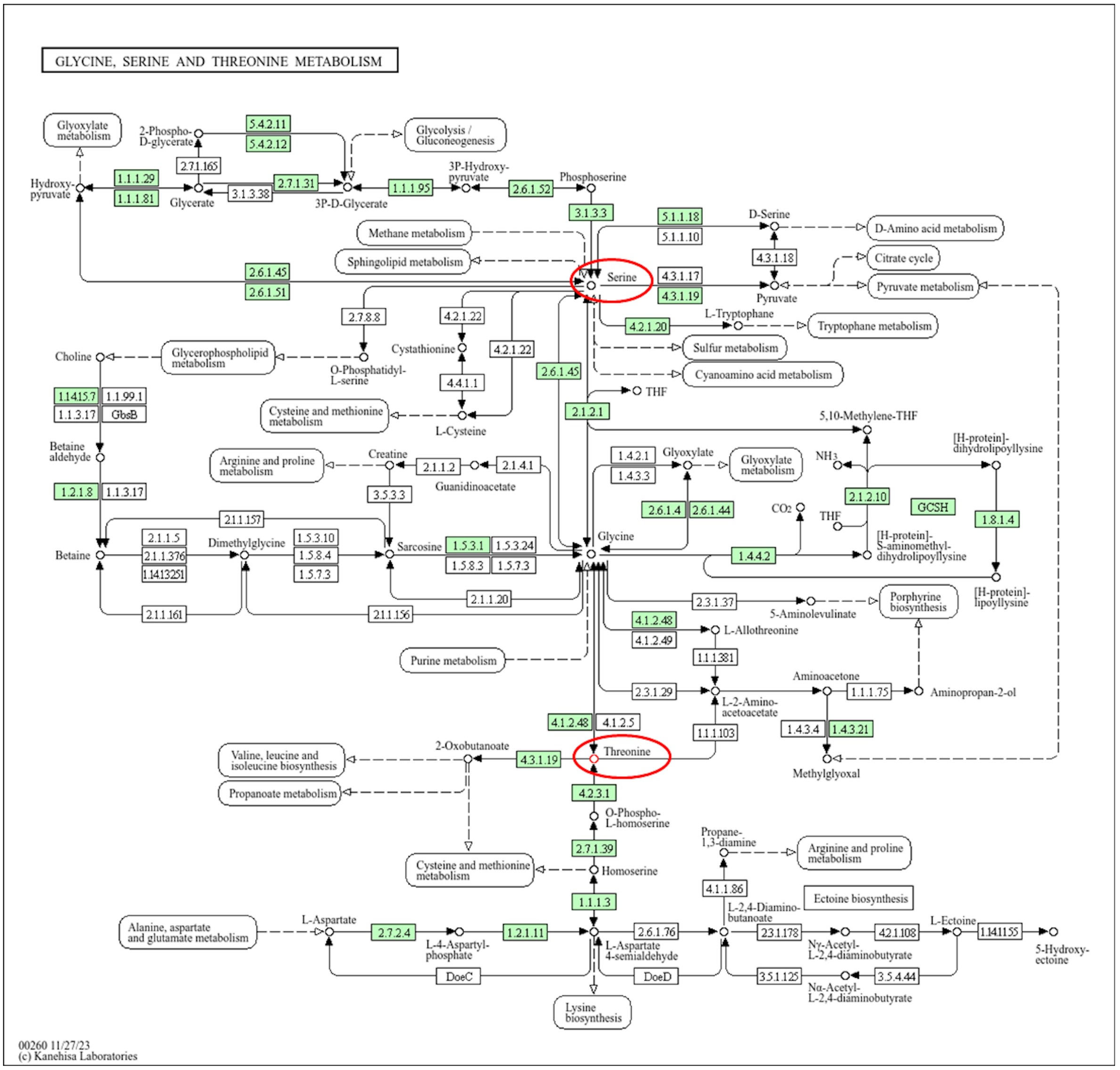
3.3. Identification of Metabolites Associated with SAR

The results of metabolomic analysis revealed that L-serine was consistently detected across the plants treated with 0, 100, or 200 mg/mL SA at 0, 1, and 2 weeks post-treatment, and that there were no significant differences among the different treatment groups. Previous studies have demonstrated that L-serine serves as a primary substrate in plant tissues [59,60]. The results of KEGG analysis revealed that L-serine is a component of the glycine, serine, and threonine metabolism pathway; however, this pathway does not directly regulate SAR and likely contributes to myricetin synthesis.
The levels of L-threonine and myricetin were upregulated in cassava plants treated with 100 or 200 mg/mL SA at 1 and 2 weeks post-treatment; however, they were not detectable in untreated cassava plants. Statistical analyses revealed that there were no significant differences in the levels of L-threonine and myricetin among the different SA-treated groups (p < 0.05; Figure 5). The results of KEGG pathway analysis revealed that L-threonine was involved in glycine, serine, and threonine metabolism (Figure 6), while myricetin was associated with the flavonoid biosynthesis pathway.
Although limited, existing studies suggest that threonine may enhance resistance to pathogens, thereby potentially serving as a precursor for homoserine, which regulates the ET, JA, and SA pathways in response to downy mildew (Hyaloperonospora arabidopsis) in Arabidopsis sp. [61]. This implies that amino acids may trigger the defense response; however, the precise underlying mechanisms remain unclear. Our findings suggest that treatment with SA may also induce the defense response via amino acids. L-threonine also plays a crucial role in plant growth, development, stress tolerance, and gene regulation [62].
The results of KEGG pathway analysis indicated that myricetin is synthesized through the flavonoid biosynthesis pathway, which originates from kaempferol, a derivative of p-coumaroyl-CoA in the phenylpropanoid biosynthesis pathway (PPP) (Figure 7). L-phenylalanine was detected in all the SA-treated plants, and its levels increased 2 weeks post-treatment (Figure 8). Plant viruses activate SA biosynthesis and the production of other defense metabolites via PPP to initiate SAR. For instance, sugarcane mosaic virus triggers this response in maize [63]. Previous studies have identified various antiviral metabolites in TMV-infected tobacco and tomato plants [63,64]. In particular, it has been reported that 5-O-caffeoylquinic acid and quercetin accumulate at the site of TMV infection, and kaempferol was detected in distant tissues exhibiting SAR [65].

4. Conclusions

SA plays an important role in stimulating plant defenses against various pathogens via activating key signaling molecules in the plant immune system. SA induces a critical mechanism referred to as SAR, which enhances resistance to pathogens by inducing the expression of PRs. This response reduced the severity of CMD in infected cassava plants, as visually indicated by the absence of foliar symptoms at one and two weeks post-treatment. Additionally, SA treatment significantly reduced SLCMV loads in infected cassava plants. The alterations in gene expression following SA treatment suggest that SA induces the expression of immune-related genes, including HSP90, WRKY59, SRS1, and PR9e, in diseased cassava plants. In conclusion, 200 mg/mL SA was the most effective concentration for reducing viral particles in cassava plants, whereas 100 mg/mL SA was particularly effective in upregulating the expression of immune-related genes compared to control plants. Furthermore, SA enhances the regulation of secondary metabolite pathways involving L-serine within the glycine, serine, and threonine metabolism pathway, as well as the PPP. The findings obtained herein provide valuable insights for enhancing plant immunity and mitigating viral infections, especially in regions such as Thailand where there is a scarcity of healthy planting materials, and cassava farmers continue to utilize infected stems. The strategic application of SA at optimal concentrations could serve as a viable strategy for disease management and ensuring the sustainable cultivation of cassava.

Supplementary Materials

The following supporting information can be downloaded at: https://www.mdpi.com/article/10.3390/metabo15040261/s1, Table S1: List of primer sequences. Figure S1: The alterations in the count of SLCMV particles at 0, 1, 2, 3, and 4 weeks post-treatment with SA were analyzed through regression analysis, and the results are presented as the unstandardized beta (b) ± standard error (SE). The cassava plants were infected with SLCMV after spraying 0 mg/mL SA (A), 100 mg/mL SA (B), or 200 mg/mL SA (C). Figure S2: The alterations in the expression patterns of Hsf8, SYP121, and PR7 genes at 0, 1, 2, 3, and 4 weeks post-treatment with SA were analyzed through regression analysis, and represented as the unstandardized beta (b) ± standard error (SE). The cassava plants were infected with SLCMV after spraying 0 mg/mL SA (A), 100 mg/mL SA (B), or 200 mg/mL SA (C). Figure S3: The alterations in the expression patterns of PDF2 and PR1 genes at 0, 1, 2, 3, and 4 weeks post-treatment with SA were analyzed through regression analysis and represented as the unstandardized beta (b) ± standard error (SE). The cassava plants were infected with SLCMV after spraying 0 mg/mL SA (A), 100 mg/mL SA (B), or 200 mg/mL SA (C). Figure S4: The alterations in the expression patterns of WRKY59, Hsp90.9, SRS1, and PR9 genes at 0, 1, 2, 3, and 4 weeks post-treatment with SA were analyzed through regression analysis and represented as the unstandardized beta (b) ± standard error (SE). The cassava plants were infected with SLCMV after spraying 0 mg/mL SA (A), 100 mg/mL SA (B), or 200 mg/mL SA (C).

Author Contributions

The authors confirm contribution to the paper as follows: study conception and design: W.S., S.M. and C.P.; data collection: C.P., N.V., N.H. and S.C.; analysis and interpretation of results: W.S., S.M., A.P. and C.P.; draft manuscript preparation: C.P. and W.S. All authors have read and agreed to the published version of the manuscript.

Funding

This research was supported by Kasetsart University Research and Development (KURDI), FF(KU)18.65.

Institutional Review Board Statement

Not applicable.

Informed Consent Statement

Not applicable.

Data Availability Statement

The data that support the findings of this study are available from the corresponding author upon reasonable request.

Acknowledgments

We thank Thai Tapioca Development Institute (TTDI) for support with the cassava planting materials and extend special thanks to the Tropical Animal Genetic Special Research Unit, Faculty of Agriculture, Kasetsart University, for their support with statistical analysis during this experiment.

Conflicts of Interest

The authors declare no conflicts of interest.

Abbreviations

The following abbreviations are used in this manuscript:
ANOVAAnalysis and one-way analysis of variance
CMDCassava mosaic disease
CTABCetyl trimethylammonium bromide
HESIHeated electrospray ionization
HRHypersensitive response
IRInduced resistance
JAJasmonic acid
KEGGKyoto Encyclopedia of Genes and Genomes
LSDLeast significant difference
PRPathogenesis-related
ROSReactive oxygen species
SASalicylic acid
SARSystemic acquired resistance
SLCMVSri Lankan cassava mosaic virus
TFTranscription factors
TMVTobacco mosaic virus
TYLCVTomato yellow leaf curl virus

References

  1. Wanapat, M.; Kang, S. Cassava chip (Manihot esculenta Crantz) as an energy source for ruminant feeding. Anim. Nutr. 2015, 1, 266–270. [Google Scholar] [CrossRef]
  2. Legg, J.; Alvarez, E. Diseases Affecting Cassava. In Achieving Sustainable Cultivation of Cassava; Burleigh Dodds Science Publishing: Cambridge, UK, 2017; Volume 2, pp. 213–244. [Google Scholar] [CrossRef]
  3. Chikoti, P.C.; Mulenga, R.M.; Tembo, M.; Sseruwagi, P. Cassava mosaic disease: A review of a threat to cassava production in Zambia. J. Plant Pathol. 2019, 101, 467–477. [Google Scholar] [CrossRef] [PubMed]
  4. Brown, J.K.; Zerbini, F.M.; Navas-Castillo, J.; Moriones, E.; Ramos-Sobrinho, R.; Silva, J.C.F.; Fiallo-Olivé, E.; Briddon, R.W.; Hernández-Zepeda, C.; Idris, A.; et al. Revision of Begomovirus taxonomy based on pairwise sequence comparisons. Arch. Virol. 2015, 160, 1593–1619. [Google Scholar] [CrossRef]
  5. Siriwan, W.; Jimenez, J.; Hemniam, N.; Saokham, K.; Lopez-Alvarez, D.; Leiva, A.M.; Martinez, A.; Mwanzia, L.; Becerra Lopez-Lavalle, L.A.; Cuellar, W.J. Surveillance and diagnostics of the emergent Sri Lankan cassava mosaic virus (Fam. Geminiviridae) in Southeast Asia. Virus Res. 2020, 285, 197959. [Google Scholar] [CrossRef]
  6. Saokham, K.; Hemniam, N.; Roekwan, S.; Hunsawattanakul, S.; Thawinampan, J.; Siriwan, W. Survey and Molecular Detection of Sri Lankan Cassava Mosaic Virus in Thailand. bioRxiv 2021, 16, e0252846. [Google Scholar] [CrossRef]
  7. Wang, H.-L.; Cui, X.-Y.; Wang, X.-W.; Liu, S.-S.; Zhang, Z.-H.; Zhou, X. First Report of Sri Lankan cassava mosaic virus Infecting Cassava in Cambodia. Plant Dis. 2016, 100, 1029. [Google Scholar] [CrossRef]
  8. Mabasso, S. Tobacco Whitefly on Cassava; PlantwisePlus Knowl. Bank: Wallingford, UK, 2019. [Google Scholar] [CrossRef]
  9. Chant, S.R. Studies on the transmission of cassava mosaic virus by bemisia spp. (aleyrodidae). Ann. Appl. Biol. 1958, 46, 210–215. [Google Scholar] [CrossRef]
  10. Pradhan, B.; Vu, T.; Dey, N.; Mukherjee, S. Molecular Biology of Geminivirus DNA Replication. In Viral Replication; Avid Science: Hyderabad, India, 2017; pp. 2–31. [Google Scholar]
  11. Houngue, J.A.; Houédjissin, S.S.; Ahanhanzo, C.; Pita, J.S.; Houndénoukon, M.S.E.; Zandjanakou-Tachin, M. Cassava mosaic disease (CMD) in Benin: Incidence, severity and its whitefly abundance from field surveys in 2020. Crop. Prot. 2022, 158, 106007. [Google Scholar] [CrossRef]
  12. Bennett, A.; Agbandje-McKenna, M. Geminivirus structure and assembly. Adv. Virus Res. 2020, 108, 1–32. [Google Scholar] [CrossRef]
  13. Legg, J.P.; Owor, B.; Sseruwagi, P.; Ndunguru, J. Cassava Mosaic Virus Disease in East and Central Africa: Epidemiology and Management of A Regional Pandemic. In Advances in Virus Research; Academic Press: Cambridge, MA, USA, 2006; Volume 67, pp. 355–418. [Google Scholar]
  14. Dos Santos, C.; Franco, O.L. Pathogenesis-Related Proteins (PRs) with Enzyme Activity Activating Plant Defense Responses. Plants 2023, 12, 2226. [Google Scholar] [CrossRef]
  15. Mahawer, S.; Arya, S.; Kabdal, T.; Kumar, R.; Prakash, O.; Chitara, M.; Koli, P. Plant Defense Systems: Mechanism of Self-Protection by Plants Against Pathogens. In Plant Protection: From Chemicals to Biologicals Edited; De Gruyter: Berlin, Germany; Boston, MA, USA, 2022; pp. 115–140. [Google Scholar]
  16. Aboulila, A.A. Efficiency of plant growth regulators as inducers for improve systemic acquired resistance against stripe rust disease caused by Puccinia striiformis f. sp. tritici in wheat through up-regulation of PR-1 and PR-4 genes expression. Physiol. Mol. Plant Pathol. 2022, 121, 101882. [Google Scholar] [CrossRef]
  17. Ali, S.; Ganai, B.A.; Kamili, A.N.; Bhat, A.A.; Mir, Z.A.; Bhat, J.A.; Tyagi, A.; Islam, S.T.; Mushtaq, M.; Yadav, P.; et al. Pathogenesis-related proteins and peptides as promising tools for engineering plants with multiple stress tolerance. Microbiol. Res. 2018, 212–213, 29–37. [Google Scholar] [CrossRef] [PubMed]
  18. Yamaguchi, I.; Cohen, J.D.; Culler, A.H.; Quint, M.; Slovin, J.P.; Nakajima, M.; Yamaguchi, S.; Sakakibara, H.; Kuroha, T.; Hirai, N.; et al. 4.02—Plant Hormones. In Comprehensive Natural Products II; Liu, H.-W., Mander, L., Eds.; Elsevier: Oxford, UK, 2010; pp. 9–125. [Google Scholar]
  19. Raskin, I. Role of Salicylic Acid in Plants. Annu. Rev. Plant Biol. 1992, 43, 439–463. [Google Scholar] [CrossRef]
  20. Ullah, C.; Chen, Y.-H.; Ortega, M.A.; Tsai, C.-J. The diversity of salicylic acid biosynthesis and defense signaling in plants: Knowledge gaps and future opportunities. Curr. Opin. Plant Biol. 2023, 72, 102349. [Google Scholar] [CrossRef]
  21. Chen, Z.; Zheng, Z.; Huang, J.; Lai, Z.; Fan, B. Biosynthes©is of salicylic acid in plants. Plant Signal. Behav. 2009, 4, 493–496. [Google Scholar] [CrossRef]
  22. Ge, Y.; Bi, Y.; Guest, D.I. Defence responses in leaves of resistant and susceptible melon (Cucumis melo L.) cultivars infected with Colletotrichum lagenarium. Physiol. Mol. Plant Pathol. 2013, 81, 13–21. [Google Scholar] [CrossRef]
  23. Sangpueak, R.; Phansak, P.; Thumanu, K.; Siriwong, S.; Wongkaew, S.; Buensanteai, N. Effect of Salicylic AcidFormulations on Induced Plant Defense against Cassava Anthracnose Disease. Plant Pathol. J. 2021, 37, 356–364. [Google Scholar] [CrossRef]
  24. Wang, F.; Feng, G.; Chen, K. Burdock fructooligosaccharide induces resistance to tobacco mosaic virus in tobacco seedlings. Physiol. Mol. Plant Pathol. 2009, 74, 34–40. [Google Scholar] [CrossRef]
  25. Király, L.; Künstler, A.; Höller, K.; Fattinger, M.; Juhász, C.; Müller, M.; Gullner, G.; Zechmann, B. Sulfate supply influences compartment specific glutathione metabolism and confers enhanced resistance to Tobacco mosaic virus during a hypersensitive response. Plant Physiol. Biochem. 2012, 59, 44–54. [Google Scholar] [CrossRef]
  26. Wang, P.; Sun, S.; Peng, R.; Li, N.; Liu, K.; Wang, L.; Geng, X.; Hu, B.; Wang, H.; Afzal, A.J. Physiological and transcriptomic analyses revealed gene networks involved in heightened resistance against tomato yellow leaf curl virus infection in salicylic acid and jasmonic acid treated tomato plants. Front. Microbiol. 2022, 13, 970139. [Google Scholar] [CrossRef]
  27. Obata, T.; Klemens, P.A.W.; Rosado-Souza, L.; Schlereth, A.; Gisel, A.; Stavolone, L.; Zierer, W.; Morales, N.; Mueller, L.A.; Zeeman, S.C.; et al. Metabolic profiles of six African cultivars of cassava (Manihot esculenta Crantz) highlight bottlenecks of root yield. Plant J. 2020, 102, 1202–1219. [Google Scholar] [CrossRef] [PubMed]
  28. Chaowongdee, S.; Malichan, S.; Pongpamorn, P.; Paemanee, A.; Siriwan, W. Metabolic profiles of Sri Lankan cassava mosaic virus-infected and healthy cassava (Manihot esculenta Crantz) cultivars with tolerance and susceptibility phenotypes. BMC Plant Biol. 2023, 23, 178. [Google Scholar] [CrossRef] [PubMed]
  29. Sseruwagi, P.; Sserubombwe, W.S.; Legg, J.P.; Ndunguru, J.; Thresh, J.M. Methods of surveying the incidence and severity of cassava mosaic disease and whitefly vector populations on cassava in Africa: A review. Virus Res. 2004, 100, 129–142. [Google Scholar] [CrossRef] [PubMed]
  30. Olasanmi, B.; Kyallo, M.; Yao, N. Marker-assisted selection complements phenotypic screening at seedling stage to identify cassava mosaic disease-resistant genotypes in African cassava populations. Sci. Rep. 2021, 11, 2850. [Google Scholar] [CrossRef]
  31. Doyle, J.J.; Doyle, J.L. A rapid DNA isolation procedure for small quantities of fresh leaf tissue. Phytochem. Bull. 1987, 19, 11–15. [Google Scholar]
  32. Behnam, B.; Bohorquez-Chaux, A.; Castaneda-Mendez, O.F.; Tsuji, H.; Ishitani, M.; Becerra Lopez-Lavalle, L.A. An optimized isolation protocol yields high-quality RNA from cassava tissues (Manihot esculenta Crantz). FEBS Open Bio 2019, 9, 814–825. [Google Scholar] [CrossRef]
  33. Rao, X.; Huang, X.; Zhou, Z.; Lin, X. An improvement of the 2ˆ(-delta delta CT) method for quantitative real-time polymerase chain reaction data analysis. Biostat. Bioinforma Biomath. 2013, 3, 71–85. [Google Scholar]
  34. Xia, J.; Psychogios, N.; Young, N.; Wishart, D.S. MetaboAnalyst: A web server for metabolomic data analysis and interpretation. Nucleic Acids Res. 2009, 37, W652–W660. [Google Scholar] [CrossRef]
  35. Kanehisa, M.; Goto, S. KEGG: Kyoto encyclopedia of genes and genomes. Nucleic Acids Res. 2000, 28, 27–30. [Google Scholar] [CrossRef]
  36. Sofy, A.R.; Sofy, M.R.; Hmed, A.A.; Dawoud, R.A.; Refaey, E.E.; Mohamed, H.I.; El-Dougdoug, N.K. Molecular Characterization of the Alfalfa mosaic virus Infecting Solanum melongena in Egypt and the Control of Its Deleterious Effects with Melatonin and Salicylic Acid. Plants 2021, 10, 459. [Google Scholar] [CrossRef]
  37. Sambandham, V.T.; Shankar, P.; Mukhopadhaya, S. Early Onset Yellow Rust Detection Guided By Remote Sensing Indices. Agriculture 2022, 12, 1206. [Google Scholar] [CrossRef]
  38. Basit, A.; Farhan, M.; Mo, W.-D.; Ding, H.-X.; Ikram, M.; Farooq, T.; Ahmed, S.; Yang, Z.-F.; Wang, Y.; Hashem, M.; et al. Enhancement of resistance by poultry manure and plant hormones (salicylic acid & citric acid) against tobacco mosaic virus. Saudi J. Biol. Sci. 2021, 28, 3526–3533. [Google Scholar] [CrossRef] [PubMed]
  39. Li, T.; Huang, Y.; Xu, Z.-S.; Wang, F.; Xiong, A.-S. Salicylic acid-induced differential resistance to the Tomato yellow leaf curl virus among resistant and susceptible tomato cultivars. BMC Plant Biol. 2019, 19, 173. [Google Scholar] [CrossRef]
  40. Radojičić, A.; Li, X.; Zhang, Y. Salicylic Acid: A Double-Edged Sword for Programed Cell Death in Plants. Front. Plant Sci. 2018, 9, 1133. [Google Scholar] [CrossRef]
  41. Singh, D.P.; Moore, C.A.; Gilliland, A.; Carr, J.P. Activation of multiple antiviral defence mechanisms by salicylic acid. Mol. Plant Pathol. 2004, 5, 57–63. [Google Scholar] [CrossRef]
  42. Herrera-Vásquez, A.; Salinas, P.; Holuigue, L. Salicylic acid and reactive oxygen species interplay in the transcriptional control of defense genes expression. Front. Plant Sci. 2015, 6, 171. [Google Scholar] [CrossRef]
  43. Vlot, A.C.; Dempsey, D.A.; Klessig, D.F. Salicylic Acid, a Multifaceted Hormone to Combat Disease. Annu. Rev. Phytopathol. 2009, 47, 177–206. [Google Scholar] [CrossRef]
  44. Gao, Q.-M.; Zhu, S.; Kachroo, P.; Kachroo, A. Signal regulators of systemic acquired resistance. Front. Plant Sci. 2015, 6, 228. [Google Scholar] [CrossRef]
  45. Singh, A.; Lim, G.H.; Kachroo, P. Transport of chemical signals in systemic acquired resistance. J. Integr. Plant Biol. 2017, 59, 336–344. [Google Scholar] [CrossRef]
  46. Wendehenne, D.; Gao, Q.-m.; Kachroo, A.; Kachroo, P. Free radical-mediated systemic immunity in plants. Curr. Opin. Plant Biol. 2014, 20, 127–134. [Google Scholar] [CrossRef]
  47. Kim, T.-J.; Lim, G.-H. Salicylic Acid and Mobile Regulators of Systemic Immunity in Plants: Transport and Metabolism. Plants 2023, 12, 1013. [Google Scholar] [CrossRef] [PubMed]
  48. Mishra, S.; Roychowdhury, R.; Ray, S.; Hada, A.; Kumar, A.; Sarker, U.; Aftab, T.; Das, R. Salicylic acid (SA)-mediated plant immunity against biotic stresses: An insight on molecular components and signaling mechanism. Plant Stress 2024, 11, 100427. [Google Scholar] [CrossRef]
  49. Malichan, S.; Vannatim, N.; Chaowongdee, S.; Pongpamorn, P.; Paemanee, A.; Siriwan, W. Comparative analysis of salicylic acid levels and gene expression in resistant, tolerant, and susceptible cassava varieties following whitefly-mediated SLCMV infection. Sci. Rep. 2023, 13, 13610. [Google Scholar] [CrossRef]
  50. Wei, Y.; Zhu, B.; Liu, W.; Cheng, X.; Lin, D.; He, C.; Shi, H. Heat shock protein 90 co-chaperone modules fine-tune the antagonistic interaction between salicylic acid and auxin biosynthesis in cassava. Cell Rep. 2021, 34, 108717. [Google Scholar] [CrossRef]
  51. Loon, L.C.v.; Rep, M.; Pieterse, C.M.J. Significance of Inducible Defense-related Proteins in Infected Plants. Annu. Rev. Phytopathol. 2006, 44, 135–162. [Google Scholar] [CrossRef]
  52. Irigoyen, M.L.; Garceau, D.C.; Bohorquez-Chaux, A.; Lopez-Lavalle, L.A.B.; Perez-Fons, L.; Fraser, P.D.; Walling, L.L. Genome-wide analyses of cassava Pathogenesis-related (PR) gene families reveal core transcriptome responses to whitefly infestation, salicylic acid and jasmonic acid. BMC Genom. 2020, 21, 93. [Google Scholar] [CrossRef]
  53. Pandey, S.P.; Somssich, I.E. The Role of WRKY Transcription Factors in Plant Immunity. Plant Physiol. 2009, 150, 1648–1655. [Google Scholar] [CrossRef]
  54. Deslandes, L.; Olivier, J.; Theulieres, F.; Hirsch, J.; Feng, D.X.; Bittner-Eddy, P.; Beynon, J.; Marco, Y. Resistance to Ralstonia solanacearum in Arabidopsis thaliana is conferred by the recessive RRS1-R gene, a member of a novel family of resistance genes. Proc. Natl. Acad. Sci. USA 2002, 99, 2404–2409. [Google Scholar] [CrossRef]
  55. Oh, S.K.; Baek, K.H.; Park, J.M.; Yi, S.Y.; Yu, S.H.; Kamoun, S.; Choi, D. Capsicum annuum WRKY protein CaWRKY1 is a negative regulator of pathogen defense. New Phytol. 2008, 177, 977–989. [Google Scholar] [CrossRef]
  56. Jiang, Y.; Guo, L.; Ma, X.; Zhao, X.; Jiao, B.; Li, C.; Luo, K. The WRKY transcription factors PtrWRKY18 and PtrWRKY35 promote Melampsora resistance in Populus. Tree Physiol. 2017, 37, 665–675. [Google Scholar] [CrossRef]
  57. Wang, H.; Hao, J.; Chen, X.; Hao, Z.; Wang, X.; Lou, Y.; Peng, Y.; Guo, Z. Overexpression of rice WRKY89 enhances ultraviolet B tolerance and disease resistance in rice plants. Plant Mol. Biol. 2007, 65, 799–815. [Google Scholar] [CrossRef] [PubMed]
  58. Wu, Y.; Zhang, D.; Chu, J.Y.; Boyle, P.; Wang, Y.; Brindle, I.D.; De Luca, V.; Després, C. The Arabidopsis NPR1 protein is a receptor for the plant defense hormone salicylic acid. Cell Rep. 2012, 1, 639–647. [Google Scholar] [CrossRef] [PubMed]
  59. Holeček, M. Serine Metabolism in Health and Disease and as a Conditionally Essential Amino Acid. Nutrients 2022, 14, 1987. [Google Scholar] [CrossRef]
  60. Ros, R.; Muñoz-Bertomeu, J.; Krueger, S. Serine in plants: Biosynthesis, metabolism, and functions. Trends Plant Sci. 2014, 19, 564–569. [Google Scholar] [CrossRef]
  61. Stuttmann, J.; Hubberten, H.M.; Rietz, S.; Kaur, J.; Muskett, P.; Guerois, R.; Bednarek, P.; Hoefgen, R.; Parker, J.E. Perturbation of Arabidopsis amino acid metabolism causes incompatibility with the adapted biotrophic pathogen Hyaloperonospora arabidopsidis. Plant Cell 2011, 23, 2788–2803. [Google Scholar] [CrossRef]
  62. Muthuramalingam, P.; Krishnan, S.R.; Pandian, S.; Mareeswaran, N.; Aruni, W.; Pandian, S.K.; Ramesh, M. Global analysis of threonine metabolism genes unravel key players in rice to improve the abiotic stress tolerance. Sci. Rep. 2018, 8, 9270. [Google Scholar] [CrossRef]
  63. Yuan, W.; Jiang, T.; Du, K.; Chen, H.; Cao, Y.; Xie, J.; Li, M.; Carr, J.P.; Wu, B.; Fan, Z. Maize phenylalanine ammonia-lyases contribute to resistance to Sugarcane mosaic virus infection, most likely through positive regulation of salicylic acid accumulation. Mol. Plant Pathol. 2019, 20, 1365–1378. [Google Scholar] [CrossRef]
  64. Creager, A.N.; Scholthof, K.-B.G.; Citovsky, V.; Scholthof, H.B. Tobacco mosaic virus: Pioneering research for a century. Plant Cell 1999, 11, 301–308. [Google Scholar] [CrossRef]
  65. Choi, Y.H.; Kim, H.K.; Linthorst, H.J.; Hollander, J.G.; Lefeber, A.W.; Erkelens, C.; Nuzillard, J.-M.; Verpoorte, R. NMR metabolomics to revisit the tobacco mosaic virus infection in nicotiana t abacum leaves. J. Nat. Prod. 2006, 69, 742–748. [Google Scholar] [CrossRef]
Figure 1. Determination of disease severity using SLCMVinfected cassava leaves following the foliar application of SA. Error bars indicated standard deviation, and columns not significant (p > 0.05) in the disease severity by one-way analysis.
Metabolites 15 00261 g001
Figure 2. Phenotype of SLCMV−infected cassava plants following treatment with SA at 1 and 2 weeks post−treatment. Red arrows indicate symptoms on the same leaf in 0, 100 and 200 mg/mL SA.
Metabolites 15 00261 g002
Figure 3. SLCMV particles in CMD−infected cassava plants following treatment with SA as determined by qPCR. Error bars indicated standard deviation, and columns with different letters indicate significant difference (p ≤ 0.05).
Metabolites 15 00261 g003
Figure 4. Expression patterns of nine genes related to the induction of plant immunity in SLCMV−infected cassava plants following the application of SA. Error bars indicated standard deviation.
Metabolites 15 00261 g004
Figure 5. Expression patterns of L−threonine and myricetin metabolites in SLCMV−infected cassava plants following the application of SA. Error bars indicated standard deviation, and columns with different letters indicate significant difference (p ≤ 0.05).
Metabolites 15 00261 g005
Figure 6. The glycine, serine, and threonine metabolism pathway illustrated using the KEGG Orthology within the KEGG pathway database. The red circles indicate the secondary compounds detected in the samples and the green boxes indicate to organism-specific pathway in KEGG pathway.
Metabolites 15 00261 g006
Figure 7. Flavonoid biosynthesis pathway illustrated using KEGG Orthology within the KEGG pathway database. The red circles indicate the secondary compounds detected in the samples.
Metabolites 15 00261 g007
Figure 8. Expression patterns of L−phenylalanine metabolites in SLCMV−infected cassava plants following the application of SA. Error bars indicated standard deviation, and columns with different letters indicate significant difference (p ≤ 0.05).
Metabolites 15 00261 g008
Disclaimer/Publisher’s Note: The statements, opinions and data contained in all publications are solely those of the individual author(s) and contributor(s) and not of MDPI and/or the editor(s). MDPI and/or the editor(s) disclaim responsibility for any injury to people or property resulting from any ideas, methods, instructions or products referred to in the content.

