Article

Metabolomic Profiling of the Striatum in Shank3 Knockout ASD Rats: Effects of Early Swimming Regulation

1
Department of Physical Education and Research, China University of Mining and Technology—Beijing, Beijing 100083, China
2
College of P.E and Sports, Beijing Normal University, Beijing 100875, China
*
Authors to whom correspondence should be addressed.
Metabolites 2025, 15(2), 134; https://doi.org/10.3390/metabo15020134
Submission received: 25 November 2024 / Revised: 27 January 2025 / Accepted: 5 February 2025 / Published: 16 February 2025
(This article belongs to the Special Issue Interactions between Exercise Physiology and Metabolism)

Abstract

:
Objectives: This study aimed to investigate the regulatory impact of early swimming intervention on striatal metabolism in Shank3 gene knockout ASD model rats. Methods: Shank3 gene knockout exon 11–21 male 8-day-old SD rats were used as experimental subjects and randomly divided into the following three groups: a Shank3 knockout control group (KC), a wild-type control group (WC) from the same litter, and a Shank3 knockout swimming group (KS). The rats in the exercise group received early swimming intervention for 8 weeks starting at 8 days old. LC-MS metabolism was employed to detect the changes in metabolites in the striatum. Results: There were 17 differential metabolites (14 down-regulated) between the KC and WC groups, 19 differential metabolites (18 up-regulated) between the KS and KC groups, and 22 differential metabolites (18 up-regulated) between the KS and WC groups. Conclusions: The metabolism of striatum in Shank3 knockout ASD model rats is disrupted, involving metabolites related to synaptic morphology, and the Glu and GABAergic synapses are abnormal. Early swimming intervention regulated the striatal metabolome group of the ASD model rats, with differential metabolites primarily related to nerve development, synaptic membrane structure, and synaptic signal transduction.

1. Introduction

Autism spectrum disorder (ASD) is a complex neurodevelopmental condition marked by social impairments, repetitive and stereotyped behaviors, and a spectrum of symptoms encompassing cognitive deficits, motor dysfunction, intellectual disability, and attention deficit hyperactivity disorder (ADHD) [1,2]. The global incidence of ASD has risen significantly, posing a major public health challenge that affects children’s well-being. According to a 2021 report by the Centers for Disease Control and Prevention (CDC), the prevalence of ASD among 8-year-olds was 1 in 44, representing a staggering 240% increase from 1 in 150 in 2000 [3]. In China, nearly 1% of children suffer from varying degrees of ASD, with the prevalence rate continuing to climb annually [4]. Given the high incidence and lifelong disability associated with ASD, early diagnosis and intervention are pivotal in its prevention and management [5].
The etiology of ASD is multifaceted, involving intricate interactions between genetic and environmental factors. While the precise causes remain unknown for at least 60% of ASD cases [6], extensive research has established that ASD originates in the early stages of brain development and persists throughout life. The genetic underpinnings of ASD are complex, characterized by widespread variations in genetic networks. Mutations or deletions in numerous genes, notably the Shank3 gene, have been implicated in the pathogenesis of ASD [7,8]. The protein encoded by the Shank3 gene serves as a vital scaffolding component in the postsynaptic density (PSD) of excitatory synapses, playing a crucial role in synapse formation and stabilization and the regulation of synaptic transmission. Disruptions in the Shank3 gene can impair normal synaptic function, leading to aberrant neuronal connections and, subsequently, triggering ASD symptoms [9]. Consequently, the genetic knockout of Shank3 has emerged as a valuable animal model for studying autism.
In recent years, research into the pathophysiology of ASD has intensified, leading to a growing interest in exercise intervention as a non-pharmacological treatment option. Swimming, a whole-body exercise that combines motor stimulation with multi-sensory stimulation from a rich environment, is recognized as an effective means of promoting neurodevelopment and neural plasticity [10]. Appropriate swimming exercise not only bolsters cardiopulmonary function and enhances physical fitness but also exerts positive effects on the central nervous system, improving neurotransmitter release and fostering synaptic plasticity [11]. Studies on swimming interventions for ASD patients have documented its beneficial impact on ASD symptoms. Fragala-Pinkham et al. reported that a 14-week swimming program improved cardiopulmonary endurance and physical fitness in children with disabilities, including those with ASD [12]. Pan’s research demonstrated that aquatic therapy significantly enhanced muscle strength, endurance, and social interaction skills in children with ASD [13]. Furthermore, Ennis et al. found that swimming interventions improved social, emotional, and learning outcomes in children with ASD [14]. These findings suggest that swimming exercise may ameliorate ASD symptoms by promoting neural plasticity and enhancing synaptic function. However, despite promising initial evidence, the specific mechanisms underlying the benefits of swimming intervention in ASD treatments remain incompletely understood. Particularly in the context of the Shank3 gene knockout ASD animal model, further investigation is needed to determine whether swimming interventions can modulate striatal synaptic function to alleviate ASD symptoms. The striatum, a key component of the central nervous system, plays a crucial role in regulating various functions, including movement, cognition, and emotion. Structural and functional abnormalities in the striatum have been widely documented in individuals with ASD [15]. Therefore, exploring the effects of swimming interventions on striatal synaptic function in Shank3 gene knockout rats with ASD is of paramount importance in elucidating the mechanisms underlying the therapeutic benefits of swimming.
In a previous study, Shank3/ rats exhibited social impairments and stereotypic behaviors. Notably, these behavioral abnormalities showed improvement when the rats underwent early swimming exercise interventions [9]. A transcriptomic analysis of the striatum revealed differential gene expression related to synaptic structure and function between the Shank3/ and Shank3+/+ rats, further highlighting the differences between the Shank3/ control rats and those subjected to swimming interventions [10]. The altered striatal transcriptome is likely to exert widespread influences on neurotransmitter release within the striatum. Abnormalities in neurotransmission, including glutamate, GABA, dopamine, serotonin, opioids, and oxytocin, have been reported in ASD patients [16,17].
Metabolites are small molecules produced as byproducts of enzymatic reactions and may serve as final or intermediate products. The abundance and composition of metabolites within a tissue directly reflect changes in the genome, transcriptome, and proteome [18]. Consequently, an emerging area of central nervous system (CNS) research has focused on the study and characterization of the metabolome. Among the metabolites relevant to CNS research are energy substrates, neurotransmitters, neurochemicals, and structural lipids. In this study, we employed a metabolomic approach to further investigate the effects of Shank3 knockout and early swimming interventions on the metabolite profiles in rat striatum.

2. Materials and Methods

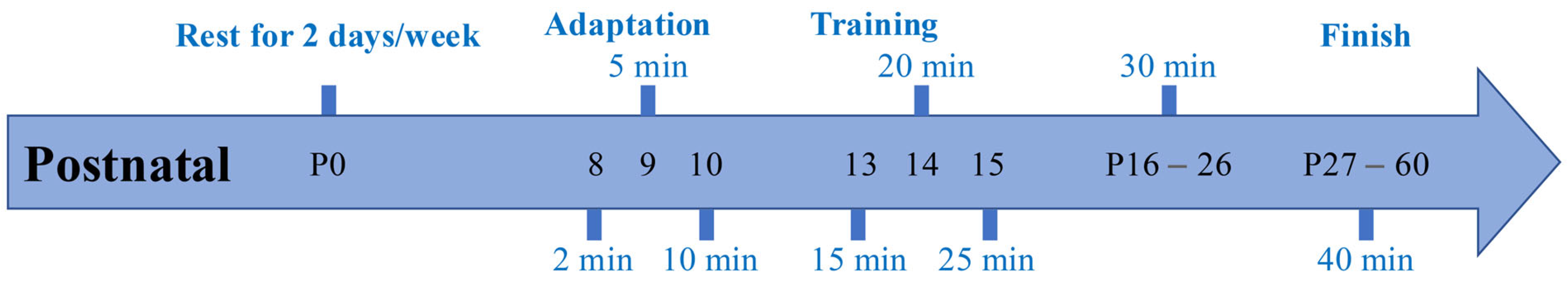
2.1. Experimental Animals and Grouping

Male specific-pathogen-free (SPF) Sprague Dawley (SD) Shank3/ rats at the age of 8 days old were obtained from the Peking University School of Medicine. The germline deletion of Shank3, specifically targeting exon 11–21, was generated using CRISPR-Cas9 technology [19]. The Shank3/+ rats, both male and female, were generated by backcrossing Shank3/ rats with Shank3+/+ SD rats. Subsequently, the Shank3/+ rats were bred to obtain both SD Shank3+/+ and Shank3/ rats for the experimental groups. The rats were randomly allocated into the following three groups based on their genotypes and treatments: the Shank3/ control group (KC; n = 3), Shank3+/+ littermates (WC; n = 3), and the Shank3/ swimming group (KS; n = 3).
The rats were housed in cages containing 3 animals each, with free access to food and water, in a room maintained at a temperature of 20–25 °C, with 45–55% humidity and a 12-h light/dark cycle. All animal procedures and care were conducted in strict accordance with the ethical standards and protocols outlined by the National Institutes of Health (US) [cite PMID 21595115] and were approved by the Biomedical Ethics Committee of Peking University (ethics number LA2021552). After the experiment, all the animals were euthanized by intraperitoneal injection of barbiturates.

2.2. Early Swimming Exercise Program

The swimming protocol employed in this study was adapted from a prior study by Muniz [20] and was performed on the rats in the KS group as depicted in Figure 1. An initial adaptation to swimming was implemented for the rats in the KS group, as illustrated in Figure 1 [10,21]. The initial acclimation to swimming took place during the post-natal days (PND) 8–10 in a cage measuring 485 × 350 × 200 mm that was filled with water maintained at a temperature of 32 ± 1 °C. The duration of each session was 2 min on PND 8, 5 min on PND 9, and 10 min on PND 10. On PND8, the water level was at the height of the rats’ legs. On PND9, the water level was at the height of the rats’ stomachs. On PND10, the water level was at the height of the rats’ necks. Starting from PND 13, the swimming intervention was conducted in a circular tank (150 cm in diameter and 100 cm in height) filled with temperature-controlled water (32 ± 1 °C) to a depth of 50 cm, a setup that remained consistent for the remainder of the experiment. The duration of each session was 15 min on PND 13, 20 min on PND 14, and 25 min on PND 15. Between PND16–26, the rats engaged in 30 min of swimming per day, with 5 consecutive days of swimming followed by 2 days of rest per week. The duration of the swimming sessions was extended to 40 min per day from PND 27 to 60.

2.3. Striatal Metabolite Analysis

2.3.1. Chemicals

This experiment utilized a variety of solutions and reagents. Liquid chromatography-mass-spectrometry-grade methanol and acetonitrile were both purchased from Thermo (Waltham, MA, USA). We sourced 2-chlorophenylalanine from Aladdin (Carlsbad, CA, USA). High-performance liquid-chromatography-grade formic acid was obtained from TCI (Tokyo, Japan). Liquid-chromatography-grade ammonium formate was purchased from Sigma (Darmstadt, Germany). The experimental water was ultrapure water with a resistivity of 18.2 MΩ/cm, and it was purified using a Millipore ultrapure water system (Burlington, NJ, USA). Additionally, ACS-grade chloroform was sourced from Wokai (Beijing, China). These solutions and reagents provided a solid guarantee for the accurate conduct of the experiment.

2.3.2. Metabolite Extraction

The rats were euthanized using pentobarbital sodium at a dose of 150 mg/kg, followed by the careful removal of their brains. To access the striatum, forceps were applied to the medial cortex. Approximately 100 mg (±2%) of striatal tissue was then transferred into a high-throughput tissue grinder (Meibi, Jiaxing, China) containing 1 mL of tissue extraction buffer (composed of 75% methanol/chloroform 9:1 and 25% H2O) kept at −20 °C. Three steel balls with diameters of 3 mm were added to facilitate the grinding process. The tissue was ground at 50 Hz for 60 s, which was repeated twice, followed by sonication at room temperature for 30 min. Subsequently, the samples were centrifuged at 12,000 rpm for 10 min at 4 °C, and 850 μL of the resultant supernatant was carefully extracted and concentrated under vacuum until it reached a dry state. For further analysis by liquid chromatography-mass spectrometry (LC-MS), 200 μL of a solution consisting of 50% acetonitrile in 2-chloro phenylalanine (20 ppm) was added to 200 μL of 50% acetonitrile. The resulting mixture was then filtered through a 0.22 μm membrane.

2.3.3. Chromatographic Conditions

The liquid chromatography (LC)-tandem mass spectrometry (MS/MS) system consists of a Waters UPLC (Dublin, Ireland) separation module coupled to a Thermo LTQ (Waltham, MA, USA) ion trap mass spectrometer. An ACQUITY UPLC® HSS T3 (2.1 × 150 mm, 1.8 µm, Waters, Dublin, Ireland) column was used with a sample temperature of 8 °C at room temperature and 40 °C at a flow rate of 0.25 mL/min. A gradient elution was performed using a mobile phase consisting of 0.1% formic acid in water (A2) and 0.1% formic acid in acetonitrile (B2). The gradient elution procedure consisted of the following steps: ① 0–1 min, 2–50% B2; ② 9.5–14 min, 50–98% B2; ③ 14–15 min, 98% B2; ④ 15–15.5 min, 98–2% B2; and ⑤ 15.5–17 min, 2% B2.

2.3.4. Mass Spectrometric Conditions

An electrospray ionization source (ESI) was employed using both the positive and negative ionization modes. The positive ion spray voltage was set at 4.80 kV, while the negative ion spray voltage was set at 4.50 kV. The sheath gas was maintained at 45 arb, and an auxiliary gas was used at 15 arb. The capillary temperature was set at 325 °C. A full scan was conducted at a resolution of 60,000, with a scan range of 89–1000 for positive ions and 114–1000 for negative ions. High-energy collision-induced dissociation (HCD) was applied for secondary cleavage, utilizing a collision voltage of 30 eV. Additionally, dynamic exclusion was implemented to eliminate unnecessary MS/MS information.

2.3.5. Data Analysis

Peak identification was performed using the XCMS package (version v3.8.2) of the Proteowizard software (version v3.0.8789) and R (version v3.3.2). Additionally, dynamic exclusion was implemented to eliminate unnecessary MS/MS information. This analysis provided essential information, including the mass-to-nucleus ratio (m/z), retention time (rt), and peak area (intensity), which was then normalized using sum peak area normalization. The complete dataset can be accessed at MetaboLights (MTBLS8577).
Metabolite identification was conducted utilizing public spectral databases such as HMDB, MassBank, LipidMaps, mzCloud, and KEGG, as well as the proprietary standard compound library established by Nuomi Metabolomics. The parameter was set with a mass deviation of less than 30 ppm to obtain qualitative metabolite results. The specific principle involved determining the molecular weights of the metabolites based on the m/z of the parent ions in the primary mass spectrum. Molecular formulas were predicted using information on the mass deviation and adduct ions, and then they were matched against the databases to achieve primary metabolite identification. Simultaneously, for the metabolites with detected secondary mass spectra listed in the quantification results, fragment ions and other relevant information were matched with each metabolite in the databases to achieve secondary metabolite identification.
The supervised pattern recognition method was used, and a partial least squares-discriminant analysis (PLS-DA) was performed on each group of data. The parameters of R2X, R2Y, and Q2 were extracted. For data processing, the pheatmap package in R was used for scaling and plotting, applying agglomerative hierarchical clustering with the Euclidean distance as the distance metric. Hierarchical clustering was performed based on the relative values of the metabolites under different experimental conditions, and the results were presented in heat maps. A log10 transformation was applied to the raw data, and clustering for both rows and columns was carried out using the Canberra algorithm. The metabolites were considered significantly different at a p-value < 0.05 and a variable importance in projection score (VIP) of >1. The MetPA database was used to analyze the relevant metabolic pathways for each group of differential metabolites, the hypergeometric test algorithm was used for the data analysis, and relative-betweenness centrality was used for the pathway topology analysis.

3. Results

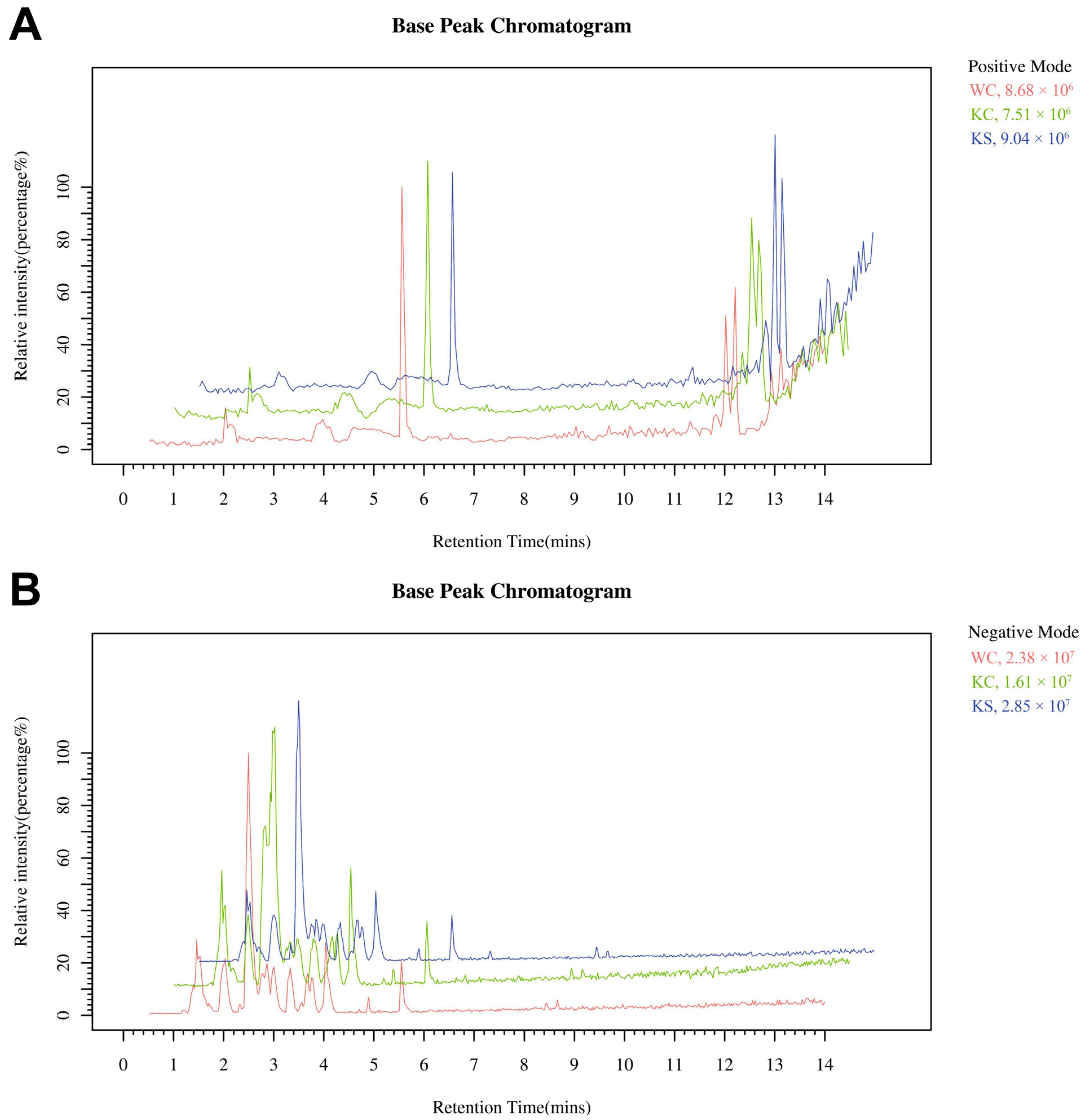
3.1. LC-MS Analysis of the Striatal Metabolome

In this study, LC-MS metabolomics was employed to analyze the metabolite compositions and contents in striatal tissue samples obtained from three distinct groups of rats. These groups included Shank3/ rats that underwent early-life swimming exercise intervention, as well as Shank3/ and Shank3+/+ control rats that did not receive the intervention.
The BPC trend was consistent for all QC samples, indicating perfect reproducibility and reliable data (Figure 2). Remarkably, the retention times and intensity profiles of the sample peaks exhibited excellent reproducibility, indicating both the high quality of the samples and the reliability of our analytical methodology. In addition, a visual inspection of the chromatograms revealed discernible differences among the various experimental groups, warranting further in-depth analysis and investigation.

3.2. PLS-DA

The effectiveness of the current PLS-DA model was assessed using permutation graphs to determine the risk of overfitting. The results, as illustrated in Figure 3, indicated significant differences in the combined positive and negative ions as well as the metabolite profiles among the WC, KC, and KS groups. Specifically, the combined positive and negative ion PLS-DA model exhibited R2Y (cum) = 1 and Q2 (cum) = 0.66. These values indicated a highly robust and reliable fit, with R2Y (cum) = 1 suggesting a perfect explanation of the variance in the response variable by the model and Q2 (cum) = 0.66 indicating a good predictive power and model validity.

3.3. Differential Metabolite Screening

Differential metabolite screening was conducted using stringent criteria, specifically, a p-value < 0.05 and a VIP of >1. These criteria were applied to both the positive and negative ion pattern metabolites in the following three rat striatal metabolism groups: WC, KC, and KS. Subsequently, heat maps representing the differential metabolites of the ions were generated (Figure 4). Following an annotation analysis of the ion pattern differential compounds, a comprehensive list of the differential metabolites was compiled. In the KC and WC groups, a total of 17 differential metabolites were identified, with 14 metabolites showing down-regulation. In the KS and KC groups, 19 differential metabolites were detected, with 18 metabolites exhibiting up-regulation. Similarly, in the KS and WC groups, 22 differential metabolites were found, with 18 metabolites displaying up-regulation. A summary of these findings is presented in Table 1.

3.4. Metabolic Network Analysis of the Differential Metabolites

The metabolic pathway analysis of the differential metabolite enrichment is depicted in Figure 5. In the KC and WC groups, the enrichment of the differential metabolites was observed in several key pathways, including the glutamatergic synapse, the GABAergic synapse, alanine, aspartate and glutamate metabolism, D-glutamine and D-glutamate metabolism, ferroptosis, proximal tubule bicarbonate reclamation, taurine and hypo taurine metabolism, nitrogen metabolism, arginine biosynthesis, and glutathione metabolism. Comparatively, the KS vs. KC and KS vs. WC groups exhibited similar patterns of metabolite enrichment pathways, as observed in the KC vs. WC group. However, the KS vs. KC group displayed an additional increase in the central carbon metabolism in cancer pathway, while the KS vs. WC group exhibited an increase in the glucagon-signaling pathway.

4. Discussion

4.1. Metabolomic Analysis of the Striatum in Shank3 Knockout Rats

ASD represents a complex set of neurodevelopmental disorders with etiological origins rooted in a combination of genetic and environmental factors. Metabolic abnormalities have been identified in several monogenic animal models of ASD, and changes in metabolite levels are emerging as crucial indicators of altered pathway activity in the pathogenesis of ASD. For instance, Rett syndrome, a monogenetic form of ASD resulting from inactivation of the X-linked MECP2 gene encoding the transcription factor methyl cytosine phosphoribosyl guanine binding protein, has been studied extensively [22]. A metabolic analysis of brain extracts from Mecp2-deficient mice, conducted using 1H-NMR, has revealed significant alterations in the D-glutamine and glutamate metabolism [23]. Fragile X syndrome (FXS), the most common genetic cause of ASD, is associated with intellectual disability and results from a recessive X-linked mutation in the fragile X messenger ribonucleoprotein 1 (FMR1) gene [24]. Comprehensive metabolomic studies on the Fmr1-KO mouse model of FXS have demonstrated region-specific metabolic profiles, with the cerebellum and cerebral cortex being particularly affected [25]. These studies have highlighted disruptions in various neurotransmitters, including GABA, glutamate, taurine, and acetylcholine. A metabolite set enrichment analysis (MSEA) revealed significant impacts on the metabolism of D-glutamine, D-glutamate, arginine, and proline [26]. Elevated levels of lipid oxides detected in the cortex of Fmr1-KO mice have indicated increased oxidative stress [26], consistent with previous observations [27,28]. Notably, this metabolic profile exhibited a striking overlap with the pattern detected in the Rett mouse model [23].
In this study, we conducted the first-ever metabolomic analysis of striatal brain regions in a Shank3 knockout animal model. A comparison between the KC group and the WC group revealed that absence of Shank3 led to alterations in 17 differential metabolites. Among these, 14 metabolites exhibited down-regulation while 3 metabolites showed up-regulation. Consistent with previous findings for the central differential metabolites observed in animal models of Rett and FXS syndromes, the striatum of the Shank3 knockout rat model displayed disruptions in key metabolites, including glutamate, glutamine, and oxidized glutathione. These alterations suggest disturbances in synaptic signaling and an increase in oxidative stress. Furthermore, our analysis identified disruptions in metabolites such as taurine, threonine, tyrosine, and glucose, indicating potential abnormalities in neurodevelopmental processes. A functional enrichment analysis of these differential metabolites, combined with transcriptomics [10], revealed functional abnormalities associated with neurodevelopmental processes and oxidative stress. Notably, the disruptions in the GABAergic and glutamatergic synaptic function hint at an imbalance in excitatory-inhibitory (E-I) signaling, a factor closely linked to the pathogenesis of ASD [29].
Our analysis further revealed that several key metabolic pathways were enriched in the striatum of the Shank3 knockout rats compared to the wild-type controls. These pathways, namely, the glutamatergic synapse and ferroptosis, hold significant biological relevance to the physiological conditions of ASD. The glutamatergic synapse pathway involves various metabolites crucial for glutamate synthesis, storage, release, and uptake [30]. Disruptions in glutamatergic signaling have been widely reported in ASD, leading to imbalances in E-I balance and synaptic transmission [31]. Our finding that the glutamatergic synapse pathway was enriched in the Shank3 knockout rats aligned with previous studies showing alterations in glutamate-related metabolites in ASD models. SHANK3 proteins are essential for the formation and stabilization of synapses, particularly glutamatergic synapses [32]. Therefore, the absence of Shank3 likely disrupts glutamate signaling, contributing to the ASD phenotype. The ferroptosis pathway, a form of cell death driven by iron-dependent lipid peroxidation, involves multiple metabolites that regulate cellular redox balance and lipid homeostasis [33]. Recent studies have implicated ferroptosis in the pathogenesis of several neurodevelopmental disorders, including ASD [34]. Oxidative stress, a hallmark of ASD, can trigger ferroptosis by overwhelming cellular antioxidant defenses [35]. Our results indicate that the ferroptosis pathway is enriched in Shank3 knockout rats, suggesting that oxidative stress-induced cell death may play a role in the striatal abnormalities observed in this ASD model. The disruption of Shank3-mediated synaptic functions may lead to increased oxidative stress, promoting ferroptosis and contributing to neuronal damage and dysfunction.

4.2. Metabolomic Changes in the Striatum of Rats After Early Swimming Interventions

Previous research has explored the potential therapeutic benefits of swimming interventions in animal models of neurological disorders. These studies have unveiled possible mechanisms through which swimming can mitigate neurological disorders, including up-regulation of neurotrophic factors within the central nervous system [36,37,38], alterations in synaptic structural proteins [39], changes in synaptic receptor expression [37,40], modifications in neurotransmitter release [41], and adjustments in multiple pathways that affect synaptic plasticity [39,42,43].
In our earlier work, our team conducted transcriptomic studies demonstrating that early swimming interventions not only upregulated genes associated with neural development, synaptic transmission, and synaptic morphology but also modulated various signaling pathways [10]. To delve deeper into the changes in striatal neurotransmission in Shank3 knockout rats following early swimming interventions and to understand the mechanisms behind the positive behavioral effects observed in animal models of ASD, we extended our previous findings by investigating alterations in striatal metabolomics after swimming interventions in Shank3 knockout rats. After early swimming intervention, 19 metabolites were changed in the KS group compared with the KC group, of which 18 metabolites were up-regulated and only 1 metabolite was down-regulated. Crucial metabolites related to neurodevelopment, such as taurine and threonine, as well as signaling molecules like glutamate and glutamine, which were down-regulated in the striatum samples due to the Shank3 knockout, were found to be restored following early swimming interventions. This indicated that early swimming interventions could mitigate aspects of abnormal neurodevelopment and synaptic signaling resulting from Shank3 gene dysfunction. A subsequent pathway enrichment analysis of these differential metabolites revealed up-regulation in the metabolites associated with taurine and subtaurine metabolism, alanine and aspartate metabolism, arginine biosynthesis, D-glutamine and D-glutamate metabolism, and glutathione metabolism, all of which are linked to neurodevelopment and synaptic transmission, particularly in glutamatergic and GABAergic synapses. The modulatory effects of the early swimming interventions on neurodevelopment and E-I balance were further corroborated. Additionally, a combined analysis of the metabolomics and transcriptomics in the KS and KC groups unveiled functions related to energy metabolism, including glycolysis/gluconeogenesis, and functions related to ASD disease development, such as purine metabolism.
Furthermore, an analysis of the differential metabolites between the KS and WC groups was conducted, revealing 22 differential metabolites, with 18 exhibiting up-regulation. Of particular note, metabolites such as taurine, creatine, glutamine, and glutamate, which had been down-regulated in the striatum samples from the Shank3 knockout rats, were found to be restored following early swimming interventions. These findings strongly suggest that abnormalities in related metabolites caused by Shank3 knockout can be effectively reversed by early swimming interventions, potentially leading to improvements in neural development and signaling processes.
The differences in the metabolic enrichment pathways observed between the KS vs. KC and KS vs. WC groups provided deeper insights into the mechanisms by which early swimming interventions may alleviate ASD-like symptoms in Shank3 knockout rats. In the KS vs. KC group comparison, the primary enrichment pathways included those related to synaptic function (e.g., glutamatergic synapse and GABAergic synapse), energy metabolism (e.g., central carbon metabolism in cancer), and antioxidant pathways (e.g., glutathione metabolism). The up-regulation of metabolites involved in the glutamatergic and GABAergic synapses suggests that early swimming interventions help restore the balance of excitatory and inhibitory neurotransmission, which is vital for normal brain function [44]. Moreover, the enrichment in the central carbon metabolism in the cancer pathway may have indicated enhanced energy metabolism in the striatum samples from the rats undergoing swimming exercise, potentially supporting neuronal recovery and synaptic plasticity [45]. The increase in the glutathione metabolism pathway could reflect a reduction in oxidative stress, a recognized factor in ASD pathogenesis [46]. In the KS vs. WC group comparison, a similar pattern of metabolic enrichment was observed, with additional emphasis on pathways such as glucagon signaling. The restoration of metabolites related to synaptic function and energy metabolism, as well as the reduction in oxidative stress, aligned with the improvements seen in the KS vs. KC comparison. The enrichment in the glucagon signaling pathway, which plays a role in glucose homeostasis and energy metabolism, may indicate that early swimming interventions not only improve synaptic function but also optimize overall metabolic health in the striatum [47]. These differences in the metabolic enrichment pathways have profound implications for the study’s outcomes. The restoration of synaptic function and energy metabolism pathways in the KS group, relative to both the KC and WC groups, suggests that early swimming interventions exert a therapeutic effect on the striatal metabolism of Shank3 knockout rats. The up-regulation of antioxidants and the reduction in oxidative stress further support the neuroprotective role of swimming exercise in this ASD model.
Collectively, these findings indicate that early swimming interventions can effectively reverse some of the metabolic abnormalities caused by Shank3 knockout, potentially leading to improvements in synaptic function, neuronal health, and, ultimately, behavioral outcomes in Shank3 knockout rats. This not only offers a mechanistic explanation for the beneficial effects of swimming interventions in ASD but also identifies the key metabolic pathways that may serve as targets for future therapeutic interventions. Furthermore, the differences in metabolic enrichment between the KS vs. KC and KS vs. WC groups underscore the importance of utilizing both knockout and wild-type control groups in metabolomic studies. While comparisons with knockout controls are essential for isolating the effects of an intervention, comparisons with wild-type controls provide additional insights into the extent to which the intervention can restore normal metabolic function. Overall, the comparative analysis of the metabolic enrichment pathways in this study revealed that early swimming interventions have profound impacts on striatal metabolism in Shank3 knockout rats, suggesting a potential therapeutic role for swimming exercise in ASD.

5. Conclusions

In summary, the metabolomic findings indicate that Shank3 knockout results in abnormal striatal neurodevelopment and disrupted glutamatergic and GABAergic synaptic function in rats. Additionally, concerning neurotransmitter release, there is a potential disruption in the excitatory–inhibitory (E-I) balance of synaptic signaling. Early swimming interventions effectively reversed the down-regulation of some essential metabolites associated with neurodevelopment and synaptic signaling, which had been negatively impacted by Shank3 knockout. This reversal could be a key factor contributing to the improvement in the behavior of Shank3 knockout rats following early swimming interventions.

Author Contributions

Conceptualization, Z.Z.; formal analysis, Y.M.; data curation, Y.X.; writing—original draft preparation, Y.M.; writing—review and editing, Y.H.; funding acquisition, Z.Z. All authors have read and agreed to the published version of the manuscript.

Funding

This work was supported by the Beijing Natural Science Foundation (7232239) and the Humanities and Social Sciences Project of the Ministry of Education (23YJC890027).

Institutional Review Board Statement

The animal study procedure was agreed to by the Animal Care and Use Committee of Peking University (approval no. LA2021552, approved in October 2021).

Informed Consent Statement

Not applicable.

Data Availability Statement

The complete dataset can be accessed at MetaboLights (MTBLS8577).

Conflicts of Interest

The authors declare no conflicts of interest.

References

  1. Knopf, A. Autism prevalence increases from 1 in 60 to 1 in 54: CDC. Brown Univ. Child Adolesc. Behav. Lett. 2020, 36, 4. [Google Scholar] [CrossRef]
  2. Edition, F. Diagnostic and statistical manual of mental disorders. Am. Psychiatr. Assoc. 2013, 21, 591–643. [Google Scholar]
  3. Zhao, M.; Chen, S. The Effects of Structured Physical Activity Program on Social Interaction and Communication for Children with Autism. BioMed Res. Int. 2018, 2018, 1825046. [Google Scholar] [CrossRef] [PubMed]
  4. Jiang, X.; Chen, X.; Su, J.; Liu, N. Prevalence of autism spectrum disorder in mainland china over the past 6 years: A systematic review and meta-analysis. BMC Psychiatry 2024, 24, 404. [Google Scholar] [CrossRef]
  5. Shahat, A.R.S.; Greco, G. The Economic Costs of Childhood Disability: A Literature Review. Int. J. Environ. Res. Public Health 2021, 18, 3531. [Google Scholar] [CrossRef]
  6. Qiu, S.; Qiu, Y.; Li, Y.; Cong, X. Genetics of autism spectrum disorder: An umbrella review of systematic reviews and meta-analyses. Transl. Psychiatry 2022, 12, 249. [Google Scholar] [CrossRef]
  7. Ioannidis, V.; Pandey, R.; Bauer, H.F.; Schön, M.; Bockmann, J.; Boeckers, T.M.; Lutz, A.K. Disrupted extracellular matrix and cell cycle genes in autism-associated Shank3 deficiency are targeted by lithium. Mol. Psychiatry 2024, 29, 704–717. [Google Scholar] [CrossRef]
  8. Monteiro, P.; Feng, G.P. SHANK proteins: Roles at the synapse and in autism spectrum disorder. Nat. Rev. Neurosci. 2017, 18, 147–157. [Google Scholar] [CrossRef]
  9. Xu, D.; Meng, Y.; An, S.; Meng, W.; Li, H.; Zhang, W.; Xue, Y.; Lan, X.; Wang, X.; Li, M.; et al. Swimming exercise is a promising early intervention for autism-like behavior in Shank3 deletion rats. CNS Neurosci. Ther. 2023, 29, 78–90. [Google Scholar] [CrossRef]
  10. Meng, Y.; Xu, D.; Zhang, W.; Meng, W.; Lan, X.; Wang, X.; Li, M.; Zhang, X.; Zhao, Y.; Yang, H.; et al. Effect of Early Swimming on the Behavior and Striatal Transcriptome of the Shank3 Knockout Rat Model of Autism. Neuropsychiatr. Dis. Treat. 2022, 18, 681–694. [Google Scholar] [CrossRef]
  11. Lee, S.S.; Kim, C.J.; Shin, M.S.; Lim, B.V. Treadmill exercise ameliorates memory impairment through ERK-Akt-CREB-BDNF signaling pathway in cerebral ischemia gerbils. J. Exerc. Rehabil. 2020, 16, 49–57. [Google Scholar] [CrossRef] [PubMed]
  12. Fragala-Pinkham, M.; Haley, S.M.; O’Neil, M.E. Group aquatic aerobic exercise for children with disabilities. Dev. Med. Child Neurol. 2008, 50, 822–827. [Google Scholar] [CrossRef] [PubMed]
  13. Pan, C.Y. The efficacy of an aquatic program on physical fitness and aquatic skills in children with and without autism spectrum disorders. Res. Autism Spectr. Disord. 2011, 5, 657–665. [Google Scholar] [CrossRef]
  14. Ennis, E. The effects of a physical therapy-directed aquatic program on children with autism spectrum disorders. J. Aquat. Phys. Ther. 2011, 19, 4–10. [Google Scholar]
  15. Kim, H.; Woo, R.S.; Yang, E.J.; Kim, H.B.; Jo, E.H.; Lee, S.; Im, H.; Kim, S.; Kim, H.S. Transcriptomic analysis in the striatum reveals the involvement of Nurr1 in the social behavior of prenatally valproic acid-exposed male mice. Transl. Psychiatry 2022, 12, 324. [Google Scholar] [CrossRef]
  16. Montanari, M.; Martella, G.; Bonsi, P.; Meringolo, M. Autism Spectrum Disorder: Focus on Glutamatergic Neurotransmission. Int. J. Mol. Sci. 2022, 23, 3861. [Google Scholar] [CrossRef]
  17. Roberts, B.M.; Lopes, E.F.; Cragg, S.J. Axonal modulation of striatal dopamine release by local γ-aminobutyric acid (GABA) signalling. Cells 2021, 10, 709. [Google Scholar] [CrossRef]
  18. Nicholson, J.K.; Connelly, J.; Lindon, J.C.; Holmes, E. Metabonomics: A platform for studying drug toxicity and gene function. Nat. Rev. Drug Discov. 2002, 1, 153–161. [Google Scholar] [CrossRef]
  19. Song, T.J.; Lan, X.Y.; Wei, M.P.; Zhai, F.J.; Boeckers, T.M.; Wang, J.N.; Yuan, S.; Jin, M.Y.; Xie, Y.F.; Dang, W.W.; et al. Altered Behaviors and Impaired Synaptic Function in a Novel Rat Model With a Complete Shank3 Deletion. Front. Cell. Neurosci. 2019, 13, 111. [Google Scholar] [CrossRef]
  20. de Santana Muniz, G.; da Silva, A.M.A.; Cavalcante, T.C.F.; da Silva França, A.K.; Ferraz, K.M.; do Nascimento, E. Early physical activity minimizes the adverse effects of a low-energy diet on growth and development parameters. Nutr. Neurosci. 2013, 16, 113–124. [Google Scholar] [CrossRef]
  21. Meng, W.; Xu, D.; Meng, Y.; Zhang, W.; Xue, Y.; Zhen, Z.; Gao, Y. Changes in the urinary proteome in rats with regular swimming exercise. PeerJ 2021, 9, e12406. [Google Scholar] [CrossRef] [PubMed]
  22. Weng, S.-M.; Bailey, M.E.; Cobb, S.R. Rett syndrome: From bed to bench. Pediatr. Neonatol. 2011, 52, 309–316. [Google Scholar] [CrossRef] [PubMed]
  23. Viola, A.; Saywell, V.; Villard, L.; Cozzone, P.J.; Lutz, N.W. Metabolic fingerprints of altered brain growth, osmoregulation and neurotransmission in a Rett syndrome model. PLoS ONE 2007, 2, e157. [Google Scholar] [CrossRef]
  24. Penagarikano, O.; Mulle, J.G.; Warren, S.T. The pathophysiology of fragile x syndrome. Annu. Rev. Genom. Hum. Genet. 2007, 8, 109–129. [Google Scholar] [CrossRef]
  25. Mientjes, E.; Nieuwenhuizen, I.; Kirkpatrick, L.; Zu, T.; Hoogeveen-Westerveld, M.; Severijnen, L.; Rifé, M.; Willemsen, R.; Nelson, D.; Oostra, B. The generation of a conditional Fmr1 knock out mouse model to study Fmrp function in vivo. Neurobiol. Dis. 2006, 21, 549–555. [Google Scholar] [CrossRef]
  26. Davidovic, L.; Navratil, V.; Bonaccorso, C.M.; Catania, M.V.; Bardoni, B.; Dumas, M.-E. A metabolomic and systems biology perspective on the brain of the fragile X syndrome mouse model. Genome Res. 2011, 21, 2190–2202. [Google Scholar] [CrossRef]
  27. Bechara, E.G.; Didiot, M.C.; Melko, M.; Davidovic, L.; Bensaid, M.; Martin, P.; Castets, M.; Pognonec, P.; Khandjian, E.W.; Moine, H. A novel function for fragile X mental retardation protein in translational activation. PLoS Biol. 2009, 7, e1000016. [Google Scholar] [CrossRef]
  28. de Diego-Otero, Y.; Romero-Zerbo, Y.; Bekay, R.; Decara, J.; Sanchez, L.; Fonseca, F.R.-d.; Arco-Herrera, I.d. α-tocopherol protects against oxidative stress in the fragile X knockout mouse: An experimental therapeutic approach for the Fmr1 deficiency. Neuropsychopharmacology 2009, 34, 1011–1026. [Google Scholar] [CrossRef]
  29. Dickinson, A.; Bruyns-Haylett, M.; Jones, M.; Milne, E. Increased peak gamma frequency in individuals with higher levels of autistic traits. Eur. J. Neurosci. 2015, 41, 1095–1101. [Google Scholar] [CrossRef]
  30. Mahmoud, S.; Gharagozloo, M.; Simard, C.; Gris, D. Astrocytes Maintain Glutamate Homeostasis in the CNS by Controlling the Balance between Glutamate Uptake and Release. Cells 2019, 8, 184. [Google Scholar] [CrossRef]
  31. Nisar, S.; Bhat, A.A.; Masoodi, T.; Hashem, S.; Akhtar, S.; Ali, T.A.; Amjad, S.; Chawla, S.; Bagga, P.; Frenneaux, M.P.; et al. Genetics of glutamate and its receptors in autism spectrum disorder. Mol. Psychiatry 2022, 27, 2380–2392. [Google Scholar] [CrossRef] [PubMed]
  32. Kim, Y.; Ko, T.H.; Jin, C.; Zhang, Y.; Kang, H.R.; Ma, R.; Li, H.; Choi, J.I.; Han, K. The emerging roles of Shank3 in cardiac function and dysfunction. Front. Cell Dev. Biol. 2023, 11, 1191369. [Google Scholar] [CrossRef] [PubMed]
  33. Tang, D.; Chen, X.; Kang, R.; Kroemer, G. Ferroptosis: Molecular mechanisms and health implications. Cell Res. 2021, 31, 107–125. [Google Scholar] [CrossRef]
  34. Jiang, P.; Zhou, L.; Zhao, L.; Fei, X.; Wang, Z.; Liu, T.; Tang, Y.; Li, D.; Gong, H.; Luo, Y.; et al. Puerarin attenuates valproate-induced features of ASD in male mice via regulating Slc7a11-dependent ferroptosis. Neuropsychopharmacology 2024, 49, 497–507. [Google Scholar] [CrossRef]
  35. Liu, X.; Lin, J.; Zhang, H.; Khan, N.U.; Zhang, J.; Tang, X.; Cao, X.; Shen, L. Oxidative Stress in Autism Spectrum Disorder-Current Progress of Mechanisms and Biomarkers. Front. Psychiatry 2022, 13, 813304. [Google Scholar] [CrossRef]
  36. Sigwalt, A.R.; Budde, H.; Helmich, I.; Glaser, V.; Ghisoni, K.; Lanza, S.; Cadore, E.L.; Lhullier, F.L.R.; de Bem, A.F.; Hohl, A.; et al. Molecular aspects involved in swimming exercise training reducing anhedonia in a rat model of depression. Neuroscience 2011, 192, 661–674. [Google Scholar] [CrossRef]
  37. Boracı, H.; Kirazlı, Ö.; Gülhan, R.; Yıldız Sercan, D.; Şehirli, Ü.S. Neuroprotective effect of regular swimming exercise on calretinin-positive striatal neurons of Parkinsonian rats. Anat. Sci. Int. 2020, 95, 429–439. [Google Scholar] [CrossRef]
  38. Gyorkos, A.M.; McCullough, M.J.; Spitsbergen, J.M. Glial cell line-derived neurotrophic factor (GDNF) expression and NMJ plasticity in skeletal muscle following endurance exercise. Neuroscience 2014, 257, 111–118. [Google Scholar] [CrossRef]
  39. Liu, W.; Xue, X.; Xia, J.; Liu, J.; Qi, Z. Swimming exercise reverses CUMS-induced changes in depression-like behaviors and hippocampal plasticity-related proteins. J. Affect. Disord. 2018, 227, 126–135. [Google Scholar] [CrossRef]
  40. Ko, I.-G.; Kim, S.-E.; Kim, T.-W.; Ji, E.-S.; Shin, M.-S.; Kim, C.-J.; Hong, M.-H.; Bahn, G.H. Swimming exercise alleviates the symptoms of attention-deficit hyperactivity disorder in spontaneous hypertensive rats. Mol. Med. Rep. 2013, 8, 393–400. [Google Scholar] [CrossRef]
  41. Leite, H.R.; Mourão, F.A.G.; Drumond, L.E.; Ferreira-Vieira, T.H.; Bernardes, D.; Silva, J.F.; Lemos, V.S.; Moraes, M.F.D.; Pereira, G.S.; Carvalho-Tavares, J.; et al. Swim training attenuates oxidative damage and promotes neuroprotection in cerebral cortical slices submitted to oxygen glucose deprivation. J. Neurochem. 2012, 123, 317–324. [Google Scholar] [CrossRef] [PubMed]
  42. Aguiar Jr, A.S.; Castro, A.A.; Moreira, E.L.; Glaser, V.; Santos, A.R.; Tasca, C.I.; Latini, A.; Prediger, R.D. Short bouts of mild-intensity physical exercise improve spatial learning and memory in aging rats: Involvement of hippocampal plasticity via AKT, CREB and BDNF signaling. Mech. Ageing Dev. 2011, 132, 560–567. [Google Scholar] [CrossRef] [PubMed]
  43. Anand, S.; Devi, S.A.; Ravikiran, T. Differential expression of the cerebral cortex proteome in physically trained adult rats. Brain Res. Bull. 2014, 104, 88–91. [Google Scholar] [CrossRef] [PubMed]
  44. Harmon, T.C.; McLean, D.L.; Raman, I.M. Integration of Swimming-Related Synaptic Excitation and Inhibition by olig2(+) Eurydendroid Neurons in Larval Zebrafish Cerebellum. J. Neurosci. 2020, 40, 3063–3074. [Google Scholar] [CrossRef]
  45. Newman, A.C.; Maddocks, O.D.K. One-carbon metabolism in cancer. Br. J. Cancer 2017, 116, 1499–1504. [Google Scholar] [CrossRef]
  46. Kwon, D.H.; Cha, H.J.; Lee, H.; Hong, S.H.; Park, C.; Park, S.H.; Kim, G.Y.; Kim, S.; Kim, H.S.; Hwang, H.J.; et al. Protective Effect of Glutathione against Oxidative Stress-induced Cytotoxicity in RAW 264.7 Macrophages through Activating the Nuclear Factor Erythroid 2-Related Factor-2/Heme Oxygenase-1 Pathway. Antioxidants 2019, 8, 82. [Google Scholar] [CrossRef]
  47. Jiang, G.; Zhang, B.B. Glucagon and regulation of glucose metabolism. Am. J. Physiol. Endocrinol. Metab. 2003, 284, E671–E678. [Google Scholar] [CrossRef]
Figure 1. Early swimming intervention program [10]. The protocol included swimming from postnatal day (P) 8 to 60 and always at the same time (17:00 to 18:00), with rest periods for two consecutive rest days by week. The pups practiced swimming for 2, 5, and 10 min on P8, 9, and 10, respectively, and rested on P11 and 12. On P13, the rats swam for 15 and 20 min on P14 and 25 min on P15. From P16 to 26, the rats swam for 30 min/day, 5 times per week. From days 27 to 60, the rats swam for 40 min/day, 5 times per week.
Metabolites 15 00134 g001
Figure 2. Ion-mode base peak chromatogram. (A) A chromatogram of the positive ion mode base peak, and (B) a chromatogram of the negative ion mode base peak.
Metabolites 15 00134 g002
Figure 3. Plots of the PLS-DA scores, loadings, and replacement tests for each comparison group in the positive and negative ion modes.
Metabolites 15 00134 g003
Figure 4. Heat map of the ionic pattern differences in the metabolites.
Metabolites 15 00134 g004
Figure 5. Differential metabolite enrichment pathways in the striatum samples of each comparison group. (A) A bubble diagram of the differential metabolite enrichment pathway in the KC vs. WC groups; (B) a bubble diagram of the differential metabolite enrichment pathway in the KS vs. KC groups; and (C) a bubble diagram of the differential metabolite enrichment pathway in the KS vs. WC groups.
Metabolites 15 00134 g005
Table 1. Differential metabolite profiles of the striatal tissue samples in the WC, KC, and KS groups.
No.Metabolitesm/zRT (s)FormulaKEGGTypeFold Change
KC/WCKS/KCKS/WC
1Taurine124.0189.03C2H7NO3SC00245[M−H]−0.92 *1.23 *1.13 *
2Pyroglutamic acid128.04177.84C5H7NO3C01879[M−H]−0.881.57 *1.39 *
3(E)-Glutaconate129.02103.77C5H6O4C02214[M−H]−0.31 *4.53 *1.43 *
4Creatine130.0699.11C4H9N3O2C00300[M−H]−0.88 *1.29 *1.14 *
5L-Malic acid133.01119.43C4H6O5C00149[M−H]−0.84 *1.62 *1.36 *
6Threonic acid135.0397.76C4H8O5C01620[M−H]−0.74 *2.15 *1.59 *
7L-Glutamine145.0688.52C5H10N2O3C00064[M−H]−0.86 *1.58 *1.36 *
8L-Glutamic acid146.0592.43C5H9NO4C00025[M−H]−0.86 *1.53 *1.32 *
93-Hydroxymethylglutaric acid161.05239.48C6H10O5C03761[M−H]−0.78 *2.66 *2.08 *
10Myo-Inositol161.05556.35C6H12O6C00137[M−H2O−H]−0.61 *1.250.76 *
11L-Phenylalanine166.09280.18C9H11NO2C00079[M+H]+1.062.16 *2.29 *
12(2R)-2-Hydroxy-3-propanoate166.98105.63C3H7O7PC00197[M−H2O−H]−1.052.04 *2.14 *
13N-Acetyl-L-aspartic acid174.04151.21C6H9NO5C01042[M−H]−1.41 *1.95 *2.76 *
14Alpha-D-Glucose179.06763.79C6H12O6C00267[M−H]−0.46 *1.310.61 *
15L-Iditol181.07665.47C6H14O6C01507[M−H]−0.76 *1.210.92
16(S)-beta-Tyrosine181.07808.76C9H11NO3C21308[M]−0.29 *1.200.35 *
17N-Acetylglutamic acid188.06200.43C7H11NO5C00624[M−H]−0.842.28 *1.92 *
18Citric acid191.02167.31C6H8O7C00158[M−H]−0.991.85 *1.82 *
19Sebacic acid201.11565.53C10H18O4C08277[M−H]−0.967.66 *7.32 *
20Pantothenic acid218.10294.11C9H17NO5C00864[M−H]−0.59 *1.64 *0.97
21Gamma-Glutamylcysteine248.9681.79C8H14N2O5SC00669[M−H]−0.84 *1.180.98
22N2-gamma-Glutamylglutamine274.10101.94C10H17N3O6C05283[M−H]−1.101.77 *1.95 *
23Dehydroepiandrosterone288.29724.49C19H28O2C01227[M]+1.89 *0.51 *0.96
24Oxidized glutathione611.14219.81C20H32N6O12S2C00127[M−H]−2.20 *2.05 *4.52 *
25Theophylline179.06643.65C7H8N4O2C07130[M−H]−0.880.850.74 *
26Hypoxanthine135.03170.48C5H4N4OC00262[M−H]−1.241.291.60 *
“*” indicates a significant difference between the two groups (p < 0.05).
Disclaimer/Publisher’s Note: The statements, opinions and data contained in all publications are solely those of the individual author(s) and contributor(s) and not of MDPI and/or the editor(s). MDPI and/or the editor(s) disclaim responsibility for any injury to people or property resulting from any ideas, methods, instructions or products referred to in the content.

