Review

Targeting Metabolic Dysregulation in Obesity and Metabolic Syndrome: The Emerging Role of N-Acetylcysteine

by
Dorota Magdalena Radomska-Leśniewska
1,
Justyna Niderla-Bielińska
1,
Marek Kujawa
2 and
Ewa Jankowska-Steifer
1,*
1
Department of Histology and Embryology, Faculty of Medicine, Medical University of Warsaw, Chalubinskiego 5 Str, 02-004 Warsaw, Poland
2
Department of Histology and Embryology, Faculty of Medicine, Lazarski University, 02-662 Warsaw, Poland
*
Author to whom correspondence should be addressed.
Metabolites 2025, 15(10), 645; https://doi.org/10.3390/metabo15100645
Submission received: 22 August 2025 / Revised: 16 September 2025 / Accepted: 22 September 2025 / Published: 26 September 2025
(This article belongs to the Special Issue Metabolic Modulators in Cardiovascular Disease Management)

Abstract

Obesity and metabolic syndrome (MetS), growing global health concerns, are closely linked to the development of insulin resistance, type 2 diabetes, steatotic liver disease, and cardiovascular diseases (CVDs). An increase in visceral adipose tissue, the main symptom of MetS, contributes to systemic metabolic dysfunction, resulting in disturbances in glucose and lipid metabolism, mitochondrial dysfunction, and redox imbalance, which creates a vicious cycle of inflammation and oxidative stress, accelerating comorbidities. N-acetylcysteine (NAC), a precursor to glutathione, with antioxidant and anti-inflammatory properties, is described as a potent metabolic modulator that restores metabolic homeostasis. NAC’s ability to modulate oxidative stress and inflammation may be particularly valuable in preventing or mitigating cardiovascular complications of MetS. The aim of this narrative review is to summarize current evidence from cellular, animal, and human studies on NAC’s impact on metabolic health. MetS affects nearly one-third of the global population; therefore, there is a pressing need for accessible therapeutic strategies. NAC appears to offer potential benefits as an adjunctive agent for individuals with metabolic disturbances, but further research is needed to confirm its efficacy and establish its role in clinical practice.

1. Introduction

Overweight and obesity are currently among the world’s major health problems. According to the World Health Organization (WHO) European Regional Obesity Report 2022, nearly two-thirds of adults and one in three children in the WHO European Region are overweight or obese, and unfortunately, this trend is increasing compared to previous years. Given this upward trend, it is estimated that by 2050, nearly two in three adults aged 25 and older will be overweight or obese [1]. The consequence of obesity is an increase in visceral adipose tissue, which, due to its specific metabolic activity, has a negative impact on the functioning of the body. Visceral adipose tissue accumulation is the most common and key component of metabolic syndrome (MetS), which is characterized by the coexistence of abdominal obesity, hyperglycemia, dyslipidemia, insulin resistance, and hypertension [2]. In clinical practice, MetS is typically diagnosed using criteria such as waist circumference (≥102 cm in men, ≥88 cm in women), elevated triglycerides (≥150 mg/dL), reduced HDL cholesterol, high blood pressure, and fasting glucose ≥5.6 mmol/L. To better assess visceral adiposity and cardiometabolic risk, composite indices like the Lipid Accumulation Product (LAP) and Cardiometabolic Index (CMI) have been proposed, with cut-off values of LAP ≥ 49.87 or CMI ≥ 0.56 for women and LAP ≥ 52.76 or CMI ≥ 0.70 for men. These markers show superior diagnostic accuracy compared to traditional measures such as body mass index (BMI) or waist circumference alone [3]. Visceral obesity itself is an independent component of the MetS, and the severity of this obesity is directly related to the prognosis of this condition [4,5]. Obesity predisposes to the development of insulin resistance, type 2 diabetes (T2DM), non-alcoholic fatty liver disease (now known as metabolic dysfunction-associated steatotic liver disease—MASLD), and cardiovascular diseases (CVDs) [6]. CVDs are one of the leading causes of death in the world, and among the most important risk factors for these diseases are metabolic disorders resulting from abdominal obesity, which may lead to the development of hypertension, insulin resistance, and atherogenic dyslipidemia. The pathophysiological mechanisms linking abdominal obesity with CVD are complex and multifaceted [7,8].
Given the complexity of metabolic disturbances in the pathogenesis of CVD, restoring normal metabolism seems to be a viable strategy for improving heart function or preventing further damage. Compounds capable of influencing key metabolic pathways—such as glucose and lipid metabolism, mitochondrial function, or redox homeostasis—in a way that helps restore metabolic balance are referred to as metabolic modulators [9]. To date, metabolic modulators in CVD have been used to modify cardiac energy metabolism, particularly in heart failure (HF) [10], and modulate glucose and lipid transporters, mitochondrial function, and inflammatory signaling [11]. Metabolic modulators encompass a wide range of compounds, including peroxisome proliferator-activated receptor (PPAR) agonists, fibroblast growth factor 21 (FGF21) analogues, thyroid hormone receptor β agonists, and glucagon-like peptide-1 (GLP-1) receptor agonists [12,13,14,15]. Currently, one of the most widely studied and promising metabolic modulators is N-acetylcysteine (NAC) due to its broad spectrum of biological activity relevant to cardiometabolic health. According to numerous studies, NAC may improve insulin sensitivity, reduce oxidative stress, and support the proper function of adipose tissue, which may be beneficial for MetS patients [16]. Therefore, the aim of this paper is to present current information regarding NAC’s impact on MetS symptoms in cell, animal, and human studies. Since MetS and obesity affect more and more patients, it is extremely important to develop new therapeutic strategies not only to effectively manage MetS comorbidities but also to prevent metabolic deregulation. A healthy lifestyle combined with metabolic modulators, such as NAC, may be a future option for high-risk patients, although more clinical trials are needed to evaluate the efficacy of this substance.

2. Methodology

This narrative review was conducted to synthesize current knowledge on the role of NAC in MetS, obesity-related metabolic disturbances, and cardiovascular health. We searched the PubMed database using keywords including “NAC”, “metabolic syndrome”, “obesity”, “cardiovascular health”, “insulin sensitivity”, “oxidative stress”, “visceral adipose tissue”, and “leptin signaling”. Particular attention was given to studies published within the last five years, including original research articles, reviews, and experimental studies in both animal and human models. The selection of the literature was guided by relevance to the topic, with emphasis on studies exploring NAC’s biochemical actions, its influence on adipose tissue function, and its potential therapeutic role in mitigating cardiometabolic risk. Additionally, older foundational studies were included when they provided essential background information or mechanistic insights relevant to the topic. The review is structured to first present the pathophysiological mechanisms underlying metabolic disturbances in obesity and MetS, followed by a discussion of NAC’s effects on cellular metabolism and cardiovascular outcomes. Images were created by the authors with the use of image elements provided by Servier Medical Art (https://smart.servier.com/) accessed on 12 August 2025, licensed under CC BY 4.0 (https://creativecommons.org/licenses/by/4.0/).

3. MetS and Metabolic Disruption

3.1. Visceral Adipose Tissue

There are two types of white adipose tissue, based on its anatomical location: subcutaneous adipose tissue (SAT) and visceral adipose tissue (VAT). SAT is located under the skin, primarily in the abdominal region, buttocks, thighs, and upper limbs. VAT, also known as ectopic fat, accumulates deep in the abdominal cavity, surrounding vital organs such as the liver, intestines, heart (pericardium, epicardium), and skeletal muscle. It can also occur in areas of the body not typically associated with fat storage and containing only small amounts of fat, such as the renal sinus, pancreas, thoracic cage, periaortic sinus, and perivascular areas [8,17]. VAT and SAT differ in their cellular and molecular characteristics, metabolic functions, clinical implications, and prognostic significance [18]. Compared with SAT, VAT consists of larger adipocytes due to enlarged lipid droplets. It is better vascularized and innervated [19]. Adipocyte hypertrophy in VAT is highly correlated with the accumulation of proinflammatory adipokines and immune cells [20]. All types of VAT are metabolically active, insulin-resistant due to a low number of insulin receptors, highly lipolytic, and resistant to the antilipolytic effects of insulin. Compared with SAT, VAT responds better to adrenergic stimulation and has a greater capacity for free fatty acid (FFA) release and glucose uptake. Its metabolic profile contributes to systemic insulin resistance and increases the risk of T2DM, obstructive sleep apnea, and CVD [8,19]. It should be remembered that adipose tissue also accumulates around the heart as epicardial adipose tissue (EAT), which occurs between the outer wall of the myocardium and the visceral layer of the pericardium. Under physiological conditions, EAT provides the heart with mechanical, thermogenic, and metabolic support and releases cytokines and chemokines into blood vessels [21]. In obesity, EAT becomes dysfunctional and contributes to myocardial connective tissue hypertrophy and fibrosis, and through the secretion of proinflammatory factors, reduces the synthesis of adiponectin, an important regulator of glucose and lipid metabolism [8].

3.2. Lipid Metabolism and Its Deregulation

Dietary fats are absorbed by enterocytes in the form of FFA and monoglycerides, and then converted into triglycerides (TGs), which, together with cholesterol, phospholipids, and apolipoproteins (primarily ApoB-48), form lipoproteins called chylomicrons, which are distributed through the lymph and blood. TGs in chylomicrons are broken down by lipoprotein lipase into FFA and glycerol, enabling their uptake by target cells, such as adipocytes and muscle cells. Free fatty acids are used for energy production through beta-oxidation in mitochondria [22,23]. Fatty acids in the form of TG are stored in adipocytes, and in the event of glucose deficiency, they are converted into ketone bodies in the liver. The degraded chylomicron remnants are taken up by the liver. Cholesterol is also absorbed and synthesized in the intestines and transported in lipoproteins of varying density [24,25]. Cholesterol is a structural component of cell membranes and is used to produce hormones, vitamin D, or stored as cholesterol esters [26]. A consequence of obesity and VAT accumulation is atherogenic dyslipidemia—a lipid disorder characterized by abnormal levels of cholesterol and TG in the blood. Triglyceride levels increase, while high-density lipoprotein (HDL) cholesterol (which reduces the risk of CVD) decreases. The lipid-overloaded liver produces more small low-density lipoprotein (LDL) particles (key transmitter of cholesterol to the vascular artery wall), which are directly associated with the risk of atherosclerotic cardiovascular events [27,28,29]. Preliminary estimates indicate that approximately 60–70% of obese individuals and 50–60% of overweight individuals suffer from dyslipidemia, a significant risk factor for CVD [30,31].

3.3. Glucose Metabolism and Its Deregulation

Dietary carbohydrates, digested into simple, soluble sugars, are transported through enterocytes in the intestinal wall into the circulatory system and then to various tissues [32]. Glucose uptake in skeletal muscle and adipose tissue is regulated by insulin, which translocates the GLUT4 glucose transporter to the cell membrane [33]. Glucose catabolism, leading to ATP production, involves three sequential processes: glycolysis, the tricarboxylic acid cycle (Krebs cycle), and finally oxidative phosphorylation [34]. When excess glucose is available, insulin activates glycogen synthase, initiating glycogenesis in muscle and liver [35]. High blood glucose levels often coexist with insulin resistance—a condition in which cells (especially muscle and adipocytes) respond poorly to insulin. The body compensates by increasing insulin secretion from the pancreas, leading to hyperinsulinemia. Chronic hyperglycemia leads to cell dysfunction and apoptosis, resulting in T2DM [36]. People with T2DM have a higher risk of developing and dying from CVD (such as myocardial infarction, stroke, and heart failure) [37].

3.4. Adipokine Profile of Visceral Adiposity

VAT, through its paracrine activity and the secretion of bioactive molecules, i.e., adipokines, participates in the regulation of metabolic, inflammatory, and cardiovascular processes [38]. Obesity disrupts their production, which contributes to the development of many diseases, including CVD [39]. Pro-inflammatory adipokines, including leptin, resistin, visfatin, and chemerin, contribute to the development and progression of chronic inflammation. In contrast, adipokines such as adiponectin, vaspin, apelin, omentin, and isthmin-1 exert anti-inflammatory effects and are associated with improved metabolic homeostasis [40]. Adiponectin is the main cardioprotective adipokine and exerts a protective effect against the development of obesity-related CVD. Its high levels correlate with a lower risk of atherosclerosis, hypertension, and coronary artery disease, while in obesity, its deficiency contributes to the development of chronic inflammation and vascular dysfunction. It has been documented that circulating adiponectin levels are reduced in CVD [41]. Obesity increases VAT volume, which is the main source of leptin, which typically plays a key role in regulating appetite, energy homeostasis, and body weight. However, despite high leptin levels, many obese individuals experience leptin resistance, and high circulating leptin levels can lead to endothelial dysfunction, which is a precursor and major contributor to hypertension, atherosclerosis, and coronary artery disease [8,42].

3.5. Integrated Metabolic Disruption in CVD

Metabolism is a multifactorial phenomenon in which individual components are interrelated and exert reciprocal effects on one another, creating, in pathological conditions, a self-perpetuating cycle. Dyslipidemia and hyperglycemia are closely interconnected metabolic disturbances commonly observed in obesity, and together they significantly contribute to the development of CVD [43]. Elevated levels of FFA in obesity promote insulin resistance, which impairs glucose uptake and leads to chronic hyperglycemia [44]. In turn, hyperglycemia exacerbates oxidative stress and inflammatory signaling, further disrupting lipid metabolism and promoting atherogenic lipid profiles. Insulin resistance causes increased lipolysis of TG from adipose tissue and increased glucose production in the liver [22]. Moreover, hyperglycemia can modify lipoproteins through non-enzymatic glycation, making them more prone to oxidation and less efficiently cleared from circulation. These modified lipoproteins contribute to endothelial dysfunction and accelerate atherosclerotic plaque formation, a risk factor of atherosclerotic CVD [45,46]. Hyperglycemia, lipid accumulation in tissues, chronic low-grade inflammation, elevated leptin levels, endothelial dysfunction, and impaired mitochondrial function can lead to increased production of reactive oxygen species (ROS) [47]. ROS are integral components of many cellular pathways. Under physiological conditions, in small amounts, ROS exhibit antimicrobial and signaling properties and are neutralized by antioxidant systems containing glutathione (GSH) [48,49]. However, in the course of many disorders, including obesity, their excessive production occurs, leading to oxidative stress. The main sources of cellular ROS are mitochondria and NADPH oxidases [50]. It has been shown that in endothelial cells (ECs) and vascular myocytes, increased levels of glucose and FFA can stimulate ROS production via phosphokinase C (PKC)-dependent activation of NAD(P)H oxidase [51].
The coexistence of dyslipidemia, hyperglycemia, and oxidative stress creates a pro-inflammatory environment that plays a key role in the development of CVD [52]. Progressive hypertrophy of VAT leads to adipocyte stress and local hypoxia, which activates the hypoxia inducible factor 1 (HIF-1) gene. This predisposes to adipocyte necrosis and macrophage infiltration [53]. M1 macrophages secrete pro-inflammatory cytokines (e.g., interleukins (IL-1β and IL-6), monocyte chemoattractant protein-1 (MCP-1), and tumor necrosis factor α (TNF-α)), contributing to local and systemic inflammation and insulin resistance [54].
Lipoproteins are now increasingly recognized as important modulators of vascular inflammation and atherosclerosis. HDL not only removes accumulated cholesterol and phospholipids but also has potent anti-inflammatory effects, while triglyceride-rich lipoproteins such as LDL can stimulate inflammation and foam cell formation [55]. The expansion of adipose tissue is accompanied by an increased release of adipokines into circulation, encompassing both pro-inflammatory and anti-inflammatory mediators. Leptin, a pro-inflammatory adipokine, can directly stimulate the production of various inflammatory mediators—including cytokines such as IL-6, IL-12, IL-18, and TNFα; chemokines like IL-8 and MCP-1; and lipid-derived molecules such as prostaglandin E2 (PGE2), cysteinyl leukotrienes (cysLT), and leukotriene B4 (LTB4) [56,57].

3.6. Mitochondrial Dysfunction in Obesity, MetS, and CVD

Dyslipidemia, hyperglycemia, and insulin resistance disrupt normal mitochondrial function, and their dysfunction becomes a central element in the pathogenesis of MetS and its cardiovascular complications. In the healthy heart, more than 95% of ATP is produced by oxidative phosphorylation in mitochondria. The remaining energy comes mainly from glycolysis, with a small contribution from direct ATP generation in the Krebs cycle [58]. In CVD, mitochondrial dysfunction is characterized by impaired oxidative phosphorylation, excessive production of ROS, impaired mitochondrial dynamics, and impaired calcium metabolism. These changes contribute to endothelial dysfunction, myocardial remodeling, and progression of HF [59].
At the molecular level, mitochondrial dysfunction can be considered in relation to several signaling pathways. Elevated ROS levels activate the nuclear factor kappa-light-chain enhancer of activated B cells (NF-κB), which promotes chronic inflammation and contributes to atherosclerosis and cardiac remodeling [60].
Cellular stress leads to activation of the mitogen-activated protein kinase (MAPK) pathway, which, under oxidative conditions, drives cardiomyocyte hypertrophy and apoptosis [61]. Inhibition of the phosphoinositide 3-kinase/protein kinase B (PI3K/AKT) pathway during oxidative stress increases cellular damage and disrupts mitochondrial integrity [62]. Dysfunction of AMP-activated protein kinase (AMPK), a key regulator of energy homeostasis, impairs lipid and glucose metabolism and reduces mitochondrial biogenesis [63]. Reduced activity of sirtuin 3 (SIRT3), a mitochondrial deacetylase that normally maintains redox balance and regulates respiratory enzymes, is associated with increased oxidative stress and impaired mitochondrial function [64,65].
Metabolic disruption in MetS is summarized in Figure 1.

4. N-Acetylcysteine

NAC, an acetylated form of the amino acid L-cysteine, has been used clinically since the 1960s as a mucolytic agent and since the 1980s as an effective antidote for acetaminophen overdose. Its therapeutic efficacy is primarily attributed to its antioxidant and anti-inflammatory properties, which help restore cellular redox balance and protect against damage caused by ROS. In addition to these effects, NAC exhibits cytoprotective, antibacterial, and antimicrobial effects, making it a versatile treatment option for various human diseases [66]. These include metabolic disorders such as diabetes and obesity, cancer, inflammatory bowel disease, neurological conditions, hypertension, as well as pulmonary diseases, autoimmune diseases, and CVDs [67]. Oxidative stress and inflammation are key drivers in the pathogenesis and progression of these disorders. NAC’s ability to modulate these processes has positioned it as a promising therapeutic agent in conditions characterized by disrupted redox homeostasis [16].
NAC exerts its biological effects through several mechanisms. Most importantly, NAC serves as a precursor for the intracellular synthesis of GSH, the body’s most potent endogenous antioxidant [66]. After administration, NAC is metabolized into L-cysteine, which increases intracellular cysteine levels and thereby enhances the rate of GSH synthesis. GSH levels are often reduced during oxidative stress and chronic inflammation, conditions commonly seen in aging and disease. By restoring GSH levels, NAC helps rebalance the cellular redox state. This enables NAC to influence redox-sensitive signaling pathways and transcriptional activity, which are crucial for regulating inflammation, cell survival, and stress responses [68].
The strong antioxidant properties of NAC are also related to its structure. It is a sulfhydryl substance, possessing free SH groups, thanks to which it directly neutralizes ROS (namely hydroxyl radicals (•OH)) and hypochlorous acid (HOCl), but does not react directly with hydrogen peroxide (H2O2) or superoxide anions (O2•−) [69]. NAC’s chemical structure allows it to act as a nucleophile, meaning it can donate electrons to other molecules. This property enables NAC to bind to and neutralize electrophilic toxins, heavy metals, and ROS. By donating electrons, NAC helps to detoxify these substances, preventing them from damaging cells and tissues. This electron-donating ability is a key part of NAC’s antioxidant and protective role in the body [70]. NAC also exerts indirect antioxidant action through conversion into hydrogen sulfide and sulfane sulfur species, which expose antioxidant and cytoprotective properties [71]. NAC also influences the activity of enzymes like superoxide dismutase (SOD) and catalase, which transform ROS into less harmful substances. It should also be noted that the effect of NAC can sometimes be the opposite, i.e., it can have pro-oxidant properties as a result of the autoxidation process, causing formation of H2O2 in the presence of O2 [72]. NAC is primarily an antioxidant; however, it may, rarely, exhibit pro-oxidant activity under certain conditions. First and foremost, high NAC concentrations can disrupt the redox balance and promote the formation of ROS. Furthermore, the presence of transition metals such as iron or copper can trigger reactions, generating harmful radicals. Also, impaired GSH synthesis can lead to NAC accumulation and oxidative stress. Pathological conditions, such as chronic inflammation or cancer, have also been reported to alter the redox properties of NAC [73].
By restoring redox balance, NAC influences redox-sensitive transcription factors and pathways that regulate, among others, inflammation, cell survival, and apoptosis. It is known to inhibit the activation of stress-responsive kinases such as c-Jun N-terminal kinase (JNK) and MAPK, while promoting extracellular signal-regulated kinase (ERK) signaling, which supports neuroprotection and cell survival [74]. NAC also activates the PI3K/AKT pathway, which plays a central role in preventing oxidative stress-induced hypertrophy and apoptosis in cardiomyocytes [75,76]. Furthermore, NAC suppresses pro-inflammatory signaling cascades, including NFκB, activator protein 1 (AP-1), stress-activated protein kinases/cyclin-dependent kinase inhibitor (SAPK/INK), c-Fos, JNK, and signal transducers and activators of transcription (STATs), leading to reduced expression of cytokines such as IL-6, IL-8, and TNF-α [77,78,79,80]. In addition, NAC protects mitochondrial and genomic integrity by reducing oxidative damage and regulating apoptotic enzymes, including caspases 3, 8, and 9, particularly in cardiac and neuronal cells [81].
NAC has demonstrated protective effects on DNA damage [70]. It reduces oxidative DNA damage, as NAC administration lowers the levels of 8-hydroxyguanine (8-OH-G), a key biomarker of ROS-induced genotoxicity. NAC also influences gene expression through epigenetic mechanisms, including modulation of DNA methylation [82], and by altering the activity of histone-modifying enzymes such as histone deacetylases (HDACs) and histone acetyltransferases (HATs), which affect chromatin structure and gene accessibility [83]. Additionally, NAC supports the expression of non-coding RNAs, including microRNAs (e.g., miR-141, miR-3, miR-13) and long non-coding RNAs (e.g., lncRNA-EN-181), which are involved in regulating cellular responses to oxidative stress [84,85]. These combined actions contribute to NAC’s role in protecting cells from oxidative damage and maintaining genomic stability.

5. NAC and MetS—Evidence from Cellular, Animal, and Human Studies

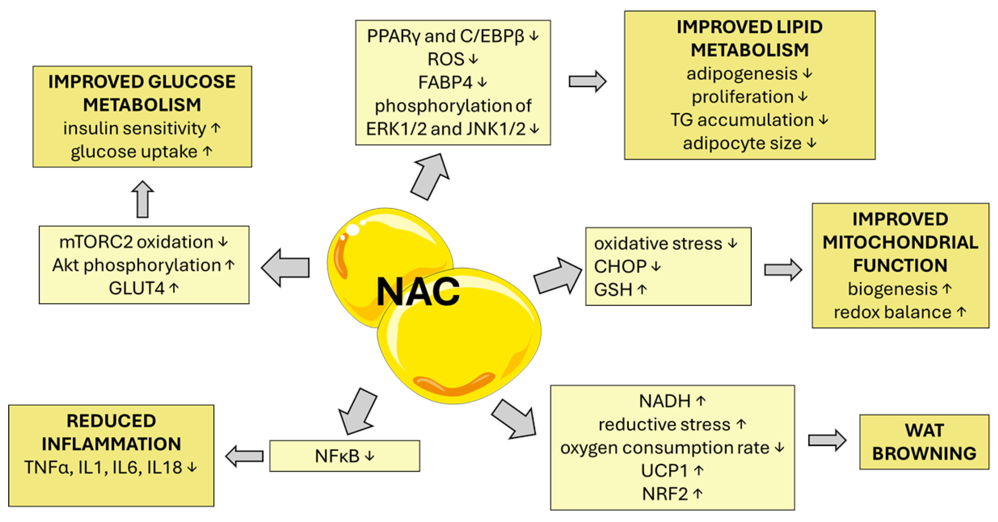
Standard therapeutic management for patients with obesity, MetS, and CVD includes lifestyle changes (weight reduction, diet, physical activity), pharmacotherapy (statins, angiotensin converting enzyme (ACE) inhibitors, beta-blockers, antidiabetic medications), and interventional treatment for acute myocardial infarction (e.g., coronary angioplasty). Lately, there has also been growing interest in metabolic modulators—compounds that can help to restore metabolic balance [86]. Growing evidence from both cell culture, animal, and human studies suggests that NAC may offer therapeutic benefits in MetS-associated comorbidities, including CVD, by improving redox balance, glucose metabolism, insulin sensitivity, lipid metabolism, and inflammation, although some data seem contradictory, and a more in-depth analysis is surely necessary to fully understand NAC’s actions (summarized in Table 1). Additionally, NAC is often combined with other compounds, e.g., glycine, apocynin, betaine, L-carnitine, methionine, or pyridoxine [87,88,89,90]. In vitro and in vivo experiments show that NAC can inhibit lipid accumulation by targeting adipogenic transcription factors such as peroxisome proliferator-activated receptor gamma (PPARγ) and CCAAT/enhancer binding protein beta (C/EBPβ) and improve insulin sensitivity through augmenting the PI3K/AKT pathway (summarized in Table 2 and reviewed by [91]).

5.1. Effect of NAC on Glucose Metabolism and Its Deregulation

A lot of studies based on in vitro and animal models have shown that NAC may exert significant effects on glucose metabolism and insulin signaling. In Olson et al.’s study [98], NAC was used to investigate its ability to reverse insulin resistance in 3T3-L1 adipocytes with silenced peroxiredoxin-3 (Prdx3), a mitochondrial antioxidant enzyme, which mimics oxidative stress conditions [98]. Treatment with NAC restored insulin sensitivity and stimulated glucose uptake via a reduction in mechanistic target of rapamycin complex 2 (mTORC2) oxidation and upregulation of AKT phosphorylation. Moreover, NAC seemed to protect mitochondrial function without triggering endoplasmic reticulum (ER) stress or mitochondrial unfolded protein response (mtUPR). Similar results were observed in skeletal muscle fibers isolated from high-fat-diet (HFD) mice by Russell-Guzman et al. [116]. The treatment with NAC significantly reduced oxidative stress markers, including lipid peroxidation and peroxiredoxin 2 dimerization, and inhibited activation of the ROS/TXNIP/NLRP3 inflammasome pathway. Moreover, NAC reduced caspase-1 activity and restored insulin-dependent glucose uptake and AKT phosphorylation, but at the same time impaired insulin signaling in control (non-insulin-resistant) muscle fibers, suggesting its effects are context-dependent and influenced by the redox state [116]. In Alnahdi et al.’s investigation [107], NAC was used to mitigate glucolipotoxicity-induced damage in pancreatic β-cells. Cells exposed to high glucose and palmitic acid showed increased apoptosis, mitochondrial dysfunction, and oxidative stress. NAC treatment significantly reduced cell death, preserved mitochondrial membrane potential and ATP production, and restored redox balance. It also reversed suppression of autophagy by increasing autophagy-related protein light chain 3 (LC3-II) expression and modulating mTOR/AMPK signaling [107].
Animal studies also show the beneficial impact of NAC on insulin sensitivity in insulin-responsive tissues, mainly via restoration of proper levels of phosphorylated AKT. In Argaev-Frenkel et al.’s study [117], in diabetic KK-Ay mice, NAC showed tissue-specific, dose-dependent effects on glucose metabolism and insulin signaling. It preserved isolated pancreatic islet function but induced reductive stress in insulin-responsive tissues, impairing insulin signaling and glucose uptake via reduced AKT phosphorylation. In vivo, NAC improved glucose tolerance in diabetic mice but disrupted insulin signaling in healthy C57BL/6 mice, indicating its effects depend on tissue oxidative status [117]. Schuurman et al. [118] showed that NAC improves glucose metabolism and β-cell function when administered before and during HFD-induced diabetes in mice. Long-term treatment was essential, as short-term (10 weeks) NAC had no effect. Benefits were linked to its antioxidant and anti-inflammatory actions, including GSH precursor activity and NF-κB inhibition [118]. NAC, administered to obese Swiss mice, significantly reduced fasting blood glucose and improved insulin sensitivity via restoration of phosphorylated AKT levels, lowered nitrite and protein carbonyl levels, indicating reduced oxidative damage, and increased glutathione peroxidase (GPx) protein levels, enhancing antioxidant defense [100]. Similar results were reported by Pieri et al. [97], who showed that NAC, administered orally, significantly improved insulin sensitivity and reduced fasting glucose levels without affecting body weight, food intake, or adiposity. The treatment led to a reduction in both oxidative stress and inflammation markers linked to obesity. Importantly, NAC increased phosphorylation of insulin receptor substrate 1 (IRS1) and AKT, and enhanced IRS1/PI3K association, suggesting improved insulin signaling [97]. Kaneto et al. [119] demonstrated, in a study performed on diabetic db/db (leptin receptor-deficient) mice, that intravenous perfusion of NAC during a hyperglycemic clamp improves insulin sensitivity and increases peripheral glucose uptake as well as upregulation of pancreatic-duodenal homeobox-1 (PDX-1) amounts, responsible for transcription of the insulin gene [119]. In the study by Straub et al. [87], NAC was used in combination with apocynin (a potent antioxidant) to investigate its effects on glucose metabolism and insulin resistance in inducible fat-specific insulin receptor knockout (iFIRKO) mice. Treatment with an antioxidant cocktail delayed the onset of hyperglycemia and reduced its severity, lowering blood glucose and improving insulin sensitivity, but also reducing food intake and water consumption, suggesting a systemic metabolic impact. In leptin-deficient ob/ob mice, antioxidant supplementation rapidly reduced hyperglycemia and normalized hyperphagia, showing antioxidant therapy as a successful tool to improve glucose homeostasis in both acute and chronic models of diabetes and obesity [87]. Similar effects, although tissue-specific, were shown in insulin resistance induced by prolonged elevation of plasma FFA (Intralipid plus heparin infusion) in Wistar rats. While NAC administration did not prevent hepatic insulin resistance caused by FFA, it effectively prevented peripheral insulin resistance, since skeletal muscle showed increased oxidative stress markers after FFA elevation, including protein carbonyls, which NAC successfully suppressed [120]. Markers of oxidative stress in the liver (protein carbonyls, malondialdehyde (MDA), and reduced glutathione/oxidised glutathione (GSH/GSSG) ratio) were not elevated by FFA, suggesting oxidative stress was not the primary driver of hepatic insulin resistance in this model [120].
It is worth mentioning that antioxidant therapy, including NAC, may interfere with glucose meter readings. A study by Grzych et al. [121] discussed the clinical implications for patients undergoing antioxidant therapy, particularly those with MetS. NAC was shown to cause falsely elevated glucose readings on several commercial glucose meters, especially at mid and high plasma concentrations, which could lead to inappropriate administration of glucose-lowering drugs [121].

5.2. From Oxidative Stress and Inflammation to Senescence and Aging

The interplay between MetS, CVD, and aging is increasingly attributed to chronic inflammation and oxidative stress, which promote cellular senescence and tissue dysfunction [122,123]. Visceral obesity, a hallmark of MetS, contributes to systemic metabolic derangement by inducing oxidative stress and elevating proinflammatory cytokines in adipose tissue, leading to the senescence-associated secretory phenotype (SASP) [122,124,125]. Senescent cells accumulate in metabolically active tissues, including the heart, where they exacerbate fibrosis, hypertrophy, and mitochondrial dysfunction—features shared by both obesity and aging [123]. These overlapping mechanisms support the concept of obesity as a driver of premature aging and cardiovascular decline.
In VAT from obese patients (BMI > 40), NAC reduced SA-β-gal activity and expression of p16, p21, and IL-6, indicating anti-senescent and anti-inflammatory effects. Samples were collected during bariatric surgery and treated ex vivo [126]. In a similar study of 40 obese individuals (BMI > 35), 4-week NAC supplementation (600 mg/day) reduced SA-β-gal activity and expression of p16 and IL-6 in VAT, but had no effect on lipid profiles or anthropometric measures [127].
In a pilot study with eight older adults (71–80 years), 24-week GlyNAC supplementation (glycine + NAC) corrected GSH deficiency, reduced oxidative stress (e.g., TBARS), improved mitochondrial function, lowered pro-inflammatory markers (IL-6, TNF-α, CRP), and enhanced endothelial function, genomic stability, cognitive performance, and physical capacity (gait speed, grip strength, and exercise tolerance). Fat mass and waist circumference decreased, though muscle mass remained unchanged. Most benefits declined after 12 weeks of discontinuation, indicating the need for ongoing supplementation [88]. A randomized placebo-controlled clinical trial confirmed GlyNAC’s ability to reverse multiple aging-related defects in older adults. In addition to restoring GSH levels and reducing oxidative stress and inflammation, GlyNAC significantly lowered insulin resistance and fasting insulin. NAC contributed to improved nutrient sensing and increased GLUT4 expression. Genomic stability was enhanced, as shown by reduced levels of 8-OH-G and Phospho-H2A Histone Family Member X (pH2AX), markers of DNA damage [128].

5.3. Effect of NAC on Adipogenesis, Visceral Obesity, Lipid Metabolism, and Its Deregulation

Obesity is characterized by excessive TG accumulation in VAT and SAT, leading to enlarged adipocytes, inflammation, and altered adipokine secretion. It seems that NAC may influence adipocyte differentiation and lipid metabolism through redox-sensitive transcriptional regulation. NAC was shown to inhibit adipocyte differentiation in 3T3-L1 cells, which are used for adipocyte differentiation studies, by downregulating PPARγ and C/EBPβ, the main adipogenic transcription factors. Moreover, NAC reduced intracellular ROS levels and suppressed the activities of antioxidant enzymes SOD and GPx. It significantly decreased TG accumulation and elevated intracellular GSH content early in the differentiation process, suggesting its antioxidant effect was mediated via GSH. NAC did not interfere with insulin-induced AKT phosphorylation, indicating its action was not through insulin receptor disruption [129]. Additionally, NAC suppressed the expression of key obesity-related proteins, including fatty acid binding protein 4 (FABP4), monoamine oxidase A (MAOA), heat shock protein 70 (HSP70), aminoacylase-1 (ACY-1), and transketolase (TKT) in 3T3-L1 cells [92]. Similar results were obtained by Soto et al. [113], where incubation of 3T3-L1 cells with NAC inhibited the phosphorylation of ERK1/2 and JNK1/2, key kinases involved in proliferation and adipogenesis. Additionally, NAC increased the expression of mitochondrial enzymes citrate synthase and fumarate hydratase, suggesting enhanced aerobic respiration, while decreasing the expression of MAOA, a mitochondrial source of ROS. These molecular changes correlated with reduced TG accumulation and adipogenic marker expression [113]. Consistent findings were observed in studies on mouse embryonic fibroblasts, where NAC significantly decreased lipid content and TG accumulation, and similarly inhibited ERK1/2 and JNK1/2 phosphorylation, supporting its anti-adipogenic effects [130]. This is in line with results obtained by Li et al. [131], where NAC was used as an ROS scavenger to investigate its role in adipocyte differentiation in 3T3-L1 cells. The study found that knockdown of metallothionein 3 (MT3), a protein known for its antioxidant properties, led to elevated ROS levels during early adipogenesis, and treatment with NAC effectively reversed this increase in ROS, suggesting its capacity to mitigate oxidative stress in differentiating adipocytes. This reduction in ROS correlated with a suppression of adipocyte differentiation, indicating that NAC can interfere with the early stages of adipogenesis. The study also showed that NAC treatment restored the expression of ROS scavenging genes such as catalase, Gpx1, Sod1, and Sod2, which were downregulated by MT3 overexpression [131].
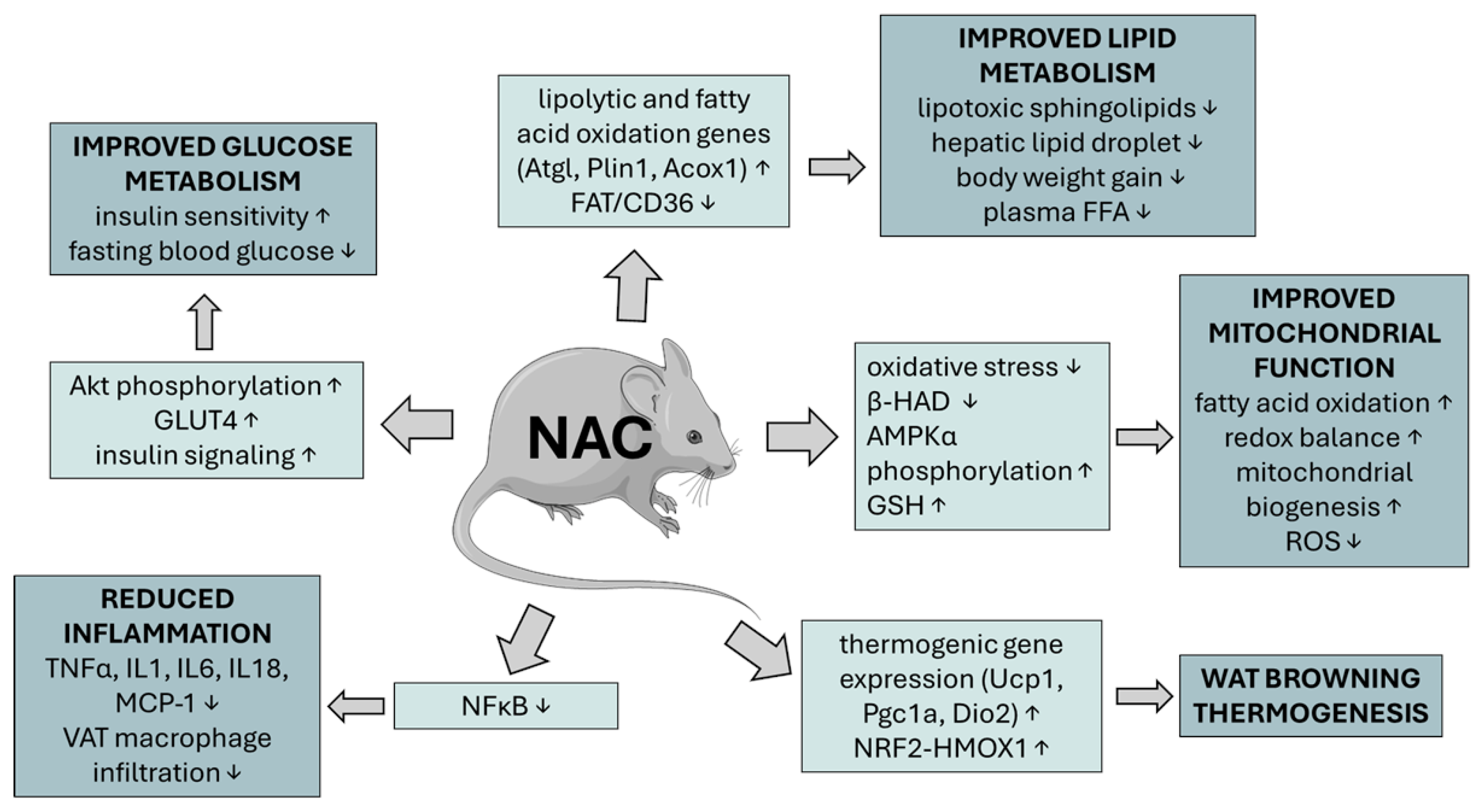
Results obtained in animal models also confirm the positive impact of NAC on lipid metabolism and adiposity. In an HFD-induced obese mouse model, multi-ingredient metabolic cofactor (MC) therapy—including NAC—reduced weight gain, improved glucose and insulin tolerance, and lowered fasting glucose, insulin, and HOMA-IR scores. It enhanced lipid oxidation, reduced adipocyte hypertrophy in VAT, and upregulated lipolytic and fatty acid oxidation genes such as adipose triglyceride lipase (Atgl), perilipin 1 (Plin1), and acyl-CoA oxidase 1 (Acox1). In BAT, NAC reduced fat accumulation and increased thermogenic gene expression (Ucp1, Pgc1a, and Dio2). The cocktail also improved lipid profiles, though NAC’s specific contribution remains unclear due to synergistic effects of all components [89].
Metabolic disorders may also affect bone marrow adipose tissue (BMAT), which is considered to be an important source of paracrine factors that may influence bone turnover, protect osteoblasts from lipotoxicity, and maintain bone marrow hematopoietic function and niche integrity [132,133]. In human bone marrow stromal cells differentiated into adipocytes, NAC reduced lipid droplet accumulation and enhanced antioxidant activity, as shown by increased heme oxygenase-1 (HO-1) expression and reduced ROS. NAC upregulated key metabolic and anti-inflammatory markers, including SIRT1, HO-1, adiponectin, and PPARα/δ, while downregulating IL-6. It also reversed the suppression of diacylglycerol acyltransferase 1 (DGAT1) and PPARγ and increased FABP4 expression, indicating improved lipid metabolism and reduced lipotoxicity, supporting NAC’s role in restoring BMAT function [93].
NAC’s influence on WAT is summarized in Figure 2.

5.4. Effect of NAC on Hepatic Lipid Accumulation

MetS is often associated with MASLD. MASLD, driven by metabolic dysfunction and hepatic fat accumulation, is closely associated with an increased risk of CVD due to shared inflammatory and metabolic pathways [134]. In a rat model of MASLD induced by a high-fat diet, NAC administered at 500 mg/kg/day for eight weeks reduced hepatic levels of lipotoxic sphingolipids, such as ceramide and dihydroceramide, while increasing sphingosine-1-phosphate (S1P) and sphinganine-1-phosphate (SA1P), which are linked to improved insulin sensitivity. NAC modulated key enzymes in sphingolipid metabolism and enhanced insulin signaling via increased phosphorylation of AKT, glycogen synthase kinase-3α/β (GSK3α/β), and P70 S6 kinase. Histological analysis showed reduced lipid accumulation and fibrosis markers, indicating improved liver function and reduced inflammation [110]. A randomized controlled pilot trial was designed to evaluate the effect of MetioNac®, which consists of S-adenosyl-L-methionine, NAC, thioctic acid, and vitamin B6, in 15 patients with MetS at risk for MASLD for 3 months. Patients and the control group were also prescribed a semi-personalized Mediterranean diet for weight loss. MetioNac® normalized lipid parameters in the study group, as evidenced by reduced TG and very low-density lipoprotein (VLDL) levels. Both the study and control groups demonstrated a visible weight loss. The authors concluded that MetioNac® may be effective in overweight patients with MetS who suffer from hyperlipidemia and insulin resistance, although the risk of bias was calculated as moderate for this study due to the small sample size, high dropout rate, lack of blinding, and rather exploratory design [90].

5.5. Effect of NAC on Mitochondrial Dysfunction in Obesity, MetS, and CVD

A key mechanism of NAC involves its role as a precursor to glutathione, a potent endogenous antioxidant that plays a critical role in neutralizing ROS. NAC supplementation may mitigate oxidative stress and also inflammation, which are among the key drivers of metabolic dysfunction on the cellular level. In cell culture models, NAC consistently reduced ROS levels in adipocytes and preadipocytes exposed to high glucose or palmitic acid [96,107]. NAC was shown to mitigate oxidative stress and reduce C/EBP homologous protein (CHOP) accumulation in adipocytes under metabolic stress. High glucose conditions increase ROS production, activating the nuclear factor erythroid 2-related factor 2 (NRF2) pathway, which plays a crucial role in the protection against oxidative stress and is activated by different stressors. Upregulating HO-1, an antioxidant marker, via NAC treatment decreased intracellular fumarate levels in adipocytes matured in high glucose, which in turn lowered protein succination—a marker of mitochondrial stress. NAC also restored GSH levels and reduced ROS accumulation, contributing to improved redox balance. This antioxidant effect led to decreased CHOP stability and reduced expression of HO-1. Importantly, NAC restored the secretion of IL-13, an anti-inflammatory cytokine suppressed by CHOP accumulation, indicating a reversal of CHOP-mediated transcriptional repression [96]. Similarly, NAC reversed oxidative stress and inflammation in neuregulin 4-deficient adipocytes, restoring insulin receptor and GLUT4 protein levels and inhibiting NFκB activation. Treatment with NAC for 24 hours significantly reduced H2O2 levels and completely abrogated TNFα gene expression. NAC also increased IκB protein levels, inhibiting NFκB-mediated inflammation, and partially restored insulin receptor and GLUT4 protein content [105].
Animal studies corroborate these findings, although some data are contradictory. NAC supplementation in HFD rats reduced body weight, normalized plasma FFA, and decreased adipocyte hypertrophy and macrophage infiltration in both VAT and SAT, indicating reduced inflammation. NAC decreased expression of fatty acid transporters such as fatty acid translocase (FAT/CD36) and fatty acid binding protein (FABPpm) in VAT and SAT, and also reduced long-chain fatty acid transport protein 1 (FATP1) expression in SAT on both mRNA and protein levels, although these results were inconsistent, probably due to posttranscriptional modifications. NAC also enhanced the expression of β-hydroxyacyl-CoA dehydrogenase (β-HAD), a marker of mitochondrial β-oxidation, especially in VAT, indicating increased fatty acid oxidation [95]. Similarly, prolonged treatment of adipocytes with NAC induced browning via the NRF2/HO-1 signaling axis, marked by elevated expression of uncoupling protein 1 (UCP1), peroxisome proliferator-activated receptor gamma coactivator 1-alpha (PGC1α/β), and mitochondrial biogenesis genes. In vivo, in the HFD mouse model, high-dose NAC reduced fat mass gain, lowered insulin levels, and enhanced mitochondrial oxygen consumption rate in inguinal WAT. These effects were absent in adipocyte-specific NRF2 knockout mice, indicating that NRF2 mediates NAC-induced mitochondrial adaptations [111]. On the contrary, NAC inhibited UCP1 expression and thermogenesis in adipocytes by blocking p38 MAPK activation. NAC, along with other antioxidants like butylated hydroxyanisole (BHA), suppressed both basal and cold- or cAMP-induced UCP1 expression in cultured brown adipocytes and in vivo. This suppression was linked to inhibition of ROS accumulation and subsequent p38 MAPK activation. NAC treatment reduced mitochondrial uncoupling and thermogenic capacity, leading to increased lipid accumulation in brown adipose tissue (BAT). A similar outcome was observed in PG-Sn2 mice (engineered to overexpress sestrin2 in adipose tissue). Sestrin2 is a stress-inducible protein that suppresses ROS accumulation and regulates AMPK/mTORC1 signaling, and loss of this protein leads to insulin resistance, fat accumulation, and mitochondrial dysfunction. Sestrin2 inhibited UCP1 expression, leading to reduced thermogenesis and increased fat accumulation. This suggests that ROS are necessary for proper UCP1 expression and BAT function, and although antioxidants like NAC can reduce oxidative stress, they may inadvertently impair beneficial metabolic processes such as thermogenesis [135].
In catalase-knockout mice fed HFD, which are prone to obesity due to elevated oxidative stress, NAC restored AMPKα phosphorylation and improved mitochondrial function. NAC treatment significantly reduced body weight gain, fat mass, and improved lean mass. It also lowered H2O2 concentrations in epididymal fat and downregulated nicotinamide adenine dinucleotide phosphate oxidase 4 (NOX4) protein level, which are associated with ROS production. In vitro, NAC reversed H2O2-induced lipogenesis in 3T3-L1 adipocytes, supporting its role in mitigating oxidative stress-driven adipocyte hypertrophy [108].
As mentioned earlier, NAC may rarely have an oxidative effect instead of an antioxidant effect under certain conditions, especially in high concentrations [73]. Chronic NAC treatment was found to induce reductive stress in adipocytes, increasing intracellular GSH but failing to prevent ROS accumulation after β3-adrenergic stimulation. NAC enhanced mitochondrial ROS production, reduced oxygen consumption, and increased lactate levels, indicating a shift toward glycolytic metabolism. In vivo, in healthy mice, NAC suppressed browning of WAT and reduced mitochondrial activity in BAT, even without adrenergic input. It also upregulated mitochondrial antioxidant proteins, suggesting elevated mitochondrial oxidative stress. These effects were linked to activation of the NRF2 pathway and impaired fatty acid clearance, resulting in increased fat pad size and dysfunctional adipose metabolism. The findings caution against chronic antioxidant use in metabolically healthy individuals [136].

5.6. Effects of NAC on Chronic Metabolic Inflammation (Metaflammation) and Immune Modulation

NAC’s anti-inflammatory effects are closely tied to its antioxidant properties. In human macrophages, hypoxia amplified palmitate (PA)-induced inflammation by increasing IL-6 and IL-1β expression, linked to activation of JNK and p38 MAPK pathways. This effect was independent of ER stress. NAC and the mitochondria-targeted antioxidant MitoTEMPO suppressed hypoxia-enhanced cytokine expression and MAPK activation, but had no effect under normoxic conditions. Conditioned medium from hypoxic, palmitate-treated macrophages triggered inflammatory responses in adipocytes, increasing IL-6 and MCP-1, and reducing adiponectin. These effects were mitigated by NAC or JNK inhibition, highlighting NAC’s selective anti-inflammatory role under hypoxia [104]. Similar results were observed in 3T3-L1 adipocytes treated with high glucose. The addition of NAC significantly reduced the elevated levels of ROS and 8-OH-G and normalized the aberrant expression and secretion of pro-inflammatory adipokines—IL-18, MCP-1, and plasminogen activator inhibitor-1 (PAI-1)—and restored adiponectin levels. These effects were observed in both stable and intermittent glucose conditions, with intermittent exposure showing more pronounced dysregulation [101]. In adipocytes with knockdown of the SelenoM gene, NAC partially reversed oxidative stress and inflammatory responses. NAC suppressed the expression of pro-inflammatory cytokines such as TNF-α, IL-6, IL-1β, and inducible nitric oxide synthase (iNOS) in both SelenoM-deficient adipocytes and co-cultured macrophages, by NF-κB deactivation, a key driver of inflammation in macrophages. NAC also improved glucose metabolism by restoring phosphorylation of IRS1, AKT, and GSK3β, and increased GLUT4 expression in SelenoM-knockdown mice. It diminished M1 macrophage polarization and cytokine release, indicating its anti-inflammatory potential [115].
In DNA polymerase η knockout (pol η−/−) mice, NAC was used to investigate its effects on DNA damage-induced adipocyte senescence and metabolic abnormalities. NAC significantly reduced oxidative DNA damage in adipose tissue, as measured by comet assay and markers like H2A histone family member X (γ-H2AX), ataxia-telangiectasia mutated (ATM), p53, and p21. NAC also suppressed NF-κB activation and lowered proinflammatory cytokines IL-6, TNF-α, and MCP-1. Importantly, NAC attenuated adipocyte hypertrophy and senescence markers such as SA–β-gal and p16, and improved insulin sensitivity and glucose tolerance. Interestingly, NAC had minimal impact on wild-type mice [109]. Protective effects of NAC against insulin resistance and adipose tissue inflammation were shown in advanced glycated albumin (AGE-albumin) treated healthy rats. Chronic administration of AGE-albumin to healthy rats led to increased macrophage infiltration in adipose tissue, reduced GLUT4 protein levels, and impaired insulin sensitivity, despite no changes in body weight or plasma lipids. NAC treatment counteracted these effects by reducing lipid peroxidation, decreasing macrophage numbers (without significant changes in M1 (CD11b) or M2 (CD206) markers), and enhancing the expression of GLUT4 and PPAR-α. Although NAC did not fully restore GLUT4 protein levels, it improved glucose tolerance and reduced inflammatory gene expression [94].
NAC significantly reduced arachidonic acid (AA) levels in myocardial phospholipid and TG fractions in male Wistar rats fed an HFD, indicating a suppression of lipid-driven inflammation. It downregulated cyclooxygenase-2 (COX-2) and arachidonate 5-lipoxygenase (5-LOX) expression, leading to decreased levels of proinflammatory eicosanoids such as PGE2, LTB4, and leukotriene C4 (LTC4), while increasing anti-inflammatory lipoxin A4 (LXA4). NAC also inhibited NF-κB activation, resulting in reduced secretion of inflammatory cytokines, including TNF-α and IL-1α. Furthermore, NAC enhanced antioxidant defences by elevating GSH and catalase levels and reduced oxidative damage markers like MDA and 4-hydroxynonenal (4-HNE). These effects were observed both in HFD-fed and standard diet-fed rats [106]. Similar results were obtained in the diabetic-wound-healing rat model. Diabetic rats exhibited elevated ROS levels and increased expression of nod-like receptor protein 3 (NLRP3), caspase-1, apoptosis-associated speck-like proteins (ASCs), and IL-1β, contributing to persistent inflammation and delayed wound closure. NAC effectively reduced ROS production, suppressed NLRP3 inflammasome activation, decreased IL-1β secretion, and improved wound healing, including faster re-epithelialization and reduced healing time [112].
NAC’s impact on animal MetS models is summarized in Figure 3.

5.7. Obesity and the Brain: A Self-Perpetuating Neuro-Metabolic Disorder

NAC can also exert a positive effect on HFD-induced neurodegeneration by targeting oxidative stress, inflammation, and cell death mechanisms, as well as improve cognition and reduce addiction-like behaviors towards high-fat and high-sugar diets. In Wistar rats fed an HFD and treated with NAC, weight gain, BMI, hyperglycemia, and insulin resistance were significantly reduced. NAC improved cerebral cortex redox status by increasing GSH level and catalase activity while reducing MDA, peroxynitrite, and myeloperoxidase activity. NAC also attenuated neuroinflammation and necroptosis by lowering levels of receptor-interacting protein kinase-3 (RIPK3) and mixed lineage kinase domain-like protein (MLKL), necroptotic signaling molecules that activate the inflammasome, as well as NLRP3 and ASC, an inflammasome component protein. NAC also downregulated pyroptosis-related cytokine IL-18. Histologically, NAC preserved cerebral cortex architecture and reduced astrogliosis, as evidenced by decreased glial fibrillary acidic protein (GFAP), iNOS, and matrix metalloproteinase 9 (MMP-9) immunoreactivity [103].
NAC or taurine administered to female mice fed HFD prevented HFD-induced memory impairment in both novel object and novel location recognition tasks. It also preserved hippocampal levels of N-acetylaspartate, lactate, and the phosphocreatine-to-creatine ratio, which are markers of neuronal health and energy metabolism. Unlike taurine, NAC increased hippocampal concentrations of glutamate, glutamine, GABA, and glutathione [137]. NAC can also modulate addiction-like behaviors toward high-fat, high-sugar (HFHS) food. Both systemic and intracerebroventricular administration of NAC prior to limited access to a highly palatable Western diet (WD) significantly reduced WD consumption without affecting intake of standard chow, indicating a selective suppression of hedonic feeding rather than general appetite. Importantly, NAC did not induce conditioned taste aversion, suggesting its effects were not due to malaise [138]. Similarly, NAC significantly reduced compulsive-like food-seeking behavior in DIO (diet-induced obesity) rats, particularly persistent lever pressing during reward-unavailable periods, and prevented escalation of HFHS intake. The treatment did not affect general locomotion, anxiety-like behavior, or re-acquisition of operant responding, suggesting specificity for compulsive eating [139].

5.8. NAC and Its Role in Cardiovascular Protection

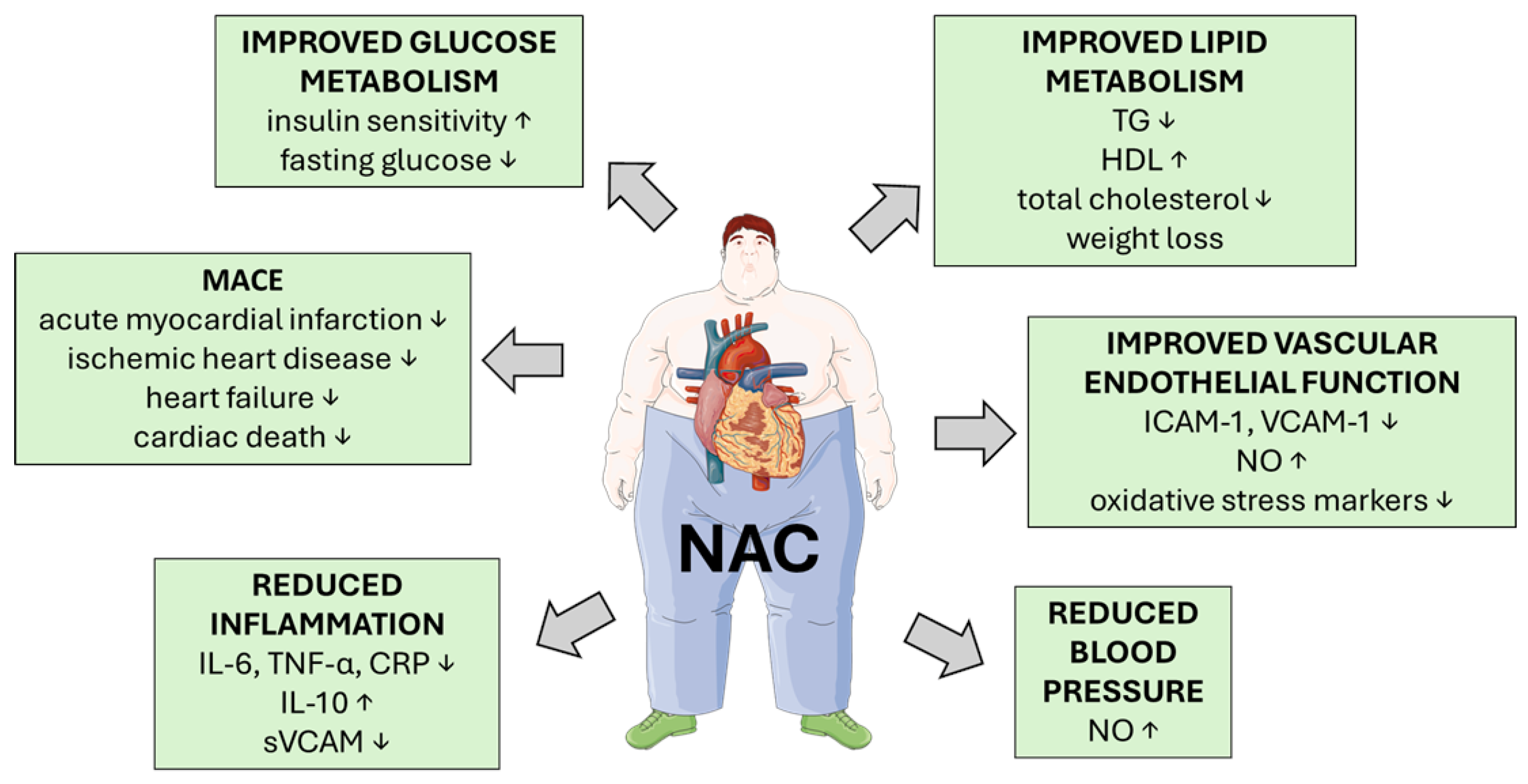
NAC’s cardioprotective effects are evident in multiple models. It is believed that the mechanism of NAC action in myocardial infarction and HF relies on ROS inhibition, antiplatelet, anti-inflammatory, and anti-apoptotic activity, reduction in obesity-related cardiac insulin resistance, as well as inhibition of angiotensin-converting enzyme (ACE) [140]. Also, NAC may have a beneficial effect on EC dysfunction, which arises from oxidative stress, eNOS uncoupling, leukocyte adhesion, and apoptosis. These processes are exacerbated by MetS components such as hyperglycemia, dyslipidemia, and insulin resistance [141,142]. There are several human studies and clinical trials involving NAC and its use in the mitigation of CVD symptoms, although most of the trials do not involve MetS patients. Nevertheless, it is worth describing these studies since most of the enrolled individuals suffer from one or more MetS symptoms, such as obesity, hypertension, and atherosclerosis (Summarized in Table 3). NAC enhances intracellular GSH levels, reducing oxidative stress and inflammation. In the comprehensive review by Xu et al. [142], NAC is highlighted as a potent antioxidant with therapeutic potential in mitigating endothelial dysfunction caused by oxidative stress. Oxidative stress is a key driver of endothelial dysfunction in MetS, contributing to impaired nitric oxide (NO) bioavailability, inflammation, and vascular permeability. NAC’s ability to scavenge ROS and restore redox balance directly supports endothelial integrity and function. Moreover, NAC is noted to inhibit leukocyte adhesion, reduce NADPH oxidase (NOX) expression, suppress inflammatory cytokine secretion and improve endothelial barrier function [142] (Figure 4).
NAC supplementation to Wistar rats fed either a standard or HFD significantly reduced body mass, plasma glucose and insulin levels, improving insulin sensitivity, but also lowered ceramide accumulation in cardiac tissue by downregulating key enzymes involved in ceramide synthesis, including serine palmitoyltransferase long chain base subunit 2 (SPTLC2), ceramide synthase 5 (LASS5), neutral ceramidase (ASAH2), alkaline sphingomyelinase (Alk-SMase), and neutral sphingomyelinase (N-SMase). This reduction in ceramide was accompanied by an increase in S1P levels and an elevated S1P/ceramide ratio, indicating a shift toward a more protective, anti-apoptotic sphingolipid profile. NAC also improved insulin signaling by enhancing phosphorylation of AKT and upregulation of GLUT4 expression in the left ventricle, thereby promoting glucose uptake. These effects were observed both in rats on a standard diet and those subjected to lipid overload via an HFD [99].
In a rat model of diabetic cardiac hypertrophy induced by alloxan, treatment with NAC markedly reduced blood glucose levels, restored body weight, and improved both heart weight-to-body weight and heart weight-to-tibia length ratios. Histological analysis revealed that NAC preserved myocardial architecture, reduced cardiomyocyte hypertrophy, and prevented fibrosis. Moreover, NAC effectively lowered ROS levels and lipid peroxidation, while enhancing antioxidant enzymes activation, including SOD and catalase. At the molecular level, NAC downregulated pro-apoptotic markers such as dynamin-related protein 1 (Drp1), p53-upregulated modulator of apoptosis (PUMA), and cytochrome c. Concurrently, it upregulated anti-apoptotic and mitochondrial maintenance genes, including Bcl-2 and mitofusin-2 (MFN-2) mitochondrial fusion marker. Additionally, NAC improved lipid profiles by reducing serum TG and cholesterol [143]. Unfortunately, human studies do not show the beneficial effects of NAC on cardiac architecture. In HALT-HCM (Hypertrophy Regression With N-Acetylcysteine in Hypertrophic Cardiomyopathy), a randomized, double-blind, placebo-controlled pilot study, 42 patients with MetS comorbidities such as hypertension, dyslipidemia, and diabetes, with established hypertrophic cardiomyopathy (HCM), received high-dose NAC or placebo for 12 months, and the study found only negligible to modest effects on echocardiographic and cardiac MRI indices of hypertrophy and fibrosis. Moreover, no significant improvements in clinical symptoms or exercise tolerance were observed to evaluate the effects of NAC. Since this was a pilot study with the risk of bias calculated as moderate to high due to the small sample size and exploratory design, more and better conducted clinical trials are necessary to evaluate NAC’s impact on cardiac architecture [144].
Some data show that NAC may prevent diastolic dysfunction in. The treatment of HFHS-fed rats with NAC inhibited the rise in E/DT ratio, an echocardiographic marker of diastolic dysfunction and elevated end-diastolic pressure, without reversing right ventricular hypertrophy [145]. In the estrogen-deprived obese insulin-resistant female rat model, NAC monotherapy partially improved metabolic parameters, including reductions in plasma glucose TG, but did not significantly improve insulin resistance or cardiac function. However, when combined with low-dose estrogen, NAC showed efficacy comparable to regular-dose estrogen in improving left ventricular function, heart rate variability, and reducing cardiac ischemia-reperfusion (I/R) injury. The low-dose estrogen with NAC reduced oxidative stress markers like MDA and increased anti-apoptotic Bcl-2 expression, though it did not suppress pro-apoptotic Bax as effectively as regular-dose estrogen [146]. Treatment with NAC and allopurinol (ALP) in the diabetic rat model synergistically mitigated cardiac damage. NAC and ALP increased cardiac adiponectin and its receptor AdipoR2 expression, restored phosphorylation of key survival pathways—PI3K/AKT and Jak2/STAT3—and upregulated eNOS activation. These molecular changes correlated with reduced infarct size and lower creatine kinase-MB (CK-MB) release [114].
NAC also showed anti-atherosclerotic effects in diabetic ApoE/ mice, which exhibit elevated levels of methylglyoxal (MG), a key precursor in the formation of AGEs and a marker of carbonyl stress. Following 12 weeks of NAC treatment, MG levels were significantly reduced. NAC also restored GSH levels in the aorta, thereby enhancing MG elimination and reducing oxidative stress markers such as serum MDA, and upregulated antioxidant enzyme expression, including SOD-1 and GPX-1. Endothelial function was improved, as evidenced by increased phosphorylation of AKT and eNOS, along with elevated serum NO levels [102]. NAC seems to delay cellular senescence in ECs isolated from patients with severe CAD by reducing oxidative stress and activating telomerase (hTERT), leading to telomere stabilization. NAC also decreased lipid peroxidation, lowered DNA damage markers (ATM, H2AX, p53, promyelocytic leukemia protein—PML). These molecular changes were accompanied by extended cell replication and delayed onset of senescence [147].
NAC lowers plasma homocysteine (Hcy) levels, a known risk factor for MetS. Increased levels of Hcy are associated with CVD, stroke, and obesity, although some authors underline iatrogenic elevation of Hcy in MetS, since most hypolipidemic, antidiabetic, and antihypertensive drugs raise circulating Hcy levels [148]. NAC may reduce Hcy blood levels by increasing urinary excretion, as was shown in a single-arm interventional study. Intravenous NAC administration to healthy individuals led to a rapid and significant reduction in plasma Hcy levels, which was accompanied by a marked increase in urinary excretion, and the mechanism was thought to involve NAC displacing thiols from protein-binding sites, forming mixed disulphides that are more readily cleared renally. Unfortunately, the risk of bias for this study was estimated as moderate due to the small sample size, lack of control group, and short duration [149]. A double-blind, randomized, placebo-controlled crossover trial investigated the effects of oral NAC on plasma Hcy and lipoprotein levels in patients with elevated cardiovascular risk markers. Eleven subjects with high plasma lipoprotein levels were recruited, and NAC was administered at a high dose over a two-week period. While NAC had no significant effect on lipoprotein levels, it caused a substantial reduction in plasma Hcy. The treatment was well tolerated, with only mild side effects like flatulence and bad taste, and the risk of bias for this study was estimated as low due to rigorous design, validated assays, and appropriate statistical analysis [150]. Hildebrandt et al. [151] reanalyzed two randomized, double-blind, placebo-controlled trials to assess the long-term effects of oral NAC on plasma Hcy and blood pressure in normolipidemic and hyperlipidemic men, stratified by smoking status. NAC supplementation significantly reduced plasma Hcy concentrations, independent of lipid profile or smoking status. This reduction was accompanied by increased plasma cysteine levels and improved intracellular redox status, particularly in smokers. NAC also significantly lowered systolic blood pressure in both groups, with a notable reduction in diastolic pressure observed only in hyperlipidemic subjects [151]. Supporting these findings, a comprehensive meta-analysis of 28 studies confirmed that NAC significantly reduces Hcy levels, as well as oxidative stress markers such as MDA and proinflammatory cytokines, including IL-8, IL-6, and TNF-α [152].
NAC may also stabilize levels of NO, since it supplies free sulfhydryl groups. This prevents NO degradation and enhances vasodilation, and finally improves vascular endothelial function and reduces nitrate tolerance [140]. In a randomized, double-blind, placebo-controlled, four-arm trial, NAC was evaluated for its potential to enhance the long-term treatment of unstable angina pectoris, since it is known to potentiate the vasodilatory effects of nitroglycerin and prevent nitrate tolerance. Although the inclusion criteria did not include MetS, but unstable angina, some of the individuals exhibited MetS symptoms, such as diabetes and hypertension. The combination of nitroglycerin and NAC significantly reduced the incidence of adverse cardiac events (death and MI), presumably due to enhanced NO bioavailability and antiplatelet effects. However, the combination therapy was associated with a higher incidence of severe headaches, leading to more frequent drug discontinuation, although the risk of bias for this study was estimated as low, due to robust design, standardized protocols, and good compliance [153]. NAC improves blood pressure control not only by enhancing NO bioavailability, but also by inhibiting ACE. Clinical studies also showed reductions in systolic and diastolic pressure in patients with T2DM and hypertension. There was a randomized, double-blind, placebo-controlled trial to evaluate the effects of NAC and L-arginine (ARG) on endothelial function in hypertensive male patients with T2DM. The study focused on NO bioavailability, oxidative stress, and vascular health. The NAC+ARG group showed significant reductions in systolic and diastolic blood pressure, total and LDL cholesterol, oxidized LDL, CRP, nitrotyrosine, biomarker of nitrosative stress, fibrinogen, and adhesion molecules (ICAM-1, VCAM-1, and PAI-1). These changes were accompanied by improved intima-media thickness and increased plasma nitrites/nitrates levels. Unfortunately, the risk of bias for this study was estimated to be moderate due to the small sample size and short duration, despite rigorous design and comprehensive biomarker analysis [154]. In an open-label, within-patient crossover study, NAC significantly reduced both 24-hour and daytime systolic and diastolic blood pressure compared to the ACE inhibitor (captopril or enalapril) alone in hypertensive smokers. In this study, the patients were not suffering from MetS, although hypertension is one of its symptoms, and therefore results may translate into such populations. The effect was attributed to NAC’s function as a sulfhydryl donor, enhancing NO bioavailability and protecting it from oxidative degradation. The risk of bias for this study was estimated as moderate due to the small sample size, short duration, and open-label design, but the crossover structure strengthened internal validity [155]. As was stated above, NAC also enhances the antiplatelet and hemodynamic effects of nitrates and reduces nitrate tolerance. Therefore, it may improve outcomes in patients with coronary artery disease, having overlapping risk factors such as hypertension, diabetes, and hyperlipidemia [156].
Several studies support NAC’s role in reducing oxidative stress and improving cardiac function in major adverse cardiac events (MACEs). Although MetS was not explicitly studied in most of these trials, many participants had overlapping risk factors such as hypertension, diabetes, and hyperlipidemia. Population-based cohort study with propensity score matching used Taiwan’s National Health Insurance Research Database to evaluate the long-term effects of NAC on MACE in patients with T2DM. The study included 46,718 T2DM patients, with 23,359 receiving long-term NAC therapy and 23,359 matched controls. NAC users exhibited a significantly lower incidence of MACE compared to non-users. NAC was associated with reduced risks of acute myocardial infarction, ischemic heart disease, cerebrovascular accidents, HF, and cardiac death, although the risk of bias for this study was estimated to be moderate due to the observational design that limits causal inference [157]. A double-blind, randomized, placebo-controlled clinical trial involved ninety-eight patients with ST-elevation myocardial infarction (STEMI) who received either NAC or placebo for three days. NAC significantly reduced serum levels of MMP-2 and MMP-9 after 72 hours. Patients treated with NAC also had a lower incidence of MACE at one-year follow-up, including fewer reinfarctions, but the small sample size and short NAC exposure reduce the robustness of the results [158]. A controlled, non-randomized clinical trial involving patients with acute myocardial infarction (AMI) treated with streptokinase demonstrated that intravenous NAC significantly reduced plasma MDA levels and improved left ventricular (LV) function, including ejection fraction and wall motion, despite no change in infarct size, but due to the small sample size and short NAC exposure, outcomes are subject to bias [159]. Similar results were obtained in a randomized, open-label, safety-focused pilot trial with AMI patients treated with streptokinase and nitroglycerin combined with NAC, carrying a comparable risk of bias [160]. A randomized, controlled, open-label pilot study reported reduced lipid hydroperoxides and transient neutrophil suppression with NAC while investigating the effects of NAC as adjunct therapy in patients with AMI undergoing fibrinolytic treatment with streptokinase. NAC administration significantly reduced plasma lipid hydroperoxide (ROOH) levels after 24 hours and transiently reduced circulating PMN counts. The risk of bias for this study was estimated as moderate due to the small sample and limited endpoints [161]. In LIPSIA-N-ACC (Leipzig Immediate Percutaneous Coronary Intervention Acute Myocardial Infarction N-Acetylcysteine), a randomized, single-blind, placebo-controlled trial, NAC reduced oxidative biomarkers (advanced oxidation protein products and oxidized LDL), but it did not improve clinical outcomes such as infarct size, the incidence of CIN, myocardial salvage index (MSI), or microvascular obstruction in unselected patients with STEMI undergoing primary percutaneous coronary intervention (PCI). The low risk of bias strengthens the validity of the findings [162]. In contrast, the NACIAM (N-Acetylcysteine in Acute Myocardial Infarction), a randomized, double-blind, placebo-controlled, multicenter trial, showed that high-dose NAC combined with low-dose nitroglycerin significantly reduced infarct size and enhanced myocardial salvage in patients with STEMI undergoing primary PCI, with sustained benefits at 3 months. NAC treatment resulted in a significant absolute reduction in infarct size by 5. 5% compared to placebo, and late cardiac magnetic resonance (CMR) imaging at 3 months confirmed sustained infarct size reduction. This study also demonstrates a low risk of bias, making its findings more reliable [163].
The results from human studies are summarized in Table 3.
Table 3. Human studies involving NAC use in management of MetS and its comorbid conditions.
DesignPopulationDose/RouteDurationComparatorsEffect SizesSafetyRisk of Bias
Randomized, double-blind, placebo-controlled clinical trial
[127]
40 obese adults (BMI ≥ 35 kg/m2), aged 25–50, candidates for bariatric surgery600 mg/day oral NAC4 weeks before bariatric surgeryPlaceboAdipose tissue sample: SA-β-gal ↓ staining (p = 0.001), IL-6 ↓ (p = 0.014), P16 ↓ (p = 0.047) gene expression
Blood: hsCRP, IL-6 FSB, insulin ↓ (p < 0.001)
HOMA-IR ↓ (p < 0.001)
No adverse events reportedLow
Randomized, double-blind, placebo-controlled clinical trial
[128]
24 older adults (61–80 years, BMI > 27), 12 young adults (21–40 years)GlyNAC: glycine (100 mg/kg/day) + NAC (100 mg/kg/day), oral16 weeks (YA received GlyNAC for 2 weeks)PlaceboMuscle sample: GSH ↑ (p < 0.001)
Blood: RBC total GSH ↑ (p < 0.001), TBARS ↓ (p < 0.001), IL-6 ↓ (p < 0.001), TNFα ↓ (p < 0.001), hsCRP ↓ (p < 0.001), IL-10 ↑ (p < 0.001), sICAM1 ↓ (p < 0.001), sVCAM1 ↓ (p < 0.001), HOMA-IR ↓ (p < 0.001), improved cognition, gait speed
No adverse events reportedLow
Double-blind, randomized, placebo-controlled crossover trial
[150]
11 adults (mean age 58), with elevated plasma lipoprotein(a) 2 g NAC twice daily, oral2 weeks NAC, 2 weeks placebo, with 2-week washout betweenPlaceboHomocysteine ↓ (p < 0.0001); Cysteinyl glycine ↓ (p = 0.0001); Cysteine ↓ (p = 0.013)Well tolerated; minor side effects (flatulence, bad taste) Low
Randomized, double-blind, placebo-controlled, multicenter trial (NACIAM)
[163]
112 STEMI patients undergoing PCINAC 29 g IV over 48 h + nitroglycerin 7.2 mg IV over 48 h48 h treatment; 3-month follow-upPlacebo + nitroglycerinInfarct size ↓ (p = 0.02); Myocardial salvage ↑ (p < 0.01); Late infarct size ↓ (p = 0.02)No increase in hypotension, bleeding, or nephropathy; 2 deaths in placebo group, none in NAC groupLow
Randomized, double-blind, placebo-controlled, 4-arm trial
[153]
200 patients with unstable angina not requiring emergency revascularizationNAC 600 mg orally, 3× daily; Nitroglycerin 10 mg transdermal patch daily4 monthsPlacebo, Nitroglycerin alone, NAC alone, Nitroglycerin + NACCombined therapy reduced outcome events (death, MI, refractory angina): vs. placebo (OR 0.25; 95% CI 0.07–0.7; p = 0.0022); vs. Nitroglycerin (OR 0.31; 95% CI 0.098–0.90; p = 0.018); vs. NAC (OR 0.19; 95% CI 0.06–0.57; p = 0.0008)High incidence of severe headache in combined group (31% vs. 19% in Nitroglycerin alone); 33% discontinued due to side effectsLow
Randomized, single-blind, placebo-controlled trial (LIPSIA-N-ACC)
[162]
251 STEMI patients undergoing PCINAC 6 g IV over 48 h (1.2 g bolus + 2 × 1.2 g/day)48 h treatment; 6-month follow-upPlacebo + hydrationNo significant difference in CIN, oxidative stress markers ↓ (p < 0.05)No adverse eventsLow
Open-label clinical trial
[88]
8 OA (71–80 years), compared to 8 YA (21–30 years)GlyNAC: glycine (1.33 mmol/kg/day) + NAC (0.81 mmol/kg/day), oral24 weeks supplementation + 12 weeks withdrawalYoung adults (baseline comparison)Blood: RBC-reduced GSH ↑ (p = 0.0000)
TBARS ↓ (p = 0.0000), IL-6 ↓ (p = 0.0000), TNF-α ↓ (p = 0.0000), hsCRP ↓ (p = 0.0000), IL-10 ↑ (p = 0.0000), sICAM1 ↓ (p = 0.0000), sVCAM1 ↓ (p = 0.0002), glucose ↓ (p = 0.04), HOMA-IR ↓ (p = 0.0000), improved cognition, gait speed
No adverse events reportedModerate
Randomized controlled pilot trial
[90]
27 adults (45–65 years) with MetS and at risk of MASLD MetioNac®: 3 capsules/day (each capsule: SAMe 200 mg, NAC 100 mg, thioctic acid 75 mg, vitamin B6 0.65 mg), oral3 monthsControl: semipersonalized MDBlood: TG ↓ (p = 0.043), VLDL ↓ (p = 0.048)No adverse events reportedModerate
Randomized, double-blind, placebo-controlled trial
[154]
24 male patients with T2DM and hypertensionNAC 600 mg orally twice daily + L-arginine 1200 mg orally once daily6 monthsPlaceboSignificant reductions in SBP and DBP (p = 0.05), total cholesterol (p = 0.01), LDL (p = 0.005), ox-LDL (p = 0.05), hsCRP (p = 0.05), sICAM (p = 0.05), sVCAM (p = 0.01)No adverse effects Moderate
Population-based cohort study with propensity score matching
[157]
46,718 patients with T2DMOral NAC; average daily dose ≥ 600 mgUp to 13 years (2008–2021)Non-NAC usersOverall MACE: average dose aHR 0.84 (95% CI: 0.81–0.86, p < 0.0001); highest dose aHR 0.61 (95% CI: 0.58–0.64, p < 0.0001)No adverse effects reportedModerate
Open-label, within-patient crossover study
[155]
18 hypertensive smokers (15 males, 3 females; mean age 69 ± 5 years) on ACEi (captopril or enalapril)NAC 600 mg orally three times daily (1800 mg/day) added to ACEi21 days per treatment arm (ACEi alone vs. ACEi + NAC), with 5-day washoutACEi alone24 h SBP ↓ (p < 0.05); 24 h DBP ↓ (p = 0.01); Daytime SBP and DBP ↓ (both p < 0.05)No adverse effects reportedModerate
Double-blind, randomized, placebo-controlled clinical trial
[158]
98 patients with STEMINAC 600 mg twice daily, oral3 days (acute phase), with 1-year follow-up for MACEPlacebo72 h: MMP-9 ↓ (p = 0.014); MMP-2 ↓ (p = 0.045); MACE: 14% (NAC) vs. 25% (placebo), p = 0.024; Reinfarction: 4% vs. 16.7%, p = 0.007No adverse effects reportedModerate
Controlled, non-randomized clinical trial
[159]
30 patients with acute MI, admitted within 6 h of symptom onsetNAC 15 g IV over 24 h, combined with streptokinase and nitroglycerin24 h for oxidative stress markers; 3 months for echocardiographic follow-upStreptokinase + nitroglycerinMDA ↓ at 4 h and 24 h in NAC group (p < 0.01); LVEF ↑ at day 3 and month 3 (p < 0.05); LVESD ↓ at day 3 and month 3 (p < 0.001); WMSI ↓ at day 3 and month 3 (p < 0.05)No adverse effects reported Moderate
Randomized, open-label, safety-focused pilot trial
[160]
27 AMI patients treated with streptokinase and nitroglycerinNAC 15 g IV over 24 h24 h treatment; 7-day follow-upStreptokinase + nitroglycerinGSH:GSSG ratio ↑ (p < 0.05); MDA ↓ over first 8h (p < 0.001); Cardiac index ↑ (p = 0.009)No deaths; minor bleeding in 3 NAC patients; headache in 4; no hypotensionModerate
Randomized, controlled, open-label pilot study
[161]
16 AMI patients (mean age 52)NAC 15 g IV over 24 h + nitroglycerin + streptokinase48 h treatment; 6-month follow-upNitroglycerin + streptokinasePlasma hydroperoxides ↓ at 24 h (p = 0.039); PMN count ↓ at 6–24 h (p < 0.01)No adverse eventsModerate
Randomized, double-blind, placebo-controlled pilot study
[144]
42 adults with HCM, LV wall thickness ≥ 15 mmNAC: 600 mg twice daily for 3 months, then 1200 mg twice daily for 9 months, oral capsules12 monthsPlacebo LV mass index: Δ = 5.99 g/m2 (95% CI: −19.10, 31.10), wall thickness: Δ = 0.79 mm (95% CI: −2.97, 1.39)6 serious adverse events in NAC group (e.g., pneumonia, CVA, seizure), none attributed to NACModerate to high
Abbreviations: NAC: N-acetylcysteine; BMI: body mass index; SA-β-gal: senescence associated β-galactosidase; IL-6: interleukin 6; hsCRP: high sensitivity C-reactive protein; FSB: fasting blood sugar; HOMA-IR: Homeostatic Model Assessment of Insulin Resistance; OA: older adult; YA: young adult; RBC: red blood cell; GSH: glutathione; TNF-α: tumor necrosis factor alpha; sICAM: soluble intercellular adhesion molecule-1; sVCAM: soluble vascular cell adhesion molecule; TBARS: thiobarbituric acid-reactive substances; MetS: metabolic syndrome; MASLD: metabolic dysfunction-associated steatotic liver disease; SAMe: S-adenosyl-L-methionine; TG: triglycerides; VLDL: very low-density lipoprotein; HCM: hypertrophic cardiomyopathy; LV: left ventricle; CVA: cerebrovascular accident; MI: myocardial infarction; MD: mediterranean diet; T2DM: Type 2 Diabetes Mellitus; SBP: systolic blood pressure; DBP: diastolic blood pressure; LDL: low-density lipoprotein; ox-LDL: oxidized low-density lipoprotein; ACEi: angiotensin-converting enzyme inhibitor; MACE: major adverse cardiac events; OR: odds ratio; aHR: adjusted hazard ratio; STEMI: St-elevation myocardial infarction; MDA: malondialdehyde; LVESD: Left Ventricular End-Systolic Diameter; WMSI: Wall Motion Scoring Index; AMI: acute myocardial infarction; GSH:GSSG: glutathione/oxidised glutathione; PMN: polymorphonuclear neutrophil; CIN: contrast-induced nephropathy; PCI: percutaneous coronary intervention.

6. Concluding Remarks, Clinical Implications, and Limitations

NAC has emerged as a promising adjunctive agent in the therapeutic management of MetS and its associated comorbidities, including TM2D and CVD. Through replenishment of intracellular GSH and modulation of redox-sensitive signaling pathways, NAC exerts beneficial effects on insulin sensitivity, lipid metabolism, and chronic inflammation. Additionally, its capacity to enhance endothelial function, reduce oxidative stress, and improve lipid profiles underscores its potential in mitigating cardiovascular complications.
From a clinical perspective, NAC’s favorable safety profile, accessibility, and broad spectrum of biological activity support its integration into existing treatment strategies, particularly in patients with early-stage metabolic dysfunction or multiple risk factors. The context-dependent nature of its effects—such as stress reduction under specific metabolic conditions—highlights the importance of individualized dosing, timing, and patient selection. Furthermore, combination therapies involving NAC, such as GlyNAC or MetioNac®, may offer enhanced efficacy, especially in aging populations or those with complex metabolic profiles.
Despite these encouraging findings, several limitations must be acknowledged. The majority of available data derive from preclinical studies or small-scale clinical trials, which restrict the generalizability of results. Heterogeneity in study design, dosage, and treatment duration complicates direct comparisons and consensus on optimal therapeutic protocols. Moreover, long-term safety data and potential interactions with standard pharmacotherapies remain insufficiently explored.
To establish NAC’s definitive role in clinical practice, future research should prioritize large, well-controlled randomized trials. These studies should aim to standardize dosing regimens, evaluate long-term outcomes, and define NAC’s place within personalized treatment frameworks. Until such evidence is available, NAC should be considered a valuable adjunct rather than a standalone therapy in the management of MetS and its cardiovascular complications.

Author Contributions

Conceptualization, D.M.R.-L., J.N.-B. and E.J.-S.; writing—original draft preparation, D.M.R.-L., J.N.-B. and E.J.-S.; writing—review and editing, D.M.R.-L., J.N.-B., M.K. and E.J.-S. All authors have read and agreed to the published version of the manuscript.

Funding

This research received no external funding.

Institutional Review Board Statement

Not applicable.

Informed Consent Statement

Not applicable.

Data Availability Statement

No new data were created or analyzed in this study.

Conflicts of Interest

The authors declare no conflicts of interest.

Abbreviations

The following abbreviations are used in this manuscript:
WHOWorld Health Organisation
MetsMetabolic Syndrome
LAPLipid Accumulation Product
CMICardiometabolic Index
BMIBody Mass Index
CvdCardiovascular Disease
NacN-Acetylcysteine
MasldMetabolic Dysfunction-Associated Steatotic Liver Disease
HFheart failure
PparPeroxisome Proliferator-Activated Receptor
Fgf21Fibroblast Growth Factor 21
Glp-1Glucagon-Like Peptide-1
SatSubcutaneous Adipose Tissue
VatVisceral Adipose Tissue
FfaFree Fatty Acid
EatEpicardial Adipose Tissue
TgTriglycerides
HDLHigh-Density Lipoprotein
LDLLow-Density Lipoprotein
RosReactive Oxygen Species
Hif-1Hypoxia Inducible Factor 1
Mcp-1Monocyte Chemoattractant Protein-1
TNF-ATumor Necrosis Factor A
Prdx3Peroxiredoxin-3
CysltCysteinyl Leukotriene
Ltb4Leukotriene B4
NF-ΚbNuclear Factor Kappa-Light-Chain Enhancer Of Activated B Cells
MapkMitogen-Activated Protein Kinase
Pi3k/AktPhosphoinositide 3-Kinase/Protein Kinase B
AmpkAmp-Activated Protein Kinase
Sirt3Sirtuin 3
GshGlutathione
AceAngiotensin Converting Enzyme
SodSuperoxide Dismutase
JnkC-Jun N-Terminal Kinase
ErkExtracellular Signal-Regulated Kinase
Ap-1Activator Protein 1
Sapk/InkStress-Activated Protein Kinases/Cyclin-Dependent Kinase Inhibitor
StatsSignal Transducers And Activators Of Transcription
8-Oh-G8-Hydroxyguanine
HdacsHistone Deacetylases
HatsHistone Acetyltransferases
PparγPeroxisome Proliferator-Activated Receptor Gamma
C/EbpβCCAAT/Enhancer Binding Protein Beta
Mtorc2Mechanistic Target Of Rapamycin Complex 2
ErEndoplasmic Reticulum
MtuprMitochondrial Unfolded Protein Response
HfdHight Fat Diet
Lc3-IiAutophagy-Related Protein Light Chain 3
GpxGlutathione Peroxidase
Irs1Insulin Receptor Substrate 1
Pdx-1Pancreatic-Duodenal Homeobox-1
MdaMalondialdehyde
Gsh/GssgGlutathione/Oxidised Glutathione
SaspSenescence Secretory Phenotype
CMRCardiac magnetic resonance
TbarsThiobarbituric Acid-Reactive Substances
Ph2axPhospho-H2A Histone Family Member X
Fabp4Fatty Acid Binding Protein 4
Hsp70Heat Shock Protein 70
MaoaMonoamine Oxidase A
Acy-1Aminoacylase-1
TktTransketolase
Mt3Metallothionein 3
McMetabolic Cofactor
Homa-IrHomeostasis Model Assessment Of Insulin Resistance
AtglAdipose Triglyceride Lipase
Plin1Perilipin 1
Acox1Acyl-Coa Oxidase 1
HscrpHigh-Sensitivity C-Reactive Protein
BmatBone Marrow Adipose Tissue
Ho-1Heme Oxygenase-1
Dgat1Diacylglycerol Acyltransferase
S1pSphingosine-1-Phosphate
Sa1pSphinganine-1-Phosphate
GSK3α/ΒGlycogen Synthase Kinase-3α/Β
S6rpS6 Ribosomal Protein
ChopC/Ebp Homologous Protein
Nrf2Nuclear Factor Erythroid 2-Related Factor 2
Fat/Cd36Fatty Acid Translocase
FabppmFatty Acid Binding Protein
Fatp1Long-Chain Fatty Acid Transport Protein 1
Β-HADΒ-Hydroxyacyl-Coa Dehydrogenase
Ucp1Uncoupling Protein 1
PGC1α/ΒPeroxisome Proliferator-Activated Receptor Gamma Coactivator 1-Alpha
BhaButylated Hydroxyanisole
BatBrown Adipose Tissue
Nox4Nicotinamide Adenine Dinucleotide Phosphate Oxidase 4
PaPalmitate
Grp78Er Stress Markers 78 Kda Glucose-Regulated Protein
Ire1Inositol-Requiring Protein 1
VLDLVery Low-Density Lipoprotein
MitoTEMPOMitochondria-Targeted Superoxide Dismutase Mimetic
Pai-1Plasminogen Activator Inhibitor-1
InosInducible Nitric Oxide Synthase
GSK3β glycogen synthase kinase 3 beta
Γ-H2AXH2A Histone Family Member X
AtmAtaxia-Telangiectasia Mutated
AGE-AlbuminAdvanced Glycated Albumin
AaArachidonic Acid
Cox-2Cyclooxygenase-2
5-Lox5-Lipoxygenase
Pge2Prostaglandin E2
Ltc4Leukotriene C4
Lxa4Lipoxin A4
4-Hne4-Hydroxynonenal
Ripk3Receptor-Interacting Protein Kinase-3
Nlrp3Nod Like Receptor Protein 3
MlklMixed Lineage Kinase Domain-Like Protein
AscApoptosis-Associated Speck-Like Proteins
GfapGlial Fibrillary Acidic Protein
Mmp-9Matrix Metalloproteinase 9
WdWestern Diet
HfhsHigh-Fat, High-Sugar
DIOdiet-induced obesity
EcEndothelial Cell
AceAngiotensin-Converting Enzyme
NoNitric Oxide
NoxNadph Oxidase
Sptlc2Serine Palmitoyltransferase Long Chain Base Subunit 2
Lass5Ceramide Synthase 5
Asah2Neutral Ceramidase
Alk-SmaseAlkaline Sphingomyelinase
N-SmaseNeutral Sphingomyelinase
Drp1Dynamin-Related Protein 1
PumaP53-Upregulated Modulator Of Apoptosis
Mfn-2Mitofusin-2
AlpAllopurinol
MgMethylglyoxal
HtertTelomerase
PmlPromyelocytic Leukemia Protein
HcyHomocysteine
SvcamSoluble Vascular Cell Adhesion Molecule
ArgL-Arginine
MaceMajor Adverse Cardiac Events
AMIAcute Myocardial Infarction
StemiSt-Elevation Myocardial Infarction
RoohLipid Hydroperoxide
PmnPolymorphonuclear Neutrophil
CinContrast-Induced Nephropathy
PciPercutaneous Coronary Intervention
MsiMyocardial Salvage Index
T2dmType 2 Diabetes Mellitus
PkcPhosphokinase C
IlInterleukin
Prdx2Peroxiredoxin 2
MtorMammalian Target Of Rapamycin
SA-Β-GalSenescence Activated Β-Galactosidase

References

  1. Collaborators, G.B.D.A.B. Global, regional, and national prevalence of adult overweight and obesity, 1990–2021, with forecasts to 2050: A forecasting study for the Global Burden of Disease Study 2021. Lancet 2025, 405, 813–838. [Google Scholar] [CrossRef]
  2. Pigeot, I.; Ahrens, W. Epidemiology of metabolic syndrome. Pflug. Arch. 2025, 477, 669–680. [Google Scholar] [CrossRef]
  3. Chen, X.; Zhao, Y.; Sun, J.; Jiang, Y.; Tang, Y. Identification of metabolic syndrome using lipid accumulation product and cardiometabolic index based on NHANES data from 2005 to 2018. Nutr. Metab. 2024, 21, 96. [Google Scholar] [CrossRef]
  4. Kwon, J.H.; Jang, H.Y.; Oh, M.J.; Rho, J.S.; Jung, J.H.; Yum, K.S.; Han, J.W. Association of visceral fat and risk factors for metabolic syndrome in children and adolescents. Yonsei Med. J. 2011, 52, 39–44. [Google Scholar] [CrossRef]
  5. Shuster, A.; Patlas, M.; Pinthus, J.H.; Mourtzakis, M. The clinical importance of visceral adiposity: A critical review of methods for visceral adipose tissue analysis. Br. J. Radiol. 2012, 85, 1–10. [Google Scholar] [CrossRef]
  6. Lee, M.J.; Kim, J. The pathophysiology of visceral adipose tissues in cardiometabolic diseases. Biochem. Pharmacol. 2024, 222, 116116. [Google Scholar] [CrossRef] [PubMed]
  7. Silveira Rossi, J.L.; Barbalho, S.M.; Reverete de Araujo, R.; Bechara, M.D.; Sloan, K.P.; Sloan, L.A. Metabolic syndrome and cardiovascular diseases: Going beyond traditional risk factors. Diabetes Metab. Res. Rev. 2022, 38, e3502. [Google Scholar] [CrossRef] [PubMed]
  8. Cesaro, A.; De Michele, G.; Fimiani, F.; Acerbo, V.; Scherillo, G.; Signore, G.; Rotolo, F.P.; Scialla, F.; Raucci, G.; Panico, D.; et al. Visceral adipose tissue and residual cardiovascular risk: A pathological link and new therapeutic options. Front. Cardiovasc. Med. 2023, 10, 1187735. [Google Scholar] [CrossRef]
  9. Lopaschuk, G.D. Metabolic Modulators in Heart Disease: Past, Present, and Future. Can. J. Cardiol. 2017, 33, 838–849. [Google Scholar] [CrossRef] [PubMed]
  10. Rehberger-Likozar, A.; Sebestjen, M. Influence of trimetazidine and ranolazine on endothelial function in patients with ischemic heart disease. Coron. Artery Dis. 2015, 26, 651–656. [Google Scholar] [CrossRef]
  11. Minano, S.; Gonzalez-Correa, C.; Moleon, J.; Duarte, J. Metabolic Modulators in Cardiovascular Complications of Systemic Lupus Erythematosus. Biomedicines 2023, 11, 3142. [Google Scholar] [CrossRef] [PubMed]
  12. Byrne, C.D.; Targher, G.; Tilg, H. Thyroid hormone receptor-beta agonists: New MASLD therapies on the horizon. Gut 2024, 73, 573–581. [Google Scholar] [CrossRef]
  13. Cheng, H.S.; Tan, W.R.; Low, Z.S.; Marvalim, C.; Lee, J.Y.H.; Tan, N.S. Exploration and Development of PPAR Modulators in Health and Disease: An Update of Clinical Evidence. Int. J. Mol. Sci. 2019, 20, 5055. [Google Scholar] [CrossRef]
  14. Loomba, R.; Sanyal, A.J.; Kowdley, K.V.; Bhatt, D.L.; Alkhouri, N.; Frias, J.P.; Bedossa, P.; Harrison, S.A.; Lazas, D.; Barish, R.; et al. Randomized, Controlled Trial of the FGF21 Analogue Pegozafermin in NASH. N. Engl. J. Med. 2023, 389, 998–1008. [Google Scholar] [CrossRef]
  15. Zheng, Z.; Zong, Y.; Ma, Y.; Tian, Y.; Pang, Y.; Zhang, C.; Gao, J. Glucagon-like peptide-1 receptor: Mechanisms and advances in therapy. Signal Transduct. Target. Ther. 2024, 9, 234. [Google Scholar] [CrossRef]
  16. Tenorio, M.; Graciliano, N.G.; Moura, F.A.; Oliveira, A.C.M.; Goulart, M.O.F. N-Acetylcysteine (NAC): Impacts on Human Health. Antioxidants 2021, 10, 967. [Google Scholar] [CrossRef] [PubMed]
  17. Ying, W.; Sharma, K.; Yanek, L.R.; Vaidya, D.; Schar, M.; Markl, M.; Subramanya, V.; Soleimani, S.; Ouyang, P.; Michos, E.D.; et al. Visceral adiposity, muscle composition, and exercise tolerance in heart failure with preserved ejection fraction. ESC Heart Fail. 2021, 8, 2535–2545. [Google Scholar] [CrossRef]
  18. Ibrahim, M.M. Subcutaneous and visceral adipose tissue: Structural and functional differences. Obes. Rev. 2010, 11, 11–18. [Google Scholar] [CrossRef]
  19. Khawaja, T.; Nied, M.; Wilgor, A.; Neeland, I.J. Impact of Visceral and Hepatic Fat on Cardiometabolic Health. Curr. Cardiol. Rep. 2024, 26, 1297–1307. [Google Scholar] [CrossRef]
  20. Ouchi, N.; Parker, J.L.; Lugus, J.J.; Walsh, K. Adipokines in inflammation and metabolic disease. Nat. Rev. Immunol. 2011, 11, 85–97. [Google Scholar] [CrossRef] [PubMed]
  21. Iacobellis, G. Local and systemic effects of the multifaceted epicardial adipose tissue depot. Nat. Rev. Endocrinol. 2015, 11, 363–371. [Google Scholar] [CrossRef]
  22. Bhat, N.; Mani, A. Dysregulation of Lipid and Glucose Metabolism in Nonalcoholic Fatty Liver Disease. Nutrients 2023, 15, 2323. [Google Scholar] [CrossRef]
  23. Ko, C.W.; Qu, J.; Black, D.D.; Tso, P. Regulation of intestinal lipid metabolism: Current concepts and relevance to disease. Nat. Rev. Gastroenterol. Hepatol. 2020, 17, 169–183. [Google Scholar] [CrossRef]
  24. Kersten, S. The impact of fasting on adipose tissue metabolism. Biochim. Biophys. Acta Mol. Cell Biol. Lipids 2023, 1868, 159262. [Google Scholar] [CrossRef]
  25. Deng, L.; Vrieling, F.; Stienstra, R.; Hooiveld, G.J.; Feitsma, A.L.; Kersten, S. Macrophages take up VLDL-sized emulsion particles through caveolae-mediated endocytosis and excrete part of the internalized triglycerides as fatty acids. PLoS Biol. 2022, 20, e3001516. [Google Scholar] [CrossRef]
  26. Schade, D.S.; Shey, L.; Eaton, R.P. Cholesterol Review: A Metabolically Important Molecule. Endocr. Pract. 2020, 26, 1514–1523. [Google Scholar] [CrossRef]
  27. Gutierrez, D.A.; Puglisi, M.J.; Hasty, A.H. Impact of increased adipose tissue mass on inflammation, insulin resistance, and dyslipidemia. Curr. Diab Rep. 2009, 9, 26–32. [Google Scholar] [CrossRef] [PubMed]
  28. Bentzon, J.F.; Otsuka, F.; Virmani, R.; Falk, E. Mechanisms of plaque formation and rupture. Circ. Res. 2014, 114, 1852–1866. [Google Scholar] [CrossRef] [PubMed]
  29. Dronkers, J.; van Veldhuisen, D.J.; van der Meer, P.; Meems, L.M.G. Heart Failure and Obesity: Unraveling Molecular Mechanisms of Excess Adipose Tissue. J. Am. Coll. Cardiol. 2024, 84, 1666–1677. [Google Scholar] [CrossRef]
  30. Trandafir, L.M.; Dodi, G.; Frasinariu, O.; Luca, A.C.; Butnariu, L.I.; Tarca, E.; Moisa, S.M. Tackling Dyslipidemia in Obesity from a Nanotechnology Perspective. Nutrients 2022, 14, 3774. [Google Scholar] [CrossRef] [PubMed]
  31. Bays, H.E.; Toth, P.P.; Kris-Etherton, P.M.; Abate, N.; Aronne, L.J.; Brown, W.V.; Gonzalez-Campoy, J.M.; Jones, S.R.; Kumar, R.; La Forge, R.; et al. Obesity, adiposity, and dyslipidemia: A consensus statement from the National Lipid Association. J. Clin. Lipidol. 2013, 7, 304–383. [Google Scholar] [CrossRef]
  32. Han, H.S.; Kang, G.; Kim, J.S.; Choi, B.H.; Koo, S.H. Regulation of glucose metabolism from a liver-centric perspective. Exp. Mol. Med. 2016, 48, e218. [Google Scholar] [CrossRef]
  33. Satoh, T. Molecular mechanisms for the regulation of insulin-stimulated glucose uptake by small guanosine triphosphatases in skeletal muscle and adipocytes. Int. J. Mol. Sci. 2014, 15, 18677–18692. [Google Scholar] [CrossRef]
  34. Bonora, M.; Patergnani, S.; Rimessi, A.; De Marchi, E.; Suski, J.M.; Bononi, A.; Giorgi, C.; Marchi, S.; Missiroli, S.; Poletti, F.; et al. ATP synthesis and storage. Purinergic Signal. 2012, 8, 343–357. [Google Scholar] [CrossRef]
  35. Halse, R.; Bonavaud, S.M.; Armstrong, J.L.; McCormack, J.G.; Yeaman, S.J. Control of glycogen synthesis by glucose, glycogen, and insulin in cultured human muscle cells. Diabetes 2001, 50, 720–726. [Google Scholar] [CrossRef]
  36. Thomas, D.D.; Corkey, B.E.; Istfan, N.W.; Apovian, C.M. Hyperinsulinemia: An Early Indicator of Metabolic Dysfunction. J. Endocr. Soc. 2019, 3, 1727–1747. [Google Scholar] [CrossRef] [PubMed]
  37. Arsenault, B.J.; Carpentier, A.C.; Poirier, P.; Despres, J.P. Adiposity, type 2 diabetes and atherosclerotic cardiovascular disease risk: Use and abuse of the body mass index. Atherosclerosis 2024, 394, 117546. [Google Scholar] [CrossRef] [PubMed]
  38. Feijoo-Bandin, S.; Aragon-Herrera, A.; Morana-Fernandez, S.; Anido-Varela, L.; Tarazon, E.; Rosello-Lleti, E.; Portoles, M.; Moscoso, I.; Gualillo, O.; Gonzalez-Juanatey, J.R.; et al. Adipokines and Inflammation: Focus on Cardiovascular Diseases. Int. J. Mol. Sci. 2020, 21, 7711. [Google Scholar] [CrossRef]
  39. Clemente-Suarez, V.J.; Redondo-Florez, L.; Beltran-Velasco, A.I.; Martin-Rodriguez, A.; Martinez-Guardado, I.; Navarro-Jimenez, E.; Laborde-Cardenas, C.C.; Tornero-Aguilera, J.F. The Role of Adipokines in Health and Disease. Biomedicines 2023, 11, 1290. [Google Scholar] [CrossRef] [PubMed]
  40. Hemat Jouy, S.; Mohan, S.; Scichilone, G.; Mostafa, A.; Mahmoud, A.M. Adipokines in the Crosstalk between Adipose Tissues and Other Organs: Implications in Cardiometabolic Diseases. Biomedicines 2024, 12, 2129. [Google Scholar] [CrossRef]
  41. Peng, J.; Chen, Q.; Wu, C. The role of adiponectin in cardiovascular disease. Cardiovasc. Pathol. 2023, 64, 107514. [Google Scholar] [CrossRef]
  42. Mellott, E.; Faulkner, J.L. Mechanisms of leptin-induced endothelial dysfunction. Curr. Opin. Nephrol. Hypertens. 2023, 32, 118–123. [Google Scholar] [CrossRef]
  43. Bays, H.E.; Kirkpatrick, C.; Maki, K.C.; Toth, P.P.; Morgan, R.T.; Tondt, J.; Christensen, S.M.; Dixon, D.; Jacobson, T.A. Obesity, dyslipidemia, and cardiovascular disease: A joint expert review from the Obesity Medicine Association and the National Lipid Association 2024. Obes. Pillars 2024, 10, 100108. [Google Scholar] [CrossRef]
  44. Tian, X.; Chen, S.; Wang, P.; Xu, Q.; Zhang, Y.; Luo, Y.; Wu, S.; Wang, A. Insulin resistance mediates obesity-related risk of cardiovascular disease: A prospective cohort study. Cardiovasc. Diabetol. 2022, 21, 289. [Google Scholar] [CrossRef] [PubMed]
  45. Luciani, L.; Pedrelli, M.; Parini, P. Modification of lipoprotein metabolism and function driving atherogenesis in diabetes. Atherosclerosis 2024, 394, 117545. [Google Scholar] [CrossRef] [PubMed]
  46. Cui, Y.; Zhu, Q.; Hao, H.; Flaker, G.C.; Liu, Z. N-Acetylcysteine and Atherosclerosis: Promises and Challenges. Antioxidants 2023, 12, 2073. [Google Scholar] [CrossRef] [PubMed]
  47. Manna, P.; Jain, S.K. Obesity, Oxidative Stress, Adipose Tissue Dysfunction, and the Associated Health Risks: Causes and Therapeutic Strategies. Metab. Syndr. Relat. Disord. 2015, 13, 423–444. [Google Scholar] [CrossRef]
  48. Panday, S.; Talreja, R.; Kavdia, M. The role of glutathione and glutathione peroxidase in regulating cellular level of reactive oxygen and nitrogen species. Microvasc. Res. 2020, 131, 104010. [Google Scholar] [CrossRef]
  49. Incalza, M.A.; D’Oria, R.; Natalicchio, A.; Perrini, S.; Laviola, L.; Giorgino, F. Oxidative stress and reactive oxygen species in endothelial dysfunction associated with cardiovascular and metabolic diseases. Vasc. Pharmacol. 2018, 100, 1–19. [Google Scholar] [CrossRef]
  50. Dan Dunn, J.; Alvarez, L.A.; Zhang, X.; Soldati, T. Reactive oxygen species and mitochondria: A nexus of cellular homeostasis. Redox Biol. 2015, 6, 472–485. [Google Scholar] [CrossRef]
  51. Inoguchi, T.; Li, P.; Umeda, F.; Yu, H.Y.; Kakimoto, M.; Imamura, M.; Aoki, T.; Etoh, T.; Hashimoto, T.; Naruse, M.; et al. High glucose level and free fatty acid stimulate reactive oxygen species production through protein kinase C--dependent activation of NAD(P)H oxidase in cultured vascular cells. Diabetes 2000, 49, 1939–1945. [Google Scholar] [CrossRef]
  52. Battineni, G.; Sagaro, G.G.; Chintalapudi, N.; Amenta, F.; Tomassoni, D.; Tayebati, S.K. Impact of Obesity-Induced Inflammation on Cardiovascular Diseases (CVD). Int. J. Mol. Sci. 2021, 22, 4798. [Google Scholar] [CrossRef]
  53. Mirabelli, M.; Misiti, R.; Sicilia, L.; Brunetti, F.S.; Chiefari, E.; Brunetti, A.; Foti, D.P. Hypoxia in Human Obesity: New Insights from Inflammation towards Insulin Resistance-A Narrative Review. Int. J. Mol. Sci. 2024, 25, 9802. [Google Scholar] [CrossRef] [PubMed]
  54. Longo, M.; Zatterale, F.; Naderi, J.; Parrillo, L.; Formisano, P.; Raciti, G.A.; Beguinot, F.; Miele, C. Adipose Tissue Dysfunction as Determinant of Obesity-Associated Metabolic Complications. Int. J. Mol. Sci. 2019, 20, 2358. [Google Scholar] [CrossRef] [PubMed]
  55. Schwartz, E.A.; Reaven, P.D. Lipolysis of triglyceride-rich lipoproteins, vascular inflammation, and atherosclerosis. Biochim. Biophys. Acta 2012, 1821, 858–866. [Google Scholar] [CrossRef]
  56. Tilg, H.; Moschen, A.R. Adipocytokines: Mediators linking adipose tissue, inflammation and immunity. Nat. Rev. Immunol. 2006, 6, 772–783. [Google Scholar] [CrossRef]
  57. Mancuso, P. The role of adipokines in chronic inflammation. Immunotargets Ther. 2016, 5, 47–56. [Google Scholar] [CrossRef] [PubMed]
  58. Heath, D.F. Subcellular aspects of the response to trauma. Br. Med. Bull. 1985, 41, 240–245. [Google Scholar] [CrossRef]
  59. Lopaschuk, G.D.; Karwi, Q.G.; Tian, R.; Wende, A.R.; Abel, E.D. Cardiac Energy Metabolism in Heart Failure. Circ. Res. 2021, 128, 1487–1513. [Google Scholar] [CrossRef]
  60. Gutierrez-Cuevas, J.; Sandoval-Rodriguez, A.; Meza-Rios, A.; Monroy-Ramirez, H.C.; Galicia-Moreno, M.; Garcia-Banuelos, J.; Santos, A.; Armendariz-Borunda, J. Molecular Mechanisms of Obesity-Linked Cardiac Dysfunction: An Up-Date on Current Knowledge. Cells 2021, 10, 629. [Google Scholar] [CrossRef]
  61. Zhang, W.; Elimban, V.; Nijjar, M.S.; Gupta, S.K.; Dhalla, N.S. Role of mitogen-activated protein kinase in cardiac hypertrophy and heart failure. Exp. Clin. Cardiol. 2003, 8, 173–183. [Google Scholar] [PubMed]
  62. Li, J.; Sun, M.; Tang, M.; Song, X.; Zheng, K.; Meng, T.; Li, C.; Du, L. Mechanism of PI3K/Akt-mediated mitochondrial pathway in obesity-induced apoptosis (Review). Biomed. Rep. 2025, 22, 40. [Google Scholar] [CrossRef]
  63. Wu, S.; Zou, M.H. AMPK, Mitochondrial Function, and Cardiovascular Disease. Int. J. Mol. Sci. 2020, 21, 4987. [Google Scholar] [CrossRef]
  64. Sun, W.; Liu, C.; Chen, Q.; Liu, N.; Yan, Y.; Liu, B. SIRT3: A New Regulator of Cardiovascular Diseases. Oxid. Med. Cell Longev. 2018, 2018, 7293861. [Google Scholar] [CrossRef]
  65. Zhang, Q.; Siyuan, Z.; Xing, C.; Ruxiu, L. SIRT3 regulates mitochondrial function: A promising star target for cardiovascular disease therapy. Biomed. Pharmacother. 2024, 170, 116004. [Google Scholar] [CrossRef]
  66. Tieu, S.; Charchoglyan, A.; Paulsen, L.; Wagter-Lesperance, L.C.; Shandilya, U.K.; Bridle, B.W.; Mallard, B.A.; Karrow, N.A. N-Acetylcysteine and Its Immunomodulatory Properties in Humans and Domesticated Animals. Antioxidants 2023, 12, 1867. [Google Scholar] [CrossRef]
  67. Hou, Y.; Wang, L.; Yi, D.; Wu, G. N-acetylcysteine and intestinal health: A focus on its mechanism of action. Front. Biosci. 2015, 20, 872–891. [Google Scholar] [CrossRef] [PubMed]
  68. de Andrade, K.Q.; Moura, F.A.; dos Santos, J.M.; de Araujo, O.R.; de Farias Santos, J.C.; Goulart, M.O. Oxidative Stress and Inflammation in Hepatic Diseases: Therapeutic Possibilities of N-Acetylcysteine. Int. J. Mol. Sci. 2015, 16, 30269–30308. [Google Scholar] [CrossRef]
  69. Mlejnek, P.; Dolezel, P.; Kriegova, E.; Pastvova, N. N-acetylcysteine Can Induce Massive Oxidative Stress, Resulting in Cell Death with Apoptotic Features in Human Leukemia Cells. Int. J. Mol. Sci. 2021, 22, 12635. [Google Scholar] [CrossRef] [PubMed]
  70. Galicia-Moreno, M.; Monroy-Ramirez, H.C.; Caloca-Camarena, F.; Arceo-Orozco, S.; Muriel, P.; Sandoval-Rodriguez, A.; Garcia-Banuelos, J.; Garcia-Gonzalez, A.; Navarro-Partida, J.; Armendariz-Borunda, J. A new opportunity for N-acetylcysteine. An outline of its classic antioxidant effects and its pharmacological potential as an epigenetic modulator in liver diseases treatment. Naunyn Schmiedebergs Arch. Pharmacol. 2025, 398, 2365–2386. [Google Scholar] [CrossRef]
  71. Ezerina, D.; Takano, Y.; Hanaoka, K.; Urano, Y.; Dick, T.P. N-Acetyl Cysteine Functions as a Fast-Acting Antioxidant by Triggering Intracellular H(2)S and Sulfane Sulfur Production. Cell Chem. Biol. 2018, 25, 447–459.e444. [Google Scholar] [CrossRef]
  72. Lee, S.J.; Noh, H.J.; Sung, E.G.; Song, I.H.; Kim, J.Y.; Kwon, T.K.; Lee, T.J. Berberine sensitizes TRAIL-induced apoptosis through proteasome-mediated downregulation of c-FLIP and Mcl-1 proteins. Int. J. Oncol. 2011, 38, 485–492. [Google Scholar] [CrossRef]
  73. Gutteridge, J.M.; Mumby, S.; Quinlan, G.J.; Chung, K.F.; Evans, T.W. Pro-oxidant iron is present in human pulmonary epithelial lining fluid: Implications for oxidative stress in the lung. Biochem. Biophys. Res. Commun. 1996, 220, 1024–1027. [Google Scholar] [CrossRef]
  74. Yu, N.; Wu, X.; Zhang, C.; Qin, Q.; Gu, Y.; Ke, W.; Liu, X.; Zhang, Q.; Liu, Z.; Chen, M.; et al. NADPH and NAC synergistically inhibits chronic ocular hypertension-induced neurodegeneration and neuroinflammation through regulating p38/MAPK pathway and peroxidation. Biomed. Pharmacother. 2024, 175, 116711. [Google Scholar] [CrossRef] [PubMed]
  75. Chao, S.P.; Cheng, W.L.; Yi, W.; Cai, H.H.; Deng, K.; Cao, J.L.; Zeng, Z.; Wang, H.; Wu, X. N-Acetylcysteine Alleviates Phenylephrine-Induced Cardiomyocyte Dysfunction via Engaging PI3K/AKT Signaling Pathway. Am. J. Hypertens. 2024, 37, 230–238. [Google Scholar] [CrossRef]
  76. Li, W.; Li, W.; Leng, Y.; Xiong, Y.; Xue, R.; Chen, R.; Xia, Z. Mechanism of N-acetylcysteine in alleviating diabetic myocardial ischemia reperfusion injury by regulating PTEN/Akt pathway through promoting DJ-1. Biosci. Rep. 2020, 40, BSR20192118. [Google Scholar] [CrossRef]
  77. Radomska-Lesniewska, D.M.; Sadowska, A.M.; Van Overveld, F.J.; Demkow, U.; Zielinski, J.; De Backer, W.A. Influence of N-acetylcysteine on ICAM-1 expression and IL-8 release from endothelial and epithelial cells. J. Physiol. Pharmacol. 2006, 57 (Suppl. 4), 325–334. [Google Scholar] [PubMed]
  78. Sadowska, A.M.; Manuel-y-Keenoy, B.; Vertongen, T.; Schippers, G.; Radomska-Lesniewska, D.; Heytens, E.; De Backer, W.A. Effect of N-acetylcysteine on neutrophil activation markers in healthy volunteers: In vivo and in vitro study. Pharmacol. Res. 2006, 53, 216–225. [Google Scholar] [CrossRef] [PubMed]
  79. Radomska-Lesniewska, D.M.; Skopinska-Rozewska, E.; Jankowska-Steifer, E.; Sobiecka, M.; Sadowska, A.M.; Hevelke, A.; Malejczyk, J. N-acetylcysteine inhibits IL-8 and MMP-9 release and ICAM-1 expression by bronchoalveolar cells from interstitial lung disease patients. Pharmacol. Rep. 2010, 62, 131–138. [Google Scholar] [CrossRef]
  80. Fan, H.; Le, J.W.; Zhu, J.H. Protective Effect of N-Acetylcysteine Pretreatment on Acute Kidney Injury in Septic Rats. J. Surg. Res. 2020, 254, 125–134. [Google Scholar] [CrossRef]
  81. Liu, X.; Wang, L.; Cai, J.; Liu, K.; Liu, M.; Wang, H.; Zhang, H. N-acetylcysteine alleviates H2O2-induced damage via regulating the redox status of intracellular antioxidants in H9c2 cells. Int. J. Mol. Med. 2019, 43, 199–208. [Google Scholar] [CrossRef]
  82. Yuan, C.; Wang, L.; Zhu, L.; Ran, B.; Xue, X.; Wang, Z. N-acetylcysteine alleviated bisphenol A-induced testicular DNA hypermethylation of rare minnow (Gobiocypris rarus) by increasing cysteine contents. Ecotoxicol. Environ. Saf. 2019, 173, 243–250. [Google Scholar] [CrossRef]
  83. Aksit, H.; Bildik, A. Determination of DNA damage in experimental liver intoxication and role of N-acetyl cysteine. Cell Biochem. Biophys. 2014, 70, 1119–1125. [Google Scholar] [CrossRef]
  84. Wang, L.L.; Huang, Y.H.; Yan, C.Y.; Wei, X.D.; Hou, J.Q.; Pu, J.X.; Lv, J.X. N-acetylcysteine Ameliorates Prostatitis via miR-141 Regulating Keap1/Nrf2 Signaling. Inflammation 2016, 39, 938–947. [Google Scholar] [CrossRef]
  85. Yang, W.; Guo, R.; Pi, A.; Ding, Q.; Hao, L.; Song, Q.; Chen, L.; Dou, X.; Na, L.; Li, S. Long non-coding RNA-EN_181 potentially contributes to the protective effects of N-acetylcysteine against non-alcoholic fatty liver disease in mice. Br. J. Nutr. 2022, 1–15. [Google Scholar] [CrossRef]
  86. Kolanu, N.D.; Syeda, Z.R.; Joshi, N.; Singh, P.; Erukulla, M. The Differential Impact of Medical Therapy and Lifestyle Modification on Cardiovascular Health and Risk of Adverse Cardiovascular Events: A Narrative Review. Cureus 2024, 16, e57742. [Google Scholar] [CrossRef] [PubMed]
  87. Straub, L.G.; Efthymiou, V.; Grandl, G.; Balaz, M.; Challa, T.D.; Truscello, L.; Horvath, C.; Moser, C.; Rachamin, Y.; Arnold, M.; et al. Antioxidants protect against diabetes by improving glucose homeostasis in mouse models of inducible insulin resistance and obesity. Diabetologia 2019, 62, 2094–2105. [Google Scholar] [CrossRef] [PubMed]
  88. Kumar, P.; Liu, C.; Hsu, J.W.; Chacko, S.; Minard, C.; Jahoor, F.; Sekhar, R.V. Glycine and N-acetylcysteine (GlyNAC) supplementation in older adults improves glutathione deficiency, oxidative stress, mitochondrial dysfunction, inflammation, insulin resistance, endothelial dysfunction, genotoxicity, muscle strength, and cognition: Results of a pilot clinical trial. Clin. Transl. Med. 2021, 11, e372. [Google Scholar] [CrossRef]
  89. Quesada-Vazquez, S.; Antolin, A.; Colom-Pellicer, M.; Aragones, G.; Herrero, L.; Del Bas, J.M.; Caimari, A.; Escote, X. Reduction of Obesity and Insulin Resistance through Dual Targeting of VAT and BAT by a Novel Combination of Metabolic Cofactors. Int. J. Mol. Sci. 2022, 23, 14923. [Google Scholar] [CrossRef] [PubMed]
  90. Garicano Vilar, E.; Sanz Rojo, S.; Lopez Oliva, S.; Martinez, S.; Terren Lora, A.; San Mauro Martin, I. Effect of MetioNac(R) in patients with metabolic syndrome who are at risk of metabolic dysfunction associated fatty liver disease: A randomized controlled trial. Nutr. Hosp. 2023, 40, 755–762. [Google Scholar] [CrossRef]
  91. Dludla, P.V.; Mazibuko-Mbeje, S.E.; Nyambuya, T.M.; Mxinwa, V.; Tiano, L.; Marcheggiani, F.; Cirilli, I.; Louw, J.; Nkambule, B.B. The beneficial effects of N-acetyl cysteine (NAC) against obesity associated complications: A systematic review of pre-clinical studies. Pharmacol. Res. 2019, 146, 104332. [Google Scholar] [CrossRef]
  92. Calzadilla, P.; Gomez-Serrano, M.; Garcia-Santos, E.; Schiappacasse, A.; Abalde, Y.; Calvo, J.C.; Peral, B.; Guerra, L.N. N-Acetylcysteine affects obesity-related protein expression in 3T3-L1 adipocytes. Redox Rep. 2013, 18, 210–218. [Google Scholar] [CrossRef]
  93. Raffaele, M.; Barbagallo, I.; Licari, M.; Carota, G.; Sferrazzo, G.; Spampinato, M.; Sorrenti, V.; Vanella, L. N-Acetylcysteine (NAC) Ameliorates Lipid-Related Metabolic Dysfunction in Bone Marrow Stromal Cells-Derived Adipocytes. Evid. Based Complement. Altern. Med. 2018, 2018, 5310961. [Google Scholar] [CrossRef]
  94. da Silva, K.S.; Pinto, P.R.; Fabre, N.T.; Gomes, D.J.; Thieme, K.; Okuda, L.S.; Iborra, R.T.; Freitas, V.G.; Shimizu, M.H.M.; Teodoro, W.R.; et al. N-acetylcysteine Counteracts Adipose Tissue Macrophage Infiltration and Insulin Resistance Elicited by Advanced Glycated Albumin in Healthy Rats. Front. Physiol. 2017, 8, 723. [Google Scholar] [CrossRef]
  95. Wolosowicz, M.; Dajnowicz-Brzezik, P.; Lukaszuk, B.; Zebrowska, E.; Maciejczyk, M.; Zalewska, A.; Kasacka, I.; Chabowski, A. Diverse impact of N-acetylcysteine or alpha-lipoic acid supplementation during high-fat diet regime on fatty acid transporters in visceral and subcutaneous adipose tissue. Adv. Med. Sci. 2022, 67, 216–228. [Google Scholar] [CrossRef]
  96. Manuel, A.M.; Walla, M.D.; Dorn, M.T.; Tanis, R.M.; Piroli, G.G.; Frizzell, N. Fumarate and oxidative stress synergize to promote stability of C/EBP homologous protein in the adipocyte. Free Radic. Biol. Med. 2020, 148, 70–82. [Google Scholar] [CrossRef]
  97. Pieri, B.; Rodrigues, M.S.; Farias, H.R.; Silveira, G.B.; Ribeiro, V.; Silveira, P.C.L.; De Souza, C.T. Role of Oxidative Stress on Insulin Resistance in Diet-Induced Obesity Mice. Int. J. Mol. Sci. 2023, 24, 12088. [Google Scholar] [CrossRef]
  98. Olson, D.H.; Burrill, J.S.; Kuzmicic, J.; Hahn, W.S.; Park, J.M.; Kim, D.H.; Bernlohr, D.A. Down regulation of Peroxiredoxin-3 in 3T3-L1 adipocytes leads to oxidation of Rictor in the mammalian-target of rapamycin complex 2 (mTORC2). Biochem. Biophys. Res. Commun. 2017, 493, 1311–1317. [Google Scholar] [CrossRef]
  99. Hodun, K.; Sztolsztener, K.; Chabowski, A. Antioxidants Supplementation Reduces Ceramide Synthesis Improving the Cardiac Insulin Transduction Pathway in a Rodent Model of Obesity. Nutrients 2021, 13, 3413. [Google Scholar] [CrossRef]
  100. Rodrigues, M.S.; Pieri, B.; Silveira, G.B.; Zaccaron, R.P.; Venturini, L.M.; Comin, V.H.; Luiz, K.D.; Silveira, P.C.L. Reduction of oxidative stress improves insulin signaling in cardiac tissue of obese mice. Einstein 2020, 18, eAO5022. [Google Scholar] [CrossRef]
  101. Sun, J.; Xu, Y.; Dai, Z.; Sun, Y. Intermittent high glucose stimulate MCP-l, IL-18, and PAI-1, but inhibit adiponectin expression and secretion in adipocytes dependent of ROS. Cell Biochem. Biophys. 2009, 55, 173–180. [Google Scholar] [CrossRef]
  102. Fang, X.; Liu, L.; Zhou, S.; Zhu, M.; Wang, B. N-acetylcysteine inhibits atherosclerosis by correcting glutathione-dependent methylglyoxal elimination and dicarbonyl/oxidative stress in the aorta of diabetic mice. Mol. Med. Rep. 2021, 23, 201. [Google Scholar] [CrossRef]
  103. Keshk, W.A.; Ibrahim, M.A.; Shalaby, S.M.; Zalat, Z.A.; Elseady, W.S. Redox status, inflammation, necroptosis and inflammasome as indispensable contributors to high fat diet (HFD)-induced neurodegeneration; Effect of N-acetylcysteine (NAC). Arch. Biochem. Biophys. 2020, 680, 108227. [Google Scholar] [CrossRef]
  104. Snodgrass, R.G.; Boss, M.; Zezina, E.; Weigert, A.; Dehne, N.; Fleming, I.; Brune, B.; Namgaladze, D. Hypoxia Potentiates Palmitate-induced Pro-inflammatory Activation of Primary Human Macrophages. J. Biol. Chem. 2016, 291, 413–424. [Google Scholar] [CrossRef]
  105. Diaz-Saez, F.; Balcells, C.; Rossello, L.; Lopez-Soldado, I.; Romero, M.; Sebastian, D.; Lopez-Soriano, F.J.; Busquets, S.; Cascante, M.; Ricart, W.; et al. Neuregulin 4 Downregulation Alters Mitochondrial Morphology and Induces Oxidative Stress in 3T3-L1 Adipocytes. Int. J. Mol. Sci. 2024, 25, 11718. [Google Scholar] [CrossRef]
  106. Sztolsztener, K.; Bzdega, W.; Hodun, K.; Chabowski, A. N-Acetylcysteine Decreases Myocardial Content of Inflammatory Mediators Preventing the Development of Inflammation State and Oxidative Stress in Rats Subjected to a High-Fat Diet. Int. J. Inflam. 2023, 2023, 5480199. [Google Scholar] [CrossRef]
  107. Alnahdi, A.; John, A.; Raza, H. Mitigation of Glucolipotoxicity-Induced Apoptosis, Mitochondrial Dysfunction, and Metabolic Stress by N-Acetyl Cysteine in Pancreatic beta-Cells. Biomolecules 2020, 10, 239. [Google Scholar] [CrossRef]
  108. Shin, S.K.; Cho, H.W.; Song, S.E.; Im, S.S.; Bae, J.H.; Song, D.K. Oxidative stress resulting from the removal of endogenous catalase induces obesity by promoting hyperplasia and hypertrophy of white adipocytes. Redox Biol. 2020, 37, 101749. [Google Scholar] [CrossRef]
  109. Chen, Y.W.; Harris, R.A.; Hatahet, Z.; Chou, K.M. Ablation of XP-V gene causes adipose tissue senescence and metabolic abnormalities. Proc. Natl. Acad. Sci. USA 2015, 112, E4556–E4564. [Google Scholar] [CrossRef]
  110. Sztolsztener, K.; Michalak, D.; Chabowski, A. N-acetylcysteine influence on PI3K/Akt/mTOR and sphingolipid pathways in rats with MASLD induced by HFD: A promising new therapeutic purpose. Mol. Cell Endocrinol. 2025, 603, 112545. [Google Scholar] [CrossRef]
  111. Bauza-Thorbrugge, M.; Peris, E.; Zamani, S.; Micallef, P.; Paul, A.; Bartesaghi, S.; Benrick, A.; Wernstedt Asterholm, I. NRF2 is essential for adaptative browning of white adipocytes. Redox Biol. 2023, 68, 102951. [Google Scholar] [CrossRef]
  112. Dai, J.; Zhang, X.; Wang, Y.; Chen, H.; Chai, Y. ROS-activated NLRP3 inflammasome initiates inflammation in delayed wound healing in diabetic rats. Int. J. Clin. Exp. Pathol. 2017, 10, 9902–9909. [Google Scholar] [PubMed]
  113. Soto, D.; Gomez-Serrano, M.; Pieralisi, A.; Calvo, J.C.; Peral, B.; Guerra, L.N. N-acetylcysteine inhibits kinase phosphorylation during 3T3-L1 adipocyte differentiation. Redox Rep. 2017, 22, 265–271. [Google Scholar] [CrossRef]
  114. Wang, T.; Mao, X.; Li, H.; Qiao, S.; Xu, A.; Wang, J.; Lei, S.; Liu, Z.; Ng, K.F.; Wong, G.T.; et al. N-Acetylcysteine and allopurinol up-regulated the Jak/STAT3 and PI3K/Akt pathways via adiponectin and attenuated myocardial postischemic injury in diabetes. Free Radic. Biol. Med. 2013, 63, 291–303. [Google Scholar] [CrossRef]
  115. Cai, J.; Huang, J.; Li, D.; Zhang, X.; Shi, B.; Liu, Q.; Fang, C.; Xu, S.; Zhang, Z. Hippo-YAP/TAZ-ROS signaling axis regulates metaflammation induced by SelenoM deficiency in high-fat diet-derived obesity. J. Adv. Res. 2025, 71, 603–620. [Google Scholar] [CrossRef]
  116. Russell-Guzman, J.; Americo-Da Silva, L.; Cadagan, C.; Maturana, M.; Palomero, J.; Estrada, M.; Barrientos, G.; Buvinic, S.; Hidalgo, C.; Llanos, P. Activation of the ROS/TXNIP/NLRP3 pathway disrupts insulin-dependent glucose uptake in skeletal muscle of insulin-resistant obese mice. Free Radic. Biol. Med. 2024, 222, 187–198. [Google Scholar] [CrossRef]
  117. Argaev-Frenkel, L.; Rosenzweig, T. Complexity of NAC Action as an Antidiabetic Agent: Opposing Effects of Oxidative and Reductive Stress on Insulin Secretion and Insulin Signaling. Int. J. Mol. Sci. 2022, 23, 2965. [Google Scholar] [CrossRef]
  118. Schuurman, M.; Wallace, M.; Sahi, G.; Barillaro, M.; Zhang, S.; Rahman, M.; Sawyez, C.; Borradaile, N.; Wang, R. N-acetyl-L-cysteine treatment reduces beta-cell oxidative stress and pancreatic stellate cell activity in a high fat diet-induced diabetic mouse model. Front. Endocrinol. 2022, 13, 938680. [Google Scholar] [CrossRef]
  119. Kaneto, H.; Kajimoto, Y.; Miyagawa, J.; Matsuoka, T.; Fujitani, Y.; Umayahara, Y.; Hanafusa, T.; Matsuzawa, Y.; Yamasaki, Y.; Hori, M. Beneficial effects of antioxidants in diabetes: Possible protection of pancreatic beta-cells against glucose toxicity. Diabetes 1999, 48, 2398–2406. [Google Scholar] [CrossRef]
  120. Pereira, S.; Shah, A.; George Fantus, I.; Joseph, J.W.; Giacca, A. Effect of N-acetyl-l-cysteine on insulin resistance caused by prolonged free fatty acid elevation. J. Endocrinol. 2015, 225, 1–7. [Google Scholar] [CrossRef]
  121. Grzych, G.; Pekar, J.D.; Chevalier-Curt, M.J.; Decoin, R.; Vergriete, P.; Henry, H.; Odou, P.; Maboudou, P.; Brousseau, T.; Vamecq, J. Antioxidants other than vitamin C may be detected by glucose meters: Immediate relevance for patients with disorders targeted by antioxidant therapies. Clin. Biochem. 2021, 92, 71–76. [Google Scholar] [CrossRef]
  122. Ajoolabady, A.; Pratico, D.; Bahijri, S.; Eldakhakhny, B.; Tuomilehto, J.; Wu, F.; Ren, J. Hallmarks and mechanisms of cellular senescence in aging and disease. Cell Death Discov. 2025, 11, 364. [Google Scholar] [CrossRef]
  123. Owesny, P.; Grune, T. The link between obesity and aging—Insights into cardiac energy metabolism. Mech. Ageing Dev. 2023, 216, 111870. [Google Scholar] [CrossRef]
  124. Salvestrini, V.; Sell, C.; Lorenzini, A. Obesity May Accelerate the Aging Process. Front. Endocrinol. 2019, 10, 266. [Google Scholar] [CrossRef]
  125. Ou, M.Y.; Zhang, H.; Tan, P.C.; Zhou, S.B.; Li, Q.F. Adipose tissue aging: Mechanisms and therapeutic implications. Cell Death Dis. 2022, 13, 300. [Google Scholar] [CrossRef]
  126. Behtaj, D.; Ghorbani, A.; Eslamian, G.; Malekpour Alamdari, N.; Abbasi, M.; Zand, H.; Shakery, A.; Shimi, G.; Sohouli, M.H.; Fazeli Taherian, S. Ex vivo Anti-Senescence Activity of N-Acetylcysteine in Visceral Adipose Tissue of Obese Volunteers. Obes. Facts 2024, 17, 355–363. [Google Scholar] [CrossRef]
  127. Sohouli, M.H.; Eslamian, G.; Malekpour Alamdari, N.; Abbasi, M.; Fazeli Taherian, S.; Behtaj, D.; Zand, H. Effects of N-acetylcysteine on aging cell and obesity complications in obese adults: A randomized, double-blind clinical trial. Front. Nutr. 2023, 10, 1237869. [Google Scholar] [CrossRef]
  128. Kumar, P.; Liu, C.; Suliburk, J.; Hsu, J.W.; Muthupillai, R.; Jahoor, F.; Minard, C.G.; Taffet, G.E.; Sekhar, R.V. Supplementing Glycine and N-Acetylcysteine (GlyNAC) in Older Adults Improves Glutathione Deficiency, Oxidative Stress, Mitochondrial Dysfunction, Inflammation, Physical Function, and Aging Hallmarks: A Randomized Clinical Trial. J. Gerontol. A Biol. Sci. Med. Sci. 2023, 78, 75–89. [Google Scholar] [CrossRef]
  129. Calzadilla, P.; Sapochnik, D.; Cosentino, S.; Diz, V.; Dicelio, L.; Calvo, J.C.; Guerra, L.N. N-acetylcysteine reduces markers of differentiation in 3T3-L1 adipocytes. Int. J. Mol. Sci. 2011, 12, 6936–6951. [Google Scholar] [CrossRef]
  130. Pieralisi, A.; Martini, C.; Soto, D.; Vila, M.C.; Calvo, J.C.; Guerra, L.N. N-acetylcysteine inhibits lipid accumulation in mouse embryonic adipocytes. Redox Biol. 2016, 9, 39–44. [Google Scholar] [CrossRef]
  131. Li, Y.; Lee, S.H.; Piao, M.; Kim, H.S.; Lee, K.Y. Metallothionein 3 Inhibits 3T3-L1 Adipocyte Differentiation via Reduction of Reactive Oxygen Species. Antioxidants 2023, 12, 640. [Google Scholar] [CrossRef]
  132. Devlin, M.J.; Rosen, C.J. The bone-fat interface: Basic and clinical implications of marrow adiposity. Lancet Diabetes Endocrinol. 2015, 3, 141–147. [Google Scholar] [CrossRef]
  133. Ferraro, F.; Lymperi, S.; Mendez-Ferrer, S.; Saez, B.; Spencer, J.A.; Yeap, B.Y.; Masselli, E.; Graiani, G.; Prezioso, L.; Rizzini, E.L.; et al. Diabetes impairs hematopoietic stem cell mobilization by altering niche function. Sci. Transl. Med. 2011, 3, 104ra101. [Google Scholar] [CrossRef] [PubMed]
  134. Wen, W.; Li, H.; Wang, C.; Chen, C.; Tang, J.; Zhou, M.; Hong, X.; Cheng, Y.; Wu, Q.; Zhang, X.; et al. Metabolic dysfunction-associated fatty liver disease and cardiovascular disease: A meta-analysis. Front. Endocrinol. 2022, 13, 934225. [Google Scholar] [CrossRef]
  135. Ro, S.H.; Nam, M.; Jang, I.; Park, H.W.; Park, H.; Semple, I.A.; Kim, M.; Kim, J.S.; Park, H.; Einat, P.; et al. Sestrin2 inhibits uncoupling protein 1 expression through suppressing reactive oxygen species. Proc. Natl. Acad. Sci. USA 2014, 111, 7849–7854. [Google Scholar] [CrossRef]
  136. Peris, E.; Micallef, P.; Paul, A.; Palsdottir, V.; Enejder, A.; Bauza-Thorbrugge, M.; Olofsson, C.S.; Wernstedt Asterholm, I. Antioxidant treatment induces reductive stress associated with mitochondrial dysfunction in adipocytes. J. Biol. Chem. 2019, 294, 2340–2352. [Google Scholar] [CrossRef]
  137. Garcia-Serrano, A.M.; Vieira, J.P.P.; Fleischhart, V.; Duarte, J.M.N. Taurine and N-acetylcysteine treatments prevent memory impairment and metabolite profile alterations in the hippocampus of high-fat diet-fed female mice. Nutr. Neurosci. 2023, 26, 1090–1102. [Google Scholar] [CrossRef]
  138. Hurley, M.M.; Resch, J.M.; Maunze, B.; Frenkel, M.M.; Baker, D.A.; Choi, S. N-acetylcysteine decreases binge eating in a rodent model. Int. J. Obes. 2016, 40, 1183–1186. [Google Scholar] [CrossRef] [PubMed]
  139. Sketriene, D.; Battista, D.; Perry, C.J.; Sumithran, P.; Lawrence, A.J.; Brown, R.M. N-acetylcysteine reduces addiction-like behaviour towards high-fat high-sugar food in diet-induced obese rats. Eur. J. Neurosci. 2021, 54, 4877–4887. [Google Scholar] [CrossRef] [PubMed]
  140. John, P.; Marenco, R.E.F. The Clinical Use of N-Acetylcysteine in Cardiology, 1st ed.; Adis Singapore: Singapore, 2018; p. 394. [Google Scholar]
  141. Bartkowiak, K.; Bartkowiak, M.; Jankowska-Steifer, E.; Ratajska, A.; Kujawa, M.; Aniolek, O.; Niderla-Bielinska, J. Metabolic Syndrome and Cardiac Vessel Remodeling Associated with Vessel Rarefaction: A Possible Underlying Mechanism May Result from a Poor Angiogenic Response to Altered VEGF Signaling Pathways. J. Vasc. Res. 2024, 61, 151–159. [Google Scholar] [CrossRef]
  142. Xu, S.; Ilyas, I.; Little, P.J.; Li, H.; Kamato, D.; Zheng, X.; Luo, S.; Li, Z.; Liu, P.; Han, J.; et al. Endothelial Dysfunction in Atherosclerotic Cardiovascular Diseases and Beyond: From Mechanism to Pharmacotherapies. Pharmacol. Rev. 2021, 73, 924–967. [Google Scholar] [CrossRef] [PubMed]
  143. Mushtaq, I.; Bashir, Z.; Sarwar, M.; Arshad, M.; Ishtiaq, A.; Khan, W.; Khan, U.; Tabassum, S.; Ali, T.; Fatima, T.; et al. N-Acetyl Cysteine, Selenium, and Ascorbic Acid Rescue Diabetic Cardiac Hypertrophy via Mitochondrial-Associated Redox Regulators. Molecules 2021, 26, 7285. [Google Scholar] [CrossRef]
  144. Marian, A.J.; Tan, Y.; Li, L.; Chang, J.; Syrris, P.; Hessabi, M.; Rahbar, M.H.; Willerson, J.T.; Cheong, B.Y.; Liu, C.Y.; et al. Hypertrophy Regression With N-Acetylcysteine in Hypertrophic Cardiomyopathy (HALT-HCM): A Randomized, Placebo-Controlled, Double-Blind Pilot Study. Circ. Res. 2018, 122, 1109–1118. [Google Scholar] [CrossRef]
  145. Kelley, R.C.; Lapierre, S.S.; Muscato, D.R.; Hahn, D.; Christou, D.D.; Ferreira, L.F. Cardiac and respiratory muscle responses to dietary N-acetylcysteine in rats consuming a high-saturated fat, high-sucrose diet. Exp. Physiol. 2022, 107, 1312–1325. [Google Scholar] [CrossRef] [PubMed]
  146. Sivasinprasasn, S.; Chattipakorn, K.; Pratchayasakul, W.; Chattipakorn, S.C.; Chattipakorn, N. N-Acetylcysteine enhances low-dose estrogen efficacy against ischemia-reperfusion injury in estrogen-deprived obese insulin-resistant rats. Menopause 2025, 32, 81–90. [Google Scholar] [CrossRef]
  147. Voghel, G.; Thorin-Trescases, N.; Farhat, N.; Mamarbachi, A.M.; Villeneuve, L.; Fortier, A.; Perrault, L.P.; Carrier, M.; Thorin, E. Chronic treatment with N-acetyl-cystein delays cellular senescence in endothelial cells isolated from a subgroup of atherosclerotic patients. Mech. Ageing Dev. 2008, 129, 261–270. [Google Scholar] [CrossRef]
  148. Ulloque-Badaracco, J.R.; Hernandez-Bustamante, E.A.; Alarcon-Braga, E.A.; Al-Kassab-Cordova, A.; Cabrera-Guzman, J.C.; Herrera-Anazco, P.; Benites-Zapata, V.A. Vitamin B12, folate, and homocysteine in metabolic syndrome: A systematic review and meta-analysis. Front. Endocrinol. 2023, 14, 1221259. [Google Scholar] [CrossRef]
  149. Ventura, P.; Panini, R.; Pasini, M.C.; Scarpetta, G.; Salvioli, G. N -Acetyl-cysteine reduces homocysteine plasma levels after single intravenous administration by increasing thiols urinary excretion. Pharmacol. Res. 1999, 40, 345–350. [Google Scholar] [CrossRef]
  150. Wiklund, O.; Fager, G.; Andersson, A.; Lundstam, U.; Masson, P.; Hultberg, B. N-acetylcysteine treatment lowers plasma homocysteine but not serum lipoprotein(a) levels. Atherosclerosis 1996, 119, 99–106. [Google Scholar] [CrossRef]
  151. Hildebrandt, W.; Sauer, R.; Bonaterra, G.; Dugi, K.A.; Edler, L.; Kinscherf, R. Oral N-acetylcysteine reduces plasma homocysteine concentrations regardless of lipid or smoking status. Am. J. Clin. Nutr. 2015, 102, 1014–1024. [Google Scholar] [CrossRef] [PubMed]
  152. Faghfouri, A.H.; Zarezadeh, M.; Tavakoli-Rouzbehani, O.M.; Radkhah, N.; Faghfuri, E.; Kord-Varkaneh, H.; Tan, S.C.; Ostadrahimi, A. The effects of N-acetylcysteine on inflammatory and oxidative stress biomarkers: A systematic review and meta-analysis of controlled clinical trials. Eur. J. Pharmacol. 2020, 884, 173368. [Google Scholar] [CrossRef]
  153. Ardissino, D.; Merlini, P.A.; Savonitto, S.; Demicheli, G.; Zanini, P.; Bertocchi, F.; Falcone, C.; Ghio, S.; Marinoni, G.; Montemartini, C.; et al. Effect of transdermal nitroglycerin or N-acetylcysteine, or both, in the long-term treatment of unstable angina pectoris. J. Am. Coll. Cardiol. 1997, 29, 941–947. [Google Scholar] [CrossRef] [PubMed]
  154. Martina, V.; Masha, A.; Gigliardi, V.R.; Brocato, L.; Manzato, E.; Berchio, A.; Massarenti, P.; Settanni, F.; Della Casa, L.; Bergamini, S.; et al. Long-term N-acetylcysteine and L-arginine administration reduces endothelial activation and systolic blood pressure in hypertensive patients with type 2 diabetes. Diabetes Care 2008, 31, 940–944. [Google Scholar] [CrossRef]
  155. Barrios, V.; Calderon, A.; Navarro-Cid, J.; Lahera, V.; Ruilope, L.M. N-acetylcysteine potentiates the antihypertensive effect of ACE inhibitors in hypertensive patients. Blood Press. 2002, 11, 235–239. [Google Scholar] [CrossRef] [PubMed]
  156. Marchetti, G.; Lodola, E.; Licciardello, L.; Colombo, A. Use of N-acetylcysteine in the management of coronary artery diseases. Cardiologia 1999, 44, 633–637. [Google Scholar] [PubMed]
  157. Sun, M.; Lu, Z.; Chen, W.M.; Lv, S.; Fu, N.; Yang, Y.; Wang, Y.; Miao, M.; Wu, S.Y.; Zhang, J. N-acetylcysteine therapy reduces major adverse cardiovascular events in patients with type 2 diabetes mellitus. Atherosclerosis 2025, 402, 119117. [Google Scholar] [CrossRef]
  158. Talasaz, A.H.; Khalili, H.; Fahimi, F.; Jenab, Y.; Broumand, M.A.; Salarifar, M.; Darabi, F. Effects of N-acetylcysteine on the cardiac remodeling biomarkers and major adverse events following acute myocardial infarction: A randomized clinical trial. Am. J. Cardiovasc. Drugs 2014, 14, 51–61. [Google Scholar] [CrossRef]
  159. Yesilbursa, D.; Serdar, A.; Senturk, T.; Serdar, Z.; Sag, S.; Cordan, J. Effect of N-acetylcysteine on oxidative stress and ventricular function in patients with myocardial infarction. Heart Vessel. 2006, 21, 33–37. [Google Scholar] [CrossRef]
  160. Arstall, M.A.; Yang, J.; Stafford, I.; Betts, W.H.; Horowitz, J.D. N-acetylcysteine in combination with nitroglycerin and streptokinase for the treatment of evolving acute myocardial infarction. Safety and biochemical effects. Circulation 1995, 92, 2855–2862. [Google Scholar] [CrossRef]
  161. Sajkowska, A.; Wykretowicz, A.; Szczepanik, A.; Kempa, M.; Minczykowski, A.; Wysocki, H. Fibrinolytic therapy and n-acetylocysteine in the treatment of patients with acute myocardial infarction: Its influence on authentic plasma hydroperoxide levels and polymorphonuclear neutrophil oxygen metabolism. Cardiology 1999, 91, 60–65. [Google Scholar] [CrossRef]
  162. Thiele, H.; Hildebrand, L.; Schirdewahn, C.; Eitel, I.; Adams, V.; Fuernau, G.; Erbs, S.; Linke, A.; Diederich, K.W.; Nowak, M.; et al. Impact of high-dose N-acetylcysteine versus placebo on contrast-induced nephropathy and myocardial reperfusion injury in unselected patients with ST-segment elevation myocardial infarction undergoing primary percutaneous coronary intervention. The LIPSIA-N-ACC (Prospective, Single-Blind, Placebo-Controlled, Randomized Leipzig Immediate PercutaneouS Coronary Intervention Acute Myocardial Infarction N-ACC) Trial. J. Am. Coll. Cardiol. 2010, 55, 2201–2209. [Google Scholar] [CrossRef] [PubMed]
  163. Pasupathy, S.; Tavella, R.; Grover, S.; Raman, B.; Procter, N.E.K.; Du, Y.T.; Mahadavan, G.; Stafford, I.; Heresztyn, T.; Holmes, A.; et al. Early Use of N-acetylcysteine With Nitrate Therapy in Patients Undergoing Primary Percutaneous Coronary Intervention for ST-Segment-Elevation Myocardial Infarction Reduces Myocardial Infarct Size (the NACIAM Trial [N-acetylcysteine in Acute Myocardial Infarction]). Circulation 2017, 136, 894–903. [Google Scholar] [CrossRef] [PubMed]
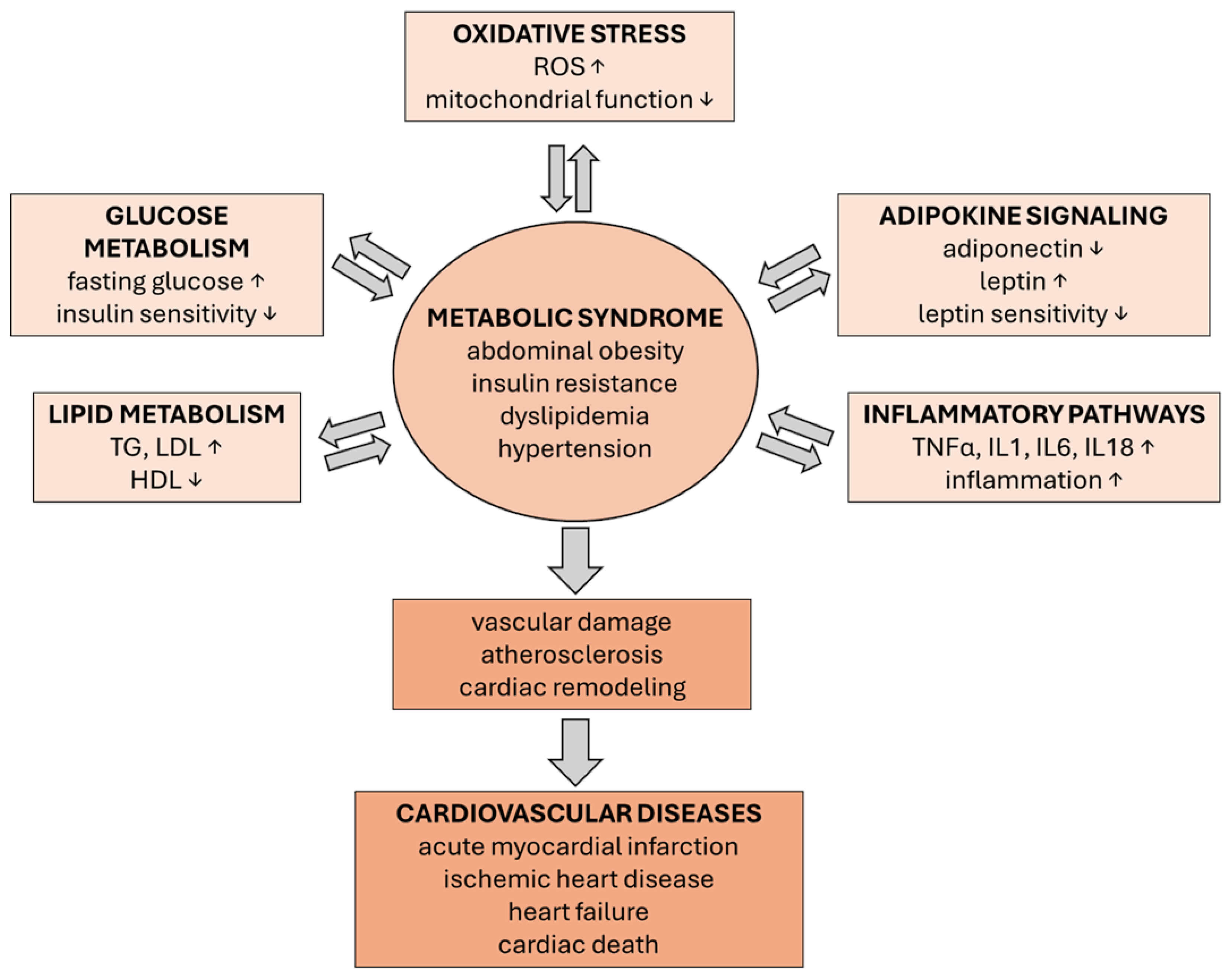
Figure 1. Metabolic disruption in MetS and its comorbidities.
Metabolites 15 00645 g001
Figure 2. Effect of NAC on adipose tissue—evidence from cellular and animal studies.
Metabolites 15 00645 g002
Figure 3. Effect of NAC on obesity and MetS animal models.
Metabolites 15 00645 g003
Figure 4. NAC and MetS/obesity related cardiovascular complications.
Metabolites 15 00645 g004
Table 1. NAC and the modulation of metabolic pathways in the management of MetS and its comorbid conditions.
Metabolic PathwaysPhysiological FunctionEffect of DysregulationImpact of NACModel/Dosage
LipidmetabolismFatty acid oxidation, energy balanceDyslipidemia, ectopic fat accumulationIncreases fatty acid oxidation, reduces lipogenesis, normalizes lipid profilescultured 3T3-L1
preadipocytes, 10 μM NAC [92]
BMAT in vitro, 10 mM NAC [93]
Wistar rats injected
AGE-albumin, NAC in
drinking water (600 mg/L) [94]
HFD rat, 500 mg/kg NAC [95]
Glucose metabolismMaintaining energy homeostasis via ATP production, glucose synthesis, redox balance, and storageDysregulated glucose flux, lactate accumulation, oxidative stress, insulin resistanceImproves insulin signaling, reduces oxidative stress, enhances glycogen synthesis, modulates glycolysis, improves glucose tolerance and uptake3T3-L1 murine fibroblasts, 2.5 or 5 mM NAC [96]
iFIRKO mice, 15 mmol/L NAC in drinking water [87]
HFD mice, 50 mg/kg NAC [97]
Insulin signalingGlucose uptake, lipid metabolismInsulin resistance, hyperglycemiaImproves insulin sensitivity, reduces inflammation in adipose tissue3T3-L1 fibroblasts, 50 μM NAC [98]
HFD rat, 500 mg/kg NAC [99]
HFD mice, 50 mg/kg NAC [100]
Oxidative stress responseROS detoxification, redox balanceIncreased accumulation of ROS—endothelial dysfunction, mitochondrial damageIncreases GSH, scavenges ROS, protects mitochondria and vascular endothelium3T3-L1 adipocytes,
1.0 mmol/L NAC [101]
ApoE-/- mice, 2 mmol/L NAC in drinking water [102]
HFD rat, 150 mg/kg NAC [103]
MetaflammationImmune regulation, tissue repairChronic low-grade
Inflammation, elevated proinflammatory cytokines such tnf-α, il-6
Reduces pro-inflammatory cytokines, attenuates chronic low-grade inflammation, improves adiponectin levelshuman ATM, 5 mM NAC [104]
3T3-L1 adipocytes, 1 mM NAC [105]
HFD rat, 500 mg/kg NAC [106]
Mitochondrial metabolismEnergy production, apoptosis regulationMitochondrial dysfunction, reduced membrane potentialImproves energy metabolism and prevents mitochondrial dysfunction in adipose tissue and cardiomyocytes, restores mitochondrial membrane potentialpancreatic-βcells, 10 mM NAC [107]
HFD mice, 60 mg/kg NAC [108]
Molecular hallmarks of agingIncrease in activity with ageIncrease in sa–β-gal and p16, p21 expressionReduces SA–β-gal activity and expression of p16, p21pol η−/− mice, 1 mg/mL NAC in drinking water [109]
ApoptosisCell growth, metabolism, and survivalHypertrophy and apoptosis of cardiomyocytesReduces oxidative stress-induced hypertrophy and cardiomyocyte apoptosispancreatic-βcells, 10 mM NAC [107]
NRCM cardiomyocytes, 100 µM NAC [75]
Abbreviations: NAC: N-acetylcysteine; MetS: metabolic syndrome; PI3K: phosphoinositide 3-kinase; ROS: reactive oxygen species; GSH: glutathione; TNF-α: tumor necrosis factor alpha; IL-6: interleukin 6; SA–β-gal: senescence-associated beta-galactosidase; ER: endoplasmic reticulum; NRCM: primary cultured neo-natal rat cardiomyocytes; HFD: high-fat diet.
Table 2. NAC-mediated modulation of cellular signaling in obesity/MetS-related cardiometabolic disorders.
Cellular SignalingPhysiological RoleEffect of DysregulationImpact of NACModel/Dosage
AMPKEnergy sensor, catabolic pathways activation at low ATPImpaired energy balance, lipid accumulationIndirectly supports AMPK activation, restores redox balance,pancreatic-βcells, 10 mM NAC [107]
HFD mice, 60 mg/kg NAC [108]
m-TORCell growth, protein synthesis regulation, and nutrient sensingLipogenesis promotion, insulin resistanceReduces oxidative stress, may normalize mTOR signalingpancreatic-βcells, 10 mM NAC [107]
HFD rat, 500 mg/kg NAC [110]
ROS/NRF2cellular resistance to oxidation regulationInflammation, cellular damageBoosts glutathione, activates NRF2, reduces ROS3T3-L1 murine fibro-blasts, 2.5 or 5 mM NAC [96]
HFD mice, 0.5, 2 or
10 g/L NAC in drinking water [111]
ROS–NLRP3Inflammatory response triggered by mitochondrial DAMPsChronic inflammation and tissue injury, insulin resistance in skeletal muscleInhibits NLRP3 activation by lowering ROS restored insulin-dependent glucose uptakeHFD rat, 150 mg/kg NAC [103]
HFD diabetic rat, 20 mg/kg NAC [112]
PI3K/AKTInsulin signaling mediation, glucose uptake regulationInsulin resistance, hyperglycemia, metabolic imbalance, oxidative stress and inflammation increase, mitochondrial dysfunction and apoptosis of cardiomyocytes, pathological cardiac remodeling and progression to HFImproves insulin sensitivity by reducing oxidative and ER stress, enhances AKT expression, elevates phosphorylated AKT levelsNRCM cardiomyocytes, 100 µM NAC [75]
3T3-L1 fibroblasts, 50 μM NAC [98]
HFD mice, 50 mg/kg NAC [97]
HFD mice, 50 mg/kg NAC [100]
SIRT1/
SIRT3
Mitochondrial function oxidative stress, and metabolism regulationMitochondrial dysfunction and ROS accumulationIncreases the NAD+/NADH ratio, supports SIRT1/3 activity, reduces inflammationBMAT in vitro, 10 mM NAC [93]
NFκB/
AP-1/
JNK
Inflammatory response regulationChronic inflammation; insulin resistance, endothelial dysfunctionInhibits NFκB-mediated inflammation3T3-L1 adipocytes, 1 mM NAC [105]
HFD rat, 500 mg/kg NAC [110]
MAPK (ERK/JNK)Cellular proliferation, differentiation, and stress response regulationAdipogenesis and inflammation activationInhibits ERK1/2 and JNK1/2 phosphorylation; suppresses adipocyte differentiation and inflammatory signaling3T3-L1 preadipocytes, 0.01 or 1 mM NAC [113]
STAT3Cytokine signaling and immune response regulationChronic inflammation and metabolic dysfunction activationInhibits STAT3 activation via antioxidant and anti-inflammatory mechanismsHFD diabetic rat, 1.5 g/kgNAC [114]
GSK3βGlycogen synthesis and insulin signaling regulationAltered glucose metabolism and defective insulin signalingRestores GSK3β phosphorylation; improves insulin signalingHFD rat, 500 mg/kg NAC [110]
T3-L1 preadipocytes, 100 µM NAC
[115]
IRS1/PI3KInsulin signaling cascade initiationGlucose uptake dysfunction, insulin resistanceEnhances IRS1/PI3K association, improves insulin signallingHFD mice, 50 mg/kg NAC [97]
CHOPER stress marker, apoptosis promotionER stress, mitochondrial dysfunction, inflammatory activationReduces CHOP accumulation; restores anti-inflammatory cytokine secretion3T3-L1 murine fibroblasts, 2.5 or 5 mM NAC [96]
UCP1/
PGC-1α
Thermogenic response and mitochondrial biogenesis inductionLowered energy output and adiposity developmentInduces browning of WAT via NRF2/HO-1 axis; upregulates UCP1 and PGC-1αHFD mice, 400 mg/kg NAC (with other cofactors) [89]
HFD mice, 0.5, 2, or
10 g/L NAC in drinking water 111 [111]
Abbreviations: NAC: N-acetylcysteine; AMPK: AMP-activated protein kinase; mTOR: mechanistic target of rapamycin; ROS: reactive oxygen species; HF: heart failure; NRF2: nuclear factor erythroid 2–related factor 2; NLRP3: NOD-, LRR- and pyrin domain-containing protein 3; PI3K: phosphoinositide 3-kinase; AKT: protein kinase B; ER: endoplasmic reticulum; SIRT1/SIRT3: sirtuin 1/sirtuin 3; NAD+/NADH: nicotinamide adenine dinucleotide (oxidized/reduced forms); NF-κB: nuclear factor kappa-light-chain enhancer of activated B cells; AP-1: activator protein 1: JNK: c-Jun N-terminal kinase; STAT: signal transducer and activator of transcription 3; MAPK: mitogen-activated protein kinase; ERK: extracellular signal-regulated kinase; GSK3β: glycogen synthase kinase 3 beta; IRS1: insulin receptor substrate 1; CHOP: C/EBP homologous protein; UCP1: uncoupling protein 1; HO-1: heme oxygenase 1; PPARγ: peroxisome proliferator-activated receptor gamma; C/EBPβ: CCAAT/enhancer binding protein beta.
Disclaimer/Publisher’s Note: The statements, opinions and data contained in all publications are solely those of the individual author(s) and contributor(s) and not of MDPI and/or the editor(s). MDPI and/or the editor(s) disclaim responsibility for any injury to people or property resulting from any ideas, methods, instructions or products referred to in the content.