Article

Metabolic Deficits in the Retina of a Familial Dysautonomia Mouse Model

1
Department of Chemistry and Biochemistry, Montana State University—Bozeman, Bozeman, MT 59717, USA
2
Department of Microbiology and Cell Biology, Montana State University—Bozeman, Bozeman, MT 59717, USA
3
Department of Biological and Physical Sciences, Montana State University—Billings, Billings, MT 59102, USA
*
Authors to whom correspondence should be addressed.
These authors contributed equally to this work.
Metabolites 2024, 14(8), 423; https://doi.org/10.3390/metabo14080423

Submission received: 8 May 2024 / Revised: 15 July 2024 / Accepted: 29 July 2024 / Published: 31 July 2024

Abstract

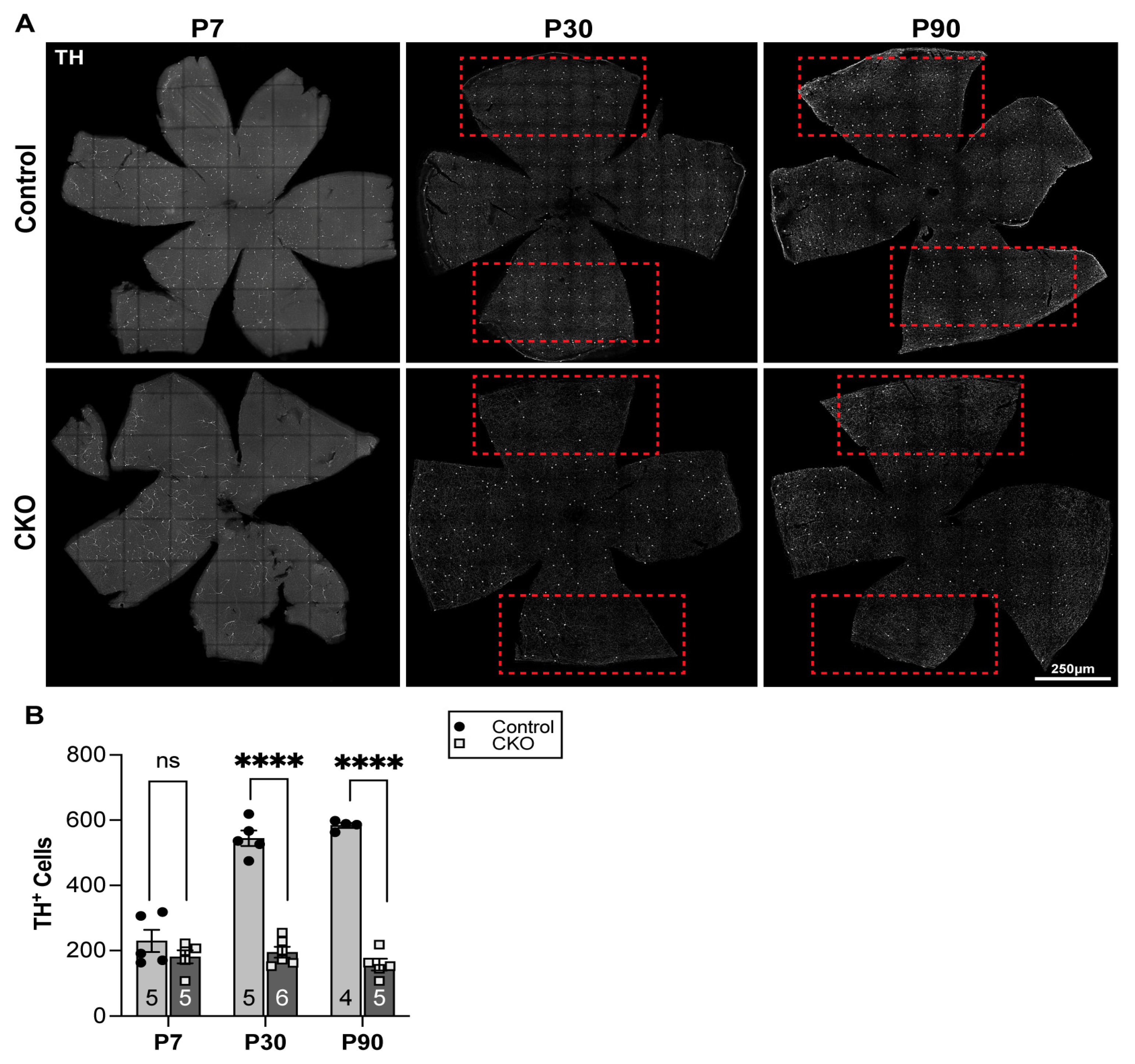
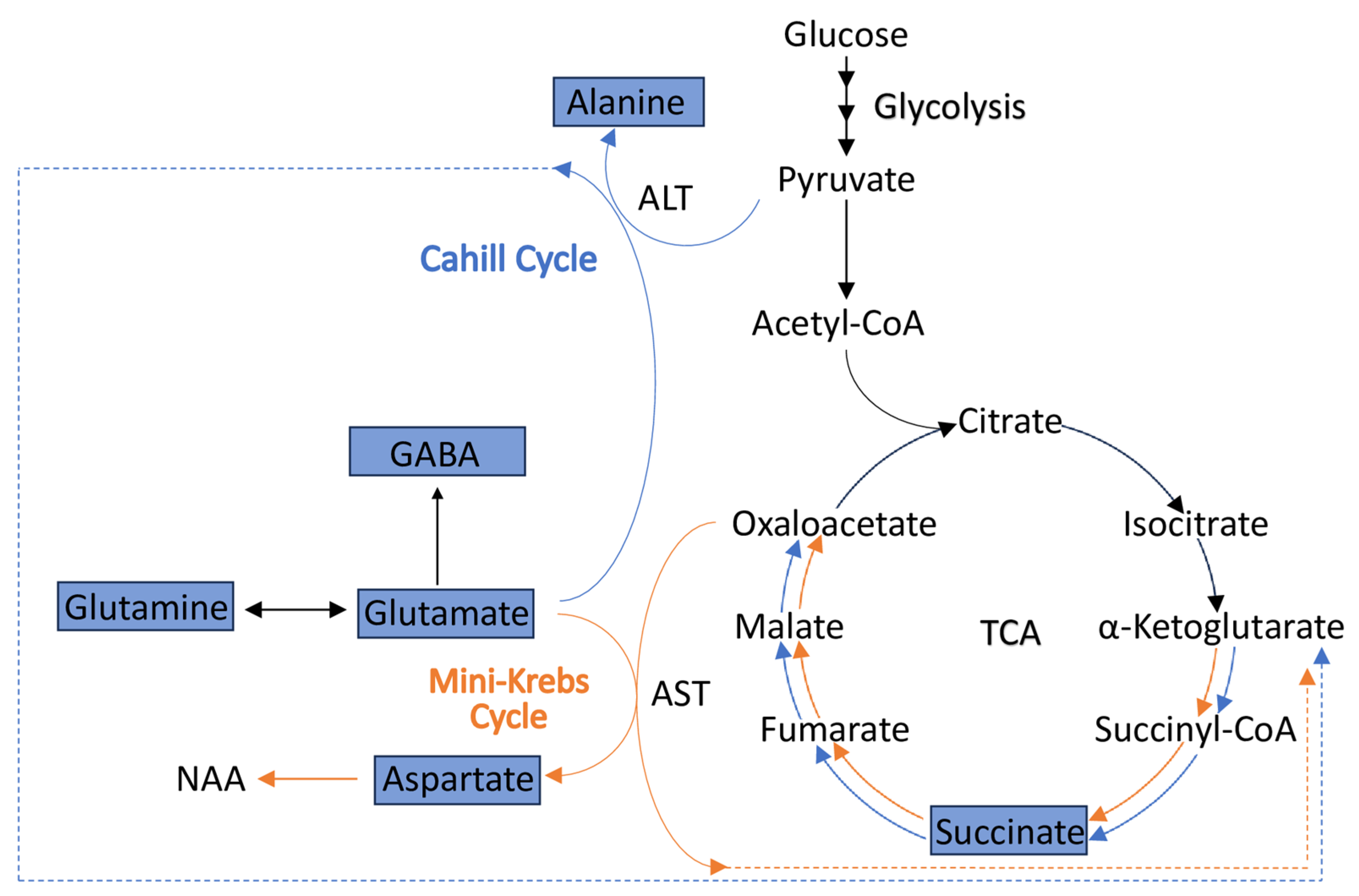
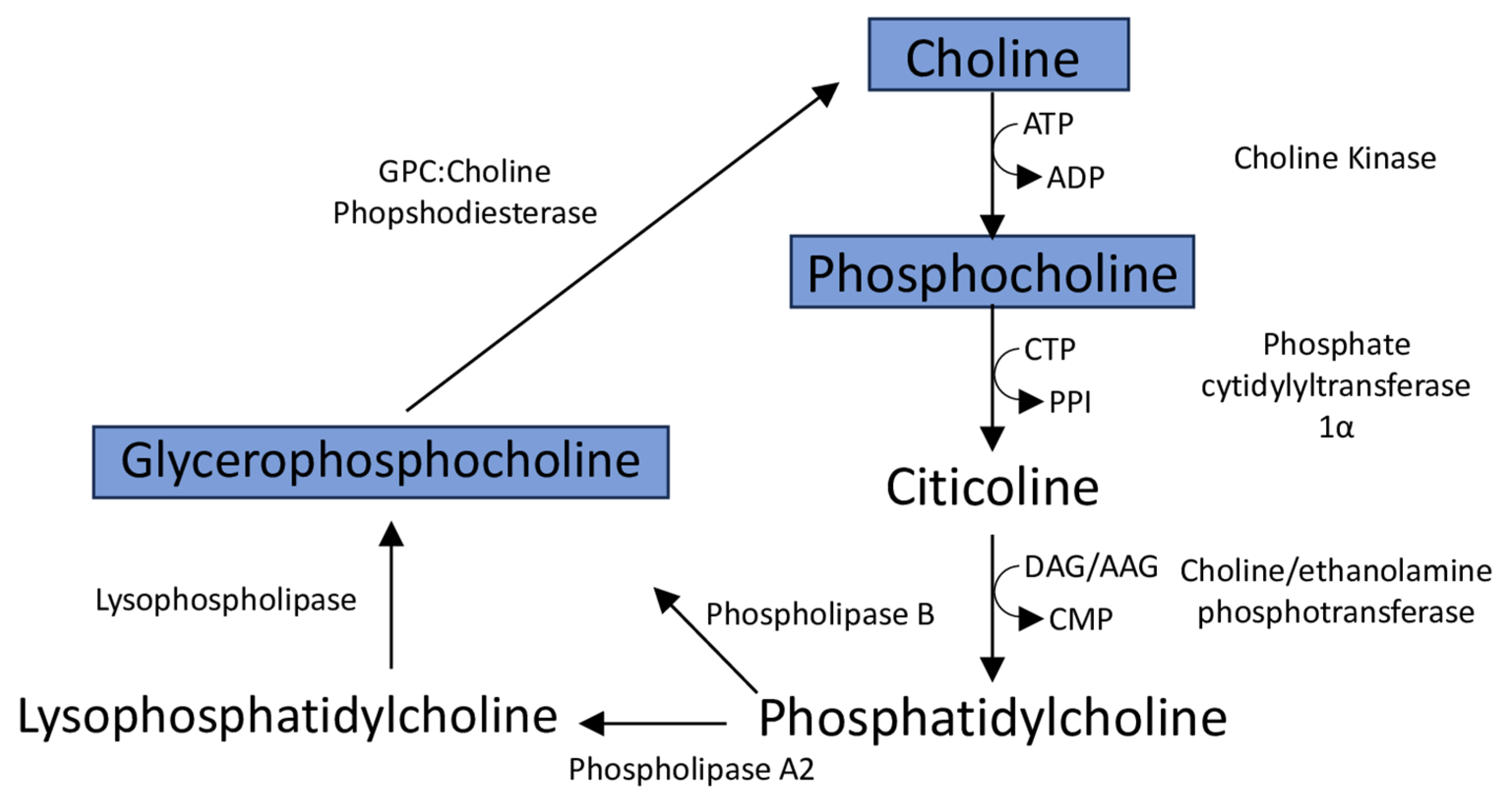
Neurodegenerative retinal diseases such as glaucoma, diabetic retinopathy, Leber’s hereditary optic neuropathy (LHON), and dominant optic atrophy (DOA) are marked by progressive death of retinal ganglion cells (RGC). This decline is promoted by structural and functional mitochondrial deficits, including electron transport chain (ETC) impairments, increased oxidative stress, and reduced energy (ATP) production. These cellular mechanisms associated with progressive optic nerve atrophy have been similarly observed in familial dysautonomia (FD) patients, who experience gradual loss of visual acuity due to the degeneration of RGCs, which is thought to be caused by a breakdown of mitochondrial structures, and a disruption in ETC function. Retinal metabolism plays a crucial role in meeting the elevated energetic demands of this tissue, and recent characterizations of FD patients’ serum and stool metabolomes have indicated alterations in central metabolic processes and potential systemic deficits of taurine, a small molecule essential for retina and overall eye health. The present study sought to elucidate metabolic alterations that contribute to the progressive degeneration of RGCs observed in FD. Additionally, a critical subpopulation of retinal interneurons, the dopaminergic amacrine cells, mediate the integration and modulation of visual information in a time-dependent manner to RGCs. As these cells have been associated with RGC loss in the neurodegenerative disease Parkinson’s, which shares hallmarks with FD, a targeted analysis of the dopaminergic amacrine cells and their product, dopamine, was also undertaken. One dimensional (1D) proton (1H) nuclear magnetic resonance (NMR) spectroscopy, mass spectrometry, and retinal histology methods were employed to characterize retinae from the retina-specific Elp1 conditional knockout (CKO) FD mouse model (Pax6-Cre; Elp1LoxP/LoxP). Metabolite alterations correlated temporally with progressive RGC degeneration and were associated with reduced mitochondrial function, alterations in ATP production through the Cahill and mini-Krebs cycles, and phospholipid metabolism. Dopaminergic amacrine cell populations were reduced at timepoints P30–P90, and dopamine levels were 25–35% lower in CKO retinae compared to control retinae at P60. Overall, this study has expanded upon our current understanding of retina pathology in FD. This knowledge may apply to other retinal diseases that share hallmark features with FD and may help guide new avenues for novel non-invasive therapeutics to mitigate the progressive optic neuropathy in FD.
Keywords: conditional knockout (CKO) FD mouse model; eye health; familial dysautonomia (FD); metabolomics; nuclear magnetic resonance; mass spectrometry; retinal immunohistology; retinal metabolism; retinal ganglion cells; dopaminergic amacrine cells; univariate and multivariate statistical analyses conditional knockout (CKO) FD mouse model; eye health; familial dysautonomia (FD); metabolomics; nuclear magnetic resonance; mass spectrometry; retinal immunohistology; retinal metabolism; retinal ganglion cells; dopaminergic amacrine cells; univariate and multivariate statistical analyses