Article

Urinary 1H NMR Metabolomic Analysis of Prenatal Maternal Stress Due to a Natural Disaster Reveals Metabolic Risk Factors for Non-Communicable Diseases: The QF2011 Queensland Flood Study

1
Canadian Centre for Behavioural Neuroscience, Department of Neuroscience, University of Lethbridge, 4401 University Drive, Lethbridge, AB T1K 3M4, Canada
2
Southern Alberta Genome Sciences Centre, University of Lethbridge, 4401 University Drive, Lethbridge, AB T1K 3M4, Canada
3
Department of Chemistry and Biochemistry, University of Lethbridge, 4401 University Drive, Lethbridge, AB T1K 3M4, Canada
4
Midwifery Research Unit, Mater Research Institute, University of Queensland, Brisbane, QLD 4072, Australia
5
School of Psychology, University of Queensland, Brisbane, QLD 4072, Australia
6
Molly Wardaguga Research Centre, Faculty of Health, Charles Darwin University, Alice Springs, NT 0870, Australia
7
Perinatal and Woman’s Health Unit, University of New South Wales, Sydney, NSW 2052, Australia
8
Centre for Child Development and Mental Health, Lady Davis Institute for Medical Research, Jewish General Hospital, 4335 Chemin de la Côte-Sainte-Catherine, Montreal, QC H3T 1E4, Canada
9
Department of Psychiatry, Douglas Mental Health University Institute, McGill University, 6875 LaSalle Boulevard, Montreal, QC H4H 1R3, Canada
*
Authors to whom correspondence should be addressed.
Metabolites 2023, 13(4), 579; https://doi.org/10.3390/metabo13040579
Submission received: 23 March 2023 / Revised: 15 April 2023 / Accepted: 17 April 2023 / Published: 21 April 2023
(This article belongs to the Topic Metabolism and Health)

Abstract

:
Prenatal stress alters fetal programming, potentially predisposing the ensuing offspring to long-term adverse health outcomes. To gain insight into environmental influences on fetal development, this QF2011 study evaluated the urinary metabolomes of 4-year-old children (n = 89) who were exposed to the 2011 Queensland flood in utero. Proton nuclear magnetic resonance spectroscopy was used to analyze urinary metabolic fingerprints based on maternal levels of objective hardship and subjective distress resulting from the natural disaster. In both males and females, differences were observed between high and low levels of maternal objective hardship and maternal subjective distress groups. Greater prenatal stress exposure was associated with alterations in metabolites associated with protein synthesis, energy metabolism, and carbohydrate metabolism. These alterations suggest profound changes in oxidative and antioxidative pathways that may indicate a higher risk for chronic non-communicable diseases such obesity, insulin resistance, and diabetes, as well as mental illnesses, including depression and schizophrenia. Thus, prenatal stress-associated metabolic biomarkers may provide early predictors of lifetime health trajectories, and potentially serve as prognostic markers for therapeutic strategies in mitigating adverse health outcomes.

1. Introduction

Human populations are increasingly exposed to physical and psychological stress associated with climate change, including natural disasters, forced migration, starvation, or exposure to environmental toxins [1,2,3]. If pregnant mothers are exposed to these adverse events, the ensuing stress response may induce excessive glucocorticoid release that has the potential to affect the health of the developing fetus [4,5,6]. Accordingly, prenatal maternal stress (PNMS) has been shown to affect birth outcomes and potentially predispose the child to adverse mental and physical health later in life [1,3,7]. For example, children who experienced greater maternal stress from the 2008 Iowa Flood in utero displayed higher body mass index (BMI) and increased risks for adiposity at age 2.5 years [8,9], along with higher cortisol reactivity [10]. Similar results were also obtained due to prenatal exposure to high maternal stress caused by the 1998 Quebec Ice Storm, with the children displaying higher BMI and adiposity at ages 5.5, 8.5, 13.5, and 15.5 years [11], poorer language abilities [12], and altered testosterone and cortisol reactivity [13]. Notably, the mother’s objective hardship and her subjective experience are associated with different outcomes. Greater maternal subjective distress was associated with diminished cognitive functioning in the toddler, whereas both objective hardship and subjective distress were linked to reduced language-related development in children who experienced the 2008 Iowa Flood in utero [14]. Furthermore, exposure to high objective hardship during the 1998 Quebec Ice Storm was associated with lower intelligence quotients at 2 and 5.5 years of age [12,15].
The transmission of maternal stress to the fetus is complex and multifaceted. The placenta represents a protective barrier to the transmission of maternal stress to the fetus. During gestation, the placenta and fetal brain display an overexpression of the enzyme 11β-hydroxysteroid dehydrogenase (11β-HSD) type 2 [16,17,18]. 11β-HSD type 2 converts cortisol to cortisone [19], thereby protecting the developing fetus from the growth-inhibiting and pro-apoptotic effects of cortisol. Placental impairment in adaptive regulation of 11β-HSD expression in response to stress [20] has been associated with growth restriction of the infant and the development of psychopathologies in later life, including anxiety traits and schizophrenia [18,21,22,23,24]. Thus, maternal stress may alter fetal development via changes in the placenta, a process known as fetal programming [25].
Importantly, outcomes linked to severe PNMS can last into adolescence and adulthood. For example, individuals who were prenatally exposed to the 1976 Tangshan earthquake had a significantly higher incidence of depression in adulthood [26]. Prenatal exposure to earthquakes was also shown to raise the risk of schizophrenia spectrum disorders [27]. Moreover, the increased severity of maternal stress during pregnancy caused by the 1998 Quebec Ice Storm was associated with greater offspring insulin secretion in adolescence, suggesting a potential risk of insulin resistance and metabolic disease [28]. This finding was also supported by urinary metabolomic analysis of offspring exposed to the 1998 Quebec Ice Storm in utero, suggesting a potential risk for type 2 diabetes mellitus [29]. Indeed, one retrospective study found that maternal bereavement during pregnancy was significantly associated with offspring development of type 2 diabetes mellitus in adulthood [30]. Notably, the risk for metabolic disease shows sex-specific programming [31].
A series of floods impacted Queensland, Australia, beginning in November 2010 and lasting until January 2011. The 2011 Queensland Flood (QF2011) Australian study commenced following significant flooding that forced thousands to evacuate their homes, caused more than $2.3 billion AUD in reported damages, and resulted in 33 deaths [4]. The 2011 Queensland Flood represented an environmental stressor with a measurable objective and subjective impacts on pregnant women [4]. Previous work on the QF2011 cohort found that high maternal subjective distress in later pregnancy significantly reduced placental corticotropin-releasing hormone (CRH) concentrations in girls only, indicating sex-specific influences [31]. Importantly, higher CRH has been linked to reduced fetal adaptation to the uterine environment, resulting in lower birth weight and slower fetal growth [1]. In addition, work on the same study found an association between low levels of maternal hardship, high hair copper levels, and language developmental deficits in 4-year-old boys [32].
Differences in the outcomes of PNMS may vary due to changes in placental function [33] and an altered metabolome [5]. Metabolic profiling via proton nuclear magnetic resonance (1H NMR) spectroscopy has emerged as a particularly useful tool for characterizing pathophysiological consequences of fetal-environment interactions [29,34,35,36]. Urinary metabolic profiles represent the downstream physiological consequences of these interactions [37]. Although 1H NMR spectroscopy is less sensitive (micromolar concentrations) than other metabolomics approaches, such as liquid or gas chromatography-mass spectroscopy (nanomolar concentrations), NMR spectroscopy is advantageous when working with samples of limited availability. As 1H NMR is non-destructive, samples can be reused for additional analyses using other methods. Furthermore, 1H NMR spectroscopy is a high-throughput technique that requires minimal sample preparation and provides access to quantify the most metabolites in urine when compared to mass-spectroscopy based techniques (209 vs. 179, respectively) [37].
The present study utilized 1H NMR to investigate the metabolic alterations arising from PNMS in urine. The analysis of males and females was carried out separately, as there are significant sex differences in the urine metabolome [38]. Although PNMS is known to impact offspring development and life-long health risks [7,39], the mechanisms by which these changes occur remain unclear [40,41,42]. By including objective hardship as a stressor, this study considers stress independently of parental influences or family characteristics such as socio-economic status. This feature facilitates the ability to determine how objective and subjective factors, separately or synergistically, influence child development. Our aim was to identify metabolic profiles in male and female offspring that reflect their mother’s levels of objective hardship and subjective distress in relation to the 2011 Queensland Flood and provide insight into potential long-term health risks and their mechanisms of action. Overall, this study highlights the potential role of maternal experience in shaping the metabolic responses in children and suggests potential targets for early diagnosis, intervention, and prevention strategies.

2. Materials and Methods

2.1. Study Design

2.1.1. Post-Flood Recruitment

In January 2011 Queensland, Australia experienced its worst flooding in 30 years. The recruitment of study participants began once ethics approval was obtained in April 2011 and continued up to 12 months post-flood with full details outlined in the protocol [4]. Many participants were re-recruited from an unrelated study (the M@NGO study; [43,44]) at the Mater Mothers Hospital, while others were recruited by their midwives, or by research assistants, or responded to flyers at the hospital. Women included in the study were 18 years of age or older, residing in the area of Brisbane on 10 January 2011, spoke fluent English, and were pregnant or became pregnant within days of peak flooding with a single offspring. All 230 women had given birth within the first 12 months after the beginning of the flood [45]. Mothers’ mean age at childbirth was 31.12 years (SD = 5.28; range = 19.52–47.33) and their mean socio-economic status was 1050.77 (SD = 59.13) [46]. Maternal socio-economic status was determined using the Socio-Economic Index For Area (SEIFA) scores, and was based on Australian postcodes and census data. A detailed description of the study protocol can be found in King et al. [4], and maternal SES can be found in Chen et al. [46].

2.1.2. Assessment of Objective Hardship and Composite Subjective Distress

Upon recruitment, the 230 participants were administered the QFOSS200 (Queensland Flood Objective Stress Survey), the IES-R (Impact of Events Scale—Revised; [47]), the Peritraumatic Distress Inventory (PDI; [48]), and the Peritraumatic Dissociative Experiences Questionnaire (PDEQ; [49]). Participants completed all surveys again at 12 months post-flood.
The QFOSS200 questionnaire assessed pregnant women’s flood-related objective hardship in terms of threat, loss, scope, and change. Each dimension had a maximum of 50 points for a total maximum score of 200 points, with higher scores reflecting greater objective hardship [4,45]. The QFOSS200 survey was based on an existing objective hardship survey used to assess the flood experiences of pregnant mothers during the 2008 Iowa flood [50]. The QFOSS200 survey was administered upon recruitment, and again 12 months post-flood to update participants’ experiences with insurance companies and financial loss [4].
The IES-R, PDI, and PDEQ were used to identify potential flood-related adverse mental health symptoms. The 10-item PDEQ assessed the mother’s dissociative-like experiences, whereas the 13-item PDI assessed panic-like reactions [51]. The PDI and PDEQ questionnaires were used to assess how the participants recalled having felt at the time of the flood. The IES-R is a 22-item assessment, commonly used to identify symptoms of post-traumatic stress disorder [47]. This questionnaire addressed how participants in the QF2011 study currently felt—at recruitment and 12 months post-flood—to assess the ongoing psychological impact resulting from the flood [4].
A composite score for mothers’ subjective distress (COSMOSS) was created utilizing Principal Component Analysis (PCA) of the three subjective distress scores, with the algorithm deriving: 0.358 × IESR + 0.397 × PDI + 0.387 × PDEQ. COSMOSS scores represent a variable derived from the PCA that described 76.27% of the total subjective distress variance, standardized to a mean of 0, and a standard deviation of 1.0 [45].
For the purposes of analyses in the present study, maternal scores were divided at the median into high vs. low objective hardship groups and high vs. low composite subjective distress groups.

2.2. Sample Collection and Preparation

Of the 230 mothers who took part in the original study, urine samples were only provided from 89 of the 4-year-old offspring (male: n = 50; female: n = 39) and these samples were collected at the first passage of the day and stored at −80 °C. Frozen samples were transported to the University of Lethbridge for metabolomic analysis. The following procedure was carried out for all 89 samples. A total of 160 µL of buffer, 40 µL of 0.02709% w/v D2O with trimethylsilyl propanoic acid (TSP), and 400 µL of urine was added to each microcentrifuge tube. The buffer consisted of a 4:1 ratio of dibasic potassium phosphate (K2HPO4) to monobasic potassium phosphate (KH2PO4) with a concentration of 0.625 M and contained 3.75 mM of NaN3 as an antimicrobial agent and 3.75 mM of potassium fluoride (KF). The centrifuge tube was then vortexed and centrifuged at 12,000 rpm at 4 °C for 5 min. A 550 µL aliquot of the resulting supernatant was transferred to an NMR tube. All samples were checked for a pH of 7.4 ± 0.05 before 1H NMR analysis.

2.3. NMR Data Acquisition

Spectra were collected on a 700 MHz Bruker Avance III HD spectrometer (Bruker Ltd., Milton, ON, Canada), using a 1-D NOESY gradient water suppression pulse sequence. Samples were run for 128 scans to a total acquisition size of 128 k, zero filled to 256 k, automatically phased, baseline corrected, and line-broadened by 0.3 Hz. The spectra were then exported to MATLAB (MathWorks, Natick, MA, USA) to undergo recursive segment wise peak alignment [52] and subsequently binned using Dynamic Adaptive Binning, followed by manual correction [53]. Prior to modeling, the data were normalized, log-transformed, and pareto-scaled [54]. All spectra were referenced to the TSP peak (0.00δ).

2.4. Statistical Analyses

A Shapiro-Wilk test was used to assess the normality of the data and all bins were determined to be non-parametric. The Wilcoxon-Mann-Whitney U (MW) test was employed following the result of non-parametric data [55]. All univariate statistical tests underwent Bonferroni-Holm correction to correct for multiple comparisons and a p-value equal to or less than 0.05 was considered significant. Variable Importance Analysis based on random Variable Combination (VIAVC) was utilized to identify significantly altered bins between groups for use in metabolite identification [56]. The VIAVC algorithm employs binary matrix resampling, ensuring each variable has an equal chance of being selected. The importance of each variable is assessed based on the percent increase or decrease in the area under the curve (AUC) corresponding to the receiver operator characteristic curve (ROC) when added or removed from the respective subsets. This process is repeated until only bins that result in the best class separation remain. Orthogonal Projection to Latent Structures—Discriminant Analysis (OPLS-DA), a supervised multivariate test [57], was carried out to maximise observed differences between high vs. low maternal composite subjective distress as well as high vs. low maternal objective hardship. All multivariate models were validated by double ten-fold cross-validation and permutation testing using 2000 permutations [58,59]. OPLS-DA analysis was carried out utilizing bins identified as significant by VIAVC, as they led to the most observed separation between comparison groups. Males and females were analyzed separately, as there are significant sex differences in the urinary metabolome [38].

2.5. Metabolite Identification and Pathway Analysis

Chenomx 8.2 NMR suite software (Chenomx Inc., Edmonton, AB, Canada), in conjunction with the Human Metabolome Database (HMDB) of urinary metabolites [60,61,62], was used to identify the metabolites corresponding to bins found to be significantly altered by either VIAVC or MW testing. Metaboanalyst was used to perform pathway topology analysis using the metabolites identified as significant by VIAVC. The homo sapiens pathway library was utilized for all comparisons, the hypergeometric test was selected for the over-representation analysis, and relative-betweenness centrality was chosen for the pathway topology analysis. Pathways with a p-value equal to or less than 0.05 were labeled and considered to be significant.

3. Results

3.1. Maternal Objective Hardship and Subjective Distress Produce Unique Metabolome Profiles

According to the median split of the QFOSS200 objective hardship questionnaire results, 26 of the 50 male offspring mothers experienced high objective hardship and the remaining 24 experienced low objective hardship. COSMOSS results showed that 25 mothers of male offspring had high composite subjective distress scores and 25 had low composite subjective distress scores.
QFOSS200 scores classified 20 of the 39 mothers of female offspring as having experienced high objective hardship, the remaining 19 were categorized as having experienced low objective hardship. COSMOSS data resulted in 23 mothers of females with high composite subjective distress, whereas the remaining 16 scored low composite subjective distress.
Comparisons were conducted separately for high versus low objective hardship in males, high versus low subjective distress in males, high versus low objective hardship in females, and high versus low subjective distress in females. Differences in urinary metabolite concentrations that were statistically significant between comparison groups were identified using VIAVC. Out of 382 bins, 26 unique metabolites previously found in urine were significantly altered in males associated with maternal objective hardship, whereas 13 were altered in males associated with maternal composite subjective distress (Table 1). Thirty-one metabolites were significantly altered in females because of maternal objective hardship, and twenty-five metabolites were altered in females when considering maternal composite subjective distress (Table 2) [61].
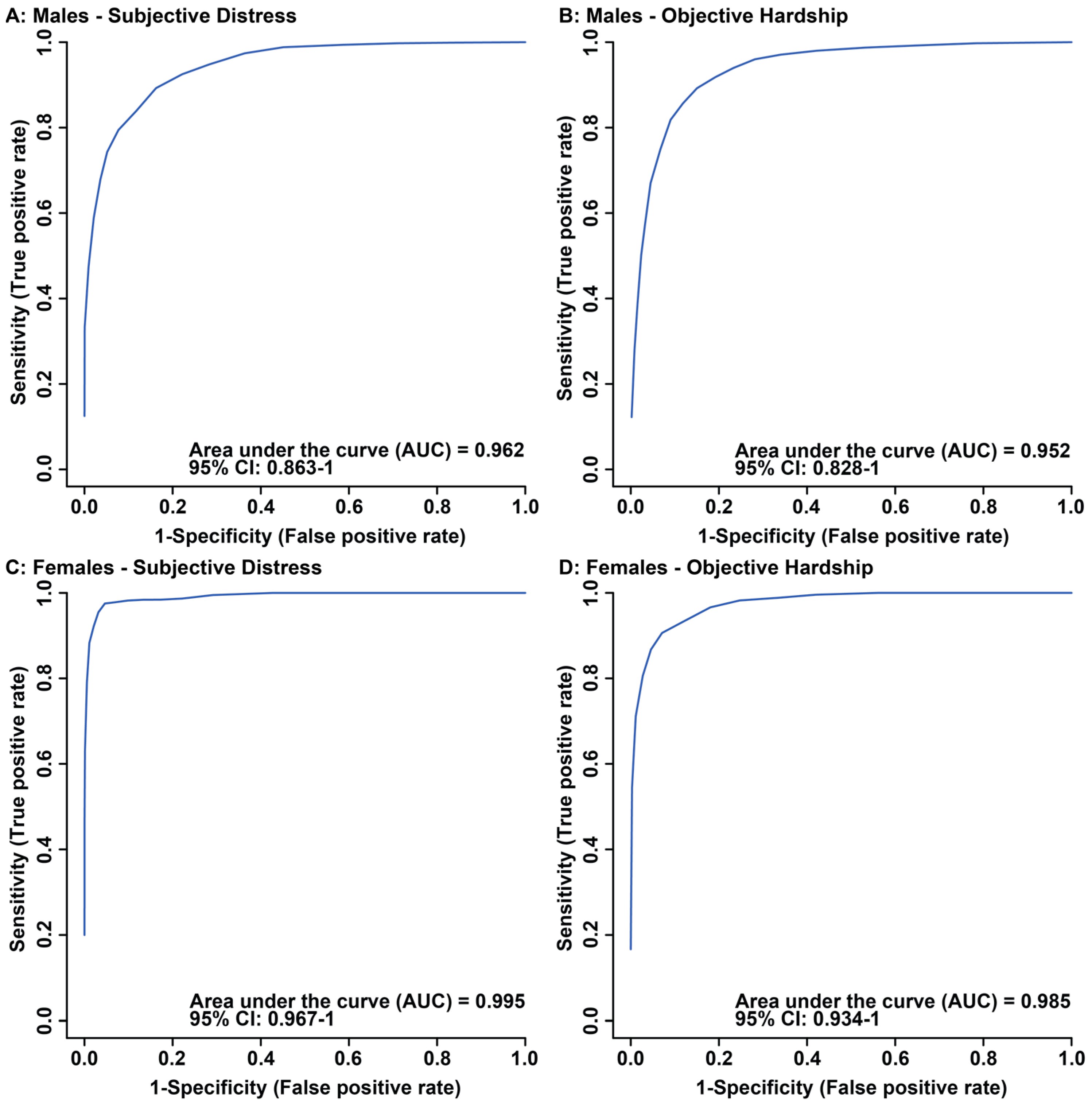
OPLS-DA scores plots of males showed significant separation in maternal composite subjective distress and maternal objective hardship groups, when comparing median-split high vs. low scores (Figure 1A,B). Cross validation and permutation testing confirmed the observed supervised separation in maternal composite subjective distress (p = 0.00004, Q2: 0.594, R2: 0.757, Figure 1A) and objective hardship (p = 0.00004, Q2: 0.635, R2: 0.837, Figure 1B). ROC analysis validated the identified metabolites as contributing to alterations in males due to maternal composite subjective distress (AUC: 0.962, CI: 0.863–1, Predictive accuracy: 87.2%, Figure 2A) and objective hardship (AUC: 0.952, CI: 0.828–1, Predictive accuracy: 87.6%, Figure 2B) group comparisons. In females, supervised OPLS-DA scores plots display significant separation between high vs. low groupings for both composite subjective distress and objective hardship (Figure 1C and Figure 1D, respectively). Cross validation and permutation testing confirmed the observed supervised separation in both maternal composite subjective distress (p = 0.00004, Q2: 0.84, R2: 0.912, Figure 1C) and objective hardship (p = 0.00004, Q2: 0.791, R2: 0.935, Figure 1D). ROC analysis validated the identified metabolites as contributing to alterations in females due to maternal composite subjective distress (AUC: 0.995, CI: 0.967–1, Predictive accuracy: 97.2%, Figure 2C) and objective hardship group comparisons (AUC: 0.985, CI: 0.934–1, Predictive accuracy: 90.3%, Figure 2D).

3.2. Maternal Objective Hardship and Subjective Distress Program Protein Synthesis, Energy Metabolism, and Carbohydrate Metabolism

High maternal objective hardship in males downregulated 20/32 (62.5%) bins identified as significant by VIAVC (Table 1). In contrast, 9/14 (64%) significant bins were upregulated due to high maternal composite subjective distress in males (Table 1).
In females, 23/39 (58.9%) significant bins were upregulated due to high maternal objective hardship (Table 2), and 13/25 (52%) bins were downregulated in females of high maternal composite subjective distress (Table 2).
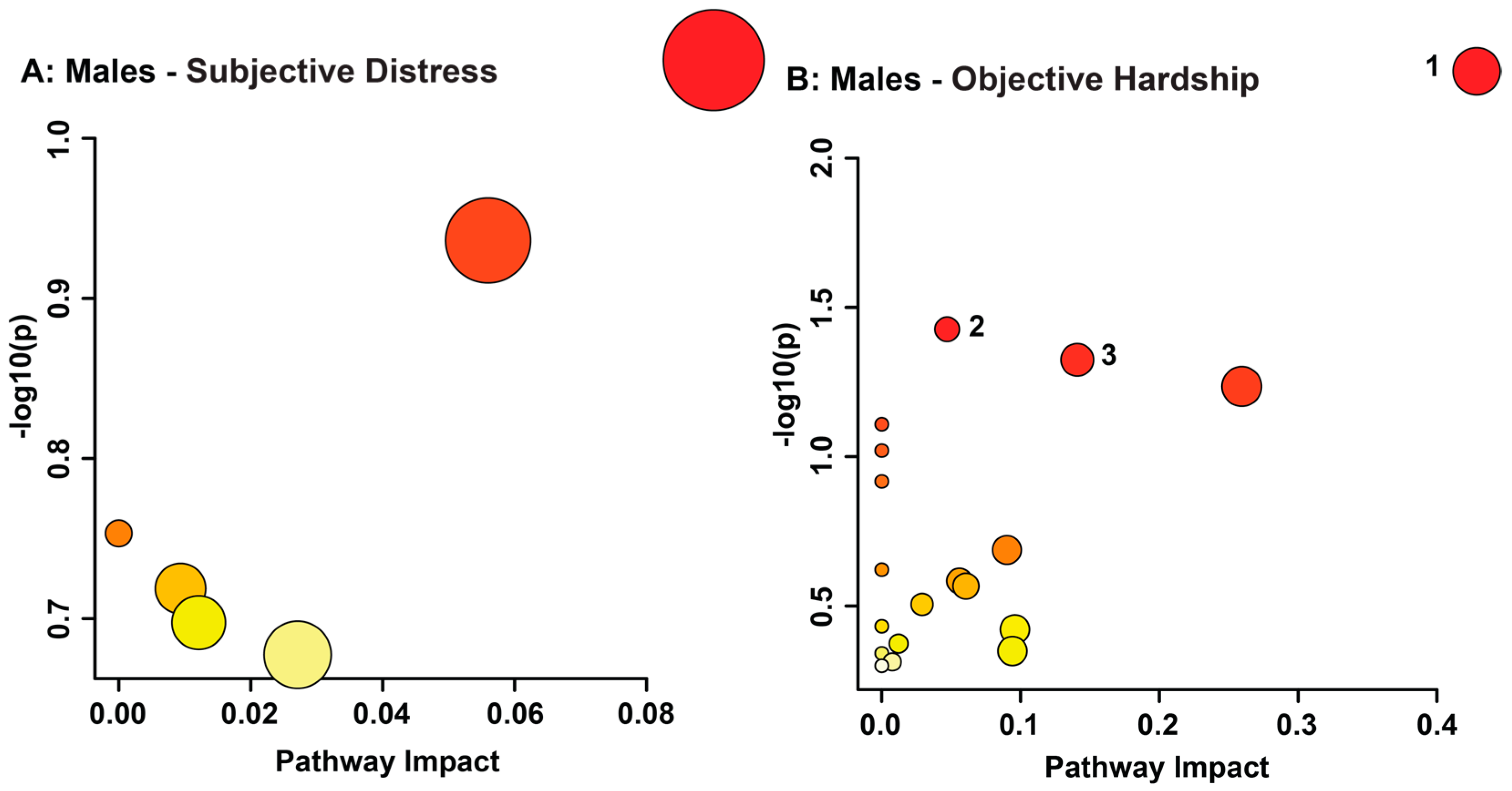
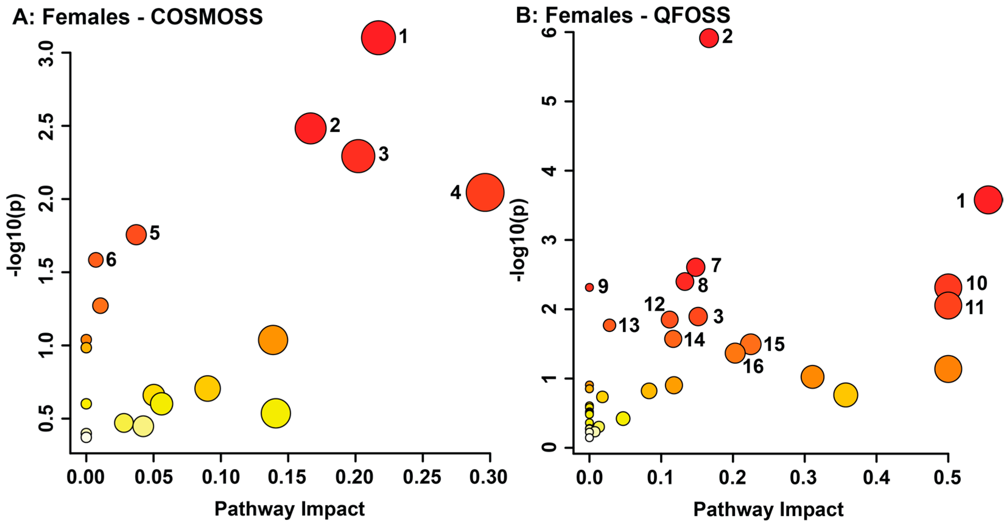
Changes in metabolite concentration deemed significant by exploratory statistical analysis were grouped by biological metabolic pathway. In order to predict the metabolic pathways that were potentially altered in both sexes due to the effects of maternal objective hardship and composite subjective distress, all significant metabolites identified by VIAVC were utilized (Figure 3 and Figure 4).
Maternal composite subjective distress in males did not indicate any metabolic pathway as potentially altered (Figure 3A, Table S1). Males’ maternal objective hardship pathway topology analysis revealed the taurine and hypotaurine metabolism (p = 0.0051158, impact = 0.42857), pentose phosphate pathway (p = 0.037427, impact = 0.04712), and lysine degradation (p = 0.047378, impact = 0.14085) as potentially altered metabolic pathways (Figure 3B, Table S1).
Maternal composite subjective distress in females presented potentially altered metabolic pathways including glycine, serine, and threonine metabolism (p = 0.00079075, impact = 0.21707); aminoacyl-tRNA biosynthesis (p = 0.003291, impact = 0.16667); galactose metabolism (p = 0.0050899, impact = 0.20208); cysteine and methionine metabolism (p = 0.009009, impact = 0.2963); tyrosine metabolism (p = 0.017523, impact = 0.03714); and pantothenate and CoA biosynthesis (p = 0.02603, impact = 0.00714; Figure 4A, Table S1). Pathway topology analysis for objective hardship in females showed the following metabolic pathways as potentially impacted: aminoacyl-tRNA biosynthesis (p = 1.22 × 10⁻⁶, impact = 0.16667); glycine, serine, and threonine metabolism (p = 0.00026396, impact = 0.55577); glyoxylate and dicarboxylate metabolism (p = 0.0024763, impact = 0.14815); starch and sucrose metabolism (p = 0.0039813, impact = 0.13303); nitrogen metabolism (p = 0.0048415, impact = 0); D-glutamine and D-glutamate metabolism (p = 0.0048415, impact = 0.5); ascorbate and aldarate metabolism (p = 0.0088294, impact = 0.5); galactose metabolism (p = 0.012742, impact = 0.15148); glutathione metabolism (p = 0.014093, impact = 0.11182); porphyrin and chlorophyll metabolism (p = 0.017032, impact = 0.02799); arginine biosynthesis (p = 0.026767, impact = 0.11675); arginine and proline metabolism (p = 0.032014, impact = 0.22479); and pentose and glucuronate interconversions (p = 0.042975, impact = 0.20312; Figure 4B, Table S1).

4. Discussion

Here we show that high and low flood-related composite subjective distress and objective hardship are associated with significant differences in offspring urinary metabolome measured at four years of age. This study revealed significant alterations in metabolic pathways linked to oxidative stress. Oxidative stress reflects an imbalance between reactive oxygen species (ROS) and antioxidants, leading to cellular apoptosis and damage to DNA and proteins. Furthermore, psychological distress in adults [63], as well as PNMS [64], can upregulate oxidative stress via the HPA axis, primarily through inflammatory cytokines [65]. Modulation of oxidative stress by psychological or physical stress has been linked to a greater imbalance in oxidation, thereby contributing to a higher risk for oxidative stress related diseases later in life [63,64,66]. Interestingly, many of the pathways involved in the antioxidant response play crucial roles in protein synthesis, energy metabolism, and carbohydrate metabolism. This study found significant alterations in antioxidant metabolic pathways including glutathione metabolism, taurine and hypotaurine metabolism, and ascorbate and aldarate metabolism, suggesting a link between early adverse experiences and the risk of metabolic illness later in life [67,68,69,70].
Cysteine, a non-essential amino acid important for protein synthesis, detoxification, and the energy metabolism, was significantly downregulated in males as a result of maternal objective hardship. Moreover, cysteine concentrations were reduced in females prenatally exposed to both high objective hardship and subjective distress. The cysteine and methionine metabolism, dysregulated in females with high maternal composite subjective distress, describes the synthesis of cysteine and its transfer to other metabolic pathways. Cysteine plays a vital role in the synthesis of key antioxidants, including glutathione and taurine, both a part of phase II metabolism [71,72]. The glutathione metabolism, part of the energy metabolism, was significantly altered in females with high maternal objective hardship and presented with a high impact in males prenatally exposed to high maternal objective hardship (p = 0.05819, impact = 0.25939). Glutathione was upregulated in males with high levels of maternal objective hardship and plays a vital role in the body’s toxic waste disposal by protecting against peroxidation and conjugation reactions by binding with toxic chemicals to detoxify and remove them from the body. Glutathione reduces to form cysteine-S-conjugates, such as cysteine-S-sulfate seen in both males and females with high maternal objective hardship, with ROS before being excreted through urine or bile [73]. Thus, cysteine concentrations may be lowered due to the increased synthesis of glutathione for detoxification, producing cysteine-S-conjugates. These findings suggest that glutathione metabolism is modulated by PNMS-related ROS in both sexes due to maternal objective hardship.
Taurine and hypotaurine metabolism presented as significant in males with high maternal objective hardship when compared to low maternal objective hardship. The upregulation of taurine and the downregulation of one of its components, cysteine, may result from oxidative stress due to toxicant insults in the liver [72,74]. Indeed, increases in urinary excretion of taurine are seen in many disease states related to oxidative stress, including diabetes, as well as cardiovascular diseases such as atherosclerosis [75,76,77]. In addition to taurine’s antioxidant properties, it may also play a role in modulating glutathione synthesis, preventing decreases in glutathione during oxidative stress [78]. The upregulation of taurine found in males of high maternal objective hardship may explain the increases in glutathione as well.
The ascorbate and aldarate metabolism and the pentose and glucuronate interconversions pathway, both significant in females with high maternal objective hardship, provide another form of phase II metabolism, glucuronidation. Glucuronidation occurs via the conversion of UDP-glucose into D-glucuronate, and functions to make a variety of substances, including toxicants and hormones, more water-soluble for transport or elimination from the body [79,80,81]. The significance of glucuronidation in females exposed to high maternal objective hardship implies increased detoxification occurring due to elevated ROS concentrations. The alterations observed in the taurine and hypotaurine metabolism and glutathione in males and ascorbate and aldarate metabolism in females suggest that gender may have a role in PNMS-related impacts on oxidative pathways. Indeed, significant gender differences in oxidative stress have been observed. Previous literature shows that males are considerably more susceptible to oxidative stress than females [82]. Our study mirrors this finding with males displaying upregulation of the antioxidant glutathione, but not females. Higher estrogen concentrations in females are thought to mediate their greater resilience to oxidative stress [82]. This may account for the significance of glucuronidation and glutathione metabolism in females only, providing a mechanism through which PNMS reduces the risk for oxidative stress-related disorders.
3-Chlorotyrosine, a potential marker for oxidative damage and inflammation, was significantly altered in males exposed to high maternal composite subjective distress and in both female comparisons. The only known producer of 3-chlorotyrosine in the human body is hypochlorous acid, which is produced by myeloperoxidases released by neutrophil degranulation [83,84]. Due to its link with immunity, 3-chlorotyrosine is present in those with atherogenesis, respiratory distress, as well as severely asthmatic patients [85,86]. Higher concentrations of 3-chlorotyrosine in infancy may serve as a potential biomarker for the future development of chronic lung diseases. Interestingly, exposure to prenatal and early life stress increases the risk for childhood respiratory disorders, suggesting a potential link [87,88,89]. The elevation of 3-chlorotyrosine as seen in this study is consistent with previous results of PNMS due to the 1998 Quebec Ice Storm [29].
The glycine, serine, and threonine metabolic pathway describes the process of serine’s conversion into cystathionine, cysteine, and glycine and was potentially altered in females prenatally exposed to high maternal objective hardship and subjective distress. This pathway, part of the energy metabolism, is frequently observed in stress-related studies [29,34,90,91,92]. Previous literature report that reduced glycine concentrations in plasma may often reflect elevated urinary glycine excretion [93]. Additionally, long-term insufficiencies in plasma glycine are a significant factor in the etiology of metabolic diseases [93]. These results reflect metabolic changes associated with obesity, insulin resistance, and type 2 diabetes [75,93,94,95,96,97]. Our findings indicate similar metabolic alterations in females who experienced high maternal objective hardship exhibiting elevated urinary glycine paired with decreased levels of serine, a constituent of glycine synthesis. Furthermore, these alterations in glycine and serine are also associated with the development of cardiovascular diseases, such as atherosclerosis, and Alzheimer’s disease, as these disorders share a similar etiology with metabolic syndromes [76,98,99]. These findings may provide a link between PNMS and a risk of developing these disorders later in life. In contrast, maternal subjective distress decreased serine paired with an increase in cystathionine in females. This suggests a potential vitamin B6 deficiency, that when severe, presents with seizures in younger individuals, and is implicated in depression, heart disease, cancer, and cognitive decline [100,101,102,103,104].
Females whose mothers experienced high objective hardship presented high potential impacts on the D-glutamate and D-glutamine metabolism. Glutamate is a vital excitatory neurotransmitter that plays a significant role in cognition, memory, learning, and synaptic plasticity. Excess extracellular glutamate can lead to neuronal damage and cell death via excitotoxicity and has been associated with diseases such as Alzheimer’s disease, chronic kidney disease, and schizophrenia [105,106,107]. Furthermore, increased intracellular glutamate leads to increased concentration of ROS [108]. Therefore, the upregulation of glutamate may play a role in the antioxidant pathways identified in females whose mothers experienced high objective hardship. An upregulation in urinary glutamine may implicate an impaired liver capacity to detoxify ammonia, thereby relying on glutamine synthesis. An oversaturation of the urea cycle may be caused by muscle protein breakdown due to impaired amino acid metabolism and increased proinflammatory cytokines [109,110,111]. Indeed, increased proinflammatory cytokines were reported previously in PNMS in the offspring of mothers who experienced the 1998 Quebec ice storm [112]. This is also supported by the possible dysregulation of amino acid metabolisms in females whose mothers experienced high objective hardship seen here through the glycine, serine, and threonine metabolism, arginine biosynthesis, and arginine and proline metabolism. Thus, females whose mothers experienced high objective hardship may present with impaired ammonia detoxification, thereby increasing their risk of kidney disorders, such as chronic kidney disease [113].
In contrast to females, males whose mothers experienced high objective hardship saw possible alterations in taurine and hypotaurine metabolism, lysine degradation, and pentose phosphate pathway; however, the potential impact on the pentose phosphate pathway was minimal. We observed no significant alterations in metabolic pathways in males whose mothers experienced high subjective distress, which is not consistent with previous findings in a similar disaster-related PNMS study [29]. This may be due to the use of only VIAVC significant bins, as only 14 bins, relating to 11 distinct metabolites, were utilized for pathway analysis. However, with only 14 bins a predictive accuracy of 87.2% was achieved. Lysine degradation presented as significant in males with high levels of maternal objective hardship due to the increased concentrations of 5-hydroxylysine and 2-aminoadipate. Elevations in hydroxylysines are found in almost all chronic degenerative diseases and are involved in the synthesis of 2-aminoadipate [114]. An increase in the concentration of 2-aminoadipate has been shown to occur due to the breakdown of lysine by oxidative stress. Furthermore, elevated levels of 2-aminoadipate due to the degradation of lysine have been implicated as a risk factor for the development of type 2 diabetes, atherosclerosis, and organic aciduria, leading to general metabolic acidosis, potentially triggering kidney abnormalities and liver damage [115,116,117,118]. These results may provide an additional mechanism for the elevated risk of chronic diseases in males who experience PNMS.
The dysregulation of aminoacyl-tRNA biosynthesis, a key mechanism of protein biosynthesis, and the galactose metabolism, a central part of cellular energy production, was potentially impacted in females prenatally exposed to either high objective hardship or subjective distress. Potential disruptions in the various energy metabolisms seen here may alter mitochondrial energy pathways, leading to an overabundance of oxidative stress compounds. This induction of oxidative stress can impact aminoacyl-tRNA biosynthesis by promoting mitochondrial tRNA mutations, common in type 2 diabetes mellitus [75,99,119]. Indeed, the alterations in protein biosynthesis and energy metabolism seen here support previous findings that adolescents exposed to high levels of objective hardship during the 1998 Quebec Ice Storm were at a higher risk of developing obesity [11,29,120]. Altogether, the present results suggest that PNMS increases ROS, altering antioxidant, energy, protein, and carbohydrate metabolisms. The gender differences in resiliency to ROS may explain the results and the increased risk of cardiovascular diseases in males.
Lastly, stress exposure frequently leads to adverse mental health, outcomes, which is also observed in offspring exposed in-utero to PNMS [121,122,123]. Results from this study support the increased risk of future adverse mental health outcomes. Taurine, upregulated in males whose mothers experienced high objective hardship, has been implicated in mental illness and is upregulated in individuals with autism and depression [124,125,126,127]. Previous research has also shown that glycine, serine, and threonine metabolism is associated with schizophrenia, autism, and depression [124,126,128]. Furthermore, oxidative stress provides another potential causal link between stress and mental health disorders [129]. These alterations present a prospective mechanistic role for adverse fetal-environment interactions in the development of adverse mental health outcomes later in life.
Metabolic profiling by 1H NMR spectroscopy in this study was able to distinguish unique metabolic profiles in males and females that potentially reflect their mother’s levels of objective hardship and/or subjective distress levels resulting from the 2011 Queensland Flood. Furthermore, considerable differences in the effects of PNMS on males and females were observed. Females displayed different oxidative stress and antioxidant responses as well as greater impacts on protein synthesis, carbohydrate metabolism, and energy metabolism than males. Females whose mothers experienced high objective hardship displayed the most dysregulation in these pathways due to an increase in glucuronidation and oxidative stress-related breakdown. However, males whose mothers experienced high maternal objective hardship were also observed to have potential impacts on energy metabolism and antioxidant pathways, suggesting a greater antioxidant response via the taurine and hypotaurine metabolism. Taken together, the present findings provide evidence that both maternal flood-related objective hardship and subjective distress increased oxidative stress in the ensuing offspring, altering key metabolic pathways. Our findings support the causal role of PNMS-related alterations in fetal development, thereby increasing the risk of offspring developing diseases related to oxidative stress. It is worth noting that the metabolic fingerprints identified in this study stem from a single timepoint, and therefore, have not yet been validated longitudinally. Furthermore, the risk of developing non-communicable diseases and mental illness due to PNMS is complex, and may be mitigated through social supports, coping strategies, and innate resiliency, among other factors [130,131,132]. Additionally, as this study only used 89 participants, the results from this study need to be verified in a larger population. Lastly, other factors not considered in this study may play a role in the present results, including environmental toxicant exposure due to the flood, lifestyle choices (diet, smoking, drinking, and exercise), mother’s health status, gestational age, and newborn health. These factors should also be accounted for when working with a larger population. Nonetheless, the identified metabolites may create new opportunities for personalized risk prediction and intervention for non-communicable chronic diseases in vulnerable populations.

Supplementary Materials

The following supporting information can be downloaded at: https://www.mdpi.com/article/10.3390/metabo13040579/s1, Table S1: Pathway topology analysis of statistically significant metabolic pathways generated utilizing metabolites identified as significant by VIAVC in the comparison of high and low maternal objective hardship for both males and females, and high and low maternal composite subjective distress for females. Analysis of maternal composite subjective distress for males is not reported, as there were no metabolic pathways identified as significant (p-value less than or equal to 0.05). Pathways are displayed by order of p-value. A −Log10(p) of 1.3 or greater is equivalent to a p-value of 0.05 or less.

Author Contributions

Conceptualization, G.A.S.M., S.K. (Sue Kildea), S.K. (Suzanne King) and D.P.L.; methodology, J.P.H., R.R.M., N.S.B. and T.M.; software, T.M.; formal analysis, J.P.H., R.R.M., N.S.B. and T.M.; writing—original draft preparation, J.P.H., R.R.M., N.S.B., T.M. and G.A.S.M.; writing—review and editing, J.P.H., T.M. and G.A.S.M.; supervision, T.M. and G.A.S.M.; funding acquisition, S.K. (Sue Kildea), S.K. (Suzanne King), T.M. and G.A.S.M.; project administration, G.S. and M.-P.A. All authors have read and agreed to the published version of the manuscript.

Funding

This research was funded by the Natural Sciences and Engineering Research Council of Canada (NSERC), grant number #05628 (G.A.S.M.) and the Canadian Institute of Health Research (CIHR), grant number # MOP-1150067 (S.K. (Sue Kildea), S.K. (Suzanne King), and colleagues and the Mater Research Institute and the Mater Health Service).

Institutional Review Board Statement

The study was conducted in accordance with the Declaration of Helsinki, and approved by the Ethics Committee of Mater Research Institute (# 1844 M, approved 4 April 2011), the University of Queensland (# 2013001236, approved 1 April 2011), and the University of Lethbridge (## 2015-075, approved 9 June 2021).

Informed Consent Statement

Informed consent was obtained from all subjects involved in the study.

Data Availability Statement

The data presented in this study are available on request from the corresponding authors. The data are not publicly available due to privacy reasons.

Acknowledgments

The authors would like to express their appreciation to the families who participated in this research. We also thank Vanessa Cobham, Mia McLean, and Gabrielle Simcock for collecting the data, Donna Amaraddio for study administration, and Michael Opyr for his assistance with both the data processing scripts and the spectrometer. We also thank the University of Lethbridge for use of the Magnetic Resonance Facility, which provided access to and training on the 700 MHz NMR spectrometer.

Conflicts of Interest

The authors declare no conflict of interest.

References

  1. Muldera, E.J.H.; Medinaa, P.G.R.d.; Huizinkb, A.C.; Bergh, B.R.H.V.d.; Buitelaar, J.K.; Visser, G.H.A. Prenatal maternal stress: Effects on pregnancy and the (unborn) child. Early Hum. Dev. 2002, 70, 3–14. [Google Scholar] [CrossRef] [PubMed]
  2. Van Aalst, M.K. The impacts of climate change on the risk of natural disasters. Disasters 2006, 30, 5–18. [Google Scholar] [CrossRef] [PubMed]
  3. Lafortune, S.; Laplante, D.P.; Elgbeili, G.; Li, X.; Lebel, S.; Dagenais, C.; King, S. Effect of Natural Disaster-Related Prenatal Maternal Stress on Child Development and Health: A Meta-Analytic Review. Int. J. Environ. Res. Public Health 2021, 18, 8332. [Google Scholar] [CrossRef] [PubMed]
  4. King, S.; Kildea, S.; Austin, M.-P.; Brunet, A.; Cobham, V.E.; Dawson, P.A.; Harris, M.; Hurrion, E.M.; Laplante, D.P.; McDermott, B.M.; et al. QF2011: A protocol to study the effects of the Queensland flood on pregnant women, their pregnancies, and their children’s early development. BMC Pregnancy Childbirth 2015, 15, 109. [Google Scholar] [CrossRef] [PubMed]
  5. Cao-Lei, L.; Laplante, D.P.; King, S. Prenatal Maternal Stress and Epigenetics: Review of the Human Research. Curr. Mol. Biol. Rep. 2016, 2, 16–25. [Google Scholar] [CrossRef]
  6. Cao-Lei, L.; de Rooij, S.R.; King, S.; Matthews, S.G.; Metz, G.A.S.; Roseboom, T.J.; Szyf, M. Prenatal stress and epigenetics. Neurosci. Biobehav. Rev. 2020, 117, 198–210. [Google Scholar] [CrossRef] [PubMed]
  7. Van den Bergh, B.R.H.; van den Heuvel, M.I.; Lahti, M.; Braeken, M.; de Rooij, S.R.; Entringer, S.; Hoyer, D.; Roseboom, T.; Raikkonen, K.; King, S.; et al. Prenatal developmental origins of behavior and mental health: The influence of maternal stress in pregnancy. Neurosci. Biobehav. Rev. 2020, 117, 26–64. [Google Scholar] [CrossRef]
  8. Dancause, K.N.; Laplante, D.P.; Hart, K.J.; O’Hara, M.W.; Elgbeili, G.; Brunet, A.; King, S. Prenatal stress due to a natural disaster predicts adiposity in childhood: The Iowa Flood Study. J. Obes. 2015, 2015, 570541. [Google Scholar] [CrossRef]
  9. Kroska, E.B.; O’Hara, M.W.; Elgbeili, G.; Hart, K.J.; Laplante, D.P.; Dancause, K.N.; King, S. The impact of maternal flood-related stress and social support on offspring weight in early childhood. Arch. Womens Ment. Health 2018, 21, 225–233. [Google Scholar] [CrossRef]
  10. Yong Ping, E.; Laplante, D.P.; Elgbeili, G.; Hillerer, K.M.; Brunet, A.; O’Hara, M.W.; King, S. Prenatal maternal stress predicts stress reactivity at 2(1/2) years of age: The Iowa Flood Study. Psychoneuroendocrinology 2015, 56, 62–78. [Google Scholar] [CrossRef]
  11. Liu, G.T.; Dancause, K.N.; Elgbeili, G.; Laplante, D.P.; King, S. Disaster-related prenatal maternal stress explains increasing amounts of variance in body composition through childhood and adolescence: Project Ice Storm. Environ. Res. 2016, 150, 1–7. [Google Scholar] [CrossRef] [PubMed]
  12. Laplante, D.P.; Barr, R.G.; Brunet, A.; Galbaud du Fort, G.; Meaney, M.L.; Saucier, J.F.; Zelazo, P.R.; King, S. Stress during pregnancy affects general intellectual and language functioning in human toddlers. Pediatr. Res. 2004, 56, 400–410. [Google Scholar] [CrossRef] [PubMed]
  13. Nguyen, T.V.; Jones, S.L.; Elgbeili, G.; Monnier, P.; Yu, C.; Laplante, D.P.; King, S. Testosterone-cortisol dissociation in children exposed to prenatal maternal stress, and relationship with aggression: Project Ice Storm. Dev. Psychopathol. 2018, 30, 981–994. [Google Scholar] [CrossRef] [PubMed]
  14. Laplante, D.P.; Hart, K.J.; O’Hara, M.W.; Brunet, A.; King, S. Prenatal maternal stress is associated with toddler cognitive functioning: The Iowa Flood Study. Early Hum. Dev. 2018, 116, 84–92. [Google Scholar] [CrossRef]
  15. Laplante, D.P.; Brunet, A.; Schmitz, N.; Ciampi, A.; King, S. Project Ice Storm: Prenatal maternal stress affects cognitive and linguistic functioning in 5 1/2-year-old children. J. Am. Acad. Child Adolesc. Psychiatry 2008, 47, 1063–1072. [Google Scholar] [CrossRef]
  16. Brown, R.W.; Diaz, R.; Robson, A.C.; Kotelevtsev, Y.V.; Mullins, J.J.; Kaufman, M.H.; Seckl, J.R. The ontogeny of 11 beta-hydroxysteroid dehydrogenase type 2 and mineralocorticoid receptor gene expression reveal intricate control of glucocorticoid action in development. Endocrinology 1996, 137, 794–797. [Google Scholar] [CrossRef]
  17. Brown, R.W.; Chapman, K.E.; Kotelevtsev, Y.; Yau, J.L.; Lindsay, R.S.; Brett, L.; Leckie, C.; Murad, P.; Lyons, V.; Mullins, J.J.; et al. Cloning and production of antisera to human placental 11 beta-hydroxysteroid dehydrogenase type 2. Biochem. J. 1996, 313 Pt 3, 1007–1017. [Google Scholar] [CrossRef]
  18. Wyrwoll, C.S.; Holmes, M.C.; Seckl, J.R. 11beta-hydroxysteroid dehydrogenases and the brain: From zero to hero, a decade of progress. Front. Neuroendocr. 2011, 32, 265–286. [Google Scholar] [CrossRef]
  19. Seckl, J.R.; Walker, B.R. Minireview: 11beta-hydroxysteroid dehydrogenase type 1- a tissue-specific amplifier of glucocorticoid action. Endocrinology 2001, 142, 1371–1376. [Google Scholar] [CrossRef]
  20. Welberg, L.A.; Thrivikraman, K.V.; Plotsky, P.M. Chronic maternal stress inhibits the capacity to up-regulate placental 11beta-hydroxysteroid dehydrogenase type 2 activity. J. Endocrinol. 2005, 186, R7–R12. [Google Scholar] [CrossRef]
  21. Monk, C.; Lugo-Candelas, C.; Trumpff, C. Prenatal Developmental Origins of Future Psychopathology: Mechanisms and Pathways. Annu. Rev. Clin. Psychol. 2019, 15, 317–344. [Google Scholar] [CrossRef] [PubMed]
  22. Thayer, Z.M.; Wilson, M.A.; Kim, A.W.; Jaeggi, A.V. Impact of prenatal stress on offspring glucocorticoid levels: A phylogenetic meta-analysis across 14 vertebrate species. Sci. Rep. 2018, 8, 4942. [Google Scholar] [CrossRef] [PubMed]
  23. Krontira, A.C.; Cruceanu, C.; Binder, E.B. Glucocorticoids as Mediators of Adverse Outcomes of Prenatal Stress. Trends Neurosci. 2020, 43, 394–405. [Google Scholar] [CrossRef] [PubMed]
  24. Jensen Pena, C.; Monk, C.; Champagne, F.A. Epigenetic effects of prenatal stress on 11beta-hydroxysteroid dehydrogenase-2 in the placenta and fetal brain. PLoS ONE 2012, 7, e39791. [Google Scholar] [CrossRef] [PubMed]
  25. Weinstock, M. The potential influence of maternal stress hormones on development and mental health of the offspring. Brain Behav. Immun. 2005, 19, 296–308. [Google Scholar] [CrossRef]
  26. Lu, W.T.; Zhao, X.C.; Wang, R.; Li, N.; Song, M.; Wang, L.; Yu, L.L.; Gao, Y.Y.; Wang, Y.M.; An, C.X.; et al. Long-term effects of early stress due to earthquake exposure on depression symptoms in adulthood: A cross-sectional study. Injury 2023, 54, 207–213. [Google Scholar] [CrossRef]
  27. Guo, C.; He, P.; Song, X.; Zheng, X. Long-term effects of prenatal exposure to earthquake on adult schizophrenia. Br. J. Psychiatry 2019, 215, 730–735. [Google Scholar] [CrossRef]
  28. Dancause, K.N.; Veru, F.; Andersen, R.E.; Laplante, D.P.; King, S. Prenatal stress due to a natural disaster predicts insulin secretion in adolescence. Early Hum. Dev. 2013, 89, 773–776. [Google Scholar] [CrossRef]
  29. Paxman, E.J.; Boora, N.S.; Kiss, D.; Laplante, D.P.; King, S.; Montina, T.; Metz, G.A.S. Prenatal Maternal Stress from a Natural Disaster Alters Urinary Metabolomic Profiles in Project Ice Storm Participants. Sci. Rep. 2018, 8, 12932. [Google Scholar] [CrossRef]
  30. Virk, J.; Li, J.; Vestergaard, M.; Obel, C.; Kristensen, J.K.; Olsen, J. Prenatal exposure to bereavement and type-2 diabetes: A Danish longitudinal population based study. PLoS ONE 2012, 7, e43508. [Google Scholar] [CrossRef]
  31. St-Pierre, J.; Laplante, D.P.; Elgbeili, G.; Dawson, P.A.; Kildea, S.; King, S.; Vaillancourt, C. Natural disaster-related prenatal maternal stress is associated with alterations in placental glucocorticoid system: The QF2011 Queensland Flood Study. Psychoneuroendocrinology 2018, 94, 38–48. [Google Scholar] [CrossRef] [PubMed]
  32. Ambeskovic, M.; Laplante, D.P.; Kenney, T.; Elgbeili, G.; Beaumier, P.; Azat, N.; Simcock, G.; Kildea, S.; King, S.; Metz, G.A.S. Elemental analysis of hair provides biomarkers of maternal hardship linked to adverse behavioural outcomes in 4-year-old children: The QF2011 Queensland Flood Study. J. Trace Elem. Med. Biol. 2022, 73, 127036. [Google Scholar] [CrossRef] [PubMed]
  33. Nugent, B.M.; Bale, T.L. The omniscient placenta: Metabolic and epigenetic regulation of fetal programming. Front. Neuroendocr. 2015, 39, 28–37. [Google Scholar] [CrossRef] [PubMed]
  34. Kiss, D.; Ambeskovic, M.; Montina, T.; Metz, G.A. Stress transgenerationally programs metabolic pathways linked to altered mental health. Cell Mol. Life Sci. 2016, 73, 4547–4557. [Google Scholar] [CrossRef]
  35. Joseph, S.; Walejko, J.M.; Zhang, S.; Edison, A.S.; Keller-Wood, M. Maternal hypercortisolemia alters placental metabolism: A multiomics view. Am. J. Physiol. Endocrinol. Metab. 2020, 319, E950–E960. [Google Scholar] [CrossRef]
  36. Mohammad, S.; Bhattacharjee, J.; Vasanthan, T.; Harris, C.S.; Bainbridge, S.A.; Adamo, K.B. Metabolomics to understand placental biology: Where are we now? Tissue Cell 2021, 73, 101663. [Google Scholar] [CrossRef]
  37. Bouatra, S.; Aziat, F.; Mandal, R.; Guo, A.C.; Wilson, M.R.; Knox, C.; Bjorndahl, T.C.; Krishnamurthy, R.; Saleem, F.; Liu, P.; et al. The human urine metabolome. PLoS ONE 2013, 8, e73076. [Google Scholar] [CrossRef]
  38. Costanzo, M.; Caterino, M.; Sotgiu, G.; Ruoppolo, M.; Franconi, F.; Campesi, I. Sex differences in the human metabolome. Biol. Sex Differ. 2022, 13, 30. [Google Scholar] [CrossRef]
  39. Metz, G.A.; Ng, J.W.; Kovalchuk, I.; Olson, D.M. Ancestral experience as a game changer in stress vulnerability and disease outcomes. Bioessays 2015, 37, 602–611. [Google Scholar] [CrossRef]
  40. Casas, E.; Vavouri, T. Mechanisms of epigenetic inheritance of variable traits through the germline. Reproduction 2020, 159, R251–R263. [Google Scholar] [CrossRef]
  41. Blaze, J.; Roth, T.L. Evidence from clinical and animal model studies of the long-term and transgenerational impact of stress on DNA methylation. Semin. Cell Dev. Biol. 2015, 43, 76–84. [Google Scholar] [CrossRef] [PubMed]
  42. Azar, N.; Booij, L. DNA methylation as a mediator in the association between prenatal maternal stress and child mental health outcomes: Current state of knowledge. J. Affect. Disord. 2022, 319, 142–163. [Google Scholar] [CrossRef] [PubMed]
  43. Tracy, S.K.; Hartz, D.; Hall, B.; Allen, J.; Forti, A.; Lainchbury, A.; White, J.; Welsh, A.; Tracy, M.; Kildea, S. A randomised controlled trial of caseload midwifery care: M@NGO (Midwives @ New Group practice Options). BMC Pregnancy Childbirth 2011, 11, 82. [Google Scholar] [CrossRef] [PubMed]
  44. Tracy, S.K.; Hartz, D.L.; Tracy, M.B.; Allen, J.; Forti, A.; Hall, B.; White, J.; Lainchbury, A.; Stapleton, H.; Beckmann, M.; et al. Caseload midwifery care versus standard maternity care for women of any risk: M@NGO, a randomised controlled trial. Lancet 2013, 382, 1723–1732. [Google Scholar] [CrossRef] [PubMed]
  45. Simcock, G.; Laplante, D.P.; Elgbeili, G.; Kildea, S.; Cobham, V.; Stapleton, H.; King, S. Infant Neurodevelopment is Affected by Prenatal Maternal Stress: The QF2011 Queensland Flood Study. Infancy 2017, 22, 282–302. [Google Scholar] [CrossRef] [PubMed]
  46. Chen, T.; Laplante, D.P.; Elgbeili, G.; Brunet, A.; Simcock, G.; Kildea, S.; King, S. Coping during Pregnancy Following Exposure to a Natural Disaster: The QF2011 Queensland Flood Study. J. Affect. Disord. 2020, 273, 341–349. [Google Scholar] [CrossRef]
  47. Weiss, D.S.; Marmar, C.R. The Impact of Event Scale-Revised. In Assessing Psychological Trauma and PTSD: A Practitioner’s Handbook; Wilson, J.P., Keane, T.M., Eds.; The Guilford Press: New York, NY, USA, 1997; pp. 399–411. [Google Scholar]
  48. Brunet, A.; Weiss, D.S.; Metzler, T.J.; Best, S.R.; Neylan, T.C.; Rogers, C.; Fagan, J.; Marmar, C.R. The Peritraumatic Distress Inventory: A proposed measure of PTSD criterion A2. Am. J. Psychiatry 2001, 158, 1480–1485. [Google Scholar] [CrossRef]
  49. Marmar, C.R.; Weiss, D.S.; Metzler, T.J. The Peritraumatic Dissociative Experiences Questionnaire. In Assessing Psychological Trauma and PTSD; The Guilford Press: New York, NY, USA, 1997; pp. 412–428. [Google Scholar]
  50. Brock, R.L.; O’Hara, M.W.; Hart, K.J.; McCabe-Beane, J.E.; Williamson, J.A.; Brunet, A.; Laplante, D.P.; Yu, C.; King, S. Peritraumatic Distress Mediates the Effect of Severity of Disaster Exposure on Perinatal Depression: The Iowa Flood Study. J. Trauma Stress 2015, 28, 515–522. [Google Scholar] [CrossRef]
  51. Boudou, M.; Sejourne, N.; Chabrol, H. Childbirth pain, perinatal dissociation and perinatal distress as predictors of posttraumatic stress symptoms. Gynecol. Obs. Fertil. 2007, 35, 1136–1142. [Google Scholar] [CrossRef]
  52. Veselkov, K.A.; Lindon, J.C.; Ebbels, T.M.; Crockford, D.; Volynkin, V.V.; Holmes, E.; Davies, D.B.; Nicholson, J.K. Recursive segment-wise peak alignment of biological (1)h NMR spectra for improved metabolic biomarker recovery. Anal. Chem. 2009, 81, 56–66. [Google Scholar] [CrossRef]
  53. Anderson, P.E.; Mahle, D.A.; Doom, T.E.; Reo, N.V.; DelRaso, N.J.; Raymer, M.L. Dynamic adaptive binning: An improved quantification technique for NMR spectroscopic data. Metabolomics 2010, 7, 179–190. [Google Scholar] [CrossRef]
  54. Craig, A.; Cloarec, O.; Holmes, E.; Nicholson, J.K.; Lindon, J.C. Scaling and normalization effects in NMR spectroscopic metabonomic data sets. Anal. Chem. 2006, 78, 2262–2267. [Google Scholar] [CrossRef] [PubMed]
  55. Goodpaster, A.M.; Romick-Rosendale, L.E.; Kennedy, M.A. Statistical significance analysis of nuclear magnetic resonance-based metabonomics data. Anal. Biochem. 2010, 401, 134–143. [Google Scholar] [CrossRef] [PubMed]
  56. Yun, Y.-H.; Liang, F.; Deng, B.-C.; Lai, G.-B.; Vicente Gonçalves, C.M.; Lu, H.-M.; Yan, J.; Huang, X.; Yi, L.-Z.; Liang, Y.-Z. Informative metabolites identification by variable importance analysis based on random variable combination. Metabolomics 2015, 11, 1539–1551. [Google Scholar] [CrossRef]
  57. Worley, B.; Powers, R. Multivariate Analysis in Metabolomics. Curr. Metab. 2013, 1, 92–107. [Google Scholar] [CrossRef]
  58. Szymanska, E.; Saccenti, E.; Smilde, A.K.; Westerhuis, J.A. Double-check: Validation of diagnostic statistics for PLS-DA models in metabolomics studies. Metabolomics 2012, 8, 3–16. [Google Scholar] [CrossRef]
  59. Westerhuis, J.A.; Hoefsloot, H.C.J.; Smit, S.; Vis, D.J.; Smilde, A.K.; van Velzen, E.J.J.; van Duijnhoven, J.P.M.; van Dorsten, F.A. Assessment of PLSDA cross validation. Metabolomics 2008, 4, 81–89. [Google Scholar] [CrossRef]
  60. Wishart, D.S.; Knox, C.; Guo, A.C.; Eisner, R.; Young, N.; Gautam, B.; Hau, D.D.; Psychogios, N.; Dong, E.; Bouatra, S.; et al. HMDB: A knowledgebase for the human metabolome. Nucleic Acids Res. 2009, 37, D603–D610. [Google Scholar] [CrossRef]
  61. Wishart, D.S.; Jewison, T.; Guo, A.C.; Wilson, M.; Knox, C.; Liu, Y.; Djoumbou, Y.; Mandal, R.; Aziat, F.; Dong, E.; et al. HMDB 3.0--The Human Metabolome Database in 2013. Nucleic Acids Res. 2013, 41, D801–D807. [Google Scholar] [CrossRef]
  62. Wishart, D.S.; Tzur, D.; Knox, C.; Eisner, R.; Guo, A.C.; Young, N.; Cheng, D.; Jewell, K.; Arndt, D.; Sawhney, S.; et al. HMDB: The Human Metabolome Database. Nucleic Acids Res. 2007, 35, D521–D526. [Google Scholar] [CrossRef]
  63. Salim, S. Oxidative stress: A potential link between emotional wellbeing and immune response. Curr. Opin. Pharm. 2016, 29, 70–76. [Google Scholar] [CrossRef] [PubMed]
  64. Dowell, J.; Elser, B.A.; Schroeder, R.E.; Stevens, H.E. Cellular stress mechanisms of prenatal maternal stress: Heat shock factors and oxidative stress. Neurosci. Lett. 2019, 709, 134368. [Google Scholar] [CrossRef] [PubMed]
  65. Hayashi, T. Conversion of psychological stress into cellular stress response: Roles of the sigma-1 receptor in the process. Psychiatry Clin. Neurosci. 2015, 69, 179–191. [Google Scholar] [CrossRef] [PubMed]
  66. Eick, S.M.; Barrett, E.S.; van ‘t Erve, T.J.; Nguyen, R.H.N.; Bush, N.R.; Milne, G.; Swan, S.H.; Ferguson, K.K. Association between prenatal psychological stress and oxidative stress during pregnancy. Paediatr. Perinat. Epidemiol. 2018, 32, 318–326. [Google Scholar] [CrossRef] [PubMed]
  67. Wittenbecher, C.; Guasch-Ferre, M.; Haslam, D.E.; Dennis, C.; Li, J.; Bhupathiraju, S.N.; Lee, C.H.; Qi, Q.; Liang, L.; Eliassen, A.H.; et al. Changes in metabolomics profiles over ten years and subsequent risk of developing type 2 diabetes: Results from the Nurses’ Health Study. EBioMedicine 2022, 75, 103799. [Google Scholar] [CrossRef]
  68. Wang, H.; Wang, Y.; Li, X.; Deng, X.; Kong, Y.; Wang, W.; Zhou, Y. Machine learning of plasma metabolome identifies biomarker panels for metabolic syndrome: Findings from the China Suboptimal Health Cohort. Cardiovasc. Diabetol. 2022, 21, 288. [Google Scholar] [CrossRef]
  69. Chen, M.; Yang, Z.; Gan, H.; Wang, Y.; Li, C.; Gao, Y. Investigation into potential mechanisms of metabolic syndrome by integrative analysis of metabolomics and proteomics. PLoS ONE 2022, 17, e0270593. [Google Scholar] [CrossRef]
  70. Hernandez-Baixauli, J.; Quesada-Vázquez, S.; Mariné-Casadó, R.; Gil Cardoso, K.; Caimari, A.; Del Bas, J.M.; Escoté, X.; Baselga-Escudero, L. Detection of Early Disease Risk Factors Associated with Metabolic Syndrome: A New Era with the NMR Metabolomics Assessment. Nutrients 2020, 12, 806. [Google Scholar] [CrossRef]
  71. Pompella, A.; Visvikis, A.; Paolicchi, A.; De Tata, V.; Casini, A.F. The changing faces of glutathione, a cellular protagonist. Biochem. Pharmacol. 2003, 66, 1499–1503. [Google Scholar] [CrossRef]
  72. Ripps, H.; Shen, W. Review: Taurine: A “very essential” amino acid. Mol. Vis. 2012, 18, 2673–2686. [Google Scholar]
  73. Hanna, P.E.; Anders, M.W. The mercapturic acid pathway. Crit. Rev. Toxicol. 2019, 49, 819–929. [Google Scholar] [CrossRef] [PubMed]
  74. Waterfield, C.J.; Turton, J.A.; Scales, M.D.; Timbrell, J.A. Taurine, a possible urinary marker of liver damage: A study of taurine excretion in carbon tetrachloride-treated rats. Arch. Toxicol. 1991, 65, 548–555. [Google Scholar] [CrossRef] [PubMed]
  75. Connor, S.C.; Hansen, M.K.; Corner, A.; Smith, R.F.; Ryan, T.E. Integration of metabolomics and transcriptomics data to aid biomarker discovery in type 2 diabetes. Mol. Biosyst. 2010, 6, 909–921. [Google Scholar] [CrossRef] [PubMed]
  76. Martin-Lorenzo, M.; Zubiri, I.; Maroto, A.S.; Gonzalez-Calero, L.; Posada-Ayala, M.; de la Cuesta, F.; Mourino-Alvarez, L.; Lopez-Almodovar, L.F.; Calvo-Bonacho, E.; Ruilope, L.M.; et al. KLK1 and ZG16B proteins and arginine-proline metabolism identified as novel targets to monitor atherosclerosis, acute coronary syndrome and recovery. Metabolomics 2015, 11, 1056–1067. [Google Scholar] [CrossRef] [PubMed]
  77. Zhao, L.; Qiu, X.; Wang, R.; Wang, D. (1)H NMR-based metabolomics study of the dynamic effect of Xue-Fu-Zhu-Yu capsules on coronary heart disease rats induced by high-fat diet, coronary artery ligation. J. Pharm. Biomed. Anal. 2021, 195, 113869. [Google Scholar] [CrossRef] [PubMed]
  78. Oudit, G.Y.; Trivieri, M.G.; Khaper, N.; Husain, T.; Wilson, G.J.; Liu, P.; Sole, M.J.; Backx, P.H. Taurine supplementation reduces oxidative stress and improves cardiovascular function in an iron-overload murine model. Circulation 2004, 109, 1877–1885. [Google Scholar] [CrossRef] [PubMed]
  79. Mackenzie, P.I.; Gardner-Stephen, D.A.; Miners, J.O. UDP-Glucuronosyltransferases*. In Comprehensive Toxicology; McQueen, C.A., Ed.; Elsevier: Oxford, UK, 2010; pp. 413–434. [Google Scholar]
  80. Sanchez, R.I.; Kauffman, F.C. Regulation of Xenobiotic Metabolism in the Liver. In Comprehensive Toxicology; McQueen, C.A., Ed.; Elsevier: Oxford, UK, 2010; pp. 109–128. [Google Scholar]
  81. Testa, B.; Clement, B. Biotransformation Reactions and their Enzymes. In The Practice of Medicinal Chemistry; Wermuth, C.G., Aldous, D., Raboisson, P., Rognan, D., Eds.; Academic Press: San Diego, CA, USA, 2015; pp. 561–584. [Google Scholar]
  82. Kander, M.C.; Cui, Y.; Liu, Z. Gender difference in oxidative stress: A new look at the mechanisms for cardiovascular diseases. J. Cell. Mol. Med. 2017, 21, 1024–1032. [Google Scholar] [CrossRef]
  83. Buss, I.H.; Senthilmohan, R.; Darlow, B.A.; Mogridge, N.; Kettle, A.J.; Winterbourn, C.C. 3-Chlorotyrosine as a marker of protein damage by myeloperoxidase in tracheal aspirates from preterm infants: Association with adverse respiratory outcome. Pediatr. Res. 2003, 53, 455–462. [Google Scholar] [CrossRef]
  84. Mita, H.; Higashi, N.; Taniguchi, M.; Higashi, A.; Kawagishi, Y.; Akiyama, K. Urinary 3-bromotyrosine and 3-chlorotyrosine concentrations in asthmatic patients: Lack of increase in 3-bromotyrosine concentration in urine and plasma proteins in aspirin-induced asthma after intravenous aspirin challenge. Clin. Exp. Allergy 2004, 34, 931–938. [Google Scholar] [CrossRef]
  85. MacPherson, J.C.; Comhair, S.A.; Erzurum, S.C.; Klein, D.F.; Lipscomb, M.F.; Kavuru, M.S.; Samoszuk, M.K.; Hazen, S.L. Eosinophils are a major source of nitric oxide-derived oxidants in severe asthma: Characterization of pathways available to eosinophils for generating reactive nitrogen species. J. Immunol. 2001, 166, 5763–5772. [Google Scholar] [CrossRef]
  86. Hazen, S.L.; Heinecke, J.W. 3-Chlorotyrosine, a specific marker of myeloperoxidase-catalyzed oxidation, is markedly elevated in low density lipoprotein isolated from human atherosclerotic intima. J. Clin. Investig. 1997, 99, 2075–2081. [Google Scholar] [CrossRef]
  87. Rosa, M.J.; Lee, A.G.; Wright, R.J. Evidence establishing a link between prenatal and early-life stress and asthma development. Curr. Opin. Allergy Clin. Immunol. 2018, 18, 148–158. [Google Scholar] [CrossRef] [PubMed]
  88. Rosenberg, S.L.; Miller, G.E.; Brehm, J.M.; Celedon, J.C. Stress and asthma: Novel insights on genetic, epigenetic, and immunologic mechanisms. J. Allergy Clin. Immunol. 2014, 134, 1009–1015. [Google Scholar] [CrossRef] [PubMed]
  89. Toskala, E.; Kennedy, D.W. Asthma risk factors. Int. Forum Allergy Rhinol. 2015, 5 (Suppl. S1), S11–S16. [Google Scholar] [CrossRef] [PubMed]
  90. Ambeskovic, M.; Ilnytskyy, Y.; Kiss, D.; Currie, C.; Montina, T.; Kovalchuk, I.; Metz, G.A.S. Ancestral stress programs sex-specific biological aging trajectories and non-communicable disease risk. Aging 2020, 12, 3828–3847. [Google Scholar] [CrossRef] [PubMed]
  91. Poplawski, J.; Radmilovic, A.; Montina, T.D.; Metz, G.A.S. Cardiorenal metabolic biomarkers link early life stress to risk of non-communicable diseases and adverse mental health outcomes. Sci. Rep. 2020, 10, 13295. [Google Scholar] [CrossRef]
  92. Scott, H.D.; Buchan, M.; Chadwick, C.; Field, C.J.; Letourneau, N.; Montina, T.; Leung, B.M.Y.; Metz, G.A.S. Metabolic dysfunction in pregnancy: Fingerprinting the maternal metabolome using proton nuclear magnetic resonance spectroscopy. Endocrinol. Diabetes Metab. 2021, 4, e00201. [Google Scholar] [CrossRef]
  93. Alves, A.; Bassot, A.; Bulteau, A.L.; Pirola, L.; Morio, B. Glycine Metabolism and Its Alterations in Obesity and Metabolic Diseases. Nutrients 2019, 11, 1356. [Google Scholar] [CrossRef]
  94. Messana, I.; Forni, F.; Ferrari, F.; Rossi, C.; Giardina, B.; Zuppi, C. Proton nuclear magnetic resonance spectral profiles of urine in type II diabetic patients. Clin. Chem. 1998, 44, 1529–1534. [Google Scholar] [CrossRef]
  95. Salek, R.M.; Maguire, M.L.; Bentley, E.; Rubtsov, D.V.; Hough, T.; Cheeseman, M.; Nunez, D.; Sweatman, B.C.; Haselden, J.N.; Cox, R.D.; et al. A metabolomic comparison of urinary changes in type 2 diabetes in mouse, rat, and human. Physiol. Genom. 2007, 29, 99–108. [Google Scholar] [CrossRef]
  96. Xie, B.; Waters, M.J.; Schirra, H.J. Investigating potential mechanisms of obesity by metabolomics. J. Biomed. Biotechnol. 2012, 2012, 805683. [Google Scholar] [CrossRef] [PubMed]
  97. Yousri, N.A.; Mook-Kanamori, D.O.; Selim, M.M.; Takiddin, A.H.; Al-Homsi, H.; Al-Mahmoud, K.A.; Karoly, E.D.; Krumsiek, J.; Do, K.T.; Neumaier, U.; et al. A systems view of type 2 diabetes-associated metabolic perturbations in saliva, blood and urine at different timescales of glycaemic control. Diabetologia 2015, 58, 1855–1867. [Google Scholar] [CrossRef] [PubMed]
  98. Fonteh, A.N.; Harrington, R.J.; Tsai, A.; Liao, P.; Harrington, M.G. Free amino acid and dipeptide changes in the body fluids from Alzheimer’s disease subjects. Amino Acids 2007, 32, 213–224. [Google Scholar] [CrossRef] [PubMed]
  99. Amin, A.M.; Mostafa, H.; Arif, N.H.; Abdul Kader, M.A.S.; Kah Hay, Y. Metabolomics profiling and pathway analysis of human plasma and urine reveal further insights into the multifactorial nature of coronary artery disease. Clin. Chim. Acta 2019, 493, 112–122. [Google Scholar] [CrossRef] [PubMed]
  100. Bird, R.P. Chapter Four—The Emerging Role of Vitamin B6 in Inflammation and Carcinogenesis. In Advances in Food and Nutrition Research; Eskin, N.A.M., Ed.; Academic Press: Cambridge, MA, USA, 2018; Volume 83, pp. 151–194. [Google Scholar]
  101. Hellmann, H.; Mooney, S. Vitamin B6: A molecule for human health? Molecules 2010, 15, 442–459. [Google Scholar] [CrossRef]
  102. Smith, A.D.; Refsum, H.; Bottiglieri, T.; Fenech, M.; Hooshmand, B.; McCaddon, A.; Miller, J.W.; Rosenberg, I.H.; Obeid, R. Homocysteine and Dementia: An International Consensus Statement. J. Alzheimers Dis. 2018, 62, 561–570. [Google Scholar] [CrossRef] [PubMed]
  103. Tong, Y. Seizures caused by pyridoxine (vitamin B6) deficiency in adults: A case report and literature review. Intractable Rare Dis. Res. 2014, 3, 52–56. [Google Scholar] [CrossRef] [PubMed]
  104. Ueland, P.M.; McCann, A.; Midttun, O.; Ulvik, A. Inflammation, vitamin B6 and related pathways. Mol. Asp. Med. 2017, 53, 10–27. [Google Scholar] [CrossRef]
  105. Posada-Ayala, M.; Zubiri, I.; Martin-Lorenzo, M.; Sanz-Maroto, A.; Molero, D.; Gonzalez-Calero, L.; Fernandez-Fernandez, B.; de la Cuesta, F.; Laborde, C.M.; Barderas, M.G.; et al. Identification of a urine metabolomic signature in patients with advanced-stage chronic kidney disease. Kidney Int. 2014, 85, 103–111. [Google Scholar] [CrossRef]
  106. Wang, R.; Reddy, P.H. Role of Glutamate and NMDA Receptors in Alzheimer’s Disease. J. Alzheimers Dis. 2017, 57, 1041–1048. [Google Scholar] [CrossRef] [PubMed]
  107. Yang, J.; Chen, T.; Sun, L.; Zhao, Z.; Qi, X.; Zhou, K.; Cao, Y.; Wang, X.; Qiu, Y.; Su, M.; et al. Potential metabolite markers of schizophrenia. Mol. Psychiatry 2013, 18, 67–78. [Google Scholar] [CrossRef] [PubMed]
  108. Xin, H.; Cui, Y.; An, Z.; Yang, Q.; Zou, X.; Yu, N. Attenuated glutamate induced ROS production by antioxidative compounds in neural cell lines. RSC Adv. 2019, 9, 34735–34743. [Google Scholar] [CrossRef] [PubMed]
  109. Spate, U.; Schulze, P.C. Proinflammatory cytokines and skeletal muscle. Curr. Opin. Clin. Nutr. Metab. Care 2004, 7, 265–269. [Google Scholar] [CrossRef]
  110. Zoico, E.; Roubenoff, R. The role of cytokines in regulating protein metabolism and muscle function. Nutr. Rev. 2002, 60, 39–51. [Google Scholar] [CrossRef] [PubMed]
  111. van Hall, G. Cytokines: Muscle protein and amino acid metabolism. Curr. Opin. Clin. Nutr. Metab. Care 2012, 15, 85–91. [Google Scholar] [CrossRef] [PubMed]
  112. Veru, F.; Dancause, K.; Laplante, D.P.; King, S.; Luheshi, G. Prenatal maternal stress predicts reductions in CD4+ lymphocytes, increases in innate-derived cytokines, and a Th2 shift in adolescents: Project Ice Storm. Physiol. Behav. 2015, 144, 137–145. [Google Scholar] [CrossRef] [PubMed]
  113. Weiner, I.D.; Verlander, J.W. Recent advances in understanding renal ammonia metabolism and transport. Curr. Opin. Nephrol. Hypertens. 2016, 25, 436–443. [Google Scholar] [CrossRef]
  114. Srivastava, A.K.; Khare, P.; Nagar, H.K.; Raghuwanshi, N.; Srivastava, R. Hydroxyproline: A Potential Biochemical Marker and Its Role in the Pathogenesis of Different Diseases. Curr. Protein Pept. Sci. 2016, 17, 596–602. [Google Scholar] [CrossRef]
  115. Yuan, W.; Zhang, J.; Li, S.; Edwards, J.L. Amine metabolomics of hyperglycemic endothelial cells using capillary LC-MS with isobaric tagging. J. Proteome Res. 2011, 10, 5242–5250. [Google Scholar] [CrossRef]
  116. Wijekoon, E.P.; Skinner, C.; Brosnan, M.E.; Brosnan, J.T. Amino acid metabolism in the Zucker diabetic fatty rat: Effects of insulin resistance and of type 2 diabetes. Can. J. Physiol. Pharm. 2004, 82, 506–514. [Google Scholar] [CrossRef]
  117. Lin, H.; Levison, B.S.; Buffa, J.A.; Huang, Y.; Fu, X.; Wang, Z.; Gogonea, V.; DiDonato, J.A.; Hazen, S.L. Myeloperoxidase-mediated protein lysine oxidation generates 2-aminoadipic acid and lysine nitrile in vivo. Free. Radic. Biol. Med. 2017, 104, 20–31. [Google Scholar] [CrossRef] [PubMed]
  118. Gray, R.G.; O’Neill, E.M.; Pollitt, R.J. Alpha-aminoadipic aciduria: Chemical and enzymatic studies. J. Inherit. Metab. Dis. 1980, 2, 89–92. [Google Scholar] [CrossRef] [PubMed]
  119. Park, S.G.; Schimmel, P.; Kim, S. Aminoacyl tRNA synthetases and their connections to disease. Proc. Natl. Acad. Sci. USA 2008, 105, 11043–11049. [Google Scholar] [CrossRef] [PubMed]
  120. Dancause, K.N.; Laplante, D.P.; Fraser, S.; Brunet, A.; Ciampi, A.; Schmitz, N.; King, S. Prenatal exposure to a natural disaster increases risk for obesity in 5(1/2)-year-old children. Pediatr. Res. 2012, 71, 126–131. [Google Scholar] [CrossRef]
  121. Lipner, E.; Murphy, S.K.; Ellman, L.M. Prenatal Maternal Stress and the Cascade of Risk to Schizophrenia Spectrum Disorders in Offspring. Curr. Psychiatry Rep. 2019, 21, 99. [Google Scholar] [CrossRef]
  122. Beydoun, H.; Saftlas, A.F. Physical and mental health outcomes of prenatal maternal stress in human and animal studies: A review of recent evidence. Paediatr. Perinat. Epidemiol. 2008, 22, 438–466. [Google Scholar] [CrossRef]
  123. Ronald, A.; Pennell, C.E.; Whitehouse, A.J. Prenatal Maternal Stress Associated with ADHD and Autistic Traits in early Childhood. Front. Psychol. 2010, 1, 223. [Google Scholar] [CrossRef]
  124. Chen, J.J.; Zhou, C.J.; Zheng, P.; Cheng, K.; Wang, H.Y.; Li, J.; Zeng, L.; Xie, P. Differential urinary metabolites related with the severity of major depressive disorder. Behav. Brain Res. 2017, 332, 280–287. [Google Scholar] [CrossRef]
  125. Liang, Y.; Xiao, Z.; Ke, X.; Yao, P.; Chen, Y.; Lin, L.; Lu, J. Urinary Metabonomic Profiling Discriminates Between Children with Autism and Their Healthy Siblings. Med. Sci. Monit 2020, 26, e926634. [Google Scholar] [CrossRef]
  126. Mavel, S.; Nadal-Desbarats, L.; Blasco, H.; Bonnet-Brilhault, F.; Barthelemy, C.; Montigny, F.; Sarda, P.; Laumonnier, F.; Vourc’h, P.; Andres, C.R.; et al. 1H-13C NMR-based urine metabolic profiling in autism spectrum disorders. Talanta 2013, 114, 95–102. [Google Scholar] [CrossRef]
  127. Tian, J.S.; Peng, G.J.; Gao, X.X.; Zhou, Y.Z.; Xing, J.; Qin, X.M.; Du, G.H. Dynamic analysis of the endogenous metabolites in depressed patients treated with TCM formula Xiaoyaosan using urinary (1)H NMR-based metabolomics. J. Ethnopharmacol. 2014, 158 Pt A, 1–10. [Google Scholar] [CrossRef]
  128. Cai, H.L.; Li, H.D.; Yan, X.Z.; Sun, B.; Zhang, Q.; Yan, M.; Zhang, W.Y.; Jiang, P.; Zhu, R.H.; Liu, Y.P.; et al. Metabolomic analysis of biochemical changes in the plasma and urine of first-episode neuroleptic-naive schizophrenia patients after treatment with risperidone. J. Proteome Res. 2012, 11, 4338–4350. [Google Scholar] [CrossRef] [PubMed]
  129. Bouayed, J.; Rammal, H.; Soulimani, R. Oxidative stress and anxiety: Relationship and cellular pathways. Oxid Med. Cell Longev. 2009, 2, 63–67. [Google Scholar] [CrossRef]
  130. Hornor, G. Resilience. J. Pediatr. Health Care 2017, 31, 384–390. [Google Scholar] [CrossRef] [PubMed]
  131. Osorio, C.; Probert, T.; Jones, E.; Young, A.H.; Robbins, I. Adapting to Stress: Understanding the Neurobiology of Resilience. Behav. Med. 2017, 43, 307–322. [Google Scholar] [CrossRef] [PubMed]
  132. McCreary, J.K.; Metz, G.A.S. Environmental enrichment as an intervention for adverse health outcomes of prenatal stress. Environ. Epigenetics 2016, 2, dvw013. [Google Scholar] [CrossRef]
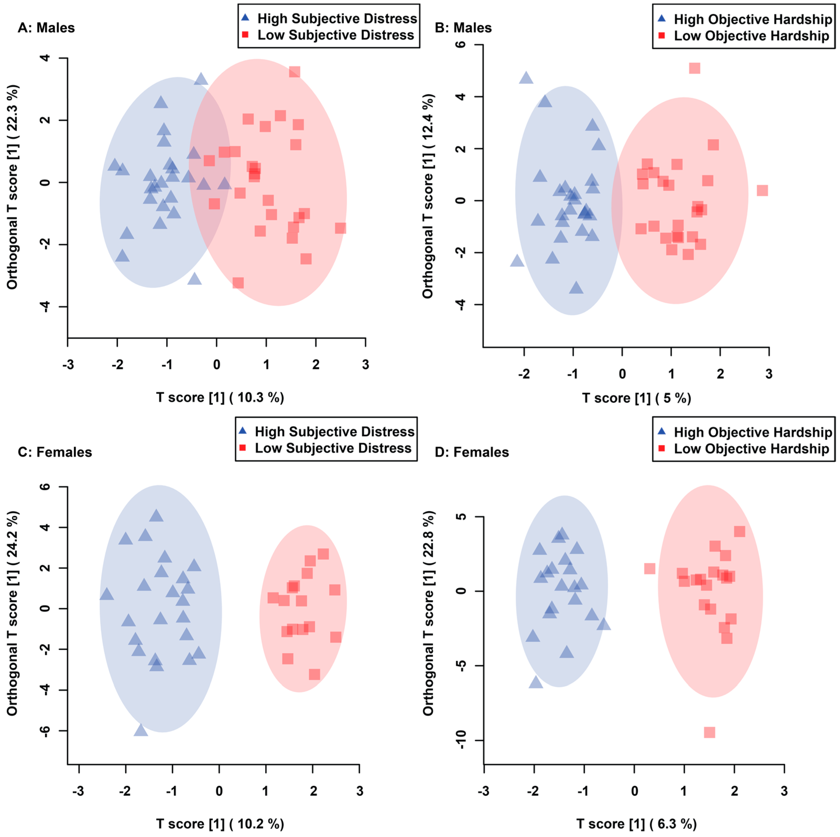
Figure 1. Orthogonal Projections to Latent Structures Discriminant Analysis (OPLS-DA) score plots representing the statistically significant supervised separation of maternal composite subjective distress in males (A), maternal objective hardship in males (B), maternal composite subjective distress in females (C) and maternal objective hardship in females (D) comparisons. Each triangle and square represent one individual, plotted using urinary metabolites found to be statistically significant by VIAVC testing. Shaded ellipses represent the 95% confidence interval. The x-axis represents between-group (predictive) variation, whereas the y-axis represents within-group (orthogonal) variation.
Metabolites 13 00579 g001
Figure 2. Receiver Operator Characteristic (ROC) curves of maternal composite subjective distress in males (A), maternal objective hardship in males (B), maternal composite subjective distress in females (C), and maternal objective hardship in females (D) comparisons. The area under the curve (AUC) and confidence interval (CI) corresponding to the comparison are indicated in each figure. Each figure was created utilizing metabolites identified as significant by VIAVC best subset.
Metabolites 13 00579 g002
Figure 3. Metabolomic Pathway Topology Analysis of males whose mothers experienced composite subjective distress (A) and objective hardship (B) conducted utilizing bins identified as significantly altered by VIAVC. A higher value on the y-axis relates to a lower p-value, with 1.3 referring to a p-value of 0.05. The x-axis provides a measure of the predicted impact on the metabolic pathway due to the identified metabolites. The color of each circle is indicative of its p-value, whereas the size of each circle relates to its impact factor, with a darker color and larger circle indicating a lower p-value and greater impact factor, respectively. Only the pathways with a significant p-value (less than or equal to 0.05) are numbered and refer to the following metabolic pathways: (1) Taurine and hypotaurine metabolism; (2) Pentose phosphate pathway; and (3) Lysine degradation.
Metabolites 13 00579 g003
Figure 4. Metabolomic Pathway Topology Analysis of females whose mothers experienced composite subjective distress (A) and objective hardship (B) conducted utilizing bins identified as significantly altered by VIAVC. A higher value on the y-axis relates to a lower p-value, with 1.3 referring to a p-value of 0.05. The x-axis provides a measure of the predicted impact on the metabolic pathway due to the identified metabolites. The color of each circle is indicative of its p-value, whereas the size of each circle relates to its impact factor, with a darker color and larger circle indicating a lower p-value and greater impact factor, respectively. Only the pathways with a significant p-value (less than or equal to 0.05) are numbered and refer to the following metabolic pathways: (1) Glycine, serine, and threonine metabolism; (2) Aminoacyl-tRNA biosynthesis; (3) Galactose metabolism; (4) Cysteine and methionine metabolism; (5) Tyrosine metabolism; (6) Pantothenate and CoA biosynthesis; (7) Glyoxylate and dicarboxylate metabolism; (8) Starch and sucrose metabolism; (9) Nitrogen metabolism; (10) D-Glutamine and D-glutamate metabolism; (11) Ascorbate and aldarate metabolism; (12) Glutathione metabolism; (13) Porphyrin and chlorophyll metabolism; (14) Arginine biosynthesis; (15) Arginine and proline metabolism; (16) Pentose and glucuronate interconversions.
Metabolites 13 00579 g004
Table 1. Males: Statistically significant urinary metabolites identified by VIAVC or MW testing in the comparison of high and low maternal composite subjective distress and objective hardship in males. Significance was determined using a p-value equal to or less than 0.05. Metabolites are displayed in order of VIAVC p-value. Regulation relates to the high-strM Metabolites with more than one NMR resonance peak are represented in order of ppm as metabolite.1, metabolite.2, etc.
GroupMetaboliteNMR Chemical Shift Range of Bin (ppm)VIAVCMann-Whitney U TestRegulation
Male Subjective Distress
(COSMOSS)
3-Hydroxyisovalerate1.281–1.275.98 × 10−122Not sig.Up
Indole-3-lactate.17.285–7.2633.56 × 10−82Not sig.Up
Creatine.13.947–3.9291.32 × 10−651.04 × 10⁻²Down
Glycylproline3.929–3.9163.92 × 10−53Not sig.Down
Methylguanidine2.875–2.866.83 × 10−48Not sig.Down
Citramalic acid2.75–2.7335.51 × 10−37Not sig.Down
Creatine.23.045–3.032.19 × 10−362.57 × 10⁻²Down
Carnosine2.671–2.6623.09 × 10−30Not sig.Up
Carnitine3.227–3.2151.20 × 10−29Not sig.Down
3-Chlorotyrosine7.129–7.1163.25 × 10−27Not sig.Down
3-Methylhistamine3.723–3.7121.40 × 10−232.17 × 10⁻³Up
Indole-3-lactate.24.383–4.332.10 × 10−232.04 × 10⁻³Up
(S)-3-Hydroxyisobutyric acid1.116–1.1052.98 × 10−23Not sig.Down
O-Phosphocholine3.241–3.2274.17 × 10−194.45 × 10⁻⁴Down
Male Objective Hardship (QFOSS)Xanthurenate7.105–7.0951.41 × 10−83Not sig.Up
Cysteine3.011–2.9962.01 × 10−65Not sig.Down
3-Aminoisobutyrate.13.118–3.1013.02 × 10−48Not sig.Down
Creatine.13.947–3.9295.98 × 10−47Not sig.Down
Creatinine.13.066–3.0451.06 × 10−41Not sig.Down
Erythritol, Glycylproline3.643–3.6041.78 × 10−35Not sig.Up
Gluconate.14.053–4.035.96 × 10−348.03 × 10⁻³Up
Dimethyl sulfone3.164–3.1562.37 × 10−33Not sig.Up
Acetate1.932–1.9193.58 × 10−30Not sig.Up
Creatine.23.045–3.035.73 × 10−28Not sig.Down
5-Hydroxylysine.11.963–1.9531.11 × 10−24Not sig.Up
Creatinine.24.081–4.0539.63 × 10−24Not sig.Down
2-Methylbutyroylcarnitine.10.8867–0.87685.24 × 10−23Not sig.Down
5-Hydroxylysine.2, Glutathione2.963–2.9423.40 × 10−22Not sig.Up
Carnosine.12.702–2.6842.02 × 10−19Not sig.Up
5-Hydroxylysine.33.172–3.1647.56 × 10−19Not sig.Up
3-Aminoisobutyrate.21.196–1.1851.49 × 10−18Not sig.Up
4-Pyridoxic acid2.358–2.3397.62 × 10−17Not sig.Up
Vanylglycol3.375–3.3661.19 × 10−15Not sig.Up
Gluconate.24.157–4.1341.66 × 10−154.05 × 10⁻²Up
Carnosine.22.671–2.6622.03 × 10−15Not sig.Up
2-Aminoadipate2.26–2.2472.10 × 10−15Not sig.Up
Ribose4.134–4.1063.55 × 10−14Not sig.Up
Allantoin5.401–5.3813.92 × 10−14Not sig.Down
Taurine.13.448–3.4375.86 × 10−14Not sig.Up
Taurine.23.426–3.4186.32 × 10−14Not sig.Up
Citramalic acid2.75–2.7331.32 × 10−13Not sig.Up
2-Methylbutyroylcarnitine.20.8768–0.8661.82 × 10−12Not Sig.Down
3-Phenyllactate7.333–7.3163.02 × 10−124.45 × 10⁻²Down
3-indoxylsulfate7.513–7.4952.10 × 10−10Not sig.Up
Cysteine-S-sulfate3.524–3.5111.01 × 10−09Not sig.Down
3,4,5-Trimethoxycinnamic acid, Kynurenine6.815–6.7761.39 × 10−09Not sig.Down
Table 2. Females: Statistically significant urinary metabolites identified by VIAVC or MW testing in the comparison of high and low maternal composite subjective distress and objective hardship in females. Significance was determined using a p-value equal to or less than 0.05. Metabolites are displayed in order of VIAVC p-value. Regulation relates to the high-stress group. Metabolite with more than one NMR resonance peak are represented in order of ppm as metabolite.1, metabolite.2, etc.
GroupMetaboliteNMR Chemical Shift Range of Bin (ppm)VIAVCMann-Whitney U TestRegulation
Female Subjective Distress (COSMOSS)Proline.13.409–3.41.56 × 10−99Not sig.Up
Lactose.14.699–4.6762.63 × 10−801.45 × 10⁻³Down
Epinephrine2.797–2.7792.41 × 10−77Not sig.Down
5-Aminolevulinic acid2.806–2.7976.22 × 10−771.35 × 10⁻²Down
Cysteine, Serine3.966–3.9471.68 × 10−692.50 × 10⁻²Down
Galactose5.29–5.2762.05 × 10−501.77 × 10⁻³Down
4-Hydroxyproline3.498–3.4891.74 × 10−492.89 × 10⁻²Down
Lactose.25.261–5.2391.94 × 10−241.85 × 10⁻²Down
Tyramine, Carnosine3.252–3.2412.47 × 10−233.11 × 10⁻²Up
Sucrose3.489–3.4821.24 × 10−22Not sig.Down
Cystathionine2.208–2.1988.34 × 10−22Not sig.Up
2-Aminoadipate.12.26–2.2475.47 × 10−21Not sig.Up
Chlorogenate 5.357–5.3521.11 × 10−162.15 × 10⁻²Down
5-Methoxytryptamine6.972–6.9644.92 × 10−164.12 × 10⁻²Up
Homovanillic acid6.776–6.7392.43 × 10−15Not sig.Up
3-Chlorotyrosine6.984–6.9724.27 × 10−14Not sig.Up
3-Nitrotyrosine7.028–7.021.27 × 10−134.41 × 10⁻²Up
Indole-3-lactate7.775–7.7622.25 × 10−13Not sig.Up
Proline.23.4–3.3932.44 × 10−11Not sig.Up
Pantothenate0.9397–0.92738.19 × 10−11Not sig.Down
Isobutyrate1.085–1.0751.50 × 10−8Not sig.Down
2-Hydroxy-3-methylvalerate0.8528–0.84313.12 × 10−8Not sig.Down
Isoleucine1.012–1.0033.29 × 10−8Not sig.Up
2-Aminoadipate.21.695–1.6531.08 × 10−6Not sig.Up
Pyroglutamate2.391–2.3851.14 × 10−6Not sig.Up
Female Objective Hardship (QFOSS)Epinephrine2.797–2.7792.33 × 10−61Not sig.Down
Cysteine, Serine.13.966–3.9478.49 × 10−537.29 × 10⁻³Down
Glycine3.586–3.5643.61 × 10−52Not sig.Up
Serine.23.869–3.8634.24 × 10−52Not sig.Down
(S)-3-Hydroxybutyric acid1.254–1.2451.55 × 10−49Not sig.Up
Glutamine.12.163–2.1521.11 × 10−413.89 × 10⁻²Up
Serine.34.002–3.9948.30 × 10−39Not sig.Down
Cysteine-S-sulfate3.536–3.5249.29 × 10−37Not sig.Down
3-Chlorotyrosine7.129–7.1168.67 × 10−32Not sig.Up
Isobutyrate.11.085–1.0752.59 × 10−30Not sig.Down
Erythritol.13.801–3.7841.06 × 10−294.45 × 10⁻²Down
Theophylline7.949–7.9371.65 × 10−29Not sig.Up
4-Hydroxyproline3.498–3.4893.55 × 10−29Not sig.Down
Glucuronate4.66–4.6496.10 × 10−27Not sig.Up
Erythritol.2, Lactose.13.643–3.6045.15 × 10−23Not sig.Down
3-Phenyllactate2.887–2.8758.49 × 10−22Not sig.Up
Isobutyrate.21.075–1.0657.82 × 10−19Not sig.Down
Glutamine.22.474–2.4654.10 × 10−18Not sig.Up
Lactose.25.261–5.2391.81 × 10−17Not sig.Down
Isoleucine1.036–1.0121.02 × 10−16Not sig.Up
Fucose1.265–1.2543.11 × 10−16Not sig.Down
Phenylalanine4.03–4.0215.43 × 10−133.32 × 10⁻³Down
Symmetric dimethylarginine2.85–2.8172.32 × 10−12Not sig.Down
Saccharopine.12.425–2.4171.14 × 10−11Not sig.Down
Sucrose3.489–3.4821.32 × 10−10Not sig.Down
Maltose5.407–5.4014.81 × 10−10Not sig.Down
Saccharopine.22.417–2.4042.44 × 10−9Not sig.Down
Proline3.418–3.4096.91 × 10−9Not sig.Up
Ethanolamine3.146–3.149.59 × 10−9Not sig.Up
Glutamate2.329–2.3191.12 × 10−8Not sig.Up
UDP-glucose4.397–4.3834.25 × 10−83.39 × 10⁻²Down
5-Aminolevulinic acid2.806–2.7977.56 × 10−8Not sig.Down
Sarcosine2.768–2.758.93 × 10−8Not sig.Down
3,4-Dihydroxybenzeneacetic acid6.739–6.7152.07 × 10−7Not sig.Up
Unidentified Metabolite @ 8.65 ppm8.663–8.6462.85 × 10−6Not sig.Down
Malonate3.133–3.1183.07 × 10−6Not sig.Down
Unidentified Metabolite @ 0.55 ppm0.5574–0.5353.29 × 10−6Not sig.Up
N-Acetylglutamine2.036–2.031.32 × 10−5Not sig.Up
Arabinose4.527–4.521.76 × 10−5Not sig.Up
Disclaimer/Publisher’s Note: The statements, opinions and data contained in all publications are solely those of the individual author(s) and contributor(s) and not of MDPI and/or the editor(s). MDPI and/or the editor(s) disclaim responsibility for any injury to people or property resulting from any ideas, methods, instructions or products referred to in the content.