Abstract
Environmental metabolomics is a promising approach to study pollutant impacts to target organisms in both terrestrial and aquatic environments. To this end, both nuclear magnetic resonance (NMR)- and mass spectrometry (MS)-based methods are used to profile amino acids in different environmental metabolomic studies. However, these two methods have not been compared directly which is an important consideration for broader comparisons in the environmental metabolomics field. We compared the quantification of 18 amino acids in the tissue extracts of Daphnia magna, a common model organism used in both ecotoxicology and ecology, using both 1H NMR spectroscopy and liquid chromatography with tandem MS (LC-MS/MS). 1H NMR quantification of amino acids agreed with the LC-MS/MS quantification for 17 of 18 amino acids measured. We also tested both quantitative methods in a D. magna sub-lethal exposure study to copper and lithium. Again, both NMR and LC-MS/MS measurements showed agreement. We extended our analyses with extracts from the earthworm Eisenia fetida and the plant model Nicotiana tabacum. The concentrations of amino acids by both 1H NMR and LC-MS/MS, agreed and demonstrated the robustness of both techniques for quantitative metabolomics. These findings demonstrate the compatibility of these two analytical platforms for amino acid profiling in environmentally relevant model organisms and emphasizes that data from either method is robust for comparisons across studies to further build the knowledge base related to pollutant exposure impacts and toxic responses of diverse environmental organisms.
1. Introduction
Environmental metabolomics is a rapidly growing area of research that has contributed to novel findings in many various disciplines by providing rapid and holistic data about metabolic perturbations due to disease or some external stressor [1,2,3]. Environmental metabolomic studies focuses on environmental perturbations, such as pollutants and climate change, that impart stress on organisms dwelling in aquatic and terrestrial ecosystems [4]. Yet, environmental metabolomics faces unique challenges because of the diverse and large number of model organisms that can be studied and the lack of a priori knowledge about the metabolome, genome or proteome for environmentally-relevant organisms [4,5]. As such, non-targeted metabolomic studies using nuclear magnetic resonance (NMR) spectroscopy were more prevalent due to the ease in sample preparation and the ability of these techniques to yield information about a wide range of metabolites [5,6,7,8]. 1H NMR spectroscopy has been the primary tool with validation of metabolite structural assignments using two-dimensional (2-D) experiments and readily available database information [9,10,11,12]. In addition, 1H NMR spectroscopy is fully quantitative and highly reproducible [8,13]. Studies have reported that analysis of small molecules yield relative standard deviations of <1% [14,15]. NMR also has a large dynamic range which facilitates the analysis of complex mixtures with components of varying concentration [13,14,16].
More recently, the field of environmental metabolomics has grown to include sensitive mass spectrometric (MS) methods based on the knowledge gained from earlier NMR studies. For example, with the keystone water flea, Daphnia magna, the tiny size (high µg to low mg range, depending on age and life stage) necessitates that more than one organism be pooled for extraction prior to NMR analysis [12]. There are other techniques available to study the D. magna metabolome of individuals, such as in vivo NMR spectroscopy [17] however, these approaches are difficult to apply to large sets of organisms and with multiple environmental variables. Therefore, there is a need to use targeted MS/MS techniques for the quantification of endogenous metabolites in environmentally relevant organisms or samples of limited mass, such as fish mucus [18], in addition to metabolic profiling by NMR spectroscopy [19,20]. A particular group of metabolites of interest include free amino acids which are commonly used to assess changes in the metabolic profile of various organisms [4], including earthworms [21,22,23,24], Daphnia [25,26,27,28], and various fish [29,30,31,32,33,34,35] and plant species [10,36,37,38,39,40,41,42,43,44,45]. Amino acids are commonly analyzed because changes in their concentrations can be used to examine mechanisms such as oxidative stress, disruptions to energy metabolism and protein degradation. Consequently, amino acids are a key group of metabolites that can be used with other endogenous metabolites to form an understanding of the molecular-level perturbations due to various environmental stressors, such as sub-lethal pollutant exposure or nutritional stress [27,46].
Environmental metabolomic investigations provide critical information about molecular-level perturbations but one of the challenges related to this area of research is the shear number of model organisms that are used in addition to the use of different analytical platforms. There is a need to examine quantification of amino acids using different techniques, to determine the extent of compatibility across species and analytical approaches for improved standardization of workflows and results [2]. As such, the objective of this study was to compare the quantification of amino acids in three model organisms using both LC-MS/MS and 1H NMR spectroscopy on the same polar extracts. The model organisms included: D. magna which is a commonly used model in ecotoxicology and metabolomics [47], the earthworm, Eisenia fetida, a model organism for assessing soil pollution [4] and the well-studied tobacco plant, Nicotiana tabacum which is commonly used for assessing environmental variations on plant biochemistry [48,49,50,51,52]. The overall objective was to compare quantification of amino acids by 1H NMR and LC-MS/MS for environmentally relevant model organisms to determine if the two platforms agree and produce data that is transferable which is important for standardizing environmental metabolomic methods that use different approaches. This approached involved two main experiments (Figure 1) where we first examined different quantification methods using an environmental metabolomics exposure study with D. magna and then applied the approach to a comparison of amino acid profiling in three different model organisms (D. magna, E. fetida, and N. tabacum). This cross-platform comparison is also important for establishing baseline knowledge related to further development and use of metabolomic methods for broadly assessing environmental impacts to target organisms in different environmental compartments. Given the diversity of model organisms used in environmental metabolomics, a direct comparison of results is important for establishing standard approaches and data evaluation.
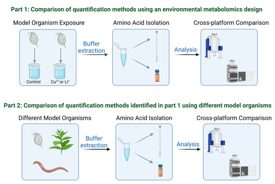
Figure 1.
Schematic outlining the two part approach used in this study to compare amino acid quantification methods using nuclear magnet resonance (NMR) spectroscopy and liquid chromatography with tandem mass spectrometry (LC-MS/MS). Created with www.biorender.com.
2. Materials and Methods
Our study included a two part experiment as outlined in Figure 1. Part 1 examines different quantification approaches for assessing amino acid concentrations after exposure to sub-lethal concentrations of copper and lithium. Based on the results of part 1, we applied the same cross-platform comparison to three different model organisms to more broadly assess the results between different samples.
2.1. Daphnia magna Amino Acid Extraction
Daphnia magna neonates (<24 h old) were isolated from the ongoing culture maintained at the Ontario Ministry of the Environment, Conservation and Parks as detailed in Nagato et al. [53]. Sub-lethal exposure to copper and lithium were carried out over 48 h. Neonates were exposed to either copper or lithium at 50% of the lethal concentration that results in mortality to 50% of the population (LC50). The LC50 values were determined to be 24.8 µg/L and 2300 µg/L for copper and lithium respectively and exposure concentrations for copper and lithium of 12.4 µg/L and 1150 µg/L were used (see Nagato et al., [53] for full details). Control (unexposed) neonates were also isolated for comparisons to metal-exposed D. magna.
After exposure, amino acids were extracted using a polar metabolite extraction previously optimized for NMR-based metabolomics that has high reproducibility and isolates a range of polar metabolites [12,53]. D. magna neonates (~10 mg, dry weight) were flash frozen in liquid nitrogen, lyophilized and extracted by adding 750 µL of a 0.2 M monobasic sodium phosphate buffer solution (NaH2PO4•2H2O; 99.3%; Fisher Chemicals; Ottawa, ON, Canada) with 0.1% w/v sodium azide (99.5%; Sigma Aldrich, Oakville, ON, Canada) as a preservative and 10 mg/L of sodium 2,2-dimethyl-2-silapentane-5-sulfonate (DSS; 97%, Sigma Aldrich) as an internal calibrant/standard for 1H NMR analysis. Buffer solution was made with D2O (99.9%, Cambridge Isotope Laboratories Inc., Tewksbury, MA, USA) and adjusted to a pD of 7.4 using NaOD (30% w/w in 99.5% D2O; Cambridge Isotope Laboratories Inc.). Samples were vortexed for 30 s and sonicated for 15 min. Samples were then centrifuged for 20 min at 14,000 rpm (~15,000 g) and the supernatant was decanted into a new 1.5 mL centrifuge tube. The centrifugation procedure was repeated once more to remove any remaining particulate material. 50 µL of the resulting extract was reserved for LC-MS/MS analysis and the remaining solution (700 µL) was transferred to a 5 mm High Throughputplus NMR tube (Norell Inc., Morganton, NC, USA) for 1H NMR analysis. In total, eight replicate extractions were performed (n = 8) for D. magna.
2.2. Eisenia fetida and Nicotiana tabacum Amino Acid Extraction
Mature E. fetida earthworms were isolated from a culture where earthworms have been maintained for metabolomic experiments since 2006 [54]. Twelve mature E. fetida earthworms (50–70 mg per earthworm, dry weight), each with visible clitellum were individually flash frozen in liquid nitrogen, lyophilized, and homogenized in a 1.5 mL centrifuge tube using a 5 mm stainless steel spatula. Wild-type (WT) tobacco (N. tabacum L. cv Petit Havana SR1) was grown as described by Alber et al. [52]. Three whole leaves (10–20 mg per leaf, dry weight) from each of four mature plants were severed and immediately flash-frozen in liquid nitrogen. The frozen leaves were then ground into a fine powder with a liquid nitrogen cooled mortar and pestle and lyophilized. Because proteins may interfere with the use of an internal standard by 1H NMR spectroscopy, a modified method was used for isolating free amino acids which includes steps that result in protein precipitation. Lyophilized earthworm and tobacco samples were extracted by adding 400 µL of methanol and 85 µL of water and vortexing for 15 s [55]. Next, 200 µL water and 400 µL chloroform were then added and the samples vortexed for 1 min. The samples were then allowed to sit at 4 °C for 10 min, and then centrifuged for 10 min at 12,000 rpm (~11,000 g). The methanol:water layer was decanted and dried under a steady stream of nitrogen gas. The samples were then reconstituted in 750 µL of the same D2O phosphate buffer as described previously. Of this extract, 50 µL was reserved for LC-MS/MS analysis and the remaining extract (700 µL) was transferred to a 5 mm High Throughputplus NMR tube (Norell Inc., Morganton, NC, USA) for 1H NMR analysis. For both E. fetida and N. tabacum, twelve replicate extractions were performed (n = 12).
2.3. Amino Acid Profiling via 1H NMR
Amino acid standards were purchased from BioShop (Burlington, ON, Canada) or Sigma-Aldrich (Oakville, ON, Canada) and were all >98.5% purity. Amino acid standards as well as a mixture of standards were prepared in the same D2O buffer (as described previously) and analyzed by 1H NMR spectroscopy. Amino acid solutions were analyzed using a BrukerBioSpin Avance III 500 MHz spectrometer (Ettlingen, Germany) equipped with a 1H-19F-15N-13C 5 mm Quadruple Resonance Inverse (QXI) probe fitted with an actively shielded Z gradient. 1H NMR experiments were performed using Presaturation Using Relaxation Gradients and Echoes (PURGE) water suppression [56] which was previously developed within our laboratory specifically for environmental metabolomics applications using NMR spectroscopy. Data were collected using 128 scans, and 64 K time domain points. Preliminary experiments were performed to measure the spin-lattice relaxation time (T1) of amino acid standards and samples to ensure that nuclei were fully relaxed before subsequent NMR pulses (the recycle delay was 5 × T1) [57]. All 1H NMR spectra were apodized through multiplication with an exponential decay corresponding to 0.3 Hz line broadening in the transformed spectrum, and a zero filling factor of 2. All spectra were manually phased and calibrated to the trimethylsilyl group of the DSS internal reference (δ = 0.00 ppm). Amino acid peaks in samples were identified in each spectrum by comparison with individually analyzed standards and compared to NMR spectra available from the Madison Metabolomics Consortium Database and published studies [9,12]. The NMR resonances used for quantification are listed in Table S1. These resonances did not overlap with 1H resonance from other amino acids or other compounds that may have been co-extracted from the model organisms and have been confirmed using 2-D NMR spectroscopy in D. magna [12] and E. fetida [11]. Integration of these specific amino acid resonances in 1H NMR spectra was performed using the multi-integration package of AMIX (version 3.8.4). The integrated resonance areas were then imported into Microsoft Excel (Microsoft Corporation, Redmond, WA, USA) and concentrations in the samples were calculated using an external calibration curve constructed from the integrated resonances of the standards. 1H NMR spectra of amino acid standards as compared to resonances found in sample extracts are shown in Figure S1. Internal standard quantification was also evaluated for D. magna extracts. The same integrated resonances for amino acids were used as with external standard quantification. Resonance areas were quantified based on the integrated area for the trimethylsilyl resonance of DSS (δ = 0.00 ppm).
2.4. Amino Acid Profiling via LC-MS/MS
Amino acid standards and samples were analyzed using a Sciex API 4000 QTrap mass spectrometer (Applied Biosystems/MDS Sciex, Concord, ON, Canada) coupled to an Agilent 1200 LC system equipped with an Ultra Aqueous C18 LC column (10 mm × 4.6 mm, 3 µm, Restek Corp.) which is designed for the separation of low retention compounds. The analysis involved a linear methanol:water gradient, with 0.15% formic acid added as a modifier to both solvents. The developed LC gradient consisted of: 0–1 min. hold at 5:95 methanol:water; 1–3 min. move to 20:80 methanol:water; 3–6 min. move to 40:60 methanol:water; 6–6.5 min. move to 95:5 methanol:water; 6.5–8 min. hold at 95:5 methanol:water and then return to initial conditions. All solvents were LC-MS grade purity (Sigma Aldrich, Oakville, ON, Canada). The LC flow rate was set at 0.5 mL/min and an injection volume of 10 µL was used. The LC column temperature was set at 40 °C. Electrospray ionization in positive ion mode and multiple reaction monitoring (MRM) was used based on transitions reported in the literature [58]. The ion source temperature was set to 750 °C. Table S2 lists the MRM ions used for detection and the retention times of each amino acid measured. Repeat injections (n = 3) for each amino acid standard yielded a relative standard deviation from 2.3–11% (Table S2). NMR D2O buffer extracts (50 µL) were diluted 100–500 times with LC-MS grade water (Sigma Aldrich, Oakville, ON, Canada). Amino acids were quantified by external calibration with standards, except glycine which was calibrated by internal calibration using 13C 2-glycine (Cambridge Isotope Laboratories, Inc., Tewksbury, MA, USA). Analyst (version 1.5.1) was used for peak integration and amino acid quantification.
2.5. Statistical Analysis and Comparison of Method Agreement
A t-test (two-tailed, equal variances) was used to compare the absolute concentrations of amino acids between the two methods to determine statistical differences at α = 0.05. A t-test (two-tailed, equal variances) was also used to compare the relative changes in the absolute concentrations of amino acids obtained by 1H NMR and LC-MS/MS in D. magna after copper and lithium exposure at sub-lethal concentrations.
We used methods recommended by Bland and Altman [59] to determine the extent of agreement between 1H NMR and LC-MS/MS methods. Measuring agreement between two independent measurements where the true value is not known can be established by plotting the difference of the mean measurements against the average measurement. The mean of the difference along with the standard deviation are also tabulated and plotted to establish a region of agreement (near the mean but not higher than the mean ± 2 × the standard deviation). This approach was applied to the D. magna concentrations measured by 1H NMR and LC-MS/MS using external standard quantification for both methods. For selected amino acids, we also performed regression analysis from all three model organisms which is an indicator of correlations between measurement [59].
3. Results
An overall comparison of the measured concentrations of amino acids in D. magna extracts is shown in Figure 2A. As a quantitative detector NMR has a large dynamic range, producing linear calibrations that span several orders of magnitude [14,16,60], whereas with MS, the dynamic range is analyte dependent and dependent on ionization and matrix effects [61,62]. Other considerations when examining quantification by NMR include the relaxation time (T1) of the sample components. Appropriate external and internal calibration methods require that the analytes of interest exhibit similar relaxation characteristics between the samples and standards and the results shown in Figure 2A demonstrates that this requirement was met for most amino acids analyzed. Histidine could not be quantified with certainty in these extracts by 1H NMR because the only proton resonances for histidine that did not overlap with other resonances were those on the carbons of the imidazole functional group at 8.03 ppm and 7.4 ppm (Figure S1). This would appear ideal as few other endogenous metabolites have resonances in this region. However, as the nitrogen atoms on the imidazole ring have pKa values around 6 [63] using a buffering system at pD 7.4 (~pH 7) can result in different charge states with very small changes in pH, which in turn causes the resonances for the C-H protons to shift slightly. The issue is confounded as the standards tended to shift downfield to 8.06 and 7.15 ppm, while the D. magna samples shifted upfield to 7.96 and 7.11 ppm. As evident from Figure 2A, there is a difference between 1H NMR results obtained using an internal (DSS) standard versus external standards for quantification. For some amino acids (Figure 2A), the concentrations determined using DSS alone were statistically different than those observed using external standard quantification by both 1H NMR and LC-MS/MS. But in other cases, both internal and external standard quantification methods for 1H NMR were similar with each other (e.g., glycine; Figure 2A).
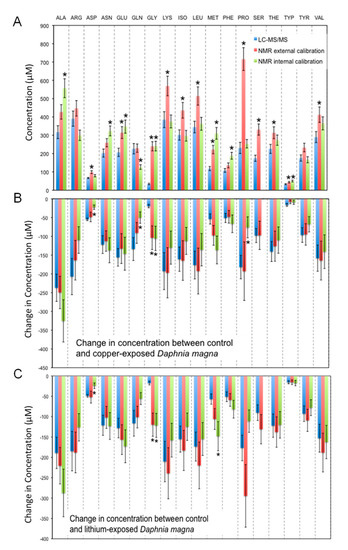
Figure 2.
Concentrations of 18 amino acids observed in extracts of Daphnia magna by 1H NMR and LC-MS/MS (histidine was not quantified) expressed as (A) absolute concentrations observed in the extracts of control D. magna, or differences in amino acid concentrations observed between control D. magna and those exposed to (B) copper or (C) lithium. Any significant differences (α = 0.05) between measured concentrations are denoted with an asterisk (*). ALA = alanine, ARG = arginine, ASP = aspartate, ASN = asparagine, GLU = glutamate, GLN = glutamine, GLY = glycine, ILE = isoleucine, LEU = leucine, LYS = lysine, MET = methionine, PHE = phenylalanine, PRO = proline, SER = serine, THR = threonine, TRP = tryptophan, TYR = tyrosine, VAL = valine.
This excellent agreement is likely due to the concentration of these amino acids in D. magna as well as a lack of matrix effects or other interferences. For example, the 1H NMR spectra of a D. magna extract is shown together with the 1H NMR spectra of a 100 μg/L mixed amino acid standard in Figure 3, where the x-axis is focused on the resonances used for quantification of glutamine, alanine and proline. Four unobstructed resonances for glutamine with no elevation or disturbance of the baseline between the samples and the standard are visible between 2.41 and 2.47 ppm in Figure 3A.
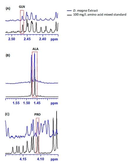
Figure 3.
A comparison of the 1H NMR spectra of a D. magna extract and a mixed amino acid standard (100 μg/L), both in D2O buffer, highlighting the regions used for the quantification of (A) glutamine (GLN), (B) alanine (ALA), and (C) proline (PRO). A red dotted box indicates the specific resonance used for quantification for each amino acid.
To further evaluate the agreement between LC-MS/MS and 1H NMR spectroscopy measurements using external calibration of amino acids D. magna extracts, we compared the difference of the average measurements versus the average concentration of measurements (Figure 4), based on the method of Bland and Altman [59]. This method is employed when the true measurement value is not known and avoids bias when comparing two independent measurements [59]. Measurements are considered to be in good agreement if the difference in concentration between the measurements is within two standard deviations of their mean value [59]. By applying this method to evaluate agreement all amino acids except for proline fall within the agreement range (Figure 4). The difference between measurements (Figure 4; Table S3) for arginine, aspartate, glutamate, methionine, phenylalanine, tryptophan, tyrosine, and valine are closest to zero (mean differences ranged from -10 to 31 µM; Figure 4). The amino acids alanine, asparagine, glutamate, isoleucine, leucine, serine, and threonine exhibit greater average differences between measurements (Figure 4; Table S3) but are still within two standard deviations and considered to be in agreement [59]. For some of these amino acids, the underlying baseline is slightly elevated, suggesting small underlying interferences, as can be seen for alanine between 1.45 and 1.50 ppm in Figure 3B, and for the other amino acids (Figure S1). The overall similarity between sample and standard suggests no underlying interferences in the D. magna 1H NMR spectra for glutamine, which supports the excellent agreement observed (Figure 4; Table S3).
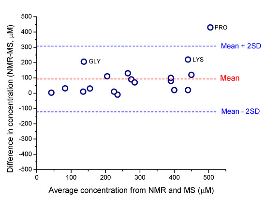
Figure 4.
Evaluation of measurement agreement between 1H NMR and LC-MS/MS based on Bland and Altman [59]. The difference of the mean measurement plotted against the average measured concentration compares the agreement between the measurements within the calculated standard deviation (SD). Concentration data are listed in Table S3. GLY = glycine, LYS = lysine and PRO = proline.
The focus of our study was on the quantification of amino acids found in D. magna for metabolomic studies. However, to explore if quantification by both LC-MS/MS and 1H NMR would yield the same results in extracts from other organism models, we also analyzed selected amino acids using external calibration methods by both 1H NMR and LC-MS/MS for E. fetida earthworms and the model plant N. tabacum. As mentioned previously, we observed some broadening of the NMR internal standard (DSS) so we did not proceed with internal quantification of amino acids in E. fetida and N. tabacum extracts. Overall, we found good agreement for detectable amino acids in all three organisms (Figure 5). The correlation coefficients (r2 values) are higher than 0.70 except for two correlations (aspartate in D. manga and valine in N. tabacum). This is likely due to the low concentrations of both amino acids which may have been below the limit of quantification. When amino acids were present above the limit of quantification, then the accuracy of detection improved, and we observed strong linear correlations between absolute concentrations measured by both 1H NMR and LC-MS/MS for all three model organisms (Figure 5).
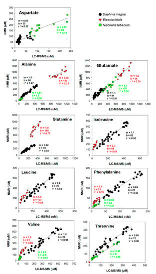
Figure 5.
Comparison of amino acid quantification by 1H NMR and LC-MS/MS in polar extracts from three different environmentally relevant model organisms.
4. Discussion
Overall, we generally found good agreement across platforms and with extracts from different organism models. We did observe some differences, depending on the approach, which will be discussed in detail in this section. Using an internal standard for quantification of 1H NMR spectroscopy data agreed less for D. magna extracts (Figure 2). There are several challenges with using internal standards for quantification in 1H NMR spectroscopy. The internal standard must not have overlapping resonances with compounds of interest which is why we chose DSS as it has an unobstructed resonance at δ = 0.00 ppm which also serves to calibrate the chemical shift. However, the internal standard may interact with other components in the sample matrix and this can result in changes in relaxation or line broadening of the internal standard. DSS is a commonly used internal calibrant because it is highly soluble in D2O and typically does not interfere with other compounds of interest. Sodium 3-trimethylsilyltetradeuteropropionate (TSP), is another NMR calibrant that is also used but both DSS and TSP can bind to proteins that may be present in sample extracts and this can result in broadening and/or loss of signal intensity [64,65]. Therefore, as an internal standard, this may result in an overestimation of metabolite concentrations. Also, such interactions may alter the T1 value of the internal standard making quantification less accurate for some compounds. With Daphnia extracts, we did not observe broadening of DSS which suggests that internal calibration is a viable method for quantitative amino acid profiling. However, in other organisms, the co-extraction of peptides may limit the use of DSS as an internal standard. Interestingly, proline quantification was most variable of the amino acids (Figure 2A). This is likely due to differences in proline relaxation behavior when in a sample matrix as compared to a standard. To test this, we extended the delay time and observed that the concentration of proline decreased from 720 ± 60 µM to 560 ± 50 µM. This suggests that there is sample interference with proline that may limit the quantitative reliability in Daphnia extracts by 1H NMR analysis. We also noted that proline may have some interference due to the proximity to other resonances in the 1H NMR spectrum (Figure 3C). However, this would also manifest in the internal quantification results as well. As such, it is more likely that the relaxation of proline is variable and differs than the external standards. Further investigation is warranted to better understand the source of this variability and limitations associated with the absolute quantification of proline in Daphnia extracts.
We also found some differences in quantification when using different 1H resonances for integration. In the D. magna extracts the amino acid methionine gives three resonances that are not overlapped, the two downfield peaks of a triplet at 2.6 ppm (MET1 and MET2) and a singlet at 2.1 ppm (MET3) from the three protons of the methyl thioether (Figure S2). By LC-MS/MS the methionine concentration was found to be 140 ± 10 µM (Figure 2A). Using the three 1H NMR peaks individually the concentration of methionine by external NMR calibration were: 170 ± 20 µM (MET1, 21% difference), 190 ± 20 µM (MET2, 36% difference), and 170 ± 20 µM (MET3, 21% difference), all of which agree reasonably well with the LC-MS/MS data (140 ± 10 µM). By internal NMR calibration these same resonances yield concentrations of: 310 ± 30 µM (MET1, 120% difference), 180 ± 20 µM (MET2, 29% difference), and 1100 ± 100 µM (MET3, 690% difference), where only MET2 gives a comparable concentration. Reasons for these differences likely involve the relaxation properties of the protons in the molecules, particularly the significant over-estimation when using the resonance from the methyl thioether functional group. Furthermore, selecting one concentration for the internal standard may be challenging given the range of concentrations of amino acids found in these samples. However, with Daphnia extracts, this method provided good agreement overall. As indicated previously, DSS broadening due to interactions with proteins that are co-extracted with amino acids may reduce the reliability of internal standard methods. With Daphnia, we did not observe DSS broadening but with the other two extracts included in our study (E. fetida and N. tabacum), broadening was observed despite that we attempted to remove proteins prior to isolation of the free amino acids. As such, the remainder of our discussion will focus on comparisons made with external calibration. However, we note here that quantitative NMR techniques such as Electronic REference To access In vivo Concentrations (ERETIC), can be employed for internal standard quantification [66,67] by NMR that may alleviate issues we observed here. In our study, because our aim was to compare two specific analytical platforms, we focused more on quantification methods that would apply directly to LC-MS/MS and not only 1H NMR spectroscopy.
When comparing external quantification methods (Figure 4), we noted that some amino acids, alanine, asparagine, glutamate, isoleucine, leucine, serine, and threonine, exhibited more variability between methods (Figure 4). No obvious interferences are visible for asparagine; however the concentrations were low (Table S3) and the resulting NMR resonance intensity was lower than that observed for other amino acids (Figure S1). As such, the lower sensitivity of NMR may also contribute to some of the observed concentration differences however, amino acids with the lowest concentrations (arginine, aspartate, tryptophan; Table S3) exhibited very small differences and this suggests that differences in sensitivity between 1H NMR and LC-MS/MS did not limit accurate quantification. For asparagine, the proton resonances of interest are adjacent to a chiral center, resulting in increased splitting that drives the peaks of interest closer to the baseline, which may contribute to difficulties during spectral integration. Serine in D. magna extracts also has obvious interferences via NMR, which are manifested as an elevated baseline (Figure S1) which likely increased the difference between the measured concentrations.
Three amino acids show more deviation from the mean of the differences: glycine, lysine and proline, although only the difference in proline concentrations lies outside the agreement area (> mean ± 2 × Standard Deviation). As discussed previously, proline is likely overestimated by 1H NMR spectroscopy due to interference from another compound or compounds that are altering the relaxation and spectral baseline. As shown in Figure 3C, proline was particularly difficult to quantify in D. magna extracts because the only resonance that does not overlap resides around 4.1 ppm which is in a similar region to other amino acid protons. With glycine and lysine measurement, there may be instrument-specific reasons for the discrepancy between measurements. Lysine quantification via NMR may be altered due to the inference of another nearby resonance (Figure S1). Glycine concentrations were significantly higher by 1H NMR analysis in the D. magna extracts (Figure 2 and Figure 4; Table S3) by both external and internal techniques as compared to concentrations determined by LC-MS/MS (Figure 2A). The resonance used for glycine quantification (Figure S1; Table S1) does not show any overlap or baseline distortion. Also, there do not appear to be any resonances that may be attributed to other compounds. To investigate this disparity further, we included an internal standard for glycine during our LC-MS/MS analysis (13C 2-glycine) as we suspected that glycine was underestimated by external calibration due to ion suppression from matrix effects during electrospray ionization [61,68]. Ion suppression due to matrix effects is a common disadvantage of LC-MS/MS analyses of metabolomics samples [19,68]. However, inclusion of the mass labeled glycine (13C 2-glycine) did not significantly alter the concentration of glycine determined by LC-MS/MS (without internal standard the concentration of glycine in the control D. magna D2O extracts was 30 ± 4 µM, with internal standard this increased to 34 ± 4 µM). Glycine has a retention time that is similar to other amino acids (Table S2) and this may also impact ESI efficiency. Interference and/or suppression can occur even with a mass-labelled internal standard and given that the transition ions for glycine are common (76 > 30 m/z), it is likely that LC-MS/MS may be underestimating glycine concentrations in Daphnia extracts. With ESI, the MS responses may vary with analyte concentration because of varying ionization efficiencies and varying ion suppression [69]. Another possible explanation for the high concentrations of glycine observed by 1H NMR is that, as with histidine, there are several common glycyl dipeptides [70], and it is possible that the protons from these small molecules are obstructing or overlapping with the NMR resonance for free glycine leading to the observed overestimation of the concentrations. However, 2-D NMR analysis of D. magna polar extracts did not reveal any overlap between the resonance used for glycine (δ = 3.55 ppm) quantification [12]. Furthermore, both internal and external quantification using 1H NMR show good agreement (Figure 2). As such, the observed discrepancy in concentrations determined for glycine by 1H NMR and LC-MS/MS warrants further investigation but is likely specific to sample matrix effects specific to D. magna extracts.
In environmental metabolomic studies, a common objective is to examine how metabolite concentrations fluctuate with an external stressor, such as an environmental pollutant. However, typically, only the fold change is reported (change in metabolite concentration relative to the control). This is mostly due to the vast amount of data generated in metabolomic studies and brevity in presenting such data. To investigate the use of relative changes in concentration for metabolomic data, we analyzed D. magna extracts from a companion study where D. magna were exposed to sub-lethal concentrations of copper, lithium and arsenic for 48 h [53]. Nagato et al. [53] reported a significant decrease in the concentration of all the amino acids in lithium or copper exposed D. magna relative to the controls (unexposed), whereas the amino acids were unchanged or increased in the arsenic exposures. These changes in the amino acid concentrations were attributed to a specific reaction of the D. magna to metal exposure, potentially via the incorporation of these amino acids into enzymes in response to the metal-induced stress, or it may represent changes in growth patterns between treatments. Figure 2B,C show the change in amino acid concentration observed between the copper and lithium exposure groups compared to control as determined by LC-MS/MS, and both external and internal standard NMR quantification. As with the D. magna extracts (Figure 2A), we observed agreement between the LC-MS/MS and NMR quantification. Again, internal standard use by NMR is significantly different for some amino acids. The only amino acid that has significantly different concentrations between the two techniques is glycine, which as described previously remains a topic of investigation.
One explanation for why there is better agreement between LC-MS/MS and 1H NMR when looking at concentration changes between groups of organisms as opposed to absolute concentrations is that any underlying 1H NMR baseline disruption is nullified. This hypothesis is visualized in plots of concentration by 1H NMR (external calibration) versus concentration by LC-MS/MS in Figure 5. The slope (m) for most of the amino acids, especially for the D. magna samples, is close to unity, which reflects the ability of 1H NMR to characterize differences in amino acid concentrations between samples. However, the values for the y-intercepts (b) are large, which may reflect underlying interferences that do not scale with changes in the amino acid concentration as well as differences in the detection limits of both analytical platforms. For example, threonine has a slope of 0.99, but an intercept of 70 µM. The concentration of threonine observed in the control D. magna was 250 ± 20 µM by LC-MS/MS and 320 ± 30 µM by 1H NMR, a difference of 70 µM which is similar to the y-intercept. However, this difference decreased when comparing the change in concentration between the control and copper-exposed animals, which was 150 ± 30 µM by LC-MS/MS and 130 ± 40 µM by NMR. The underlying interference, which is reflected in the elevated y-intercept, is almost entirely cancelled out when considering differences in concentration between two sample sets.
This analysis of the comparison of the changes in the amino acid concentrations between NMR and LC-MS/MS methods provides justification for the utility of NMR quantification in applications, such as metabolomics, where relative concentrations between related sample sets are more relevant than absolute concentrations. Furthermore, these observations highlight the excellent agreement between LC-MS/MS and 1H NMR quantification methods and demonstrate that these analytical platforms enable the same result. This is important as researchers often select one platform over the other and rarely use both in tandem [19]. Both techniques have advantages and disadvantages but when data acquisition is optimized, we found that the data are in excellent agreement with each other and for the most part, data are transferable from one platform to the other. These results are highly promising and demonstrate that both analytical platforms are robust and capable of quantifying amino acids in a variety of environmental samples. We note that we did not optimize the extraction methods, especially for N. tabacum, so the method of amino acid extraction used in our study may require further optimization for metabolomic studies with N. tabacum. Nonetheless, of the amino acids that were quantified, we report excellent agreement using both LC-MS/MS and NMR and demonstrate excellent promise for future studies which aim to quantify amino acids in these model organisms.
5. Conclusions
Overall, our study showed that 1H NMR quantification of amino acids generally agreed with LC-MS/MS quantification except for proline in D. magna extracts only. We also confirmed that accurate quantification of amino acids by both methods is dependent on the complexity of the biological matrix and the concentration of the amino acids in the extracts. D. magna extracts had the simplest biological matrix and the highest concentration of amino acids per mass biomass extracted, where both techniques agreed for all amino acids studied except for proline. The more complex E. fetida and N. tabacum extracts, which had a lower concentration of amino acids per biomass extracted, enabled the quantification of only selected amino acids. Our study confirmed that 1H NMR quantification via external calibration is an easy and reliable method to quantify metabolites present in extracts from environmentally relevant organisms. We also illustrated that both analytical platforms detect metabolic shifts similarly when used to quantify amino acids in an environmental metabolomics study of D. magna exposure to two metal pollutants. Because small organisms, such as D. magna, need to be pooled for 1H NMR-based metabolomics, the higher sensitivity afforded by LC-MS/MS is advantageous. We demonstrate that there is agreement in amino acid profiling by LC-MS/MS and 1H NMR and data collected by both methods are directly comparable. Our results are significant because it demonstrates that the direct translation of data across different model organisms and analytical platforms with certainty. This is critical for the future advancement of the use of metabolomics in environmental toxicology and biomonitoring programs, especially given the diversity of organisms and available analytical platforms for such investigations. Our work provides confidence in the measurement of endogenous metabolites across platforms and demonstrate the ubiquity of metabolomic investigations for ascertaining molecular-level perturbations with environmental stressors.
Supplementary Materials
The following supporting information can be downloaded at: https://www.mdpi.com/article/10.3390/metabo13030402/s1, Figure S1: Comparison of the 1H NMR spectra of amino acid standards and of the tissue extracts of Daphnia magna, the earthworm Eisenia fetida, and the tobacco species Nicotiana tabacum. Resonances used for quantification are highlighted by a dashed box and also listed in Table S1.; Figure S2: 1H NMR spectra of a D. magna extract, a 100 μg/L individual methionine standard solution in D2O buffer and a mixture of amino acids (100 μg/L) in D2O buffer, highlighting the section highlighting the resonances that are attributed to methionine.; Table S1: The 1H NMR regions used for quantification of amino acids. Resonances are based on metabolic profiling using both 1H and two-dimensional NMR spectroscopy for D. magna extracts; Table S2: LC-MS/MS multiple reaction monitoring (MRM) mass transitions and relative standard deviation (%) of triplicate injections of amino acid standards.; Table S3: Comparison of amino acid concentrations measured using LC-MS/MS and 1H NMR spectroscopy on Daphnia magna D2O buffer extracts. Values are based on external standard quantification and expressed as averages (n = 8) with associated standard errors.
Author Contributions
Conceptualization, J.C.D., B.P.L., M.J.S.; methodology, J.C.D., B.P.L., A.J.S., E.J.R., D.G.P., G.C.V. and M.J.S.; resources, A.J.S., E.J.R., D.G.P., G.C.V. and M.J.S.; data curation, J.C.D., B.P.L., A.J.S. and E.J.R.; writing—original draft preparation, J.C.D., B.P.L. and M.J.S.; writing—review and editing, J.C.D., B.P.L., A.J.S., E.J.R., D.G.P., G.C.V. and M.J.S. All authors have read and agreed to the published version of the manuscript.
Funding
The authors acknowledge funding from the Krembil Foundation. M.J.S acknowledges generous support from the Natural Sciences and Engineering Research Council (NSERC) of Canada via a Tier 1 Canada Research Chair in Integrative Molecular Biogeochemistry.
Institutional Review Board Statement
Not applicable.
Informed Consent Statement
Not applicable.
Data Availability Statement
All data are available in the main article or in the supporting materials. Raw data files (instrument files) can be obtained upon request from the corresponding author because of the privacy.
Conflicts of Interest
The authors declare no conflict of interest.
References
- Viant, M.R. Applications of metabolomics to the environmental sciences. Metabolomics 2009, 5, 1–2. [Google Scholar] [CrossRef]
- Kim, H.M.; Kang, J.S. Metabolomic Studies for the Evaluation of Toxicity Induced by Environmental Toxicants on Model Organisms. Metabolites 2021, 11, 485. [Google Scholar] [CrossRef] [PubMed]
- Zhang, L.-J.; Qian, L.; Ding, L.-Y.; Wang, L.; Wong, M.H.; Tao, H.-C. Ecological and toxicological assessments of anthropogenic contaminants based on environmental metabolomics. Environ. Sci. Ecotechnology 2021, 5, 100081. [Google Scholar] [CrossRef] [PubMed]
- Lankadurai, B.P.; Nagato, E.G.; Simpson, M.J. Environmental metabolomics: An emerging approach to study organism responses to environmental stressors. Environ. Rev. 2013, 21, 180–205. [Google Scholar] [CrossRef]
- Viant, M.R.; Sommer, U. Mass spectrometry based environmental metabolomics: A primer and review. Metabolomics 2012, 9, 144–158. [Google Scholar] [CrossRef]
- Wishart, D.S. Quantitative metabolomics using NMR. TrAC 2008, 27, 228–237. [Google Scholar] [CrossRef]
- Simpson, M.J.; Bearden, D.W. Environmental Metabolomics: NMR Techniques. eMagRes 2013, 2, 549–560. [Google Scholar] [CrossRef]
- Viant, M.R.; Bearden, D.W.; Bundy, J.G.; Burton, I.W.; Collette, T.W.; Ekman, D.R.; Ezernieks, V.; Karakach, T.K.; Lin, C.-Y.; Rochfort, S.; et al. International NMR-Based Environmental Metabolomics Intercomparison Exercise. Environ. Sci. Technol. 2009, 43, 219–225. [Google Scholar] [CrossRef]
- Cui, Q.; Lewis, I.A.; Hegeman, A.D.; Anderson, M.E.; Li, J.; Schulte, C.F.; Westler, W.M.; Eghbalnia, H.R.; Sussman, M.R.; Markley, J.L. Metabolite identification via the Madison Metabolomics Consortium Database. Nat. Biotechnol. 2008, 26, 162–164. [Google Scholar] [CrossRef] [PubMed]
- Larive, C.K.; Barding, G.A.; Dinges, M.M. NMR Spectroscopy for Metabolomics and Metabolic Profiling. Anal. Chem. 2015, 87, 133–146. [Google Scholar] [CrossRef]
- Brown, S.A.; Simpson, A.J.; Simpson, M.J. Evaluation of sample preparation methods for nuclear magnetic resonance metabolic profiling studies with eisenia fetida. Environ. Toxicol. Chem. 2008, 27, 828–836. [Google Scholar] [CrossRef] [PubMed]
- Nagato, E.G.; Lankadurai, B.P.; Soong, R.; Simpson, A.J.; Simpson, M.J. Development of an NMR microprobe procedure for high-throughput environmental metabolomics of Daphnia magna. Magn. Reson. Chem. 2015, 53, 745–753. [Google Scholar] [CrossRef] [PubMed]
- Markley, J.L.; Brüschweiler, R.; Edison, A.S.; Eghbalnia, H.R.; Powers, R.; Raftery, D.; Wishart, D.S. The future of NMR-based metabolomics. Curr. Opin. Biotechnol. 2017, 43, 34–40. [Google Scholar] [CrossRef]
- Burton, I.W.; Quilliam, M.A.; Walter, J.A. Quantitative 1H NMR with External Standards: Use in Preparation of Calibration Solutions for Algal Toxins and Other Natural Products. Anal. Chem. 2005, 77, 3123–3131. [Google Scholar] [CrossRef]
- Caligiani, A.; Acquotti, D.; Palla, G.; Bocchi, V. Identification and quantification of the main organic components of vinegars by high resolution 1H NMR spectroscopy. Anal. Chim. Acta 2007, 585, 110–119. [Google Scholar] [CrossRef] [PubMed]
- Rizzo, V.; Pinciroli, V. Quantitative NMR in synthetic and combinatorial chemistry. J. Pharm. Biomed. Anal. 2005, 38, 851–857. [Google Scholar] [CrossRef]
- Majumdar, R.D.; Akhter, M.; Fortier-McGill, B.; Soong, R.; Liaghati-Mobarhan, Y.; Simpson, A.J.; Spraul, M.; Schmidt, S.; Heumann, H. In Vivo Solution-State NMR-Based Environmental Metabolomics. eMagRes 2017, 6, 133–148. [Google Scholar] [CrossRef]
- Ekman, D.R.; Skelton, D.M.; Davis, J.M.; Villeneuve, D.L.; Cavallin, J.E.; Schroeder, A.; Jensen, K.M.; Ankley, G.T.; Collette, T.W. Metabolite Profiling of Fish Skin Mucus: A Novel Approach for Minimally-Invasive Environmental Exposure Monitoring and Surveillance. Environ. Sci. Technol. 2015, 49, 3091–3100. [Google Scholar] [CrossRef]
- Marshall, D.D.; Powers, R. Beyond the paradigm: Combining mass spectrometry and nuclear magnetic resonance for metabolomics. Prog. Nucl. Magn. Reson. Spectrosc. 2017, 100, 1–16. [Google Scholar] [CrossRef]
- Gowda, G.A.N.; Raftery, D. Recent Advances in NMR-Based Metabolomics. Anal. Chem. 2017, 89, 490–510. [Google Scholar] [CrossRef]
- Lankadurai, B.P.; Wolfe, D.M.; Whitfield Åslund, M.L.; Simpson, A.J.; Simpson, M.J. 1H NMR-based metabolomic analysis of polar and non-polar earthworm metabolites after sub-lethal exposure to phenanthrene. Metabolomics 2012, 9, 44–56. [Google Scholar] [CrossRef]
- Lankadurai, B.P.; Simpson, A.J.; Simpson, M.J. 1H NMR metabolomics of Eisenia fetida responses after sub-lethal exposure to perfluorooctanoic acid and perfluorooctane sulfonate. Environ. Chem. 2012, 9, 502–511. [Google Scholar] [CrossRef]
- McKelvie, J.R.; Yuk, J.; Xu, Y.; Simpson, A.J.; Simpson, M. 1H NMR and GC/MS metabolomics of earthworm responses to sub-lethal DDT and endosulfan exposure. Metabolomics 2008, 5, 84–94. [Google Scholar] [CrossRef]
- Gao, Y.; Wang, L.; Zhang, X.; Shi, C.; Ma, L.; Zhang, X.; Wang, G. Similarities and differences among the responses to three chlorinated organophosphate esters in earthworm: Evidences from biomarkers, transcriptomics and metabolomics. Sci. Total. Environ. 2022, 815, 152853. [Google Scholar] [CrossRef]
- Nagato, E.G.; Simpson, A.J.; Simpson, M.J. Metabolomics reveals energetic impairments in Daphnia magna exposed to diazinon, malathion and bisphenol-A. Aquat. Toxicol. 2016, 170, 175–186. [Google Scholar] [CrossRef]
- Poynton, H.C.; Lazorchak, J.M.; Impellitteri, C.A.; Blalock, B.J.; Rogers, K.; Allen, H.J.; Loguinov, A.; Heckman, J.L.; Govindasmawy, S. Toxicogenomic Responses of Nanotoxicity in Daphnia magna Exposed to Silver Nitrate and Coated Silver Nanoparticles. Environ. Sci. Technol. 2012, 46, 6288–6296. [Google Scholar] [CrossRef]
- Taylor, N.; Weber, R.J.; Southam, A.; Payne, T.; Hrydziuszko, O.; Arvanitis, T.; Viant, M. A new approach to toxicity testing in Daphnia magna: Application of high throughput FT-ICR mass spectrometry metabolomics. Metabolomics 2009, 5, 44–58. [Google Scholar] [CrossRef]
- Wang, P.; Li, Q.-Q.; Hui, J.; Xiang, Q.-Q.; Yan, H.; Chen, L.-Q. Metabolomics reveals the mechanism of polyethylene microplastic toxicity to Daphnia magna. Chemosphere 2022, 307, 135887. [Google Scholar] [CrossRef]
- Ekman, D.R.; Teng, Q.; Villeneuve, D.L.; Kahl, M.D.; Jensen, K.M.; Durhan, E.J.; Ankley, G.T.; Collette, T.W. Investigating Compensation and Recovery of Fathead Minnow (Pimephales promelas) Exposed to 17α-Ethynylestradiol with Metabolite Profiling. Environ. Sci. Technol. 2008, 42, 4188–4194. [Google Scholar] [CrossRef]
- Ekman, D.R.; Teng, Q.; Villeneuve, D.L.; Kahl, M.D.; Jensen, K.M.; Durhan, E.J.; Ankley, G.T.; Collette, T.W. Profiling lipid metabolites yields unique information on sex- and time-dependent responses of fathead minnows (Pimephales promelas) exposed to 17α-ethynylestradiol. Metabolomics 2008, 5, 22–32. [Google Scholar] [CrossRef]
- Teng, Q.; Ekman, D.R.; Huang, W.; Collette, T.W. Impacts of 17α-ethynylestradiol exposure on metabolite profiles of zebrafish (Danio rerio) liver cells. Aquat. Toxicol. 2013, 130, 184–191. [Google Scholar] [CrossRef] [PubMed]
- Viant, M.R.; Rosenblum, E.S.; Tjeerdema, R.S. NMR-Based Metabolomics: A Powerful Approach for Characterizing the Effects of Environmental Stressors on Organism Health. Environ. Sci. Technol. 2003, 37, 4982–4989. [Google Scholar] [CrossRef] [PubMed]
- Collette, T.W.; Teng, Q.; Jensen, K.M.; Kahl, M.D.; Makynen, E.A.; Durhan, E.J.; Villeneuve, D.L.; Martinović-Weigelt, D.; Ankley, G.T.; Ekman, D.R. Impacts of an Anti-Androgen and an Androgen/Anti-Androgen Mixture on the Metabolite Profile of Male Fathead Minnow Urine. Environ. Sci. Technol. 2010, 44, 6881–6886. [Google Scholar] [CrossRef] [PubMed]
- Wang, Y.; Zhu, W.; Wang, D.; Teng, M.; Yan, J.; Miao, J.; Zhou, Z. 1H NMR-based metabolomics analysis of adult zebrafish (Danio rerio) after exposure to diniconazole as well as its bioaccumulation behavior. Chemosphere 2017, 168, 1571–1577. [Google Scholar] [CrossRef] [PubMed]
- Gómez-Canela, C.; Prats, E.; Piña, B.; Tauler, R. Assessment of chlorpyrifos toxic effects in zebrafish (Danio rerio) metabolism. Environ. Pollut. 2017, 220, 1231–1243. [Google Scholar] [CrossRef]
- Barding, G.A.; Orr, D.J.; Larive, C.K. Plant Metabolomics. eMagRes 2011, 1, 85–99. [Google Scholar] [CrossRef]
- White, R.A.; Borkum, M.I.; Rivas-Ubach, A.; Bilbao, A.; Wendler, J.P.; Colby, S.M.; Köberl, M.; Jansson, C. From data to knowledge: The future of multi-omics data analysis for the rhizosphere. Rhizosphere 2017, 3, 222–229. [Google Scholar] [CrossRef]
- Tomita, S.; Ikeda, S.; Tsuda, S.; Someya, N.; Asano, K.; Kikuchi, J.; Chikayama, E.; Ono, H.; Sekiyama, Y. A survey of metabolic changes in potato leaves by NMR-based metabolic profiling in relation to resistance to late blight disease under field conditions. Magn. Reson. Chem. 2017, 55, 120–127. [Google Scholar] [CrossRef]
- Bijttebier, S.; Van der Auwera, A.; Foubert, K.; Voorspoels, S.; Pieters, L.; Apers, S. Bridging the gap between comprehensive extraction protocols in plant metabolomics studies and method validation. Anal. Chim. Acta 2016, 935, 136–150. [Google Scholar] [CrossRef]
- Fabres, P.J.; Collins, C.; Cavagnaro, T.R.; Rodríguez López, C.M. A Concise Review on Multi-Omics Data Integration for Terroir Analysis in Vitis vinifera. Front. Plant Sci. 2017, 8, 1065–1073. [Google Scholar] [CrossRef]
- Jorge, T.F.; Rodrigues, J.A.; Caldana, C.; Schmidt, R.; van Dongen, J.T.; Thomas-Oates, J.; António, C. Mass spectrometry-based plant metabolomics: Metabolite responses to abiotic stress. Mass Spectrom. Rev. 2015, 35, 620–649. [Google Scholar] [CrossRef]
- Yuk, J.; Simpson, M.J.; Simpson, A.J. 1-D and 2-D NMR-based metabolomics of earthworms exposed to endosulfan and endosulfan sulfate in soil. Environ. Pollut. 2013, 175, 35–44. [Google Scholar] [CrossRef] [PubMed]
- Moradi, P.; Ford-Lloyd, B.; Pritchard, J. Metabolomic approach reveals the biochemical mechanisms underlying drought stress tolerance in thyme. Anal. Biochem. 2017, 527, 49–62. [Google Scholar] [CrossRef] [PubMed]
- Sampaio, B.L.; Edrada-Ebel, R.; Da Costa, F.B. Effect of the environment on the secondary metabolic profile of Tithonia diversifolia: A model for environmental metabolomics of plants. Sci. Rep. 2016, 6, 29265–29276. [Google Scholar] [CrossRef] [PubMed]
- Wang, Y.; Bai, J.; Wen, L.; Wang, W.; Zhang, L.; Liu, Z.; Liu, H. Phytotoxicity of microplastics to the floating plant Spirodela polyrhiza (L.): Plant functional traits and metabolomics. Environ. Pollut. 2023, 322, 121199. [Google Scholar] [CrossRef]
- Wagner, N.D.; Lankadurai, B.P.; Simpson, M.J.; Simpson, A.J.; Frost, P.C. Metabolomic Differentiation of Nutritional Stress in an Aquatic Invertebrate. Physiol. Biochem. Zool. 2015, 88, 43–52. [Google Scholar] [CrossRef]
- Edison, A.S.; Hall, R.D.; Junot, C.; Karp, P.D.; Kurland, I.J.; Mistrik, R.; Reed, L.K.; Saito, K.; Salek, R.M.; Steinbeck, C.; et al. The Time Is Right to Focus on Model Organism Metabolomes. Metabolites 2016, 6, 8–15. [Google Scholar] [CrossRef]
- Sun, H.; Liu, X.; Li, F.; Li, W.; Zhang, J.; Xiao, Z.; Shen, L.; Li, Y.; Wang, F.; Yang, J. First comprehensive proteome analysis of lysine crotonylation in seedling leaves of Nicotiana tabacum. Sci. Rep. 2017, 7, 3013–3027. [Google Scholar] [CrossRef]
- Mhlongo, M.I.; Steenkamp, P.A.; Piater, L.A.; Madala, N.E.; Dubery, I.A. Profiling of Altered Metabolomic States in Nicotiana tabacum Cells Induced by Priming Agents. Front. Plant Sci. 2016, 7, 1527–1543. [Google Scholar] [CrossRef]
- Walter, A.; Schurr, U. The modular character of growth in Nicotiana tabacum plants under steady-state nutrition. J. Exp. Bot. 1999, 50, 1169–1177. [Google Scholar] [CrossRef]
- Wang, J.; Cheung, M.; Rasooli, L.; Amirsadeghi, S.; Vanlerberghe, G.C. Plant respiration in a high CO2 world: How will alternative oxidase respond to future atmospheric and climatic conditions? Can. J. Plant Sci. 2014, 94, 1091–1101. [Google Scholar] [CrossRef]
- Alber, N.A.; Sivanesan, H.; Vanlerberghe, G.C. The occurrence and control of nitric oxide generation by the plant mitochondrial electron transport chain. Plant Cell Environ. 2016, 40, 1074–1085. [Google Scholar] [CrossRef]
- Nagato, E.G.; D’Eon, J.C.; Lankadurai, B.P.; Poirier, D.G.; Reiner, E.J.; Simpson, A.J.; Simpson, M.J. 1H NMR-based metabolomics investigation of Daphnia magna responses to sub-lethal exposure to arsenic, copper and lithium. Chemosphere 2013, 93, 331–337. [Google Scholar] [CrossRef]
- Åslund, M.W.; Celejewski, M.; Lankadurai, B.P.; Simpson, A.J.; Simpson, M.J. Natural variability and correlations in the metabolic profile of healthy Eisenia fetida earthworms observed using 1H NMR metabolomics. Chemosphere 2011, 83, 1096–1101. [Google Scholar] [CrossRef] [PubMed]
- Wu, H.; Southam, A.D.; Hines, A.; Viant, M.R. High-throughput tissue extraction protocol for NMR- and MS-based metabolomics. Anal. Biochem. 2008, 372, 204–212. [Google Scholar] [CrossRef] [PubMed]
- Simpson, A.J.; Brown, S.A. Purge NMR: Effective and easy solvent suppression. J. Magn. Reson. 2005, 175, 340–346. [Google Scholar] [CrossRef] [PubMed]
- Kostidis, S.; Addie, R.D.; Morreau, H.; Mayboroda, O.A.; Giera, M. Quantitative NMR analysis of intra- and extracellular metabolism of mammalian cells: A tutorial. Anal. Chim. Acta 2017, 980, 1–24. [Google Scholar] [CrossRef]
- Gu, L.; Jones, A.D.; Last, R.L. LC−MS/MS Assay for Protein Amino Acids and Metabolically Related Compounds for Large-Scale Screening of Metabolic Phenotypes. Anal. Chem. 2007, 79, 8067–8075. [Google Scholar] [CrossRef]
- Bland, J.M.; Altman, D.G. Statistical methods for assessing agreement between two methods of clinical measurement. Lancet 1986, 327, 307–310. [Google Scholar] [CrossRef]
- Malz, F.; Jancke, H. Validation of quantitative NMR. J. Pharm. Biomed. Anal. 2005, 38, 813–823. [Google Scholar] [CrossRef]
- Taylor, P.J. Matrix effects: The Achilles heel of quantitative high-performance liquid chromatography–electrospray–tandem mass spectrometry. Clin. Biochem. 2005, 38, 328–334. [Google Scholar] [CrossRef]
- Matuszewski, B.K.; Constanzer, M.L.; Chavez-Eng, C.M. Strategies for the Assessment of Matrix Effect in Quantitative Bioanalytical Methods Based on HPLC−MS/MS. Anal. Chem. 2003, 75, 3019–3030. [Google Scholar] [CrossRef] [PubMed]
- Horton, H.R.; Moran, L.A.; Scrimgeour, K.G.; Perry, M.D.; Rawn, J.D. Principles of Biochemistry, 4th ed.; Pearson Prentice Hall: Hoboken, NJ, USA, 2006. [Google Scholar]
- Nowick, J.S.; Khakshoor, O.; Hashemzadeh, M.; Brower, J.O. DSA: A New Internal Standard for NMR Studies in Aqueous Solution. Org. Lett. 2003, 5, 3511–3513. [Google Scholar] [CrossRef] [PubMed]
- Alum, M.F.; Shaw, P.A.; Sweatman, B.C.; Ubhi, B.K.; Haselden, J.N.; Connor, S.C. 4,4-Dimethyl-4-silapentane-1-ammonium trifluoroacetate (DSA), a promising universal internal standard for NMR-based metabolic profiling studies of biofluids, including blood plasma and serum. Metabolomics 2008, 4, 122–127. [Google Scholar] [CrossRef]
- Akoka, S.; Barantin, L.; Trierweiler, M. Concentration Measurement by Proton NMR Using the ERETIC Method. Anal. Chem. 1999, 71, 2554–2557. [Google Scholar] [CrossRef] [PubMed]
- Cullen, C.H.; Ray, G.J.; Szabo, C.M. A comparison of quantitative nuclear magnetic resonance methods: Internal, external, and electronic referencing. Magn. Reson. Chem. 2013, 51, 705–713. [Google Scholar] [CrossRef] [PubMed]
- Han, W.; Li, L. Matrix effect on chemical isotope labeling and its implication in metabolomic sample preparation for quantitative metabolomics. Metabolomics 2015, 11, 1733–1742. [Google Scholar] [CrossRef]
- Wu, Y.; Li, L. Sample normalization methods in quantitative metabolomics. J. Chromatogr. A 2016, 1430, 80–95. [Google Scholar] [CrossRef] [PubMed]
- Sänger van de Griend, C. Enantiomeric Separation of Glycyl Dipeptides by Capillary Electrophoresis with Cyclodextrins as Chiral Selectors. Electrophoresis 1999, 20, 3417–3423. [Google Scholar] [CrossRef]
Disclaimer/Publisher’s Note: The statements, opinions and data contained in all publications are solely those of the individual author(s) and contributor(s) and not of MDPI and/or the editor(s). MDPI and/or the editor(s) disclaim responsibility for any injury to people or property resulting from any ideas, methods, instructions or products referred to in the content. |
© 2023 by the authors. Licensee MDPI, Basel, Switzerland. This article is an open access article distributed under the terms and conditions of the Creative Commons Attribution (CC BY) license (https://creativecommons.org/licenses/by/4.0/).