Article

The Role of Indoor Microbiome and Metabolites in Shaping Children’s Nasal and Oral Microbiota: A Pilot Multi-Omic Analysis

1
Guangdong Provincial Key Laboratory of Protein Function and Regulation in Agricultural Organisms, College of Life Sciences, South China Agricultural University, Guangzhou 510642, China
2
School of Public Health, Fudan University, Shanghai 200032, China
3
Children’s Hospital of Fudan University, Shanghai 201102, China
4
Guangdong Provincial Engineering Research Center of Public Health Detection and Assessment, School of Public Health, Guangdong Pharmaceutical University, Guangzhou 510006, China
5
Key Laboratory of Public Health Safety of the Ministry of Education, NHC Key Laboratory of Health Technology Assessment (Fudan University), Shanghai Typhoon Institute/CMA, Shanghai Key Laboratory of Meteorology and Health, Shanghai 200030, China
*
Authors to whom correspondence should be addressed.
These authors contributed equally to this work.
Metabolites 2023, 13(10), 1040; https://doi.org/10.3390/metabo13101040
Submission received: 31 August 2023 / Revised: 21 September 2023 / Accepted: 27 September 2023 / Published: 27 September 2023
(This article belongs to the Special Issue Environmental Toxicology and Metabolism)

Abstract

:
Maintaining a diverse and well-balanced nasal and oral microbiota is vital for human health. However, the impact of indoor microbiome and metabolites on nasal and oral microbiota remains largely unknown. Fifty-six children in Shanghai were surveyed to complete a questionnaire about their personal and environmental characteristics. The indoor microbiome and metabolites from vacuumed indoor dust were profiled via shotgun metagenomics and untargeted liquid chromatography-mass spectrometry (LC–MS). The nasal and oral microbiota in children was characterized using full-length 16S rRNA sequencing from PacBio. Associations between personal/environmental characteristics and the nasal/oral microbiota were calculated using PERMANOVA and regression analyses. We identified 6247, 431, and 342 microbial species in the indoor dust, nasal, and oral cavities, respectively. The overall nasal and oral microbial composition showed significant associations with environmental tobacco smoke (ETS) exposure during pregnancy and early childhood (p = 0.005 and 0.03, respectively), and the abundance of total indoor flavonoids and two mycotoxins (deoxynivalenol and nivalenol) (p = 0.01, 0.02, and 0.03, respectively). Notably, the abundance of several flavonoids, such as baicalein, eupatilin, isoliquiritigenin, tangeritin, and hesperidin, showed positive correlations with alpha diversity and the abundance of protective microbial taxa in nasal and oral cavities (p < 0.02), suggesting their potential beneficial roles in promoting nasal/oral health. Conversely, high carbohydrate/fat food intake and ETS exposure diminished protective microorganisms while augmenting risky microorganisms in the nasal/oral cavities. Further, potential microbial transfer was observed from the indoor environment to the childhood oral cavity (Moraxella catarrhalis, Streptococcus mitis, and Streptococcus salivarius), which could potentially increase virulence factors related to adherence and immune modulation and vancomycin resistance genes in children. This is the first study to reveal the association between the indoor microbiome/metabolites and nasal/oral microbiota using multi-omic approaches. These findings reveal potential protective and risk factors related to the indoor microbial environment.

1. Introduction

The nasal cavity, a critical respiratory organ in humans and animals, facilitates essential respiratory functions such as air exchange, filtering, humidifying, and warming. It habitats a diverse microbial community encompassing numerous bacterial phyla, including Firmicutes, Actinobacteria, Bacteroidetes, Proteobacteria, and Fusobacteria [1,2]. The stability of this nasal microbiota plays a fundamental role in maintaining respiratory health, and disturbances or dysbiosis of this microbial community have been implicated in a variety of acute and chronic respiratory diseases, such as rhinosinusitis [3,4] and asthma [5]. For instance, an increased abundance of the bacterial genus Moraxella has been associated with exacerbations of asthma and chronic obstructive pulmonary disease [6]. Conversely, certain microbes like Corynebacterium and Dolosigranulum are thought to have protective roles against such respiratory diseases [7,8]. Furthermore, high microbial alpha diversity/richness within the nasal microbiome has been suggested to confer protective effects against nasal diseases [9].
The oral cavity, acting as the secondary external opening for the respiratory tract and the initial site of the digestive tract, is similarly colonized by a complex microbiota. Unlike the nasal cavity, which acts as a filter and often captures environmental species that do not establish long-term colonization, the oral cavity has a higher abundance of truly residential microorganisms. There are roughly eight hundred microbial species that have been characterized in the oral cavity [10]. This oral microbiota exhibits substantial spatial compositional variation across different oral niches [11]. Dysbiosis within this community has been correlated with numerous oral diseases, including gingivitis, periodontitis, and dental caries [12,13,14], significantly impacting medical and economic burdens and lowering the overall quality of life. Importantly, alterations in the oral microbiome have also been linked to respiratory diseases, such as pneumonia and chronic obstructive pulmonary disease (COPD), owing to the aspiration of oral pathogens into the lower respiratory tract [15]. A decreased oral microbial alpha diversity has been associated with severe respiratory conditions, such as lung cancer [16]. Thus, maintaining a healthy nasal and oral microbiome is of paramount importance for human health.
Understanding the factors influencing the nasal and oral microbiota is vital to improving human health. Several personal and environmental characteristics, including host genetic background, age and gender, air pollution, volatile environmental chemicals surrounding greenness, antibiotic usage, environmental tobacco smoke (ETS), and geographic regions [17,18,19,20,21,22,23,24,25], have been reported to impact the nasal and oral microbial composition. However, the effects of indoor microbiome/metabolites on the diversity and abundance of health-related nasal/oral microbiota remain largely unexplored. The indoor microbiome and microbial metabolites have been linked to several acute and chronic respiratory diseases, including respiratory infections, asthma, rhinitis, and sick-building syndrome [26,27,28,29,30,31,32], underscoring the necessity for in-depth research to elucidate potential interactions and connections between the indoor microbiome/metabolites and nasal/oral microbiota.
In this study, we conducted a comprehensive assessment of the association between personal and environmental characteristics and children’s nasal and oral microbiomes. Specifically, we explored the impact of indoor microbial exposure (microbiome and metabolites), characteristics of the living environment, air pollution, and personal characteristics (including personal, family, and food intake information) on the diversity of nasal/oral microbiome and health-related microorganisms. Additionally, we investigated the potential transference of microbial taxa and functional genes (including virulence genes and antimicrobial resistance genes) from the indoor environment to the nasal/oral cavities. To our knowledge, this is the first study to report the association between indoor microbiome/metabolome exposure and nasal/oral microbiota, thereby offering novel insights into human health.

2. Materials and Methods

2.1. Data and Sample Collection

From December 2019 to April 2020, we recruited 56 healthy children, mainly aged between 4 and 6 years, who had resided in Shanghai, China, for at least one year. Participants were identified and recruited through local schools and community centers. A screening questionnaire was administered to parents to ensure that the children met the eligibility criteria, including being in good health and having lived in Shanghai for the requisite period. To determine the health status of the participants, children were required to have no history of major illnesses, chronic conditions, or antibiotic usage in the past year. Although our sample size is limited, the demographic characteristics of the participants closely match those of the broader population of 4–6-year-old children living in Shanghai, thereby enhancing the generalizability of our findings. A self-reported questionnaire was used to gather information on personal details, family and parental background, environmental factors, and frequency of food intake. The questionnaire was adapted from the International Study of Asthma and Allergies in Children [33]. Additionally, the annual outdoor air pollutants were determined using the inverse distance weighted (IDW) method, which utilized data from local air monitoring stations [34,35]. Prior to data collection, the purpose of the study was explained to the children and their guardians, and informed consent was obtained from all participants. The study design and protocol were approved by the ethical committee of the School of Public Health at Fudan University (IRB#2019-09-0778).
To collect indoor dust samples, we employed a standardized vacuuming procedure across all participating households. A sterile sampler with a 6 µm filter pore was equipped with a vacuum cleaner with 20 kPa pressure. For each home, the sampler was run for four minutes on the living room floor and the child’s bed to collect dust. In instances where siblings shared a room, only the proband (child participating in the study) bed was sampled. The vacuum cleaner was placed in direct contact with the surfaces, ensuring that dust particles were effectively captured. The four-minute sampling length was determined based on previous research indicating that this duration is sufficient for obtaining a representative sample of indoor dust [30,36]. The sampling length was strictly adhered to for all households to maintain consistency in the sampling procedure. The fine dust was obtained by sieving the vacuumed dust through a 0.3 mm mesh screen and stored at −80 °C in a freezer. Nasal samples were collected from both nasal cavities using an Isohelix nasal cotton swab (rotating 15 times), which was then stored in a microtube at −80 °C until DNA extraction. Oral samples were collected using a saliva tube (SARSTEDT AG & Co., KG Sarstedtstraße, Nümbrecht, Germany). The children rinsed their mouths with water three to five times before chewing on a sponge for 60 s. The chewed sponge was then centrifuged at 1000 r/min for 2 min at 4 °C, and the saliva samples were stored at −80 °C until DNA extraction and sequencing. The collection of indoor vacuum dust, nasal swabs, and oral samples was conducted within a single day.

2.2. DNA Extraction, High-Throughput Sequencing and Microbiome Analysis

We utilized the Dneasy PowerSoil Kit from QIAGEN, Hilden, Germany, to extract a fifty-milligram sample of indoor dust for whole-genome shotgun metagenomic sequencing. The sequencing was performed by Personal Biotechnology Co., Ltd., a sequencing service provider based in Shanghai, China. The TruSeq DNA High-throughput Library Preparation Kit was used to construct a sequencing library with 2 × 150 bp paired-end reads and an insert size of 400 bp. A dual-indexed code was added to the constructed read for multiplexing. The prepared library was sequenced on the Illumina HiSeq X-ten platform (Illumina, San Diego, CA, USA). We deposited the raw sequencing data in the Genome Sequence Archive [37] under the accession number PRJCA008482. Cutadapt (v1.2.1), KneadData (v.0.9.0), and BMTagger (v3.1.01) were used to remove low-quality, chimeric, and human-derived reads. The clean reads were then assembled using MEGAHIT (v1.0.5) [38]. MetaGeneMark (v3.25) was used to predict coding sequences with a length > 300 bp [39]. To annotate the indoor microbial taxonomy, we compared the assembled sequences to the NCBI-NT database using BLASTN, setting an e-value threshold of less than 0.001. We then processed the resulting BLAST hits using MEGAN (v6.0), employing the Lowest Common Ancestor (LCA) assignment algorithm [40]. The virulence factors (VFs) and antimicrobial resistance genes (ARGs) were annotated by searching the clean reads in functional gene databases VFDB (Virulence Factor Database) [41] and CARD (Comprehensive Antibiotic Resistance Database) [42]. We measured the abundance of VFs and ARGs as reads per kilobase per million mapped reads (RPKM). To ensure the accuracy of our findings, we established a minimum threshold for sequence identity (70% in nucleotide sequences) and alignment length (50%). This helped us confirm significant matches and eliminate false positives or matches resulting from random sequence similarity.
Full-length bacterial 16S rRNA amplicon sequencing was conducted for nasal and oral saliva samples. DNA was extracted with a QIAamp DNA Microbiome Kit (QIAGEN, Hilden, Germany). Poly adenine and indexed read adapters were added at both ends, and the library was purified using Ampure PB beads. The SMRTbellTM Template Prep Kit 1.0 (PacBio, Menlo Park, CA, USA) was used to construct the sequencing library and sequenced on the PacBio Sequel II platform using the circular consensus sequencing (CCS) technique [43]. The following analysis was mainly conducted by the QIIME2 platform [44]. Raw sequencing reads were denoised by DADA2 [45]. The OTU taxonomy assignment was conducted based on searching a downloaded human oral microbiome database (HOMD database, version 15.23) [10]. Finally, we calculated the nasal and oral microbial compositional variation (beta diversity) using Bray–Curtis distance metrics.

2.3. Indoor Dust LC/MS for Metabolomics Profiling

The fine dust was also subjected to metabolic profiling using LC–MS on a Vanquish UHPLC system coupled with a QE-HF-X Orbitrap mass spectrometer from Thermo Fisher Scientific (Waltham, MA, USA). This process involved adding 2-chloropheylalanine to the fine dust and rotating the mixture for 30 s at −20 °C. We then filtered the supernatant through a 0.22 μm pore-sized membrane. Chromatographic separation was conducted by a column with a size of 150 × 2.1 mm × 1.8 μm (ACQUITY UPLC® HSS T3) at 40 °C, and analytes were detected in water and acetonitrile at a flow rate of 0.25 mL/min. The mass spectrometer was set to scan over the mass range of 81–1000 m/z at a resolution of 60,000. Unnecessary information was removed from the MS/MS spectra by dynamic exclusion. The measurements were recorded as relative abundances presented as intensity values. Lastly, we aligned the analytes to several databases, including the Human Metabolome Database, METLIN, MoNA, mzCloud, and MassBank.

2.4. Environmental Characteristics and Association Analysis

We analyzed over 40 environmental characteristics in the association analysis, which can be broadly categorized into six major categories:
(1)
Personal and family data, including, but not limited to, gender, age to start kindergarten, breastfeeding duration, age of child, premature delivery, type of delivery (premature or cesarean section), presence of siblings, and parental income and education level. These variables were self-reported and treated as categorical (e.g., gender) or continuous (e.g., age, income).
(2)
Living environment characteristics, including room cleaning frequency, the age of the residential building, number of cohabitants, proximity to heavy traffic, rivers, parks, or gardens, presence of indoor pets, presence of indoor plants, maternal and child exposure to smoking, visible mold/dampness, and family history of Helicobacter pylori infection. Data were self-reported and categorized based on pre-defined criteria.
(3)
Food intake frequency, including intake of meat, milk, eggs, seafood, fruits, salad, cooked vegetables, juice, soft drinks, fries, rice/pasta/bread. The data were self-reported and treated as ordinal variables based on intake frequency.
(4)
The concentration of annual average outdoor air pollutants, including SO2, NO2, CO, O3, PM10, and PM2.5. The daily average values of atmospheric pollutants were collected from the environmental monitoring station closest to the children’s residences over a period of one year preceding the collection of biological samples.
(5)
Indoor microbial exposure, including indoor microbial abundance and diversity, including the alpha diversity of the indoor microbiome (Shannon index, Chao1, observed a number of species), microbial virulence factors, antimicrobial resistance genes, and NIAID-defined pathogen species https://www.niaid.nih.gov/research/emerging-infectious-diseases-pathogens (accessed on 26 September 2023). These data were calculated from indoor shotgun metagenomics sequencing.
(6)
Indoor metabolites come from four classes, including keto acids, indoles, flavonoids, and mycotoxins. These data were calculated from indoor metabolomic profiling. To address the compositional nature of the microbiome and metabolome data, we employed a centered log-ratio (CLR) transformation prior to conducting regression analyses.
In the statistical analyses, each of the independent variables was tested individually against the dependent variables. Specifically, we employed linear regression analyses using SPSS (Statistics 21) and StataIC15, adjusting for the children’s age and gender. The command line used for these analyses was “regress dependent–variable on independent–variable, age, and gender”. Independent variables were drawn from six categories of personal and environmental characteristics, and dependent variables included the diversity and relative abundance of risk-associated and protective microbial taxa within the nasal and oral cavities. In addition to the linear regression analyses, a permutational MANOVA (PERMANOVA) was conducted using the Adonis function in the R package with 10,000 permutations.

2.5. Potential Microbial Transfer between Indoor Environment to Nasal/Oral Cavities

We defined the potential transfer species from the indoor environment to the nasal/oral cavities as follows: (1) microbial indoor abundance > 0.2%; (2) microbial indoor abundance > microbial abundance in the nasal/oral cavities. Species with an indoor abundance lower than that in the nasal/oral cavities may indicate a reverse transfer from these cavities to the indoor environment; (3) a positive Spearman’s rank correlation (rho) with a p-value < 0.10. The VFs and ARGs of the three potential transfer species (M. catarrhalis, S. mitis, and S. salivarius) were characterized by searching against the CARD and VFDB databases.

3. Results

3.1. Personal Information, Environmental Characteristics and Dietary Frequency

In this study, a total of 56 children were randomly selected from twelve distinct districts in Shanghai, China. An assortment of personal, environmental (including indoor characteristics and air pollutants), and dietary characteristics were collected from the participating children, as detailed in Table 1. Among the cohort, approximately 59% were girls. Birth histories revealed that 12% of these children were born prematurely, while 45% were delivered via cesarean section. For environmental tobacco smoke (ETS) exposure, we found that 18% of the children were exposed during pregnancy, 14% during early childhood, and 18% in the preceding ten months.

3.2. Indoor Microbiome, VFs, ARGs, and Metabolites

We characterized 6247, 431, and 342 microbial species from the indoor dust, nasal, and oral cavities, respectively. Actinobacteria, Bacilli, and Gammaproteobacteria were identified as the most prevalent indoor microbial classes, followed by Alphaproteobacteria, Clostridia, Betaproteobacteria, Bacteroidia, and Tissierella (Figure 1A). In terms of species, Cutibacterium acnes (5.26%), Staphylococcus aureus (4.72%), Micrococcus luteus (2.91%), and Staphylococcus epidermidis (2.16%) were found to be the most abundant (Figure 1B). Compared to bacteria, fungi were less abundant in the indoor environment. The most prevalent fungal species included Malassezia restricta (0.46%), Malassezia globosa (0.22%), and Alternaria alternata (0.21%). We also detected potential pathogens in the indoor dust, including Clostridium perfringens (0.01%), Salmonella enterica (0.50%), Listeria monocytogenes (0.01%), Toxoplasma gondii (0.01%), and Mycobacterium tuberculosis (1.47%) (Table 1).
The top VF categories identified were nutritional and metabolic factors (average RPKM 998.3, mainly including pyoverdine, FbpABC, and Acinetobactin), immune modulation (RPKM 641.7, including LOS, LPS, and Capsule I), and adherence (RPKM 433.4, including Type IV pili, Type 3 fimbriae, and Type 1 fimbriae). The most dominant ARG category was antibiotic efflux (RPKM 2661.9, including macB, tetA, evgS, ranA, bcrA, and novA), followed by antibiotic inactivation (RPKM 345.9, including nmcR) and antibiotic target alteration (RPKM 851.0, including basS).
A variety of indoor metabolites were detected, such as indole and keto acid derivatives, flavonoids, and mycotoxins. Table 1 shows the relative abundance (represented as the intensity value) of highly abundant representative indole and keto acid derivatives, flavonoids, and mycotoxins in the indoor dust.

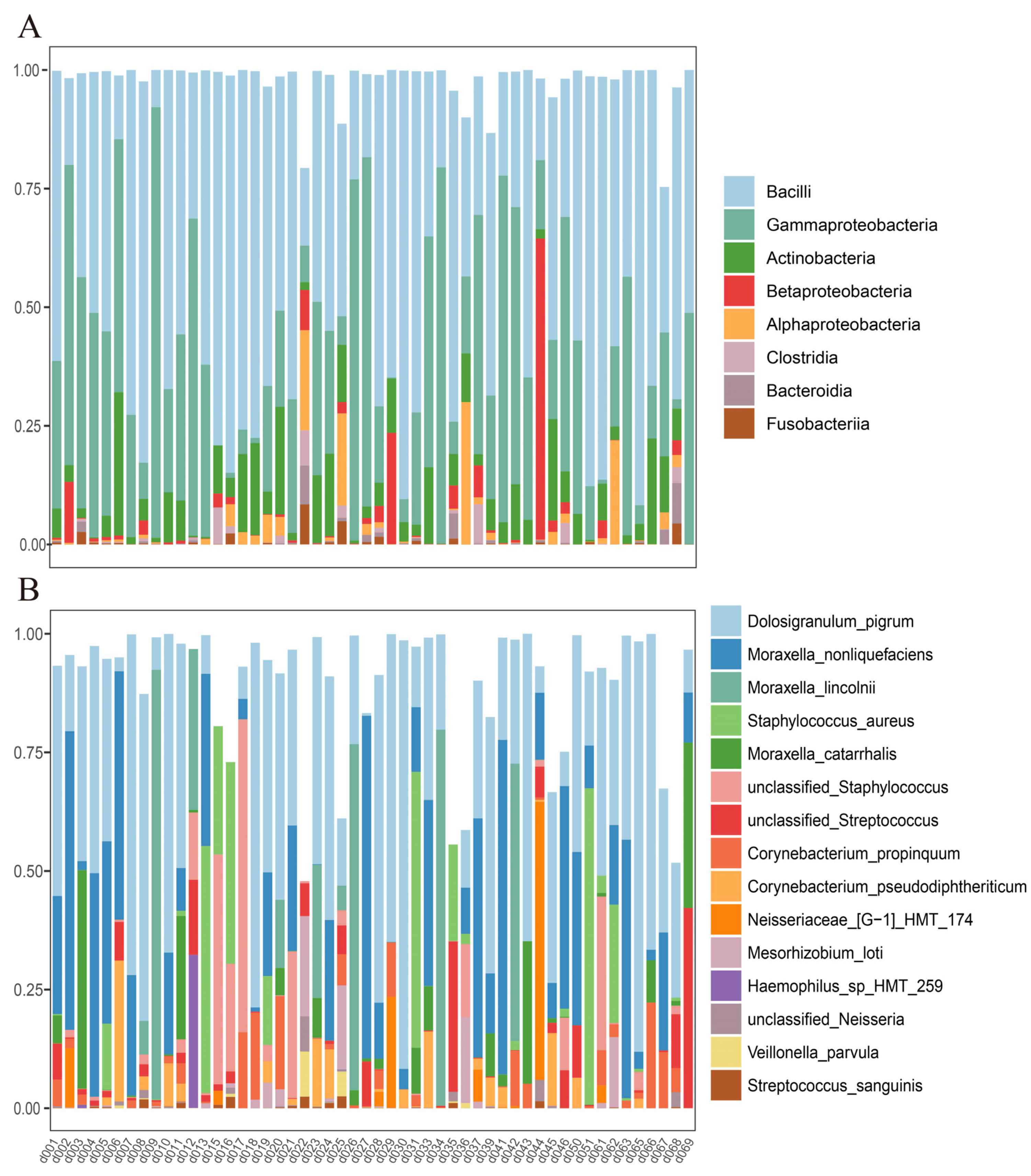
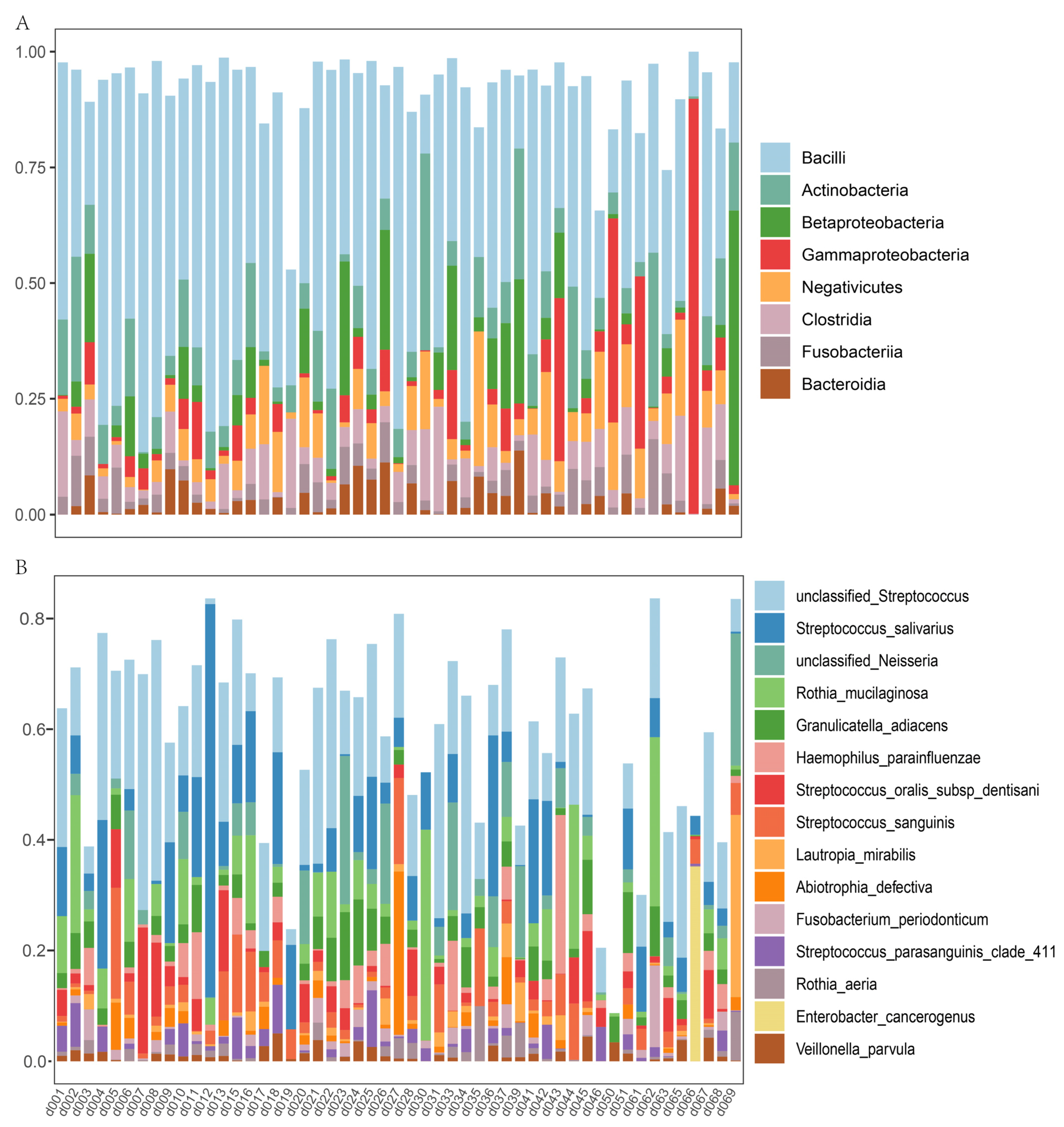
3.3. Nasal and Oral Microbial Composition

In the nasal microbiome, Bacilli, Gammaproteobacteria, Actinobacteria, and Betaproteobacteria were highly abundant microbial classes (Figure 2A). The top nasal microbial species included Dolosigranulum pigrum, Moraxella nonliquefaciens, Moraxella lincolnii, Staphylococcus aureus, Moraxella catarrhalis, an unclassified Staphylococcus, and an unclassified Streptococcus (Figure 2B). In oral saliva, the top microbial classes were Bacilli, Actinobacteria, Betaproteobacteria, and Gammaproteobacteria. The top oral species included an unclassified Streptococcus, Streptococcus salivarius, an unclassified Neisseria, Rothia mucilaginosa, Granulicatella adiacens, Haemophilus parainfluenzae, Streptococcus oralis, Streptococcus sanguinis, and Lautropia mirabilis (Figure 3B).

3.4. Impact of Environmental Variables on Overall Nasal and Oral Microbial Composition

We used PERMANOVA to calculate the impact of environmental characteristics on the beta diversity (composition) of the nasal and oral microbiota (Table 1). To control for false positive results, only associations with a p-value < 0.02 were considered significant and presented in bold fonts. Age (p = 0.008, R2 = 4.2) and education level of the father (p = 0.04, R2 = 2.8) were significantly associated with the nasal microbial composition. Exposure to ETS during the mother’s pregnancy and early childhood was associated with nasal and oral microbial composition (p = 0.005 and 0.03, R2 = 4.63 and 1.74). Food intake, outdoor air pollution, the abundance of indoor pathogens, and the indoor total abundance of VFs and ARGs were not associated with the beta diversity of the nasal and oral microbiota. The abundance of total flavonoids was associated with the nasal microbial composition (p = 0.01, R2 = 3.90). Indole derivatives 3-methylindole (p = 0.01, R2 = 3.57), mycotoxin deoxynivalenol (p = 0.017, R2 = 3.26), and nivalenol (p = 0.028, R2 = 2.77) were also associated with nasal and oral microbial composition.

3.5. Impact of Environmental Variables on Alpha Diversity and the Abundance of Risky/Protective Nasal and Oral Microorganisms

Regression analyses were performed to evaluate the relationship between environmental variables and nasal alpha diversity, as characterized by the Shannon index and the observed species (Table 2). The presence of siblings (p = 0.008, β = 0.69) and exposure to an indoor flavonoid baicalein (p = 0.004, β = 0.72) were positively associated with the Shannon index. The presence of siblings (p = 0.01, β = 29.9) and eupatilin (p = 0.0037, β = 0.68; a flavonoid) were positively associated with the observed number of species. A prior study [7] identified a set of nasal microorganisms that offer protection against chronic rhinosinusitis, which were hence defined as protective nasal microorganisms in this study. The abundance of protective nasal microorganisms was positively associated with isoliquiritigenin (p = 0.0018, β = 0.002; a flavonoid) and serotonin (p = 0.0005, β = 0.62; an indole) but negatively associated with daily intake of rice/pasta/bread (p = 0.0091, β = −0.062).
As for oral microbiota, age starting kindergarten (p = 0.002, β = 0.48) was positively correlated with the Shannon index. The observed species showed significant associations with two flavonoids, tangeritin (p = 0.014, β = 2.29) and hesperidin (p = 0.005, β = 1.40). In line with a prior study [14] that identified protective and risky oral microorganisms for periodontitis, we defined these microorganisms as such in our study. The abundance of these risky oral microorganisms showed a positive association with early childhood exposure to smoking (p = 0.013, β = 0.005) and frequent consumption of fries (p = 0.019, β = 0.005). However, pyruvic acid abundance (p = 0.0173, β = −0.056) was negatively associated with risky oral microorganisms, while the total abundance of keto acids (p = 0.0147, β = 2.300) was positively correlated with oral protective microorganisms.
Regarding indoor metabolites, flavonoids are predominantly plant-derived, whereas keto acids and indoles can be produced by indoor microorganisms. Therefore, neural network analysis [46] was employed to ascertain the co-occurrence probability of the top indoor microorganisms with serotonin and keto acids. The analysis revealed that certain indoor microorganisms, including Staphylococcus epidermidis, Mycolicibacerium iranicum, and Corynebacterium (Figure 4), co-occurred with these metabolites, indicating that these indoor microorganisms may potentially produce these metabolites.

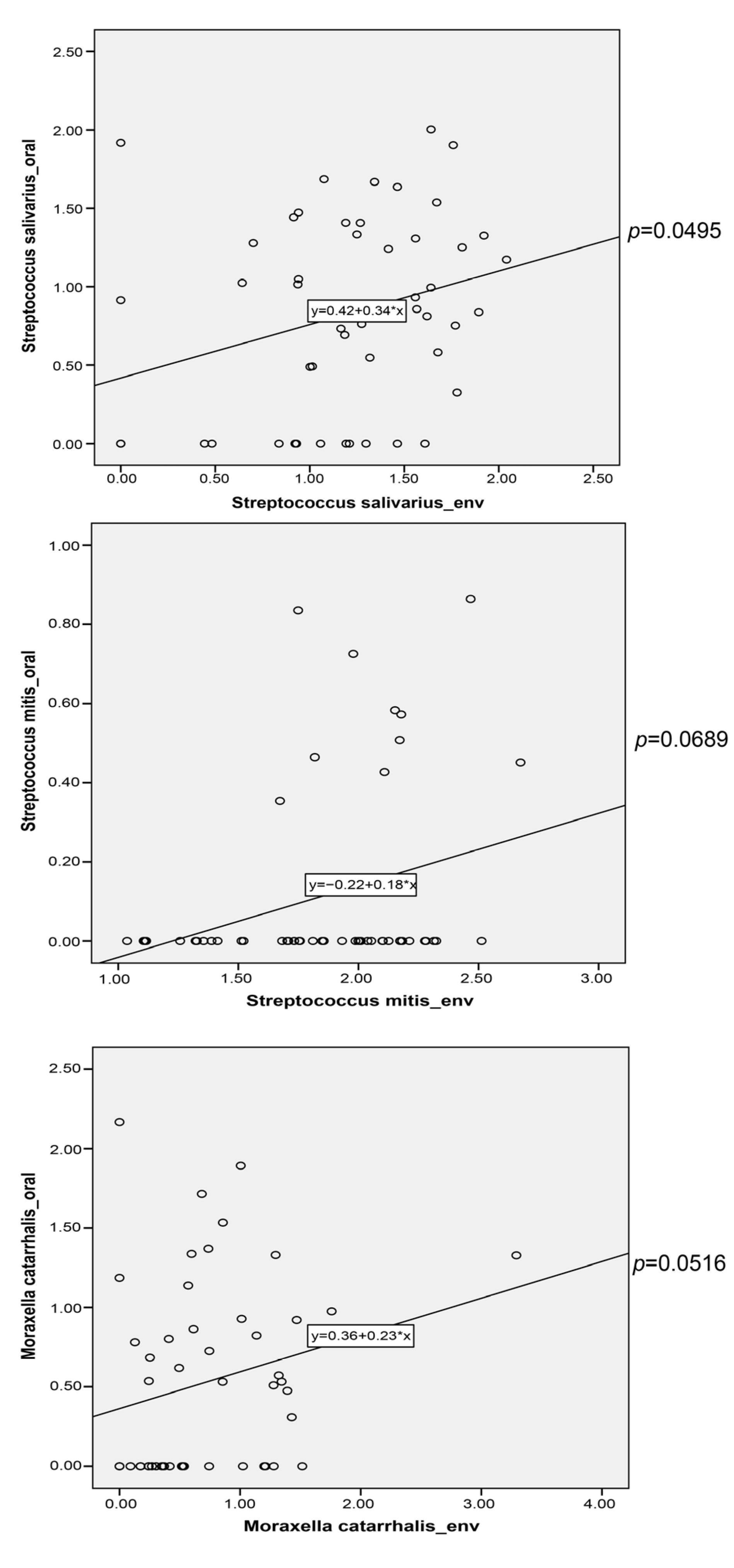
3.6. Potential Microbial Transfer from Indoor Environment to Nasal/Oral Cavity

To investigate the potential transfer of microbes from the indoor environment to the nasal/oral cavity, we conducted a correlation analysis between major indoor and nasal/oral species. Three oral species, including Moraxella catarrhalis (rho = 0.2768, p = 0.0516), Streptococcus mitis (rho = 0.2564, p = 0.0689), and Streptococcus salivarius (rho = 0.2793, p = 0.0495), were positively correlated with the indoor species and could potentially be transmitted from an indoor environment to the oral cavity (Figure 5). We further analyze the VFs and ARGs in these species. In total, 8, 5, and 9 potential VFs were presented (RPKM > 0.5) in M. catarrhalis (tufA, katA, clpE, hhuA, htpB, clpC, ccmF, carB), S. mitis (pavB, hasC, cbpA, pspA), and S. salivarius (pavB, hasC, psaA, fbp54, lap, cps4B, clpE, clpC, tufA). These VFs were mainly involved in adherence, immune modulation, nutritional/metabolic factors, and stress survival. Similarly, 3, 3, and 2 potential ARGs were presented (RPKM > 0.5) in M. catarrhalis (bro-1, icr-mc, vanY), S. mitis (patA, patB, vanY), and S. salivarius (vanY, vanT). Interestingly, vancomycin resistance genes were widely distributed in these species, suggesting a potential acquisition of these ARGs in the oral cavity from the indoor environment.

4. Discussion

In the present study, we conducted a multi-omic analysis to explore the associations between the indoor microbiome/metabolites and the nasal/oral microbiota in children. We identified specific indoor flavonoids and mycotoxins, such as deoxynivalenol and nivalenol, that were significantly associated with microbial diversity and abundance in these cavities. Additionally, our data suggest that certain microorganisms may be transferred from the indoor environment to children’s oral cavities, potentially influencing virulence factors and antibiotic resistance profiles. These observations provide valuable insights into the complex interplay between the indoor environment and microbiota, opening new avenues for future research aimed at promoting human health.

4.1. Strengths and Limitations of the Study

This study has several notable strengths. Firstly, it pioneers the investigation of the relationship between indoor microbiome/metabolites and nasal/oral microbiota. Over 40 environmental and personal characteristics were collected, providing a comprehensive understanding of the effect of environmental factors on oral and nasal microbiota. Second, culture-independent multi-omics were utilized, including metagenomics, full-length 16S rRNA sequencing, and untargeted LC/MS. Multi-omic profiling has been widely conducted in intestinal gut studies [47] but is seldom reported in the indoor environment [32]. Therefore, our study can also provide resources for future analysis, such as comparing indoor microbiome/metabolite profiling with other environments. Thirdly, we used shotgun metagenomics to assess indoor functional genes, including VFs and ARGs.
Despite its contributions, this study has some limitations that warrant discussion. First, our study has a relatively small sample size of 56 healthy children, which was determined based on logistical constraints such as funding and available resources for in-depth multi-omics analyses. Although this sample size aligns with similar exploratory or pilot studies in this emerging research area, it does restrict the study’s statistical power for association analyses. Furthermore, the small sample size precluded us from applying stringent corrections for multiple testing, such as false discovery rate (FDR) control, as it would significantly reduce our ability to detect true associations. Therefore, our results should be interpreted as exploratory and will require validation in larger cohorts. Second, we used untargeted metabolomics for assessing indoor metabolites. While this approach allows for the characterization of a large number of metabolites, it yields only relative abundance, not absolute concentration. This restricts our ability to draw conclusions about the impacts of specific metabolite concentrations on the nasal and oral microbiota. In future studies, targeted metabolomics may enable more precise assessments of the impacts of specific indoor metabolites on the nasal/oral microbiota. Third, only one dust sample per child’s home was collected, raising questions about reproducibility over time and sensitivity to random variation. However, it should be noted that the vacuuming technique employed in this study is designed to collect large biomass samples. Prior research has shown that such samples are representative of long-term exposure and less susceptible to random variation compared to swab sampling [27]. Fourth, we did not conduct dental examinations to assess the oral health status of the participants. Thus, we cannot definitively conclude the precise oral health condition of our cohort. However, to ascertain the health status of the participants, the guardians of the children were interviewed and answered a comprehensive questionnaire. This method of data collection should lend some accuracy to our understanding of the participants’ health. Fifth, the source organisms for VFs and ARGs were identified using a bioinformatics approach, which may have inherent limitations that could lead to false positives. Similarly, the detection of species DNA in both oral and indoor environments does not necessarily indicate the viability or transferability of these organisms. Further studies investigating the survival and viability of these species in environmental conditions would be needed to confirm the actual transfer. Sixth, there is a methodological discrepancy in our approach, wherein we employed shotgun metagenomics for indoor samples and 16S rRNA sequencing for children’s nasal and oral samples. Shotgun metagenomics provides a comprehensive view of all the genetic material in the samples, including bacteria, viruses, fungi, and archaea, allowing for the identification of VFs, ARGs, and specific microbial genes. In contrast, 16S rRNA sequencing, while robust and cost-effective, specifically targets bacterial communities, potentially missing out on broader microbial diversity and specific gene-level information. This difference in methodologies could impact the comparability of the datasets and might have introduced biases in our analysis.

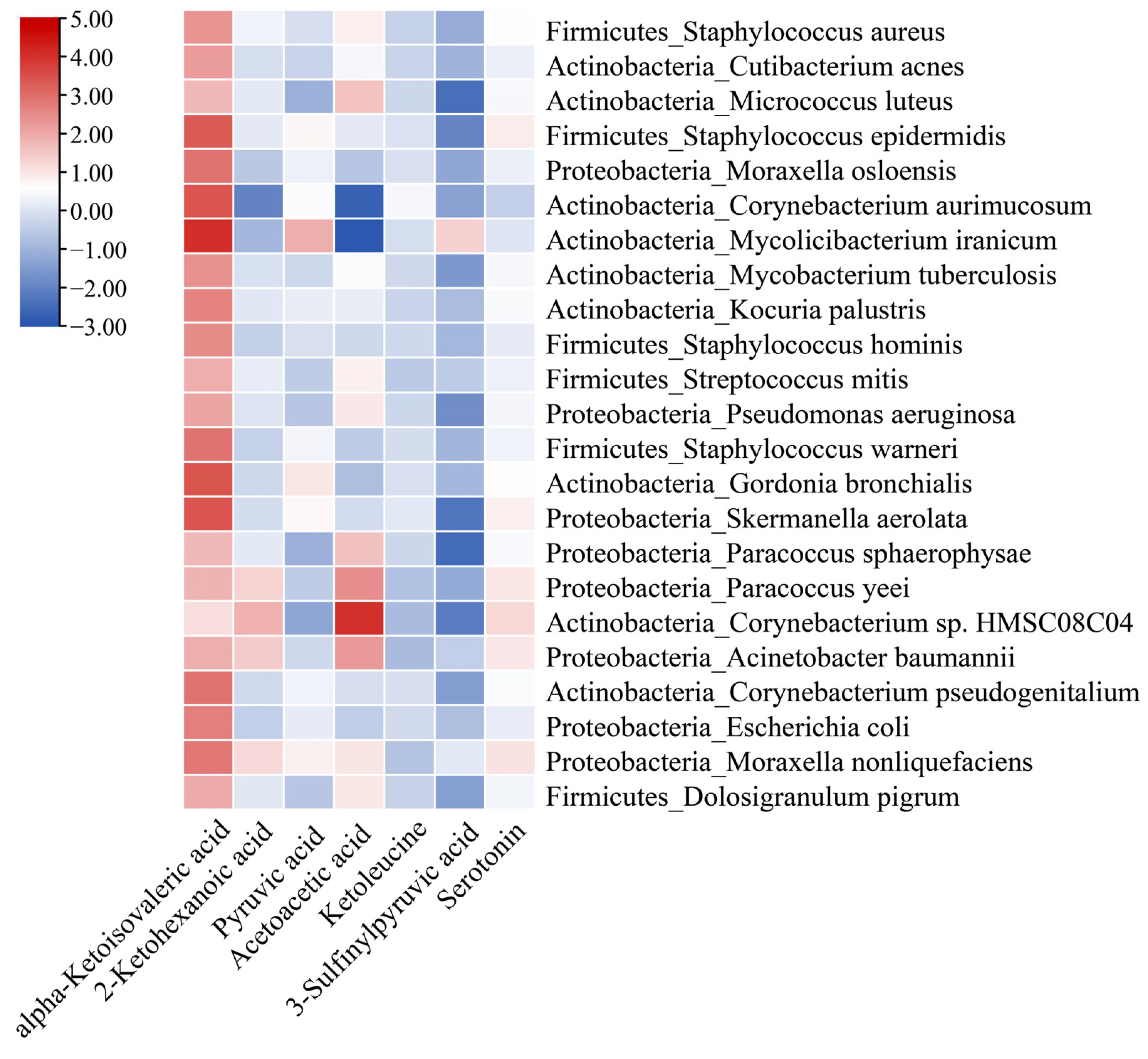
4.2. Indoor Metabolites and Nasal/Oral Microbiota

In this study, we found that indoor flavonoids (baicalein, eupatilin, isoliquiritigenin, tangeritin, and hesperidin) were associated with multiple nasal/oral microbial features, including the overall nasal microbial community variation, increased nasal/oral microbial alpha diversity, and an increased abundance of protective microorganisms in the oral cavity. These observations suggest a possible role for indoor flavonoid exposure in influencing nasal/oral health. Flavonoids are plant metabolites that possess several protective health effects, such as anti-bacterial, anti-oxidant, anti-inflammatory, anti-cancer, and anti-aging effects [48,49,50]. However, the mechanisms through which they increase nasal/oral diversity and the abundance of protective microbiota remain unclear. A possible mechanism is that flavonoids may suppress the biofilm formation and aggregation of pathogenic microorganisms. For instance, baicalein can protect oral health by inhibiting microbial biofilm formation, which prevents dental caries [51]. The suppression of pathogenic species may facilitate the growth of potentially beneficial microorganisms in the oral cavity.
Regarding the source of these flavonoids in the indoor environment, multiple possibilities exist. They could originate from outdoor plants, or they could be remnants from the consumption of flavonoid-rich foods and beverages within the household. However, it is important to clarify that the detected levels of dust might not directly indicate a health impact. Their presence might be due to higher consumption of flavonoid-rich foods in these environments, serving more as markers of certain dietary habits than direct influencers of health. Comparative analysis of flavonoid types and concentrations in outdoor plants and consumed foods would be essential for a more precise understanding of their origin and relevance.
In addition to flavonoids, we found that pyruvic acid (a keto acid derivative) and serotonin (an indole derivative) were associated with decreased risk microorganisms and increased protective microorganisms in the nasal/oral cavity. Pyruvic acid is a cellular metabolite produced at the end of glycolysis and has been widely used as a precursor for food, cosmetics, pharmaceuticals, and agricultural applications [52]. It has been shown to provide a wide range of beneficial health effects, including protection against oxidative stress and zinc toxicity [53,54]. However, their protective effects on the nasal/oral microbiota require further exploration. Serotonin is a monoamine neurotransmitter with multifaceted functions in modulating mood, cognition, learning, and memory. Approximately 90% of serotonin is produced in the human gut with the help of microorganisms, and therefore, it is one of the major signal transmitters in the gut–brain axis. A recent study in Malaysia reported that serotonin was present in high abundance in the dust of schools with low asthma prevalence [32], indicating that it may also play a role in respiratory health.
We found that nivalenol and deoxynivalenol in indoor dust were associated with the overall microbial composition in the nasal and oral cavities. Nivalenol and deoxynivalenol are mycotoxins that belong to the trichothecene group and are structurally similar. Fusarium species mainly produce these mycotoxins, and they are among the most widespread mycotoxins in food and feedstuffs [55]. Exposure to deoxynivalenol through food intake causes gastrointestinal inflammation and can lead to changes in the diversity, integrity, and composition of the gut microbiota, posing persistent threats to human and animal health [56]. However, the impact of nivalenol and deoxynivalenol on the nasal and oral microbiota has not been reported. In this study, we characterized these two mycotoxins in all samples, indicating their widespread presence in indoor dust. The resuspension of indoor dust and human inhalation may lead to nasal and oral exposure to these mycotoxins, which may further affect the nasal/oral microbiota.

4.3. Other Environmental and Personal Characteristics and Nasal/Oral Microbiota

In our study, we found a positive association between the presence of siblings and increased nasal microbial diversity. This finding is consistent with the Dutch Microbiome Project, which demonstrated that the number of cohabitants is the main factor in shaping the human microbiota [57]. Our results also showed that frequent intake of rice, pasta, bread, and French fries decreased the abundance of protective nasal microorganisms and increased the abundance of risky oral microorganisms. Although no previous studies have reported the association between food frequency and nasal/oral microbiota, our results are consistent with previous studies on gut microbiota. A high-glycemic and carbohydrate diet has been shown to decrease protective microorganisms and increase the risk of microorganisms in the gut microbiota [58,59]. Additionally, a high-fat diet, such as french fries, has been shown to promote the development of pro-inflammatory microbiota and alter the microbial composition of beneficial and pathogenic bacteria [60,61].
In our study, we did not find an association between air pollution and microbiota composition and diversity in the nasal and oral cavities, which is inconsistent with previous observations [17,18]. One potential explanation for this discrepancy is that we only sampled children in Shanghai for this study. The concentration of air pollutants in Shanghai was homogeneous across the entire city.

5. Conclusions

This study sheds light on the potential influence of the indoor microbiome and metabolites on the nasal and oral microbiota in children. The evidence points towards the crucial role of environmental factors, such as exposure to tobacco smoke and indoor flavonoids, in modulating the microbiota. Moreover, this study uncovers potential microbial transfer routes from indoor environments to the human body, warranting further research in this area. These findings underscore the critical need for a deeper understanding of our indoor microbial environments to promote health and prevent disease.

Author Contributions

Conceptualization, Z.Z. and Y.S.; methodology, H.T.; formal analysis, M.Z., H.T. and Y.Y.; resources, Z.Z.; data curation, H.T., Z.C. and Y.X.; writing—original draft preparation, M.Z. and Y.S.; writing—review and editing, X.F., Z.Z. and Y.S.; visualization, M.Z., Z.O. and Y.Y.; supervision, Z.Z. and Y.S.; funding acquisition, X.F. and Z.Z. All authors have read and agreed to the published version of the manuscript.

Funding

This study was funded by the National Natural Science Foundation of China (grant numbers 81861138005 and 42377106), the Natural Science Foundation of Guangdong Province (2021A1515010492), and the Shanghai 3-year Public Health Action Plan (grant number GWV-10.1-XK11).

Institutional Review Board Statement

This study was conducted in accordance with the Declaration of Helsinki and approved by the Ethics Committee of the School of Public Health at Fudan University (IRB#2019-09-0778, protocol code 200032, and date of approval: 27 September 2019).

Informed Consent Statement

Informed consent was obtained from all subjects involved in the study.

Data Availability Statement

The sequencing data are in the Genome Sequence Archive https://ngdc.cncb.ac.cn/gsa/ (accessed on 26 September 2023) under the accession number PRJCA008482.

Conflicts of Interest

The authors declare no conflict of interest.

References

  1. Lemon, K.P. Human nasal microbiota. Curr. Biol. 2020, 30, R1118–R1119. [Google Scholar] [CrossRef] [PubMed]
  2. De Steenhuijsen Piters, W.A.; Sanders, E.A.; Bogaert, D. The role of the local microbial ecosystem in respiratory health and disease. Philos. Trans. R. Soc. B Biol. Sci. 2015, 370, 20140924. [Google Scholar] [CrossRef] [PubMed]
  3. Cho, D.Y.; Hunter, R.C.; Ramakrishnan, V.R. The Microbiome and Chronic Rhinosinusitis. Immunol. Allergy Clin. N. Am. 2020, 40, 251–263. [Google Scholar] [CrossRef]
  4. Shah, R.; Bunyavanich, S. The airway microbiome and pediatric asthma. Curr. Opin. Pediatr. 2021, 33, 639–647. [Google Scholar] [CrossRef] [PubMed]
  5. Huang, Y.J.; Boushey, H.A. The microbiome in asthma. J. Allergy Clin. Immunol. 2015, 135, 25–30. [Google Scholar] [CrossRef]
  6. Heinrich, A.; Heyl, K.A.; Klaile, E.; Müller, M.M.; Klassert, T.E.; Wiessner, A.; Fischer, K.; Schumann, R.R.; Seifert, U.; Riesbeck, K.; et al. Moraxella catarrhalis induces CEACAM3-Syk-CARD9-dependent activation of human granulocytes. Cell. Microbiol. 2016, 18, 1570–1582. [Google Scholar] [CrossRef]
  7. Hoggard, M.; Waldvogel-Thurlow, S.; Zoing, M.; Chang, K.; Radcliff, F.J.; Wagner Mackenzie, B.; Biswas, K.; Douglas, R.G.; Taylor, M.W. Inflammatory Endotypes and Microbial Associations in Chronic Rhinosinusitis. Front. Immunol. 2018, 9, 2065. [Google Scholar] [CrossRef]
  8. Bomar, L.; Brugger, S.D.; Yost, B.H.; Davies, S.S.; Lemon, K.P. Corynebacterium accolens Releases Antipneumococcal Free Fatty Acids from Human Nostril and Skin Surface Triacylglycerols. mBio 2016, 7, e01725-15. [Google Scholar] [CrossRef]
  9. Shi, Z.; Li, X.; Wang, X.; Zhang, L.; Li, L.; Fu, X.; Sun, Z.; Li, Z.; Zhang, X.; Zhang, M. Characteristics and Clinical Implications of the Nasal Microbiota in Extranodal NK/T-Cell Lymphoma, Nasal Type. Front. Cell. Infect. Microbiol. 2021, 11, 686595. [Google Scholar] [CrossRef]
  10. Escapa, I.F.; Huang, Y.; Chen, T.; Lin, M.; Kokaras, A.; Dewhirst, F.E.; Lemon, K.P. Construction of habitat-specific training sets to achieve species-level assignment in 16S rRNA gene datasets. Microbiome 2020, 8, 65. [Google Scholar] [CrossRef]
  11. Lloyd-Price, J.; Mahurkar, A.; Rahnavard, G.; Crabtree, J.; Orvis, J.; Hall, A.B.; Brady, A.; Creasy, H.H.; McCracken, C.; Giglio, M.G.; et al. Strains, functions and dynamics in the expanded Human Microbiome Project. Nature 2017, 550, 61–66. [Google Scholar] [CrossRef] [PubMed]
  12. Lamont, R.J.; Koo, H.; Hajishengallis, G. The oral microbiota: Dynamic communities and host interactions. Nat. Rev. Microbiol. 2018, 16, 745–759. [Google Scholar] [CrossRef] [PubMed]
  13. Mira, A.; Simon-Soro, A.; Curtis, M.A. Role of microbial communities in the pathogenesis of periodontal diseases and caries. J. Clin. Periodontol. 2017, 44, S23–S38. [Google Scholar] [CrossRef] [PubMed]
  14. Curtis, M.A.; Diaz, P.I.; Van Dyke, T.E. The role of the microbiota in periodontal disease. Periodontology 2000 2020, 83, 14–25. [Google Scholar] [CrossRef] [PubMed]
  15. Peng, X.; Cheng, L.; You, Y.; Tang, C.; Ren, B.; Li, Y.; Xu, X.; Zhou, X. Oral microbiota in human systematic diseases. Int. J. Oral Sci. 2022, 14, 14. [Google Scholar] [CrossRef] [PubMed]
  16. Hosgood, H.D.; Cai, Q.; Hua, X.; Long, J.; Shi, J.; Wan, Y.; Yang, Y.; Abnet, C.; Bassig, B.A.; Hu, W.; et al. Variation in oral microbiome is associated with future risk of lung cancer among never-smokers. Thorax 2021, 76, 256. [Google Scholar] [CrossRef]
  17. Mariani, J.; Favero, C.; Spinazzè, A.; Cavallo, D.M.; Carugno, M.; Motta, V.; Bonzini, M.; Cattaneo, A.; Pesatori, A.C.; Bollati, V. Short-term particulate matter exposure influences nasal microbiota in a population of healthy subjects. Environ. Res. 2018, 162, 119–126. [Google Scholar] [CrossRef]
  18. Gisler, A.; Korten, I.; de Hoogh, K.; Vienneau, D.; Frey, U.; Decrue, F.; Gorlanova, O.; Soti, A.; Hilty, M.; Latzin, P.; et al. Associations of air pollution and greenness with the nasal microbiota of healthy infants: A longitudinal study. Environ. Res. 2021, 202, 111633. [Google Scholar] [CrossRef]
  19. Irizar, H.; Chun, Y.; Arditi, Z.; Do, A.; Grishina, G.; Grishin, A.; Vicencio, A.; Bunyavanich, S. Examination of host genetic effects on nasal microbiome composition. J. Allergy Clin. Immunol. 2022, 150, 1232–1236. [Google Scholar] [CrossRef]
  20. Zeineldin, M.; Aldridge, B.; Blair, B.; Kancer, K.; Lowe, J. Microbial shifts in the swine nasal microbiota in response to parenteral antimicrobial administration. Microb. Pathog. 2018, 121, 210–217. [Google Scholar] [CrossRef]
  21. Ma, G.; Qiao, Y.; Shi, H.; Zhou, J.; Li, Y. Comparison of the Oral Microbiota Structure among People from the Same Ethnic Group Living in Different Environments. BioMed Res. Int. 2022, 2022, 6544497. [Google Scholar] [CrossRef] [PubMed]
  22. Cheung, M.K.; Chan, J.Y.K.; Wong, M.C.S.; Wong, P.Y.; Lei, P.; Cai, L.; Lan, L.; Ho, W.C.S.; Yeung, A.C.M.; Chan, P.K.S.; et al. Determinants and Interactions of Oral Bacterial and Fungal Microbiota in Healthy Chinese Adults. Microbiol. Spectr. 2022, 10, e0241021. [Google Scholar] [CrossRef] [PubMed]
  23. Bomar, L.; Brugger, S.D.; Lemon, K.P. Bacterial microbiota of the nasal passages across the span of human life. Curr. Opin. Microbiol. 2018, 41, 8–14. [Google Scholar] [CrossRef] [PubMed]
  24. Jia, Y.J.; Liao, Y.; He, Y.Q.; Zheng, M.Q.; Tong, X.T.; Xue, W.Q.; Zhang, J.B.; Yuan, L.L.; Zhang, W.L.; Jia, W.H. Association Between Oral Microbiota and Cigarette Smoking in the Chinese Population. Front. Cell. Infect. Microbiol. 2021, 11, 658203. [Google Scholar] [CrossRef]
  25. Vallès, Y.; Inman, C.K.; Peters, B.A.; Wareth, L.A.; Abdulle, A.; Alsafar, H.; Anouti, F.A.; Dhaheri, A.A.; Galani, D.; Haji, M.; et al. Incense Burning is Associated with Human Oral Microbiota Composition. Sci. Rep. 2019, 9, 10039. [Google Scholar] [CrossRef]
  26. Sun, Y.; Meng, Y.; Ou, Z.; Li, Y.; Zhang, M.; Chen, Y.; Zhang, Z.; Chen, X.; Mu, P.; Norbäck, D.; et al. Indoor microbiome, air pollutants and asthma, rhinitis and eczema in preschool children—A repeated cross-sectional study. Environ. Int. 2022, 161, 107137. [Google Scholar] [CrossRef]
  27. Fu, X.; Ou, Z.; Sun, Y. Indoor microbiome and allergic diseases: From theoretical advances to prevention strategies. Eco-Environ. Health 2022, 1, 133–146. [Google Scholar] [CrossRef]
  28. Fu, X.; Ou, Z.; Zhang, M.; Meng, Y.; Li, Y.; Chen, Q.; Jiang, J.; Zhang, X.; Norbäck, D.; Zhao, Z.; et al. Classroom microbiome, functional pathways and sick-building syndrome (SBS) in urban and rural schools—Potential roles of indoor microbial amino acids and vitamin metabolites. Sci. Total Environ. 2021, 795, 148879. [Google Scholar] [CrossRef]
  29. Fu, X.; Yuan, Q.; Zhu, X.; Li, Y.; Meng, Y.; Hashim, J.H.; Hashim, Z.; Ali, F.; Zheng, Y.W.; Lai, X.X.; et al. Associations between the indoor microbiome, environmental characteristics and respiratory infections in junior high school students of Johor Bahru, Malaysia. Environ. Sci. Process. Impacts 2021, 23, 1171–1181. [Google Scholar] [CrossRef]
  30. Fu, X.; Norbäck, D.; Yuan, Q.; Li, Y.; Zhu, X.; Hashim, J.H.; Hashim, Z.; Ali, F.; Zheng, Y.-W.; Lai, X.-X.; et al. Indoor microbiome, environmental characteristics and asthma among junior high school students in Johor Bahru, Malaysia. Environ. Int. 2020, 138, 105664. [Google Scholar] [CrossRef]
  31. Fu, X.; Li, Y.; Meng, Y.; Yuan, Q.; Zhang, Z.; Norbäck, D.; Deng, Y.; Zhang, X.; Sun, Y. Associations between respiratory infections and bacterial microbiome in student dormitories in Northern China. Indoor Air 2020, 30, 816–826. [Google Scholar] [CrossRef] [PubMed]
  32. Sun, Y.; Zhang, M.; Ou, Z.; Meng, Y.; Chen, Y.; Lin, R.; Hashim, J.H.; Hashim, Z.; Wieslander, G.; Chen, Q.; et al. Indoor microbiome, microbial and plant metabolites, chemical compounds and asthma symptoms in junior high school students: A multicentre association study in Malaysia. Eur. Respir. J. 2022, 60, 2200260. [Google Scholar] [CrossRef] [PubMed]
  33. Crane, J.; Mallol, J.; Beasley, R.; Stewart, A.; Asher, M.I. Agreement between written and video questions for comparing asthma symptoms in ISAAC. Eur. Respir. J. 2003, 21, 455–461. [Google Scholar] [CrossRef]
  34. Bell, M.L. The use of ambient air quality modeling to estimate individual and population exposure for human health research: A case study of ozone in the Northern Georgia Region of the United States. Environ. Int. 2006, 32, 586–593. [Google Scholar] [CrossRef] [PubMed]
  35. Norbäck, D.; Lu, C.; Zhang, Y.; Li, B.; Zhao, Z.; Huang, C.; Zhang, X.; Qian, H.; Sun, Y.; Sundell, J.; et al. Onset and remission of childhood wheeze and rhinitis across China—Associations with early life indoor and outdoor air pollution. Environ. Int. 2019, 123, 61–69. [Google Scholar] [CrossRef]
  36. Sun, Y.; Fu, X.; Li, Y.; Yuan, Q.; Ou, Z.; Lindgren, T.; Deng, Y.; Norbäck, D. Shotgun Metagenomics of Dust Microbiome from Flight Deck and Cabin in Civil Aviation Aircraft. Indoor Air 2020, 30, 1199–1212. [Google Scholar] [CrossRef]
  37. Wang, Y.; Song, F.; Zhu, J.; Zhang, S.; Yang, Y.; Chen, T.; Tang, B.; Dong, L.; Ding, N.; Zhang, Q.; et al. GSA: Genome Sequence Archive. Genom. Proteom. Bioinform. 2017, 15, 14–18. [Google Scholar] [CrossRef]
  38. Li, D.; Luo, R.; Liu, C.M.; Leung, C.M.; Ting, H.F.; Sadakane, K.; Yamashita, H.; Lam, T.W. MEGAHIT v1.0: A fast and scalable metagenome assembler driven by advanced methodologies and community practices. Methods 2016, 102, 3–11. [Google Scholar] [CrossRef] [PubMed]
  39. Zhu, W.; Lomsadze, A.; Borodovsky, M. Ab initio gene identification in metagenomic sequences. Nucleic Acids Res. 2010, 38, e132. [Google Scholar] [CrossRef]
  40. Huson, D.H.; Auch, A.F.; Qi, J.; Schuster, S.C. MEGAN analysis of metagenomic data. Genome Res. 2007, 17, 377–386. [Google Scholar] [CrossRef]
  41. Liu, B.; Zheng, D.; Jin, Q.; Chen, L.; Yang, J. VFDB 2019: A comparative pathogenomic platform with an interactive web interface. Nucleic Acids Res. 2019, 47, D687–D692. [Google Scholar] [CrossRef] [PubMed]
  42. Alcock, B.P.; Raphenya, A.R.; Lau, T.T.Y.; Tsang, K.K.; Bouchard, M.; Edalatmand, A.; Huynh, W.; Nguyen, A.V.; Cheng, A.A.; Liu, S.; et al. CARD 2020: Antibiotic resistome surveillance with the comprehensive antibiotic resistance database. Nucleic Acids Res. 2020, 48, D517–D525. [Google Scholar] [CrossRef] [PubMed]
  43. Eid, J.; Fehr, A.; Gray, J.; Luong, K.; Lyle, J.; Otto, G.; Peluso, P.; Rank, D.; Baybayan, P.; Bettman, B.; et al. Real-time DNA sequencing from single polymerase molecules. Science 2009, 323, 133–138. [Google Scholar] [CrossRef] [PubMed]
  44. Caporaso, J.G.; Kuczynski, J.; Stombaugh, J.; Bittinger, K.; Bushman, F.D.; Costello, E.K.; Fierer, N.; Pena, A.G.; Goodrich, J.K.; Gordon, J.I.; et al. QIIME allows analysis of high-throughput community sequencing data. Nat. Methods 2010, 7, 335–336. [Google Scholar] [CrossRef] [PubMed]
  45. Callahan, B.J.; McMurdie, P.J.; Rosen, M.J.; Han, A.W.; Johnson, A.J.A.; Holmes, S.P. DADA2: High-resolution sample inference from Illumina amplicon data. Nat. Methods 2016, 13, 581–583. [Google Scholar] [CrossRef]
  46. Morton, J.T.; Aksenov, A.A.; Nothias, L.F.; Foulds, J.R.; Quinn, R.A.; Badri, M.H.; Swenson, T.L.; Van Goethem, M.W.; Northen, T.R.; Vazquez-Baeza, Y.; et al. Learning representations of microbe–metabolite interactions. Nat. Methods 2019, 16, 1306–1314. [Google Scholar] [CrossRef]
  47. Lloyd-Price, J.; Arze, C.; Ananthakrishnan, A.N.; Schirmer, M.; Avila-Pacheco, J.; Poon, T.W.; Andrews, E.; Ajami, N.J.; Bonham, K.S.; Brislawn, C.J.; et al. Multi-omics of the gut microbial ecosystem in inflammatory bowel diseases. Nature 2019, 569, 655–662. [Google Scholar] [CrossRef]
  48. Sato, M.; Fujiwara, S.; Tsuchiya, H.; Fujii, T.; Iinuma, M.; Tosa, H.; Ohkawa, Y. Flavones with antibacterial activity against cariogenic bacteria. J. Ethnopharmacol. 1996, 54, 171–176. [Google Scholar] [CrossRef]
  49. Yan, X.; Qi, M.; Li, P.; Zhan, Y.; Shao, H. Apigenin in cancer therapy: Anti-cancer effects and mechanisms of action. Cell Biosci. 2017, 7, 50. [Google Scholar] [CrossRef]
  50. Tan, Y.; Zhang, X.; Cheang, W.S. Isoflavones daidzin and daidzein inhibit lipopolysaccharide-induced inflammation in RAW264.7 macrophages. Chin. Med. 2022, 17, 95. [Google Scholar] [CrossRef]
  51. Chen, H.; Xie, S.; Gao, J.; He, L.; Luo, W.; Tang, Y.; Weir, M.D.; Oates, T.W.; Xu, H.H.K.; Yang, D. Flavonoid Baicalein Suppresses Oral Biofilms and Protects Enamel Hardness to Combat Dental Caries. Int. J. Mol. Sci. 2022, 23, 10593. [Google Scholar] [CrossRef] [PubMed]
  52. Maleki, N.; Eiteman, M.A. Recent Progress in the Microbial Production of Pyruvic Acid. Fermentation 2017, 3, 8. [Google Scholar] [CrossRef]
  53. Mazzio, E.A.; Soliman, K.F. Cytoprotection of pyruvic acid and reduced beta-nicotinamide adenine dinucleotide against hydrogen peroxide toxicity in neuroblastoma cells. Neurochem. Res. 2003, 28, 733–741. [Google Scholar] [CrossRef] [PubMed]
  54. Yoo, M.H.; Lee, J.-Y.; Lee, S.E.; Koh, J.-Y.; Yoon, Y.H. Protection by Pyruvate of Rat Retinal Cells against Zinc Toxicity In Vitro, and Pressure-Induced Ischemia In Vivo. Investig. Ophthalmol. Vis. Sci. 2004, 45, 1523–1530. [Google Scholar] [CrossRef]
  55. Sun, Y.; Jiang, J.; Mu, P.; Lin, R.; Wen, J.; Deng, Y. Toxicokinetics and metabolism of deoxynivalenol in animals and humans. Arch. Toxicol. 2022, 96, 2639–2654. [Google Scholar] [CrossRef]
  56. Lin, R.; Sun, Y.; Mu, P.; Zheng, T.; Mu, H.; Deng, F.; Deng, Y.; Wen, J. Lactobacillus rhamnosus GG supplementation modulates the gut microbiota to promote butyrate production, protecting against deoxynivalenol exposure in nude mice. Biochem. Pharmacol. 2020, 175, 113868. [Google Scholar] [CrossRef]
  57. Gacesa, R.; Kurilshikov, A.; Vich Vila, A.; Sinha, T.; Klaassen, M.A.Y.; Bolte, L.A.; Andreu-Sánchez, S.; Chen, L.; Collij, V.; Hu, S.; et al. Environmental factors shaping the gut microbiome in a Dutch population. Nature 2022, 604, 732–739. [Google Scholar] [CrossRef]
  58. Hur, H.J.; Wu, X.; Yang, H.J.; Kim, M.J.; Lee, K.-H.; Hong, M.; Park, S.; Kim, M.-S. Beneficial Effects of a Low-Glycemic Diet on Serum Metabolites and Gut Microbiota in Obese Women with Prevotella and Bacteriodes Enterotypes: A Randomized Clinical Trial. Front. Nutr. 2022, 9, 861880. [Google Scholar] [CrossRef]
  59. Zhang, Y.; Liang, X.-F.; He, S.; Chen, X.; Wang, J.; Li, J.; Zhu, Q.; Zhang, Z.; Li, L.; Alam, M.S. Effects of High Carbohydrate Diet-Modulated Microbiota on Gut Health in Chinese Perch. Front. Microbiol. 2020, 11, 575102. [Google Scholar] [CrossRef]
  60. Bibbò, S.; Ianiro, G.; Giorgio, V.; Scaldaferri, F.; Masucci, L.; Gasbarrini, A.; Cammarota, G. The role of diet on gut microbiota composition. Eur. Rev. Med. Pharmacol. Sci. 2016, 20, 4742–4749. [Google Scholar]
  61. Wei, G.; Ye, Y.; Yan, X.; Chao, X.; Yang, F.; Wang, M.; Zhang, W.; Yuan, C.; Zeng, Q. Effect of banana pulp dietary fibers on metabolic syndrome and gut microbiota diversity in high-fat diet mice. J. Food Biochem. 2020, 44, e13362. [Google Scholar] [CrossRef] [PubMed]
Figure 1. (A) The top 8 microbial classes and (B) the top 15 microbial species of the indoor environment. The x-axis lists individual sample identifiers, while the y-axis denotes the relative abundance of microbial taxa in the respective samples.
Metabolites 13 01040 g001
Figure 2. (A) The top 8 microbial classes and (B) the top 15 microbial species of the nasal microbiota. The x-axis lists individual sample identifiers, while the y-axis denotes the relative abundance of microbial taxa in the respective samples.
Metabolites 13 01040 g002
Figure 3. (A) The top 8 microbial classes and (B) the top 15 microbial species of the oral microbiota. The x-axis lists individual sample identifiers, while the y-axis denotes the relative abundance of microbial taxa in the respective samples.
Metabolites 13 01040 g003
Figure 4. Co-occurrence probability of the top 25 indoor microbial species, keto acid derivatives, and serotonin.
Metabolites 13 01040 g004
Figure 5. Spearman’s correlation of oral and indoor microbials in the indoor environment.
Metabolites 13 01040 g005
Table 1. Associations between personal and environmental characteristics and the overall nasal and oral microbiota community of children (β-diversity). Metabolite abundances in the table are represented as intensity values derived from our untargeted metabolomics approach. The microbiota β-diversity was calculated using the Bray–Curtis distance indices. The associations were calculated using a 10,000 permutation bivariate Adonis/PERMANOVA analysis in R. Only associations with a p-value < 0.05 are shown in bold font.
NasalOral
R2p-ValueR2p-Value
Personal characteristicsQ1–Q3 percentile
Age (year)4–64.200.0080.820.57
Breastfeeding duration (year)0.5–10.820.491.020.38
Number of cohabitants3–50.920.421.440.16
Age start kindergarten3–41.450.211.230.25
Girl59%1.860.131.360.17
Preterm delivery12%0.380.820.580.75
Cesarean section45%0.330.840.950.46
Presence of siblings51%1.230.280.530.92
High parents income14%0.260.901.440.18
High education level for mother (graduate and postgraduate)50%1.000.380.510.83
High education level for father (graduate and postgraduate)54%2.820.040.510.89
Food intake frequencyWeekly/daily
Juice and soda drink86%/14%2.100.090.980.41
Fries90%/10%0.900.430.490.81
Rice/pasta/bread37%/63%1.920.120.940.47
Fruits, vegetables4%/96%1.420.230.560.83
Eggs, milk, fish, meat, and sea foods22%/78%0.170.970.820.57
Outdoor air pollution
SO2 (μg/m3)4.75–10.781.400.230.570.78
NO2 (μg/m3)29.35–43.101.660.171.200.25
CO (mg/m3)0.59–0.761.150.320.670.74
O3 (μg/m3)63.15–81.921.340.250.630.72
PM10 (μg/m3)38.53–48.102.470.060.810.59
PM2.5 (μg/m3)22.03–32.870.580.641.050.34
Living environment characteristics
Adjacent to heavy traffic41%0.680.561.920.04
Adjacent to river/park/garden22%0.740.520.470.91
ETS mother—pregnancy18%4.630.0051.740.09
ETS children—early childhood (<1 year)14%0.960.402.430.03
ETS children—previous 10 months18%1.870.131.170.30
Presence of pets/plants indoors—early childhood (<1 year old)27%1.550.191.290.22
Presence of pets/plants indoors—previous 10 months59%1.060.361.480.13
Visible mold/dampness—pregnancy20%1.410.221.390.18
Visible mold/dampness—early childhood (<1 year)27%1.010.370.770.65
Visible mold/dampness—previous 10 months25%1.060.350.800.61
Frequent room cleaning38%1.570.190.730.67
Building age (years)10–400.400.790.420.96
Abundance of potential pathogens indoor0.05–0.321.280.270.670.73
Clostridium perfringens0.01%0.920.420.350.86
Salmonella enterica0.50%0.470.730.880.48
Listeria monocytogenes0.01%1.140.311.150.28
Toxoplasma gondii0.01%0.520.690.740.56
Mycobacterium tuberculosis1.47%1.570.180.380.95
Total abundance of VFs indoors (RPKM)2.3 × 10³–5.2 × 1030.920.420.610.81
Total abundance of ARGs indoors (RPKM)2.4 × 10³–5.5 × 1030.910.420.670.75
Abundance of flavonoids indoors 3.910.011.080.35
Baicalein0–2.86 × 10100.840.461.100.34
Daidzein2.46 × 105–1.18 × 1081.960.113.880.03
Tangeritin1.92 × 106–5.01 × 1081.580.180.690.60
Isoliquiritigenin5.03 × 106–2.85 × 1080.790.491.070.37
Apigenin2.65 × 106–5.31 × 1082.380.060.550.62
(2S)-Liquiritigenin1.59 × 107–2.22 × 1090.370.821.190.25
Hesperidin2.31 × 106–1.12 × 1092.450.071.780.09
Eupatilin3.41 × 105–1.32 × 1080.580.641.490.16
Abundance of indoles indoors 0.780.500.950.46
3-Methylindole7.33 × 107–1.64 × 1093.570.010.390.86
Serotonin8.37 × 105–6.52 × 1080.580.650.410.84
Indole1.04 × 109–5.03 × 1091.050.350.390.93
L-Tryptophan1.67 × 109–3.95 × 10100.730.542.140.04
Indole-3-carboxylic acid7.50 × 105–1.51 × 1071.040.360.280.99
Abundance of keto acids indoors 0.740.530.490.89
Pyruvic acid3.51 × 107–1.41 × 1090.750.520.990.37
Ketoleucine1.43 × 107–4.40 × 1082.740.041.170.26
2-Ketohexanoic acid3.20 × 106–2.95 × 1081.690.163.490.04
Acetoacetic acid8.16 × 107–1.28 × 10100.150.970.590.58
alpha-Ketoisovaleric acid2.87 × 107–9.61 × 1081.200.300.970.40
Abundance of mycotoxins indoors 1.830.141.410.17
Vomitoxin (deoxynivalenol)7.30 × 10⁴–5.49 × 1073.260.021.340.19
Nivalenol2.38 × 105–1.81 × 1070.420.782.770.03
Tentoxin8.60 × 107–1.56 × 1092.140.091.780.10
Diacetoxyscirpenol4.83 × 104–1.75 × 1071.240.281.540.15
Table 2. Associations between personal information, environmental characteristics, α-diversity index, protective/risk nose, and oral microbial. The associations were calculated by linear regression adjusted for children’s age and gender. Only microorganisms with an abundance > 0.1% and metabolites with an abundance > 1 × 10⁶ (intensity) were included in the analysis. Associations with a p-value < 0.02 are presented in this table. Protective nasal microorganisms were defined based on a previous study in chronic rhinosinusitis [7] and included Corynebacterium, Finegoldia, Anaerococcus, Peptoniphilus, and Staphylococcus. Protective and risky oral microorganisms were defined based on a previous publication in periodontitis [14]. Risky oral microorganisms included Treponema maltophilum, Treponema socranskii, Porphyromonas gingivalis, Tannerella forsythia, Treponema denticola, Parvimonas micra, and Selenomonas sputigena, while protective oral microorganisms included Rothia dentocariosab and Streptococcus sanguinis.
Coefficientp-Value95%CI
Nasal
Shannon index
Presence of siblings0.690.0080.191.19
Baicalein (flavonoid)0.720.0040.241.20
Observed number of species
Presence of siblings29.90.017.4952.3
Eupatilin (flavonoid) 0.680.0040.231.12
Protective microorganisms
Rice/pasta/bread−0.060.009−0.11−0.02
Isoliquiritigenin (flavonoid)0.0020.0020.0010.003
Serotonin (indole)0.620.00050.030.95
Oral
Shannon index
Age start kindergarten0.480.0020.180.77
Observed_OTU_env−0.00090.011−0.002−0.0002
Observed number of species
Tangeritin (flavonoid)2.290.0140.494.09
Hesperidin (flavonoid)1.400.0070.392.40
Risk microorganisms
ETS children—early childhood (<1 year)0.0050.0130.0010.009
Fries0.0050.0190.0010.01
Pyruvic acid (keto acid)−0.0560.017−0.10−0.01
Protective microorganisms
Total keto acid2.300.0150.474.10
Disclaimer/Publisher’s Note: The statements, opinions and data contained in all publications are solely those of the individual author(s) and contributor(s) and not of MDPI and/or the editor(s). MDPI and/or the editor(s) disclaim responsibility for any injury to people or property resulting from any ideas, methods, instructions or products referred to in the content.

