Article

Prognostic Value of 18F-FDG PET/CT Volume-Based Metabolic Parameters in Patients with Node-Negative Stage II Esophageal Squamous Cell Carcinoma

1
Department of Nuclear Medicine, Kaohsiung Veterans General Hospital, Kaohsiung 813, Taiwan
2
School of Medicine, University of California, Davis, Sacramento, CA 95817, USA
3
Department of Biological Sciences, National Sun Yat-Sen University, Kaohsiung 804, Taiwan
4
Graduate Institute of Animal Vaccine Technology, College of Veterinary Medicine, National Pingtung University of Science and Technology, Pingtung 912, Taiwan
5
Department of Medical Education and Research, Kaohsiung Veterans General Hospital, Kaohsiung 813, Taiwan
6
Center of General Education, Shu-Zen Junior College of Medicine and Management, Kaohsiung 821, Taiwan
7
Department of Medical Imaging and Radiological Sciences, Kaohsiung Medical University, Kaohsiung 807, Taiwan
8
Department of Nuclear Medicine, Kaohsiung Medical University Hospital, Kaohsiung 807, Taiwan
9
Graduate Institute of Medicine, College of Medicine, Kaohsiung Medical University, Kaohsiung 807, Taiwan
10
Institute of Medical Science and Technology, National Sun Yat-Sen University, Kaohsiung 804, Taiwan
11
Department of Medical Research, Kaohsiung Medical University Hospital, Kaohsiung 807, Taiwan
12
Center for Cancer Research, Kaohsiung Medical University, Kaohsiung 807, Taiwan
13
Research Center for Environmental Medicine, Kaohsiung Medical University, Kaohsiung 807, Taiwan
*
Authors to whom correspondence should be addressed.
Metabolites 2022, 12(1), 7; https://doi.org/10.3390/metabo12010007
Submission received: 30 November 2021 / Revised: 14 December 2021 / Accepted: 18 December 2021 / Published: 22 December 2021
(This article belongs to the Special Issue Metabolic Volume Measurements)

Abstract

:
Esophageal squamous cell carcinoma (ESCC) is a major cancer prevalent in Asian males. Pretreatment tumor burden can be prognostic for ESCC. We studied the prognostic value of metabolic parameters of 2-deoxy-2-[18F] fluoro-D-glucose positron emission tomography/computed tomography (18F-FDG PET/CT) and the serum squamous cell carcinoma antigen (SCC-Ag) level in node-negative stage II ESCC patients. Eighteen males underwent staging evaluation were included. The volume-based metabolic parameters derived from 18F-FDG PET/CT, including metabolic tumor volume (MTV) and total lesion glycolysis (TLG), were obtained using the PET Volume Computer Assisted Reading application. The Spearman correlation coefficients were calculated to assess the relationship between metabolic parameters and pretreatment serum SCC-Ag levels. Based on the 5-year follow-up, patients were sub-divided into the demised and the stable groups. Potential prognostic value was assessed by independent t-test and the Mann–Whitney U test. The association of overall survival was assessed using univariate and multivariate Cox regression analyses. The demised group showed significant higher values in serum SCC-Ag, as well as in MTV and TLG, but not SUVmax and SUVmean. The SUVmax, MTV, TLG, and serum SCC-Ag showed significant association with overall survival. Our findings suggest potential usage of pretreatment volume-based metabolic parameters of 18F-FDG PET/CT and serum SCC-Ag as prognostic factors for node-negative stage II ESCC patients.

Graphical Abstract

1. Introduction

Esophageal cancer is the eighth common cancer worldwide [1] and the fifth prevalent cancer in Taiwan [2]. ESCC and esophageal adenocarcinoma (EAC) are the two major histopathological subtypes of esophageal cancer. While EAC is more common in North America and certain parts of Europe [3], ESCC constitutes about 90% of esophageal cancers in Eastern Asia [4]. The treatment options for stage II ESCC patients include esophagectomy alone, chemoradiotherapy (CRT) alone, or combination of esophagectomy and CRT [5].
Integrating clinical information of primary tumor, as well as nodal and distant metastasis, the TNM staging system is the overall most reliable prognostic factor for cancers. Node metastasis has been shown to be a major prognostic factor for esophageal cancer [6]. However, largely based on tumor size and anatomy, the TNM staging system lacks tumor metabolic information and, unavoidably, contains major prognostic discrepancy when comparing patients with small-sized, metabolically aggressive tumors to patients with large-sized, metabolically indolent tumors. Despite recent medical advances, according to the nationwide population-based study of Taiwan, the five-year overall survival for stage II esophageal cancer was only about 28% [2]. Information in reliable prognostic and predictive factors is urgently needed in early, non-metastatic esophageal cancer.
The 2-deoxy-2-[18F] fluoro-D-glucose positron emission tomography/computed tomography (18F-FDG PET/CT) can provide both anatomic and metabolic information and has become an excellent staging tool for whole-body evaluation of cancer patients [7]. 18F-FDG PET/CT is also a valuable tool to assess responses after therapeutic interventions with chemotherapy and/or radiotherapy [8,9]. Among the metabolic parameters derived from 18F-FDG PET/CT, the maximum standardized uptake value (SUVmax) and mean standardized uptake values (SUVmean) are most frequently used in routine clinical settings to monitor changes in tumor metabolic activity before and after treatment. Recently, clinical applications of volume-based metabolic parameters derived from 18F-FDG PET/CT have been explored in various clinical settings [10,11,12,13,14]. Among the volume-based metabolic parameters, the metabolic tumor volume (MTV) delineates the volume of tumor with increased glycolytic activity, and the total lesion glycolysis (TLG) is calculated by multiplying MTV by SUVmean of the delineated tumor. Recent studies have shown potential usage of MTV and TLG in predicting clinical outcomes for different cancers [11,12,13,14].
Serum squamous cell carcinoma antigen (SCC-Ag) is one of tumor-associated antigens related to squamous cell carcinoma. Molecular biology studies demonstrate that SCCA belongs to the super family of serine protease inhibitors and functions as suicide substrates for cellular proteases. Serum SCC-Ag has been adapted as a marker in squamous cell carcinoma of the head and neck, lung, and esophagus [15]. The current study aimed to evaluate the prognostic value of 18F- FDG PET/CT-derived volume-based metabolic parameters and serum SCC-Ag in patients with ESCC staging II.

2. Results

2.1. Patient Characteristics

Table 1 showed characteristics of the 18 node-negative stage II ESSC patients in our study, including seven patients with T2N0 (38.9%) and 11 patients with T3N0 (61.1%) were identified. The median age of these patients was 62 years old (range 49 to 83), including four patients below 50 years of age, four patients between 50 and 60 years of age, seven patients between 60 and 70 years of age, and two patients older than 70 years of age. Interestingly, all 18 patients were of male gender, with 14 patients with history of cigarette smoking (77.8%) and 12 patients with history of heavy alcohol consumption (69.4%). Among the 18 patients, 11 patients underwent CRT (61.1%) alone, five patients underwent esopahgectomy (27.7%) alone, and two patients (11.1%) received both CRT and esopahgectomy.

2.2. Comparison of Serum SCC-Ag Level and 18F-FDG PET/CT-Derived Metabolic Parameters in the Demised versus Stable Prognostic Groups Patients with Stage II ESCC

The Spearman’s rank correlation coefficients were calculated to assess the relationship between different tumor metabolic parameters derived from 18F-FDG PET/CT and serum SCC-Ag in our patient. As shown in Table 2, SUVmax showed strong positive correlation with SUVmean (Spearman’s ρ = 0.962; 95% confidence interval [CI], 0.96–0.95), moderate positive correlation with MTV (Spearman’s ρ = 0.483; 95% confidence interval [CI], 0.42–0.59), near strong positive correlation with TLG (Spearman’s ρ = 0.682; 95% confidence interval [CI], 0.58–0.79) and near strong positive correlation with serum SCC-Ag (Spearman’s ρ = 0.646; 95% confidence interval [CI], 0.52–0.73). SUVmean showed moderate positive correlation with MTV (Spearman’s ρ = 0.527; 95% confidence interval [CI], 0.48–0.57), near strong positive correlation with TLG (Spearman’s ρ = 0.733; 95% confidence interval [CI], 0.67–0.84) and near strong positive correlation with serum SCC-Ag (Spearman’s ρ = 0.607; 95% confidence interval [CI], 0.50–0.69). MTV showed strong positive correlation with TLG (Spearman’s ρ = 0.922; 95% confidence interval [CI], 0.89–0.96) and near strong positive correlation with serum SCC-Ag (Spearman’s ρ = 0.737; 95% confidence interval [CI], 0.67–0.82).
Based on patient prognosis with 5-years follow-up, the 18 node-negative stage II ESSC patients were sub-divided into the demised (n = 8) and the stable (n = 10) groups. The independent t-test was performed to evaluate serum SCC-Ag level and 18F-FDG PET/CT-derived metabolic parameters in the two prognostic groups of patients. As shown in Table 3, the values of SUVmax (20.36 ± 12.64 vs. 11.59 ± 5.71) and SUVmean (10.7 ± 6.62 vs. 6.82 ± 3.05) showed no statistical difference between the demised group and the stable group. In contrast, significantly higher value of serum SCC-Ag level (4.29 ± 4.24 vs. 1.17 ± 0.52; p < 0.05), MTV (24.42 ± 18.26 vs. 7.59 ± 8.39; p < 0.05), and TLG (10.7 ± 5.62 vs. 6.82 ± 3.05; p < 0.05) were observed in the demised group comparing to the stable group (Table 3). Given the relative small sample size, the Mann–Whitney U test was performed to evaluate these parameters (Table 4). Again, statistical significant differences were detected for MTV and TLG (p < 0.01), but not for SUVmax or SUVmean, between the two prognostic patient groups (Table 4). However, the difference in serum SCC-Ag level became statistically insignificant (p = 0.055) with the Mann-Whitney U test analysis (Table 4).
Table 3. Comparison of the tumor metabolic parameters of demised versus stable patients by independent t-test.
ParameterDemised (n = 8)Stable (n = 10)p Value
SUVmax20.36 ± 12.6411.59 ± 5.710.101
SUVmean10.70 ± 5.626.82 ± 3.050.109
MTV24.42 ± 18.267.59 ± 8.390.038 *
TLG311.53 ± 362.4748.79 ± 47.960.036 *
SCC-Ag4.29 ± 4.241.17 ± 0.520.034 *
* p < 0.05; Figure 1 showed a pretreatment whole-body 18F-FDG PET/CT of an example case of 78 years old male patient with stage IIA (cT2N0M0) ESCC. With no smoking history, he was found to have a small-sized distal esophageal tumor with relatively high FDG uptake (Figure 1). As shown, the values of his 18F-FDG PET/CT-derived metabolic parameters are: SUVmax: 7.61, SUVmean: 4.65, MTV: 11.07 cm3 and TLG: 51.41 g/mL cm3 by the PET VCAR. He underwent CRT and, unfortunately, deceased in 3.9 months due to rapid disease progression.

2.3. Comparison of Serum SCC-Ag Level and 18F-FDG PET/CT-Derived Metabolic Parameters in Surgery versus CRT Patient Groups Patients with Stage II ESCC ESCC

Serum SCC-Ag level and 18F-FDG PET/CT-derived metabolic parameters in surgery alone (n = 5) versus CRT alone (n = 11) patients were evaluated. Comparing the surgery group to the CRT group, the independent t-test showed no significant difference in SUVmax (13.5 ± 6.2 vs. 16.4 ± 11.1), SUVmean (7.24 ± 3.66 vs. 8.91 ± 4.54), MTV (11.35 ± 7.91 vs. 13.45 ± 10.9), TLG (59.81 ± 22.73 vs. 143.55 ± 169.73), or serum SCC-Ag level (2.24 ± 2.46 vs. 2.01 ± 2.48) (data not shown).

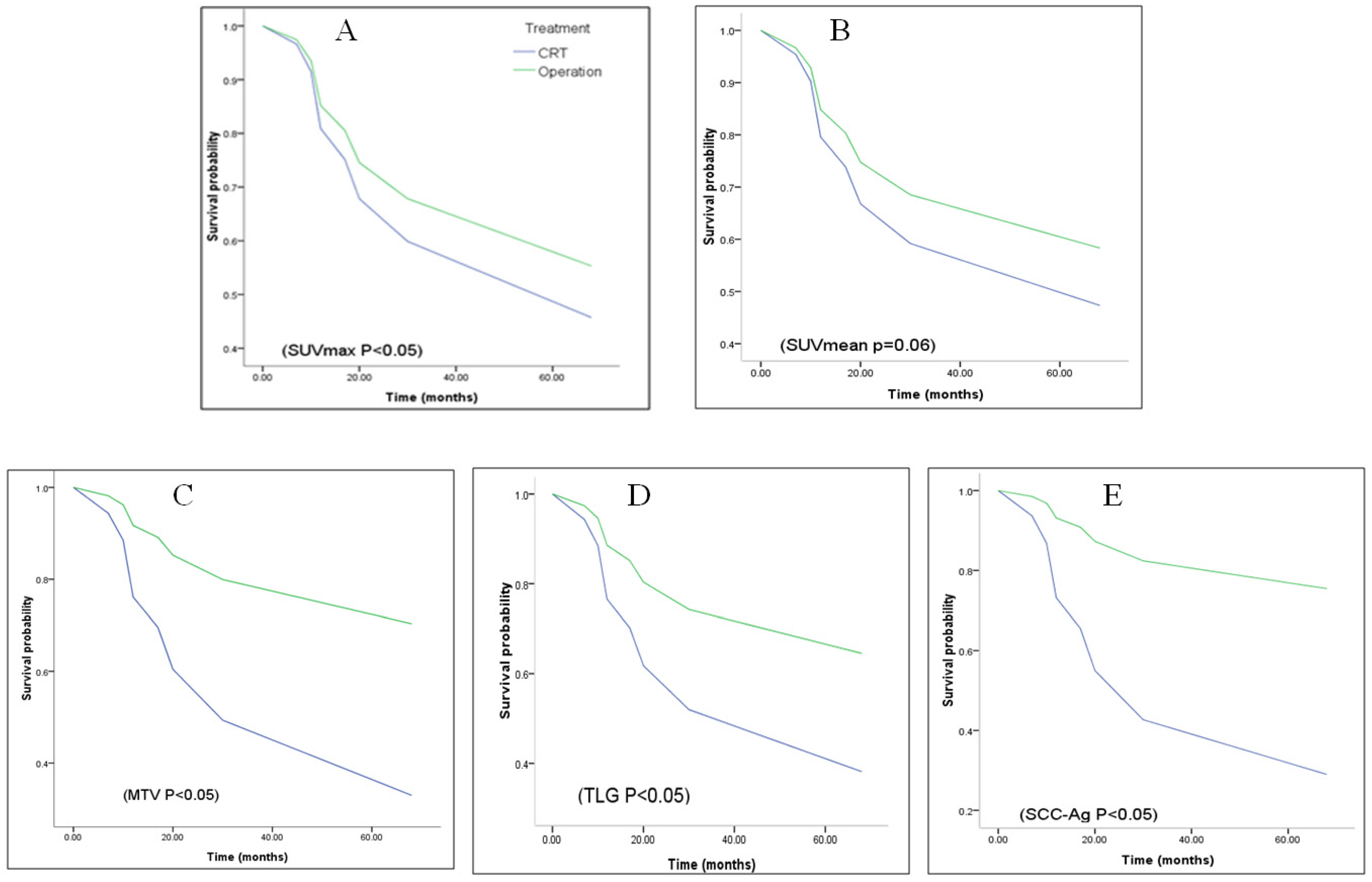
2.4. Relationships between Serum SCC-Ag Level and 18F-FDG PET/CT-Derived Metabolic Parameters with Overall Survival (OS) in ESCC Staging II Patients

The association of various metabolic parameters and serum SCC-Ag with patient overall survival was evaluated by univariate and multivariate Cox regression analyses. As shown in Table 5, SUVmax, MTV, TLG, and serum SCC-Ag, but not SUVmean, showed a significant association with OS in our studied node-negative stage II ESCC patient. Additional univariate or multivariate analyses did not show any significant association of age, smoking and treatment with OS (data not shown). Kaplan–Meier survival analysis revealed survival probability of CRT (solid line) less than operation with several parameters that showed in Figure 2.

3. Discussion

The current TNM staging system for esophageal cancer is based on only anatomic finding of tumor, without tumor metabolic information. The 18F-FDG PET/CT is a powerful noninvasive modality that can provide not only anatomic, but also metabolic information of tumor. SUVmax is the most common 18F-FDG PET/CT metabolic parameter routinely used in the clinic. Recently, there are increasing evidence to support usage of different 18F-FDG PET/CT-derived metabolic parameters in assessing pretreatment extent of disease for various cancers [14,16,17]. Different from SUVmax measuring on a single voxel and may not reflect the whole tumor metabolism, volume-based metabolic parameters such as MTV and TLG are volumetric measurements incorporated with metabolic activity throughout the entire tumor. Lately, MTV and TLG have been shown of prognostic value to predict clinical outcomes of cancer patients [11,12,18,19]. Serum SCC-Ag is a tumor marker and elevated serum SCC levels are known to be associated with advanced tumor stage, poor treatment response, and increased risk of tumor recurrence [20]. As shown in the current study, positive correlations were found among various 18F-FDG PET/CT-derived metabolic parameters and serum SCC-Ag for node-negative stage II ESCC patients. Among them, the SUVmax and SUVmean showed moderate positive correlation with MTV, and near strong positive correlation with TLG and serum SCC-Ag.
The current study investigated prognostic value of 18F-FDG PET/CT-derived metabolic parameters and serum SCC-Ag in early-stage ESCC patients. A total of 18 node-negative stage II ESCC patients with sufficient follow-up of 5-years was sub-divided into the demised (poor prognosis) and the stable (good prognosis) group. Our study showed patients in the demised group have statistically significant higher values than the stable group in MTV and TLG, but not in SUVmax and SUVmean. Previously, Mantziari et al. reported higher values of baseline metabolic parameters of 18F-FDG PET/CT were significantly related to the tumor location and presence of advanced T3 and T4 stages in esophageal cancer [14]. In their study, SUVmax > 8.25 g/mL, TLG > 41.7, and MTV > 10.7 cm3 were noted in their cT/4 stage cases. They also defined SUVmax ≥ 12.7 g/mL that can be predicted early recurrence and poor disease-free survival. In a study by Wang et al., pretreatment SUVmean was shown to be a better independent predictor of treatment response than SUVmax, MTV, and TLG in patients with locally advanced ESCC treated with concurrent chemoradiotherapy [21]. The subtle discrepancies between our findings and others can be due to differences in studied patient populations and variations in treatment modalities. Solid nodule type, poor histological grade, and larger nodule size have been reported to be associated with higher values of SUV, MTV, and TLG in stage I lung adenocarcinoma [22]. Taken together, the higher MTV and TLG values in the demised group patients of our study could be related to the poor pathological grades or other features of their tumors. Further studies are needed to establish correlations between pathological grades and 18F-FDG PET/CT-derived volume-based metabolic parameters.
Our study showed serum SCC-Ag level significant increased (4.29 ± 4.24 vs. 1.17 ± 0.52; p < 0.05) in the demised group patients, as compared to the stable group patients. It also correlated to 18F-FDG PET/CT-derived parameters of the main tumor in ESCC stage II patients. In Cox models, it was significantly associated with OS. However, the level of SCC-Ag sometimes is also observed at an elevated level in lung, cervix, head and neck cancer or benign disease, including skin disease, pelvic inflammatory disease, cystitis and renal failure [23,24,25]. Therefore, it is a limitation for serum SCC-Ag level screening in clinical routine, though it is a convenient test for pre-treatment or post-treatment surveillance of patients.
Another aim of this study was to investigate the association of various 18F-FDG PET/CT-derived tumor metabolic parameters and serum SCC-Ag with the overall survival in node-negative stage II ESCC patients. Based on the univariate and multivariate Cox regression analyses, serum SCC-Ag, SUVmax, MTV, and TLG were identified to be independent predictors of OS. SUVmean was the only exception that did not shown statistical significant association with OS by univariate (p = 0.057) and multivariate analyses (p = 0.060). In this regard, Tamandl et al. reported no correlation between various metabolic parameters and OS in 38 patients with unresectable or metastatic ESCC who had 18F-FDG PET/CT prior to palliative treatment [26]. Albeit, in the same study, MTV was shown to be predictive for OS in 33 patients with unresectable or metastatic esophageal adenocarcinoma (EAC) [26]. The discrepancy of MTV being predictive factor of OS between node-negative early-stage ESCC and advanced-stage ESCC patients is intriguing and demands additional investigations. Nevertheless, our study also identified SCC-Ag, SUVmax, and TLG being independent predictors of OS in early-stage ESCC patients. Another issue of partial volume effect (PVE) for SUVmax measurements was reported by Hatt and et al. [27]. They mentioned PVE correction did not add any value in volume-based metabolic parameters of 18F-FDG PET for prognosis of esophageal cancer, although PVE correction may increase the levels of SUVmax and SUVmean.
The difference of our study with other studies mainly included (1) to evaluate and compare the SCC-Ag level and 18F-FDG PET/CT-derived tumor metabolic parameters of pre-treatment tumor burden of demised and stable patients in early-stage ESCC. There were no factors of nodal metastasis, distant metastasis or tumor recurrence in our study; (2) to evaluate the prognostic value of SCC-Ag level and 18F-FDG PET/CT-derived tumor metabolic parameters in early-stage ESCC patients. To our knowledge, this is the first study discussing the relationship and prognostic value between serum SCC-Ag with 18F-FDG PET/CT-derived tumor metabolic parameters of tumor burden in early-stage ESCC patients. We also concluded the parameters and prognostic value of tumor burden in different clinical status. A systematic review and meta-analysis article was reported by Han and et al. [28]. In this article, they employed different staging and patient-specific treatment from several published articles for discussion. They presented that higher MTV and TLG values on pre-treatment 18F-FDG PET/CT of esophageal patients were at higher risk of adverse events or death. This finding is similar to ours. Another similar result of volume-based metabolic parameters was found as having significant prognostic factors in OS with esophageal cancer and our ESCC stage II patients. In our study, we also found serum SCC-Ag and SUVmax of pre-treatment 18F-FDG PET/CT that could be prognostic biomarkers in ESCC stage II patients. However, SUVmean was not a significant prognostic biomarker in our study.
There are limitations for the current study. Firstly, this is a retrospective study of only node-negative stage II ESCC patients that is limited by small sample size and patient selection bias. For example, all our patients are of male gender. Small sample size may cause poor statistical power and increase the error of study. Patient selection bias may affect the external validity of the study. The results are from a selected group and may not be generalized to all patients, not even to all males. Secondly, patients with tumor recurrence or nodal metastasis were excluded from the study. Therefore, our results cannot be applied for these patient groups. Thirdly, our patients received different treatment modalities, different regimens of chemotherapy and likely varying doses of radiotherapy, which could have affected the outcome.

4. Materials and Methods

4.1. Patients

From 1 January 2015 to 31 December 2016, there were 117 patients with biopsy proved ESCC underwent pre-treatment 18F-FDG PET/CT examination in our institution. After chart review, 18 patients with node-negative stage II ESSC, including 7 patients with T2N0 (38.9%) and 11 patients with T3N0 (61.1%) were identified. Patients with history of previous cancer, prior esophageal surgery, evidence of lymph node metastasis by 18F-FDG PET/CT or other examination and histological type other than SCC were excluded from the study. Based on clinical information with 5 years of follow-up, patients were sub-divided into the demised and the stable prognostic groups. The treatment of patients was decided by a multidisciplinary team, consisting with surgeons, medical oncologists and radiation oncologists. Serum samples were obtained from peripheral blood of patients, and then centrifuged for 10 min at 1300× g. The serum SCC-Ag levels were measured and recorded by Chemiluminescent Microparticle Immunoassy (CMIA) (ABBOTT GMBH & CO.KG, Wiesbaden, Germany). Data analysis was conducted from the patent product of Abbott Architect SCC (Tokyo, Japan). The study design was approved by the Institutional Review Board in our hospital (KSVGH 21-CT14-07). The characteristics of the enrolled patients are given in Table 1.

4.2. 18F-FDG PET/CT Imaging

All patients were suggested to fast for at least 6 h before 18F-FDG PET/CT imaging. An intravenous catheter was placed before the radiopharmaceutical agent injection, and patients’ blood glucose levels were measured before the tracer injection for ensuring good PET/CT imaging quality (adequate blood glucose level <150 mg/dL). Each patient was administrated with 370–555 MBq of 18F-FDG according to the body weight (7.03 MBq/kg). After injection of the 18F-FDG tracer, patients then underwent whole-body 18F-FDG PET/CT (Discovery ST-16; GE Healthcare, Milwaukee, WI, USA) from the head to the upper thigh in a supine position. A delayed image might be obtained while necessary. CT scanning was performed prior to acquisition of the PET imaging by using the subsequent parameters: 0.6 s per rotation, 120 kV, 100 mA, and 3.75 mm thick slices. An ordered subset expectation maximization iterative reconstructed algorithm was used for attenuation-corrected PET images reconstruction. The fusion imaging of PET and CT were obtained on a Xeleris image display and processing platform (GE Healthcare, Milwaukee, WI, USA).

4.3. Imaging Analysis

The PET, CT, and fused PET/CT images of each patient were independently reviewed and interpreted by three experienced nuclear medicine physicians. The FDG-avid esophageal tumor detected by PET was fused to the corresponding lesion and anatomical location on CT scan. Standardized uptake values (SUVs) were used as the metric for metabolic tumor quantification with 18F-FDG PET. The SUV was measured semi-automatically by SUV tools obtained in the Xeleris software (Version 4.0) as follows: SUV = activity in the region of interest (Bq/g)/(injected dose(Bq)/body weight (g)). The SUVmax is the highest SUV detected for the tumor. The metabolic tumor volume (MTV; cm3) is defined as total tumor volume with an SUV of ≥2.5. Total lesion glycolysis (TLG; g/mL cm3) is calculated by multiplying MTV by SUVmean of the delineated tumor. The PET VCAR (volume computer assisted reading; GE Healthcare) was used for imaging analysis. After drawing a cuboid volume of interest (VOI) covering the tumor, the software then automatically drew the FDG uptake of tumor margin according to the specific SUV threshold. MTV and TLG then were automatically computed and measured by the PET VCAR application (Advanced workstation 4.4, GE Medical System, Milwaukee, WI, USA).

4.4. Statistical Analysis

The results are shown as mean ± standard deviation. The Spearman correlation coefficients were calculated to assess the relationship between different tumor metabolic parameters derived from 18F-FDG PET/CT and serum SCC-Ag in our patients. Statistical significance of different 18F-FDG PET/CT metabolic parameters and serum SCC-Ag level between the demised and stable prognostic groups of patients was assessed by using independent t-test and Mann–Whitney U test. The univariable or multivariable Cox regression model with Breslow approximation was used to determine the hazards ratio of parameters. A p-Value of <0.05 was considered statistically significant. All statistical analysis was performed using the SPSS software package (version 17.0, Chicago, IL, USA).

5. Conclusions

In comparison to the stable prognostic group, patients in the demised prognostic group showed significant higher values in serum SCC antigen (SCC-Ag) level, as well as in the volume-based metabolic parameters MTV and TLG (p < 0.05), but not SUVmax (p = 0.10) and SUVmean (p = 0.11). By univariate and multivariate Cox regression analyses, values of SUVmax, MTV, TLG, and serum SCC-Ag, but not SUVmean, showed significant association with overall survival of studied patients. Our findings suggest potential usage of pretreatment volume-based metabolic parameters of 18F-FDG PET/CT and serum SCC-Ag as prognostic factors for node-negative stage II ESCC patients.

Author Contributions

D.H.-Y.S. and H.-P.C.: manuscript drafting; F.-R.T., C.H., and H.-Y.C.: data collection and analysis; A.Y.-N.C., C.-H.L., and K.-P.C.: drafted the work and revised it critically for important intellectual content; M.-H.Y. and Y.-C.T. prepared the manuscript and provided editorial assistance before submission. All authors have read and agreed to the published version of the manuscript.

Funding

This work was supported by research grants: MOST 109-2221-E-037-001-MY3 from the Ministry of Science and Technology, NSYSUKMU109-P012 from NSYSU-KMU Research Project, and the Research Center for Environmental Medicine, Kaohsiung Medical University, Kaohsiung, Taiwan, from The Featured Areas Research Center Program within the framework of the Higher Education Sprout Project by the Ministry of Education (MOE) in Taiwan.

Institutional Review Board Statement

This study was performed in accordance with the Declaration of Helsinki and local laws. The study design was approved by the Institutional Review Board in our hospital (KSVGH 21-CT14-07).

Informed Consent Statement

The Institutional Review Board waived the need to obtain informed consent, due to the retrospective nature of this study.

Data Availability Statement

The data presented in this study is available on request from the corresponding author. The data is not publicly available due to its proprietary nature.

Acknowledgments

The authors thank S. Sheldon MT (ASCP, Retired) of Oklahoma University Medical Center Edmond for fruitful discussions and editorial assistance.

Conflicts of Interest

The authors declare no conflict of interest. All persons who made significant contributions to this study and the manuscript are included in the author list. The first draft of the manuscript was written by Hung-Pin Chan, Ming-Hui Yang, and Yu-Chang Tyan, and no other honoraria, grants, or other forms of payment were given to anyone to produce the manuscript.

References

  1. Uhlenhopp, D.J.; Then, E.O.; Sunkara, T.; Gaduputi, V. Epidemiology of esophageal cancer: Update in global trends, etiology and risk factors. Clin. J. Gas. 2020, 13, 1010–1021. [Google Scholar] [CrossRef]
  2. Lu, H.H.; Chiu, N.C.; Tsai, M.H. Prognostic significance of pretreatment staging with 18F-FDG PET in esophageal cancer: A nationwide population-based study. Clin. Nucl. Med. 2021, 46, 647–653. [Google Scholar] [CrossRef] [PubMed]
  3. Gholipour, C.; Shalchi, R.A.; Abbasi, M. A histopathological study of esophageal cancer on the western side of the Caspian littoral from 1994 to 2003. Dis. Esophagus 2008, 21, 322–327. [Google Scholar] [CrossRef]
  4. Huang, F.L.; Yu, S.J. Esophageal cancer: Risk factors, genetic association, and treatment. Asian J. Surg. 2018, 41, 210–215. [Google Scholar] [CrossRef] [PubMed]
  5. Watanabe, M.; Otake1, R.; Kozuki1, R.; Toihata1, T.; Takahashi1, K.; Okamura1, A.; Imamura1, Y. Recent progress in multidisciplinary treatment for patients with esophageal cancer. Surg. Today 2020, 50, 12–20. [Google Scholar] [CrossRef] [Green Version]
  6. Wilson, M.; Rosato, E.L.; Chojnacki, K.A.; Chervoneva, I.; Kairys, J.C.; Cohn, H.E.; Rosato, F.E.; Berger, A.C. Prognostic significance of lymph node metastases and ratio in esophageal cancer. J. Surg. Res. 2008, 146, 11–15. [Google Scholar] [CrossRef] [Green Version]
  7. Altini, C.; Niccoli Asabella, A.; Lavelli, V.; Bianco, G.; Ungaro, A.; Pisani, A.; Merenda, N.; Ferrari, C.; Rubini, G. Role of 18F-FDG PET/CT in comparison with CECT for whole-body assessment of patients with esophageal cancer. Recenti. Prog. Med. 2019, 110, 144–150. [Google Scholar]
  8. Swisher, S.G.; Erasmus, J.; Maish, M.; Correa, A.M.; Macapinlac, H.; Ajani, J.A.; Cox, J.D.; Komaki, R.R.; Hong, D.; Lee, H.K.; et al. 2-Fluoro-2-deoxy-D-glucose positron emission tomography imaging is predictive of pathologic response and survival after preoperative chemoradiation in patients with esophageal carcinoma. Cancer 2004, 101, 1776–1785. [Google Scholar] [CrossRef] [PubMed]
  9. Flamen, P.; Van Cutsem, E.; Lerut, A.; Cambier, J.P.; Haustermans, K.; Bormans, G.; De Leyn, P.; Van Raemdonck, D.; De Wever, W.; Ectors, N.; et al. Positron-emission tomography for assessment of the response to induction radiochemotherapy in locally advanced oesophageal cancer. Ann. Oncol. 2002, 13, 361–368. [Google Scholar] [CrossRef]
  10. Wan, L.; Gao, Y.; Gu, J.; Chi, H.; Wang, Z.; Hu, Q.; Jia, J.; Liu, T.; Li, B.; Teng, J.; et al. Total metabolic lesion volume of lymph nodes measured by 18F-FDG PET/CT: A new predictor of macrophage activation syndrome in adult-onset Still’s disease. Arthritis Res. Ther. 2021, 23, 97. [Google Scholar] [CrossRef]
  11. Tosi, D.; Pieropan, S.; Cattoni, M.; Bonitta, G.; Franzi, S.; Mendogni, P.; Imperatori, A.; Rotolo, N.; Castellani, M.; Cuzzocrea, M.; et al. Prognostic value of 18F-FDG PET/CT metabolic parameters in surgically treated stage I lung adenocarcinoma patients. Clin. Nucl. Med. 2021, 46, 621–626. [Google Scholar] [CrossRef]
  12. Polverari, G.; Ceci, F.; Bertaglia, V.; Reale, M.L.; Rampado, O.; Gallio, E.; Passera, R.; Liberini, V.; Scapoli, P.; Arena, V.; et al. (18)F-FDG PET parameters and radiomics features analysis in advanced NSCLC treated with immunotherapy as predictors of therapy response and survival. Cancers 2020, 12, 1163. [Google Scholar] [CrossRef]
  13. Creff, G.; Devillers, A.; Depeursinge, A.; Palard-Novello, X.; Acosta, O.; Jegoux, F.; Castelli, J. Evaluation of the prognostic value of FDG PET/CT parameters for patients with surgically treated head and neck cancer: A systematic review. JAMA Otolaryngol. Head Neck Surg. 2020, 146, 471–479. [Google Scholar] [CrossRef] [PubMed]
  14. Mantziari, S.; Pomoni, A.; Prior, J.O.; Winiker, M.; Allemann, P.; Demartines, N.; Schäfer, M. 18F- FDG PET/CT-derived parameters predict clinical stage and prognosis of esophageal cancer. BMC Med. Imaging 2020, 20, 7. [Google Scholar] [CrossRef] [PubMed] [Green Version]
  15. Fu, J.; Wang, W.; Wang, Y.; Liu, C.; Wang, P. The role of squamous cell carcinoma antigen (SCC Ag) in outcome prediction after concurrent chemoradiotherapy and treatment decisions for patients with cervical cancer. Radiat Oncol. 2019, 14, 146. [Google Scholar] [CrossRef] [PubMed] [Green Version]
  16. Cerfolio, R.J.; Bryant, A.S. Maximum standardized uptake values on positron emission tomography of esophageal cancer predicts stage, tumor biology, and survival. Ann. Thorac Surg. 2006, 82, 391–394. [Google Scholar] [CrossRef]
  17. Lee, S.; Choi, Y.; Park, G.; Jo, S.; Lee, S.S.; Park, J.; Shim, H.Y. 18F-FDG PET/CT Parameters for predicting prognosis in esophageal cancer patients treated with concurrent chemoradiotherapy. Technol. Cancer Res. Treat. 2021, 20, 1–7. [Google Scholar] [CrossRef]
  18. Eude, F.; Toledano, M.N.; Vera, P.; Tilly, H.; Mihailescu, S.; Becker, S. Reproducibility of baseline tumour metabolic volume measurements in diffuse large B-cell lymphoma: Is there a superior method? Metabolites 2021, 11, 72. [Google Scholar] [CrossRef] [PubMed]
  19. Kandeel, A.; Saied, M.; Aldaly, M.; Darwish, H.; Alsayed, Y. Impact of 18F-2-fluoro-2-deoxy-D-glucose PET/computerized tomography on the initial staging and changing the management intent in head and neck squamous cell carcinoma. Nucl. Med. Commun. 2021, 42, 216–224. [Google Scholar] [CrossRef]
  20. Okamura, A.; Matsuda, S.; Mayanagi, S.; Kanamori, J.; Imamura, Y.; Irino, T.; Kawakubo, H.; Mine, S.; Takeuchi, H.; Kitagawa, Y.; et al. Clinical significance of pretherapeutic serum squamous cell carcinoma antigen level in patients with neoadjuvant chemotherapy for esophageal squamous cell carcinoma. Ann. Surg Oncol. 2021, 28, 1209–1216. [Google Scholar] [CrossRef] [PubMed]
  21. Wang, C.; Zhao, K.; Hu, S.; Huang, Y.; Ma, L.; Song, Y.; Li, M. A predictive model for treatment response in patients with locally advanced esophageal squamous cell carcinoma after concurrent chemoradiotherapy: Based on SUVmean and NLR. BMC Cancer 2020, 20, 544. [Google Scholar] [CrossRef] [PubMed]
  22. Wang, X.Y.; Zhao, Y.F.; Liu, Y.; Yang, Y.K.; Wu, N. Prognostic value of metabolic variables of [18F]FDG PET/CT in surgically resected stage I lung adenocarcinoma. Medicine 2017, 96, e7941. [Google Scholar] [CrossRef]
  23. Duk, J.M.; van Voorst Vader, P.C.; ten Hoor, K.A.; Hollema, H.; Doeglas, H.M.; de Bruijn, H.W. Elevated levels of squamous cell carcinoma antigen in patients with a benign disease of the skin. Cancer 1989, 64, 1652–1656. [Google Scholar] [CrossRef]
  24. Jao, M.S.; Chang, T.C.; Chang, H.P.; Wu, T.I.; Chao, A.; Lai, C.H. Long-term follow up of cervical cancer patients with unexplained squamous cell carcinoma antigen elevation after post-therapy surveillance using positron emission tomography. J. Obstet. Gynaecol. Res. 2010, 36, 1003–1008. [Google Scholar] [CrossRef] [PubMed]
  25. Cases, A.; Filella, X.; Molina, R.; Ballesta, A.M.; Lopez-Pedret, J.; Revert, L. Tumor markers in chronic renal failure and hemodialysis patients. Nephron 1991, 57, 183–186. [Google Scholar] [CrossRef] [PubMed]
  26. Tamandl, D.; Ta, J.; Schmid, R.; Preusser, M.; Paireder, M.; Schoppmann, S.F.; Haug, A.; Ba-Ssalamah, A. Prognostic value of volumetric PET parameters in unresectable and metastatic esophageal cancer. Eur. J. Radiol. 2016, 85, 540–545. [Google Scholar] [CrossRef] [PubMed]
  27. Hatt, M.; le Pogam, A.; Visvikis, D.; Pradier, O.; le Rest, C.C. Impact of partial-volume effect correction on the predictive and prognostic value of baseline 18F-FDG PET images in esophageal cancer. J. Nucl. Med. 2012, 53, 12–20. [Google Scholar] [CrossRef] [Green Version]
  28. Han, S.; Kim, Y.J.; Woo, S.; Suh, C.H.; Lee, J.J. Prognostic value of volumetric parameters of pretreatment 18F-FDG PET/CT in esophageal cancer: A systematic review and meta-analysis. Clin. Nucl. Med. 2018, 43, 887–894. [Google Scholar] [CrossRef]
Figure 1. A 78 y/o male patient was diagnosed cT2N0M0, stage II on pre-treatment 18F-FDG PET/CT. The small tumor mass with FDG hot uptake was noted on 18F-FDG PET/CT (yellow color cross indicator). Metabolic parameters showed SUVmax: 7.61, SUVmean: 4.65, MTV: 11.07 cm3, and TLG: 51.41g/mL cm3 by the PET VCAR. He underwent CRT treatment and expired 3.9 months later due to disease progression.
Metabolites 12 00007 g001
Figure 2. Kaplan–Meier curves of different tumor metabolic parameters for overall survival of patients. (A) SUVmax, (B) SUVmean, (C) MTV, (D) TLG, and (E) SCC-Ag.
Metabolites 12 00007 g002
Table 1. Characteristics of the enrolled patients in this study.
Patient CharacteristicsNumber (%) or Mean (SD)
Demographic characteristics
Age (year)62 (49–83)
Male18 (100%)
Smoking14 (77.8%)
Alcohol consumption12 (69.4%)
Clinical characteristics
T stage
T27 (38.9%)
T311 (61.1%)
Treatment
CRT only11 (61.1%)
Operation only6 (33.3%)
CRT + operation2 (11.1%)
Table 2. Spearman’s rank correlation coefficients between different tumor metabolic parameters and SCC-Ag.
Parameterρ/p ValueSUVmaxSUVmeanMTVTLG
SUVmaxρ/0.9620.4830.682
p Value/0.0000.0420.002
SUVmean /0.5270.733
/0.0250.001
MTV /0.922
/0
TLG /
/
SCC-Ag 0.6460.6070.7370.843
0.0040.0080.0000.000
SUVmax = maximum standardized uptake value, SUVmean = mean standardized uptake value, MTV = metabolic tumor volume, TLG = total lesion glycolysis, SCC-Ag = squamous cell carcinoma antigen.
Table 4. Comparison of the tumor metabolic parameters of demised versus stable patients by Mann–Whitney U test analysis.
ParameterMean rank
(Demised, n = 8)
Mean rank
(Stable, n = 10)
Z Valuep Value
SUVmax127.5−1.780.083
SUVmean11.637.8−1.510.146
MTV13.136.6−2.570.009 **
TLG13.386.4−2.750.006 **
SCC-Ag12.257.3−1.960.055
** p < 0.01.
Table 5. Univariate and multivariate Cox regression analysis for overall survival.
VariableHazard Ratio (95% CI)p Value
Univariate analysis
SUVmax1.112 (1.019–1.213)0.017
SUVmean1.173 (0.995–1.383)0.057
MTV1.035 (1.004–1.067)0.029
TLG1.002 (1.000–1.003)0.043
SCC-Ag1.127 (1.030–1.437)0.021
Multivariate analysis
SUVmax0.126 (1.023–1.248)0.016
SUVmean1.211 (0.992–1.478)0.06
MTV1.053 (1.007–1.101)0.024
TLG1.002 (1.000–1.005)0.043
SCC-Ag1.368 (1.040–1.2799)0.025
Publisher’s Note: MDPI stays neutral with regard to jurisdictional claims in published maps and institutional affiliations.

