Article

1,12-Bis-Triphenyl Phosphonium Dodecane Bromide Nanovesicles as Potential Inhibitors of MDR Staphylococcal Biofilms


1
Department of Pharmacy, University of Genoa, Viale Cembrano, 16148 Genoa, Italy
2
Hospital Hygiene Unit, E.O. Galliera Hospital, Mura delle Cappuccine 14, 16128 Genoa, Italy
3
Department of Health Sciences, University of Genoa, Via Pastore 1, 16132 Genova, Italy
4
Laboratory of Experimental Therapies in Oncology, IRCCS Istituto Giannina Gaslini, Via G. Gaslini 5, 16147 Genoa, Italy
5
Department of Surgical Sciences and Integrated Diagnostics (DISC), University of Genoa, Viale Benedetto XV, 6, 16132 Genova, Italy
*
Author to whom correspondence should be addressed.
Sci. Pharm. 2026, 94(3), 80; https://doi.org/10.3390/scipharm94030080 (registering DOI)
Submission received: 17 August 2026 / Revised: 10 September 2026 / Accepted: 13 September 2026 / Published: 19 September 2026

Abstract

Multidrug resistance (MDR) has become a major global health threat, leading to the emergence of difficult-to-treat bacterial “superbugs” among both Gram-positive and Gram-negative species. In hospital settings, biofilm (BF)-producing staphylococci further aggravate this problem by markedly increasing tolerance to conventional antibiotics, thereby promoting chronic and potentially life-threatening infections. In the present study, previously synthesized and characterized 1,12-bis-triphenyl phosphonium dodecane bromide nanovesicles (BPPB, 45 nm, water) were assayed by DLS in a medium (TSB), which was used for microbiologic tests, to assess the actual particle size (≈65 nm) existing in this biological setting, with PDI (0.37) and zeta potential (+6.1 mV). Although never investigated for its effects on the formation of staphylococcal BF, BPPB was, for the first time, evaluated as a potential novel agent to combat its development. A total of 12 highly BF-producing isolates from our collection, comprising six Staphylococcus aureus and six S. epidermidis strains, were selected because they are strong BF producers. Their full antibiogram was determined, and they were tested against BPPB to determine minimum inhibitory concentrations (MICs). Subsequently, BF inhibition activity was evaluated at ½ MIC, MIC, and 2× MIC concentrations. Vancomycin (V), used as a reference antibiotic, was tested under the same experimental conditions. BPPB exhibited MIC values ranging from 0.125 to 0.250 µg/mL, which were 1–8-fold lower than those of V. V did not inhibit BF formation by S. epidermidis at all and inhibited BF formation by Bam and Aam S. aureus isolates (96–97% inhibition) only at max concentrations (2 × MIC). Conversely, BPPB demonstrated potent and consistent inhibition activity against all strains, irrespective of species or resistance profile, as determined by VITEK. BF inhibition values of 83–99%, 95–>99%, and 98–>99% were observed at ½ MIC, MIC, and 2 × MIC, respectively. To confirm that BF inhibition did not arise from killing bacteria, determinations of bacterial colony count after BPPB treatment at 4 × MIC for 24 h were performed, establishing full vitality and a regrowth of 45% with respect to the inoculum. Once reseeded as in the control, treated bacteria grew exactly as the control. Overall, the findings confirmed the nanosized dimension of BPPB particles, which remained <100 nm, including in the complex biological medium; this highlighted the strong BF-forming capability and MDR phenotype of the selected staphylococcal isolates, as well as the remarkable antibacterial and BF inhibition efficacy of BPPB nanovesicles, significantly outperforming vancomycin. Importantly, the low cytotoxicity previously observed against eukaryotic Cos-7 and HepG2 cells, resulting in high selectivity index (SI) values (23.0–90.5), supports BPPB as a promising candidate for the development of new NM-based therapeutic strategies against MDR staphylococcal BF-associated infections.

1. Introduction

Antibiotics are frequently overprescribed, misused, or inappropriately used in both humans and animals. Incomplete treatments, poor adherence to antibiotic regimens, and unnecessary prophylactic use strongly contribute to the emergence of multidrug-resistant (MDR) microorganisms (superbugs) [1]. Superbugs continuously emerge, especially in hospital settings, showing resistance to an increasing number of antibiotics and, in some cases, even to all currently available treatments, thus representing a major therapeutic challenge.
MDR superbugs are responsible for difficult-to-treat infections, leading to prolonged hospitalization and decreased survival rates, thus increasing the global economic burden [1]. Improved hygiene and sanitation measures may help mitigate this critical scenario, but new antibacterial therapeutic options remain urgently needed [2]. Wild-type Staphylococcus aureus is a worrying pathogen for both humans and the environment, causing a wide variety of severe clinical infections, including septicaemia, endocarditis, necrotizing pneumonia, osteomyelitis, and BF-associated infections, thus representing one of the major community- and hospital-acquired pathogens worldwide [3,4]. The emergence of methicillin-resistant strains (MRSA) has further increased the clinical challenge associated with these infections [3]. Moreover, BF formation represents an additional and highly intractable virulence and resistance mechanism not only in S. aureus but also in other opportunistic staphylococci such as S. epidermidis.
BFs are structured microbial communities formed when bacteria adhere to biotic or abiotic surfaces. Microorganisms become embedded within a complex extracellular matrix composed of exopolysaccharides (EPSs), extracellular DNA/RNA, proteins, lipids, and other biomolecules [5,6,7]. Overall, BF formation represents an important survival strategy that protects microorganisms from harsh environmental conditions and significantly reduces susceptibility to antibiotic treatments [6]. Following surface adhesion and BF maturation, bacterial transition from the planktonic to the sessile state occurs, forming highly organized communities coordinated by quorum sensing (QS) systems [6]. BF-associated bacteria are markedly more tolerant to immune defences and antibiotics because the extracellular matrix limits drug penetration [8,9,10]. Bacterial BFs represent a major challenge in the management of persistent and recurrent infections [11,12]. The increasing emergence of MDR bacterial strains that also produce BFs further underscores the necessity to discover alternative or additive strategies to traditional antibiotics [13].
The necessary identification of novel compounds with BF inhibition activity, representing a highly relevant research field, includes: (i) the development of novel compounds, such as heterocyclic molecules, quaternary ammonium or phosphonium slats, NMs, and composite compounds encompassing both inorganic metals and organic fractions, which have shown promising BF inhibition properties [5,7,14,15,16]; (ii) the evaluation of their BF inhibition efficacies through standardized and reproducible methods, including crystal violet biomass assays, viability measurements, and microscopic analyses [17,18]; and (iii) the investigation of their mechanisms of action, which often requires sophisticated instrumentation, specialized laboratories, and advanced expertise [18].
In recent years, the papers present in the literature have comprised several studies reporting the synthesis and characterization of novel molecules that have displayed considerable activity against the most clinically relevant pathogens, such as the above-mentioned S. aureus, but also worrying Gram-negative superbugs, such as Pseudomonas aeruginosa, and Escherichia coli [13,15]. Comparative evaluation of these compounds and their experimental protocols is crucial for identifying candidates appropriate for clinical use or suitable for use as antimicrobial coatings for medical devices [19].

1.1. Biofilm (BF) Inhibition: Where Are We?

The extracellular polymeric substance (EPS) matrix and the altered physiology of sessile bacteria markedly reduce antibiotic susceptibility [12]. In this regard, BF formation by widespread bacteria, independent of their antibiotic resistance profiles [20,21], provides protection against environmental stress, host immune responses, and antimicrobial agents while promoting QS and metabolic adaptation [20]. Although some conventional antibiotics, such as rifampicin, may display antibiofilm activity in selected settings, their efficacy is often limited by resistance development [22]. MDR strains that are also strong BF producers represent a particularly severe clinical threat [20,21,23,24,25].
In recent years, researchers committed to studying BF inhibition have focused on unveiling the tools that provide BF-producing bacteria with tolerance and persistence, on the development of new agents capable of inhibiting BF formation and/or destroying established BF structures (mature BFs) and on exploring the mechanistic paths followed by active molecules (old or new) to prevent BF formation or disaggregate already formed ones.
In this context, NMs and hybrid systems (HSs) have increasingly attracted attention as antibiofilm platforms. Nanoconjugates encompassing silver and bacteriocin [16], nanocomposites prepared using “green” biosynthesis based on copper [14], and nanoparticles (NPs) loaded with cellulose or embedded with borax [18] were nanotechnologically prepared, which demonstrated significant effects on contrasting BF formation by common pathogens and improved antibiofilm efficacy [5,18].
Natural molecules, complex extracts and lactic fermentation of plant matrices, as sources of new agents capable of addressing BF formation, have been reported in numerous papers [19,26,27]. These reports underscore that the association between bioprocesses and synthetic chemistry can identify libraries of compounds with enhanced possibilities to treat BF efficiently.
Nowadays, assessments of the possible inhibition effects of new molecules rely on consolidated assays. These include BF biomass quantification by using staining dyes, such as crystal violet (CV), measurement of bacterial cell viability within BFs, determination of MIBCs and MBECs, which are the minimum BF inhibition and eradication concentrations, respectively, and SEM analysis of three-dimensional BF structures [11,17]. Uniform protocols are essential to unfailingly compare the efficacy of new antibiofilm compounds [12]. Collectively, a broader vision of possible strategies to prevent and/or eradicate BFs is of paramount importance for a constructive global attempt to counteract BF formation in the medical field [28].
Currently available antibiotics, including cefazolin, no longer fulfil their intended function, failing to fully remove BFs and effectively treat the phenotypic behaviour of dormant and persister BF bacteria. In this context, the investigation of new molecules by synthetic procedures represents one of the most promising tactics to overcome the limitations of conventional therapies against infections caused by the instauration of BFs [12,13,15].
Heterocyclic derivatives, quaternary ammonium and phosphonium salts (QASs and QPSs), NMs, and HSs are among the most researched structures to this end [29].
New bis-cationic compounds (b-QASs) derived from natural products, camphor derivatives containing sulphur atoms, and libraries of compounds based on 1,3-oxazole and isosteric analogues were developed [13,15,17]. Apostol et al. carried out in vitro assays and in silico studies and combined their results [17]. Recently, a biocide TPP+-containing QPS was reported to inhibit BF formation by MDR and ATCC S. aureus and S. epidermidis, irrespective of their antimicrobial resistance profiles, at concentrations significantly lower than those required for biocidal activity [7].

This Work

To meet this urgent need for novel agents capable of inhibiting BF formation, the present study evaluated the inhibition potency of the synthetic bis-quaternary triphenyl phosphonium compound (b-TPP-QPS, BPPB), which self-forms nanovesicles (45 nm) in water, using standardized experimental protocols. b-TPP-QPS has recently attracted attention because of its potent antibacterial activity (MICs 0.025–500 µg/mL) against both Gram-positive and Gram-negative clinical isolates and low haemolytic and cytotoxic effects toward erythrocyte cells (RBCs) and different types of eukaryotic cells (HepG2 and Cos-7 cells). Encouraged by these results, the capability of BPPB nanovesicles to inhibit the growth of strong BF-producing clinical isolates of S. aureus and S. epidermidis, with known antibiograms, and to inhibit their BF formation is reported in this study.
The most prolific BF producers were selected from our collection based on CV assays, and their identity and antimicrobial susceptibility profiles were confirmed by MALDI-TOF and VITEK 2 analyses, respectively.
Here, the dimensions, PDI and zeta potential of BPPB particles were assessed in a complex biological medium (TSB) used to test bacteria to determine how BPPB particles appear to microorganisms. Microbiologic experiments, including antibiofilm assays, were carried out in TSB (tryptic soy broth) supplemented with 2.5 g/L glucose, which mainly comprises enzymatic digests of casein and soybean meal, obtained by hydrolysing the original casein protein. During this process, most of the native proteins mainly present in the more complex foetal bovine serum (FBS) are broken down into smaller peptides and amino acids. It is possible that very small traces of native proteins may be present also in TSB, but generally, they are negligible.
Our pivotal work focused on evaluating the effects of BPPB and vancomycin, selected as a reference antibiotic, on selected strains of S. aureus and S. epidermidis by determining MICs, assessing the inhibition effects on BF formation and assuring that the BF inhibition observed for BPPB was real and not caused by bacterial death. Additionally, our aim was to compare its efficacy with those of several compounds reported in the literature as potential new antibiofilm agents. More detailed mechanistic investigations, including the identification of molecular targets and pathways involved in BF inhibition [5,7,30,31,32,33,34], were beyond the scope of this work and will be addressed in future dedicated studies. However, rational hypotheses for possible structure–activity relationships are reported in the last Section S4 of the Supplementary Materials. TEM images of the possible changes in BFs intrinsically produced by bacteria and the mass of that formed under BPPB administration were not possible in our laboratory, but we consider this further analysis not essential to achieve our aims.

2. Materials and Methods

2.1. Synthesis of BPPB

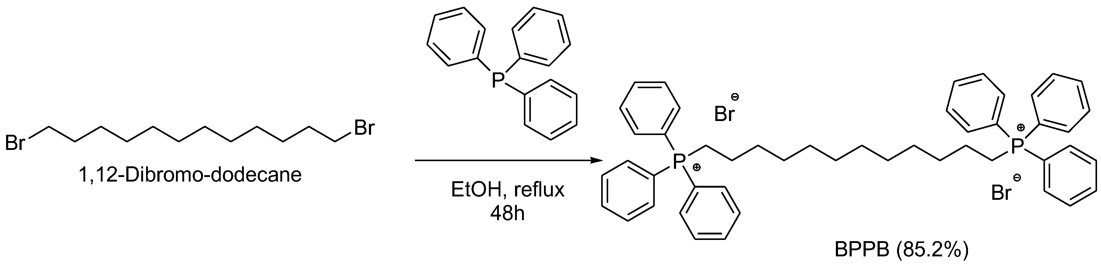
BPPB is a quaternized bis-triphenyl phosphonium salt (TPP+-QPS) capable of self-forming nanovesicles in water (45 nm), encompassing two triphenyl phosphonium groups (TPP+) linked by a twelve-methylene chain. Its synthetic synthesis procedure, as well as the methods followed to carry out its full characterization, are detailed in a previous paper [35]. The BPPB used in this study was from the same batch previously prepared and stored as powder at 4 °C for reuse. Scheme 1 reports the synthetic procedure followed to produce BPPB, including synthetic and yield details (85.2%).

2.2. DLS Analyses in Complex Medium

The hydrodynamic diameter of BPPB vesicles and their DPI were determined by dynamic light scattering (DLS) measurements in a complex biological medium (TSB) used for antibiofilm assays containing several nutritive constituents necessary for bacteria, following suggestions previously reported [36]. Precisely, BPPB was diluted (600 mL) in growth medium at max 2 × MIC (500 µg/mL) to limit interference from medium constituents with scattered light. The BPPB hydrodynamic diameter was determined using a Zetasizer Instrument previously used in our other studies [35] as follows. A nominal 5 mW HeNe laser operating at 633 nm wavelength was used. The scattered light was detected at 173°. The refractive index (n = 1.3325) and viscosity (η = 0.89) of ultrapure water at 25 °C were used in the analysis performed in automatic mode using the instrument’s software (DTS 5.0). pH was fixed at 7.3 with TSB. All measurements were done in triplicate, with 20 runs for each of the triplicates. The particle size was reported as the hydrodynamic diameter obtained as the intensity distribution by cumulant analysis. The same solutions were analysed by the same instrument to determine zeta potential (ζ-p, mV). All measurements were done in triplicate, with 20 runs for each of the triplicates. All results were expressed as the mean ± SD of three independent experiments, each one run 20 times. ZETAAVE and PDI results were corrected according to the error (12% underestimation of size) introduced by using refractive index and viscosity values different from those of TSB (η ≈ 1.00 mPa⋅× s and n = 1.335, respectively).

2.3. Microbiologic Experiments

2.3.1. Clinical MDR Staphylococcal Isolates Used in This Study

A total of twelve Staphylococcus strains from a collection of MDR Gram-positive and Gram-negative species at the University of Genova, kindly gifted by S. Martino Hospital for research, were used in this study. All were isolated from human specimens for diagnostic purposes and identified using the matrix-assisted laser desorption–ionization time-of-flight mass spectrometric technique (MALDI-TOF-MS) (BioMérieux, Firenze, Italy) according to the manufacturer’s instructions. The isolates were selected based on their strong BF-forming ability and subsequently characterized by their antimicrobial resistance profiles. The collection comprised six clinical S. aureus and six clinical S. epidermidis isolates. Antimicrobial susceptibility testing was performed using the VITEK 2 automated system (BioMérieux, Firenze, Italy) according to the manufacturer’s instructions. Susceptibility categories were assigned following EUCAST criteria.

2.3.2. Determination of MICs

To investigate the antibacterial activity of BPPB against the described pathogens, minimal inhibitory concentrations (MICs) were determined by following the microdilution procedure detailed by the European Committee on Antimicrobial Susceptibility Testing (EUCAST) [37] and reported in our previous work [38].

2.3.3. Assessment of Staphylococcal BF Inhibition Using the Crystal Violet Microtiter Plate Assay

Static BF production was detected using the microliter plate method and quantified spectrophotometrically using a method reported by Crémet et al. [39] and Stepanović et al. [40,41]. To produce BFs, stationary-phase bacterial cultures of the six S. aureus and six S. epidermidis strains previously selected (OD570 ranging from 0.73 to 3.36) were diluted 1:100 aseptically into the wells of a 96-well polystyrene tissue culture plate (Corning, Milan, Italy) containing tryptic soy broth medium supplemented with 0.25% glucose, which were incubated at 37 °C for 24 h. The inhibitory effects of BPPB and vancomycin, used as reference antibiotics, on BF formation were evaluated using the crystal violet microtiter plate assay. Each compound was added to the growth medium at the selected concentrations (½ MIC, MIC and 2 × MIC). After 24 h of incubation, the culture media were discarded, and each well was washed three times with phosphate-buffered saline (PBS) to remove non-adherent cells. The plates were air-dried in an inverted position. Adherent microorganisms were stained with 0.1% crystal violet (CV) w/v in water. Excess stain was rinsed off with running tap water, and the plates were air-dried. BF biomass was quantified spectrophotometrically by measuring the optical density at 570 nm (OD570). Each isolate was tested in triplicate, and the results were obtained from three biological replicates. Data are expressed as mean ± standard deviation (SD). Untreated cultures served as controls (CTRs). BF inhibition percentages (BFI%) and residual BF percentages (BFR%) were calculated using Formulas (1) and (2) in Section 3.2.3.

2.3.4. Monitoring of Bacterial Life over 24 Hours at 4 × MIC: Live–Dead-Like Experiments

BPPB live–dead-like assays were carried out on the different isolates of S. aureus and S. epidermidis through time-kill experiments following the recognized protocol previously reported [42]. Experiments were performed over 24 h at a BPPB concentration of four times the MIC for all strains.
A mid-logarithmic-phase culture was diluted in Mueller–Hinton (MH) broth (Merck, Darmstadt, Germany) (10 mL) containing 4 × MIC of the selected compound to give a final inoculum of 1.0 × 10⁵ CFU/mL. The same inoculum was added to cation-supplemented Mueller–Hinton broth (CSMHB) (Merck, Darmstadt, Germany) as a growth control. The tubes were incubated at 37 °C with constant shaking for 24 h. Samples of 0.20 mL were removed from each tube at 0, 2, 4, 6, and 24 h, diluted appropriately with a 0.9% sodium chloride solution to avoid carryover of the BPPB being tested, plated onto MH plates, and incubated for 24 h at 37 °C. Growth controls were run in parallel. The percentage of surviving bacterial cells was determined for each sampling time by comparing colony counts with those of standard dilutions of the growth control. The results are expressed as log 10 of viable cell numbers (CFU/mL) of surviving bacterial cells over a 24 h period. After the experiment, the bacterial fractions analysed at 6 h, when the minimum CFU/mL was reached, were reseeded and their possible regrowth was monitored for an additional 24 h. All experiments were performed at least in biological triplicate for each strain considered.

2.4. Statistical Analysis

Statistical significance was obtained using GraphPad PRISM software 8.0.1 by analysis of variance (two-way ANOVA) corrected for multiple comparisons using statistical Tukey hypothesis testing. The statistical differences of multiple comparisons are reported for each isolate, for each concentration and for each compound tested using symbols. The symbols used are specified in this text and in the Supplementary Materials. Specifically, adjusted p-values for multiple comparisons are reported for each comparison. No symbol is reported when p > 0.05. One symbol is used for p ≤ 0.05, two for p ≤ 0.01, three for p ≤ 0.001 and four for p ≤ 0.0001.

3. Results

3.1. DLS Analyses in Complex Medium; Z-AVE, PDI and ζ-p (mV)

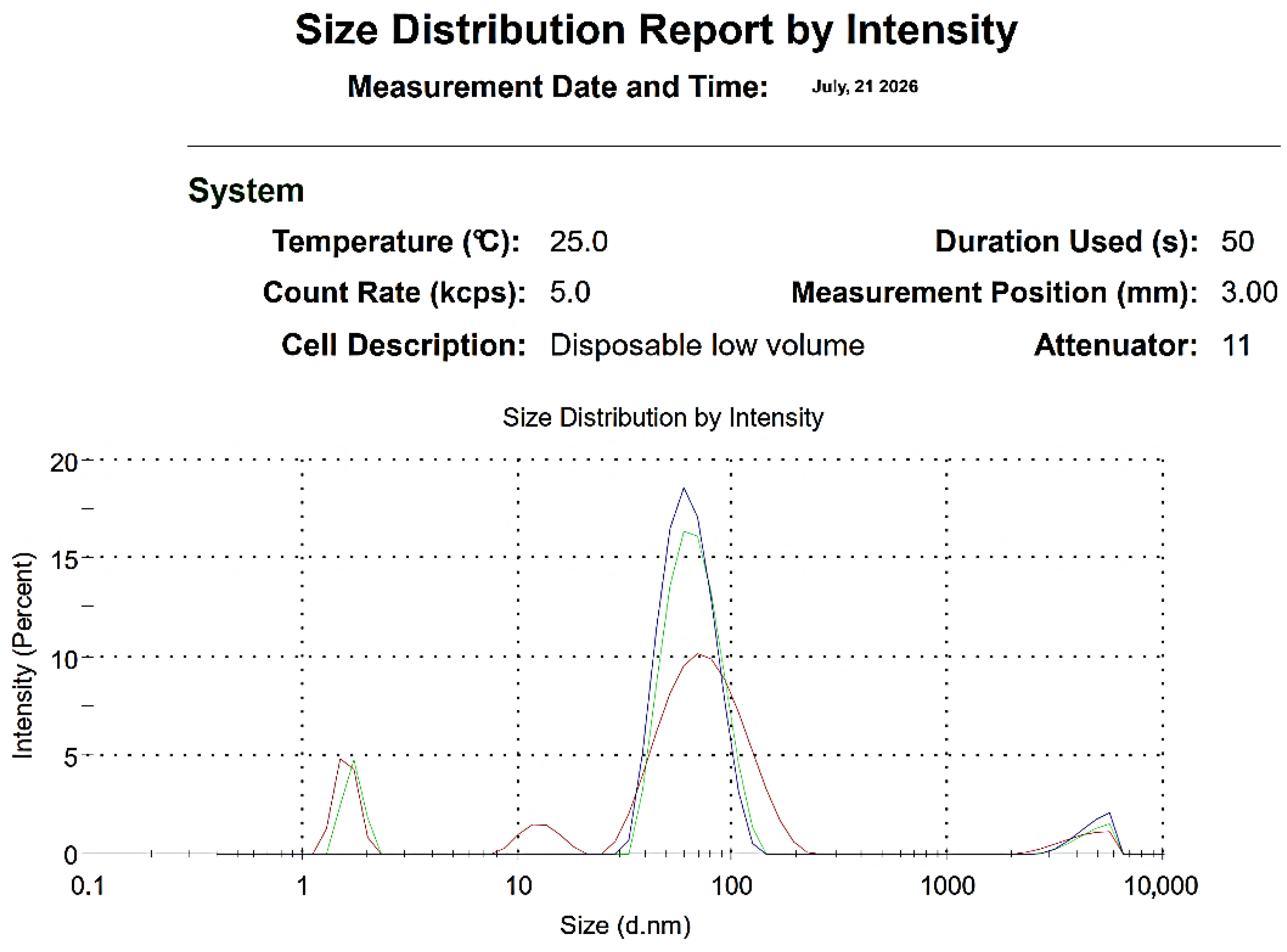
DLS experiments were newly carried out in the complex medium used for the biofilm inhibition test (TSB), at a dilution that enabled analysis, followed by filtration to remove excessively large aggregates, according to a reported protocol [36]. PDI was similar in water and reduced in TSB, while the ZAVE (nm) of our nanovesicles was similar in water and slightly augmented in TSB, with respect to the same data acquired in simple water [35]. Additionally, the obtained hydrodynamic diameters (nm) and PDI values needed to be corrected by +12% of the determined measures due to the estimated size error introduced by using the viscosity set on the instrument (water), which is different from that of TSB.
The medium alone was analysed as a blank (results not reported). Representative results are reported in Figure 1, showing ZAVE measurements by the intensity of different filtered samples at 25 °C (5 kcps). Three representative runs are reported: the blue run indicates PBBP dissolved in water, as described previously [35], while the green and red runs indicate PBBP in TSB after filtration. Table 1 reports ZAVE (nm) for all runs in Figure 1 and related PDI values as means ± SD of independent experiments, each acquired in triplicate (20 runs). Table 1 also reports DLS results corrected by +12% and results previously obtained in milli-Q water [35]. At times, a solution of BPPB 10 mM in m-Q water (312.4 Kcps) was diluted 1:2 to reach the final concentration of 5 mM (8.4 kcps) and analysed. The ζ-p value of BPPB was recorded with the same apparatus at a count rate of 20–59 kcps. The results of the experiments are presented as the means of three independent determinations, comprising 10 runs (particle size) or 12 runs (ζ-p) each, ±SDs. Intensity-based results are reported to express the particle size distribution. In this work, the ζ-p value (mV) of BPPB was recorded with the same apparatus at a count rate of 5 kcps and at 25 °C as for size analyses. The results of the experiments are presented as the means of three independent determinations, comprising 20 runs each, ± SDs.

3.2. Antibacterial and Antibiofilm Activity of BPPB Against Selected Clinical MDR Staphylococcal Isolates

3.2.1. Identification of the Strongest BF-Producing Staphylococcal Isolates

Optical density (OD) measurements at 570 nm (OD570) were used to select MDR Staphylococci strains from those in our collection for antibiofilm experiments. The results of the 1/1 dilution are reported in Figure S1 in the Supplementary Materials.
According to the results shown in Figure S1 and the biofilm (BF) production categories proposed by Stepanović (2007) [41], six S. aureus isolates (Bsp, 24, N1, 187, Bam and Aam) and six S. epidermidis strains (22, 1, 2, 3, 6 and 7) were selected for further investigation because they exhibited the highest biofilm-forming capacity in our collection, with OD570 values consistently exceeding 1, a level indicative of strong biofilm production. Among them, S. epidermidis isolate 6 was the highest producer (OD570 = 2.79), whereas S. aureus 24 exhibited the lowest BF biomass (OD570 = 1.54). Overall, S. epidermidis strains produced significantly greater amounts of BFs than S. aureus strains.

3.2.2. Antimicrobial Resistance Profiles of the S. aureus and S. epidermidis Strains Selected for This Study

Following their identification as very strong biofilm (BF) producers, the antimicrobial susceptibility profiles of the 12 selected clinical staphylococcal isolates (six S. aureus and six S. epidermidis) were determined using the VITEK® 2 system (Table 2). A total of 20 antibiotics were tested. The isolates exhibited extensive multidrug resistance (MDR) patterns, showing resistance to 3–14 antibiotics and up to three intermediate resistance phenotypes. All strains were methicillin-resistant, except for S. aureus N1, which was resistant only to benzylpenicillin, erythromycin, and clindamycin and showed intermediate susceptibility to levofloxacin. Among the isolates examined, S. aureus 187 displayed the highest level of resistance. S. aureus 187 was resistant to 14 out of the 20 antibiotics tested. It displayed resistance to cefoxitin and showed intermediate resistance to “other”. The behaviour of 187 against ceftarolin is not reported. Vancomycin susceptibility was observed in all isolates except for S. aureus 187, which exhibited resistance to both vancomycin and teicoplanin, representing a rare glycopeptide-resistant phenotype. Resistance to teicoplanin was also detected in the MRSE 6 and MRSA Bam strains. Furthermore, for all isolates except S. aureus N1, the presence of a mecA-associated phenotype was clearly detected, suggesting that altered penicillin-binding proteins (PBPs) are the mechanism underlying β-lactam resistance.

3.2.3. Antibacterial Activity and Inhibition Effects of BPPB on BF Formation Against Selected Clinical S. aureus and S. epidermidis Isolates

Antibacterial Activity of BPPB on the Selected Staphylococcal Isolates: MIC Determination
Prior to the evaluation of the effects of BPPB on BF formation, its MIC values (MICs) were determined against the selected MDR staphylococcal isolates. The obtained MICs are reported in Table 2, together with the antimicrobial susceptibility profiles of the strains, as assessed by the VITEK 2 system.
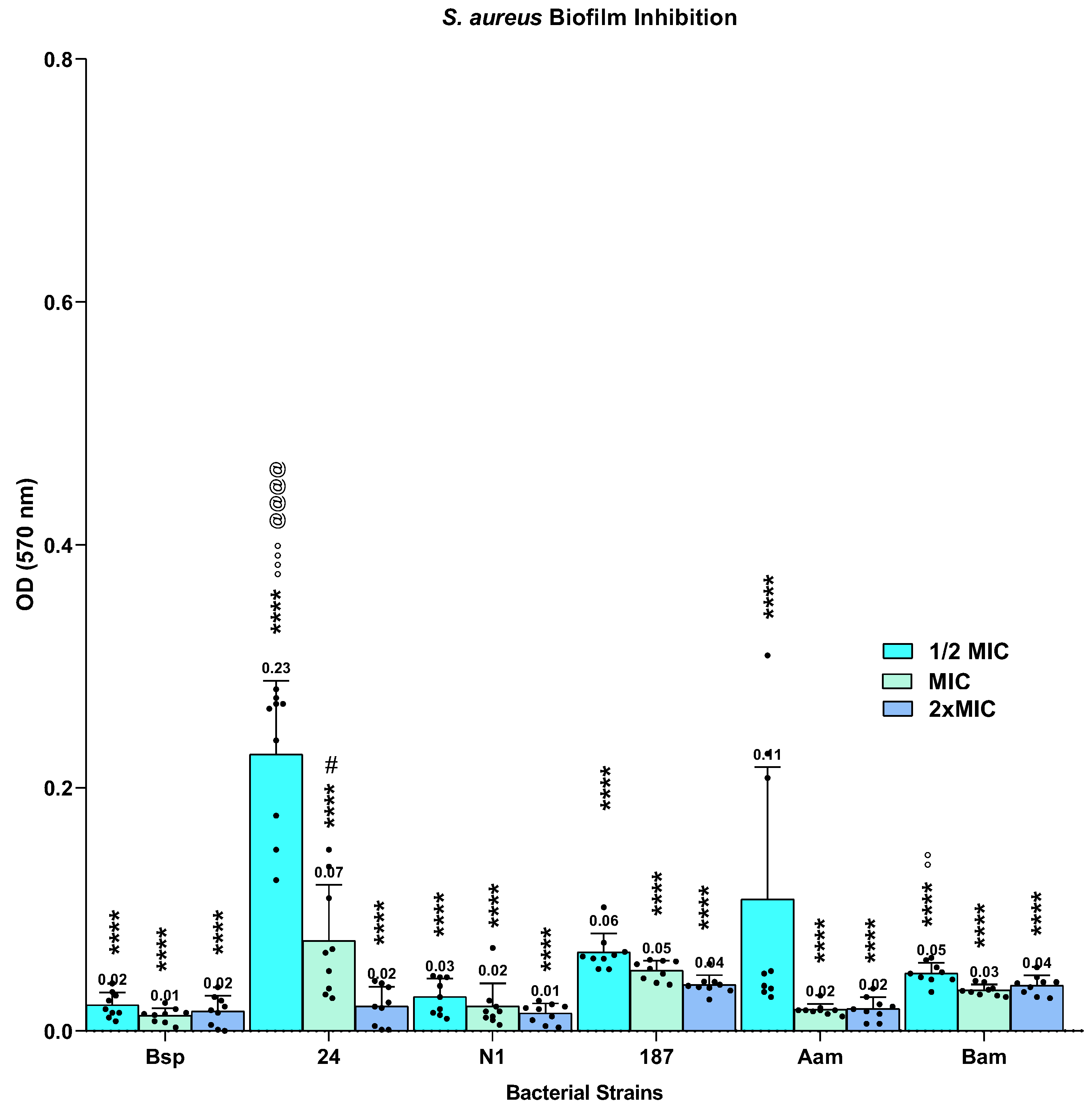
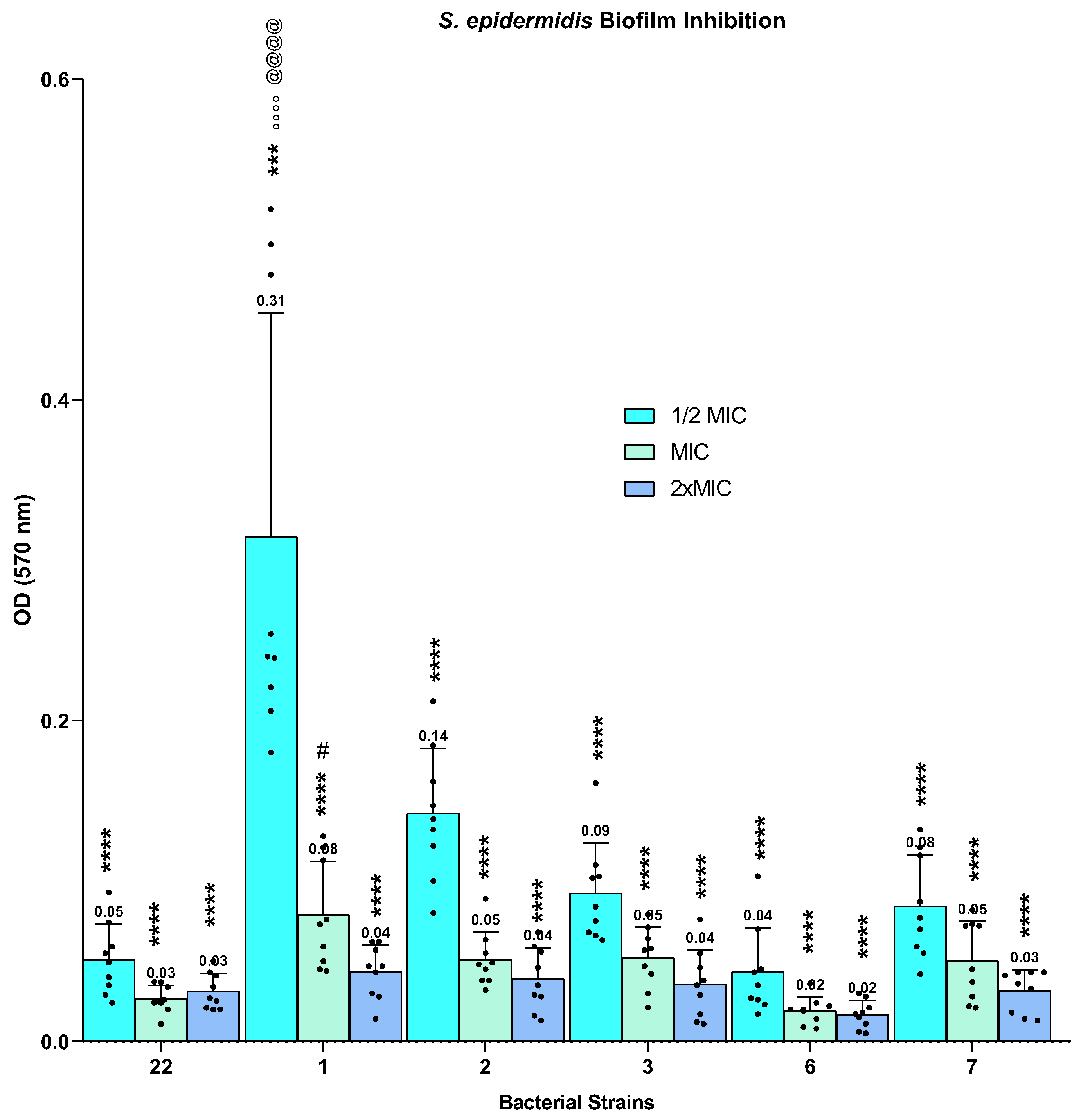
The inhibitory effects of BPPB on BF formation were evaluated at ½ MIC, MIC, and 2 × MIC against the selected staphylococcal isolates. Quantification of BF biomass by crystal violet staining and OD570 measurement revealed substantial inhibition of BF formation at all tested concentrations, as shown in Figure 2 for S. aureus isolates and in Figure 3 for S. epidermidis isolates.
Subsequently, BF inhibition percentage (%) was calculated for all strains at the same three concentrations tested (½ MIC, MIC and 2.0 × MIC) according to the following Formula (1):
BFI (%) = [(ODCTRODC)/ODCTR] × 100
where OD means optical density measured at 570 nm, CTR is the control, and C indicates the concentration considered. CTR means non-treated strains (control). BF inhibition percentages calculated for all S. aureus and S. epidermidis isolates at ½ MIC, MIC, and 2 × MIC are shown as bar graphs in Figures S2 and S3 of Section S2 in the Supplementary Materials (SMs) for S. aureus and S. epidermidis isolates, respectively. The CTR result was 0% for each strain, representing no inhibition without treatment.
Finally, the residual BF percentage (%) produced by bacteria under treatment with BPPB was calculated for all strains at the three different concentrations tested (½ MIC, MIC and 2.0 × MIC), according to the following Formula (2):
BFR (%) = (ODC/ODCTR) × 100
where BFR means residual BF, OD means optical density measured at 570 nm, CTR is the control and C indicates the concentrations considered. The result for CTR was normalized to 100% for all strains, and it is reported in the graph as white bars. The results are shown as bar graphs in Figure S4 for S. aureus isolates and in Figure S5 for S. epidermidis isolates in the Supplementary Materials (Section S2).
Monitoring of Bacterial Life over 24 Hours at 4 × MIC: Live–Dead-Like Experiments
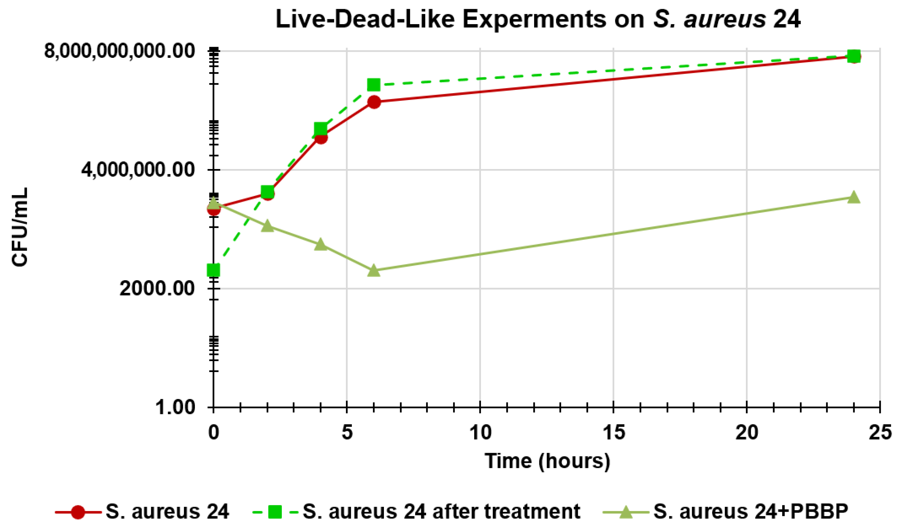
To assess the actual BPPB inhibition effects on BF formation by Staphylococci in this study, not simply the full or partial reduction in BF production due to bacterial death, live–dead-like experiments were performed on both the S. aureus and S. epidermidis strains used in this study. As described in Section 2, BPPB was administered at 4 × MIC because it is 2 times the maximum concentration used in the BF inhibition assay, ensuring that bacteria were alive and capable of replication at concentrations where we observed inhibition. The experiments were conducted for 24 h (as in the BF inhibition assay), monitoring the numbers of CFU/mL at fixed time points (2, 4, 6 and 24 h). The 24 h growth profiles of the same strains not treated with BPPB were considered the control. After treating the strains with BPPB, the fraction withdrawn at 6 h of the experiment, in which the minimum CFU/mL was counted, was reseeded in the absence of BPPB, and new colony formation (CFU/mL) was monitored up to 24 h and compared to the control. The results obtained for S. aureus 24 (MIC = 0.250 µg/mL, as 10 out of 12 strains), selected as representative of the results obtained for all other strains, which were similar, are reported as dispersion graphs in Figure 4.
Table 3 lists numbers related to the dispersion graphs in Figure 4 and percentages of non-inhibited S. aureus 24 (alive). Specifically, the percentages in column 4 relate to the numbers in the experiment on S. aureus 24 treated with BPPB 4 × MIC (column 3), with respect to inoculum, while those in the last column represent CFU/mL measured in the experiment by reseeding the fraction analysed at 6 h during treatment, containing only 1.9% of the CFU/mL observed in the control (column 2).

3.3. Antibacterial and Antibiofilm Activity of Vancomycin (V) Against Selected Clinical S. aureus and S. epidermidis Isolates

3.3.1. Antibacterial Activity of Vancomycin (V)

Regarding BPPB nanovesicles, before the evaluation of vancomycin inhibition effects on BF formation, its MICs were calculated against the MDR Staphylococcal isolates selected for investigation. The resulting MIC values, which were used to define the concentrations to be tested in subsequent BF inhibition assays, are summarized in Table 4.

3.3.2. Inhibitory Effects of Vancomycin (V) on BF Formation by Clinical S. aureus and S. epidermidis Isolates

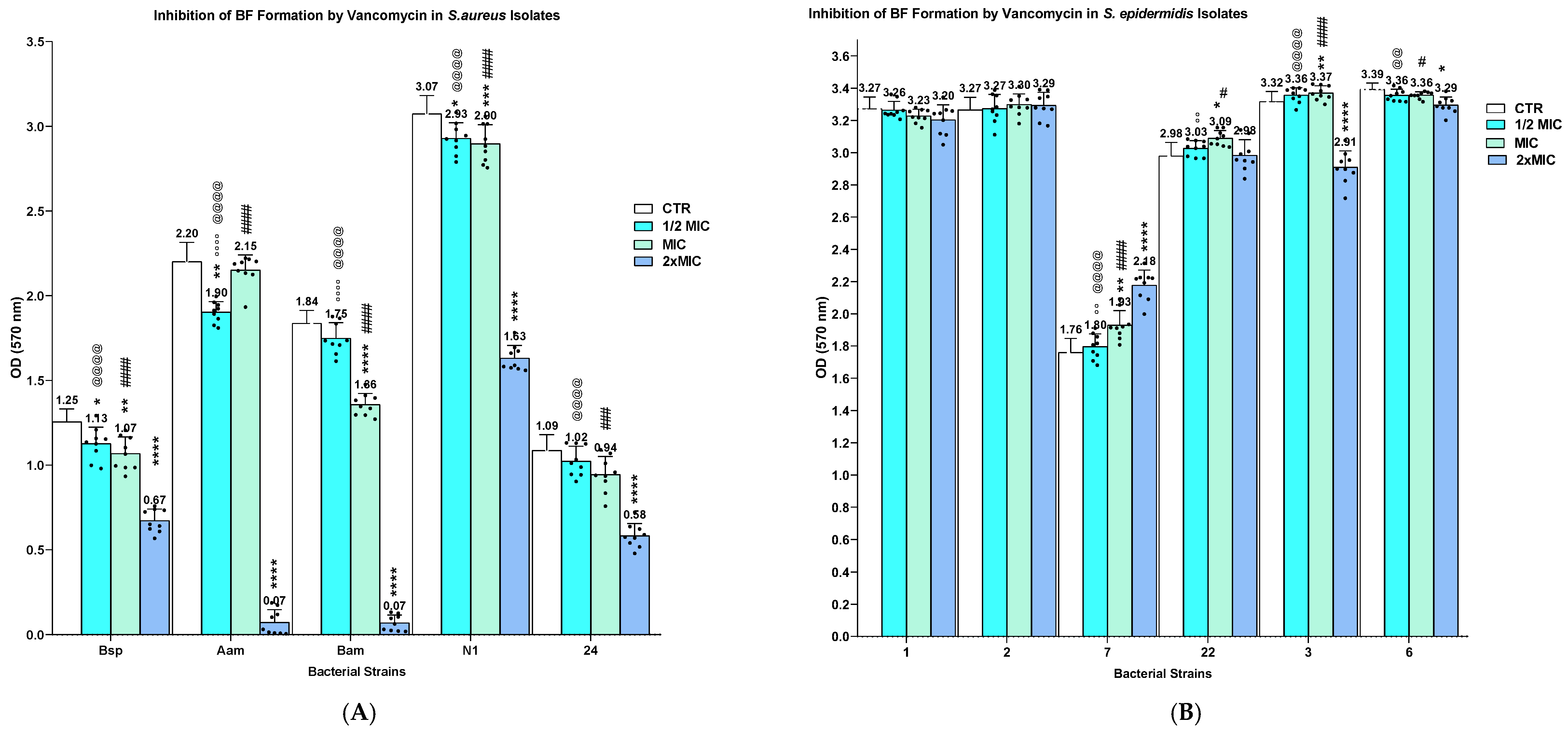
The antibiofilm activity of V was evaluated against susceptible S. aureus isolates (Bsp, 24, N1, Bam, and Aam) and all selected S. epidermidis isolates. In agreement with the experimental design adopted for BPPB, vancomycin was tested at ½ MIC, MIC, and 2 × MIC, using the MIC values previously determined for each isolate (Table 3). Depending on the strain, V concentrations ranged from 0.125 to 0.500 µg/mL at ½ MIC, from 0.250 to 1.000 µg/mL at MIC, and from 0.250 to 2.000 µg/mL at 2 × MIC. For comparison, BPPB was tested at concentrations of 0.125, 0.250, and 0.500 µg/mL for all isolates. The resulting OD570 values are shown in Figure 5A for S. aureus isolates and in Figure 5B for S. epidermidis isolates.
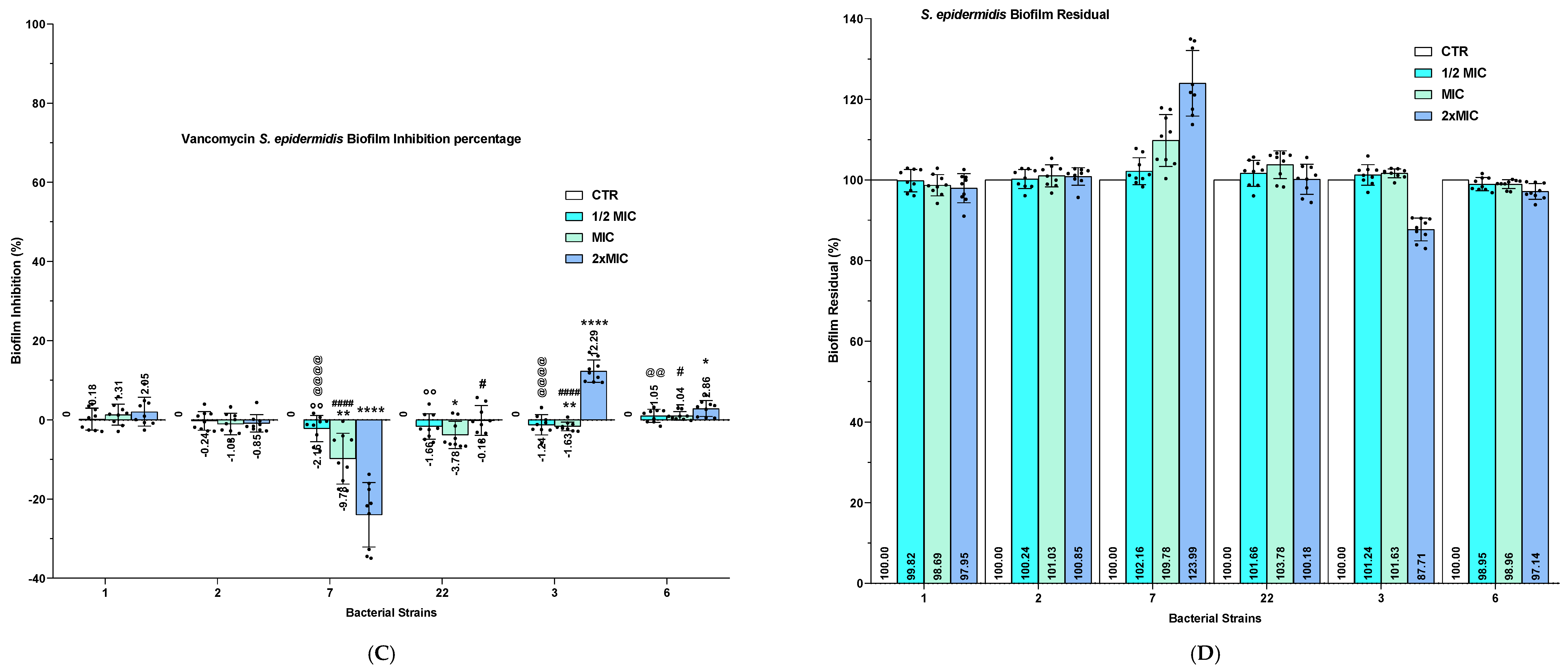
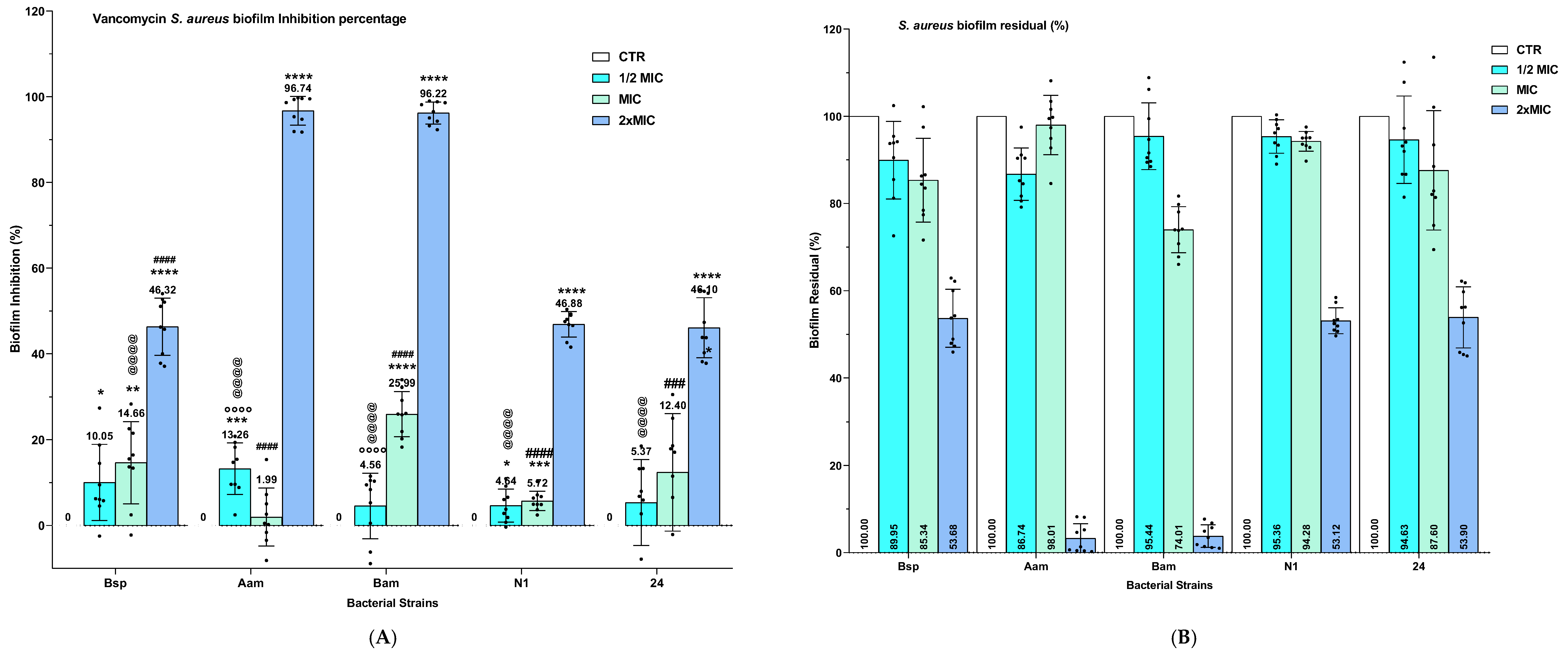
The percentages of BF inhibition and residual BF biomass were subsequently calculated for all isolates treated with vancomycin at ½ MIC, MIC, and 2 × MIC. The resulting values are reported in Figure 6.

4. Discussion

4.1. DLS Results

The previously estimated size of polycation BPPB in ultrapure water (45 nm) is smaller than 100 nm, allowing possible passive entry into a bacterial cell without endocytosis or pinocytosis processes. To study the effect of pH = 7.2 and the presence of TSB on the hydrodynamic diameter, PDI and zeta potential of BPPB, DLS analyses were carried out for the first time by dispersing BPPB in water or TSB to evaluate possible changes in how PBBP particles appear to bacteria during antibiofilm analyses. Opportunely diluted BPPB powders in water or TSB, as described in the previous sections, underwent DLS analyses, providing the dimensional distributions, polydispersity index values and Z-potential values reported in Table 1. Hydrodynamic diameter did not change substantially when acquired in water, despite the different concentrations that were selected for all experiments in this work. Collectively, the results confirmed previous data on PDI and ζ-p (Table 1) [35]. Conversely, dimensions (nm) significantly increased by +10- +25% when DLS was performed in TSB. Additional corrections of +12% applied to the obtained results further increased ZETAAVE values, which finally resulted in improvements of +27–+33% with respect to the analysis in water due to the estimated error of 12% introduced by the instrument being set for water parameters, including viscosity and refractive index. In fact, a 12% size underestimation was introduced mainly by differences in viscosity between water and TSB (η = 0.89 for water and 1.00 for TSB) following the equation [(1.00/0.98) − 1] × 100. Increases in hydrodynamic diameter (nm) in TSB can be attributed to increased solutes in the medium, which aggregated around the original BPPB nanovesicles, forming larger particles, as well as a reduced electrostatic repulsion between BPPB particles and other particles, mainly due to a decreased ζ-p (−33.3%) of new nanoparticles [43]. In this regard, it is well known that for tryptic soy broth (TSB), there is no single, universally recognized zeta potential (ζ) value, since it can depend on several factors. It is equally well known that when nanoparticles are dispersed in TSB, they can display ζ-p values that vary from a few negative mV up to tens of negative mV depending on the system studied. In the case of cationic nanoparticles based on bis-phosphonium salts dispersed in TSB, as in our case, their presence leads to compression of the double electrical layer (high ionic strength). In addition, proteins and peptides are also adsorbed on the surface of the nanoparticles, forming a sort of organic “corona”. For strongly cationic nanoparticles used as antimicrobials (ammonium quaternaries, phosphonium, polycations, etc.), as in our case, typical values observed in complex media such as TSB are often between +5 and +15 mV, while in water or low-ionic-strength buffers, they can be >+30 mV. A reasonable estimate for BPPB particles in complete TSB with 2.5 g/L glucose and at pH = 7.3 would be ζ-p ≈ +5 up to +15 mV, thus confirming the proportionally obtained of ζ p ≈ +6 mV.

4.2. Antibacterial Activity of BPPB and Inhibition of BF Formation Against Selected Staphylococci

4.2.1. Antibacterial Effects of BPPB Against Selected Staphylococci

As shown in Table 2, BPPB exhibited remarkably low MIC values (0.125–0.250 µg/mL) against all MDR staphylococcal isolates investigated in this study, regardless of their complex antimicrobial resistance profiles, further confirming the potent antibacterial activity previously reported against Staphylococcus species [35]. Although the earlier study included both different and overlapping isolates, such as S. aureus 187 and S. epidermidis 22 [35], the present findings are particularly noteworthy, as these two isolates displayed the highest and lowest numbers of antibiotic resistances, respectively (14 and three). These results further support the potential of BPPB as a promising antimicrobial candidate for the treatment of severe infections caused by Gram-positive pathogens, including members of the ESKAPE group [44,45,46]. In the present study, its activity was confirmed against staphylococcal isolates characterized by established MDR phenotypes, exhibiting resistance to more than three antibiotics. The mentioned ESKAPE pathogens are of great clinical concern because of their ability to evade the activity of currently available antimicrobial agents. They are frequently associated with healthcare settings, where immunocompromised individuals, patients with open wounds, and those carrying invasive medical devices, such as central venous catheters, are at increased risk of healthcare-associated infections, including bloodstream infections and infective endocarditis [47,48]. Among ESKAPE superbugs, MRSA represents a major global “One Health” challenge. This issue is further exacerbated by MRSA’s ability to form BFs on medical devices, which can transform otherwise manageable infections into chronic and difficult-to-treat conditions. BF-associated infections often result in prolonged hospitalization, delayed patient recovery, increased healthcare costs, and a greater risk of treatment failure. Moreover, the dissemination of MDR and BF-forming MRSA strains extends beyond healthcare settings, affecting animals, environmental reservoirs, and multiple sectors of human activity, thereby reinforcing the interconnected nature of the One Health framework [49].
Vancomycin remains one of the main therapeutic options against MRSA infections. It is bactericidal, but clinical success is achieved in only about 49% of cases [50]. Its use is further limited by the need for parenteral administration, as oral absorption is negligible. Additionally, systemic infections require intravenous treatment, while CNS and dialysis-associated infections often require specialized routes of administration [51], including intravenous [52] and intrathecal/intraventricular (IT/IVT) (IT/IVT) routes [53]. Finally, peritoneal administration is required for peritonitis associated with peritoneal dialysis [54].
Furthermore, since the late 1990s, glycopeptide-resistant MRSA strains have increasingly been reported [55]. In the present study, resistance to V and/or T was observed in S. aureus 187 (V, T), S. aureus Bam (T), and S. epidermidis 6 (T). In addition, resistance to alternative anti-MRSA agents has emerged over time, including oxazolidinones such as linezolid, for which resistant strains have been reported since 2001 [56].
In this alarming scenario, the potent antibacterial activity of BPPB, demonstrated irrespective of the complex resistance profiles of the isolates tested, further supports its potential as a promising antimicrobial candidate against MRSA and MRSE. Indeed, BPPB has previously been shown to exhibit greater antibacterial activity than most quaternary phosphonium salts developed to date [35]. However, while its conventional antibacterial properties and cytotoxicity toward eukaryotic cells have been extensively characterized to support its potential clinical development [35], its activity against staphylococcal BFs had not previously been investigated. The present study therefore expands the microbiological characterization of BPPB by evaluating, for the first time, its activity against BF formation by selected Staphylococcus isolates, chosen for their strong BF-forming ability (Figure S1 in the Supplementary Materials). Notably, all selected strains exhibited MDR phenotypes resistant to more than three antibiotics, while S. aureus 187 displayed a particularly concerning resistance profile, including resistance to both vancomycin and teicoplanin.

4.2.2. BF Inhibition Effects of BPPB Against Selected Staphylococcal Isolates

As shown in Figure 2 and Figure 3, BPPB consistently reduced the OD570 values of BFs produced by both S. aureus and S. epidermidis isolates, with significant effects already evident at ½ MIC (0.0625–0.125 µg/mL). BF inhibition ranged from 83.3% to 98.9% for S. aureus isolates (Figure S2) and from 87.9% to 98.5% for S. epidermidis isolates (Figure S3). Overall, BFs produced by S. aureus strains appeared slightly more susceptible to BPPB treatment than those formed by S. epidermidis strains. Notably, inhibition rates exceeding 99% were achieved at the MIC against S. aureus isolates Bsp, N1, and Aam. Among the S. aureus isolates, a clear concentration-dependent effect against their BFs was observed only for strain 24. In contrast, for all other isolates, such a BPPB effect remained largely unchanged across the concentrations tested, consistently approaching 99% inhibition or, in the case of strain Aam, reaching a plateau between MIC and 2 × MIC. Notably, BPPB exhibited strong inhibition effects against strain 187, despite its extensive resistance profile, which included resistance to 14 antibiotics, as well as the glycopeptides vancomycin and teicoplanin. BF inhibition rates of 97.4%, 98.0%, and 98.5% were achieved at ½ MIC, MIC, and 2 × MIC, respectively, exceeding those observed for strain 24, particularly at the lowest concentration tested. Among the S. epidermidis isolates, strain 6 was the most susceptible to BPPB, exhibiting BF inhibition rates ranging from 98.5% to 99.4%, depending on the concentration tested. Conversely, strain 1 was the least susceptible, although BPPB still produced substantial inhibition, reaching 87.9%, 97.0%, and 98.3% at ½ MIC, MIC, and 2 × MIC, respectively. A limited concentration-dependent inhibiting effect was observed for S. epidermidis strains 22 and 6, whereas a more pronounced concentration–response relationship was evident for strains 1, 2, 3, and 7. In particular, strains 2 and 1 exhibited progressive increases in BF inhibition with increasing BPPB concentrations. For strain 2, inhibition rates reached 94.1%, 97.9%, and 98.4% at ½ MIC, MIC, and 2 × MIC, respectively, while the corresponding values for strain 1 were 87.9%, 97.0%, and 98.3%. The residual BF profiles reported in Figures S4 and S5 further confirm these findings, showing only minimal amounts of residual BF following BPPB treatment. Slightly higher residual BF percentages were observed for S. aureus strains 24 (16.7%) and Aam (5.1%), as well as for S. epidermidis strains 1 (12.1%), 2 (5.9%), and 7 (4.0%), whereas all remaining isolates exhibited only negligible residual BF biomass. Over the past five years, several studies have investigated novel antibiofilm strategies against staphylococcal BFs, reporting both promising outcomes and important limitations. These findings provide a valuable framework for comparison with the results obtained for BPPB and are discussed below. Duda-Madej et al. synthesized a series of sulphur-containing camphor derivatives and evaluated their antimicrobial and antibiofilm activities against a panel of Gram-positive and Gram-negative bacteria. The tested Gram-positive strains included, among others, S. aureus and S. epidermidis strains, as in this study [13]. According to the reported data, compared with BPPB, the sulphur-containing camphor derivatives displayed substantially lower antibiofilm potency. Specifically, compound 1a inhibited S. epidermidis BF formation by only 22% at concentrations 32–512-fold higher than the corresponding ½ MIC values of BPPB, resulting in effects against BFs approximately 4.0–4.5-fold lower than those achieved by BPPB. Similarly, compound 2a inhibited BF formation by S. aureus ATCC 25923 by up to 50% at concentrations 1024–4096-fold higher than the corresponding ½ MIC values of BPPB, corresponding to an antibiofilm efficacy approximately 1.7–2.0-fold lower. Against S. epidermidis S22, compound 2a achieved only 22% BF inhibition at concentrations more than 2000-fold higher than those required for BPPB, resulting in an efficacy approximately 4.0–4.5-fold lower [13]. Recent studies by Odžak et al. suggested that the introduction of amide functionalities into the structure of quaternary ammonium salts (QASs) can lead to “soft” derivatives that, while retaining the original antimicrobial properties of conventional QASs, demonstrate a tuneable cytotoxicity [57]. In this context, Odžak et al. reported that a series of 3-amidoquinuclidine QAS derivatives displayed lower cytotoxicity than commercially available QASs, highlighting the potential of this strategy for the development of safer antimicrobial agents [57]. Although only a limited number of the newly synthesized compounds exhibited MIC values comparable to or lower than those of the corresponding commercial quaternary ammonium salts (cetylpyridinium chloride (CPC) and benzyl dimethyldodecyl ammonium bromide (BAB)) against both Gram-positive and Gram-negative bacteria, some derivatives demonstrated remarkable inhibition effects against BFs, outperforming CPC and BAB, which inhibited BF formation by less than 50% and approximately 75%, respectively [57]. Based on these findings, BPPB outperformed CPC and BAB by up to 1.3–2-fold and achieved inhibition effects on BF formation comparable to those of the most active 3-amidoquinuclidine QAS derivatives at concentrations approximately 50-fold lower. These observations further support the remarkable antibiofilm potency of BPPB. Antimicrobial peptides (AMPs) have also attracted considerable interest as a promising alternative to conventional antibiotics [58,59,60]. Their broad-spectrum antimicrobial activity and reduced susceptibility to the mechanisms responsible for bacterial antibiotic resistance make them particularly attractive candidates for the development of novel anti-infective therapies [61]. In fact, although extensive resistance has emerged against many classes of conventional antibiotics, AMPs generally target the bacterial membrane, a highly conserved and essential cellular structure. Such compounds are widely recognized as membrane-active agents capable of electrostatically interacting with the negatively charged bacterial cell surface, ultimately causing membrane disruption [7,38,42,62,63,64,65,66,67,68]. By acting primarily as non-specific membrane disruptors and inducing rapid cell death, AMPs are considered less susceptible to the development of resistance than conventional antibiotics [69]. Akhash et al. synthesized a novel peptide analogue (mKLK), derived from the D-amidated form of the sapecin B-derived peptide KLK, which is not susceptible to proteolysis as conventional AMPs [69]. The modified peptide retained the antibacterial potency of the parental KLK molecule while exhibiting markedly enhanced antibiofilm activity against both MRSA and MSSA in a murine model of catheter-associated BF infection at sub-MICs [69], thus confirming its strong potential therapeutic advantage for preventing BF-associated MRSA and MSSA infections [69]. More recently, Godoy et al. investigated the antibiofilm potential of chitosan NPs (CNPs) against S. aureus strains isolated from milk samples collected at dairy farms in southern Chile from cows diagnosed with bovine mastitis (BM) [70]. BF inhibition reached 100% at MIC, exceeded 50% at ½ MIC, and remained above 20% at ⅓ MIC, demonstrating a marked concentration-dependent effect [70]. The molecular mechanisms underlying the observed antibiofilm activity were not experimentally investigated [60], as in our case. The findings of Godoy et al. suggest that CNPs may represent a promising alternative strategy for the control, prevention, and treatment of bovine mastitis (BM) caused by S. aureus. However, when compared with BPPB, the antibacterial activity of CNPs was substantially lower, as reflected by MIC values approximately 512-fold higher than those observed for BPPB. Moreover, under antibiofilm conditions, BPPB tested at ½ MIC (0.125 µg/mL) achieved BF inhibition rates 1.7–2.0-fold greater than those obtained with CNPs at ½ MIC (64 µg/mL), despite being administered at a concentration 512-fold lower. Apostol et al. also evaluated the antimicrobial activity of a library of 49 previously synthesized compounds derived from alanine or phenylalanine and containing a 4-(4-X-phenylsulfonyl) phenyl moiety (X = H, Cl, or Br) [17]. With regard to S. epidermidis, which was also investigated in the present study through MDR clinical isolates, BPPB exhibited MIC values 22.5- to 449.6-fold lower than those of the most active compounds reported by Apostol et al. Notably, against S. epidermidis isolates 6 and 7, BPPB displayed MIC values approximately 1.2-fold lower than those reported for ciprofloxacin, despite the resistance of these isolates to levofloxacin, another fluoroquinolone antibiotic closely related to ciprofloxacin and used by the authors as the reference drug. Furthermore, even under the most favourable conditions reported by Apostol et al. (BF inhibition observed at 1/16 × MIC, based on the lowest MIC value of 14 µg/mL), the active concentrations required remained 1.8- to 3.5-fold higher than those of BPPB administered at 2 × MIC. In another study, the authors incorporated zinc oxide (ZnO), zinc oxide–borax (ZnO–borax), zinc copper oxide (ZnCuO2) NPs, and borax (Na2B4O7·10H2O) into bacterial cellulose (BC), generating a series of BC-based nanocomposites (BC-NPs). The resulting materials were evaluated for their BF inhibition and BF degradation activities using the crystal violet assay against a broad panel of clinically relevant microorganisms, including S. aureus ATCC 29213 [18]. According to data reported in the paper for BF inhibition (%) caused by the mysterious concentrations of NPs, it was evidenced that BF produced by S. aureus ATCC 29213 (CV) was inhibited by 65.53% (BC–ZnONPs), 71.74% (BC–Borax), 66.60% (BC–ZnOBorax), and 28.27% (BC–ZnOCuO2) [18], indicating moderate antibiofilm activity against non-MDR S. aureus ATCC 29213. Furthermore, considering only the highest BF inhibition (%) observed for S. aureus (28.3–71.7%) by BC-NPs, it was inferior to that exerted by BPPB at the lowest concentrations tested (0.125 µg/mL) by 1.2–3.5 times. More recently, Dutta et al. reported the development and characterization of novel NMs, evaluating their antibacterial activity and their ability to inhibit BF formation and eradicate mature BF [16]. The antibiofilm activity of biogenically synthesized bacteriocin-capped silver NPs (bac-AgNPs) against non-MDR S. aureus ATCC 23235 remained lower than that observed for BPPB against MDR MRSA. Specifically, bac-AgNPs tested at 1.7 µg/mL (corresponding to 100 × MBC) achieved BF inhibition levels approximately 1.15–1.21-fold lower than those obtained with BPPB against MDR staphylococcal isolates, despite BPPB being effective at concentrations approximately 6.8-fold lower. It should also be noted that the antibiofilm effect observed for bac-AgNPs was evaluated at concentrations greatly exceeding their bactericidal threshold. Therefore, the reduction in BF formation may have been influenced, at least in part, by extensive killing of planktonic and BF-associated bacterial cells, rather than reflecting a specific antibiofilm activity independent of bacterial viability [16]. Another recent study investigating novel antibiofilm NMs was published by Ali et al. [14]. The authors developed both free copper NPs (f-CuNPs) and immobilized copper nanocomposites (im-CuNPs). These NMs were then evaluated for their potential antibacterial and antibiofilm applications [14]. The MIC values of BPPB against the MDR S. aureus isolates investigated in the present study, including strains resistant to gentamicin (except for Bsp and N1), were comparable to or lower than those reported for gentamicin against susceptible S. aureus strains. Moreover, the highest BPPB concentration evaluated in antibiofilm assays (0.500 µg/mL) was approximately 10,000-fold lower than that used for the CuNP formulations. Despite the very high concentrations tested, both free and immobilized CuNPs exhibited only modest activity in BF formation inhibition [14]. BF inhibition achieved by the developed CuNPs against S. aureus (23–30%) remained substantially lower than that observed for BPPB, which exhibited 2.3- to 4.3-fold greater antibiofilm activity [14]. Similarly, BPPB outperformed gentamicin, achieving higher BF inhibition despite being tested at concentrations approximately two-fold lower. Rondilla et al. investigated the BF formation inhibition effects of extracts obtained with edible Scottish brown seaweeds, such as Alaria esculenta and Laminaria digitata, following lactic acid fermentation at pH 4.5 [19]. The fermented extracts were evaluated against the ciprofloxacin-resistant MRSA strain ATCC 43300 and demonstrated measurable antibiofilm activity, with fermented L. digitata exhibiting the strongest effects [19]. Both the fermented seaweed extracts and ciprofloxacin were evaluated at concentrations approximately 200-fold and 100-fold higher, respectively, than the highest concentration of BPPB tested in the present study. Both fermented seaweed extracts exhibited lower antibiofilm activity than BPPB despite being tested at concentrations approximately 200-fold higher. Similarly, ciprofloxacin achieved strong BF inhibition only at concentrations about 100-fold higher than those required for BPPB [19]. Overall, while several recent antibiofilm strategies have shown promising results, all require substantially higher concentrations than BPPB to achieve comparable effects. In this regard, although mechanistic insights could deepen our knowledge of BPPB effects and could be very relevant for research on BFs, we did not aim to identify mechanisms within the scope of this study. Although this could appear as a limitation, this study mainly aimed to identify new compounds effective against BF formation by Staphylococci, and this was successful. Furthermore, such a “limitation” affects most of the studies reported in the literature and cited here, as well as others cited in a recent review on this question [5].

4.2.3. Live–Dead-Like Experiments

In Figure 4 and Table 3, S. aureus 24 data were selected as representative of those obtained by other strains because it displayed the modal MIC of 0.250 µg/mL. When BPPB was administered at 1 µg/mL to S. aureus 24 (twice the maximum concentration in BF inhibition assays), a rapid reduction in CFU/mL was observed in the first six hours, reaching only 1.9% of non-inhibited bacteria. Furthermore, during an additional time of up to 24 h, live bacteria increased, reaching 145% with respect to the inoculum. Additionally, after seeding the fraction of bacteria treated with BPPB containing a 53-times lower amount of CFU/mL than that of the control, and monitoring bacterial life for a further 24 h, bacterial growth exceeded that of the control, reaching 294% and stabilizing at 104% at the end of 24 h. All these findings evidenced that after 24 h of treatment with BPPB, at double concentrations with respect to those used in antibiofilm experiments, Staphylococci were fully alive, augmented, and therefore fully capable of constructing BF. This established that inhibition of BFs observed with the CV assay does not depend on possible bacterial death.

4.3. Antibacterial Activity of Vancomycin (V) and Inhibition of BF Formation Against Selected Staphylococci

4.3.1. Antibacterial Effects of Vancomycin (V) Against Selected Staphylococci

Except for S. aureus 187, all isolates remained susceptible to V, exhibiting MIC values ranging from 0.25 to 1.0 µg/mL (Table 3), consistent with the susceptibility profiles commonly reported for MRSA and MRSE strains. Therefore, V was considered an appropriate reference antibiotic for comparison with BPPB in the subsequent antibiofilm assays.
Comparative Antibacterial and Antibiofilm Activities of BPPB and Vancomycin
As shown in Table 3, BPPB exhibited MIC values comparable to or lower than those of V, allowing a direct comparison of their respective BF-formation-inhibiting activities. Notably, BPPB retained potent antibacterial activity against all tested isolates, including S. aureus 187, which was resistant to V. In contrast, V was active against all other S. aureus isolates, although it was two-fold less potent than BPPB against strain 24. S. epidermidis isolates generally appeared less susceptible to V than S. aureus, resulting in a 2- to 4-fold lower activity than that observed for BPPB. Taken together, these findings, combined with the previously reported low cytotoxicity, favourable solubility, stability, and nanoscale dimensions of BPPB [35], indicate that this compound merits further investigation for the management of BF-associated infections caused by MDR staphylococci.

4.3.2. Inhibition Effects of Vancomycin on BF Formation by Selected Staphylococcal Isolates

Vancomycin was selected as the reference antibiotic because all Staphylococci are usually sensitive to vancomycin. In this study, S. aureus 187 is a rarity because we discovered that it showed resistance to 14 antibiotics, including both vancomycin and teicoplanin. However, this isolate was not considered in BF inhibition assays using vancomycin. To this end, V exhibited little or no antibiofilm activity against the isolates investigated in this study. Particularly, no relevant inhibition of BF formation was observed for S. epidermidis isolates, even at 2 × MIC, where inhibition rates ranged from only 0.18% to 2.86% for most strains and reached a maximum of 12.29% for isolate 3. Moreover, slight increases in BF biomass were observed for isolates 2, 22, and 7, suggesting a lack of antibiofilm efficacy under the tested conditions. A somewhat greater effect was observed against S. aureus isolates. However, except for strains Aam and Bam, BF inhibition remained below 50% even at 2 × MIC, with values ranging from 46.3% to 46.6%. Only Aam and Bam showed marked susceptibility to V, reaching inhibition rates of approximately 96–97% at the highest concentration tested. In contrast, BPPB consistently inhibited BF formation by all S. aureus and S. epidermidis isolates, displaying substantial activity already at ½ MIC and at concentrations generally 2–4-fold lower than those used for V. This difference was particularly evident for S. epidermidis, against which V was largely ineffective, whereas BPPB achieved high inhibition rates across all isolates. Detailed comparisons of BF inhibition and residual BF percentages obtained with BPPB and V are provided in Figures S6 and S7 (Section S3, Supplementary Materials). The mechanistic basis for the limited BF inhibition activity of vancomycin is reported in the Supplementary Materials [5,44,45,71,72,73,74,75,76,77,78,79,80,81,82,83,84,85].

5. Conclusions

BF-producing Staphylococcus spp. represents a major clinical challenge because of their enhanced tolerance to conventional antibiotics, which contributes to persistent, chronic, and sometimes life-threatening infections. In the present study, confirmation of the augmented but maintained nano dimensions of BPPB in the range of 50–60 nm, as well as in TSB used for microbiologic investigations, was achieved by carrying out new DLS analysis in TSB and correcting the size (nm) and PDI results for the estimated error (<12%) due to the differences between the viscosity of water and that of TPS. Reduced PDI and ζ-p values were also observed in this medium. BPPB nanovesicles were investigated for the first time as a potential antibiofilm agent against strong BF-producing S. aureus and S. epidermidis isolates. To ensure a rigorous evaluation, the strongest BF producers from our collection were selected based on crystal violet staining (OD570), identified by MALDI-TOF MS, and characterized by comprehensive antimicrobial susceptibility testing using the VITEK 2 system. Despite their extensive resistance profiles, all isolates remained highly susceptible to BPPB, which exhibited remarkably low MIC values (0.125–0.250 µg/mL). Subsequent evaluation of its activity against BF formation by all tested isolates at ½ MIC, MIC, and 2 × MIC demonstrated consistent and highly effective inhibition of BF formation across all isolates, irrespective of species or resistance phenotype.
BF inhibition was also confirmed by live–dead-like experiments. Non-nanosized vancomycin, used as the reference antibiotic, was evaluated in parallel. While all isolates except S. aureus 187 were susceptible to vancomycin, its MIC values were generally 1–8-fold higher than those of nanosized BPPB. Furthermore, unlike BPPB, vancomycin displayed little or no BF inhibition activity at ½ MIC and MIC and, in most cases, only partial inhibition of BF formation at 2 × MIC. Taken together, these findings, combined with the previously reported low cytotoxicity of BPPB toward Cos-7 and HepG2 cells and its favourable selectivity indices (SI = 23.0–90.5), while confirming the high potential of NMs with respect to conventional molecules, support further investigation of this compound as a potential agent for the management of BF-associated infections caused by MDR Staphylococcus spp. Comparison with the recent literature indicates that BPPB combines potent antibacterial and antibiofilm activities at concentrations substantially lower than those required for many currently available quaternary phosphonium salts, antimicrobial peptides, NMs, and other experimental antibiofilm agents. Notably, BPPB consistently inhibited BF formation by all tested isolates, including highly resistant MRSA and MRSE strains, with marked activity already evident at sub-MIC nanomolar concentrations (62.5–125.0 ng/mL). Furthermore, when compared with vancomycin, BPPB exhibited lower MIC values, retained activity against a vancomycin-resistant isolate, and displayed markedly superior antibiofilm efficacy. Although additional studies are required to clarify its mechanism of action, pharmacokinetic profile, and in vivo efficacy, the present results identify BPPB nanovesicles as a promising candidate for the development of new therapeutic strategies based on NMs targeting MDR and BF-forming staphylococcal infections.

Supplementary Materials

The following supporting information can be downloaded at: https://www.mdpi.com/article/10.3390/scipharm94030080/s1.

Author Contributions

Conceptualization, methodology, software, validation, formal analysis, investigation, resources, writing—review and editing, and project administration, S.A. and A.M.S. Writing—original draft preparation and data curation, S.A. Identification of bacterial species and their complete antibiogram against 18 antibiotics, M.L.C., M.S., G.O. and G.O. DLS analysis acquisition, G.Z. and C.R. All authors have read and agreed to the published version of the manuscript.

Funding

This research received no external funding.

Data Availability Statement

The original contributions presented in this study are included in this article/the Supplementary Material. Further inquiries can be directed to the corresponding author.

Conflicts of Interest

The authors declare no conflicts of interest.

References

  1. Chandrasekhar, D.; Joseph, C.M.; Parambil, J.C.; Murali, S.; Yahiya, M.; K, S. Superbugs: An invicible threat in post antibiotic era. Clin. Epidemiol. Glob. Health 2024, 28, 101499. [Google Scholar] [CrossRef] [Scilit]
  2. Rajendran, R. Superbug Infection. J. Drug Metab. Toxicol. 2018, 9, 238. [Google Scholar] [CrossRef]
  3. Guo, Y.; Song, G.; Sun, M.; Wang, J.; Wang, Y. Prevalence and Therapies of Antibiotic-Resistance in Staphylococcus aureus. Front. Cell. Infect. Microbiol. 2020, 10, 107. [Google Scholar] [CrossRef] [Scilit] [PubMed]
  4. Reddy, P.N.; Srirama, K.; Dirisala, V.R. An Update on Clinical Burden, Diagnostic Tools, and Therapeutic Options of Staphylococcus aureus. Infect. Dis. Res. Treat. 2017, 10. [Google Scholar] [CrossRef] [Scilit] [PubMed]
  5. Alfei, S.; Schito, G.C.; Reggio, C.; Zuccari, G. Effectiveness or Understanding: Where Does the Key to Defeating Biofilm and Produce an Impactful Article Lie? Preprints 2026. [Google Scholar] [CrossRef] [Scilit]
  6. Alfei, S.; Caviglia, D. Prevention and Eradication of Biofilm by Dendrimers: A Possibility Still Little Explored. Pharmaceutics 2022, 14, 2016. [Google Scholar] [CrossRef] [Scilit] [PubMed]
  7. Alfei, S.; Piatti, G.; Zuccari, G.; Reggio, C.; Schito, A.M. Non-Cytotoxic Benzyl Triphenyl Phosphonium Bromide Is Bactericidal on MRSA and Fully Inhibits Biofilm Formation by MRSA and MRSE. Pharmaceuticals 2026, 19, 829. [Google Scholar] [CrossRef] [Scilit] [PubMed]
  8. Kassinger, S.J.; van Hoek, M.L. Biofilm architecture: An emerging synthetic biology target. Synth. Syst. Biotechnol. 2020, 5, 1–10. [Google Scholar] [CrossRef] [Scilit] [PubMed]
  9. Dostert, M.; Trimble, M.J.; Hancock, R.E.W. Antibiofilm peptides: Overcoming biofilm-related treatment failure. RSC Adv. 2021, 11, 2718–2728. [Google Scholar] [CrossRef] [Scilit] [PubMed]
  10. Kannappan, A.; Gowrishankar, S.; Srinivasan, R.; Pandian, S.K.; Ravi, A.V. Antibiofilm activity of Vetiveria zizanioides root extract against methicillin-resistant Staphylococcus aureus. Microb. Pathog. 2017, 110, 313–324. [Google Scholar] [CrossRef] [Scilit] [PubMed]
  11. Sofy, A.R.; Aboseidah, A.A.; El-Morsi, E.-S.; Azmy, H.A.; Hmed, A.A. Evaluation of Antibacterial and Antibiofilm Activity of New Antimicrobials as an Urgent Need to Counteract Stubborn Multidrug-resistant Bacteria. J. Pure Appl. Microbiol. 2020, 14, 595–608. [Google Scholar] [CrossRef] [Scilit]
  12. Filipić, B.; Ušjak, D.; Rambaher, M.H.; Oljacic, S.; Milenković, M.T. Evaluation of novel compounds as anti-bacterial or anti-virulence agents. Front. Cell. Infect. Microbiol. 2024, 14, 1370062. [Google Scholar] [CrossRef] [Scilit] [PubMed]
  13. Duda-Madej, A.; Viscardi, S.; Pacyga, K.; Kupczyński, R.; Mączka, W.; Grabarczyk, M.; Pacyga, P.; Topola, E.; Ostrówka, M.; Bania, J.; et al. Antibiofilm and Antimicrobial Potentials of Novel Synthesized Sulfur Camphor Derivatives. Int. J. Mol. Sci. 2024, 25, 10895. [Google Scholar] [CrossRef] [Scilit] [PubMed]
  14. Ali, S.; Nadeem, Z.; Ahmad, M.U.; Khan, Q.F.; Bogale, A.; Anwar, Y.; Ullah, I. Eco-friendly optimization and green biosynthesis of Cu(II) nanocomposites from Stereum ostrea biomass extract: Enhanced antibiofilm efficacy against pathogenic bacteria. Discov. Nano 2026, 21, 38. [Google Scholar] [CrossRef] [Scilit] [PubMed]
  15. Muzychka, L.; Hodyna, D.; Metelytsia, L.; Smolii, O. In vitro evaluation of antibacterial and antibiofilm activity of new bis-quaternary ammonium compounds based on natural products. Curr. Chem. Lett. 2025, 14, 271–278. [Google Scholar] [CrossRef] [Scilit]
  16. Dutta, B.; Das, U.; Ltu, S.; Ghosh, S.; Ray, R.R. Bacteriocin-Mediated Silver Nanoconjugate: Synthesis, Characterization, and Application as an Antibiofilm Agent Against Two Common Pathogenic Bacteria. Probiotics Antimicrob. Proteins 2025, 18, 2071–2090. [Google Scholar] [CrossRef] [Scilit] [PubMed]
  17. Apostol, T.-V.; Chifiriuc, M.C.; Nitulescu, G.M.; Olaru, O.T.; Barbuceanu, S.-F.; Socea, L.-I.; Pahontu, E.M.; Karmezan, C.M.; Marutescu, L.G. In Silico and In Vitro Assessment of Antimicrobial and Antibiofilm Activity of Some 1,3-Oxazole-Based Compounds and Their Isosteric Analogues. Appl. Sci. 2022, 12, 5571. [Google Scholar] [CrossRef] [Scilit]
  18. Kart, N.B.; Sulak, M.; Mutlu, D.; Kuzucu, V.; Arslan, S.; Dogan, N.M. Evaluation of Anti-Biofilm and in Vitro Wound Healing Activity of Bacterial Cellulose Loaded with Nanoparticles and Borax. J. Polym. Environ. 2024, 32, 5654–5665. [Google Scholar] [CrossRef] [Scilit]
  19. Rondilla, R.R.; Mikneviciute, I.; Edrada-Ebel, R. Lactic acid fermentation enhances the functional metabolome and antibiofilm potential of edible Scottish seaweeds. Int. J. Food Sci. Technol. 2025, 61, vvag016. [Google Scholar] [CrossRef] [Scilit]
  20. Stewart, P.S.; Costerton, J.W. Antibiotic resistance of bacteria in biofilms. Lancet 2001, 358, 135–138. [Google Scholar] [CrossRef] [Scilit] [PubMed]
  21. Uruén, C.; Chopo-Escuin, G.; Tommassen, J.; Mainar-Jaime, R.C.; Arenas, J. Biofilms as Promoters of Bacterial Antibiotic Resistance and Tolerance. Antibiotics 2020, 10, 3. [Google Scholar] [CrossRef] [Scilit] [PubMed]
  22. Musumeci, S.; Giulieri, S.G.; Pham, T.-T. Rifampicin in staphylococcal implant infections: Precision use or pitfall? CMI Commun. 2025, 2, 105085. [Google Scholar] [CrossRef] [Scilit]
  23. Rather, M.A.; Gupta, K.; Mandal, M. Microbial biofilm: Formation, architecture, antibiotic resistance, and control strategies. Braz. J. Microbiol. 2021, 52, 1701–1718. [Google Scholar] [CrossRef] [Scilit] [PubMed]
  24. Sharma, S.; Mohler, J.; Mahajan, S.D.; Schwartz, S.A.; Bruggemann, L.; Aalinkeel, R. Microbial Biofilm: A Review on Formation, Infection, Antibiotic Resistance, Control Measures, and Innovative Treatment. Microorganisms 2023, 11, 1614. [Google Scholar] [CrossRef] [Scilit] [PubMed]
  25. Ciofu, O.; Tolker-Nielsen, T. Tolerance and Resistance of Pseudomonas aeruginosa Biofilms to Antimicrobial Agents—How P. aeruginosa Can Escape Antibiotics. Front. Microbiol. 2019, 10, 913. [Google Scholar] [CrossRef] [Scilit] [PubMed]
  26. Jeyachandran, S.; Sekar, S. Natural Anti-Biofilm Agents: A Comprehensive Review and Future Perspectives. Curr. Microbiol. 2026, 83, 115. [Google Scholar] [CrossRef] [Scilit] [PubMed]
  27. Afonso, A.C.; Sousa, M.; Simões, L.C.; Simões, M. Phytochemicals Against Drug-Resistant Bacterial Biofilms and Use of Green Extraction Solvents to Increase Their Bioactivity. In Advances in Microbiology, Infectious Diseases and Public Health; Donelli, G., Ed.; Advances in Experimental Medicine and Biology; Springer: Cham, Switzerland, 2022; Volume 1434, pp. 1–18. [Google Scholar] [CrossRef] [Scilit] [PubMed]
  28. Niedźwiadek, K.; Polak-Berecka, M.; Waśko, A. Innovations in Biofilm Prevention and Eradication in Medical Sector: An Integrative Review. Pathogens 2025, 14, 1242. [Google Scholar] [CrossRef] [Scilit] [PubMed]
  29. Urish, K.L.; DeMuth, P.W.; Kwan, B.W.; Craft, D.W.; Ma, D.; Haider, H.; Tuan, R.S.; Wood, T.K.; Davis, C.M. Antibiotic-tolerant Staphylococcus aureus Biofilm Persists on Arthroplasty Materials. Clin. Orthop. Relat. Res. 2016, 474, 1649–1656. [Google Scholar] [CrossRef] [Scilit] [PubMed]
  30. Juszczuk-Kubiak, E. Molecular Aspects of the Functioning of Pathogenic Bacteria Biofilm Based on Quorum Sensing (QS) Signal-Response System and Innovative Non-Antibiotic Strategies for Their Elimination. Int. J. Mol. Sci. 2024, 25, 2655. [Google Scholar] [CrossRef] [Scilit] [PubMed]
  31. Nguyen, A.N.X.; Thirapanmethee, K.; Audshasai, T.; Khuntayaporn, P.; Chomnawang, M.T. Insights into molecular mechanisms of phytochemicals in quorum sensing modulation for bacterial biofilm control. Arch. Microbiol. 2024, 206, 459. [Google Scholar] [CrossRef] [Scilit] [PubMed]
  32. Sedarat, Z.; Taylor-Robinson, A.W. Quorum Sensing in Biofilm. In Recent Advances in Bacterial Biofilm Studies—Formation, Regulation, and Eradication in Human Infections; IntechOpen: London, UK, 2024. [Google Scholar] [CrossRef] [Scilit]
  33. Wang, Y.; Bian, Z.; Wang, Y. Biofilm formation and inhibition mediated by bacterial quorum sensing. Appl. Microbiol. Biotechnol. 2022, 106, 6365–6381. [Google Scholar] [CrossRef] [Scilit] [PubMed]
  34. Provencher, E.A.P.; Ehrig, M.R.; Cecere, A.G.; Cousins, S.C.; Maybin, M.A.; Meredith, T.C.; Miyashiro, T.I. Inhibition of biofilm formation by a lipopolysaccharide-associated glycosyltransferase in the bacterial symbiont Vibrio fischeri. Front. Bacteriol. 2023, 2, 1254305. [Google Scholar] [CrossRef] [Scilit]
  35. Alfei, S.; Zuccari, G.; Bacchetti, F.; Torazza, C.; Milanese, M.; Siciliano, C.; Athanassopoulos, C.M.; Piatti, G.; Schito, A.M. Synthesized Bis-Triphenyl Phosphonium-Based Nano Vesicles Have Potent and Selective Antibacterial Effects on Several Clinically Relevant Superbugs. Nanomaterials 2024, 14, 1351. [Google Scholar] [CrossRef] [Scilit] [PubMed]
  36. Hachenberger, Y.U.; Rosenkranz, D.; Kromer, C.; Krause, B.C.; Dreiack, N.; Kriegel, F.L.; Koz’menko, E.; Jungnickel, H.; Tentschert, J.; Bierkandt, F.S.; et al. Nanomaterial Characterization in Complex Media—Guidance and Application. Nanomaterials 2023, 13, 922. [Google Scholar] [CrossRef] [Scilit] [PubMed]
  37. EUCAST. European Committee on Antimicrobial Susceptibility Testing. Available online: https://www.eucast.org/ast_of_bacteria/ (accessed on 20 January 2024).
  38. Bacchetti, F.; Schito, A.M.; Milanese, M.; Castellaro, S.; Alfei, S. Anti Gram-Positive Bacteria Activity of Synthetic Quaternary Ammonium Lipid and Its Precursor Phosphonium Salt. Int. J. Mol. Sci. 2024, 25, 2761. [Google Scholar] [CrossRef] [Scilit] [PubMed]
  39. Crémet, L.; Corvec, S.; Batard, E.; Auger, M.; Lopez, I.; Pagniez, F.; Dauvergne, S.; Caroff, N. Comparison of three methods to study biofilm formation by clinical strains of Escherichia coli. Diagn. Microbiol. Infect. Dis. 2013, 75, 252–255. [Google Scholar] [CrossRef] [Scilit] [PubMed]
  40. Stepanović, S.; Vuković, D.; Dakić, I.; Savić, B.; Švabić-Vlahović, M. A modified microtiter-plate test for quantification of staphylococcal biofilm formation. J. Microbiol. Methods 2000, 40, 175–179. [Google Scholar] [CrossRef] [Scilit] [PubMed]
  41. Stepanović, S.; Vuković, D.; Hola, V.; DI Bonaventura, G.; Djukić, S.; Ćirković, I.; Ruzicka, F. Quantification of biofilm in microtiter plates: Overview of testing conditions and practical recommendations for assessment of biofilm production by staphylococci. APMIS 2007, 115, 891–899. [Google Scholar] [CrossRef] [Scilit] [PubMed]
  42. Schito, A.M.; Piatti, G.; Caviglia, D.; Zuccari, G.; Alfei, S. Broad-Spectrum Bactericidal Activity of a Synthetic Random Copolymer Based on 2-Methoxy-6-(4-Vinylbenzyloxy)-Benzylammonium Hydrochloride. Int. J. Mol. Sci. 2021, 22, 5021. [Google Scholar] [CrossRef] [Scilit] [PubMed]
  43. Nimesh, S.; Thibault, M.M.; Lavertu, M.; Buschmann, M.D. Enhanced Gene Delivery Mediated by Low Molecular Weight Chitosan/DNA Complexes: Effect of pH and Serum. Mol. Biotechnol. 2010, 46, 182–196. [Google Scholar] [CrossRef] [Scilit] [PubMed]
  44. Jain, A.; Gupta, Y.; Agrawal, R.; Khare, P.; Jain, S.K. Biofilms—A Microbial Life Perspective: A Critical Review. Crit. Rev. Ther. Drug Carr. Syst. 2007, 24, 393–443. [Google Scholar] [CrossRef] [Scilit] [PubMed]
  45. Jefferson, K.K.; Goldmann, D.A.; Pier, G.B. Use of Confocal Microscopy To Analyze the Rate of Vancomycin Penetration through Staphylococcus aureus Biofilms. Antimicrob. Agents Chemother. 2005, 49, 2467–2473. [Google Scholar] [CrossRef] [Scilit] [PubMed]
  46. Miller, W.R.; Arias, C.A. ESKAPE pathogens: Antimicrobial resistance, epidemiology, clinical impact and therapeutics. Nat. Rev. Microbiol. 2024, 22, 598–616. [Google Scholar] [CrossRef] [Scilit] [PubMed]
  47. Chiang, H.-Y.; Perencevich, E.N.; Nair, R.; Nelson, R.E.; Samore, M.; Khader, K.; Chorazy, M.L.; Herwaldt, L.A.; Blevins, A.; Ward, M.A.; et al. Incidence and Outcomes Associated With Infections Caused by Vancomycin-Resistant Enterococci in the United States: Systematic Literature Review and Meta-Analysis. Infect. Control Hosp. Epidemiol. 2017, 38, 203–215. [Google Scholar] [CrossRef] [Scilit] [PubMed]
  48. De Oliveira, D.M.P.; Forde, B.M.; Kidd, T.J.; Harris, P.N.A.; Schembri, M.A.; Beatson, S.A.; Paterson, D.L.; Walker, M.J. Antimicrobial Resistance in ESKAPE Pathogens. Clin. Microbiol. Rev. 2020, 33, e00181-19. [Google Scholar] [CrossRef] [Scilit] [PubMed]
  49. Shoaib, M.; Aqib, A.I.; Muzammil, I.; Majeed, N.; Bhutta, Z.A.; Kulyar, M.F.-A.; Fatima, M.; Zaheer, C.-N.F.; Muneer, A.; Murtaza, M.; et al. MRSA compendium of epidemiology, transmission, pathophysiology, treatment, and prevention within one health framework. Front. Microbiol. 2023, 13, 1067284. [Google Scholar] [CrossRef] [Scilit] [PubMed]
  50. Alfei, S.; Brullo, C.; Caviglia, D.; Piatti, G.; Zorzoli, A.; Marimpietri, D.; Zuccari, G.; Schito, A.M. Pyrazole-Based Water-Soluble Dendrimer Nanoparticles as a Potential New Agent against Staphylococci. Biomedicines 2021, 10, 17. [Google Scholar] [CrossRef] [Scilit] [PubMed]
  51. Johnson, S.; Lavergne, V.; Skinner, A.M.; Gonzales-Luna, A.J.; Garey, K.W.; Kelly, C.P.; Wilcox, M.H. Clinical Practice Guideline by the Infectious Diseases Society of America (IDSA) and Society for Healthcare Epidemiology of America (SHEA): 2021 Focused Update Guidelines on Management of Clostridioides difficile Infection in Adults. Clin. Infect. Dis. 2021, 73, e1029–e1044. [Google Scholar] [CrossRef] [Scilit] [PubMed]
  52. Saugel, B.; Nowack, M.C.M.; Hapfelmeier, A.; Umgelter, A.; Schultheiss, C.; Thies, P.; Phillip, V.; Eyer, F.; Schmid, R.M.; Huber, W. Continuous intravenous administration of vancomycin in medical intensive care unit patients. J. Crit. Care 2013, 28, 9–13. [Google Scholar] [CrossRef] [Scilit] [PubMed]
  53. Tunkel, A.R.; Hasbun, R.; Bhimraj, A.; Byers, K.; Kaplan, S.L.; Scheld, W.M.; van de Beek, D.; Bleck, T.P.; Garton, H.J.L.; Zunt, J.R. 2017 Infectious Diseases Society of America’s Clinical Practice Guidelines for Healthcare-Associated Ventriculitis and Meningitis. Clin. Infect. Dis. 2017, 64, e34–e65. [Google Scholar] [CrossRef] [Scilit] [PubMed]
  54. Li, P.K.-T.; Chow, K.M.; Cho, Y.; Fan, S.; Figueiredo, A.E.; Harris, T.; Kanjanabuch, T.; Kim, Y.-L.; Madero, M.; Malyszko, J.; et al. ISPD peritonitis guideline recommendations: 2022 update on prevention and treatment. Perit. Dial. Int. 2022, 42, 110–153. [Google Scholar] [CrossRef] [Scilit] [PubMed]
  55. Bozdogan, B. Antibacterial susceptibility of a vancomycin-resistant Staphylococcus aureus strain isolated at the Hershey Medical Center. J. Antimicrob. Chemother. 2003, 52, 864–868. [Google Scholar] [CrossRef] [Scilit] [PubMed]
  56. Tsiodras, S.; Gold, H.S.; Sakoulas, G.; Eliopoulos, G.M.; Wennersten, C.; Venkataraman, L.; Moellering, R.C.; Ferraro, M.J. Linezolid resistance in a clinical isolate of Staphylococcus aureus. Lancet 2001, 358, 207–208. [Google Scholar] [CrossRef] [Scilit] [PubMed]
  57. Odžak, R.; Crnčević, D.; Sabljić, A.; Primožič, I.; Šprung, M. Synthesis and Biological Evaluation of 3-Amidoquinuclidine Quaternary Ammonium Compounds as New Soft Antibacterial Agents. Pharmaceuticals 2023, 16, 187. [Google Scholar] [CrossRef] [Scilit] [PubMed]
  58. Mohammadi, M.; Taheri, B.; Momenzadeh, N.; Salarinia, R.; Nabipour, I.; Farshadzadeh, Z.; Bargahi, A. Identification and Characterization of Novel Antimicrobial Peptide from Hippocampus comes by In Silico and Experimental Studies. Mar. Biotechnol. 2018, 20, 718–728. [Google Scholar] [CrossRef] [Scilit] [PubMed]
  59. Taheri, B.; Mohammadi, M.; Nabipour, I.; Momenzadeh, N.; Roozbehani, M. Identification of novel antimicrobial peptide from Asian sea bass (Lates calcarifer) by in silico and activity characterization. PLoS ONE 2018, 13, e0206578. [Google Scholar] [CrossRef] [Scilit] [PubMed]
  60. Park, S.-C.; Lee, M.-Y.; Kim, J.-Y.; Kim, H.; Jung, M.; Shin, M.-K.; Lee, W.-K.; Cheong, G.-W.; Lee, J.R.; Jang, M.-K. Anti-Biofilm Effects of Synthetic Antimicrobial Peptides Against Drug-Resistant Pseudomonas aeruginosa and Staphylococcus aureus Planktonic Cells and Biofilm. Molecules 2019, 24, 4560. [Google Scholar] [CrossRef] [Scilit] [PubMed]
  61. El Shazely, B.; Yu, G.; Johnston, P.R.; Rolff, J. Resistance Evolution Against Antimicrobial Peptides in Staphylococcus aureus Alters Pharmacodynamics Beyond the MIC. Front. Microbiol. 2020, 11, 103. [Google Scholar] [CrossRef] [Scilit] [PubMed]
  62. Alfei, S.; Piatti, G.; Caviglia, D.; Schito, G.C.; Zuccari, G.; Schito, A.M. Biocidal Cationic Macromolecules Irrespective of Bacterial Resistance: Our Best Achievements. Med. Sci. Forum 2021, 7, 10. [Google Scholar] [CrossRef] [Scilit]
  63. Alfei, S.; Schito, A.M. Positively Charged Polymers as Promising Devices against Multidrug Resistant Gram-Negative Bacteria: A Review. Polymers 2020, 12, 1195. [Google Scholar] [CrossRef] [Scilit] [PubMed]
  64. Alfei, S.; Schito, A.M. From Nanobiotechnology, Positively Charged Biomimetic Dendrimers as Novel Antibacterial Agents: A Review. Nanomaterials 2020, 10, 2022. [Google Scholar] [CrossRef] [Scilit] [PubMed]
  65. Alfei, S.; Piatti, G.; Caviglia, D.; Schito, A. Synthesis, Characterization, and Bactericidal Activity of a 4-Ammoniumbuthylstyrene-Based Random Copolymer. Polymers 2021, 13, 1140. [Google Scholar] [CrossRef] [Scilit] [PubMed]
  66. Schito, A.M.; Caviglia, D.; Piatti, G.; Alfei, S. A Highly Efficient Polystyrene-Based Cationic Resin to Reduce Bacterial Contaminations in Water. Polymers 2022, 14, 4690. [Google Scholar] [CrossRef] [Scilit] [PubMed]
  67. Graikioti, D.; Athanassopoulos, C.M.; Schito, A.M.; Alfei, S. Synthesis and Characterization of Triphenyl Phosphonium-Modified Triterpenoids with Never Reported Antibacterial Effects Against Clinically Relevant Gram-positive Superbugs. Preprints 2025. [Google Scholar] [CrossRef] [Scilit]
  68. Alfei, S.; Caviglia, D.; Piatti, G.; Zuccari, G.; Schito, A.M. Synthesis, Characterization and Broad-Spectrum Bactericidal Effects of Ammonium Methyl and Ammonium Ethyl Styrene-Based Nanoparticles. Nanomaterials 2022, 12, 2743. [Google Scholar] [CrossRef] [Scilit] [PubMed]
  69. Akhash, N.; Farajzadeh Sheikh, A.; Farshadzadeh, Z. Design of a novel analogue peptide with potent antibiofilm activities against Staphylococcus aureus based upon a sapecin B-derived peptide. Sci. Rep. 2024, 14, 2256. [Google Scholar] [CrossRef] [Scilit] [PubMed]
  70. Godoy, C.A.; Balic, I.; Moreno, A.A.; Diaz, O.; Arenas Colarte, C.; Bruna Larenas, T.; Gamboa, A.; Caro Fuentes, N. Antimicrobial and Antibiofilm Activity of Chitosan Nanoparticles Against Staphylococcus aureus Strains Isolated from Bovine Mastitis Milk. Pharmaceutics 2025, 17, 186. [Google Scholar] [CrossRef] [Scilit] [PubMed]
  71. Tian, L.; Shi, S.; Zhang, X.; Han, F.; Dong, H. Newest perspectives of glycopeptide antibiotics: Biosynthetic cascades, novel derivatives, and new appealing antimicrobial applications. World J. Microbiol. Biotechnol. 2023, 39, 67. [Google Scholar] [CrossRef] [Scilit] [PubMed]
  72. Hu, T.; Wang, L. Vancomycin resistance in gram-positive infections: Evolutionary strategies of survival. Arch. Microbiol. 2026, 208, 148. [Google Scholar] [CrossRef] [Scilit] [PubMed]
  73. Grooters, K.E.; Ku, J.C.; Richter, D.M.; Krinock, M.J.; Minor, A.; Li, P.; Kim, A.; Sawyer, R.; Li, Y. Strategies for combating antibiotic resistance in bacterial biofilms. Front. Cell. Infect. Microbiol. 2024, 14, 1352273. [Google Scholar] [CrossRef] [Scilit] [PubMed]
  74. Rivani, E.; Arfijanto, M.V.; Widodo, A.D.W. Vancomycin for methicillin-resistant Staphylococcus aureus biofilm eradication is associated with the emergence of heterogeneous vancomycin intermediate Staphylococcus aureus. Int. J. Health Sci. 2022, 6, 811–818. [Google Scholar] [CrossRef] [Scilit]
  75. Singh, R.; Sahore, S.; Kaur, P.; Rani, A.; Ray, P. Penetration barrier contributes to bacterial biofilm-associated resistance against only select antibiotics, and exhibits genus-, strain- and antibiotic-specific differences. Pathog. Dis. 2016, 74, ftw056. [Google Scholar] [CrossRef] [Scilit] [PubMed]
  76. Bowden, L.C.; Finlinson, J.; Jones, B.; Berges, B.K. Beyond the double helix: The multifaceted landscape of extracellular DNA in Staphylococcus aureus biofilms. Front. Cell. Infect. Microbiol. 2024, 14, 1400648. [Google Scholar] [CrossRef] [Scilit] [PubMed]
  77. Vergara-Irigaray, M.; Maira-Litrán, T.; Merino, N.; Pier, G.B.; Penadés, J.R.; Lasa, I. Wall teichoic acids are dispensable for anchoring the PNAG exopolysaccharide to the Staphylococcus aureus cell surface. Microbiology 2008, 154, 865–877. [Google Scholar] [CrossRef] [Scilit] [PubMed][Green Version]
  78. Pedroni, M.A.; Ribeiro, V.S.T.; Cieslinski, J.; Lopes, A.P.d.A.; Kraft, L.; Suss, P.H.; Tuon, F.F. Different concentrations of vancomycin with gentamicin loaded PMMA to inhibit biofilm formation of Staphylococcus aureus and their implications. J. Orthop. Sci. 2024, 29, 334–340. [Google Scholar] [CrossRef] [Scilit] [PubMed]
  79. Alharbi, O.; Alhazmi, K.; Gazzaz, M.; Almuhayya, S.; Aldehalan, F.; Sharif, A.; Redwan, B.; Alzain, M.; Alhazmi, W.; Altarawneh, H.; et al. A Review Vancomycin Role in Gram Positive Biofilm-Associated Infections: Challenges and Emerging Solutions. Ther. Clin. Risk Manag. 2025, 21, 1569–1578. [Google Scholar] [CrossRef] [Scilit] [PubMed]
  80. Randolph, A.G.; Xu, R.; Novak, T.; Newhams, M.M.; Wardenburg, J.B.; Weiss, S.L.; Sanders, R.C.; Thomas, N.J.; Hall, M.W.; Tarquinio, K.M.; et al. Vancomycin Monotherapy May Be Insufficient to Treat Methicillin-resistant Staphylococcus aureus Coinfection in Children With Influenza-related Critical Illness. Clin. Infect. Dis. 2019, 68, 365–372. [Google Scholar] [CrossRef] [Scilit] [PubMed]
  81. Shiri, M.; Ashrafi, F. The Bactericidal and Antibiofilm Effects of New Liposomes Containing Vancomycin Formulation Against Clinical Biofilm Positive Staphylococcus aureus Isolates. Appl. Biochem. Microbiol. 2023, 59, 824–832. [Google Scholar] [CrossRef] [Scilit]
  82. Huang, Z.; Li, Y.; Yin, W.; Raby, R.B.N.; Liang, H.; Yu, B. A magnetic-guided nano-antibacterial platform for alternating magnetic field controlled vancomycin release in staphylococcus aureus biofilm eradication. Drug Deliv. Transl. Res. 2025, 15, 1249–1264. [Google Scholar] [CrossRef] [Scilit] [PubMed]
  83. Mu, W.B.; Yao, L.Q.; Guo, Z.Y.; Ma, Y.C.; Wang, F.; Yang, J.H. Enhancing biofilm disruption and bactericidal efficiency using vancomycin-loaded microbubbles in sonodynamic therapy. JAC Antimicrob. Resist. 2025, 7, dlaf045. [Google Scholar] [CrossRef] [Scilit] [PubMed]
  84. Borges, N.H.; Suss, P.H.; Ortis, G.B.; Dantas, L.R.; Tuon, F.F. Synergistic Activity of Vancomycin and Gentamicin Against Staphylococcus aureus Biofilms on Polyurethane Surface. Microorganisms 2025, 13, 1119. [Google Scholar] [CrossRef] [Scilit] [PubMed]
  85. Bin, L.; McGiffin, D.; Nguyen, T.; Wang, L.; Sun, Y.; Ye, L.; Han, M.; Sheng, C.; Lee, T.-H.; Aguilar, M.-I.; et al. Accurate quantitation of antibiotic penetration through staphylococcal biofilms. Biofilm 2025, 10, 100316. [Google Scholar] [CrossRef] [Scilit] [PubMed]
Scheme 1. Synthetic procedure followed to produce BPPB, with synthetic and yield details (85.2%).
Scipharm 94 00080 sch001
Figure 1. Image showing ZAVE measurements by intensity of different filtered samples in the presence (green and red lines) or absence of TSB (blue line). Specifically, the blue line indicates a representative image of ZETAAVE (49.3 nm) after filtration of BPPB nanoparticles acquired without TSB; the other two green and red lines are representative images of the ZETAAVE (55–60 and 60 nm) of BPPB nanoparticles acquired with TSB, after filtration, showing additional dimensional families at 1.61 ± 0.19 nm and about 12.86 ± 2.47 nm, or only at 1.61 ± 0.19 nm, belonging, respectively, to digested casein peptones and peptides (1.6–1.8 nm) and residual native proteins of broth. All runs showed aggregates at >4698 ± 779.1 nm, despite filtration.
Scipharm 94 00080 g001
Figure 2. Inhibition of BF formation by S. aureus isolates (Bsp, 24, N1, 187, Aam and Bam), reported as OD at 570 nm (OD570), induced by BPPB administered to bacteria at ½ MIC (sky blue bars), MIC (light green bars) and 2.0 × MIC (light blue). Statistical significance was obtained using GraphPad PRISM software 8.0.1 by analysis of variance (two-way ANOVA) corrected for multiple comparisons using statistical Tukey hypothesis testing. The statistical difference is reported for each isolate and for each concentration against the control (CTR; the control bar is not reported in the graph to increase the size of the other bars and clearly show OD numbers and statistical symbols) using *. Differences between ½ MIC and MIC and 2.0 × MIC are shown using ° and @, respectively, and between MIC and 2.0 × MIC using #. Specifically, adjusted p-values for multiple comparisons are reported for each comparison, where p < 0.0001 ****, °°°°, and @@@@; p < 0.05 # and p < 0.01 °°. Numbers above the bars indicate measured OD570. Black spheres indicate the number of repetitions.
Scipharm 94 00080 g002
Figure 3. Inhibition of BF formation by S. epidermidis (22, 1, 2, 3, 6 and 7) species, reported as OD at 570 nm (OD570), induced by BPPB administered to bacteria at ½ MIC (sky blue bars), MIC (light green bars) and 2.0 × MIC (light blue). Statistical significance was obtained using GraphPad PRISM software 8.0.1 by analysis of variance (two-way ANOVA) corrected for multiple comparisons using statistical Tukey hypothesis testing. The statistical difference is reported for each isolate and for each concentration against the control (CTR; the CTR bar is not reported in the graph to increase the size of the other bars and clearly show OD numbers and statistical symbols) using *. Specifically, adjusted p-values for multiple comparisons are reported for each comparison, where p < 0.0001 means **** and p < 0.001 means ***. Analogously, significant differences between ½ MIC and MIC and 2 × MIC are reported using ° and @, whereas that between MIC and 2 × MIC is reported using #. Specifically, p < 0.05 #. p < 0.0001 °°°° and @@@@. Numbers above the bars indicate measured OD570. Black spheres indicate the number of repetitions.
Scipharm 94 00080 g003
Figure 4. Live–dead-like curves performed with PBBP (at concentrations equal to 4 × MIC) on S. aureus 24. CFU/mL, presented on a logarithmic scale.
Scipharm 94 00080 g004
Figure 5. Inhibition of BF formation by S. aureus (Bsp, Aam, Bam, N1 and 24) (A) and by S. epidermidis (22, 1, 2, 3, 6 and 7) (B) species, reported as OD at 570 nm (OD570), as induced by vancomycin administered to bacteria at ½ MIC (sky blue bars), MIC (light green bars) and 2.0 × MIC (light blue bars). Statistical significance was obtained using GraphPad PRISM software 8.0.1 by analysis of variance (two-way ANOVA) corrected for multiple comparisons using statistical Tukey hypothesis testing. The statistical difference is reported for each isolate and for each concentration against the control (CTR, white bars) using *. Specifically, adjusted p-values for multiple comparisons are reported for each comparison, where no symbol means p > 0.05; * means p < 0.05; ** means p < 0.01; *** means p < 0.001; and **** means p < 0.0001. Analogously, significant differences between ½ MIC and MIC and 2 × MIC are reported using ° and @, while the difference between MIC and 2 × MIC is reported using #. p < 0.01 °°; p < 0.0001 °°°°. p < 0.01 @@; p < 0.0001 @@@@. p < 0.05 #; p < 0.001 ###; p < 0.0001 ####. Numbers above the bars indicate measured OD570. Black spheres indicate the number of repetitions.
Scipharm 94 00080 g005
Figure 6. Inhibition of BF formation (A,C) and residual BF (%) produced (B,D) by S. aureus (A,B) Bsp, Aam, Bam, N1 and 24 species and by S. epidermidis (B,C) 22, 1, 2, 3, 6 and 7 species induced when treated with vancomycin, administered to bacteria at ½ MIC (sky blue bars), MIC (light green bars) and 2.0 × MIC (light blue bars). BF inhibition in the control (CTR) resulted in 0% (no treatment, 0% BF inhibition), while residual BF in the CTR was 100%. Statistical significance between results at all concentrations tested and the CTR, as well as between those at different concentrations, was calculated using GraphPad PRISM software 8.0.1 by analysis of variance (two-way ANOVA) corrected for multiple comparisons using statistical Tukey hypothesis testing. In panels (A,C), statistically significant differences between all concentrations and the CTR for each strain are indicated with the symbol *; specifically, no symbol means p > 0.05; * means p < 0.05; ** means p < 0.01; *** means p < 0.001; and **** means p < 0.0001. Analogously, significant differences between ½ MIC and MIC and 2 × MIC are reported using ° and @, while the difference between MIC and 2 × MIC is reported using #. p < 0.01 °°; p < 0.0001 °°°°. p < 0.01 @@; p < 0.0001 @@@@. p < 0.05 #; p < 0.001 ###; p < 0.0001 ####. Numbers above bars indicate OD570 measured. Statistical significance for panels (B,D) is identical to that observed in panels (A,C). Numerical values above the bars in panels (A,C) indicate BF inhibition (%) while those reported in panels (B,D) indicate residual BF (%). Black spheres indicate the number of repetitions. For comparative purposes, additional bar graphs showing BF inhibition (%) and residual BF (%) following treatment with BPPB or vancomycin were generated and are presented in Figures S6 and S7, respectively, in Section S3 of the Supplementary Materials.
Scipharm 94 00080 g006aScipharm 94 00080 g006b
Table 1. Results obtained from DLS analyses on BPPB 5 mM solution *: particle size (Z-ave, nm), PDI and ζ-p and same data obtained by newly DLS, acquired in water ** and TSB ***.
ExperimentMeasureBPPB NPsCorrected by +12%Refs.
Previous workZ-Ave (nm) *45.28 ± 3.99N. N.[35]
PDI *0.557 ± 0.03N. N.
ζ-p (mV) *+18.33 ± 0.98
Blue runZ-Ave (nm) **49.26 ± 17.41N. N.This work
PDI **0.5290 ± 0.1207N. N.
ζ-p (mV) **+18.33 ± 1.10
Green runZ-Ave (nm) ***55.0 ± 10.3161.6 ± 11.55
PDI ***0.2801 ± 0.02070.3137 ± 0.0232
ζ-p (mV) ***+6.11 ± 0.98
Red runZ-Ave (nm) ***60.0 ± 20.0167.2 ± 22.41
PDI ***0.3801 ± 0.03700.4257 ± 0.0414
ζ-p (mV) ***+6.11 ± 0.95
Z-Ave = average hydrodynamic diameter; * solution 5 mM in water after filtration [35]; ** in water without TSB (this work); *** in TSB (this work); N. N. = not necessary.
Table 2. MICs of BPPB against 12 MDR clinical isolates of Gram-positive Staphylococci known to be high BF producers. The modal value of at least three biological determinations is reported, expressed as µg/mL.
CompoundsBPPBResistantIntermediateSusceptible
StrainsMIC (µg/mL)
S. aureus Bsp0.250BP, M, O, L, E, CLN.D.C, CF, G, LZ, D, T, V, TR, TG, FA, R, TR/S
S. aureus 240.250BP, M, O, G, L, E, CLN.D.C, CF LZ, D, T, V, TR, TG, FA, R, TR/S
S. aureus N1 0.250BP, G, E, CLLM, O, C, CF, LZ, D, T, V, TR, TG, FA, R, TR/S
S. aureus 187 #0.250BP, M, O, P, CF *, G, L, E, CL, D, T, V, TR, RotherLZ, T, FA, TR/S
S. aureus Bam0.250BP, M, O, G, L, E, CL, T, N.D.C, CF, LZ, D, V, TR, TG, FA, R, TR/S
S. aureus Aam0.250BP, M, O, G, L, E, CL N.D.C, CF, LZ, D, T, V, TR, TG, FA, R, TR/S
S. epidermidis 22 **0.250M, O, FAL, TRG, E, CL, LZ, D, T, V, TG, R, TR/S
S. epidermidis 1 **0.250M, O, G, E, CLL, TR/SLZ, D, T, V, TR, TG, FA, R
S. epidermidis 2 **0.250M, O, G, E, CL, RL, TR/SLZ, D, T, V, TR, TG, FA
S. epidermidis 3 **0.250M, O, G, E, CLL, TR/S, TRLZ, D, T, V, TG, FA, R
S. epidermidis 6 **0.125M, O, G, E, CL, L, T, RTRLZ, D, V, TG, FA, TR/S
S. epidermidis 7 **0.125M, O, G, E, CL, L, FATR, TR/SLZ, D, T, V, TG, R
BP = benzyl penicillin; FA = fusidic acid; TR = tetracycline; R = rifampicin; CL = clindamycin; D = daptomycin; E = erythromycin; G = gentamycin; L = levofloxacin; P = pneumonia; C = cephalosporin ceftarolin; # behaviour against C is not reported; CF = cephalosporin cefoxitin; * resulted positive in cefoxitin screening; O = oxacillin; M = methicillin; V = vancomycin; T = teicoplanin; TG = tigecycline; TR/S = trimethoprim/sulfamethossazole LZ = linezolid; N.D. = not reported; ** behaviour versus P, other, C, and CF are not reported. Antibiofilm effects of BPPB.
Table 3. Numbers and percentages related to the dispersion graphs in Figure 4.
Time (h)Control *S. aureus 24 * + PBBP **Alive (%) #S. aureus 24 AF *Alive (%) §
0340,000490,00010064501.9
2880,000112,00022.91,000,000113.6
433,000,00034,0006.955,000,000166.7
6310,000,00064501.3921,000,000297.0
245,700,000,000712,000145.35,900,000,000104.0
AF = after treatment; * CFU/mL; ** 4 × MIC; alive means not inhibited; # with respect to inoculum; § with respect to control (column 2).
Table 4. MICs of vancomycin against 11 out of 12 MDR Staphylococcus clinical isolates exhibiting strong BF-forming ability. MICs are reported as the modal value of three determinations, expressed as µg/mL.
StrainsV MIC *Susceptibility Interpretation
S. aureus Bsp0.250S
S. aureus 240.500S
S. aureus N10.250S
S. aureus 187>128.0R
S. aureus Bam0.250S
S. aureus Aam0.250S
S. epidermidis 220.500S
S. epidermidis 10.500S
S. epidermidis 20.500S
S. epidermidis 31.000S
S. epidermidis 61.000S
S. epidermidis 70.500S
* µg/mL; V = vancomycin; S = susceptible, susceptibility; R = resistant.
Disclaimer/Publisher’s Note: The statements, opinions and data contained in all publications are solely those of the individual author(s) and contributor(s) and not of MDPI and/or the editor(s). MDPI and/or the editor(s) disclaim responsibility for any injury to people or property resulting from any ideas, methods, instructions or products referred to in the content.