1. Introduction
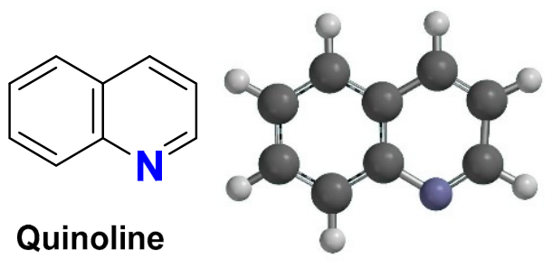
Quinoline, also known as leucoline, 1-azanaphthalene, 1-benzazine, or benzo[b]pyridine, is one of the most studied members of the aromatic heterocyclic compounds. Its chemical formula, C
9H
7N, reflects the fusion of a benzene ring with a pyridine ring, and it behaves as a weak base (pKa = 4.85) [
1]. It can act as either a nucleophile or an electrophile, and the uneven electron density between the pyridine and benzene rings gives it a distinctive reactivity profile. Quinoline forms salt complexes with acids and undergoes electrophilic and nucleophilic substitution in patterns comparable to those of pyridine and benzene (
Figure 1) [
2].
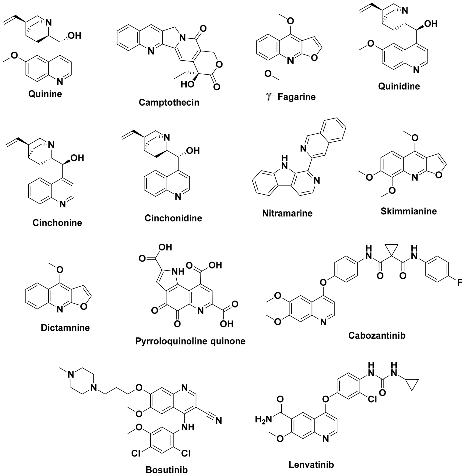
F. Runge first isolated quinoline from coal tar in 1834, and this discovery led to the later recognition of the quinoline core in a number of well-known natural alkaloids, including quinine [
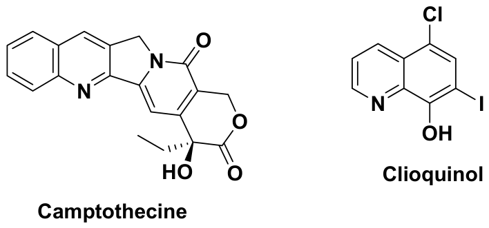
3], camptothecin [
4], and γ-fagarine [
5], all valued for their therapeutic properties. Beyond these natural sources, the quinoline core has also been deliberately incorporated into fully synthetic derivatives, including fluoroquinolone antibiotics and, more recently, kinase inhibitors developed for targeted cancer therapy, discussed further in
Section 11 (
Figure 2).
Because extracting naturally occurring quinoline compounds directly from their sources is costly and technically demanding, laboratory synthesis became the preferred alternative, making it possible not only to reproduce these molecules but also to design improved variants with better efficacy and fewer side effects. This effort led, among other things, to quinoline-based fluoroquinolones, which remain effective against a wide range of bacterial infections. Building on this interest, our laboratory has focused on a series of quinoline-based compounds, in particular 2-oxo-1,2-dihydroquinoline-4-carboxylic acid derivatives synthesized from isatin [
6,
7].
The literature search for this review was identified through systematic searches of major scientific databases, combining the terms quinoline with each therapeutic domain discussed below (e.g., “antibacterial,” “anticancer,” “antimalarial,” “kinase inhibitor,” “PROTAC”), as well as with structural and mechanistic descriptors (“structure-activity relationship,” “molecular docking,” “binding mode”) to capture studies addressing target-level interactions rather than phenotypic activity alone.
The review is organized in two parts: the first covers the main synthetic methodologies for quinoline derivatives, from classical routes to more recent, sustainable approaches, and the second covers their therapeutic applications, antibacterial [
8,
9], anticancer [
10], anti-inflammatory [
11], antimalarial [
12], antiparasitic [
13], antitubercular [
14], antiviral [
15], and antioxidant [
16] activities, along with emerging directions such as PROTACs, kinase inhibitors, and multitarget-directed ligands. Throughout, we use selected examples to illustrate structure-activity relationships and to discuss why particular quinoline-based scaffolds have proven pharmacologically relevant, not just how they are made.
2. Methods of Synthesis of Quinoline Derivatives
2.1. Conventional Methods
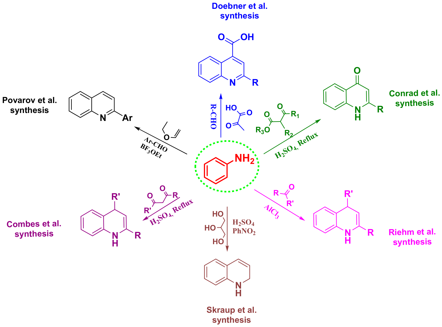
A number of classical methods have been used over the years to build the quinoline ring, each starting from different reagents and proceeding through a distinct mechanism [
17]. In the Skraup synthesis [
18], aniline is condensed with glycerol in the presence of a strong acid (H
2SO
4) and an oxidizing agent (PhNO
2), and the quinoline ring forms through subsequent cyclization and aromatization. The Doebner synthesis [
19] combines aniline with an aldehyde and pyruvic acid in a condensation-cyclization sequence to give quinoline-4-carboxylic acid derivatives. The Conrad-Limpach reaction [
20] proceeds by condensing aniline with a β-ketoester, followed by acid-catalyzed intramolecular cyclization, giving access to 4-hydroxyquinolines. The Combes synthesis [
21] works in a related way, using aniline and a β-dicarbonyl compound under acid catalysis (H
2SO
4) to form substituted quinolines. The Povarov reaction [
22], an aza-Diels–Alder-type process, involves cycloaddition between an imine (formed from aniline and an aldehyde) and an alkene, giving highly substituted tetrahydroquinolines that can then be oxidized to the aromatic quinoline. Finally, the Riehm synthesis [
23] builds functionalized quinoline frameworks from aniline derivatives and carbonyl compounds under acidic conditions (AlCl
3). Taken together, these six reactions form the classical foundation of quinoline chemistry and continue to inform the design of newer synthetic methods (
Figure 3).
2.2. Recent Synthetic Methodologies
2.2.1. Synthesis from Indoles
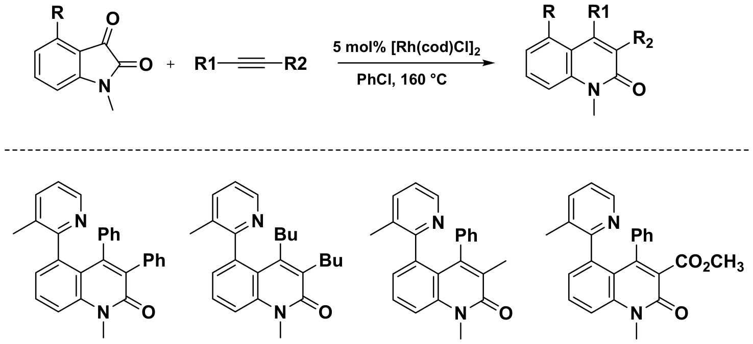
Zeng and Dong developed a rhodium-catalyzed reaction between isatins and alkynes that proceeds through an unusual decarbonylative coupling pathway [
24]. A notable feature of this method is that it activates a C–C bond selectively, rather than the arene C–H bond activation more commonly seen in this type of synthesis, achieved by controlling the orientation of the directing group. Here, the methyl substituent at the 3-position of the pyridine ring directs the rhodium catalyst toward the carbonyl group of the isatin substrate. Mechanistic work allowed the authors to isolate a rhodium intermediate, complex II (L = pyridine), which forms even at room temperature, though alkyne insertion into this intermediate only proceeds above 130 °C [
24]. This temperature dependence indicates that the alkyne insertion step, rather than formation of the rhodium intermediate, is what limits the overall catalytic cycle (
Scheme 1).
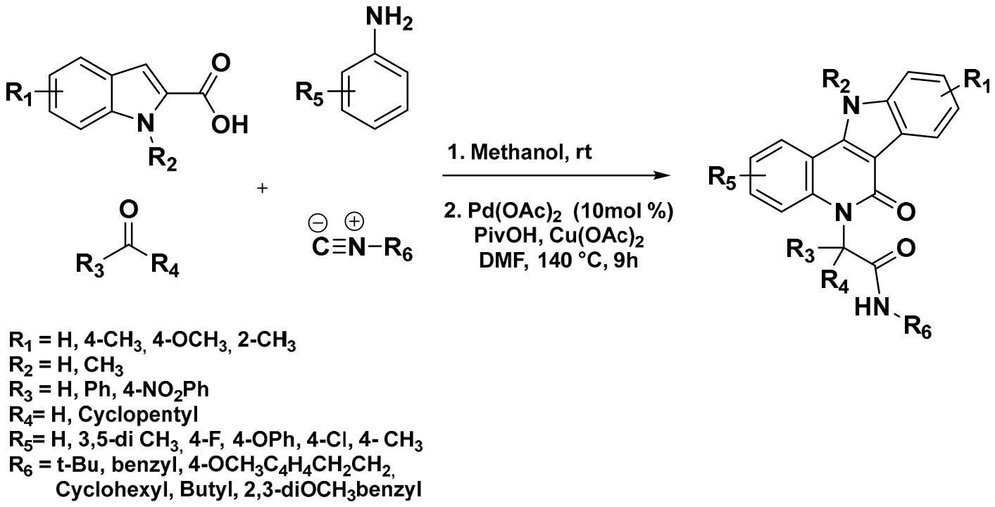
Dömling and co-workers applied the Ugi multicomponent reaction to the synthesis of fused indoloquinolone derivatives [
25]. Indole-2-carboxylic acids served as the starting point for the synthesis of the tetracyclic framework, and the resulting intermediates were then subjected to palladium-catalyzed dual C–H functionalization, giving the tetracyclic indoloquinolones in moderate yields (
Scheme 2). One advantage of this route is that it relies on readily available building blocks, which makes the overall strategy flexible and easy to adapt. The authors also evaluated the synthesized compounds by in silico molecular docking, pointing to their potential as kinase inhibitors (
Scheme 2) [
25].
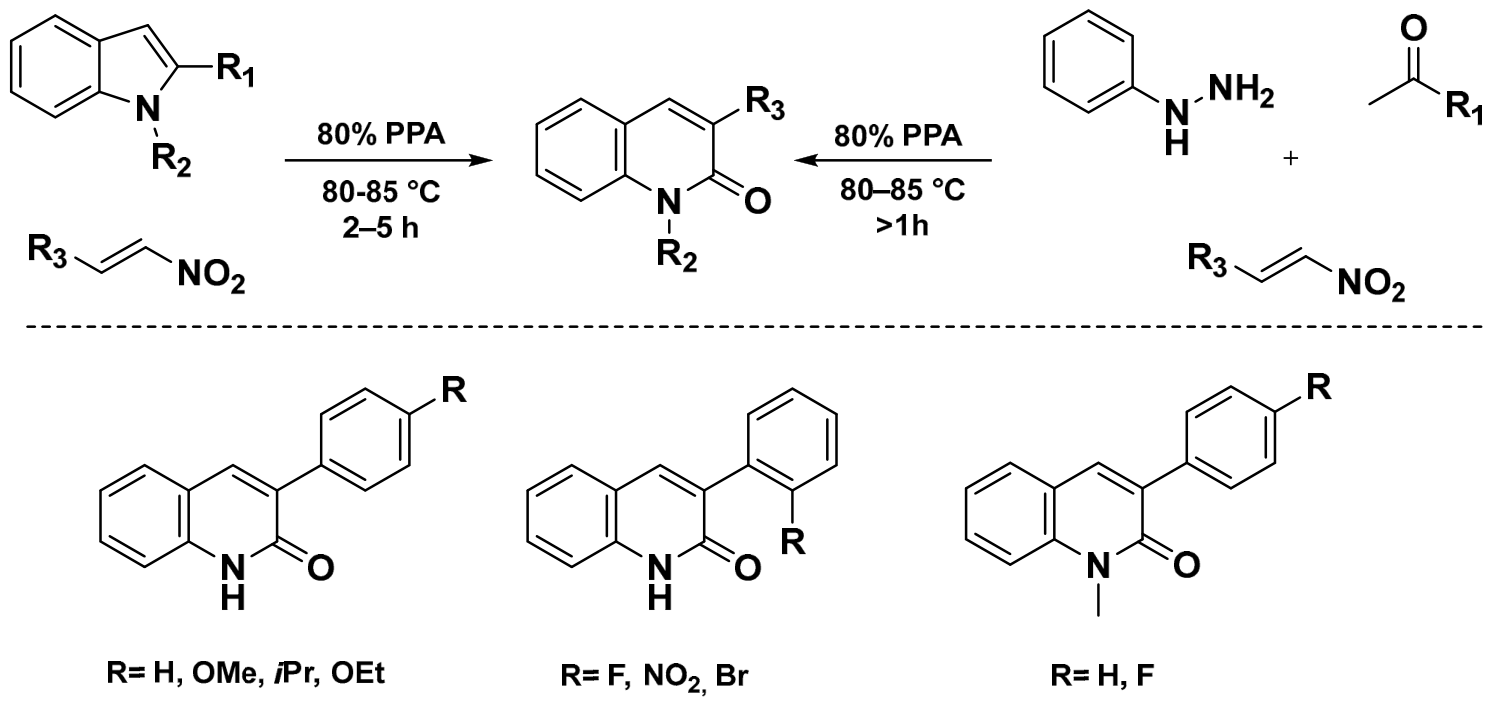
In 2015, the Aksenov research group developed an efficient method for the synthesis of 3-substituted 2-quinolones [
26]. The reaction proceeded through the formation of hydroxamic acid intermediates generated by the electrophilic alkylation of indoles in the presence of polyphosphoric acid (PPA). These intermediates subsequently underwent a ring-opening/ring-closing cascade process to afford the desired 2-quinolinones. The reaction outcome was strongly influenced by the steric properties of the nitrostyrene substrates, with ortho-substituted derivatives providing lower yields. Furthermore, the methodology was extended to a one-pot three-component process, where the indole intermediate was generated in situ through a Fischer indole synthesis before cyclization (
Scheme 3).
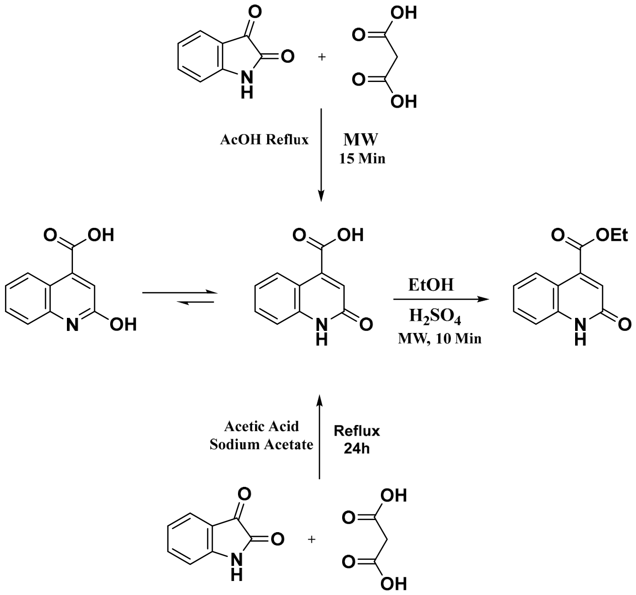
El Ashry et al. [
27] developed a microwave-assisted route to 2-quinolone-4-carboxylic acid derivatives as a faster alternative to conventional reflux. Under microwave irradiation, the target compound was obtained in 78% yield after only 15 min, and the resulting acid was then converted into ethyl 2-oxo-1,2-dihydroquinoline-4-carboxylate by microwave-promoted esterification, illustrating how microwave heating can shorten reaction times without sacrificing yield (
Scheme 4).
In our own work [
28,
29,
30], 2-quinolone-4-carboxylic acid derivatives were obtained by condensing isatin with malonic acid under reflux in acetic acid. This route is straightforward, gives the target compounds in excellent yields, and does not require specialized equipment or harsh reaction conditions, making it a convenient way to access the quinolone scaffold (
Scheme 4).
Given the wide range of biological activities associated with quinoline derivatives, finding efficient ways to synthesize these heterocycles remains an active research area [
31]. Notable progress has been made in recent years, largely through methods that are both more environmentally friendly and more cost-effective: microwave-assisted synthesis, multicomponent reactions, solvent-free transformations, ultrasound-assisted protocols, phase-transfer catalysis, and photocatalysis [
31]. Compared with traditional procedures, these approaches generally offer higher reaction efficiency and product yields, lower energy consumption, and better alignment with green chemistry principles.
2.2.2. Ultrasound-Assisted Synthesis
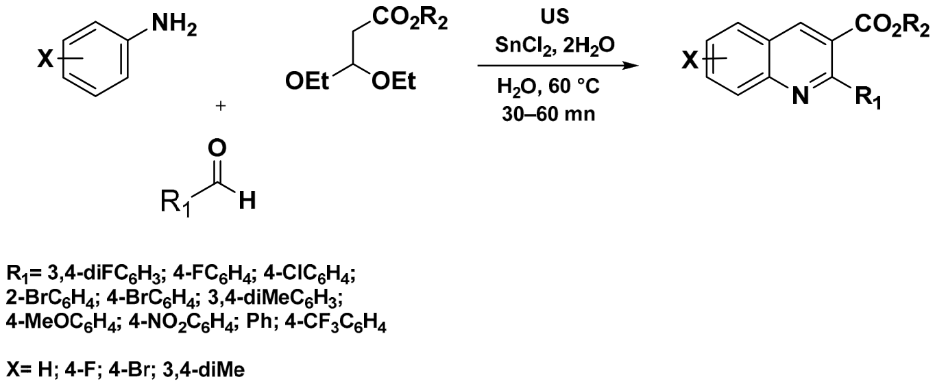
Prasad et al. reported a one-pot method for the rapid synthesis of 2-substituted quinolines using ultrasound irradiation (US) in aqueous medium [
32]. The reaction uses CuCl
2·2H
2O as a precatalyst and atmospheric oxygen as the terminal oxidant, and proceeds through a three-component condensation of aniline, an aldehyde, and ethyl 3,3-diethoxypropionate under ultrasonic activation, giving the target quinolines in satisfactory yields within relatively short reaction times. Carrying out the reaction in water under ultrasonic irradiation makes this protocol simpler to run and more environmentally friendly than conventional synthetic routes to quinolines (
Scheme 5) [
32].
In 2020, Devi et al. [
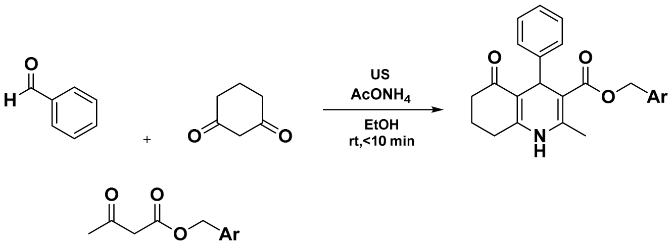
33] reported a low-cost, environmentally friendly protocol for synthesizing 2-methyl-5-oxo-hexahydroquinoline-3-carboxylate derivatives through a one-pot multicomponent reaction of substituted benzaldehydes, 1,3-cyclohexanedione, benzyl acetoacetate, and ammonium acetate in ethanol, carried out under ultrasound irradiation at room temperature. Ultrasonic activation markedly increased the reaction rate, giving the products in excellent yields (92–98%) in under 10 min. With its operational simplicity, short reaction times, and mild, environmentally benign conditions, this method offers a practical and sustainable route to hexahydroquinoline derivatives (
Scheme 6).
2.2.3. Microwave-Assisted Synthesis
In 2017, Moloi et al. [
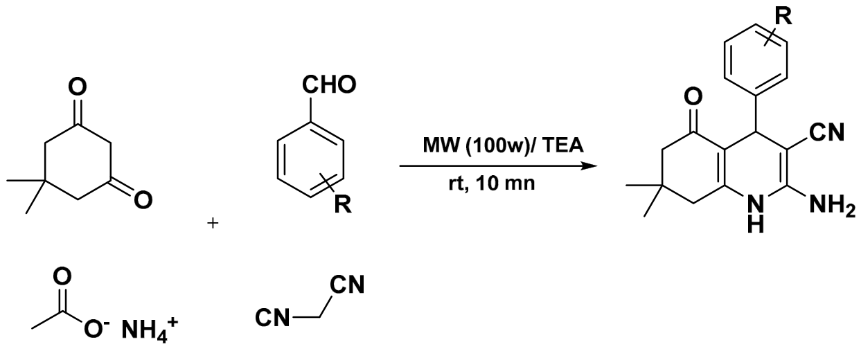
34] reported a microwave-assisted, four-component synthesis of novel quinolone derivatives from aromatic aldehydes, malononitrile, 5,5-dimethylcyclohexane-1,3-dione, and ammonium acetate. The reaction was run in ethanol with triethylamine (Et
3N) as catalyst under microwave irradiation and gave rapid access to the target quinolones in excellent yields, an example of how microwave-assisted multicomponent reactions can be a useful strategy for building biologically relevant heterocycles (
Scheme 7).
In 2019, Patel and co-workers [
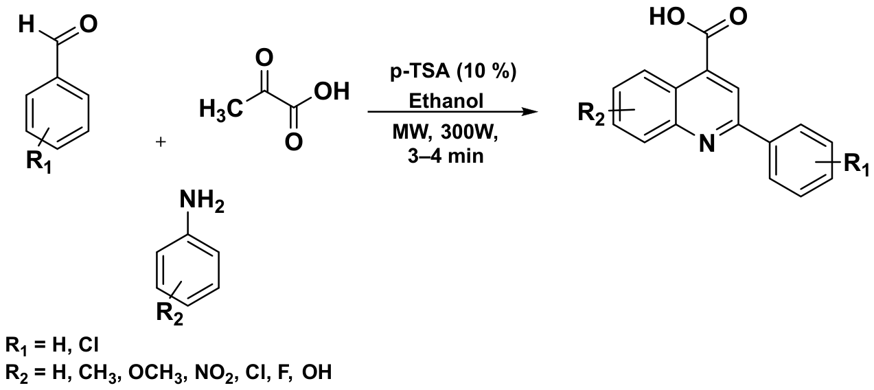
35] used p-toluenesulfonic acid (p-TSA) as an organocatalyst for the synthesis of quinoline-4-carboxylic acid derivatives, prepared through a one-pot, three-component reaction of an aromatic benzaldehyde, a substituted aniline, and pyruvic acid under microwave irradiation. The reaction reached completion in only 3–4 min and gave the target quinolines in moderate to good yields (50–80%), underlining how fast and practical microwave-assisted synthesis can be for this class of compounds (
Scheme 8).
In 2020, Tasqeeruddin et al. [
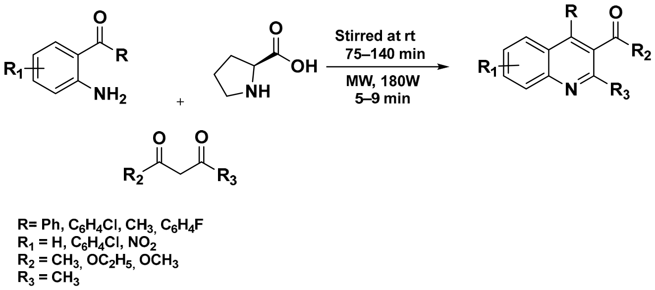
36] reported an environmentally friendly synthesis of quinoline derivatives using L-proline as an organocatalyst in a Knoevenagel condensation, in which substituted 2-aminoaryl ketones were reacted with active methylene compounds in the presence of L-proline. Carried out under microwave irradiation, the reaction was complete within 5–10 min, showing how microwave heating can speed up the cyclization step and shorten overall reaction time. This methodology combines the advantages of green chemistry with operational simplicity, providing an efficient route to quinoline-based heterocyclic frameworks (
Scheme 9).
2.2.4. Solvent-Free Reactions
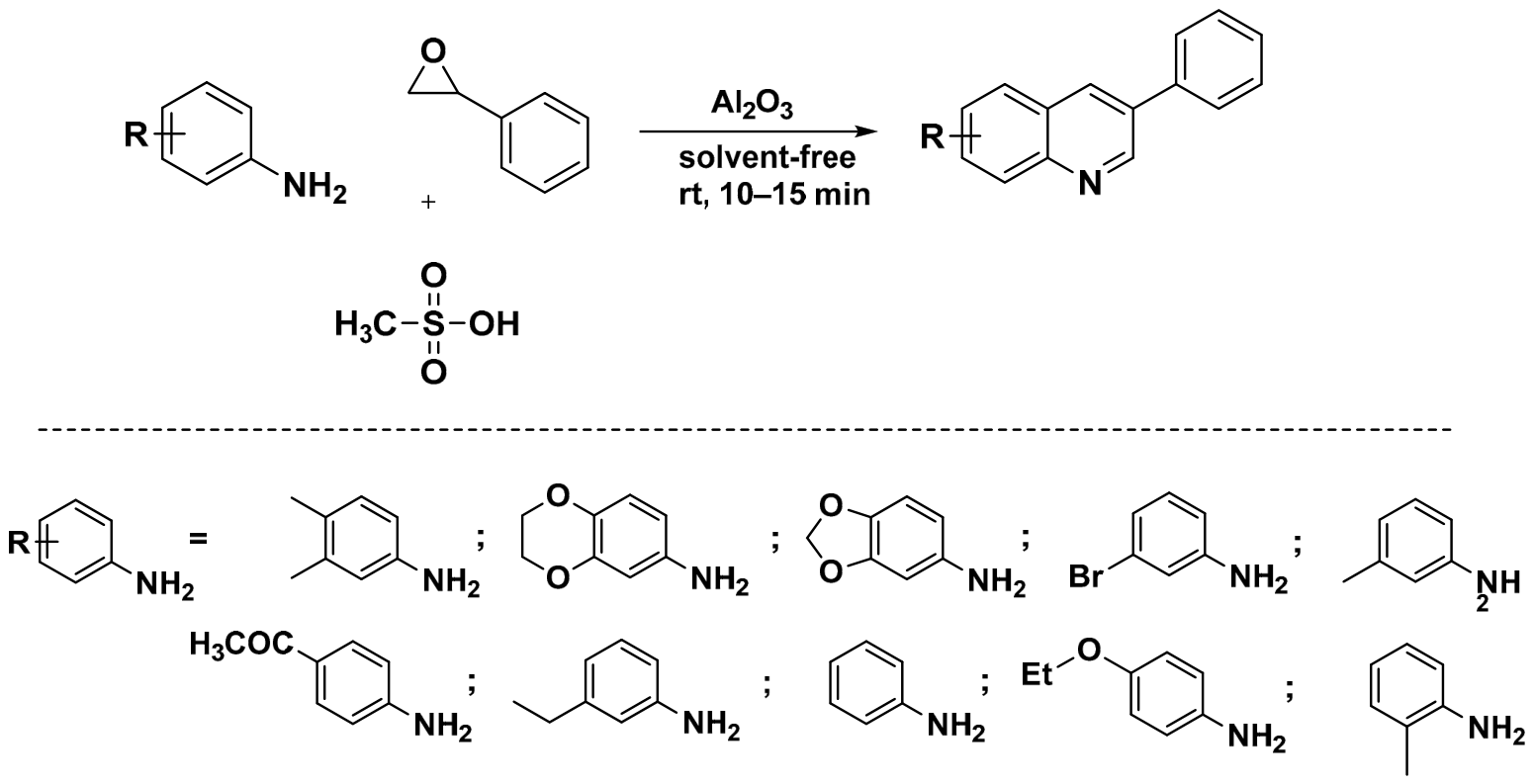
In 2017, Sharghi and colleagues [
37] developed a simple, efficient, and environmentally benign protocol for the synthesis of 3-arylquinolines. The methodology involves a one-pot reaction between anilines and styrene oxide catalyzed by activated alumina (Al
2O
3) in the presence of methanesulfonic acid (MeSO
3H). The reaction proceeds under solvent-free conditions at room temperature, providing rapid access to the desired 3-arylquinoline derivatives within 10–15 min. The method produced the target compounds in good yields and provides a practical and sustainable alternative to conventional synthetic methods, highlighting the advantages of solvent-free organic synthesis in the synthesis of quinoline-based heterocycles (
Scheme 10).
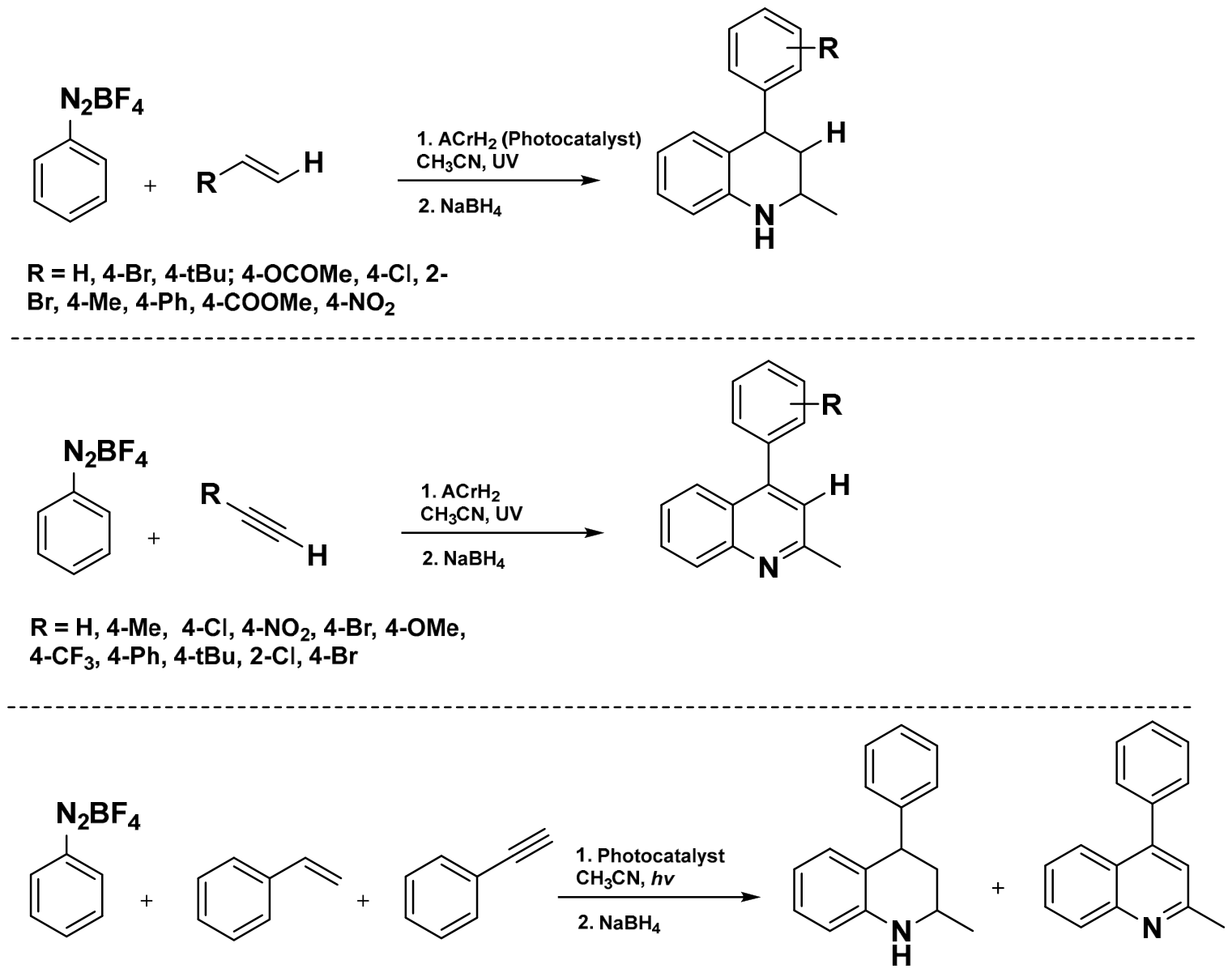
In 2018, Zhou and co-workers developed new UV light-induced synthetic methodologies for the synthesis of disubstituted quinoline derivatives [
38]. They identified 10-methyl-9,10-dihydroacridine (AcrH
2) as a highly efficient metal-free photoredox catalyst for the synthesis of quinoline derivatives through a cascade annulation process. The reaction proceeds under mild conditions at room temperature via a single-electron transfer (SET) mechanism followed by intramolecular cyclization. This photocatalytic strategy enabled the efficient synthesis of a wide range of quinoline and tetrahydroquinoline derivatives, demonstrating the potential of visible-light-driven photoredox catalysis as a sustainable and versatile approach for the preparation of nitrogen-containing heterocyclic frameworks [
38]. In conclusion, a UV light-driven and transition metal-free methodology was developed for the synthesis of quinoline and tetrahydroquinoline derivatives under mild conditions at room temperature using simple and readily accessible starting materials (
Scheme 11).
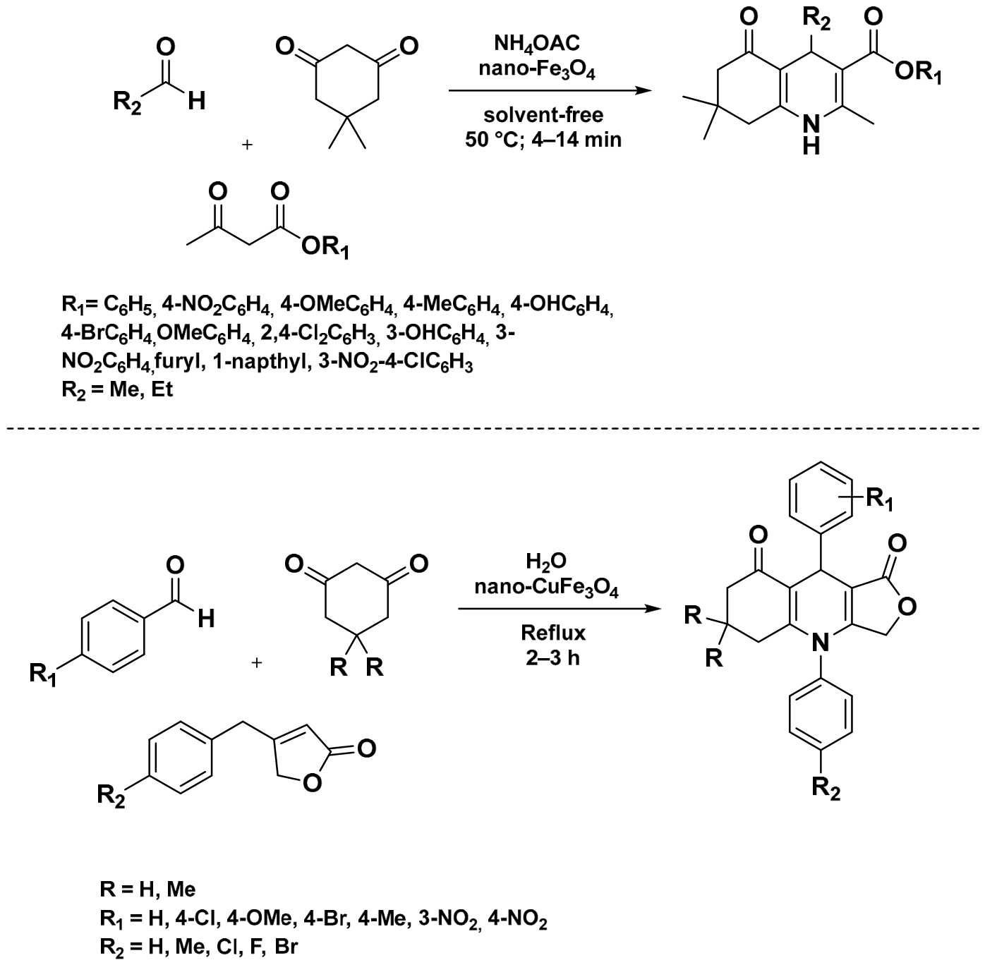
Khakyzadeh et al. [
39] reported the use of magnetite nanoparticles (Fe
3O
4) as a green, efficient, heterogeneous, and recyclable nanocatalyst for the atom-economical synthesis of hexahydroquinoline derivatives. The target compounds were prepared through a one-pot multicomponent reaction involving aryl aldehydes, dimedone, β-ketoesters, and ammonium acetate under solvent-free conditions at 50 °C.
This method gave the hexahydroquinoline derivatives in excellent yields (85–92%) within short reaction times, and worked well with both electron-donating and electron-withdrawing substituents on the aromatic aldehydes. Reaction conditions were further optimized using Response Surface Methodology, specifically a Central Composite Design, to identify the conditions giving the highest yields. Between the high catalytic efficiency, the recyclable catalyst, and the solvent-free conditions, this approach offers a sustainable route to quinoline-based heterocycles [
39].
In 2014, Farhang et al. reported an environmentally friendly synthesis of quinoline derivatives using CuFe
2O
4 nanoparticles as a heterogeneous nanocatalyst in aqueous medium [
40]. The reaction proceeds by condensing substituted anilines with ketones in water, giving the corresponding quinolines in excellent yields (84–95%) under mild conditions and with broad substrate compatibility. The CuFe
2O
4 nanoparticles were prepared through the thermal decomposition of Cu(NO
3)
2 and Fe(NO
3)
3 in an aqueous sodium hydroxide solution. Notably, the catalyst could be recovered and reused for up to five consecutive cycles without a significant decrease in catalytic activity or product yield. These findings demonstrate the potential of CuFe
2O
4 nanoparticles as a sustainable and recyclable catalyst for the green synthesis of quinoline derivatives (
Scheme 12) [
40].
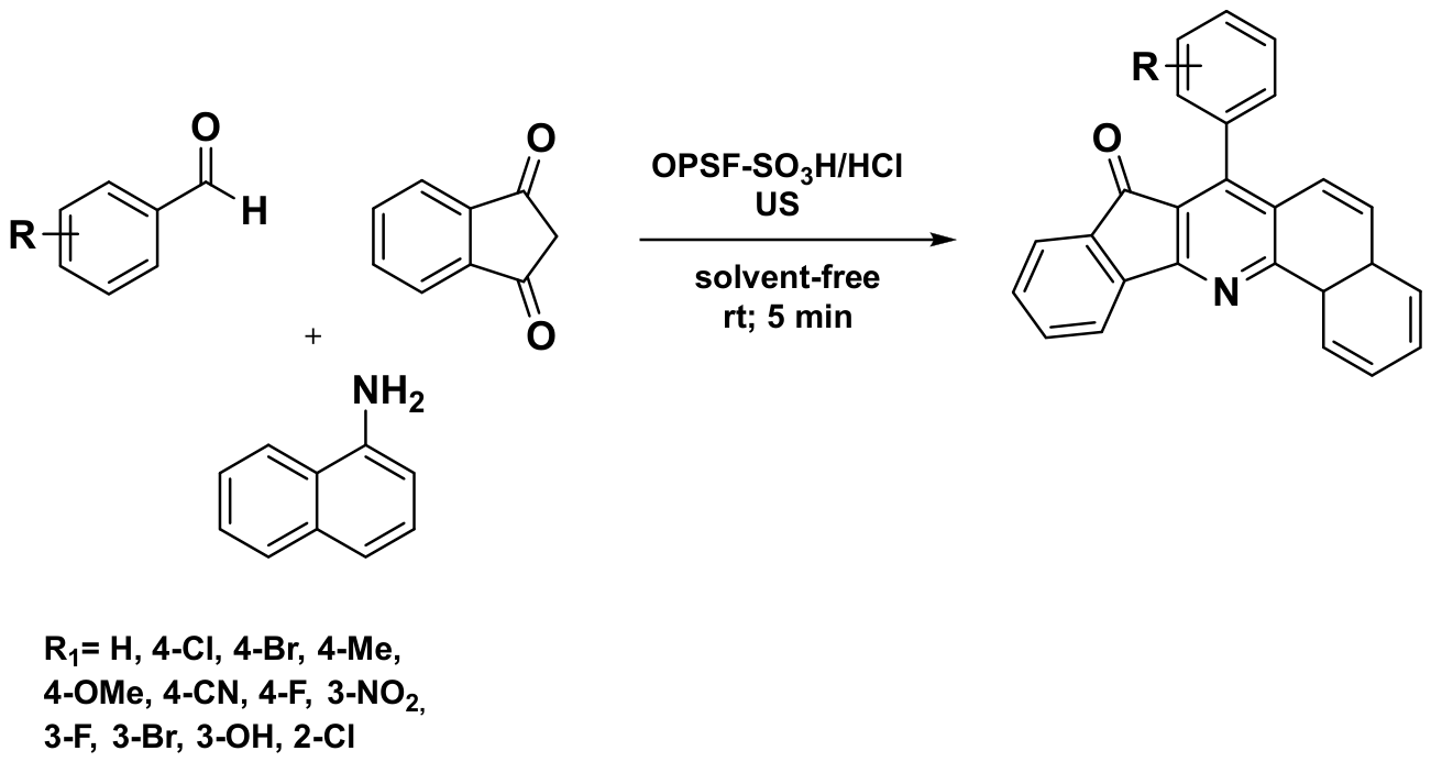
Firouzi-Haji and co-workers [
41] developed a novel hybrid magnetic organometallic nanocatalyst by immobilizing
o-phenylenediamine onto silica-coated Fe
3O
4 magnetic nanoparticles. Characterization by scanning electron microscopy (SEM) revealed a core–shell spherical morphology with a uniform particle size distribution and an average diameter of approximately 40 nm.
The catalytic performance of this nanomaterial was evaluated in the selective synthesis of 7-aryl-8H-benzo[h]indeno [1,2-b]quinoline-8-one derivatives. The target compounds were obtained in good to excellent yields ranging from 86% to 98% under solvent-free conditions and ultrasonic irradiation at room temperature. The combination of a recyclable magnetic nanocatalyst, mild reaction conditions, and high product yields highlights the efficiency and sustainability of this approach for the construction of complex quinoline-fused heterocyclic frameworks (
Scheme 13) [
41].
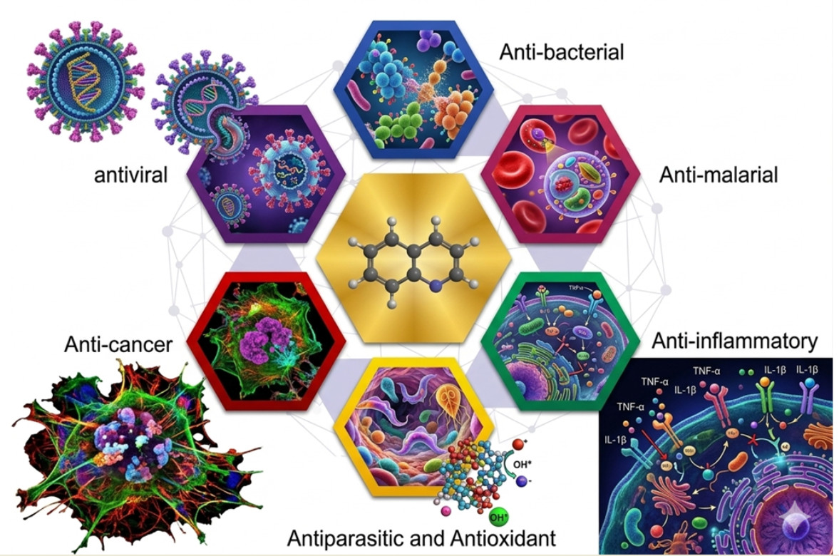
Having covered the main classical and recent synthetic approaches to quinoline derivatives, microwave- and ultrasound-assisted protocols, solvent-free reactions, photocatalytic methods, and nanocatalyst-mediated transformations, we now turn to their biological and therapeutic significance. The sections below discuss the medicinal potential of these molecules and the relationship between structural modification and pharmacological activity, supported by tables and figures summarizing target, mechanism of action, and reported potency for each activity.
3. Antibacterial Activity
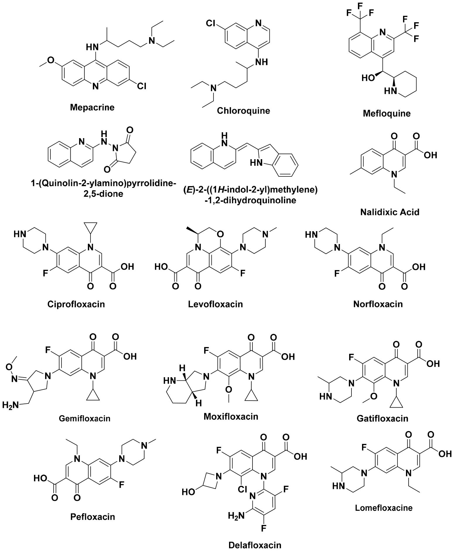
Quinoline derivatives have drawn considerable interest as antibacterial agents for their ability to interfere with essential cellular processes, including DNA replication, enzyme activity, and membrane integrity. Among the classical quinoline-based drugs, mepacrine (quinacrine) [
42] has shown inhibitory activity against several Gram-positive and Gram-negative bacteria, including
Staphylococcus aureus,
Escherichia coli, and
Pseudomonas aeruginosa.
Its antimicrobial effect is generally attributed to its capacity to intercalate into DNA and alter membrane-associated functions. Chloroquine [
43], although primarily developed as an antimalarial drug, has demonstrated moderate antibacterial activity against intracellular pathogens and has been reported to enhance the susceptibility of resistant bacterial strains when used in combination therapies [
44]. Mefloquine [
45] has exhibited significant activity against Gram-positive bacteria, particularly
S. aureus, including methicillin-resistant strains (MRSA), with minimum inhibitory concentration (MIC) values of 16 μg/mL. Moreover, mefloquine has been reported to inhibit the growth of
Enterococcus faecalis and
Streptococcus pneumoniae at low micromolar concentrations [
46]. Primaquine generally displays weaker antibacterial activity; however, its quinoline remains an attractive scaffold for structural optimization. Recent studies have demonstrated that hybrid quinoline derivatives containing indole, pyrrolidinedione, thiazolidinone, or other heterocyclic fragments exhibit enhanced antibacterial potency against clinically relevant pathogens such as
S. aureus,
Bacillus subtilis,
E. coli,
Klebsiella pneumoniae, and
P. aeruginosa. Several of these compounds showed good MIC values, highlighting the potential of quinoline-based molecules as promising candidates for the development of new antibacterial agents capable of addressing the growing challenge of antimicrobial resistance (
Figure 4 and
Table 1) [
46].
Quinolone antibiotics make up one of the most successful families of synthetic antibacterial agents derived from the quinoline scaffold. This class began with the discovery of nalidixic acid in 1958, which proved effective against Gram-negative pathogens such as
E. coli,
Proteus mirabilis,
K. pneumoniae, and
Enterobacter spp., with MIC values typically between 1 and 16 μg/mL. Its narrow antibacterial spectrum and poor tissue distribution, despite its clinical usefulness in urinary tract infections, prompted the search for more potent derivatives [
47].
The introduction of a fluorine atom at the C-6 position marked a major step forward, giving rise to the fluoroquinolones. SAR studies showed that C-6 fluorination substantially increases lipophilicity, bacterial cell penetration, and affinity for DNA gyrase and topoisomerase IV, while adding a piperazinyl or related heterocyclic group at C-7 broadens the spectrum of activity, particularly against Gram-negative organisms such as
P. aeruginosa. Together, these changes produced compounds up to 100-fold more active than the parent quinolones [
48].
Norfloxacin, among the earliest fluoroquinolones, showed strong activity against
E. coli,
Proteus spp.,
Klebsiella spp., and
P. aeruginosa, with MIC values generally under 1 μg/mL. Later optimization produced moxifloxacin and gemifloxacin, both markedly more potent against Gram-positive pathogens such as
S. pneumoniae and
S. aureus. Moxifloxacin reaches MIC
90 values of about 0.06–0.25 μg/mL against
S. pneumoniae, while gemifloxacin is active at MICs as low as 0.008–0.12 μg/mL against susceptible strains, and both retain activity against atypical respiratory pathogens including
Mycoplasma pneumoniae,
Chlamydia pneumoniae, and
Legionella pneumophila [
49,
50].
From a SAR standpoint, the 3-carboxylic acid and 4-keto groups are what make quinolones effective antibacterials, since they are required for magnesium-mediated binding to the target enzyme, while substitutions at C-6, C-7, and C-8 shape pharmacokinetics, spectrum, and resistance behavior [
51]. These structural principles still guide the design of new agents active against multidrug-resistant bacteria, including MRSA and resistant
P. aeruginosa [
52,
53,
54].
At the molecular level, fluoroquinolones work by trapping the cleavable complex that forms between DNA and type II topoisomerases, DNA gyrase in Gram-negative bacteria, topoisomerase IV in Gram-positive bacteria, once DNA strand cleavage has occurred, preventing religation and causing lethal double-strand breaks. Structural and modeling data show that the C-3 carboxylic acid and C-4 keto group chelate a Mg
2+ ion, which bridges the drug to a conserved serine and acidic residue (Ser83 and Asp87 in
E. coli GyrA numbering) through a water-mediated network [
55]. This water–metal bridge holds the quinolone within the DNA–enzyme complex, and mutations at these residues, a frequent clinical resistance mechanism, disrupt the bridge and reduce drug binding accordingly. The C-7 piperazinyl or pyrrolidinyl group points toward the GyrB/ParE subunit, and its basicity and size affect both potency against Gram-negative efflux pumps/porins and affinity for topoisomerase IV, which is part of why later agents like moxifloxacin and gemifloxacin cover Gram-positive organisms so well. The C-8 fluorine or methoxy substituent also improves anaerobic activity and lowers phototoxicity by shifting the electron density of the quinolone core [
56].
Quinoline antibiotics remain essential against resistant pathogens, and hybrid designs, such as coupling quinolines to amino acids or peptides, are currently being explored. Resistance, driven mainly by mutations in gyrase and topoisomerase IV and by efflux pump overexpression, remains the main obstacle facing this drug class. Two strategies proposed to work around this are combining quinolones with efflux pump inhibitors, and shifting the target from direct bactericidal action toward quorum-sensing disruption. On the structural side, future design will need to balance lipophilicity carefully, enough for good cell penetration, but not so much that it invites efflux recognition, while also steering clear of the cardiotoxicity linked to certain substitution patterns in this class [
57].
4. Anticancer Activity
Quinoline-based compounds received significant attention in anticancer drug discovery for their ability to interact with key molecular targets involved in cell proliferation, DNA replication, and signal transduction. Several naturally occurring and synthetic quinoline derivatives have shown potent antitumor activity and have gone on to serve as lead structures for clinically relevant anticancer agents [
58,
59].
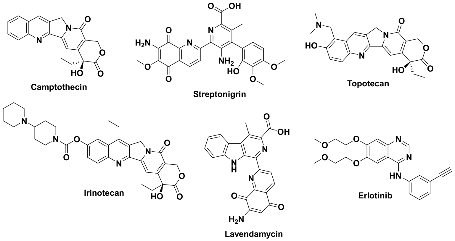
One of the earliest examples is streptonigrin, a naturally occurring aminoquinoline antibiotic isolated from
Streptomyces flocculus in 1959 [
58]. This compound exhibits potent cytotoxic activity against a broad spectrum of tumor cell lines, including murine leukemia and melanoma models, with IC
50 values typically ranging from 1 to 50 nM [
4]. Its antitumor activity is primarily attributed to DNA strand cleavage and inhibition of topoisomerase II, together with redox cycling that promotes the generation of reactive oxygen species. However, its clinical development has been severely limited by dose-limiting systemic toxicity. SAR studies have demonstrated that the quinoline-5,8-dione core is indispensable for its redox-mediated cytotoxic activity, highlighting this pharmacophore as the key structural determinant of its biological effects (
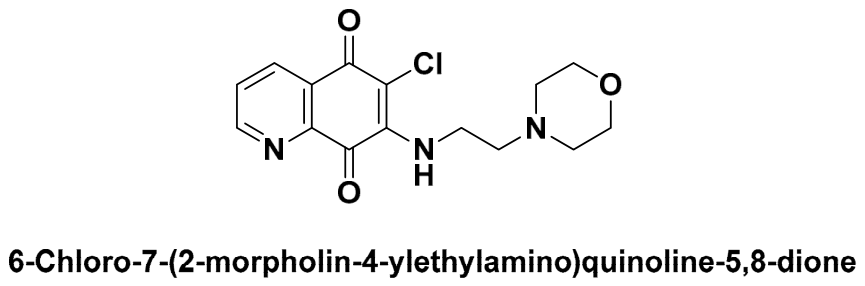
Figure 5 and
Table 2) [
59].
Camptothecin, a quinoline alkaloid isolated from
Camptotheca acuminata, inhibits DNA topoisomerase I by stabilizing the DNA–topoisomerase I cleavage complex, thereby blocking DNA religation during replication. This mechanism results in irreversible DNA damage and subsequent cell death. Camptothecin has demonstrated potent cytotoxic activity against several human cancer cell lines, including colon (HCT-116), breast (MCF-7), and lung (A549), with IC
50 values generally ranging from 5 to 100 nM. SAR studies established that the intact α-hydroxylactone E-ring is essential for activity, while modifications elsewhere on the quinoline nucleus have given derivatives with improved pharmacological profiles [
60].
Mechanistically, camptothecin stabilizes the covalent DNA–topoisomerase I cleavage complex by binding at the DNA–enzyme interface rather than directly inhibiting the catalytic activity of the enzyme. Its α-hydroxylactone E-ring forms key hydrogen-bonding interactions with active-site residues, whereas the planar pentacyclic scaffold intercalates between the DNA base pairs flanking the cleavage site. This interaction prevents DNA religation, converting transient single-strand breaks into persistent lesions that are transformed into irreversible double-strand breaks during DNA replication, ultimately triggering cell death. Hydrolysis of the α-hydroxylactone ring to the inactive carboxylate form at physiological pH destroys this hydrogen-bonding network, which is why the semi-synthetic derivatives topotecan and irinotecan were designed specifically to improve lactone stability and aqueous solubility while keeping the E-ring intact.
The poor aqueous solubility and dose-limiting toxicity of camptothecin prompted the development of semisynthetic derivatives, notably topotecan and irinotecan. Topotecan exhibits potent activity against ovarian and small-cell lung cancer, with reported IC50 values of 10–100 nM. Irinotecan is a prodrug that is converted by carboxylesterases into SN-38, a highly potent topoisomerase I inhibitor displaying subnanomolar to low-nanomolar activity against colorectal cancer cell lines. Structural modifications at positions 9 and 10 of the quinoline scaffold improve aqueous solubility, lactone stability, and pharmacokinetic properties while preserving topoisomerase I inhibition.
Another representative quinoline-containing natural product is lavendamycin, isolated from
Streptomyces lavendulae [
61]. Lavendamycin exhibits cytotoxic activity against leukemia, breast, and colon cancer cell lines, with IC
50 values generally in the low micromolar range. Its antitumor activity has been attributed to topoisomerase I inhibition together with bioreductive activation, leading to DNA damage and apoptosis.
Furthermore, gefitinib and erlotinib have revolutionized targeted cancer therapy through selective inhibition of the epidermal growth factor receptor (EGFR) tyrosine kinase. Structurally, both compounds are 4-anilinoquinazolines rather than quinolines, bearing an additional ring nitrogen relative to the quinoline core; they are discussed here because of their close structural relationship to the quinoline scaffold and because the underlying hinge-binding SAR principles apply equally to both ring systems. Erlotinib displays potency with IC50 values close to 2 nM against EGFR kinase. SAR studies have established that the 4-anilinoquinazoline pharmacophore is crucial for ATP-binding site recognition and kinase inhibition, making it one of the most successful quinazoline-based motifs in modern oncology.
The 4-anilinoquinazoline pharmacophore of gefitinib and erlotinib occupies the ATP-binding cleft of the EGFR tyrosine kinase domain, forming a key hydrogen bond between the ring nitrogen and the backbone NH of Met793 in the hinge region. The aniline substituent at C-4 extends into a hydrophobic back pocket lined by Thr790, the gatekeeper residue, and its steric and electronic complementarity to this pocket determines both potency and susceptibility to the T790M gatekeeper mutation, which sterically hinders drug binding and is the principal mechanism of acquired resistance. The solubilizing substituents at positions 6 and 7 are oriented toward solvent-exposed regions, where they mainly improve physicochemical and pharmacokinetic properties without significantly affecting target binding. This structural insight has guided the rational design of third-generation EGFR inhibitors capable of overcoming T790M-mediated resistance.
5. Anti-Inflammatory Activity
Quinoline derivatives have attracted considerable interest as anti-inflammatory agents because of their ability to modulate multiple signaling pathways involved in the inflammatory response. Numerous quinoline-based compounds have demonstrated significant anti-inflammatory activity through diverse mechanisms, including inhibition of cyclooxygenase (COX) and lipoxygenase (LOX), suppression of pro-inflammatory cytokines such as tumor necrosis factor-α (TNF-α), interleukin-1β (IL-1β), and interleukin-6 (IL-6), as well as attenuation of oxidative stress [
11].
SAR studies indicate that electron-donating substituents, particularly hydroxyl and amino groups, generally strengthen anti-inflammatory activity by improving interactions with inflammatory targets, while bulky aromatic substituents tend to favor COX-2 selectivity (
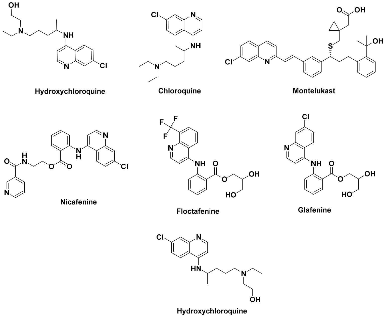
Figure 6 and
Table 3) [
62].
Among clinically relevant quinoline derivatives, chloroquine and hydroxychloroquine remain the most studied [
61]. Beyond their antimalarial use, both compounds exert immunomodulatory effects by inhibiting lysosomal acidification, antigen presentation, and cytokine production. Hydroxychloroquine is widely used in rheumatoid arthritis and systemic lupus erythematosus, where it markedly lowers serum TNF-α and IL-6, and in cellular assays it inhibits inflammatory mediator production with IC
50 values in the low micromolar range (roughly 2–10 μM), consistent with its clinical efficacy in these conditions [
61].
Chloroquine and hydroxychloroquine accumulate preferentially in acidic intracellular compartments such as lysosomes and endosomes, a consequence of their weakly basic, diprotic amine side chain, and this raises the intraluminal pH enough to impair lysosomal enzyme activity, antigen processing, and TLR9 signaling triggered by nucleic acid-containing immune complexes, a pathway particularly relevant in lupus [
61]. This mechanism differs fundamentally from the direct cyclooxygenase inhibition produced by conventional nonsteroidal anti-inflammatory drugs (NSAIDs) and accounts for the delayed onset of clinical response typically observed in autoimmune diseases.
Montelukast [
62], a quinoline-containing leukotriene receptor antagonist, represents another important anti-inflammatory agent. It selectively antagonizes the cysteinyl leukotriene receptor 1 (CysLT1), thereby suppressing leukotriene-mediated bronchoconstriction and inflammatory cell recruitment. Its high affinity for CysLT1 (IC
50 ≈ 0.5–5 nM) underlies its established efficacy in the long-term management of asthma and allergic rhinitis [
62].
Other quinoline derivatives, including nicafenine [
63], glafenine [
64], and floctafenine [
65], exhibit both analgesic and anti-inflammatory properties. Their pharmacological activity has been associated with modulation of prostaglandin biosynthesis and related inflammatory pathways. SAR studies indicate that aromatic amino substituents and appropriate substitution patterns on the quinoline nucleus play important roles in optimizing anti-inflammatory activity.
In general, these examples illustrate the remarkable versatility of the quinoline scaffold in anti-inflammatory drug discovery. Quinoline derivatives exert their therapeutic effects through distinct mechanisms, including modulation of lysosomal function, inhibition of inflammatory mediator production, antagonism of leukotriene receptors, and regulation of prostaglandin biosynthesis, highlighting the scaffold as a valuable platform for the development of anti-inflammatory agents.
6. Antimalarial Activity
Quinoline derivatives have been the cornerstone of antimalarial chemotherapy for over a century and continue to represent one of the most important classes of antiplasmodial agents [
66]. Their mechanism of action is primarily associated with the inhibition of heme detoxification within the digestive vacuole of
Plasmodium parasites, leading to the accumulation of toxic free heme and subsequent parasite death. The effectiveness of quinoline-based compounds against both chloroquine-sensitive and resistant strains has stimulated continuous efforts to develop new derivatives with improved efficacy and safety profiles (
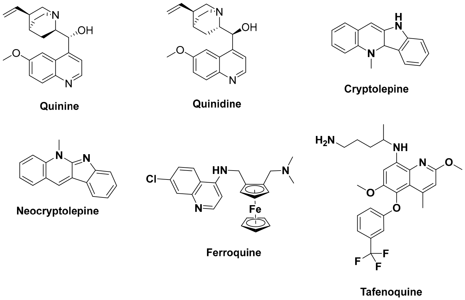
Figure 7 and
Table 4) [
67].
Quinine, the first clinically used antimalarial alkaloid isolated from the bark of Cinchona species, exhibits potent activity against Plasmodium falciparum, with reported IC
50 values generally ranging from 100 to 500 nM depending on the parasite strain. Its antimalarial activity is mainly attributed to inhibition of heme detoxification within the parasite digestive vacuole, resulting in the accumulation of toxic free heme. Although quinine remains an effective treatment for severe malaria, its long-term use is limited by adverse effects, including cinchonism and neurotoxicity. SAR studies have shown that both the quinuclidine ring and the secondary alcohol group are essential for antiplasmodial activity [
68].
Quinidine, the stereoisomer of quinine, exhibits comparable antiplasmodial potency, with IC
50 values generally ranging from 50 to 300 nM against
P. falciparum. However, its clinical use as an antimalarial has been largely restricted because of its pronounced cardiotoxic and proarrhythmic effects. Despite these limitations, quinine and quinidine have served as important lead compounds for the development of modern quinoline-based antimalarial agents [
69].
Among naturally occurring quinoline alkaloids, cryptolepine, neocryptolepine, and isocryptolepine, isolated from
Cryptolepis sanguinolenta, show strong activity against
P. falciparum. Cryptolepine in particular has IC
50 values of 20–200 nM against both chloroquine-sensitive and chloroquine-resistant strains [
70].
Its antiplasmodial activity is attributed to DNA intercalation, inhibition of hemozoin formation, and interference with nucleic acid synthesis. SAR investigations have demonstrated that substitutions on the indoloquinoline nucleus strongly influence potency and selectivity, with appropriate modifications at the nitrogen atom and aromatic ring system leading to improved antimalarial activity and reduced cytotoxicity [
70].
4-Aminoquinolines such as chloroquine accumulate within the acidic digestive vacuole of the parasite, where they bind free heme released during hemoglobin digestion and prevent its polymerization into inert hemozoin crystals [
66].
The quinoline ring stacks with the porphyrin macrocycle, while the protonated side chain at C-4 stabilizes the drug-heme complex, and the resulting adduct becomes toxic to the parasite. Mutations in the
P. falciparum chloroquine resistance transporter (PfCRT) reduce vacuolar drug accumulation through increased chloroquine efflux and represent the main resistance mechanism, which is consistent with the retained activity of ferroquine against chloroquine-resistant strains, since its ferrocenyl side chain is less readily recognized by mutant PfCRT [
67].
The remarkable efficacy of these quinoline alkaloids has established the quinoline scaffold as a privileged structure in antimalarial drug discovery. This has ultimately led to the development of clinically important agents such as chloroquine, mefloquine, and more recently tafenoquine and ferroquine, which continue to play a significant role in the fight against malaria, particularly in regions affected by drug-resistant Plasmodium strains.
7. Antiparasitic Activity
Quinoline derivatives constitute an important class of antiparasitic agents and have demonstrated efficacy against a wide variety of protozoan and helminth infections [
71]. Their antiparasitic activity is generally associated with disruption of mitochondrial function, interference with nucleic acid synthesis, inhibition of haem detoxification, and induction of oxidative stress [
13]. Owing to these diverse mechanisms of action, quinoline-based compounds continue to serve as valuable therapeutic agents and lead structures for the development of novel antiparasitic drugs (
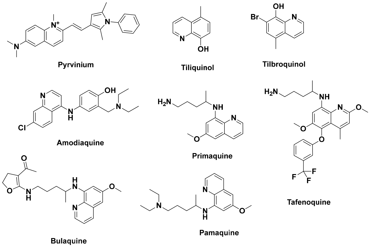
Figure 8 and
Table 5).
Among the earliest examples, pyrvinium has been widely used for the treatment of enterobiasis caused by
Enterobius vermicularis. The compound acts by inhibiting parasite mitochondrial respiration and ATP production, ultimately leading to parasite death [
72]. Tiliquinol and tilbroquinol, commonly administered in combination, exhibit potent amoebicidal activity against
Entamoeba histolytica and remain effective treatments for intestinal amoebiasis. SAR studies have shown that halogen substitution on the quinoline nucleus, particularly the bromine atom present in tilbroquinol, contributes significantly to enhanced antiparasitic activity by increasing lipophilicity and membrane permeability [
73].
The 8-aminoquinoline class, represented by primaquine, pamaquine, tafenoquine, and bulaquine, plays a pivotal role in the radical cure of malaria caused by
Plasmodium vivax and
P. ovale by eliminating dormant hepatic hypnozoites [
31]. Primaquine exhibits antiplasmodial activity with reported IC
50 values generally ranging from 100 to 500 nM, whereas tafenoquine displays greater potency, with IC
50 values frequently below 100 nM against several
P. falciparum strains. Structure–activity relationship (SAR) studies have identified the 8-aminoquinoline nucleus and its aminoalkyl side chain as key structural features required for antiplasmodial activity [
31].
In contrast to 4-aminoquinolines, which inhibit heme detoxification within the parasite digestive vacuole, 8-aminoquinolines require metabolic activation to exert their antiparasitic effects. Primaquine and tafenoquine are metabolized primarily by cytochrome P450 2D6 (CYP2D6) to reactive intermediates that induce oxidative stress through the generation of reactive oxygen species [
31]. This mechanism enables effective elimination of hepatic hypnozoites but also accounts for the risk of hemolytic anemia in individuals with glucose-6-phosphate dehydrogenase (G6PD) deficiency, limiting the use of these agents in susceptible patients.
Amodiaquine, a 4-aminoquinoline related to chloroquine, is potent against both chloroquine-sensitive and resistant strains of
Plasmodium falciparum, with IC
50 values typically between 10 and 50 nM. SAR studies show that substituting the C-4 position with aminoalkyl groups increases accumulation within the parasite’s digestive vacuole and strengthens inhibition of haem polymerization [
74]. More recently, quinoline derivatives bearing nitroaromatic and chalcone pharmacophores have shown promising activity against
Leishmania donovani,
Trypanosoma cruzi, and
Trypanosoma brucei, with IC
50 values in the low micromolar range (0.5–5 μM) [
74]. These findings highlight the importance of electron-withdrawing substituents in improving antiparasitic potency and support the continued development of quinoline-based molecular architectures as broad-spectrum antiparasitic agents.
What stands out in this class is how well the same scaffold performs across parasites that have little in common biologically: protozoa such as Plasmodium, Entamoeba, Trypanosoma, and Leishmania, and helminths such as Enterobius. This is mostly because these organisms all rely heavily on mitochondrial function and redox balance, which quinoline derivatives disrupt through fairly general mechanisms, membrane permeabilization and ROS induction, rather than by binding tightly to one specific target. That broad activity is useful in settings where precise diagnosis is not always possible, but it also means selectivity toward host mitochondria is not guaranteed and deserves more attention in future SAR work on this class.
8. Antitubercular Activity
Quinoline derivatives have emerged as valuable scaffolds in the search for new antitubercular agents, particularly in response to the growing prevalence of multidrug-resistant (MDR-TB) and extensively drug-resistant (XDR-TB) strains of
Mycobacterium tuberculosis. Their biological activity is associated with the inhibition of essential mycobacterial processes, including energy metabolism, redox homeostasis, and metal-dependent enzymatic pathways. The structural versatility of the quinoline nucleus has enabled the development of several compounds with significant activity against both replicating and dormant mycobacterial populations (
Figure 9 and
Table 6) [
75,
76].
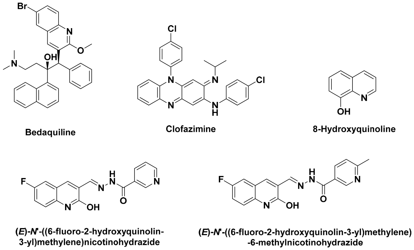
Among clinically approved drugs, bedaquiline stands out as a major breakthrough in tuberculosis therapy. This diarylquinoline derivative selectively inhibits the proton pump of mycobacterial ATP synthase, leading to depletion of intracellular ATP and bacterial death, and shows remarkable potency against
M. tuberculosis, with MIC values typically between 0.015 and 0.12 μg/mL, including activity against MDR-TB strains [
75]. SAR studies show that the diaryl and aminoalcohol substituents are essential for interaction with the ATP synthase complex and account for much of its exceptional antimycobacterial activity.
Bedaquiline, the first approved diarylquinoline for the treatment of multidrug-resistant tuberculosis, selectively inhibits mycobacterial ATP synthase by binding to the c-subunit of the Fo proton channel, thereby blocking proton translocation and ATP synthesis. This unique mechanism results in rapid depletion of intracellular ATP and potent bactericidal activity against
Mycobacterium tuberculosis. The structural differences between mycobacterial and human ATP synthase largely account for its selectivity, whereas resistance is primarily associated with mutations in the atpE gene encoding the ATP synthase c-subunit [
75].
Clofazimine, another clinically important quinoline-containing antimycobacterial agent, exhibits potent activity against
M. tuberculosis, with MIC values generally ranging from 0.06 to 0.5 μg/mL against susceptible strains. Its antimicrobial activity is mainly attributed to disruption of the mycobacterial electron transport chain and the generation of reactive oxygen species. Owing to its highly lipophilic phenazine framework, clofazimine accumulates efficiently within macrophages, contributing to its activity against persistent mycobacterial infections [
76].
8-Hydroxyquinoline and its derivatives have also attracted considerable interest because of their ability to chelate metal ions essential for mycobacterial growth, displaying MIC values of 0.5–4 μg/mL against both replicating and dormant
M. tuberculosis. In addition, Mandewale and co-workers synthesized a series of fluorinated 2-hydroxyquinoline hydrazone derivatives that completely inhibited
M. tuberculosis growth at 6.25 μg/mL. Structure–activity relationship (SAR) studies demonstrated that the combined presence of a fluorine atom at C-6, a hydroxyl group at C-2, and a pyridyl hydrazone moiety significantly enhances antimycobacterial activity [
78].
Collectively, these findings highlight the versatility of the quinoline scaffold in antitubercular drug discovery. Quinoline derivatives exert antimycobacterial activity through diverse mechanisms, including inhibition of ATP synthesis, disruption of respiratory electron transport, and metal-ion chelation, providing multiple opportunities for the development of agents active against drug-resistant and persistent M. tuberculosis strains.
9. Antioxidant Activity
Oxidative stress, resulting from an imbalance between the production of reactive oxygen species (ROS) and cellular antioxidant defenses, plays a key role in the pathogenesis of numerous chronic disorders, including cancer, cardiovascular diseases, neurodegenerative disorders, and inflammatory conditions. Consequently, the development of effective antioxidant agents has attracted considerable attention, with quinoline derivatives emerging as promising candidates because of their ability to scavenge free radicals, attenuate oxidative stress, and protect cells from oxidative damage (
Figure 10 and
Table 7) [
79].
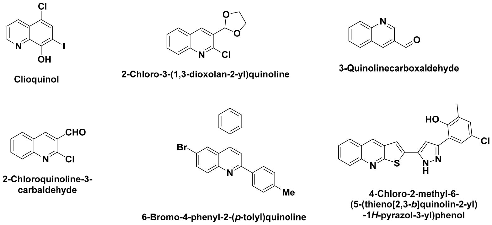
Several quinoline derivatives have demonstrated significant antioxidant activity in both chemical and cell-based assays. Clioquinol (5-chloro-7-iodo-8-hydroxyquinoline) is a representative example, exerting its antioxidant activity primarily through chelation of redox-active metal ions, thereby reducing metal-catalyzed reactive oxygen species (ROS) formation. SAR studies have identified the C-8 hydroxyl group as a key structural feature required for efficient metal coordination and antioxidant activity [
80]. In addition, 3-(1,3-dioxolan-2-yl)quinoline and chlorine-substituted 3-quinolinecarboxaldehyde derivatives have shown promising antioxidant activity in DPPH and hydrogen peroxide scavenging assays, suggesting that appropriate electron-withdrawing substituents can enhance antioxidant performance [
80].
The antioxidant properties of clioquinol arise primarily from the formation of a stable metal-chelating complex involving the C-8 hydroxyl group and the adjacent quinoline nitrogen, enabling efficient coordination of transition metal ions such as Fe
2+ and Cu
2+. By limiting the availability of these redox-active metals, clioquinol suppresses Fenton-type reactions and the subsequent generation of highly reactive hydroxyl radicals [
80].
Furthermore, Liberto and co-workers reported that appropriately substituted quinoline derivatives effectively reduce intracellular ROS levels, highlighting the importance of substitution patterns on the quinoline scaffold in optimizing antioxidant activity [
10].
Mahajan et al., in a separate study, reported a series of thiophene-based quinoline derivatives, the most active of which had an EC
50 of 12.03 ± 1.45 μg/mL in radical scavenging assays [
81]. SAR analysis suggested that the combination of a quinoline nucleus with a thiophene ring and phenolic hydroxyl substituents enhances electron-donating capacity and stabilizes radical intermediates, thereby improving antioxidant efficiency. These findings demonstrate that both the nature and position of substituents on the quinoline scaffold play a decisive role in determining antioxidant potential and support the continued development of quinoline-based antioxidants for biomedical applications [
81].
Taken as a whole, the antioxidant quinoline derivatives discussed here work through two distinct routes, metal chelation (clioquinol-type) and direct radical scavenging (thiophene/phenolic-substituted derivatives), and it is not yet clear from the current literature which of these contributes more to the biological benefit seen in cellular and animal models, an open question that future mechanistic studies on this class should address directly rather than relying solely on chemical DPPH-type assays.
10. Antiviral Activity
Beyond their well-established roles in antibacterial, antimalarial, and antiparasitic chemotherapy, quinoline derivatives have emerged as a promising class of antiviral agents with activity against coronaviruses, flaviviruses, and other RNA viruses [
82]. Their antiviral effects are mediated through multiple mechanisms, including inhibition of viral proteases, interference with viral entry and endosomal trafficking, modulation of host–cell pathways, and disruption of viral replication, highlighting the remarkable versatility of the quinoline scaffold in antiviral drug discovery [
82,
83].
Among quinoline-based antivirals, chloroquine and hydroxychloroquine have been the most extensively investigated against human coronaviruses [
82]. In a comparative evaluation of ten antimalarial quinoline derivatives against a panel of α- and β-coronaviruses, including SARS-CoV, SARS-CoV-2, HCoV-229E, and HCoV-OC43, both compounds exhibited broad-spectrum antiviral activity, with EC
50 values ranging from 0.12 to 12 μM. Chloroquine displayed the highest selectivity, achieving a selectivity index (SI) of 165 against HCoV-OC43 in HEL cells, indicating a favorable balance between antiviral potency and cytotoxicity [
82].
Amodiaquine, ferroquine, and mefloquine also displayed potent anti-coronavirus activity, although mefloquine’s activity was accompanied by substantial cytotoxicity, whereas primaquine, quinidine, quinine, and tafenoquine were only active at higher concentrations, and piperaquine lacked both antiviral and cytotoxic effects. Mechanistic studies indicated that chloroquine interferes with viral entry at a post-attachment stage and inhibits SARS-CoV-2 RNA synthesis, an effect coinciding with impaired autophagic flux, pointing to a host-directed rather than a purely virus-targeted mode of action [
82].
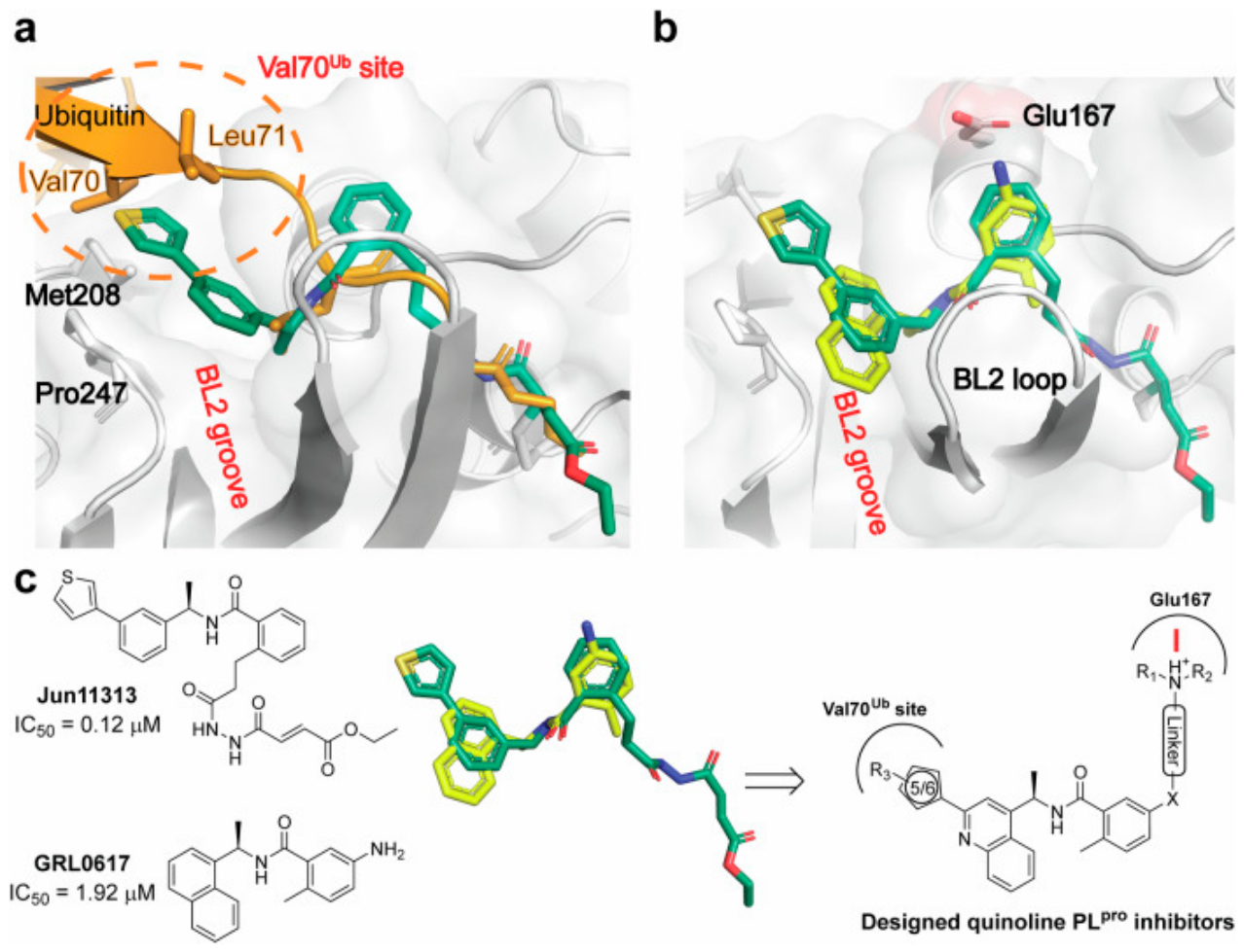
More recently, structure-based design has been applied to develop quinoline inhibitors of the SARS-CoV-2 papain-like protease (PLpro), an enzyme required for polyprotein processing and suppression of host innate immune responses [
15]. By exploiting the Val70Ub binding site on PLpro, a series of quinoline analogues was designed showing potent PLpro inhibition and antiviral activity, with X-ray structures of six lead compounds revealing that the 2-aryl substituent can occupy either the Val70Ub site or, in a flipped binding mode, the BL2 groove. The optimized lead compound, Jun13296, displayed favorable pharmacokinetic properties and potent inhibition of SARS-CoV-2 variants and nirmatrelvir-resistant mutants, and improved survival, reduced lung viral titers, and prevented lung damage in a mouse model of SARS-CoV-2 infection following oral administration. This SAR clearly identifies the Val70Ub/BL2 dual-binding capacity of the 2-aryl-quinoline substituent as a key structural determinant of potency and oral efficacy (
Figure 11) [
15].
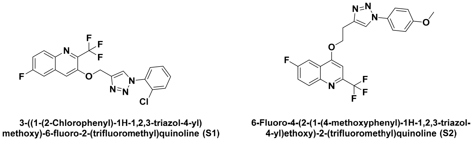
Hybridization strategies combining the quinoline core with a 1,2,3-triazole pharmacophore have also yielded promising anti-SARS-CoV-2 candidates. A series of quinoline-triazole conjugates was designed and synthesized via microwave-assisted click chemistry, among which 3-((1-(2-chlorophenyl)-1H-1,2,3-triazol-4-yl)methoxy)-6-fluoro-2-(trifluoromethyl)quinoline (
S1) and 6-fluoro-4-(2-(1-(4-methoxyphenyl)-1H-1,2,3-triazol-4-yl)ethoxy)-2-(trifluoromethyl)quinoline (
S2) showed high antiviral potency together with a favorable selectivity index relative to the chloroquine/hydroxychloroquine reference standards [
82]. SAR analysis of this series indicates that the nature of the triazole N1-aryl substituent (halogen vs. methoxy) and the length of the linker connecting it to the C-4 position of the trifluoromethylquinoline core are key modulators of both potency and selectivity (
Figure 12 and
Table 8) [
83,
84].
These findings show that the antiviral relevance of the quinoline scaffold spans several mechanistic classes, host-directed entry/autophagy modulation (chloroquine-type), direct viral protease inhibition (PLpro inhibitors), and allosteric polymerase inhibition (diaminopurine-quinoline hybrids), reinforcing its status as a versatile platform for antiviral hit-to-lead development, even though this application remains comparatively less mature than the antibacterial or antimalarial uses of the scaffold [
85].
11. Emerging Directions: PROTACs, Kinase Inhibitors, and Multitarget-Directed Ligands
Beyond the well-established therapeutic areas discussed above, the quinoline scaffold has recently been incorporated into several emerging drug-design paradigms that extend its pharmacological relevance beyond classical occupancy-based inhibition: targeted protein degradation, next-generation kinase inhibition, and multitarget-directed ligand (MTDL) design [
86,
87,
88].
11.1. PROTAC-Based Quinoline Degraders
Proteolysis-targeting chimeras (PROTACs) exploit a bifunctional architecture in which a target-binding warhead is linked, via a chemical linker, to an E3 ubiquitin ligase recruiter, inducing degradation rather than mere inhibition of the target protein [
86]. A recent first-in-class example directly employs a quinoline-dione core as the warhead: Dong et al. [
87] reported quinoline-dione-derived PROTACs as potent degraders of Cdc25 phosphatases for antitumor therapy, representing one of the first demonstrations that the quinoline scaffold itself, rather than only quinazoline-based kinase warheads, can serve as an effective degrader component (
Figure 13) [
88].
SAR consideration: unlike classical inhibitors, PROTAC potency depends not only on warhead affinity but on ternary complex formation and linker geometry; for quinoline -based warheads derived from established kinase pharmacophores, the hinge-binding nitrogen interaction identified in
Section 3 (
Table 2) is generally preserved, while the exit vector for linker attachment must avoid disrupting this key hydrogen bond, an emerging structural constraint specific to degrader design that classical SAR optimization does not need to consider.
11.2. Modern Quinoline-Containing Kinase Inhibitors
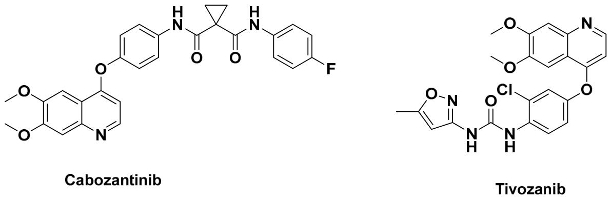
Building on the EGFR-inhibitor paradigm discussed before, several additional multi-kinase inhibitors bearing a genuine quinoline core have reached clinical use over the past decade. Cabozantinib, an oral inhibitor of VEGFR-2 and c-Met with IC
50 values of 0.035 nM and 1.3 nM, respectively, is approved for medullary thyroid carcinoma and renal cell carcinoma [
89]. Tivozanib, a quinoline-urea derivative and potent VEGFR2 inhibitor, is FDA-approved for third-line treatment of renal cell carcinoma, with its crystal structure showing that the quinoline nitrogen hydrogen-bonds with the hinge residue, while the urea moiety engages the αC-helix glutamate and the DFG-Asp motif characteristic of type-II inhibitor binding (
Figure 14) [
90].
SAR consideration: across this class, the quinoline nitrogen consistently acts as the hinge-binding hydrogen-bond acceptor (as established for fluoroquinolones and EGFR inhibitors in earlier sections), while peripheral substituents and, critically, the choice between a type-I (ATP-pocket-only, e.g., cabozantinib) or type-II (DFG-out-extending, e.g., tivozanib) binding mode determine kinase selectivity across the VEGFR/MET/RET/BTK panel.
11.3. Multitarget-Directed Ligands (MTDLs)
In place of the traditional one-drug-one-target paradigm, quinoline-based MTDLs have been designed to simultaneously engage multiple pathological targets [
91]. In neurodegenerative disease, this strategy typically combines acetylcholinesterase inhibition with metal chelation and antioxidant activity, drawing on the same C-8 hydroxyquinoline chelating motif discussed (clioquinol), reflecting the mechanistic overlap between metal dyshomeostasis, oxidative stress, and cholinergic dysfunction in Alzheimer’s disease pathology [
92]. In oncology, dual kinase/topoisomerase-directed hybrids extend the DNA-intercalating quinoline pharmacophores discussed (camptothecin-type agents) with kinase-inhibitory anilinoquinoline fragments (
Figure 15 and
Table 9).
SAR consideration: MTDL design imposes an additional constraint beyond single-target SAR, each pharmacophoric fragment must retain sufficient target engagement while the hybrid molecule remains within acceptable physicochemical (Lipinski-type) space, which in practice limits the size of the linker and often favors fusing rather than merely tethering the two pharmacophores, as is achieved when the quinoline nitrogen itself is shared between the metal-chelating and radical-scavenging functions rather than added as a separate appendage [
96,
97].
These three emerging directions indicate that the pharmacological relevance of the quinoline scaffold continues to evolve well beyond its classical applications [
98,
99]. PROTAC-based degradation offers a route to durably overcome the point-mutation resistance mechanisms that recur throughout this review; quinoline-core multi-kinase inhibitors demonstrate that the same hinge-binding chemistry validated for antibacterial and first-generation anticancer agents can be systematically re-purposed across the kinome through peripheral SAR alone; and MTDL design leverages the scaffold’s dual metal-chelating/antioxidant capacity toward multifactorial diseases where single-target inhibition has historically underperformed [
100]. Continued innovation in quinoline-based drug discovery is therefore likely to proceed along these three complementary axes, degradation, kinome-wide re-optimization, and polypharmacology, rather than through the discovery of entirely new binding mechanisms.
12. Conclusions
Quinoline derivatives are nitrogen-containing heterocycles valued for their broad range of medicinal applications and their role in treating various diseases. Their structural diversity, along with favorable chemical and physicochemical properties, has made them a scaffold of choice for developing new therapeutic agents.
This review has covered the evolution of synthetic strategies toward quinoline derivatives, from classical condensation-based routes, the Skraup, Doebner, Conrad–Limpach, Combes, Povarov, and Riehm syntheses, to more sustainable modern approaches such as microwave and ultrasound-assisted protocols, solvent-free reactions, photocatalytic methods, and nanocatalyst-mediated transformations. These methods have improved reaction efficiency and yields, reduced environmental impact, and given access to a wider range of quinoline scaffolds for biological testing.
From malaria to cancer and beyond, quinoline derivatives remain an active area of drug discovery, involving both the optimization of known scaffolds and the design of new hybrids acting through different mechanisms. We have grounded this discussion in the primary literature, covering bioactivity data, mechanistic detail, and structure-activity relationships across the antibacterial, anticancer, anti-inflammatory, antimalarial, antiparasitic, antitubercular, antioxidant, and antiviral domains, as well as the more recent directions discussed in Section (PROTACs, kinase inhibitors, and multitarget-directed ligands).
Progress in synthetic methodology, together with a clearer picture of how quinoline derivatives interact with their biological targets, keeps this scaffold relevant to drug discovery. Its continued use alongside computational design tools suggests it will remain part of therapeutic development for some time to come.
Author Contributions
Conceptualization, A.E.-M.; methodology, A.E.-M.; software, A.E.-M.; validation, A.M., A.E.-Z., R.B., A.A. and A.H.; formal analysis, A.E.-M., A.M. and A.H.; investigation, A.E.-M., A.M., A.E.-Z., R.B., A.A. and A.H.; resources, A.E.-M.; data curation, A.E.-M.; writing—original draft preparation, A.E.-M.; writing—review and editing, A.E.-M., A.M. and A.H.; visualization, A.M., A.E.-Z., R.B., A.A. and A.H.; supervision, A.H. and A.M.; project administration, A.E.-M., A.M. and A.H. All authors have read and agreed to the published version of the manuscript.
Funding
This research received no external funding.
Data Availability Statement
The original contributions presented in the study are included in the article; further inquiries can be directed to the corresponding author.
Conflicts of Interest
The authors declare no conflicts of interest.
References
- Ajani, O.O.; Iyaye, K.T.; Ademosun, O.T. Recent advances in chemistry and therapeutic potential of functionalized quinoline motifs—A review. RSC Adv. 2022, 12, 18594–18614. [Google Scholar] [CrossRef] [Scilit] [PubMed]
- Milcent, R.; Chau, F. Chimie Organique Hétérocyclique: Structures Fondamentales, Chimie et Biochimie des Principaux Composés Naturels; EDP Sciences: Les Ulis, France, 2003; p. 845. [Google Scholar]
- Dorvault, F.L.M. L’officine, Ou, Répertoire Général de Pharmacie Pratique; Nabu Press: Charleston, SC, USA, 2014. [Google Scholar]
- Wall, M.E.; Wani, M.C.; Cook, C.E.; Palmer, K.H.; McPhail, A.T.; Sim, G.A. Plant Antitumor Agents. I. The Isolation and Structure of Camptothecin, a Novel Alkaloidal Leukemia and Tumor Inhibitor from Camptotheca acuminata1,2. J. Am. Chem. Soc. 1966, 88, 3888–3890. [Google Scholar] [CrossRef] [Scilit]
- Hsiang, Y.H.; Hertzberg, R.; Hecht, S.; Liu, L.F. Camptothecin induces protein-linked DNA breaks via mammalian DNA topoisomerase I. J. Biol. Chem. 1985, 260, 14873–14878. [Google Scholar] [CrossRef] [Scilit]
- El-Mrabet, A.; Haoudi, A.; Dalbouha, S.; Skalli, M.K.; Hökelek, T.; Capet, F.; Rodi, Y.K.; Mazzah, A.; Sebbar, N.K. Crystal structure, Hirshfeld surface analysis, interaction energy and energy framework calculations, as well as density functional theory (DFT) computation, of methyl 2-oxo-1-(prop-2-ynyl)-1,2-dihydroquinoline-4-carboxylate. Acta Crystallogr. Sect. E Crystallogr. Commun. 2023, 79, 883–889. [Google Scholar] [CrossRef] [Scilit] [PubMed]
- El-Mrabet, A.; Haoudi, A.; Capet, F.; Hökelek, T.; Ahmed, M. Crystal structure and Hirshfeld surface analyses, interaction energy calculations and energy frameworks of methyl 2-[(4-cyanophenyl)methoxy]quinoline-4-carboxylate. Struct. Rep. 2025, 81, 650–656. [Google Scholar] [CrossRef] [Scilit] [PubMed]
- Dorababu, A. Recent update on antibacterial and antifungal activity of quinoline scaffolds. Arch. Pharm. 2021, 354, e2000232. [Google Scholar] [CrossRef] [Scilit] [PubMed]
- Eissa, S.I.; Farrag, A.M.; Abbas, S.Y.; El Shehry, M.F.; Ragab, A.; Fayed, E.A.; Ammar, Y.A. Novel Structural Hybrids of Quinoline and Thiazole Moieties: Synthesis and Evaluation of Antibacterial and Antifungal Activities with Molecular Modeling Studies. Bioorg. Chem. 2021, 110, 104803. [Google Scholar] [CrossRef] [Scilit] [PubMed]
- Liberto, N.A.; Simões, J.B.; Silva, S.d.P.; da Silva, C.J.; Modolo, L.V.; de Fátima, Â.; Silva, L.M.; Derita, M.; Zacchino, S.; Zuñiga, O.M.P.; et al. Quinolines: Microwave-assisted synthesis and their antifungal, anticancer and radical scavenger properties. Bioorganic Med. Chem. 2017, 25, 1153–1162. [Google Scholar] [CrossRef] [Scilit] [PubMed]
- Mukherjee, S.; Pal, M. Medicinal Chemistry of Quinolines as Emerging Anti-Inflammatory Agents: An Overview. Curr. Med. Chem. 2013, 20, 4386–4410. [Google Scholar] [CrossRef] [Scilit] [PubMed]
- Bawa, S.; Kumar, S.; Drabu, S.; Kumar, R. Structural modifications of quinoline-based antimalarial agents: Recent developments. J. Pharm. Bioallied Sci. 2010, 2, 64–71. [Google Scholar] [CrossRef] [Scilit] [PubMed]
- Senerovic, L.; Opsenica, D.; Moric, I.; Aleksic, I.; Spasić, M.; Vasiljevic, B. Quinolines and Quinolones as Antibacterial, Antifungal, Anti-Virulence, Antiviral and Anti-Parasitic Agents. In Advances in Microbiology, Infectious Diseases and Public Health; Donelli, G., Ed.; Springer International Publishing: Cham, Switzerland, 2019; pp. 37–69. [Google Scholar] [CrossRef] [Scilit] [PubMed]
- Keri, R.S.; Patil, S.A. Quinoline: A promising antitubercular target. Biomed. Pharmacother. 2014, 68, 1161–1175. [Google Scholar] [CrossRef] [Scilit] [PubMed]
- Jadhav, P.; Liang, X.; Ansari, A.; Tan, B.; Tan, H.; Li, K.; Chi, X.; Ford, A.; Ruiz, F.X.; Arnold, E.; et al. Design of quinoline SARS-CoV-2 papain-like protease inhibitors as oral antiviral drug candidates. Nat. Commun. 2025, 16, 1604. [Google Scholar] [CrossRef] [Scilit] [PubMed]
- Zeleke, D.; Eswaramoorthy, R.; Belay, Z.; Melaku, Y. Synthesis and Antibacterial, Antioxidant, and Molecular Docking Analysis of Some Novel Quinoline Derivatives. J. Chem. 2020, 1, 1324096. [Google Scholar] [CrossRef] [Scilit]
- Babita; Kanika; Srivastava, R. Contemporary Trends in the Greener Synthetic Approaches Towards Quinoline Derivatives. J. Heterocycl. Chem. 2025, 62, 1140–1170. [Google Scholar]
- Eisch, J.J.; Dluzniewski, T. Mechanism of the Skraup and Doebner-von Miller quinoline syntheses. Cyclization of .alpha.,.beta.-unsaturated N-aryliminium salts via 1,3-diazetidinium ion intermediates. J. Org. Chem. 1989, 54, 1269–1274. [Google Scholar] [CrossRef] [Scilit]
- Yamashkin, S.A.; Oreshkina, E.A. Traditional and modern approaches to the synthesis of quinoline systems by the Skraup and Doebner-Miller methods. Chem. Heterocycl. Compd. 2006, 42, 701–718. [Google Scholar] [CrossRef] [Scilit]
- Brouet, J.-C.; Gu, S.; Peet, N.P.; Williams, J.D. Survey of solvents for the Conrad–Limpach synthesis of 4-hydroxyquinolones. Synth. Commun. 2009, 39, 1563–1569. [Google Scholar] [CrossRef] [Scilit] [PubMed]
- Cheng, C.C.; Yan, S.J. The F Riedländer Synthesis of Quinolines. Org. React. 2004, 28, 37–201. [Google Scholar]
- Vinogradov, M.G.; Turova, O.V.; Zlotin, S.G. Catalytic Asymmetric Aza-Diels-Alder Reaction: Pivotal Milestones and Recent Applications to Synthesis of Nitrogen-Containing Heterocycles. Adv. Synth. Catal. 2021, 363, 1466–1526. [Google Scholar] [CrossRef] [Scilit]
- Ramann, G.A.; Cowen, B.J. Recent advances in metal-free quinoline synthesis. Molecules 2016, 21, 986. [Google Scholar] [CrossRef] [Scilit] [PubMed]
- Zeng, R.; Dong, G. Rh-catalyzed decarbonylative coupling with alkynes via C–C activation of isatins. J. Am. Chem. Soc. 2015, 137, 1408–1411. [Google Scholar] [CrossRef] [Scilit] [PubMed]
- Wang, Q.; Osipyan, A.; Konstantinidou, M.; Butera, R.; Mgimpatsang, K.C.; Shishkina, S.V.; Dömling, A. Pd-catalyzed de novo assembly of diversely substituted indole-fused polyheterocycles. J. Org. Chem. 2019, 84, 12148–12156. [Google Scholar] [CrossRef] [Scilit] [PubMed]
- Aksenov, A.V.; Smirnov, A.N.; Aksenov, N.A.; Aksenova, I.V.; Matheny, J.P.; Rubin, M. Metal-free ring expansion of indoles with nitroalkenes: A simple, modular approach to 3-substituted 2-quinolones. RSC Adv. 2015, 5, 8647–8656. [Google Scholar] [CrossRef] [Scilit]
- El Ashry, E.S.H.; Ramadan, E.S.; Abdel Hamid, H.; Hagar, M. Microwave-Assisted Synthesis of Quinoline Derivatives from Isatin. Synth. Commun. 2005, 35, 2243–2250. [Google Scholar] [CrossRef] [Scilit]
- El-Mrabet, A.; Haoudi, A.; Kandri-Rodi, Y.; Mazzah, A. An Overview of Quinolones as Potential Drugs: Synthesis, Reactivity and Biological Activities. Organics 2025, 6, 16. [Google Scholar] [CrossRef] [Scilit]
- El-Mrabet, A.; Sert, Y.; Capet, F.; Bouamri, K.; Mague, J.T.; Kandri-Rodi, Y.; Mazzah, A.; Haoudi, A.; Sebbar, N.K. Novel quinoline derivatives: Synthesis, crystal structure, structural characterization, Hirshfeld surface analysis, DFT studies, ADMET, molecular docking, and antibacterial activity. J. Mol. Struct. 2026, 1365, 145989. [Google Scholar] [CrossRef] [Scilit]
- El-Mrabet, A.; Diane, A.; Haloui, R.; El Monfalouti, H.; Alanazi, A.S.; Hefnawy, M.; Alanazi, M.M.; Kandri-Rodi, Y.; Elkhattabi, S.; Mazzah, A.; et al. Hybrid 2-Quinolone–1,2,3-triazole Compounds: Rational Design, In Silico Optimization, Synthesis, Characterization, and Antibacterial Evaluation. Antibiotics 2025, 14, 877. [Google Scholar] [CrossRef] [Scilit] [PubMed]
- Owais, M.; Kumar, A.; Hasan, S.M.; Singh, K.; Azad, I.; Hussain, A.; Suvaiv; Akil, M. Quinoline derivatives as promising scaffolds for antitubercular activity: A comprehensive review. Mini Rev. Med. Chem. 2024, 24, 1238–1251. [Google Scholar] [CrossRef] [Scilit] [PubMed]
- Prasad, A.; Rao, T.B.; Rambabu, D.; Rao, M.V.B.; Pal, M. Ultrasound assisted synthesis of quinoline derivatives in the presence of sncl2·2h2o as a precatalyst in water: Evaluation of their antibacterial activities. Mini Rev. Med. Chem. 2018, 18, 895–903. [Google Scholar] [CrossRef] [Scilit] [PubMed]
- Devi, L.; Robert, A.R.; Ganja, H.; Maddila, S.; Jonnalagadda, S.B. A Rapid, Sustainable and Environmental Friendly Protocol for the Catalyst-Free Synthesis of 2-Methyl-5-Oxo-Hexahydroquinoline-3-Carboxylate via Ultrasonic Irradiation. Chem. Data Collect. 2020, 28, 100432. [Google Scholar] [CrossRef] [Scilit]
- Amariucai-Mantu, D.; Mangalagiu, V.; Danac, R.; Mangalagiu, I.I. Microwave Assisted Reactions of Azaheterocycles Formedicinal Chemistry Applications. Molecules 2020, 25, 716. [Google Scholar] [CrossRef] [Scilit] [PubMed]
- Patel, D.B.; Rajani, D.P.; Rajani, S.D.; Patel, H.D. A Green Synthesis of Quinoline-4-Carboxylic Derivatives Using p-Toluenesulfonic Acid as an Efficient Organocatalyst under Microwave Irradiation and Their Docking, Molecular Dynamics, ADME-Tox and Biological Evaluation. J. Heterocycl. Chem. 2020, 57, 1524–1544. [Google Scholar] [CrossRef] [Scilit]
- Wang, Y.; Yao, Q.X.; He, J.R.; Liang, Z.H.; Li, X.; Cheng, H.; Li, L.L. L-Proline-Catalyzed Knoevenagel Reaction Promoted by Choline Chloride-Based Deep Eutectic Solvents. Biomass Convers. Biorefin. 2022, 12, 87–93. [Google Scholar] [CrossRef] [Scilit]
- Sharghi, H.; Shiri, P.; Aberi, M. A solvent-free and one-pot strategy for ecocompatible synthesis of substituted benzofurans from various salicylaldehydes, secondary amines, and nonactivated alkynes catalyzed by copper (I) oxide nano-particles. Synthesis 2014, 46, 2489–2498. [Google Scholar] [CrossRef] [Scilit]
- Jardim, G.A.M.; Dantas, J.A.; Barboza, A.A.; Paixão, M.W.; Ferreira, M.A.B. Light-Driven Palladium-Radical Hybrid Species: Mechanistic Aspects and Recent Examples. Synthesis 2022, 54, 4629–4645. [Google Scholar] [CrossRef] [Scilit]
- Vaishali; Sharma, S.; Sharma, P.; Das, D.K.; Vashistha, V.K.; Dhiman, J.; Sharma, R.; Kumar, R.; Singh, M.V.; Kumar, Y. Magnetic Nanoparticle-Catalysed Synthesis of Quinoline Derivatives: A Green and Sustainable Method. Heliyon 2024, 10, e40451. [Google Scholar] [CrossRef] [Scilit] [PubMed]
- Baghbanian, S.M.; Farhang, M. CuFe2O4 nanoparticles: A magnetically recoverable and reusable catalyst for the synthesis of quinoline and quinazoline derivatives in aqueous media. RSC Adv. 2014, 4, 11624–11633. [Google Scholar] [CrossRef] [Scilit]
- Firouzi-Haji, R.; Maleki, A. L-Proline-Functionalized Fe3O4 Nanoparticles as an Efficient Nanomagnetic Organocatalyst for Highly Stereoselective One-Pot Two-Step Tandem Synthesis of Substituted Cyclopropanes. ChemistrySelect 2019, 4, 853–857. [Google Scholar] [CrossRef] [Scilit]
- Oien, D.B.; Pathoulas, C.L.; Ray, U.; Thirusangu, P.; Kalogera, E.; Shridhar, V. Repurposing Quinacrine for Treatment-Refractory Cancer. Semin. Cancer Biol. 2021, 68, 21–30. [Google Scholar] [CrossRef] [Scilit] [PubMed]
- Rolain, J.-M.; Colson, P.; Raoult, D. Recycling of chloroquine and its hydroxyl analogue to face bacterial, fungal and viral infections in the 21st century. Int. J. Antimicrob. Agents 2007, 30, 297–308. [Google Scholar] [CrossRef] [Scilit] [PubMed]
- Plantone, D.; Koudriavtseva, T. Current use of chloroquine and hydroxychloroquine in infectious diseases. Infection 2018, 46, 437–448. [Google Scholar] [CrossRef] [Scilit] [PubMed]
- Kunin, C.M.; Ellis, W.Y. Antimicrobial activities of mefloquine and a series of related compounds. Antimicrob. Agents Chemother. 2000, 44, 848–852. [Google Scholar] [CrossRef] [Scilit] [PubMed]
- Claudel, M.; Schwarte, J.V.; Fromm, K.M. New Antimicrobial Strategies Based on Metal Complexes. Chemistry 2020, 2, 849–899. [Google Scholar] [CrossRef] [Scilit]
- Hooper, D.C.; Jacoby, G.A. Mechanisms of drug resistance: Quinolone resistance. Ann. N. Y. Acad. Sci. 2015, 1354, 12–31. [Google Scholar] [CrossRef] [Scilit] [PubMed]
- El-Sagheir, A.M.K.; Nekhala, I.A.; El-Gaber, M.K.A.; Aboraia, A.S.; Persson, J.; Schäfer, A.-B.; Wenzel, M.; Omar, F.A. Rational design, synthesis, molecular modeling, biological activity, and mechanism of action of polypharmacological norfloxacin hydroxamic acid derivatives. RSC Med. Chem. 2023, 14, 2593–2610. [Google Scholar] [CrossRef] [Scilit] [PubMed]
- Rusu, A.; Munteanu, A.-C.; Arbănași, E.-M.; Uivarosi, V. Overview of side-effects of antibacterial fluoroquinolones: New drugs versus old drugs, a step forward in the safety profile? Pharmaceutics 2023, 15, 804. [Google Scholar] [CrossRef] [Scilit] [PubMed]
- El-Houssni, I.; Zahidi, A.; Mortada, S.; El-Mrabet, A.; Faouzi, M.E.A.; Haoudi, A.; Khedid, K.; Hassikou, R. Wheat Landraces and Microbiome Endophytic Phytases: In Vitro/In Silico Study for Iron Bioavailability. Food Chem. Mol. Sci. 2026, 12, 100363. [Google Scholar] [CrossRef] [Scilit] [PubMed]
- Gordin, F.M.; Hackbarth, C.J.; Scott, K.G.; A Sande, M. Activities of pefloxacin and ciprofloxacin in experimentally induced Pseudomonas pneumonia in neutropenic guinea pigs. Antimicrob. Agents Chemother. 1985, 27, 452–454. [Google Scholar] [CrossRef] [Scilit] [PubMed]
- Bergogne-Bérézin, E. Antibiotic therapy in lower uncomplicated urinary tract infections: Clinical, microbiologic and pharmacologic bases. Antibiotiques 2006, 8, 51–62. [Google Scholar] [CrossRef] [Scilit]
- Keating, G.M.; Scott, L.J. Moxifloxacin: A review of its use in the management of bacterial infections. Drugs 2004, 64, 2347–2377. [Google Scholar] [CrossRef] [Scilit] [PubMed]
- Wadworth, A.N.; Goa, K.L. Lomefloxacin: A Review of its Antibacterial Activity, Pharmacokinetic Properties and Therapeutic Use. Drugs 1991, 42, 1018–1060. [Google Scholar] [CrossRef] [Scilit] [PubMed]
- Aldred, K.J.; Kerns, R.J.; Osheroff, N. Mechanism of Quinolone Action and Resistance. Biochemistry 2014, 53, 1565–1574. [Google Scholar] [CrossRef] [Scilit] [PubMed]
- Mustaev, A.; Malik, M.; Zhao, X.; Kurepina, N.; Luan, G.; Oppegard, L.M.; Hiasa, H.; Marks, K.R.; Kerns, R.J.; Berger, J.M.; et al. Fluoroquinolone-gyrase-DNA complexes: Two modes of drug binding. J. Biol. Chem. 2014, 289, 12300–12312. [Google Scholar] [CrossRef] [Scilit] [PubMed]
- Lu, X.; Wang, G.; Xie, Y.; Tang, W.; Liu, B.; Zhang, J. Efflux pump inhibitor combined with ofloxacin decreases MRSA biofilm formation by regulating the gene expression of NorA and quorum sensing. RSC Adv. 2023, 13, 2707–2717. [Google Scholar] [CrossRef] [Scilit] [PubMed]
- Bringmann, G.; Reichert, Y.; Kane, V.V. The Total Synthesis of Streptonigrin and Related Antitumor Antibiotic Natural Products. ChemInform 2004, 60, 3539–3574. [Google Scholar] [CrossRef] [Scilit]
- Pommier, Y. Topoisomerase I inhibitors: Camptothecins and beyond. Nat. Rev. Cancer 2006, 6, 789–802. [Google Scholar] [CrossRef] [Scilit] [PubMed]
- Rosing, H.; Doyle, E.; Davies, B.; Beijnen, J. High-performance liquid chromatographic determination of the novel antitumour drug topotecan and topotecan as the total of the lactone plus carboxylate forms, in human plasma. J. Chromatogr. B Biomed. Sci. Appl. 1995, 668, 107–115. [Google Scholar] [CrossRef] [Scilit] [PubMed]
- Schrezenmeier, E.; Dörner, T. Mechanisms of action of hydroxychloroquine and chloroquine: Implications for rheumatology. Nat. Rev. Rheumatol. 2020, 16, 155–166. [Google Scholar] [CrossRef] [Scilit] [PubMed]
- Capra, V.; Thompson, M.D.; Sala, A.; Cole, D.E.C.; Folco, G.; Rovati, G.E. Cysteinyl-Leukotrienes and Their Receptors in Asthma and Other Inflammatory Diseases: Critical Update and Emerging Trends. Med. Res. Rev. 2007, 27, 469–527. [Google Scholar] [CrossRef] [Scilit] [PubMed]
- Ghanim, A.M.; Girgis, A.S.; Kariuki, B.M.; Samir, N.; Said, M.F.; Abdelnaser, A.; Nasr, S.; Bekheit, M.S.; Abdelhameed, M.F.; Almalki, A.J.; et al. Design and synthesis of ibuprofen-quinoline conjugates as potential anti-inflammatory and analgesic drug candidates. Bioorg. Chem. 2022, 119, 105557. [Google Scholar] [CrossRef] [Scilit] [PubMed]
- Liang, X.; Xie, Y.; Liu, X.; Xu, H.; Ren, H.; Tang, S.; Liu, Q.; Huang, M.; Shao, X.; Li, C.; et al. Discovery of Novel Imidazo[4,5-c]quinoline Derivatives to Treat Inflammatory Bowel Disease (IBD) by Inhibiting Multiple Proinflammatory Signaling Pathways and Restoring Intestinal Homeostasis. J. Med. Chem. 2022, 65, 11949–11969. [Google Scholar] [CrossRef] [Scilit] [PubMed]
- Yadav, V.; Reang, J.; Sharma, V.; Majeed, J.; Sharma, P.C.; Sharma, K.; Giri, N.; Kumar, A.; Tonk, R.K. Quinoline derivatives as privileged scaffolds for medicinal and pharmaceutical chemists: A comprehensive review. Chem. Biol. Drug Des. 2022, 100, 389–418. [Google Scholar] [CrossRef] [Scilit] [PubMed]
- Nandal, R.; Kumar, D.; Aggarwal, N.; Kumar, V.; Narasimhan, B.; Marwaha, R.K.; Sharma, P.C.; Kumar, S.; Bansal, N.; Chopra, H.; et al. Recent Advances, Challenges and Updates on the Development of Therapeutics for Malaria. EXCLI J. 2024, 23, 672–713. [Google Scholar] [CrossRef] [Scilit] [PubMed]
- Mishra, R.; da Cunha Xavier, J.; Kumar, N.; Krishna, G.; Dhakad, P.K.; dos Santos, H.S.; Bandeira, P.N.; Rodrigues, T.H.S.; Gondim, D.R.; Ribeiro, W.H.F.; et al. Exploring Quinoline Derivatives: Their Antimalarial Efficacy and Structural Features. Med. Chem. 2025, 21, 96–121. [Google Scholar] [CrossRef] [Scilit] [PubMed]
- Achan, J.; OTalisuna, A.; Erhart, A.; Yeka, A.; Tibenderana, J.K.; Baliraine, F.N.; Rosenthal, P.J.; D’ALessandro, U. Quinine, an old anti-malarial drug in a modern world: Role in the treatment of malaria. Malar. J. 2011, 10, 144. [Google Scholar] [CrossRef] [Scilit] [PubMed]
- Gayral, P. Quinine, antipaludiques, résistance des souches de Plasmodium. Rev. Hist. Pharm. 1989, 77, 175–184. [Google Scholar] [CrossRef] [Scilit]
- Kaur, S.; Kaur, A.; Arora, A.; Kumar, V. A glimpse at the quinoline-hybridization approach for the development of new antimalarials. Mini Rev. Med. Chem. 2023, 23, 898–916. [Google Scholar] [CrossRef] [Scilit] [PubMed]
- El-Mrabet, A.; Diane, A.; Haoudi, A.; Kandri-Rodi, Y.; Mazzah, A. Exploration of Quinolone Amides as Promising Candidates for the Treatment of Human African Trypanosomiasis: In Silico Virtual Screening and Molecular Docking. Russ. J. Gen. Chem. 2025, 95, 1817–1832. [Google Scholar] [CrossRef] [Scilit]
- Hashemi, N.; Ommi, D.; Kheyri, P.; Khamesipour, F.; Setzer, W.N.; Benchimol, M. A Review Study on the Anti-Trichomonas Activities of Medicinal Plants. Int. J. Parasitol. Drugs Drug Resist. 2021, 15, 92–104. [Google Scholar] [CrossRef] [Scilit] [PubMed]
- Shrivastav, M.T.; Malik, Z.; Somlata. Revisiting Drug Development Against the Neglected Tropical Disease, Amebiasis. Front. Cell. Infect. Microbiol. 2021, 10, 628257. [Google Scholar] [CrossRef] [Scilit] [PubMed]
- Ain, Q.T.; Saleem, N.; Munawar, N.; Nawaz, R.; Naseer, F.; Ahmed, S. Quest for Malaria Management Using Natural Remedies. Front. Pharmacol. 2024, 15, 1359890. [Google Scholar] [CrossRef] [Scilit] [PubMed]
- Andries, K.; Verhasselt, P.; Guillemont, J.; Göhlmann, H.W.H.; Neefs, J.-M.; Winkler, H.; Van Gestel, J.; Timmerman, P.; Zhu, M.; Lee, E.; et al. A diarylquinoline drug active on the ATP synthase of Mycobacterium tuberculosis. Science 2005, 307, 223–227. [Google Scholar] [CrossRef] [Scilit] [PubMed]
- Yano, T.; Kassovska-Bratinova, S.; Teh, J.S.; Winkler, J.; Sullivan, K.; Isaacs, A.; Schechter, N.M.; Rubin, H. Reduction of Clofazimine by Mycobacterial Type 2 NADH:Quinone Oxidoreductase: A Pathway for the Generation of Bactericidal Levels of Reactive Oxygen Species. J. Biol. Chem. 2011, 286, 10276–10287. [Google Scholar] [CrossRef] [Scilit] [PubMed]
- Wang, Y.; Wu, P.; Liu, F.; Chen, J.; Xue, J.; Qin, Y.; Chen, F.; Wang, S.; Ji, L. Design, Synthesis, and Biological Evaluation of Membrane-Active Honokiol Derivatives as Potent Antibacterial Agents. Eur. J. Med. Chem. 2022, 240, 114593. [Google Scholar] [CrossRef] [Scilit] [PubMed]
- Fang, W.-S.; Sun, D.-Y.; Yang, S.; Cheng, C.; Moschke, K.; Li, T.; Sun, S.; Lichtenthaler, S.F.; Huang, J.; Wang, Y. Discovery of a Series of Selective and Cell Permeable Beta-Secretase (BACE1) Inhibitors by Fragment Linking with the Assistance of STD-NMR. Bioorg. Chem. 2019, 92, 103253. [Google Scholar] [CrossRef] [Scilit] [PubMed]
- Jomova, K.; Raptova, R.; Alomar, S.Y.; Alwasel, S.H.; Nepovimova, E.; Kuca, K.; Valko, M. Reactive oxygen species, toxicity, oxidative stress, and antioxidants: Chronic diseases and aging. Arch. Toxicol. 2023, 97, 2499–2574. [Google Scholar] [CrossRef] [Scilit] [PubMed]
- Subashini, R.; Mohana Roopan, S.; Nawaz Khan, F. Synthesis and Free Radical Scavenging Property of Some Quinoline Derivatives. J. Chil. Chem. Soc. 2010, 55, 317–319. [Google Scholar] [CrossRef] [Scilit]
- Mahajan, P.; Nikam, M.; Asrondkar, A.; Bobade, A.; Gill, C. Synthesis, Antioxidant, and Anti-Inflammatory Evaluation of Novel Thiophene-Fused Quinoline Based β-Diketones and Derivatives. J. Heterocycl. Chem. 2017, 54, 1415–1422. [Google Scholar] [CrossRef] [Scilit]
- Persoons, L.; Vanderlinden, E.; Vangeel, L.; Wang, X.; Do, N.D.T.; Foo, S.-Y.C.; Leyssen, P.; Neyts, J.; Jochmans, D.; Schols, D.; et al. Broad spectrum anti-coronavirus activity of a series of anti-malaria quinoline analogues. Antivir. Res. 2021, 193, 105127. [Google Scholar] [CrossRef] [Scilit] [PubMed]
- Seliem, I.A.; Panda, S.S.; Girgis, A.S.; Moatasim, Y.; Kandeil, A.; Mostafa, A.; Ali, M.A.; Nossier, E.S.; Rasslan, F.; Srour, A.M.; et al. New Quinoline-Triazole Conjugates: Synthesis, and Antiviral Properties against SARS-CoV-2. Bioorg. Chem. 2021, 114, 105117. [Google Scholar] [CrossRef] [Scilit] [PubMed]
- Al-Humaidi, J.Y.; Shaaban, M.M.; Rezki, N.; Aouad, M.R.; Zakaria, M.; Jaremko, M.; Hagar, M.; Elwakil, B.H. 1,2,3-Triazole-Benzofused Molecular Conjugates as Potential Antiviral Agents against SARS-CoV-2 Virus Variants. Life 2022, 12, 1341. [Google Scholar] [CrossRef] [Scilit] [PubMed]
- Kaur, R.; Kumar, K. Synthetic and Medicinal Perspective of Quinolines as Antiviral Agents. Eur. J. Med. Chem. 2021, 215, 113220. [Google Scholar] [CrossRef] [Scilit] [PubMed]
- Békés, M.; Langley, D.R.; Crews, C.M. PROTAC targeted protein degraders: The past is prologue. Nat. Rev. Drug Discov. 2022, 21, 181–200. [Google Scholar] [CrossRef] [Scilit] [PubMed]
- Dong, G.; Chen, M.; Jiang, X.; Shi, J.; Meng, X.; Zhan, P.; Liu, Y.; Liu, C.; Song, Y. First-in-Class Quinoline-Dione-Derived PROTACs: Potent Degraders of Cdc25 Phosphatases for Antitumor Therapy. J. Med. Chem. 2026, 69, 982–1003. [Google Scholar] [CrossRef] [Scilit] [PubMed]
- Wang, Q.; Zhu, Y.; Pei, J. Targeting EGFR with Molecular Degraders as a Promising Strategy to Overcome Resistance to EGFR Inhibitors. Future Med. Chem. 2024, 16, 1923–1944. [Google Scholar] [CrossRef] [Scilit] [PubMed]
- Roskoski, R. Properties of FDA-Approved Small Molecule Protein Kinase Inhibitors: A 2024 Update. Pharmacol. Res. 2024, 200, 107059. [Google Scholar] [CrossRef] [Scilit] [PubMed]
- Bruin, G.; Demont, D.; de Zwart, E.; Verkaik, S.; Hoogenboom, N.; van de Kar, B.; van Lith, B.; Emmelot-van Hoek, M.; Gulrajani, M.; Covey, T.; et al. Discovery of Quinoline-Based Irreversible BTK Inhibitors. Bioorg. Med. Chem. Lett. 2020, 30, 127261. [Google Scholar] [CrossRef] [Scilit] [PubMed]
- Maddeboina, K.; Yada, B.; Kumari, S.; Durden, D.L. Recent Advances in Multitarget-Directed Ligands via In Silico Drug Discovery. Drug Discov. Today 2024, 29, 103904. [Google Scholar] [CrossRef] [Scilit] [PubMed]
- Prati, F.; Bottegoni, G.; Bolognesi, M.L.; Cavalli, A. Bivalent and multitarget-directed ligands for Alzheimer’s disease: Quinoline and 8-hydroxyquinoline derivatives. J. Med. Chem. 2022, 65, 15239–15268. [Google Scholar]
- Lazo, J.S.; Aslan, D.C.; Southwick, E.C.; Cooley, K.A.; Ducruet, A.P.; Joo, B.; Vogt, A.; Wipf, P. Discovery and Biological Evaluation of a New Family of Potent Inhibitors of the Dual Specificity Protein Phosphatase Cdc25. J. Med. Chem. 2001, 44, 4042–4049. [Google Scholar] [CrossRef] [Scilit] [PubMed]
- Chen, Z.; Liu, M.; Wang, N.; Xiao, W.; Shi, J. Unleashing the Potential of Camptothecin: Exploring Innovative Strategies for Structural Modification and Therapeutic Advancements. J. Med. Chem. 2024, 67, 3244–3273. [Google Scholar] [CrossRef] [Scilit] [PubMed]
- Bareggi, S.R.; Cornelli, U. Clioquinol: Review of Its Mechanisms of Action and Clinical Uses in Neurodegenerative Disorders. CNS Neurosci. Ther. 2012, 18, 41–46. [Google Scholar] [CrossRef] [Scilit] [PubMed]
- Bolognesi, M.L. Harnessing Polypharmacology with Medicinal Chemistry. ACS Med. Chem. Lett. 2019, 10, 273–275. [Google Scholar] [CrossRef] [Scilit] [PubMed]
- Lipinski, C.A. Lead- and Drug-Like Compounds: The Rule-of-Five Revolution. Drug Discov. Today Technol. 2004, 1, 337–341. [Google Scholar] [CrossRef] [Scilit] [PubMed]
- Ramsay, R.R.; Popovic-Nikolicb, M.R.; Nikolic, K.; Uliassi, E.; Bolognesi, M.L. A Perspective on Multi-Target Drug Discovery and Design for Complex Diseases. Clin. Transl. Med. 2018, 7, 3. [Google Scholar] [CrossRef] [Scilit] [PubMed]
- Shehab, W.S.; Amer, M.M.K.; Elsayed, D.A.; Yadav, K.K.; Abdellattif, M.H. Current Progress toward Synthetic Routes and Medicinal Significance of Quinoline. Med. Chem. Res. 2023, 32, 2443–2457. [Google Scholar] [CrossRef] [Scilit]
- Cromm, P.M.; Crews, C.M. Targeted Protein Degradation: From Chemical Biology to Drug Discovery. Cell Chem. Biol. 2017, 24, 1181–1190. [Google Scholar] [CrossRef] [Scilit] [PubMed]
Figure 1.
Chemical Structure of Quinoline.
Figure 1.
Chemical Structure of Quinoline.
Figure 2.
Natural and synthetic quinoline derivatives.
Figure 2.
Natural and synthetic quinoline derivatives.
Figure 3.
Overview of conventional synthetic approaches to quinoline derivatives [
18,
19,
20,
21,
22,
23].
Figure 3.
Overview of conventional synthetic approaches to quinoline derivatives [
18,
19,
20,
21,
22,
23].
Scheme 1.
Rh-catalyzed decarbonylative coupling reaction.
Scheme 1.
Rh-catalyzed decarbonylative coupling reaction.
Scheme 2.
Preparation of indoloquinolones using four-component reaction and C–H activation.
Scheme 2.
Preparation of indoloquinolones using four-component reaction and C–H activation.
Scheme 3.
Synthesis of quinolones by reaction of 2-substituted indoles with 2-nitroalkenes under PPA.
Scheme 3.
Synthesis of quinolones by reaction of 2-substituted indoles with 2-nitroalkenes under PPA.
Scheme 4.
Synthesis of 2-quinolone-4-carboxylic acid derivatives.
Scheme 4.
Synthesis of 2-quinolone-4-carboxylic acid derivatives.
Scheme 5.
Prasad’s Method.
Scheme 5.
Prasad’s Method.
Scheme 7.
TEA-Catalyzed Reaction.
Scheme 7.
TEA-Catalyzed Reaction.
Scheme 8.
Dhaval’s Method.
Scheme 8.
Dhaval’s Method.
Scheme 9.
L-Proline-Catalyzed Knoevenagel Condensation.
Scheme 9.
L-Proline-Catalyzed Knoevenagel Condensation.
Scheme 10.
Sharghi’s Method.
Scheme 10.
Sharghi’s Method.
Scheme 11.
Zhou’s Method.
Scheme 11.
Zhou’s Method.
Scheme 12.
Role of Pure Nanoparticles in the Synthesis of Quinoline Derivatives.
Scheme 12.
Role of Pure Nanoparticles in the Synthesis of Quinoline Derivatives.
Scheme 13.
Firouzi-Haji’s Method.
Scheme 13.
Firouzi-Haji’s Method.
Figure 4.
Quinoline-Based Antibacterial Derivatives.
Figure 4.
Quinoline-Based Antibacterial Derivatives.
Figure 5.
Quinoline-Based Anticancer Derivatives.
Figure 5.
Quinoline-Based Anticancer Derivatives.
Figure 6.
Quinoline-Based Anti-inflammatory Derivatives.
Figure 6.
Quinoline-Based Anti-inflammatory Derivatives.
Figure 7.
Quinoline-Based Antimalarial Derivatives.
Figure 7.
Quinoline-Based Antimalarial Derivatives.
Figure 8.
Quinoline-Based Antiparasitic Derivatives.
Figure 8.
Quinoline-Based Antiparasitic Derivatives.
Figure 9.
Quinoline-Based Antitubercular Derivatives.
Figure 9.
Quinoline-Based Antitubercular Derivatives.
Figure 10.
Quinoline-Based Antioxidant Derivatives.
Figure 10.
Quinoline-Based Antioxidant Derivatives.
Figure 11.
Design of quinoline-based SARS-CoV-2 PLpro inhibitors. (a) Superposition of the X-ray crystal structures of SARS-CoV-2 PLpro bound to Jun11313 (green; PDB: 8UVM) and ubiquitin (orange; PDB: 6XAA). (b) Superposition of the X-ray crystal structures of SARS-CoV-2 PLpro complexed with Jun11313 (green; PDB: 8UVM) and GRL0617 (yellow; PDB: 7JRN). (c) Structure-based design strategy of quinoline-derived PLpro inhibitors inspired by Jun11313 and GRL0617.
Figure 11.
Design of quinoline-based SARS-CoV-2 PLpro inhibitors. (a) Superposition of the X-ray crystal structures of SARS-CoV-2 PLpro bound to Jun11313 (green; PDB: 8UVM) and ubiquitin (orange; PDB: 6XAA). (b) Superposition of the X-ray crystal structures of SARS-CoV-2 PLpro complexed with Jun11313 (green; PDB: 8UVM) and GRL0617 (yellow; PDB: 7JRN). (c) Structure-based design strategy of quinoline-derived PLpro inhibitors inspired by Jun11313 and GRL0617.
Figure 12.
Promising anti-SARS-CoV-2 candidates.
Figure 12.
Promising anti-SARS-CoV-2 candidates.
Figure 13.
Quinoline-dione-derived PROTACs.
Figure 13.
Quinoline-dione-derived PROTACs.
Figure 14.
Structure of Tivozanib and Cabozantinib.
Figure 14.
Structure of Tivozanib and Cabozantinib.
Figure 15.
Structure of clioquinol and camptothecin.
Figure 15.
Structure of clioquinol and camptothecin.
Table 1.
Quinoline-based antibacterial derivatives: targets, mechanisms, and potency.
Table 1.
Quinoline-based antibacterial derivatives: targets, mechanisms, and potency.
| Compound | Target | Mechanism | Reported Potency | Ref. |
|---|
| Mepacrine | S. aureus, E. coli, P. aeruginosa | DNA intercalation, topoisomerase inhibition and membrane perturbation | MIC = 2–64 μg/mL | [42] |
| Chloroquine | Intracellular bacteria (Mycobacterium spp.) | Lysosomal alkalinization, efflux pump inhibition and antibiotic potentiation | Weak direct activity (MIC > 128 μg/mL); synergistic in combination therapy | [43,44] |
| Mefloquine | S. aureus (including MRSA), E. faecalis, S. pneumoniae | Membrane perturbation and inhibition of bacterial energy metabolism | MIC = 0.2–16 μg/mL | [45,46] |
| Hybrid quinoline derivatives (indole, pyrrolidinedione, thiazolidinone) | S. aureus, B. subtilis, E. coli, K. pneumoniae, P. aeruginosa | Structure-dependent; likely DNA gyrase inhibition and membrane interaction | MIC = 0.5–16 μg/mL | [46] |
| Nalidixic acid | E. coli, P. mirabilis, K. pneumoniae, Enterobacter spp. | DNA gyrase (GyrA) inhibition | MIC = 1–16 μg/mL | [47] |
| Norfloxacin | E. coli, Proteus spp., Klebsiella spp., P. aeruginosa | DNA gyrase and topoisomerase IV inhibition | MIC = 0.03–0.5 μg/mL | [48] |
| Moxifloxacin | S. pneumoniae, S. aureus | DNA gyrase and topoisomerase IV inhibition | MIC90 = 0.06–0.25 μg/mL | [49,50] |
| Gemifloxacin | Respiratory pathogens (S. pneumoniae, M. pneumoniae, C. pneumoniae, L. pneumophila) | DNA gyrase and topoisomerase IV inhibition | MIC = 0.008–0.12 μg/mL | [49,50] |
Table 2.
Quinoline-based anticancer derivatives: targets, mechanisms, and potency.
Table 2.
Quinoline-based anticancer derivatives: targets, mechanisms, and potency.
| Compound | Target | Mechanism | Reported Potency | Refs. |
|---|
| Streptonigrin | Topoisomerase II/DNA | DNA strand cleavage; redox-mediated cytotoxicity via quinoline quinone moiety | IC50 = 1–50 nM (murine leukemia, melanoma) | [4,56,57] |
| Camptothecin | Topoisomerase I | Stabilizes cleavable complex via α-hydroxylactone E-ring | IC50 = 5–100 nM (HCT-116, MCF-7, A549) | [58] |
| Topotecan | Topoisomerase I | Semi-synthetic analogue; improved solubility | IC50 = 10–100 nM (ovarian, SCLC) | [58] |
| Irinotecan | Topoisomerase I | Prodrug metabolized to active SN-38 | Subnanomolar–low nM (colorectal cancer cell lines) | [58] |
| Lavendamycin | Topoisomerase I | Bioreductive activation; DNA damage/apoptosis | Low μM (leukemia, breast, colon) | [59] |
Table 3.
Quinoline-based anti-inflammatory derivatives: targets, mechanisms, and potency.
Table 3.
Quinoline-based anti-inflammatory derivatives: targets, mechanisms, and potency.
| Compound | Target | Mechanism | Reported Potency | Ref. |
|---|
| Chloroquine/Hydroxychloroquine | Lysosomes; endosomal TLR7/TLR9; antigen presentation | Inhibits lysosomal acidification, suppresses TLR signaling, and decreases TNF-α, IL-1β and IL-6 production | IC50 = 1–10 μM (cytokine production) | [61] |
| Montelukast | CysLT1 receptor | Selective cysteinyl leukotriene receptor antagonist | IC50 = 0.5–5 nM | [62] |
| Nicafenine | Cyclooxygenase (COX) | Non-selective NSAID; inhibits prostaglandin biosynthesis | Moderate anti-inflammatory activity | [63] |
| Glafenine | Cyclooxygenase (COX) | Non-selective NSAID; inhibits prostaglandin biosynthesis | Moderate anti-inflammatory activity | [64] |
| Floctafenine | Cyclooxygenase (COX) | Non-selective NSAID; inhibits prostaglandin biosynthesis | Moderate anti-inflammatory activity | [65] |
Table 4.
Quinoline-based antimalarial derivatives: targets, mechanisms, and potency.
Table 4.
Quinoline-based antimalarial derivatives: targets, mechanisms, and potency.
| Compound | Target | Mechanism | Reported Potency (Against P. falciparum) | Refs. |
|---|
| Quinine | Hemozoin formation (food vacuole) | Inhibits heme detoxification; the quinuclidine ring and secondary alcohol are essential for activity | IC50 = 100–500 nM | [68] |
| Quinidine | Hemozoin formation (food vacuole) | Diastereomer of quinine with similar mechanism; higher cardiotoxicity limits clinical use | IC50 = 50–300 nM | [69] |
| Cryptolepine | Hemozoin formation/DNA | Inhibits hemozoin formation and intercalates into DNA; indoloquinoline scaffold | IC50 = 20–200 nM (CQ-sensitive and CQ-resistant strains) | [70] |
| Chloroquine | Hemozoin formation (food vacuole) | 4-Aminoquinoline that accumulates in the digestive vacuole and inhibits heme polymerization | IC50 = 10–50 nM (CQ-sensitive); IC50 > 100 nM (CQ-resistant) | [66,67] |
| Tafenoquine | Liver-stage and blood-stage parasites | 8-Aminoquinoline causing oxidative stress after metabolic activation; active against hypnozoites | IC50 = 50–200 nM | [66,67] |
| Ferroquine | Hemozoin formation (food vacuole) | Ferrocenyl 4-aminoquinoline active against chloroquine-resistant parasites | IC50 = 5–30 nM | [66,67] |
Table 5.
Quinoline-based antiparasitic derivatives: targets, mechanisms, and potency.
Table 5.
Quinoline-based antiparasitic derivatives: targets, mechanisms, and potency.
| Compound | Target | Mechanism | Reported Potency | Ref. |
|---|
| Pyrvinium | Enterobius vermicularis | Inhibits mitochondrial oxidative phosphorylation and ATP production | EC50 = 20–100 nM (helminths, in vitro) | [72] |
| Tiliquinol/Tilbroquinol | Entamoeba histolytica | Amoebicidal; membrane disruption and inhibition of protozoal metabolism | MIC = 0.5–2 μg/mL | [73] |
| Primaquine | Plasmodium spp. (liver stages, gametocytes) | 8-Aminoquinoline; oxidative stress following CYP2D6-dependent bioactivation | IC50 = 100–500 nM | [31] |
| Tafenoquine | Plasmodium falciparum, P. vivax | Long-acting 8-aminoquinoline causing oxidative stress after metabolic activation | IC50 < 100 nM | [31] |
| Amodiaquine | P. falciparum (CQ-sensitive and resistant isolates) | 4-Aminoquinoline; inhibits hemozoin formation after accumulation in the digestive vacuole | IC50 = 10–50 nM | [74] |
Table 6.
Quinoline-based antitubercular derivatives: targets, mechanisms, and potency.
Table 6.
Quinoline-based antitubercular derivatives: targets, mechanisms, and potency.
| Compound | Target | Mechanism | Reported Potency | Ref. |
|---|
| Bedaquiline | Mycobacterial ATP synthase (c subunit, F0 proton rotor) | Diarylquinoline; inhibits ATP synthase by blocking proton translocation and ATP production | MIC = 0.015–0.12 μg/mL (including MDR-M. tuberculosis) | [75] |
| Clofazimine | Membrane respiratory chain (NDH-2) | Riminophenazine; redox cycling, ROS generation, and disruption of membrane electron transport | MIC = 0.06–0.5 μg/mL | [76] |
| 8-Hydroxyquinoline derivatives | Metal-dependent metalloenzymes | Metal chelation, disrupting essential metalloprotein functions | MIC = 0.5–4 μg/mL (replicating and non-replicating M. tuberculosis) | [77] |
| Fluorinated 2-hydroxyquinoline hydrazones | Metal-dependent enzymes/redox pathways | C-6 fluorine, C-2 hydroxyl and pyridine-hydrazone linker enhance antimycobacterial activity | MIC = 6.25 μg/mL | [78] |
Table 7.
Quinoline-based antioxidant derivatives: targets, mechanisms, and potency.
Table 7.
Quinoline-based antioxidant derivatives: targets, mechanisms, and potency.
| Compound | Antioxidant Mechanism | Reported Potency | Ref. |
|---|
| Clioquinol | Fe2+/Cu2+ chelation through the 8-hydroxyquinoline pharmacophore; inhibition of Fenton-type ROS generation | Active in DPPH and H2O2 scavenging assays | [80] |
| 6-Chloro-3-(1,3-dioxolan-2-yl)quinoline | Electron-withdrawing Cl substituent enhances radical-scavenging efficiency | 84.6–85.8% DPPH radical scavenging | [80] |
| 3-Quinolinecarboxaldehyde | Direct radical scavenging through the quinoline aromatic system | 92.96% DPPH radical scavenging (most active derivative) | [80] |
| 2-Chloro-3-quinolinecarboxaldehyde | Chlorine substitution improves antioxidant activity through electronic effects | 84.6–85.8% DPPH radical scavenging | [80] |
| Bromoquinoline derivative | Bromine substitution enhances electron delocalization and radical stabilization | Moderate-to-good antioxidant activity (DPPH assay) | [80] |
| Thiophene-based quinoline derivative | Phenolic OH-mediated hydrogen atom transfer (HAT) combined with quinoline–thiophene conjugation | EC50 = 12.03 ± 1.45 μg/mL | [81] |
Table 8.
Representative quinoline derivatives with antiviral activity.
Table 8.
Representative quinoline derivatives with antiviral activity.
| Compound | Viral Target | Mechanism | Reported Potency | Ref. |
|---|
| Chloroquine/hydroxychloroquine | SARS-CoV-1, SARS-CoV-2, HCoV-229E, HCoV-OC43 | Blocks post-attachment viral entry; impairs autophagic flux | EC50 0.12–12 μM (panel-wide); SI = 165 vs. HCoV-OC43 in HEL cells (most favorable of the series) | Persoons et al. [82] |
| Amodiaquine, ferroquine, mefloquine | Alpha-/beta-coronaviruses | Antimalarial quinoline analogues repurposed; entry-stage interference | Potent anti-coronavirus activity (mefloquine cytotoxic) | Persoons et al. [82] |
| Jun13296 (2-aryl-quinoline) | SARS-CoV-2 papain-like protease (PLpro) | Occupies Val70Ub site/BL2 groove of PLpro | IC50 = 0.13 μM (enzymatic), Ki = 8.8 nM, antiviral EC50 = 0.1 μM | Jadhav et al. [15] |
| Quinoline-triazole S1/S2 | SARS-CoV-2 (cytopathic effect assay) | Click-chemistry hybrid; mechanism under investigation | S1: IC50 = 0.060 mM (60 μM); most potent and selective conjugate of the series
S2: IC50 = 0.204 mM (204 μM), CC50 = 3.493 mM, SI = 17.12 | Seliem et al. [82] |
| Quinoline–2,6-diaminopurine hybrids | Dengue virus (DENV), Zika virus | Allosteric inhibition of NS5 polymerase thumb domain | DENV-2
(EC50 = 2.8 ± 1.0 μm; CC50 = 14.6 ± 7.7 μm)
ZIKVE
(EC50 = 12.2 ± 6.4 μm; CC50= >122 μm) | Kaptein et al. [85] |
Table 9.
Emerging quinoline-based therapeutic strategies.
Table 9.
Emerging quinoline-based therapeutic strategies.
| Compound | Target | Mechanism | Reported Potency | Ref. |
|---|
| Cabozantinib (XL184, Cometriq®/Cabometyx®) | VEGFR-2/c-Met/RET/KIT | Type-I ATP-competitive; quinoline N binds hinge region | IC50 = 0.035 nM (VEGFR-2); 1.3 nM (c-Met) | [89] |
| Tivozanib (AV-951, Fotivda®) | VEGFR-2 | Type-II (DFG-out) binding; quinoline-urea engages αC-helix Glu and DFG-Asp | Potent VEGFR-2 inhibition; approved 3rd-line RCC | [89] |
| 8-Hydroxyquinoline-based MTDLs | AChE, Cu2+/Zn2+, ROS | Simultaneous acetylcholinesterase inhibition, metal chelation, antioxidant activity, and inhibition of Aβ aggregation | Multi-target activity in the low-micromolar range | [92] |
| 6-chloro-7-(2-morpholin-4-ylethylamino)quinoline-5,8-dione | Cdc25A/Cdc25B2/Cdc25C phosphatases | Mixed competitive inhibition; quinoline-dione core; precursor/warhead structure for later PROTAC degraders | Ki = 29 nM (Cdc25A), 95 nM (Cdc25B2), 89 nM (Cdc25C); 20-fold selective vs. VHR, 450-fold vs. PTP1B | [93] |
| Camptothecin | DNA Topoisomerase I | Stabilizes the topoisomerase I–DNA cleavable complex via α-hydroxylactone E-ring H-bonding | IC50 = 5–100 nM (HCT-116, MCF-7, A549 cell lines) | [94] |
| Clioquinol | Zn2+/Cu2+ chelation; Aβ oligomer assembly | C-8 OH metal-protein-attenuating compound (MPAC); reduces metal-catalyzed ROS and Aβ aggregation | IC50 < 10 μM for inhibition of Aβ(1-42) oligomer assembly | [95] |
| Disclaimer/Publisher’s Note: The statements, opinions and data contained in all publications are solely those of the individual author(s) and contributor(s) and not of MDPI and/or the editor(s). MDPI and/or the editor(s) disclaim responsibility for any injury to people or property resulting from any ideas, methods, instructions or products referred to in the content. |
© 2026 by the authors. Published by MDPI on behalf of the Österreichische Pharmazeutische Gesellschaft. Licensee MDPI, Basel, Switzerland. This article is an open access article distributed under the terms and conditions of the Creative Commons Attribution (CC BY) license.



 ,
,