Article

Combined Effect of Sertraline and Capecitabine on Breast Cancer Cell Lines In Vitro and In Silico Evidence for Synergistic Interaction

Department of Biology, Faculty of Science, Akdeniz University, Antalya TR-07058, Turkey
*
Author to whom correspondence should be addressed.
Sci. Pharm. 2024, 92(3), 38; https://doi.org/10.3390/scipharm92030038
Submission received: 17 April 2024 / Revised: 3 July 2024 / Accepted: 8 July 2024 / Published: 15 July 2024

Abstract

:
Background: Depression is a common mood disorder that manifests itself simultaneously with chronic diseases. It is especially common in patients diagnosed with cancer, and when neglected, it reduces the success of cancer treatment. The fact that breast cancer is the most common type of cancer in women shows that the treatment of depression in women with cancer is very important. As a result, cancer patients undergoing chemotherapy in oncology units also use antidepressants simultaneously. It is critical to correctly understand the interactions between drugs used in combination. Method: In this study, doses were prepared for MCF7 and MDAMB-231 cell lines by serial dilution from 1000 ng/mL to 1.95 ng/mL. Cell viability was calculated with the WST-1 kit by applying the prepared doses of capecitabine and sertraline alone. In the sertraline/capecitabine combination study, cell viability was examined in MDAMB-231 and MCF-7 cells by applying doses of 300, 100, 50, 25, 10 ng/mL. Combinations that showed selective cytotoxicity after the combination were analyzed with the CompuSyn program and the combination index (CI<1 = synergism) was calculated. Studies on caspase 3-8-9, DNA fragmentation and mTOR were continued using a combination that showed a synergistic effect. Result: It was determined that compared to drug use alone, the sertraline/capecitabine combination decreased cell viability. There is no significant difference in caspase-3,-8,-9 and DNA fragmentation in cancer cells, but there is a reduction in the level of mTOR. This suggests that the death mechanism may be autophagy. Docking studies with autophagy pathway-related proteins further support our results. It is noteworthy that the AKT1-sertraline complex had the best binding affinity among the target proteins (−9.1 kcal/mol).

1. Introduction

According to 2020 world cancer statistics, the most common cancers that cause death in women are breast, lung, and cervical cancers, respectively [1]. Breast cancer accounts for 30% of all diagnosed cancers in women and causes 15% of deaths [2]. Capecitabine is an orally administered fluoropyrimidine carbamate, a chemotherapeutic agent widely used in breast cancer treatment. Capecitabine contains uracil and thymine bases and belongs to the antimetabolites, a class of chemotherapeutic drugs. Three steps detoxify capecitabine, a target-specific anticancer agent, and convert it to fluorouracil (5-FU). 5-FU is one of the most widely used chemotherapeutic agents in the treatment of solid tumors [1,2,3]. Clinical practitioners can use capecitabine as a single agent or in combination with other chemotherapeutics to treat breast and colorectal cancers [4].
SSRIs, or selective serotonin reuptake inhibitors, are the most widely used class of antidepressants, both prescription and over-the-counter. SSRIs are better tolerated and cause fewer toxic effects than first-generation antidepressants (TCAs, etc.). Therefore, the use of SSRIs is more preferred compared to first-generation antidepressants [3,5]. A cancer diagnosis is more likely to cause depression in patients than other illnesses. Depression is a comorbidity that affects more than 10% of cancer patients [6]. Depression lowers the quality of life and increases cancer patients’ mortality rates [7]. Sertraline, one of the selective serotonin reuptake inhibitor antidepressants, is a naphthylamine derivative with two chiral centers [8]. Sertraline is more tolerable than tricyclic antidepressants and other serotonin reuptake inhibitors [9].
Autophagy is important in cancer because of its dual role. It is a well-known fact that pharmacological induction of autophagy is an effective strategy in cancer therapy. Various types of cancer, including lung, breast, acute myeloid leukemia, and prostate, have shown sertraline to induce autophagy [10,11].
This study confidently evaluated the selectively cytotoxic effects of sertraline and capecitabine, alone and in combination, by measuring Caspase3-8-9 activity, DNA fragmentation, and mTOR expressions on MDAMB-231, MCF-7, and MCF10A cells. In this study, we conducted docking studies to validate the data gathered from the analysis of caspase 3-8-9, DNA fragmentation, and mTOR levels.

2. Materials and Methods

2.1. Cells and Culture Conditions

MCF-7 (ATCC® HTB-22™, estrogen receptor-positive human breast cancer), MDAMB-231 (ATCC® HTB-26™, estrogen receptor-negative human breast cancer cell line), and MCF10A (ATTC CRL-10317, human breast epithelial cell line) cells were obtained from ATCC (Rockville, MD, USA) MDAMB-231 and MCF-7 cells were grown in RPMI-1640 medium supplemented with 10% fetal bovine serum, 0.1 mM non-essential amino acids, 5% sodium pyruvate, 2 mM L-glutamine, and 10 μg/mL gentamicin in a humidified atmosphere of 5% CO2 and 95% air at 37 °C. MCF10A mammary epithelial cells were plated in MEMB (Cat no. CC-3151, Mammary Epithelial Basal Medium) medium supplemented with MEGM single-Quats (Cat no. CC-4136) growth kit and grown under appropriate cell culture conditions.

2.2. Cell Viability Assay

Cells were seeded in 96-well sterile plates with 1 × 10⁴ cells per well. At the end of the 24-h incubation period, the medium was removed, and both drugs were prepared in 1% FBS-containing medium by reducing the doses by half (1.95–1000 ng/mL), with the highest dose of 1 μg/mL. Drugs were added in 200 μL to each well and then incubated for 6, 12, 24, and 48 h. Following drug application, we determined the cell viability in a single row of wells at Time-Zero (T0). After each incubation period, the cytotoxic effects of the drugs were determined using the WST-1 cell proliferation kit (Roche, Cat. No. 11644807001). At the end of the incubation period, the absorbance values of the plates were measured and recorded in a microplate reader Thermo Scientific Multiskan Go (Thermolabsystem, Chantilly, VA, USA), at a wavelength of 450 nm [12].

2.3. Trypan Blue

The trypan blue (0.4% in phosphate buffer) test was used to determine cell viability directly microscopically and to support the cytotoxicity test results obtained. Cell viability was evaluated depending on whether trypan blue was taken into the cell or not. Cells with membrane damage were observed in blue because they were able to take the dye into the cell. The dye did not enter viable cells, resulting in a transparent color. The cells, from which the ambient medium was taken, were centrifuged at 1000 rpm for 5 min. The cells were removed with 200 ul of trypsin, added to the centrifuged eppendorf, and centrifuged at 5000 rpm for 10 min. The supernatant was removed, and the pellet was dissolved in 250 μL of Hanks solution. Cells were counted with a thoma slide [13].

2.4. Neutral Red Assay

A neutral red working solution (40 µg/mL) was prepared one day in advance and incubated at 37 °C overnight. Before use, it was centrifuged at 1800 rpm for 10 min and sterilized by filtering. After drug application, the medium of the cells, whose incubation period ended, was removed from the environment. After washing with 100 µL PBS, 150 µL NR working solution was added and incubated for 3 h. Each well was washed with 100 µL PBS, and 100 µL NR destain solution was added and kept in the dark at 25 °C for 15 min. At the end of the incubation period, the absorbance values of the plates were measured and recorded in a microplate reader Thermo Scientific Multiskan Go (Thermolabsystem, Chantilly, VA, USA),at a wavelength of 540 nm [14].

2.5. CompuSyn Combination Analysis

The most effective dose range and incubation time were determined, and combination studies were performed using this dose range and incubation time. We applied the combinations at 6, 12, 24, and 48-h incubation periods and evaluated cell viability using the WST-1 cell proliferation kit. As a result of the single use of the agents, the range deemed effective for cell viability was determined, and combinations of both agents were continued within these ranges. The combination doses were coded as follows: C1: 300, C2: 100, C3: 50, C2: 25, C5: 10 ng/mL for capecitabine, and S1: 300, S2: 100, S3: 50, S4: 25, S5: 10 ng/mL for sertraline (Table S1). The combination data obtained was evaluated using the Compusyn program developed by Ting-Chao Chou. The combination index (CI) was calculated using the data obtained from WST-1 cytotoxicity tests as a result of single and combined applications of the drugs, and the synergistic effects of the combination on cells were determined [15].

2.6. Investigation of Caspase-3-8 and 9 Enzyme Amounts

MDAMB-231 and MCF-7 cells were seeded at 1 × 10⁶/mL in small petri dishes and incubated for 24 h. After the incubation period, we removed the adherent cells’ medium and treated the cells with drugs prepared with medium containing 1% FBS at doses that demonstrated synergistic effects. At the end of each incubation period, the presence of caspase-3 in the samples was determined by following the colorimetric Elisa kit protocol (Cat no. E4804Hu), the presence of caspase-8 by following the colorimetric Elisa kit protocol (Cat no. YLA0109HU), and the presence of caspase-9 by following the colorimetric Elisa kit protocol (Cat no. YLA1683HU) [16,17].

2.7. DNA Fragmentation

MDAMB-231 and MCF-7 cells were seeded at 1 × 10⁶/mL in small petri dishes and allowed to incubate for 24 h to adhere to the surface of the petri dish. The medium of the adherent cells was removed, and the cells were treated with 1% FBS-containing medium, and the drugs were prepared with 1% FBS at doses showing synergistic effects. At the end of each incubation period, the DNA fragmentation Elisa kit (Cat no. 11 585 045 001) protocol was used to determine whether apoptosis was the mechanism of cell death [18].

2.8. Investigation of mTOR Amount

MDA-MB-231 and MCF-7 cells were seeded in small petri dishes at 1 × 10⁶/mL and incubated for 24 h. At the end of the incubation period, the medium of adherent cells was removed, and the cells were treated with drugs prepared with a medium containing 1% FBS at doses showing synergistic effects. At the end of each incubation period, the amount of mTOR in the samples obtained by centrifugation at 2000–3000 rpm for 20 min was determined by following the colorimetric Elisa kit protocol (Cat no. EK710533). The standards included in the kit were obtained by serial dilution from the 135 pg/mL master standard. After the sample and sample diluent solutions are added, they are incubated at 37 °C for 30 min. After the HRP and chromogen steps were applied, respectively, a stop solution was added and measured at 450 nm [19].

2.9. Molecular Docking

A review of the literature reveals that sertraline has been associated with mTOR in numerous studies [20,21]. These findings suggest a connection between sertraline and the PI3K/Akt/mTOR and AMPK/mTOR signaling pathways. Consequently, mTOR-related proteins were identified as potential targets for docking studies [22,23,24].
The crystal structures of AKT1 (PDB ID: 6S9X), AMPK (PDB ID: 4CFF), LKB1 (PDB ID: 4ZDR), and SIRT1 (PDB ID: 5BTR) were obtained from the Protein Data Bank (PDB). UCSF Chimera (version 1.16, UCSF, San Francisco, CA, USA) was used for the docking preparation of protein structures. The addition of polar hydrogen atoms and AMBER ff14SB partial charges to the 3D structure followed the removal of the original ligand and water. The structure of sertraline (PubChem CID: 68617) was obtained from PubChem, and using Avogadro (version 1.2.0), the energy minimization of sertraline was performed by the UFF (Universal Force Field) method. And then ligand preparation was completed using the UCSF Chimera. A grid box was generated according to the binding site of the co-crystallized ligand. Possible protein-ligand binding modes were calculated based on the grid box. Finally, molecular docking was conducted using AutoDock Vina (version 1.2.0, The Scripps Research Institute, La Jolla, CA, USA) software. Visualization of the results and analysis of protein-ligand interactions were performed with BIOVIA Discovery Studio Visualizer (version 21.1.0.20298, Dassault Systèmes, Vélizy-Villacoublay, France).
The co-crystallized ligands were extracted and re-docked at the active sites to validate the quality of the docking process. The ligands’ quality was evaluated by acquiring their respective Root Mean Square Deviation (RMSD) values. The DockRMSD webserver was used to perform the computations [25].

2.10. Statistical Analysis

Data from cytotoxicity, DNA fragmentation, and mTOR assays were evaluated using a one-way ANOVA followed by a Dunnett multiple comparison test in the GraphPad InStat statistical program. Data from caspase-3 assays were evaluated using a one-way ANOVA followed by a Bonferroni multiple comparison test in the Graph-Pad InStat statistical program (Graph-Pad Software v.3.0, San Diego, CA, USA). All data were plotted as mean ± SEM values using the Sigma Plot (v.10.0, Systat Inc., San Jose, CA, USA) program.

3. Results

3.1. Cytotoxic Effect of Sertraline and Capecitabine

Decreases in cell viability occurred after exposure to sertraline and capecitabine. Capecitabine has a more cytotoxic effect than sertraline on MCF10A epithelial cells. Sertraline showed cytotoxic effects at all doses during the 12, 24, and 48-h incubation periods of MCF-7 (p < 0.1). The determined IC50 values are given in Table 1. When examining the effects of capecitabine on MCF-7 cells, it appears that sertraline shares similarities with this cytotoxic agent. Capecitabine did not exhibit cytotoxic effects on MDA-MB-231 cells, whereas sertraline showed cytotoxic effects after 6 and 24 h of incubation (Figure 1). The Trypan blue experiment (Tables S2–S7) and neutral red assay (Figure 2) confirmed the accuracy of the WST-1 results obtained.

3.2. Synergistic Effect of the Combination of Sertraline and Capecitabine

The cytotoxic effects of the combination between MDA-MB-231 and MCF-7 cells are shown in Figure 3. The results of each combination were statistically evaluated with CIs obtained using the Compusyn synergistic effects analysis program. As a result of the single use of the agents, the range deemed effective for cell viability was determined, and combinations of both agents were continued within these ranges. The combination doses were coded as follows: C1: 300, C2: 100, C3: 50, C2: 25, C5: 10 ng/mL for capecitabine, and S1: 300, S2: 100, S3: 50, S4: 25, S5: 10 ng/mL for sertraline (Tables S8 and S9). The cytotoxic effects of capecitabine and sertraline were also evaluated on MCF10A cells. According to Table S10, certain combinations of medicines resulted in cytotoxic effects on these epithelial cells. Combination doses that caused selective cytotoxicity in breast cancer cells were determined and analyzed with the Compusyn program (Table S11). Capecitabine (c) and sertraline (s) were used at concentrations of 10, 25, 50, 100, and 300 ng/mL to create a dose-effect curve. CI values, a quantitative definition of synergism, appeared to be <1 for doses of C5S3 at 6-h incubation in MCF-7 cells and C2S3 at 12-h incubation in MDA-MB-231 cells. These results demonstrated that combined treatment with capecitabine and sertraline exhibited synergistic cytotoxic effects on breast cancer cells (Figure 4).

3.3. Colorimetric Protease (Caspase-3, -8, -9) and Cellular DNA Fragmentation Assay

Upon examining the data obtained from the application of active substances alone and in combination in breast cancer, we found no increase in caspase-3, -8, and -9 levels or DNA fragmentation (p > 0.05). The cytotoxic effect of single and combined use of active substances does not occur through the apoptosis mechanism (Figures S1–S4).

3.4. Colorimetric Autophagy (mTOR) Assay

While there was no change in caspases in MCF-7 and MDA-MB-231 cells, a statistically significant (**, p < 0.01; *, p < 0.05) suppression in the amount of mTOR was observed in MCF-7 and MDAMB-231 cells with combinations of sertraline and capecitabine. In MDAMB-231 cells, the amount of mTOR decreased 2.131-fold in capecitabine C2 (100 ng/mL), 2.99-fold in sertraline S3 (50 ng/mL) and 1.333-fold in combinations (C2S3). In MCF-7 cells, the amount of mTOR decreased 5.024-fold in capecitabine C5 (10 ng/mL), 2.75-fold in sertraline S3 (50 ng/mL) and 3.229-fold in combinations (C5S3) (Figure 5).

3.5. Docking Studies

The binding energies and poses of the compounds to AKT1, AMPK, LKB1, and SIRT1 were calculated with Autodock Vina, and the results are given in Table 2. According to docking scores, the sertraline-AKT1 complex showed the strongest binding affinity compared to other proteins. Additionally, Figure 6 presents the interaction profiles of sertraline with target proteins, as well as 2D and 3D plots of these interactions.
The targeted proteins were successfully docked with sertraline with a binding energy range of −9.9 to −7.0 kcal/mol as a result of molecular docking using Autodock. The lowest binding energy of −9.3 kcal/mol was exhibited by the AKT1-sertraline complex. These interactions involved a single bond with Gln73 and 11 pi interactions (Trp74, Leu156, Leu 210, Tyr 218, Asp 238). The binding poses of the AMPK-sertraline complex, which had a low binding energy of −7.9 kcal/mol, showed a single hydrogen bond with Lys606. In addition, LKB1 and SIRT1 docked to sertraline with strong binding affinity (−7.0 and −7.5, respectively), and 3D poses and 2D interaction diagrams are shown in Figure 6C, Figure 6D, and Figure 5E, respectively. AKT1, AMPK, and SIRT1 co-crystallized complexes with binding energies of −13.8 kcal/mol, −9.1 kcal/mol, and −7.2 kcal/mol, respectively, presented RMSD values of 1.81 Å, 0.87 Å, and 0.77 Å as a result of redocking experiments for validation. An RMSD value of less than 2 Å is considered an indication that the ligand-protein complex conformation is accurately predicted by computational methods.
Co-crystallized ligands of targeted proteins a: L1W, b: A-769662, c: resveratrol, na: not available.

4. Discussion

Patients diagnosed with cancer have many concerns, such as the feeling of not being treated, physical changes, and fear of death. Stressful situations, such as anxiety, amplify these individual concerns. Depression is a disease that manifests itself simultaneously with cancer and has a negative impact on its course [26]. Oncology units widely use SSRI antidepressants to combat depression in cancer patients [27].
Capecitabine is a thymidine phosphorylase-mediated prodrug converted to 5-fluorouracil approved for the treatment of metastatic breast cancer [28,29]. Capecitabine is used as adjuvant therapy after anthracycline or taxane-based therapy for HER2- and HER2+ breast cancer patients. Capecitabine has been used in combination with many agents, such as pyrotinib, neratinib, and lapatinib, in the treatment of HER2+ metastatic breast cancer and has been reported to be effective [30]. Thymidine phosphorylase (TP), which is highly expressed in cancer cells, is weakly expressed in MDA-MB-231 and MCF7 cells. Therefore, capecitabine cannot be converted to fluorouracil and has low cytotoxicity [31]. It has been stated that capecitabine exhibits apoptotic effects in breast cancer due to the increase in the expression of caspase3, bax, and bid and the decrease in the amount of antiapoptotic proteins such as BCL2 [32].
Studies using capecitabine yielded varying results when examining its cytotoxic effect on breast cancer cells. Some studies have reported that capecitabine causes apoptotic effects in MCF-7 breast cancer cells [28,33]. However, Pitts et al.’s study [34] reported that capecitabine showed no cytotoxic effect after 24 h of incubation. Capecitabine’s cytotoxic effects on breast cancer cells vary depending on the doses administered to the cells. In our study, capecitabine ranging between 1.95 and 1000 ng/mL caused cytotoxic effects on MCF-7 and MDAMB-231 cells at 12, 24, and 48 h of incubation. Moreover, it also causes cytotoxic effects on MCF10A breast epithelial cells at some tested doses in our range at 6 and 48 h of incubation. Therefore, we concluded that capecitabine did not show a selective cytotoxicity in the dose range we applied for 6 and 48 h. High doses of capecitabine are known to cause apoptotic effects in breast cancer cells [32,35,36,37]. Nevertheless, the administration of capecitabine within the specified dosage range between 1.95–1000 ng/mL did not result in elevated levels of caspase-3, -8, and -9, nor did it induce DNA fragmentation.
5FU, the metabolite of capecitabine, has been demonstrated to induce autophagic cell death in various cancer types, including breast cancer [38,39,40,41]. Evidence demonstrates that the PI3K/PTEN/Akt/mTORC1 kinase cascade is commonly expressed in an inappropriate manner in breast and other malignancies, leading to excessive cell growth. A clinical approach to reduce the activity of this series of events is to block the mTOR kinase, a crucial element found in the mTORC1 complex within this pathway [42]. Here in this study, significant suppression in the amount of mTOR was observed in capecitabine-treated MCF-7 and MDAMB-231 cells.
It is known that sertraline has a cytotoxic effect on many different types of cancer, especially lung and colon cancer. However, there are limited studies on breast cancer [20]. Sertraline modulates apoptotic and autophagic death by targeting proteins such as caspase 3-8-9, AMPK, mTOR, AKT, and TCTP [43,44,45]. In addition, sertraline can sensitize multidrug resistance in various types of cancer. In studies conducted with ovarian cancer, it has been stated that it sensitizes multidrug resistance by inhibiting P-glycoprotein (P-gp) extrusion pumps [46,47]. It has been determined that sertraline can increase the effects of vincristine and doxorubicin and inhibit the growth of colon cancer cells. Therefore, it is thought that sertraline may be a chemo-sensitizing agent [24]. According to the findings we obtained in this study, sertraline has a cytotoxic effect on MDAMB-231 and MCF-7 cells. Sertraline has a more cytotoxic effect on MDAMB-231 cells than on MCF-7 cells. Gwynne et al. reported that sertraline had cytotoxic effects on MCF-7 cells, which aligns with our own findings on MCF-7 cells [48].
Combinations of sertraline and chemotherapeutic agents have been studied in various cancer cell lines [18,49]. However, there are limited studies evaluating the cytotoxic effects of the sertraline combination on breast cancer cells. In this study, the cytotoxic effects of the sertraline/capecitabine combination clearly indicate that the doses used in combination preparation are crucial when targeting a selective cytotoxic effect on cancer cells. As indicated in Figures S8–S11, if the combinations are not at the right doses, they also cause cytotoxic effects on mammary epithelial cells, which hinders the targeted selectivity. Therefore, the most critical point in our study is that we focused on doses that did not cause cytotoxicity in the mammary epithelium but exhibited significant cytotoxic effects in MCF7 and MDA cells. In our study, C5S3 and C2S3 were the combination doses that did not cause cytotoxicity in epithelial cells and produced selective cytotoxicity in MCF7 and MDAMB cells, respectively. The effects of the selective cytotoxic combinations on the amount of mTOR, caspase quantity, and DNA fragmentation were evaluated on MCF7 and MDAMB cells. According to the results obtained, it was determined that the drugs used in low concentrations (1.95–1000 ng/mL) in the study did not cause a statistically significant change in caspase activity or DNA fragmentation. Therefore, this suggests that cell death is not apoptotic.
Molecular docking research is an advantageous strategy that facilitates the conduct of in vitro and ex vivo studies, especially widely used in drug research. Molecular docking experiments have the potential to study many different drug groups [50]. In many studies in the literature, it has been shown that sertraline is associated with mTOR and targets the PI3K/Akt/mTOR and AMPK/mTOR signaling pathways [20,21]. Therefore, mTOR-related proteins (AKT1, AMPK, LKB1, and SIRT1) were selected for docking studies [22,23,24]. According to the docking results obtained with Autodock Vina, the lowest binding energies for AKT1, AMPK, LKB1, and SIRT1 protein targets were calculated as −9.1 kcal/mol, −7.9 kcal/mol, −7.5 kcal/mol, and −7.0 kcal/mol, respectively. Malard, F., et al. (2022) investigated the molecular interactions between TCTP and sertraline. Their results showed that sertraline was not associated with TCTP but may be associated with mTOR [21]. Considering the predictions of Malard et al., it has been shown by calculating the binding energies that sertraline may have an effect on the mTOR protein. According to docking results, AKT1 has the highest binding energy among the other compounds tested. According to the literature, it is shown that the observation of mTOR inhibition may be associated with AKT1, AMPK, LKB1, and SIRT1 proteins [20,51,52]. Considering the binding of mTOR and AKT1, it is thought that the AKT/mTOR pathway deserves to be taken into consideration for further functional studies, and sertraline may be promising in the treatment of breast cancer.

5. Conclusions

Sertraline is used in the treatment of diseases such as depressive, obsessive-compulsive, and panic disorders. It has excellent tolerability and a favorable safety profile. Recently, there has been a focus on the anti-tumor activity of sertraline. The data of this study indicate that the administration of sertraline and capecitabine, either individually or in combination, does not have an impact on the levels of caspase 3-8-9 and DNA fragmentation. However, the sertraline/capecitabine combination causes a decrease in the amount of mTOR. According to docking results, AKT1 has the highest binding energy among the other compounds tested. Therefore, we suggest that cell death caused by combination is not apoptotic. Further studies are needed to elucidate the cytotoxic mechanism of action of Sertraline–Capecitabine combination. This study demonstrates the potential efficacy of combining sertraline with capecitabine in the treatment of breast cancer.

Supplementary Materials

The following supporting information can be downloaded at: https://www.mdpi.com/article/10.3390/scipharm92030038/s1.

Author Contributions

Conceptualization, E.A. and S.O.G. methodology, E.A. and S.O.G.; software, S.O.G. and A.K.; validation, E.A., S.O.G. and A.K.; formal analysis, E.A. and S.O.G.; investigation, E.A., S.O.G. and A.K.; resources, E.A., S.O.G. and A.K.; data curation, E.A., S.O.G. and A.K.; writing—original draft preparation, E.A., S.O.G. and A.K.; writing—review and editing, E.A., S.O.G. and A.K.; visualization, S.O.G. and A.K.; supervision, E.A.; project administration, E.A.; funding acquisition, E.A. All authors have read and agreed to the published version of the manuscript.

Funding

This research was funded by The Scientific Research Project Coordination Unit of Akdeniz University, Project number 2020-5019.

Data Availability Statement

The data presented in this study are available in the article.

Acknowledgments

The authors are thankful to The Scientific Research Project Coordination Unit of Akdeniz University, Turkey, for funding this research. The authors would like to thank Beyzanur ŞİMŞEK for their technical and scientific assistance during this study.

Conflicts of Interest

The authors declare no conflicts of interest.

References

  1. Ferlay, J.; Colombet, M.; Soerjomataram, I.; Parkin, D.M.; Piñeros, M.; Znaor, A.; Bray, F. Cancer Statistics for the Year 2020: An Overview. Int. J. Cancer 2021, 149, 778–789. [Google Scholar] [CrossRef] [PubMed]
  2. Siegel, R.L.; Miller, K.D.; Jemal, A. Cancer Statistics, 2019. CA Cancer J. Clin. 2019, 69, 7–34. [Google Scholar] [CrossRef] [PubMed]
  3. Caraci, F.; Crupi, R.; Drago, F.; Spina, E. Metabolic Drug Interactions Between Antidepressants and Anticancer Drugs: Focus on Selective Serotonin Reuptake Inhibitors and Hypericum Extract. Curr. Drug. Metab. 2011, 12, 570–577. [Google Scholar] [CrossRef] [PubMed]
  4. Saif, M.W.; Katirtzoglou, N.A.; Syrigos, K.N. Capecitabine: An Overview of the Side Effects and Their Management. Anticancer. Drugs 2008, 19, 447–464. [Google Scholar] [CrossRef] [PubMed]
  5. Rodin, G.; Lloyd, N.; Katz, M.; Green, E.; Mackay, J.A.; Wong, R.K.S. The Treatment of Depression in Cancer Patients: A Systematic Review. Support Care Cancer 2007, 15, 123–136. [Google Scholar] [CrossRef]
  6. Smith, H.R. Depression in Cancer Patients: Pathogenesis, Implications and Treatment (Review). Oncol. Lett. 2015, 9, 1509–1514. [Google Scholar] [CrossRef] [PubMed]
  7. Pinquart, M.; Duberstein, P.R. Depression and Cancer Mortality: A Meta-Analysis. Psychol. Med. 2010, 40, 1797–1810. [Google Scholar] [CrossRef] [PubMed]
  8. Mandrioli, R.; Mercolini, L.; Saracino, M.A.; Raggi, M.A. Selective Serotonin Reuptake Inhibitors (SSRIs): Therapeutic Drug Monitoring and Pharmacological Interactions. Curr. Med. Chem. 2012, 19, 1846–1863. [Google Scholar] [CrossRef] [PubMed]
  9. Hiemke, C.; Härtter, S. Pharmacokinetics of Selective Serotonin Reuptake Inhibitors. Pharmacol. Ther. 2000, 85, 11–28. [Google Scholar] [CrossRef]
  10. Amini Khodashahri, F.; Zargarnezhad, M.; Sajadi, R.; Khodashahri, A. The Cytotoxic Effects of Sertraline on Ovarian (A2780) Cancer Cells in Vitro. J. Biol. Stud. 2022, 5, 120–127. [Google Scholar] [CrossRef]
  11. He, L.; Fu, Y.; Tian, Y.; Wang, X.; Zhou, X.; Ding, R.-B.; Qi, X.; Bao, J. Antidepressants as Autophagy Modulators for Cancer Therapy. Molecules 2023, 28, 7594. [Google Scholar] [CrossRef] [PubMed]
  12. Lin, K.-L.; Chou, C.-T.; Cheng, J.-S.; Chang, H.-T.; Liang, W.-Z.; Kuo, C.-C.; Chen, I.-L.; Tseng, L.-L.; Shieh, P.; Wu, R.-F.; et al. Effect of Fluoxetine on [Ca2+]i and Cell Viability in OC2 Human Oral Cancer Cells. Chin. J. Physiol. 2014, 57, 256–264. [Google Scholar] [CrossRef] [PubMed]
  13. Liu, Q.; Sun, H.; Liu, Y.; Li, X.; Xu, B.; Li, L.; Jin, W. HTR1A Inhibits the Progression of Triple-Negative Breast Cancer via TGF-β Canonical and Noncanonical Pathways. Adv. Sci. 2022, 9, 2105672. [Google Scholar] [CrossRef] [PubMed]
  14. Repetto, G.; del Peso, A.; Zurita, J.L. Neutral Red Uptake Assay for the Estimation of Cell Viability/Cytotoxicity. Nat. Protoc. 2008, 3, 1125–1131. [Google Scholar] [CrossRef] [PubMed]
  15. Lin, M.; Pan, C.; Xu, W.; Li, J.; Zhu, X. Leonurine Promotes Cisplatin Sensitivity in Human Cervical Cancer Cells through Increasing Apoptosis and Inhibiting Drug-Resistant Proteins. Drug Des. Devel. Ther. 2020, 14, 1885–1895. [Google Scholar] [CrossRef] [PubMed]
  16. Sharifi, S.; Barar, J.; Hejazi, M.S.; Samadi, N. Doxorubicin Changes Bax /Bcl-XL, Caspase-8 and 9 in Breast Cancer Cells. Adv. Pharm. Bull. 2015, 5, 351. [Google Scholar] [CrossRef] [PubMed]
  17. Zhang, X.T.; Zhang, Y.; Zhang, Y.X.; Jiang, Z.Y.; Yang, H.; Jiang, L.; Yang, B.; Tong, J.C. Helicid Reverses the Effect of Overexpressing NCALD, Which Blocks the SGC/CGMP/PKG Signaling Pathway in the CUMS-Induced Rat Model. J. Healthc. Eng. 2021, 2021, 7168397. [Google Scholar] [CrossRef] [PubMed]
  18. Jiang, P.H.; Motoo, Y.; Sawabu, N.; Minamoto, T. Effect of Gemcitabine on the Expression of Apoptosis-Related Genes in Human Pancreatic Cancer Cells. World J. Gastroenterol. 2006, 12, 1597. [Google Scholar] [CrossRef]
  19. Chen, L.J.; Hsu, T.C.; Chan, H.L.; Lin, C.F.; Huang, J.Y.; Stewart, R.; Tzang, B.S.; Chen, V.C.H. Protective Effect of Escitalopram on Hepatocellular Carcinoma by Inducing Autophagy. Int. J. Mol. Sci. 2022, 23, 9247. [Google Scholar] [CrossRef]
  20. Duarte, D.; Vale, N. Antidepressant Drug Sertraline against Human Cancer Cells. Biomolecules 2022, 12, 1513. [Google Scholar] [CrossRef]
  21. Malard, F.; Jacquet, E.; Nhiri, N.; Sizun, C.; Chabrier, A.; Messaoudi, S.; Dejeu, J.; Betzi, S.; Zhang, X.; Thureau, A.; et al. Revisiting the Molecular Interactions between the Tumor Protein TCTP and the Drugs Sertraline/Thioridazine. ChemMedChem 2022, 17, e202100528. [Google Scholar] [CrossRef] [PubMed]
  22. Geeraerts, S.L.; Kampen, K.R.; Rinaldi, G.; Gupta, P.; Planque, M.; Louros, N.; Heylen, E.; De Cremer, K.; De Brucker, K.; Vereecke, S.; et al. Repurposing the Antidepressant Sertraline as SHMT Inhibitor to Suppress Serine/Glycine Synthesis–Addicted Breast Tumor Growth. Mol. Cancer Ther. 2021, 20, 50–63. [Google Scholar] [CrossRef] [PubMed]
  23. Shoaib, M.; Giacopuzzi, E.; Pain, O.; Fabbri, C.; Magri, C.; Minelli, A.; Lewis, C.M.; Gennarelli, M. Investigating an in Silico Approach for Prioritizing Antidepressant Drug Prescription Based on Drug-Induced Expression Profiles and Predicted Gene Expression. Pharmacogenom. J. 2021, 21, 85–93. [Google Scholar] [CrossRef] [PubMed]
  24. Bin Kanner, Y.; Teng, Q.-X.; Ganoth, A.; Peer, D.; Wang, J.-Q.; Chen, Z.-S.; Tsfadia, Y. Cytotoxicity and Reversal Effect of Sertraline, Fluoxetine, and Citalopram on MRP1- and MRP7-Mediated MDR. Front. Pharmacol. 2023, 14, 1290255. [Google Scholar] [CrossRef] [PubMed]
  25. Bell, E.W.; Zhang, Y. DockRMSD: An Open-Source Tool for Atom Mapping and RMSD Calculation of Symmetric Molecules through Graph Isomorphism. J. Cheminform. 2019, 11, 40. [Google Scholar] [CrossRef] [PubMed]
  26. Thakur, M.; Sharma, R.; Mishra, A.K.; Singh, K.R. Prevalence and Psychobiological Correlates of Depression Among Breast Cancer Patients. Indian. J. Surg. Oncol. 2021, 12, 251–257. [Google Scholar] [CrossRef] [PubMed]
  27. Matuo, R.; Sousa, F.G.; Escargueil, A.E.; Grivicich, I.; Garcia-Santos, D.; Chies, J.A.B.; Saffi, J.; Larsen, A.K.; Henriques, J.A.P. 5-Fluorouracil and Its Active Metabolite FdUMP Cause DNA Damage in Human SW620 Colon Adenocarcinoma Cell Line. J. Appl. Toxicol. 2009, 29, 308–316. [Google Scholar] [CrossRef] [PubMed]
  28. Övey, İ.S.; Güler, Y. Apoptotic Efficiency of Capecitabine and 5-Fluorouracil on Human Cancer Cells through TRPV1 Channels. Indian. J. Biochem. Biophys. 2020, 57, 64–72. [Google Scholar]
  29. Shen, M.; Pan, H.; Chen, Y.; Xu, Y.H.; Yang, W.; Wu, Z. A Review of Current Progress in Triple-Negative Breast Cancer Therapy. Open Med. 2020, 15, 1143–1149. [Google Scholar] [CrossRef]
  30. Xu, B.; Yan, M.; Ma, F.; Hu, X.; Feng, J.; Ouyang, Q.; Tong, Z.; Li, H.; Zhang, Q.; Sun, T.; et al. Pyrotinib plus Capecitabine versus Lapatinib plus Capecitabine for the Treatment of HER2-Positive Metastatic Breast Cancer (PHOEBE): A Multicentre, Open-Label, Randomised, Controlled, Phase 3 Trial. Lancet Oncol. 2021, 22, 351–360. [Google Scholar] [CrossRef]
  31. Terranova-Barberio, M.; Roca, M.S.; Zotti, A.I.; Leone, A.; Bruzzese, F.; Vitagliano, C.; Scogliamiglio, G.; Russo, D.; D’Angelo, G.; Franco, R.; et al. Valproic Acid Potentiates the Anticancer Activity of Capecitabine in Vitro and in Vivo in Breast Cancer Models via Induction of Thymidine Phosphorylase Expression. Oncotarget 2016, 7, 7715–7731. [Google Scholar] [CrossRef] [PubMed]
  32. Kaya Çakir, H.; Eroglu, O. In Vitro Anti-Proliferative Effect of Capecitabine (Xeloda) Combined with Mocetinostat (MGCD0103) in 4T1 Breast Cancer Cell Line by Immunoblotting. Iran J. Basic Med. Sci. 2021, 24, 1515–1522. [Google Scholar] [CrossRef] [PubMed]
  33. Khvatova, G.I.; Semeikin, A.V. Molecular-Biological Problems of Drug Design and Mechanism of Drug Action: Comparative Cytotoxicity of Capecitabine and Xeloda on Cultured MCF-7, HT-12, and Rat Thymocytes. Pharm. Chem. J. 2011, 44, 651–653. [Google Scholar] [CrossRef]
  34. Pitts, T.M.; Simmons, D.M.; Bagby, S.M.; Hartman, S.J.; Yacob, B.W.; Gittleman, B.; Tentler, J.J.; Cittelly, D.; Ormond, D.R.; Messersmith, W.A.; et al. Wee1 Inhibition Enhances the Anti-Tumor Effects of Capecitabine in Preclinical Models of Triple-Negative Breast Cancer. Cancers 2020, 12, 719. [Google Scholar] [CrossRef] [PubMed]
  35. Choi, E.J.; Kim, G.-H. 5-Fluorouracil Combined with Apigenin Enhances Anticancer Activity through Induction of Apoptosis in Human Breast Cancer MDA-MB-453 Cells. Oncol. Rep. 2009, 22, 1533–1537. [Google Scholar] [CrossRef] [PubMed]
  36. Ibrahim, H.A.; Abd El-Alim, A.E.-A.F.; El-Hafeez, M.A.; Metwally, M.M.M.; Khamis, T.; Galal, A.A.A. Baicalein Prevents Capecitabine-Induced Heart Damage in Female Wistar Rats and Enhances Its Anticancer Potential in MCF-7 Breast Cancer Cells. Life Sci. 2023, 319, 121523. [Google Scholar] [CrossRef] [PubMed]
  37. Wińska, P.; Karatsai, O.; Staniszewska, M.; Koronkiewicz, M.; Chojnacki, K.; Rędowicz, M.J. Synergistic Interactions of 5-Fluorouracil with Inhibitors of Protein Kinase CK2 Correlate with P38 MAPK Activation and FAK Inhibition in the Triple-Negative Breast Cancer Cell Line. Int. J. Mol. Sci. 2020, 21, 6234. [Google Scholar] [CrossRef] [PubMed]
  38. Garbar, C.; Mascaux, C.; Giustiniani, J.; Merrouche, Y.; Bensussan, A. Chemotherapy Treatment Induces an Increase of Autophagy in the Luminal Breast Cancer Cell MCF7, but Not in the Triple-Negative MDA-MB231. Sci. Rep. 2017, 7, 7201. [Google Scholar] [CrossRef] [PubMed]
  39. Yang, C.; Pan, Y. Fluorouracil Induces Autophagy-Related Gastric Carcinoma Cell Death through Beclin-1 Upregulation by MiR-30 Suppression. Tumor Biol. 2016, 37, 15489–15494. [Google Scholar] [CrossRef]
  40. Li, J.; Hou, N.; Faried, A.; Tsutsumi, S.; Kuwano, H. Inhibition of Autophagy Augments 5-Fluorouracil Chemotherapy in Human Colon Cancer in Vitro and in Vivo Model. Eur. J. Cancer 2010, 46, 1900–1909. [Google Scholar] [CrossRef]
  41. Milczarek, M.; Wiktorska, K.; Mielczarek, L.; Koronkiewicz, M.; Dąbrowska, A.; Lubelska, K.; Matosiuk, D.; Chilmonczyk, Z. Autophagic Cell Death and Premature Senescence: New Mechanism of 5-Fluorouracil and Sulforaphane Synergistic Anticancer Effect in MDA-MB-231 Triple Negative Breast Cancer Cell Line. Food Chem. Toxicol. 2018, 111, 1–8. [Google Scholar] [CrossRef] [PubMed]
  42. Steelman, L.S.; Martelli, A.M.; Cocco, L.; Libra, M.; Nicoletti, F.; Abrams, S.L.; McCubrey, J.A. The Therapeutic Potential of MTOR Inhibitors in Breast Cancer. Br. J. Clin. Pharmacol. 2016, 82, 1189–1212. [Google Scholar] [CrossRef] [PubMed]
  43. Chen, S.; Wu, Q.; Li, X.; Li, D.; Fan, M.; Ren, Z.; Bryant, M.; Mei, N.; Ning, B.; Guo, L. The Role of Hepatic Cytochrome P450s in the Cytotoxicity of Sertraline. Arch. Toxicol. 2020, 94, 2401–2411. [Google Scholar] [CrossRef] [PubMed]
  44. Chinnapaka, S.; Bakthavachalam, V.; Munirathinam, G. Repurposing Antidepressant Sertraline as a Pharmacological Drug to Target Prostate Cancer Stem Cells: Dual Activation of Apoptosis and Autophagy Signaling by Deregulating Redox Balance. Am. J. Cancer Res. 2020, 10, 2043–2065. [Google Scholar] [PubMed]
  45. Chen, S.; Xuan, J.; Wan, L.; Lin, H.; Couch, L.; Mei, N.; Dobrovolsky, V.N.; Guo, L. Sertraline, an Antidepressant, Induces Apoptosis in Hepatic Cells Through the Mitogen-Activated Protein Kinase Pathway. Toxicol. Sci. 2014, 137, 404–415. [Google Scholar] [CrossRef] [PubMed]
  46. Drinberg, V.; Bitcover, R.; Rajchenbach, W.; Peer, D. Modulating Cancer Multidrug Resistance by Sertraline in Combination with a Nanomedicine. Cancer Lett. 2014, 354, 290–298. [Google Scholar] [CrossRef] [PubMed]
  47. Mu, C.; Peng, R.-K.; Guo, C.-L.; Li, A.; Yang, X.-M.; Zeng, R.; Li, Y.-L.; Gu, J.; Ouyang, Q. Discovery of Sertraline and Its Derivatives Able to Combat Drug-Resistant Gastric Cancer Cell via Inducing Apoptosis. Bioorg. Med. Chem. Lett. 2021, 41, 127997. [Google Scholar] [CrossRef] [PubMed]
  48. Gwynne, W.D.; Hallett, R.M.; Girgis-Gabardo, A.; Bojovic, B.; Dvorkin-Gheva, A.; Aarts, C.; Dias, K.; Bane, A.; Hassell, J.A. Serotonergic System Antagonists Target Breast Tumor Initiating Cells and Synergize with Chemotherapy to Shrink Human Breast Tumor Xenografts. Oncotarget 2017, 8, 32101–32116. [Google Scholar] [CrossRef] [PubMed]
  49. Tzadok, S.; Beery, E.; Israeli, M.; Uziel, O.; Lahav, M.; Fenig, E.; Gil-Ad, I.; Weizman, A.; Nordenberg, J. In Vitro Novel Combinations of Psychotropics and Anti-Cancer Modalities in U87 Human Glioblastoma Cells. Int. J. Oncol. 2010, 37, 1043–1051. [Google Scholar] [CrossRef]
  50. Cava, C.; Castiglioni, I. Integration of Molecular Docking and In Vitro Studies: A Powerful Approach for Drug Discovery in Breast Cancer. Appl. Sci. 2020, 10, 6981. [Google Scholar] [CrossRef]
  51. Ghanem, A.; Emara, H.A.; Muawia, S.; Abd El Maksoud, A.I.; Al-Karmalawy, A.A.; Elshal, M.F. Tanshinone IIA Synergistically Enhances the Antitumor Activity of Doxorubicin by Interfering with the PI3K/AKT/MTOR Pathway and Inhibition of Topoisomerase II: In Vitro and Molecular Docking Studies. New J. Chem. 2020, 44, 17374–17381. [Google Scholar] [CrossRef]
  52. Sánchez-Castillo, A.; Heylen, E.; Hounjet, J.; Savelkouls, K.G.; Lieuwes, N.G.; Biemans, R.; Dubois, L.J.; Reynders, K.; Rouschop, K.M.; Vaes, R.D.W.; et al. Targeting Serine/Glycine Metabolism Improves Radiotherapy Response in Non-Small Cell Lung Cancer. Br. J. Cancer 2024, 130, 568–584. [Google Scholar] [CrossRef] [PubMed]
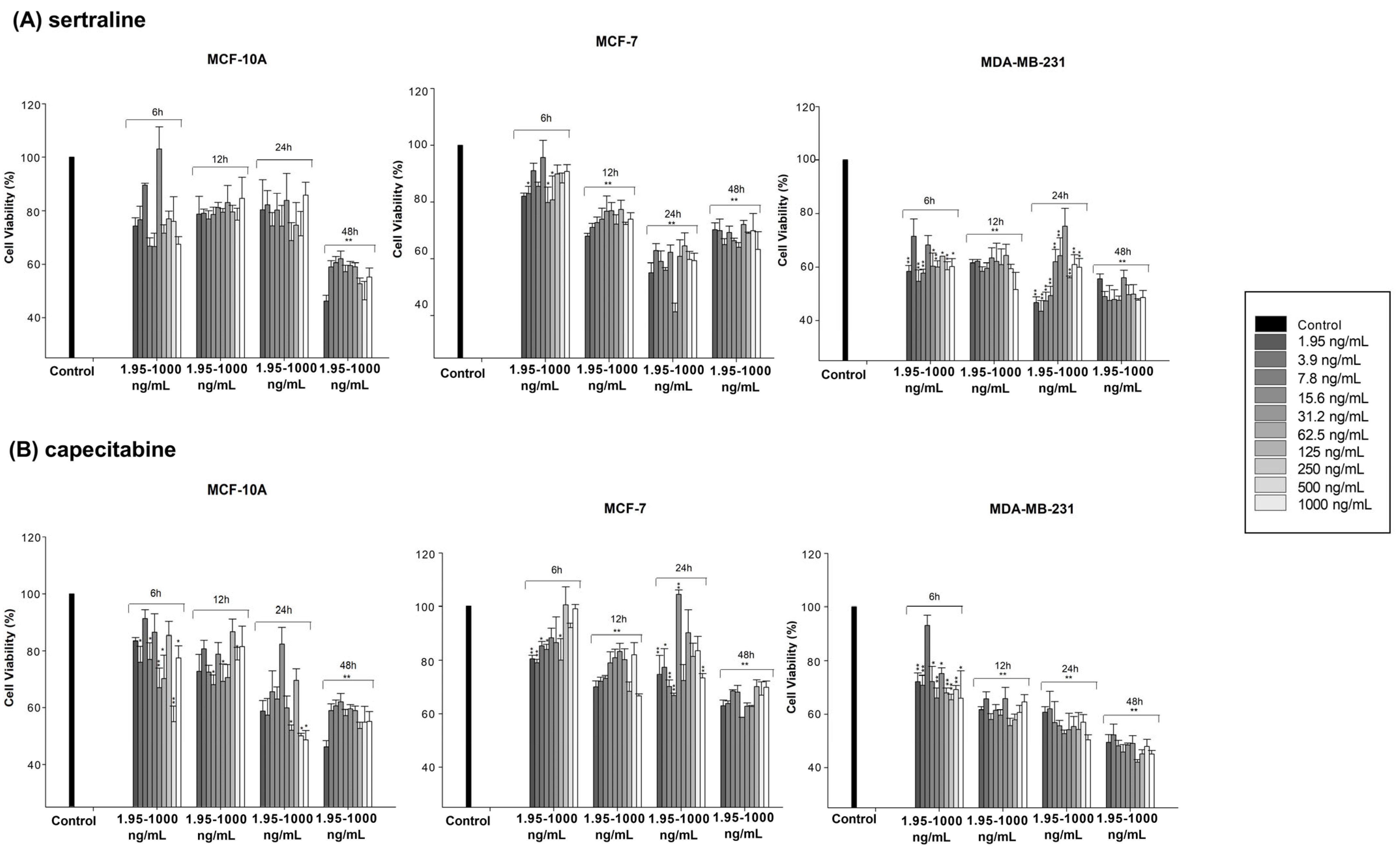
Figure 1. WST-1 Cell Proliferation Assay. (A) Cell viability (%) in MCF7, MDA-MB-231 and MCF10A cells compared to control after treatment with sertraline at doses ranging from 1.95–1000 ng/mL. (B) Cell viability (%) in cells compared to control after treatment with capecitabine at doses ranging from 1.95–1000 ng/mL. * p < 0.05 and ** p < 0.01 were considered to indicate a statistically significant differences compared to control group.
Scipharm 92 00038 g001
Figure 2. Neutral red assay. (A) Cell viability (%) in MCF7, MDA-MB-231 and MCF10A cells compared to control after treatment with sertraline at doses ranging from 1.95–1000 ng/mL. (B) Cell viability (%) in cells compared to control after treatment with capecitabine at doses ranging from 1.95–1000 ng/mL. * p < 0.05 and ** p < 0.01 were considered to indicate a statistically significant differences compared to control group.
Scipharm 92 00038 g002
Figure 3. Cell viability determined by WST-1 Cell Proliferation Assay after single and combination administration of capecitabine and sertraline (C5: 10 ng/mL, C2: 100 ng/mL; S3: 50 ng/mL). ** p < 0.01 was considered to indicate a statistically significant differences compared to control group.
Scipharm 92 00038 g003
Figure 4. CompuSyn analysis of cytotoxicity data was used to determine synergy, additivity, and antagonism between c (capecitabin) and s (sertraline) in MCF7 and MDA-MB-231 cells. The dose-effect analysis of both single compounds and their combination was determined by Compusyn software (version 1.0; ComboSyn, Paramus, NJ, USA), and the results are shown in the sigmoid concentration-effect curves. The y-axis shows the fractional survival rate (Fa) of cells. This ratio varies between 0 and 1. The x-axis indicates doses between 0 and 300 ng/mL.
Scipharm 92 00038 g004
Figure 5. (A) The amount of mTOR from the supernatant of MCF7 cells after 6 h of incubation (**, p < 0.01). (B) The amount of mTOR from the supernatant of MDAMB-231 Cells at the end of 12 h incubation (*, p < 0.05).
Scipharm 92 00038 g005
Figure 6. Visualization of the 2D and 3D protein-ligand interaction profile of Sertraline. (A) AKT1-Sertraline, (B) AMPK-Sertraline, (C) SIRT1-Sertraline, (D) LKB1-Sertraline.
Scipharm 92 00038 g006
Table 1. IC50 values calculated for MCF-7, MDA-MB-231 and MCF10A cells.
DrugMCF-7MDA-MB-231MCF10A
sertraline51.3554 ng/mL (24 h)16.86 ng/mL (24 h)
247.937 ng/mL (48 h)
362.91 ng/mL (48 h)
capecitabinend29.48 ng/mL (24 h)
8.72 ng/mL (48 h)
498.92 ng/mL (48 h)
Table 2. The binding energy of the selected compounds (kcal/mol).
AKT1AMPKSIRT1LKB1
Sertraline−9.9−7.9−7.5−7.0
Co-crystallized ligands−13.8 a−9.1 b−7.2 cna
Disclaimer/Publisher’s Note: The statements, opinions and data contained in all publications are solely those of the individual author(s) and contributor(s) and not of MDPI and/or the editor(s). MDPI and/or the editor(s) disclaim responsibility for any injury to people or property resulting from any ideas, methods, instructions or products referred to in the content.