Article

Inhibitory Effect of Mistletoe Ointment on DNCB-Induced Atopic Dermatitis in BALB/c Mice

College of Pharmacy and Research Institute of Life and Pharmaceutical Sciences, Sunchon National University, Suncheon 57922, Republic of Korea
*
Author to whom correspondence should be addressed.
Sci. Pharm. 2024, 92(1), 3; https://doi.org/10.3390/scipharm92010003
Submission received: 11 October 2023 / Revised: 18 December 2023 / Accepted: 25 December 2023 / Published: 29 December 2023
(This article belongs to the Topic Natural Products and Drug Discovery)

Abstract

:
Atopic dermatitis (AD) is increasingly prevalent globally. However, the frequent and prolonged use of corticosteroids, which are commonly employed for AD treatment, carries potential side effects. Korean mistletoe (Viscum album L. var. coloratum), a perennial parasitic plant, has demonstrated various biological effects. In this study, we conducted in vivo investigations to determine whether Korean mistletoe possesses anti-inflammatory effects that play pivotal roles in regulating the pathological mechanisms of AD. BALB/c mice with AD-like skin lesions induced by 2,4-dinitrochlorobenzene (DNCB) were utilized to explore the therapeutic effects of 1% and 2% Korean mistletoe extract (KME) ointments. The KME ointment was topically applied to the dorsal surface of the BALB/c mice, and they were categorized into four distinct groups: normal, DNCB-induced, DNCB-induced with 1% KME ointment, and DNCB-induced with 2% KME ointment. Each assessment parameter employed to evaluate the curative impact of the KME ointment displayed improvement with the application of the 1% KME ointment. While the effects observed were less pronounced than those of the 1% KME ointment, the overall therapeutic outcomes were also evident with the utilization of the 2% KME ointment. The results suggest the potential of Korean mistletoe as a viable therapeutic agent for AD. Further investigations are warranted to elucidate the underlying mechanisms of action.

1. Introduction

Characterized by incessant itching, dry skin, and a heightened proclivity for infection, atopic dermatitis (AD) is a relapsing inflammatory skin condition that varies over time in its manifested lesions [1,2]. As children grow older, the backs of their knees and fronts of their elbows are most frequently affected; however, the hands and feet tend to be the areas that experience greater prevalence in later childhood [3]. As AD’s prevalence intensifies on a global scale [4], the adult-type of this disorder has experienced noteworthy expansion in recent years [5]. To relieve its symptomatic effects, oral or topical corticosteroids and antihistamines are typically utilized. In more serious cases, phototherapy and immunosuppressive agents such as cyclosporine A may be administered [6]. Finding the pathological mechanism of AD is imperative given that it remains elusive, with no efficacious treatments addressing its fundamental cause. Many studies presently illustrate the mechanism of AD, while other investigations establish that AD may occur owing to insufficient regulation of anti-inflammatory homeostasis [7,8]. Therefore, it can be assumed that one should maintain an appropriate level of anti-inflammatory status for the treatment of AD.
The pathogenesis of AD is chiefly attributed to inflammation, a biological reaction of the host to injurious or microbial stimuli [9,10]. The properly regulated production of pro-inflammatory cytokines and inflammatory mediators serves to diminish the risk posed by pathogens. When not properly regulated, an abundance of pro-inflammatory cytokines and inflammatory mediators can lead to unpleasant situations (e.g., rash, itch, and edema) and certain illnesses [9,11,12].
Pro-inflammatory cytokines or inflammatory mediators are produced when AD incites stimuli such as scratching and infections. Interleukins (ILs), specifically IL-4, 5, and 6, promote the proliferation, maturation, migration, and adhesion of immune cells [13], as well as causing class switching of immunoglobulin (Ig)-E in B cells [14,15,16]. Recent studies have highlighted the involvement of IL-4, IL-5, and IL-6 in AD patients with elevated levels of Ig-E being its major features [17,18,19], and further revealed that tumor necrosis factor-α (TNF-α), prostaglandin E2 (PGE2), inducible nitric oxide synthase (iNOS), and cyclooxygenase-2 (COX-2) serve as immune mediators and partake in several immune responses related to AD [20,21]. Acute-phase AD is correlated with elevated levels of proinflammatory T helper 1 (Th1) and T helper 2 (Th2) cytokines. IL-4, 5, and 6 predominantly associate with skin inflammation; however, in the chronic phase of AD, skin lesions demonstrate fewer cells expressing IL-4 mRNA coupled with an increased number of cells showing TNF-α and IFN-γ mRNA expression in comparison to acute AD [1].
Acute AD requires the frequent prescription of topical corticosteroids. Corticosteroids, albeit helpful, may result in deleterious effects subsequent to extended administration, including localized skin atrophy and a heightened susceptibility to microbial invasion [22,23]. AD patients are generally worried about using corticosteroids and require treatments that have fewer side effects. With increasing interest regarding non-traditional treatments of AD, researchers have undertaken efforts to design novel treatments using natural products [24]. Natural products such as red ginseng, Sophora flavescens, Centella asiatica, Viola yedoensis, and Morinda citrifolia (Noni) have been discussed as promising remedies for AD [25,26,27,28,29].
Mistletoe, a perennial parasite that grows on various trees such as apple and oak, has been employed for centuries in traditional medicine throughout many nations, particularly those within Europe [30]. Traditionally, European mistletoe (Viscum album L., Loranthaceae) is used as a sedative, anti-spasmolytic, analgesic, cardio-tonic, and anti-cancer agent [31]. More recently, Korean mistletoe (Viscum album L. var. coloratum) has also proven its medicinal value. The main active compounds are lectins, viscotoxins, flavonoids, phenolic acids, sterols, lignans, terpenoids, phenylpropanoids, alkaloids, and fatty acids [32]. It has been observed to cause apoptosis in tumor cells [33], induce antitumor activity [34], affect immune regulation [35], and present antioxidant properties [36]. This study showed the effectiveness of Korean mistletoe for reducing atopic symptoms in vivo using an AD mouse model.

2. Materials and Methods

2.1. Experimental Animals

On obtaining 6-week-old BALB/c male mice from Sam Taco Bio (Osan-si, Republic of Korea), we ensured they were housed in a controlled environment of 23 ± 2 °C temperature, 60 ± 5 °C humidity, and 12 h cycle day/night. The mice had open access to the diets and water supplied by Sam Taco Bio, which were sterilized with an ultraviolet sterilizer and a microfiltration device. The animals were randomly divided into four groups, each containing eight mice: (1) normal control group; (2) 2,4-dinitrochlorobenzene (DNCB; Sigma-Aldrich, St. Louis, MO, USA)-treated group; (3) DNCB and 1% ointment-treated group; and (4) DNCB and 2% ointment-treated groups. The animal tests in this study were performed according to the rules of the Institutional Animal Care and Use Committee at Sunchon National University, Korea (approval code SCNU IACUC-2015-03).

2.2. Preparation of Korean Mistletoe Extract (KME) Ointment

After collecting Korean mistletoe (leaves, stems, and fruit) from Hongcheon-si, Gangwondo in South Korea, we naturally dried it before extracting 10 kg of powder using 200 kg of 70% ethanol (EtOH) at a temperature between 30 and 35 °C over a period of 48 h. The KME ointment used white petrolatum as a base; it contained either 1% or 2% by total weight in KME, respectively. We made an oil phase by mixing all components of the KME ointment (as detailed in Table 1), excluding purified water, and heating this mixture up to 70 °C. We mixed the oil phase and water phase, added KME to this mixture, stirred it with a high-speed stirrer, then cooled it down to 30 °C (Table 1).

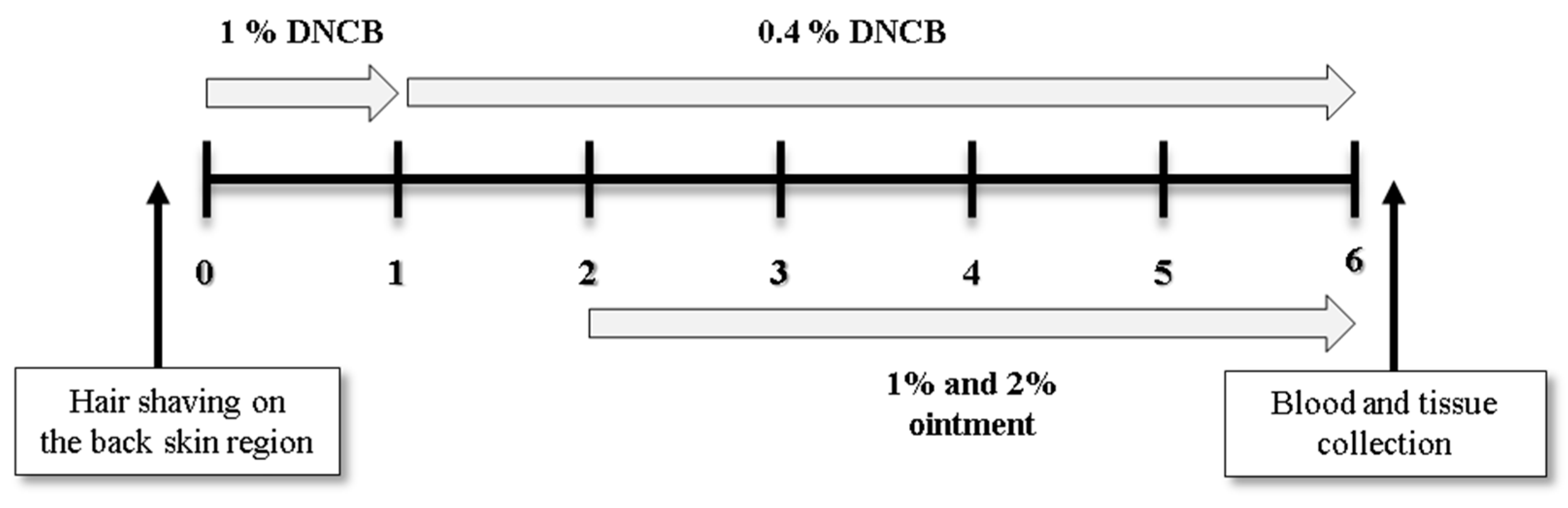
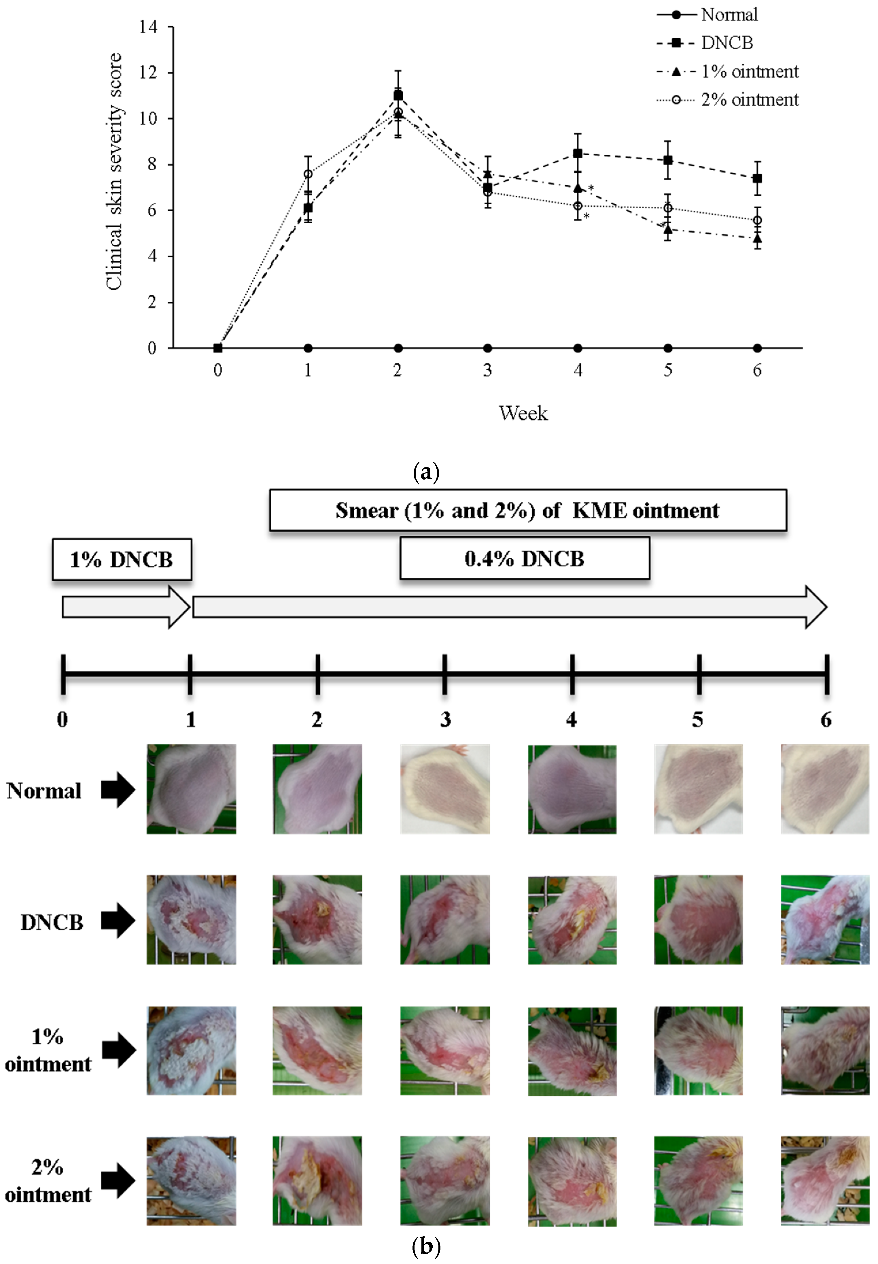
2.3. The Triggering and Treatment of AD-Like Cutaneous Lesion in Mice

We induced AD-like skin lesions in BALB/c mice by shaving and allowing their hair to heal for 24 h before administering DNCB. We administered 200 μL of 1% DNCB solution dissolved in acetone–olive oil (3:1) to the shaved dorsal surfaces of the mice three times weekly during the initial week, then shifted to four applications of 200 μL of a 0.4% DNCB solution from day eight until the concluding day of the trial. The treatment began with the application of KME ointment (1% and 2%) on induced AD-like skin lesions, administered daily sans interruption from day 15 to the last day of the trial. Mice were slaughtered at week 6, following the first treatment of DNCB (Figure 1).

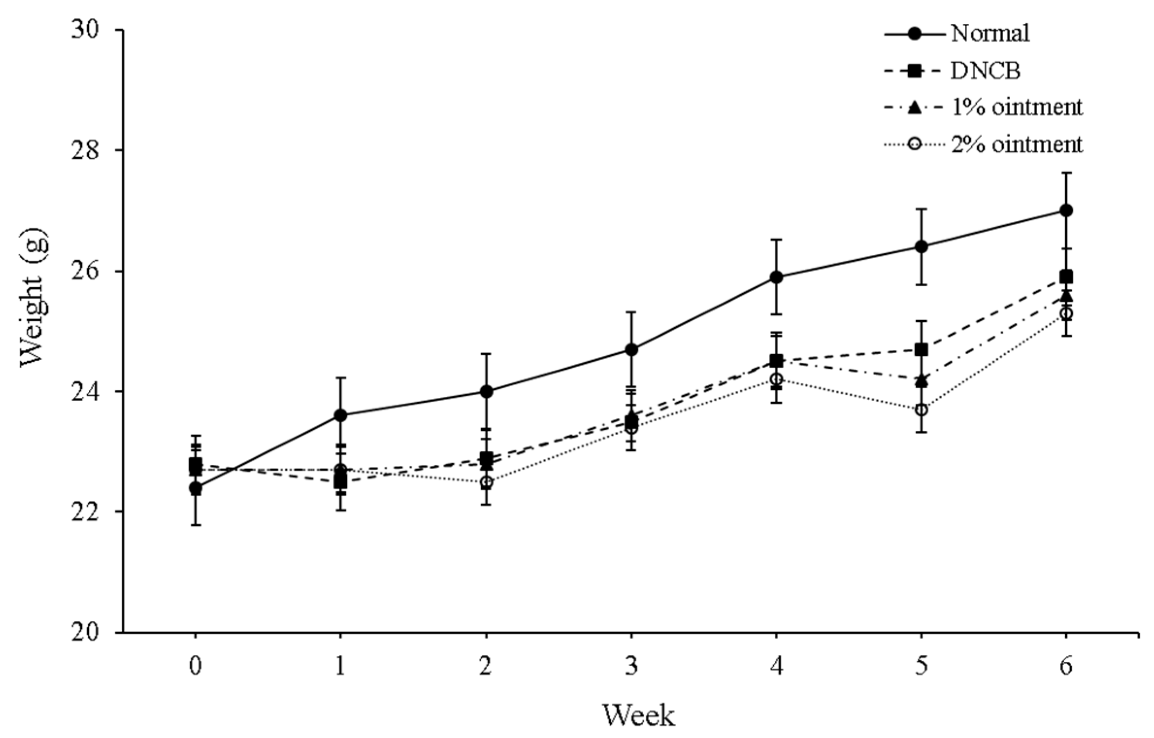
2.4. Measurement of the Weight of Mice and Their Spleens

Weekly weighing of mice was conducted over the course of 6 weeks in order to evaluate the influence of KME ointment on body weight. Furthermore, to quantify the weight of the spleen which is a secondary measure of the inflammation reaction, the mice were slaughtered at the end day of the experiment, and spleens were taken and measured.

2.5. Measurement of the Scratching Behavior Frequency

Each week following the application of DNCB and KME ointment, frequency of scratching behavior was gauged by placing mice in cages, then obtaining recordings with a video camera. We gave one point to mice that scratched continuously for a single second, and two points to those that maintained the behavior for 2 s. We monitored and tallied scratching frequencies over a 20 min period, thereby allowing us to calculate and assess each group’s average score.

2.6. Evaluation of the Cutaneous Lesion

We observed five distinct characteristics of the skin lesion for the severity of AD: erythema, pruritus and desiccation, edema and abrasion, ulceration, and lichenification. The individual indicators are rated 0 (no presence), 1 (mild, <20%), 2 (moderate, 20–60%), and 3 (severe, >60%). The cumulative score of such ratings constitutes the dermatitis score, ranging from 0 to 15.

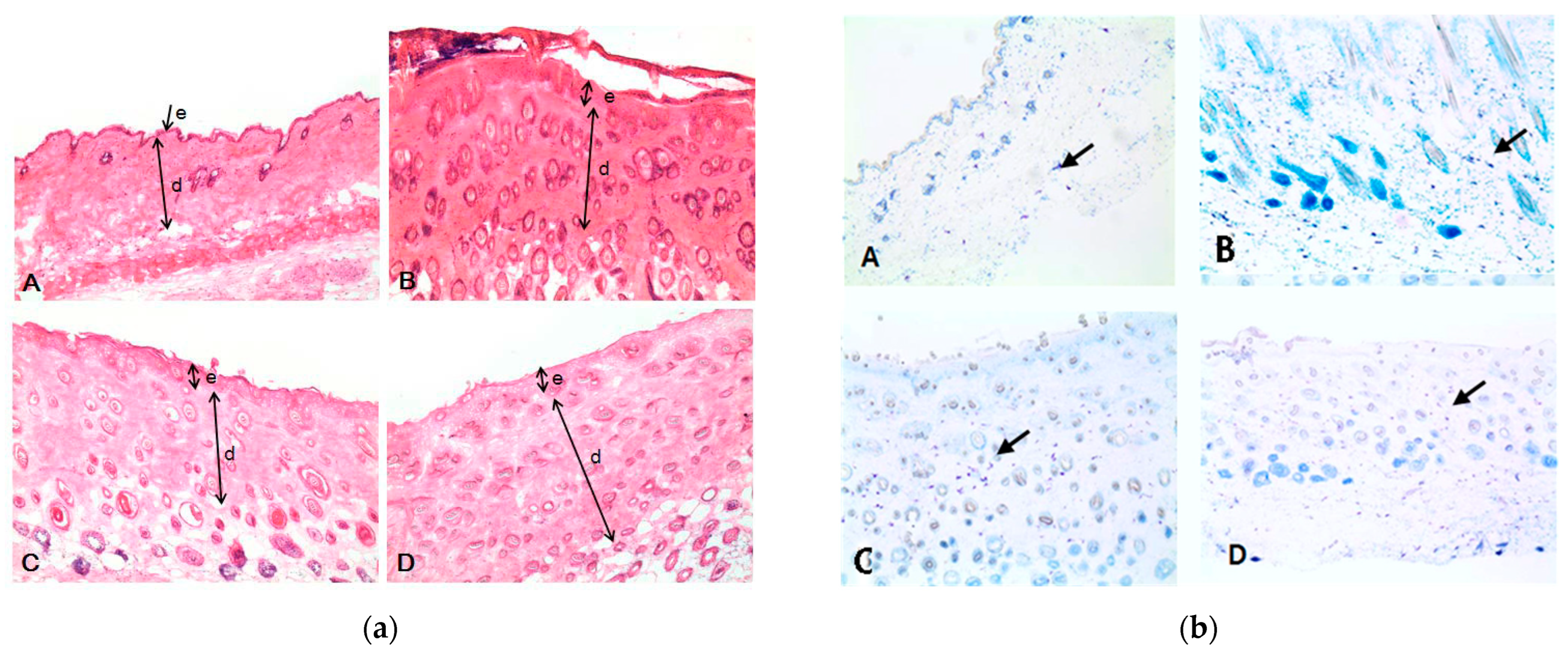
2.7. Histological Examination of Epidermal and Dermal Layers

After sacrificing the mice, we sectioned 15 μm of dorsal skin tissue utilizing a frozen method [37] and stained the segments with hematoxylin & eosin (H&E). Subsequently, we conducted histological examinations to observe the epidermal and dermal layers of the skin. Utilizing a ×400 optical microscope (Nikon, Tokyo, Japan), we measured the epidermal and dermal thickness of skin from random sections. Additionally, utilizing a ×200 optical microscope (Nikon), we stained mast cells infiltrating the dermis and hypodermis with toluidine blue and counted them from selected sections.

2.8. Measurement of the Total Serum IgE and Cytokine Levels

We administered ethyl ether anesthesia to mice, then drew serum from their hearts using a serum separator clot activator tube. Subsequently, we centrifuged the serum and used an enzyme-linked immunosorbent assay (ELISA) set (BD Biosciences, Franklin Lakes, NJ, USA) to measure total IgE and cytokine levels. Tissue Lyse II (PhileKorea, Daejeon-si, Republic of Korea) homogenized mice skin tissue with 0.1 mL tissue protein extraction reagent buffer containing protease to measure the shift in cytokine level of the mice. Utilizing an ELISA set (IL-4, IL-5, TNF-α, and IFN-γ), we measured the cytokines present in the supernatant following centrifugation of the sample. Briefly, we coated microplates with capture antibodies in a coating buffer comprising 0.1 M sodium carbonate at pH 9.0 for an overnight duration at 4 °C. After washing the plate three times with phosphate-buffered saline (PBS) containing 0.5% Tween 20, 1% bovine serum albumin (BSA) in PBS was introduced and incubated for an hour. Subsequently, the sample and standard were added into the plates and allowed to incubate for 2 h. After 5 further washings, the plate was exposed to a working detector for 1 h before being saturated with substrate solution and left undisturbed in a dark room for 30 min; thereafter, 1 M phosphoric acid stop solution was applied and color development was quantitatively evaluated using a microplate reader (Sunrise Technologies, Männedorf, Zürich, Switzerland) at 450 nm wavelength.

2.9. Statistical Analysis

All experiments were conducted in triplicate, and the results were presented as means ± standard deviation (S.D.). Statistical analysis of the data was performed using GraphPad software (version 7.00). Significant differences between groups were determined using a one-way analysis of variance (ANOVA), followed by Tukey’s multiple comparison tests for post hoc analysis. Statistical significance was defined as p < 0.05, p < 0.01, and p < 0.001, indicating the level of significance for the observed differences between groups.

3. Results

3.1. KME Ointment’s Impact on the Weight and Spleen Size of Mice

We measured the weights of mice weekly for six weeks to investigate how KME ointment influenced their weight changes. The normal group’s mice, over the course of the experiment, exhibited a consistent rise in weight and gained an average of 27.0 ± 0.9 g; however, the DNCB, 1% ointment, and 2% ointment groups all lost weight over the course of the experiment, losing an average of 1.1 g, 1.4 g, and 1.7 g, respectively. The DNCB group demonstrated a statistically significant weight loss compared to the normal group (p < 0.05), while no significant difference in weight loss was observed between the 1% ointment and 2% ointment groups (Figure 2). We evaluated spleen weights in sacrificed mice to examine the immunological responses produced by DNCB-induced AD. The DNCB mice had significantly higher spleen weights (0.17 ± 0.045 g) than the control mice (p < 0.05). However, as compared to the DNCB group, treatment with either 1% or 2% KME ointment significantly reduced spleen weight (p < 0.05). There was no significant difference in spleen weight between the 1% and 2% KME ointment groups (Table 2).

3.2. Observations in Macroscopic Change

To evaluate the effects of DNCB and KME ointment for clinical skin severity, we used DNCB for the first 3 weeks to induce an AD-like lesion in the skin of BALB/c mice, and thereafter applied 1% and 2% KME ointment on the dorsal skin from week 3 to week 6. Mice were scored on a three-point scale; zero indicated no lesions while three was indicative of the most severe skin lesions. As depicted in Figure 3, the DNCB group had more severe skin lesions upon completion of the six-week treatment than the control group (p < 0.05). One per cent or two percent KME ointment treatment significantly attenuated the increase in clinical skin severity score caused by DNCB (p < 0.05). There was no significant difference in clinical skin severity score between the 1% and 2% KME ointment groups.

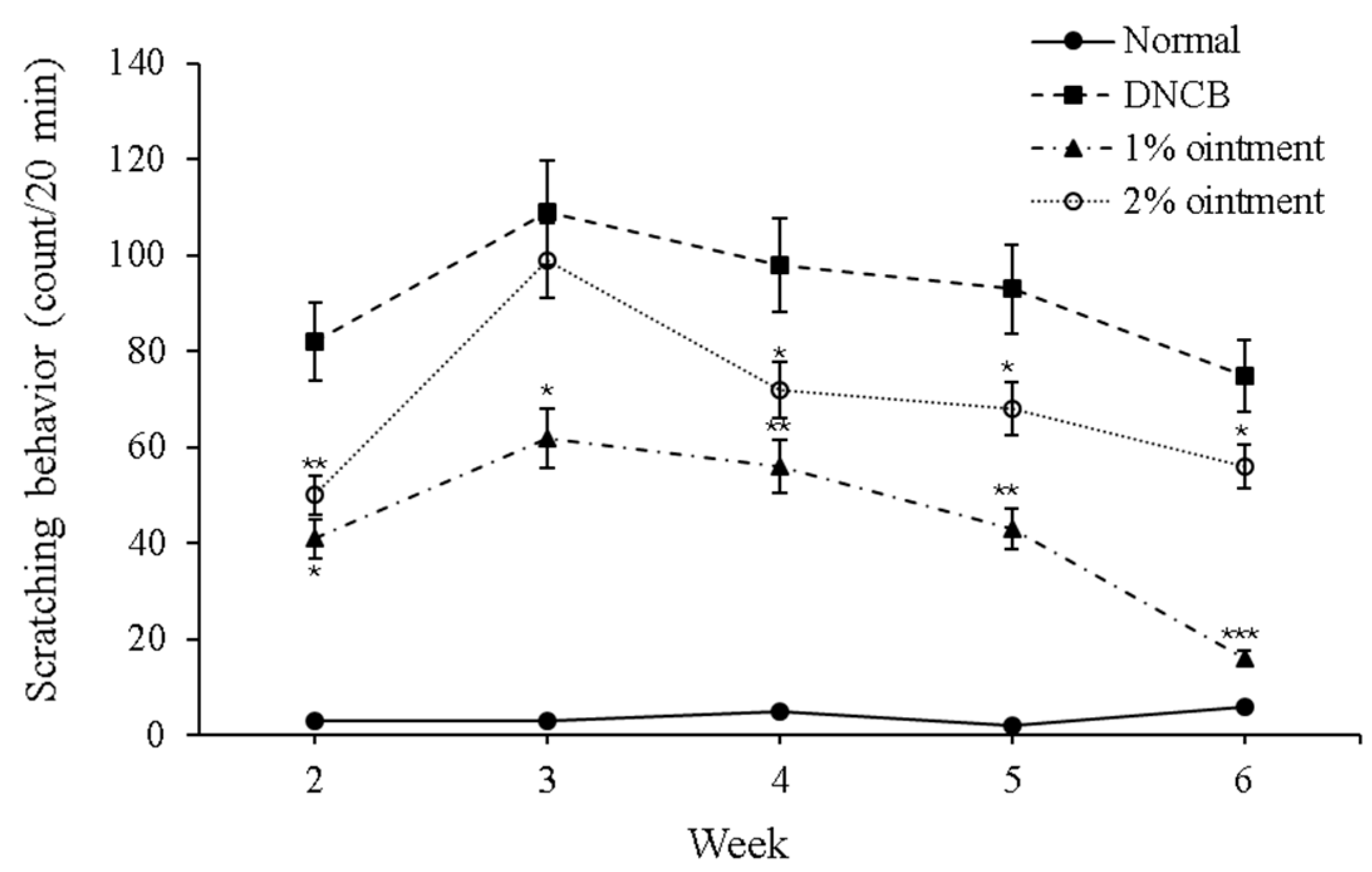
3.3. KME Ointment’s Effect on the Frequency of Scratching Behavior

In order to study the effects of KME ointment on the scratching behavior of mice with DNCB-induced AD, we counted the number of times that mice scratched their ears in a 20-min period. The results revealed that the scratching behavior among mice in the DNCB group increased significantly compared with those in the control group four weeks later (p < 0.05). On the contrary, mice with DNCB-induced AD showed a significant reduction in scratching behavior following treatment with 1% or 2% KME ointment (p < 0.05). In terms of scratching behavior, there were no significant differences between the 1% and 2% KME ointment groups (Figure 4).

3.4. Histopathological Observations Change

The skin changes were assessed by performing H&E staining and toluidine blue staining on a group of mice with DNCB-induced AD. According to H&E staining, the control group had the thinnest epidermis and dermis, while the DNCB group had the thickest. The application of 1% KME ointment resulted in a significant decrease in the thickness of both epidermal and dermal layers in mice with DNCB-induced AD compared to normal mice. However, application of 2% KME ointment decreased the thickness of epidermis only (Figure 5a and Table 3). Toluidine blue staining showed that the number of mast cells in the dermis and hypodermis was significantly higher for the DNCB group than for the normal group. Treatment with either 1% or 2% KME ointment significantly reduced the number of mast cells in mice with DNCB-induced AD (Figure 5b and Table 4).

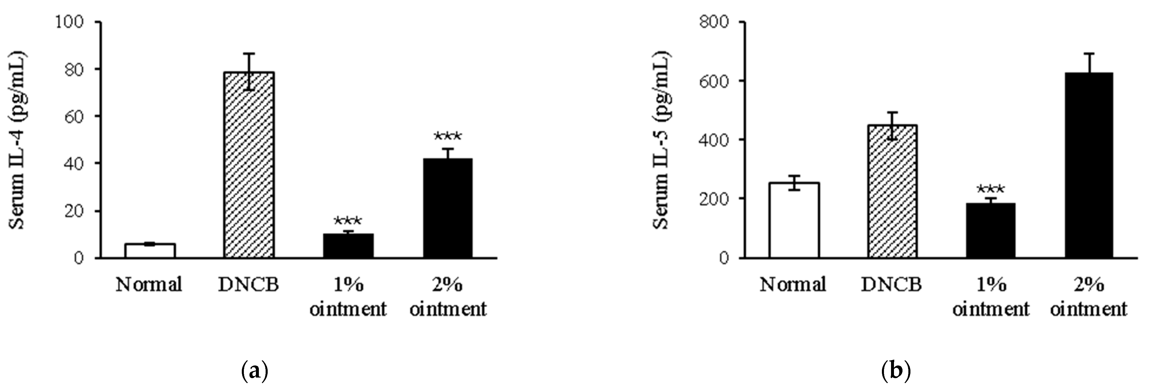
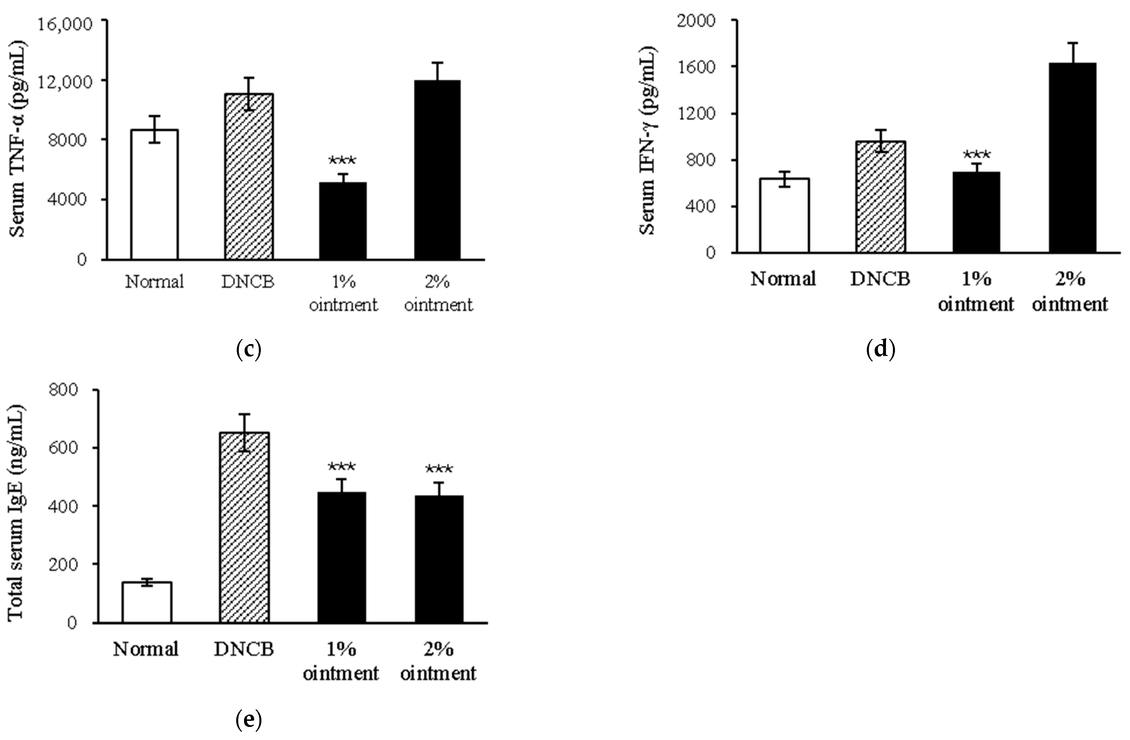
3.5. KME Ointment’s Effect on the Total Serum of IL-4, IL-5, TNF-α, IFN-γ, and IgE

To investigate the effects of KME ointment on the serum levels of Th2-associated cytokines (IL-4 and IL-5), Th1-associated cytokines (TNF-α and IFN-γ), and IgE in mice with DNCB-induced AD, we collected serum from mice in each group and measured the cytokine levels using ELISA. Compared with the controls, mice of the DNCB group demonstrated a significant increase in serum levels of all four cytokines and IgE after treatment for 4 weeks (p < 0.05). Mice with DNCB-induced AD were treated with 1% KME ointment, and all four cytokines as well as IgE serum levels were significantly reduced (p < 0.05). However, the 2% KME ointment only reduced the serum levels of IL-4 and IgE (Figure 6).

4. Discussion

The current study explored the potential of Korean mistletoe as a viable therapeutic agent for AD. The spleen, a lymphoid organ integral to immune function [38], filters blood, removes aged/damaged red blood cells, and produces immune cells [39,40,41]. Enlargement of the spleen may occur in AD owing to factors like inflammation, elevated immune cell counts, and immune cell infiltration into the organ [42]. The enlargement of the spleen can induce fatigue, abdominal pain, and anemia [43]. In DNCB-induced AD mice, KME ointment demonstrated reduced splenic mass, implying the potential to systemically suppress inflammation since the spleen generates T cells, B cells, and dendritic cells [44]. Further, AD elicits scratching mediated by histamine and pro-inflammatory mast cell products [45,46,47]. The results showed that KME ointment significantly reduced the scratching in AD mice, suggesting that KME ointment may have anti-inflammatory effects, which lead us to further investigate the secretion of inflammatory cytokines related to AD.
Inflammatory cytokines play an important part in the pathogenesis of AD. For example, IL-4 and IL-5, which are Th2-associated cytokines, are vital in explaining the progress of AD [48,49]. They facilitate the production of IgE, resulting in activation of mast cells and subsequently inflammation and itching [50]. Likewise, TNF-α and IFN-γ, which are Th1-associated cytokines, have been shown to contribute to the pathogenesis of AD [51], with the former being one of the pro-inflammatory cytokines that plays a role in the promotion of inflammation and tissue damage [52]. IFN-γ is an immunomodulatory cytokine which suppresses IgE production and mast cell activation [53]. IgE is an immunoglobulin that attaches to mast cells, which then release histamine and other pro-inflammatory mediators, leading to inflammation and itching in the skin [54]. The current investigation shows that KME ointment significantly reduced the secretion of IL-4, IL-5, TNF-α, IFN-γ, and IgE in DNCB-induced AD mice. This suggests that KME ointment may have anti-inflammatory and immunomodulatory effects. These findings are consistent with what we previously found.
Another feature of AD is histological changes in the skin [55]. The skin is composed of two thin layers, the epidermis and dermis. The epidermis is the outermost layer of skin that acts as a protective barrier towards the outside environment. Below the epidermis lies another important layer known as the dermis which is composed of connective tissue, blood vessels, and nerve endings [56,57,58]. In AD, the epidermis and dermis become thickened. There are various reasons for this, including inflammation, increased cell turnover, and the production of new collagen.
KME ointment may decrease itching, erythema, and scaling in mice with DNCB-induced AD through reducing the thickness of the epidermis and dermis. This could be further explained by the suppressive effect of KME ointment on mast cell activation which is pivotal to the development of AD. The activation of mast cells triggers the release of different inflammatory mediators, like histamine, cytokines, and proteases [59,60]. Thus, the reduction in inflammation and thickening in both epidermis and dermis by KME ointment may occur through suppressing mast cell activation. However, it remains unclear if this ointment also affects other types of inflammatory cells like eosinophils or neutrophils. These two cell types, apart from their roles in the pathogenesis of AD, actively contribute to the manifestation of AD-like symptoms in animal models [52,61,62]. Thus, it would be interesting to investigate the potential efficacy of KME ointment in suppressing eosinophil and neutrophil counts and activity in mice with DNCB-induced AD.
Our experiments revealed that 1% KME ointment applied topically yielded greater efficacy than 2% KME for mice with DNCB-induced AD. This outcome challenges the prevalent belief that higher drug concentrations lead to heightened effectiveness. One plausible rationale for this finding is that a 2% concentration of KME may be excessively irritating to the skin, potentially triggering inflammation and an increase in pro-inflammatory cytokines, which counteracts the intended anti-inflammatory impact of the KME ointment. Another conceivable explanation is that a 2% KME concentration might be too potent, potentially causing immune system suppression. This, in turn, could heighten susceptibility to infections and impaired wound healing [63,64]. Finally, it is possible that a 1% concentration of KME is the optimal choice for treating AD; hence, higher concentrations of KME may not be required to attain the desired effects. Indeed, current research acknowledges that using lower drug doses can yield superior efficacy in treating certain conditions. This is attributed to a phenomenon known as hormesis. A low-dose stimulation and a high-dose inhibition characterize hormesis, which is a dose–response phenomenon [65,66,67,68]. The effects of KME ointment on AD could potentially follow this hormetic dose–response curve. This suggests that lower doses of KME ointment potentially offer superior efficacy for treating AD, compared to their higher counterparts.
This study indicates that KME ointment has the potential to enhance the skin barrier function and reduce inflammation in AD. Further, it suggests that 1% KME ointment could be optimal for treating AD. However, more research is needed to fully understand these mechanisms.

Author Contributions

Conceptualization: C.-E.H. and S.-Y.L.; funding acquisition: S.-Y.L.; investigation: C.-E.H. and S.-Y.L.; methodology: C.-E.H.; resources: C.-E.H.; supervision: S.-Y.L.; writing—original draft: C.-E.H.; writing—review and editing: S.-Y.L. All authors have read and agreed to the published version of the manuscript.

Funding

This research was supported by the “Establishment of Economic Technology for the Cultivation of Mistletoe and Development of Strategic Export Products Using Its Functional Compounds (project No.: 313011-3)”, the Ministry of Agriculture, Food, and Rural Affairs, Republic of Korea.

Institutional Review Board Statement

Not applicable.

Informed Consent Statement

Not applicable.

Data Availability Statement

Data are contained within the article.

Conflicts of Interest

The authors declare no conflicts of interest.

References

  1. Leung, D.Y. Atopic dermatitis: New insights and opportunities for therapeutic intervention. J. Allergy Clin. Immunol. 2000, 105, 860–876. [Google Scholar] [CrossRef] [PubMed]
  2. Fishbein, A.B.; Silverberg, J.I.; Wilson, E.J.; Ong, P.Y. Update on Atopic Dermatitis: Diagnosis, Severity Assessment, and Treatment Selection. J. Allergy Clin. Immunol. Pract. 2020, 8, 91–101. [Google Scholar] [CrossRef] [PubMed]
  3. Tollefson, M.M.; Bruckner, A.L. Atopic dermatitis: Skin-directed management. Pediatrics 2014, 134, e1735–e1744. [Google Scholar] [CrossRef] [PubMed]
  4. Williams, H.; Robertson, C.; Stewart, A.; Aït-Khaled, N.; Anabwani, G.; Anderson, R.; Asher, I.; Beasley, R.; Björkstén, B.; Burr, M.; et al. Worldwide variations in the prevalence of symptoms of atopic eczema in the International Study of Asthma and Allergies in Childhood. J. Allergy Clin. Immunol. 1999, 103, 125–138. [Google Scholar] [CrossRef] [PubMed]
  5. Toyokuni, S. Reactive oxygen species-induced molecular damage and its application in pathology. Pathol. Int. 1999, 49, 91–102. [Google Scholar] [CrossRef] [PubMed]
  6. Ring, J.; Alomar, A.; Bieber, T.; Deleuran, M.; Fink-Wagner, A.; Gelmetti, C.; Gieler, U.; Lipozencic, J.; Luger, T.; Oranje, A.P.; et al. Guidelines for treatment of atopic eczema (Atopic dermatitis) Part II. J. Eur. Acad. Dermatol. Venereol. JEADV 2012, 26, 1176–1193. [Google Scholar] [CrossRef]
  7. Sharma, J.N.; Al-Omran, A.; Parvathy, S.S. Role of nitric oxide in inflammatory diseases. Inflammopharmacology 2007, 15, 252–259. [Google Scholar] [CrossRef]
  8. Kim, H.J.; Song, H.K.; Park, S.H.; Jang, S.; Park, K.S.; Song, K.H.; Lee, S.K.; Kim, T. Terminalia chebula Retz. extract ameliorates the symptoms of atopic dermatitis by regulating anti-inflammatory factors in vivo and suppressing STAT1/3 and NF-ĸB signaling in vitro. Phytomed. Int. J. Phytother. Phytopharm. 2022, 104, 154318. [Google Scholar] [CrossRef]
  9. Zamora, R.; Vodovotz, Y.; Billiar, T.R. Inducible nitric oxide synthase and inflammatory diseases. Mol. Med. 2000, 6, 347–373. [Google Scholar] [CrossRef]
  10. Tracey, K.J. The inflammatory reflex. Nature 2002, 420, 853–859. [Google Scholar] [CrossRef]
  11. Hassanshahi, A.; Moradzad, M.; Ghalamkari, S.; Fadaei, M.; Cowin, A.J.; Hassanshahi, M. Macrophage-Mediated Inflammation in Skin Wound Healing. Cells 2022, 11, 2953. [Google Scholar] [CrossRef] [PubMed]
  12. Lauritano, D.; Mastrangelo, F.; D’Ovidio, C.; Ronconi, G.; Caraffa, A.; Gallenga, C.E.; Frydas, I.; Kritas, S.K.; Trimarchi, M.; Carinci, F.; et al. Activation of Mast Cells by Neuropeptides: The Role of Pro-Inflammatory and Anti-Inflammatory Cytokines. Int. J. Mol. Sci. 2023, 24, 4811. [Google Scholar] [CrossRef] [PubMed]
  13. Brocker, C.; Thompson, D.; Matsumoto, A.; Nebert, D.W.; Vasiliou, V. Evolutionary divergence and functions of the human interleukin (IL) gene family. Hum. Genom. 2010, 5, 30–55. [Google Scholar] [CrossRef] [PubMed]
  14. Vercelli, D.; Jabara, H.H.; Arai, K.; Geha, R.S. Induction of human IgE synthesis requires interleukin 4 and T/B cell interactions involving the T cell receptor/CD3 complex and MHC class II antigens. J. Exp. Med. 1989, 169, 1295–1307. [Google Scholar] [CrossRef] [PubMed]
  15. Del Prete, G.; Maggi, E.; Parronchi, P.; Chrétien, I.; Tiri, A.; Macchia, D.; Ricci, M.; Banchereau, J.; De Vries, J.; Romagnani, S. IL-4 is an essential factor for the IgE synthesis induced in vitro by human T cell clones and their supernatants. J. Immunol. 1988, 140, 4193–4198. [Google Scholar] [CrossRef] [PubMed]
  16. Hirano, T.; Yasukawa, K.; Harada, H.; Taga, T.; Watanabe, Y.; Matsuda, T.; Kashiwamura, S.; Nakajima, K.; Koyama, K.; Iwamatsu, A.; et al. Complementary DNA for a novel human interleukin (BSF-2) that induces B lymphocytes to produce immunoglobulin. Nature 1986, 324, 73–76. [Google Scholar] [CrossRef]
  17. Tang, M.; Kemp, A.; Varigos, G. IL-4 and interferon-gamma production in children with atopic disease. Clin. Exp. Immunol. 1993, 92, 120–124. [Google Scholar] [CrossRef]
  18. Tsianakas, A.; Luger, T.A. The anti-IL-4 receptor alpha antibody dupilumab: Facing a new era in treating atopic dermatitis. Expert Opin. Biol. Ther. 2015, 15, 1657–1660. [Google Scholar] [CrossRef]
  19. Ilves, T.; Harvima, I.T. Decrease in chymase activity is associated with increase in IL-6 expression in mast cells in atopic dermatitis. Acta Derm. Venereol. 2015, 95, 411–416. [Google Scholar] [CrossRef]
  20. Hu, W.; Yang, X.; Zhe, C.; Zhang, Q.; Sun, L.; Cao, K. Puerarin inhibits iNOS, COX-2 and CRP expression via suppression of NF-κB activation in LPS-induced RAW264.7 macrophage cells. Pharmacol. Rep. 2011, 63, 781–789. [Google Scholar] [CrossRef]
  21. Fan, G.W.; Zhang, Y.; Jiang, X.; Zhu, Y.; Wang, B.; Su, L.; Cao, W.; Zhang, H.; Gao, X. Anti-inflammatory activity of baicalein in LPS-stimulated RAW264.7 macrophages via estrogen receptor and NF-κB-dependent pathways. Inflammation 2013, 36, 1584–1591. [Google Scholar] [CrossRef] [PubMed]
  22. Hengge, U.R.; Ruzicka, T.; Schwartz, R.A.; Cork, M.J. Adverse effects of topical glucocorticosteroids. J. Am. Acad. Dermatol. 2006, 54, 1–15. [Google Scholar] [CrossRef] [PubMed]
  23. Yu, S.H.; Drucker, A.M.; Lebwohl, M.; Silverberg, J.I. A systematic review of the safety and efficacy of systemic corticosteroids in atopic dermatitis. J. Am. Acad. Dermatol. 2018, 78, 733–740.e711. [Google Scholar] [CrossRef] [PubMed]
  24. Jones, V.A.; Patel, P.M.; Wilson, C.; Wang, H.; Ashack, K.A. Complementary and alternative medicine treatments for common skin diseases: A systematic review and meta-analysis. JAAD Int. 2021, 2, 76–93. [Google Scholar] [CrossRef] [PubMed]
  25. Kim, H.; Lee, M.R.; Lee, G.S.; An, W.G.; Cho, S.I. Effect of Sophora flavescens Aiton extract on degranulation of mast cells and contact dermatitis induced by dinitrofluorobenzene in mice. J. Ethnopharmacol. 2012, 142, 253–258. [Google Scholar] [CrossRef] [PubMed]
  26. Hong, C.E.; Lyu, S.Y. Anti-inflammatory and Anti-oxidative Effects of Korean Red Ginseng Extract in Human Keratinocytes. Immune Netw. 2011, 11, 42–49. [Google Scholar] [CrossRef] [PubMed]
  27. Lee, Y.; Choi, H.K.; N’deh KP, U.; Choi, Y.J.; Fan, M.; Kim, E.K.; Chung, K.H.; An, A.J.H. Inhibitory Effect of Centella asiatica Extract on DNCB-Induced Atopic Dermatitis in HaCaT Cells and BALB/c Mice. Nutrients 2020, 12, 411. [Google Scholar] [CrossRef]
  28. Fan, P.; Yang, Y.; Liu, T.; Lu, X.; Huang, H.; Chen, L.; Kuang, Y. Anti-atopic effect of Viola yedoensis ethanol extract against 2,4-dinitrochlorobenzene-induced atopic dermatitis-like skin dysfunction. J. Ethnopharmacol. 2021, 280, 114474. [Google Scholar] [CrossRef]
  29. Kim, S.H.; Seong, G.S.; Choung, S.Y. Fermented Morinda citrifolia (Noni) Alleviates DNCB-Induced Atopic Dermatitis in NC/Nga Mice through Modulating Immune Balance and Skin Barrier Function. Nutrients 2020, 12, 249. [Google Scholar] [CrossRef]
  30. Khwaja, T.A.; Dias, C.B.; Pentecost, S. Recent studies on the anticancer activities of mistletoe (Viscum album) and its alkaloids. Oncology 1986, 43 (Suppl. S1), 42–50. [Google Scholar] [CrossRef]
  31. Büssing, A.; Suzart, K.; Bergmann, J.; Pfüller, U.; Schietzel, M.; Schweizer, K. Induction of apoptosis in human lymphocytes treated with Viscum album L. is mediated by the mistletoe lectins. Cancer Lett. 1996, 99, 59–72. [Google Scholar] [CrossRef] [PubMed]
  32. Szurpnicka, A.; Zjawiony, J.K.; Szterk, A. Therapeutic potential of mistletoe in CNS-related neurological disorders and the chemical composition of Viscum species. J. Ethnopharmacol. 2019, 231, 241–252. [Google Scholar] [CrossRef] [PubMed]
  33. Lyu, S.Y.; Choi, S.H.; Park, W.B. Korean mistletoe lectin-induced apoptosis in hepatocarcinoma cells is associated with inhibition of telomerase via mitochondrial controlled pathway independent of p53. Arch. Pharmacal Res. 2002, 25, 93–101. [Google Scholar] [CrossRef] [PubMed]
  34. Yoon, T.J.; Yoo, Y.C.; Kang, T.B.; Song, S.K.; Lee, K.B.; Her, E.; Song, K.S.; Kim, J.B. Antitumor activity of the Korean mistletoe lectin is attributed to activation of macrophages and NK cells. Arch. Pharmacal Res. 2003, 26, 861–867. [Google Scholar] [CrossRef] [PubMed]
  35. Lee, C.H.; Kim, J.K.; Kim, H.Y.; Park, S.M.; Lee, S.M. Immunomodulating effects of Korean mistletoe lectin in vitro and in vivo. Int. Immunopharmacol. 2009, 9, 1555–1561. [Google Scholar] [CrossRef]
  36. Kim, B.K.; Choi, M.J.; Park, K.Y.; Cho, E.J. Protective effects of Korean mistletoe lectin on radical-induced oxidative stress. Biol. Pharm. Bull. 2010, 33, 1152–1158. [Google Scholar] [CrossRef]
  37. Yosipovitch, G.; Papoiu, A.D. What causes itch in atopic dermatitis? Curr. Allergy Asthma Rep. 2008, 8, 306–311. [Google Scholar] [CrossRef]
  38. Lewis, S.M.; Williams, A.; Eisenbarth, S.C. Structure and function of the immune system in the spleen. Sci. Immunol. 2019, 4, eaau6085. [Google Scholar] [CrossRef]
  39. Mebius, R.E.; Kraal, G. Structure and function of the spleen. Nat. Rev. Immunol. 2005, 5, 606–616. [Google Scholar] [CrossRef]
  40. McKenzie, C.V.; Colonne, C.K.; Yeo, J.H.; Fraser, S.T. Splenomegaly: Pathophysiological bases and therapeutic options. Int. J. Biochem. Cell Biol. 2018, 94, 40–43. [Google Scholar] [CrossRef]
  41. Cesta, M.F. Normal structure, function, and histology of the spleen. Toxicol. Pathol. 2006, 34, 455–465. [Google Scholar] [CrossRef] [PubMed]
  42. Lv, W.J.; Huang, J.Y.; Li, S.P.; Gong, X.P.; Sun, J.B.; Mao, W.; Guo, S.N. Portulaca oleracea L. extracts alleviate 2,4-dinitrochlorobenzene-induced atopic dermatitis in mice. Front. Nutr. 2022, 9, 986943. [Google Scholar] [CrossRef] [PubMed]
  43. Singh, C.K.; Mintie, C.A.; Ndiaye, M.A.; Chhabra, G.; Roy, S.; Sullivan, R.; Longley, B.J.; Schieke, S.M.; Ahmad, N. Protective effects of dietary grape against atopic dermatitis-like skin lesions in NC/NgaTndCrlj mice. Front. Immunol. 2022, 13, 1051472. [Google Scholar] [CrossRef] [PubMed]
  44. Mota, C.M.D.; Madden, C.J. Neural control of the spleen as an effector of immune responses to inflammation: Mechanisms and treatments. Am. J. Physiol. Regul. Integr. Comp. Physiol. 2022, 323, R375–R384. [Google Scholar] [CrossRef]
  45. Tominaga, M.; Takamori, K. Peripheral itch sensitization in atopic dermatitis. Allergol. Int. Off. J. Jpn. Soc. Allergol. 2022, 71, 265–277. [Google Scholar] [CrossRef] [PubMed]
  46. Peters, N.; Peters, A.T. Atopic dermatitis. Allergy Asthma Proc. 2019, 40, 433–436. [Google Scholar] [CrossRef]
  47. Mack, M.R.; Kim, B.S. The Itch-Scratch Cycle: A Neuroimmune Perspective. Trends Immunol. 2018, 39, 980–991. [Google Scholar] [CrossRef]
  48. Beck, L.A.; Cork, M.J.; Amagai, M.; De Benedetto, A.; Kabashima, K.; Hamilton, J.D.; Rossi, A.B. Type 2 Inflammation Contributes to Skin Barrier Dysfunction in Atopic Dermatitis. JID Innov. Ski. Sci. Mol. Popul. Health 2022, 2, 100131. [Google Scholar] [CrossRef]
  49. Huang, I.H.; Chung, W.H.; Wu, P.C.; Chen, C.B. JAK-STAT signaling pathway in the pathogenesis of atopic dermatitis: An updated review. Front. Immunol. 2022, 13, 1068260. [Google Scholar] [CrossRef]
  50. Akdis, C.A.; Arkwright, P.D.; Brüggen, M.C.; Busse, W.; Gadina, M.; Guttman-Yassky, E.; Kabashima, K.; Mitamura, Y.; Vian, L.; Wu, J.; et al. Type 2 immunity in the skin and lungs. Allergy 2020, 75, 1582–1605. [Google Scholar] [CrossRef]
  51. Sabat, R.; Wolk, K.; Loyal, L.; Döcke, W.D.; Ghoreschi, K. T cell pathology in skin inflammation. Semin. Immunopathol. 2019, 41, 359–377. [Google Scholar] [CrossRef]
  52. Yamaguchi, H.L.; Yamaguchi, Y.; Peeva, E. Role of Innate Immunity in Allergic Contact Dermatitis: An Update. Int. J. Mol. Sci. 2023, 24, 12975. [Google Scholar] [CrossRef] [PubMed]
  53. Romagnani, S. Biology of human TH1 and TH2 cells. J. Clin. Immunol. 1995, 15, 121–129. [Google Scholar] [CrossRef] [PubMed]
  54. David Boothe, W.; Tarbox, J.A.; Tarbox, M.B. Atopic Dermatitis: Pathophysiology. Adv. Exp. Med. Biol. 2017, 1027, 21–37. [Google Scholar] [CrossRef] [PubMed]
  55. Uehara, M. Clinical and histological features of dry skin in atopic dermatitis. Acta Derm. Venereol. 1985, 114, 82–86. [Google Scholar] [CrossRef]
  56. Evrard, C.; Lambert de Rouvroit, C.; Poumay, Y. Epidermal Hyaluronan in Barrier Alteration-Related Disease. Cells 2021, 10, 3096. [Google Scholar] [CrossRef] [PubMed]
  57. Choi, J.E.; Di Nardo, A. Skin neurogenic inflammation. Semin. Immunopathol. 2018, 40, 249–259. [Google Scholar] [CrossRef]
  58. Kigasawa, K.; Kajimoto, K.; Hama, S.; Saito, A.; Kanamura, K.; Kogure, K. Noninvasive delivery of siRNA into the epidermis by iontophoresis using an atopic dermatitis-like model rat. Int. J. Pharm. 2010, 383, 157–160. [Google Scholar] [CrossRef]
  59. Tong, J.; Li, Y.; Cai, X.; Lou, F.; Sun, Y.; Wang, Z.; Zheng, X.; Zhou, H.; Zhang, Z.; Fang, Z.; et al. CKBA suppresses mast cell activation via ERK signaling pathway in murine atopic dermatitis. Eur. J. Immunol. 2023, 53, e2350374. [Google Scholar] [CrossRef]
  60. Zeng, H.R.; Zhao, B.; Rui, X.; Jia, G.H.; Wu, Y.; Zhang, D.; Yu, H.N.; Zhang, B.R.; Yuan, Y. A TCM formula VYAC ameliorates DNCB-induced atopic dermatitis via blocking mast cell degranulation and suppressing NF-κB pathway. J. Ethnopharmacol. 2021, 280, 114454. [Google Scholar] [CrossRef]
  61. Lee, J.H.; Dong, L.; Noh, H.M.; Park, S.G.; Kim, S.H.; Jo, E.H.; Lee, D.S.; Park, M.C. Inhibitory Effects of Donkey Hide Gelatin on DNCB-Induced Atopic Dermatitis in NC/Nga Mice. Front. Pharmacol. 2022, 13, 896450. [Google Scholar] [CrossRef] [PubMed]
  62. Cayrol, C.; Girard, J.P. Interleukin-33 (IL-33): A critical review of its biology and the mechanisms involved in its release as a potent extracellular cytokine. Cytokine 2022, 156, 155891. [Google Scholar] [CrossRef] [PubMed]
  63. Chong, K.K.L.; Tay, W.H.; Janela, B.; Yong, A.M.H.; Liew, T.H.; Madden, L.; Keogh, D.; Barkham, T.M.S.; Ginhoux, F.; Becker, D.L.; et al. Enterococcus faecalis Modulates Immune Activation and Slows Healing During Wound Infection. J. Infect. Dis. 2017, 216, 1644–1654. [Google Scholar] [CrossRef] [PubMed]
  64. Shapouri-Moghaddam, A.; Mohammadian, S.; Vazini, H.; Taghadosi, M.; Esmaeili, S.A.; Mardani, F.; Seifi, B.; Mohammadi, A.; Afshari, J.T.; Sahebkar, A. Macrophage plasticity, polarization, and function in health and disease. J. Cell. Physiol. 2018, 233, 6425–6440. [Google Scholar] [CrossRef]
  65. Hayes, D.P. Nutritional hormesis. Eur. J. Clin. Nutr. 2007, 61, 147–159. [Google Scholar] [CrossRef]
  66. Mattson, M.P. Hormesis defined. Ageing Res. Rev. 2008, 7, 1–7. [Google Scholar] [CrossRef]
  67. Calabrese, E.J.; Baldwin, L.A. Defining hormesis. Hum. Exp. Toxicol. 2002, 21, 91–97. [Google Scholar] [CrossRef]
  68. Agathokleous, E.; Calabrese, E.J. Hormesis: A General Biological Principle. Chem. Res. Toxicol. 2022, 35, 547–549. [Google Scholar] [CrossRef]
Figure 1. Experimental protocol for induction and treatment of AD. The back skin of mice was applied repeatedly with DNCB to induce AD. KME ointment were smeared (1% and 2%) once a day for 4 weeks. AD, atopic dermatitis; DNCB, 2,4-dinitrochlorobenzene; KME, Korean mistletoe extract.
Scipharm 92 00003 g001
Figure 2. Change in body weight in DNCB and KME ointment-treated group. All data are represented by mean ± S.D. (n = 8). DNCB, 2,4-dinitrochlorobenzene; KME, Korean mistletoe extract.
Scipharm 92 00003 g002
Figure 3. Change in clinical skin severity score during KME ointment-smeared mice. (a) The clinical skin severity score was defined as the sum of scores for five clinical criteria: (1) erythema, (2) pruritus and dry skin, (3) edema and excoriation, (4) erosion, and (5) lichenification. (b) The photographs were taken once a week during 6 weeks. All data are represented by mean ± S.D. (n = 8). (* represents significant differences between DNCB and KME ointment-treated group at each week p < 0.05). DNCB, 2,4-dinitrochlorobenzene; KME, Korean mistletoe extract.
Scipharm 92 00003 g003
Figure 4. Effects of KME ointment on scratching behavior The KME ointment was smeared onto the back skin of mice for 4 weeks. All data are represented by mean ± S.D. (n = 8). (*** represents significant differences between DNCB and KME ointment-treated group at each week p < 0.001, ** p < 0.01, * p < 0.05). DNCB, 2,4-dinitrochlorobenzene; KME, Korean mistletoe extract.
Scipharm 92 00003 g004
Figure 5. (a) Histopathology of the skin epidermis and dermis. We compared the thickness of epidermis (e) and dermis (d) in each group. H&E. Mag. = ×100 for all. A: normal, B: DNCB, C: 1% ointment, D: 2% ointment. (b) Mast cells infiltrated in the dermis and hypodermis of each case (arrows). Toluidine blue stain. Mag. = ×100 for all. A: Normal, B: DNCB, C: 1% ointment, D: 2% ointment. DNCB, 2,4-dinitrochlorobenzene.
Scipharm 92 00003 g005
Figure 6. Production of serum (a) IL-4, (b) IL-5, (c) TNF-α, (d) IFN-γ, and (e) IgE of DNCB-induced AD mice. *** represents significant differences between DNCB and KME ointment-treated group p < 0.001. DNCB, 2,4-dinitrochlorobenzene.
Scipharm 92 00003 g006aScipharm 92 00003 g006b
Table 1. Composition of white petrolatum-based Korean mistletoe extracts ointment.
ComponentsPart by Weight (%)
Korean mistletoe extracts1.00/2.00
White petrolatum34.20
White wax5.40
Propylene glycol17.89
Lecithin3.60
Propyl paraben0.018
Methyl paraben0.018
Purified waterTo 100
Table 2. Spleen weights of each group. * represents significant differences between DNCB and KME ointment-treated group p < 0.05. DNCB, 2,4-dinitrochlorobenzene; KME, Korean mistletoe extract.
GroupSpleen Weight (g)
Normal0.102 ± 0.012
DNCB0.194 ± 0.026
1% ointment0.143 ± 0.003 *
2% ointment0.122 ± 0.017 *
Table 3. Spleen weights of each group. Thickness of epidermis and dermis. * represents significant differences between DNCB and KME ointment-treated group p < 0.05. DNCB, 2,4-dinitrochlorobenzene; KME, Korean mistletoe extract.
GroupNormal (μm)DNCB (μm)1% Ointment (μm)2% Ointment (μm)
Epidermis15.5 ± 2.375.4 ± 13.257.0 ± 10.1 *46.9 ± 9.6 *
Dermis344.1 ± 49.4573.8 ± 92.3457.9 ± 64.1 *512.6 ± 87.7
Table 4. Number of mast cells infiltrated in dermis and hypodermis. ** represents significant differences between DNCB and KME ointment-treated group p < 0.01, * p < 0.05.
GroupNormalDNCB1% Ointment2% Ointment
Number of mast cells17.0 ± 5.692.4 ± 49.330.6 ± 6.5 **61.0 ± 19.0 *
Disclaimer/Publisher’s Note: The statements, opinions and data contained in all publications are solely those of the individual author(s) and contributor(s) and not of MDPI and/or the editor(s). MDPI and/or the editor(s) disclaim responsibility for any injury to people or property resulting from any ideas, methods, instructions or products referred to in the content.

