Abstract
In the new antibiotic era, the exponential increase in multiresistant bacterial strains has become the main global health problem. Many researchers have focused their efforts on exploring novel or combined strategies for combating bacterial resistance. Good knowledge of the molecular mechanisms of resistance and bacterial virulence factors as key targets provides us with a good basis for resolving the problem. One particularly attractive and promising strategy is to attack the main regulatory “network” of bacterial virulence determinants known as quorum sensing (QS). The inhibition of QS signals will be a novel means of screening more effective quorum-sensing inhibitors (QSIs) and will play a key role in the use of next-generation antimicrobials in the battle against resistance. This motivated the present review to provide a comprehensive clarification of the regulatory mechanisms of quorum-sensing signaling pathways in Chromobacterium violaceum and the discovery of potential plant quorum-sensing inhibitors.
1. Introduction
One of the most significant events in human health history was the advent of antibiotics, providing people with the opportunity to treat bacterial infections. Unfortunately, in parallel with this key step came the increased risk of antibiotic resistance. Additionally, the alarming increasing frequency of the appearance of clinically resistant isolates requires the discovery of novel alternative ways to treat bacterial infections. Today, it is well known that bacterial cells have developed a regulatory system called quorum sensing (QS) for intracellular communication. The quorum-sensing process involves cell-density-dependent biochemical communication between bacteria which allow them to receive information and respond to different environments [1]. Thus, bacteria regulate gene expression, virulence potential, pathogenicity, antibiotic resistance, etc., through QS.
At present, it is known that the QS system is a promising target for inhibiting and controlling these bacterial activities. The evolution of different natural or synthetic molecules used as QS antagonists may be the next generation of therapeutic substances used to fight against antibiotic resistance [2,3]. The compounds that suppress the bacterial QS cascade in one way or another are called quorum-sensing inhibitors (QSIs). These compounds work via the interruption of signaling pathways, controlling virulence factors and microbial survival, which is the aim of any given antimicrobial strategy. QSIs are molecules with the potential to inhibit QS-regulated processes such as bioluminescence, fluorescence, biofilm formation and dispersal, pigment production, enzyme activity, and different reporters, thereby stopping bacterial communication which, in turn, leads to the control of pathogenicity. The first natural marine QSI was isolated from the Australian alga Delisea pulchra. The authors revealed that the exogenous furanones produced by this marine alga reduce QS signals and swarming motility in Serratia liquefaciens MG1 [4].
In the last few years, one of the most popular microorganisms used in QS investigations has been C. violaceum. Its indicator ability, which is related to violacein biosynthesis, a QS-regulated characteristic, makes it a suitable microorganism for identifying cell-to-cell signaling pathways. This characteristic can be helpful for the validation of various qualitative and quantitative tests for describing and characterizing bacterial communication and regulation. The knowledge of these pathways has helped in the identification of different mechanisms for interfering with bacterial virulence. For this reason, it is essential to focus scientific efforts on discovering new methods of interrupting QS. It is well known that numerous natural and synthetic compounds have the ability to disrupt QS by interacting with signal molecules or receptors [5,6,7,8]. Many studies have revealed their therapeutic and antibacterial functionalities in more detail [9,10,11,12,13], but antivirulence targets are poorly understood. To obtain a good understanding of the QSIs’ modes of action, we must answer an important question: can we interfere with or inhibit the bacterial cell-to-cell signaling network? This provoked us to summarize the recent data regarding QS mechanisms in Gram-negative bacteria, especially in the bioreporter strain Chrormobacterium violaceum, and the applications of novel natural QSIs and their main roles in this bacterial network.
2. Quorum Sensing: Bacterial Communication Network
In the 1970s, Nealson et al. discovered and described QS in two luminous marine bacterial species, Vibrio fischeri and Vibrio harveyi [14,15,16]. Since then, this bacterial feature has been found in many Gram-negative and Gram-positive species [17,18].
In essence, QS is a complex of communication mechanisms among bacteria that are based on gene expression in response to changes in cell population density [17]. This provides control over specific processes such as virulence factor expression (proteases, toxins, and adhesins), biofilm formation, sporulation, symbiosis, conjugation, the production of secondary metabolites, stress adaptation, horizontal DNA transfer, pigment and antibiotic synthesis, bioluminescence, and the synthesis of protective molecules such as biosurfactants [8,19,20,21,22,23,24]. This type of bacterial communication occurs due to the synthesis and secretion of chemical signaling molecules called autoinducers (AIs) by bacteria [17,25,26]. The concentration of AIs depends on the bacterial population’s density. In fresh cell cultures, the concentration of AIs is low, but with the increase in the cell population, their concentration increases until the threshold concentration is reached [4], which allows the signaling molecule to bind a receptor and activate a signaling cascade, leading to a coordinated change in gene expression in the population [8,18]. In Gram-negative bacteria that belong to the genus Chromobacterium, the main receptors are cytoplasmic transcription factors or transmembrane two-component histidine sensor kinases [8,22]. These QS-controlled processes are extremely ineffective and energy consuming when performed by a single cell but effective when managed by a large bacterial group [16]. One of the well-studied signals is autoinducer 2 (AI-2), which is responsible for interspecies communication and regulates motility, the production of virulence factors, and biofilm formation [27,28,29].
2.1. LuxR Receptors
The LuxR receptor group is found in Gram-negative bacteria and is subdivided into two groups known as typical LuxR-type receptors and LuxR solo receptors [8].
The typical LuxR-type receptor binds the autoinducer acyl-homoserine lactone (AHL), which is synthesized by LuxI synthase. The resulting complex activates the transcription of luciferase operon (luxICDABE) in V. fischeri [8]. AHLs are small, diffusible molecules with a core lactone ring and an acyl side chain. They are responsible for facilitating signaling in Gram-negative bacteria. In this group of receptors, binding is precise because they only bind specific ligands, ensuring proper communication in the environment. The specificity is achieved via modifications in the R groups in AHLs and the number of carbon atoms. As bacteria grow on a medium, they excrete AHLs; when the threshold concentration is reached, they return to the cells and bind to LuxR. The resulting LuxR-AHL complex binds to the Lux gene promoter, which is responsible for initiating bioluminescence and other QS-regulated functions [22].
LuxR solo receptors can modulate bacteria to adapt better to an environment or host organism by binding to AHLs or non-AHL molecules [8]. The best-studied solo receptors are QscR in P. aeruginosa, CviR in C. violaceum, and SdiA in E. coli.
The QscR receptor in P. aeruginosa is a protein with a conserved amino-terminal AHL-binding domain and a conserved carboxy-terminal DNA-binding domain. Several studies have shown the effect of this protein on the modulation of Las and Rhl regulons, particularly during the growth phase [19]. It has been discovered that QscR can auto-activate its own expression [29]. Additionally, in mixed bacterial populations, it may be activated by other non-P. aeruginosa signaling molecules, such as products from B. vietnamiensis and Roseobacter gallaeciencis [22,30]. Another feature of QscR is its dose-dependent dimerization. QscR is a monomer at low concentrations, but at high concentrations, it dimerizes, which is the active form of the receptor [19].
In C. violaceum, CviR is thought to bind to more than 20 promoters in the bacterial genome. These promoters are responsible for various functions, including gene regulation, motility, coenzyme synthesis, nutrient utilization, and virulence [31,32]. It has been observed that CviR affects chitinase production, suggesting that C. violaceum inhibits fungal growth in water or soil, providing the bacterium with a competitive advantage in its environment [30]. The ligand of CviR is a C6-homoserine lactone synthesized by CviI synthase. The CviR-CviI system is homologous to the LuxI- LuxR system first found in Vibrio fischeri [32]. The CviR-CviI complex regulates the synthesis of violacein, a purple pigment synthesized by C. violaceum [31,33]. The formation of this complex leads to an increase in CviI expression, generating positive feedback [31,32].
The SdiA receptor found in E. coli and Salmonella, like QscR from P. aeruginosa, can recognize AHL molecules synthesized by other bacterial species. Crystallographic studies have revealed that the receptor is a symmetric dimer with an N-terminal ligand-binding domain and a C-terminal DNA-binding domain [8]. Another feature, established via crystallography and molecular docking techniques, is the selectivity of SdiA for short-chain ligands [33]. The main functions of SdiA are related to the control of bacterial virulence, cell division, and biofilm formation [8].
2.2. Bicomponent Quorum-Sensing Receptors
Membrane-bound receptors have been studied best in Vibrio harveyi and Vibrio cholerae. These regulatory systems utilize two different QS signals: one of the signals is responsible for intraspecies communication, and the other is responsible for interspecies communication. In V. harveyi, three bicomponent receptors are found, LuxN, LuxPQ, and CqsS, which bind to HAI-1, AI-2, and CAI-1, respectively. Four receptors have been identified in V. cholerae—LuxPQ, CqsS, CqsR, and VpsS [8]. In V. harveyi, these receptors, after binding their ligands, undergo phosphorylation and transfer phosphate to the LuxU protein within the cell, which then transfers it to LuxO. Phosphorylated LuxO is involved in activating the expression of five small regulatory RNAs (sRNAs). These sRNAs promote the translation of AphA and inhibit the translation of LuxR [34,35]. Several years ago, scientists proved that the amount of LuxN is higher than the concentrations of LuxQ and CqsS and is further increased in the late exponential growth phase [34]. As a result of this biochemical cascade, bioluminescence, metalloproteinases, iron carriers, exopolysaccharide production, and negative type III secretion are regulated [35].
In V. cholerae, the four receptors mentioned above are histidine kinases, which regulate QS in the bacterial population via reversible phosphorylation. At low cell densities, the four kinases trigger an identical cascade to that in V. harveyi. At high cell densities, each receptor kinase binds to its AI, inhibiting phosphorylation throughout the chain and activating the translation of HapR, which is responsible for the virulence of the species. However, it remains unclear why four kinases are necessary to maintain V. cholerae colonization in hosts [36].
In Gram-negative bacteria, this bacterial communication network, in which bacteria produce and respond to specific signals and induce changes in gene expression, is the main strategy for occupying a particular niche. It is mostly used when nutrient and energy sources are limited. Most pathogenic bacteria use this “clever system” to promote infectious diseases.
This is the reason why the QS system is recognized as one of the most important targets in the search for innovative antivirulence, antibacterial, and anti-quorum-sensing inhibitors. Moreover, different reporter assays utilizing QS-regulated phenotypes (e.g., color pigments and bioluminescence) can be applied to detect appropriate inhibitors that are able to interfere with QS signals in systems such as AHL, AI-2, or AIP QS systems.
In this regard, due to its suitability for the study of such inhibitors, Chromobacterium violaceum is the most impressive bacteria because of its production of the versatile pigment violacein, a target compound for understanding or inhibiting bacterial quorum-sensing mechanisms.
3. Quorum-Sensing System in Chromobacterium violaceum
C. violaceum is a free-living, Gram-negative, facultative anaerobic, non-sporulating β-proteobacterium that was first described in the 19th century. It dominates in a variety of ecosystems in subtropical and tropical regions and is mainly found in water and soil and along the shores of the River Negro, a large part of the Brazilian Amazon [37,38]. Due to its broad distribution, it is a cosmopolitan microorganism [33]. It is a typical saprophyte that can become an aggressive opportunistic pathogen, causing severe and most of the time fatal animal and human infections with high mortality rates [38]. C. violaceum can cause respiratory and gastrointestinal infections, liver abscesses, endocarditis, meningitis, hemophagocytic syndrome, and fulminant sepsis [32] in humans, typically via entering the bloodstream through an open wound [39]. It is an oxidase- and catalase-positive microorganism with an optimal growth temperature ranging from 30 to 35 °C. C. violaceum is a rod-shaped bacterium with rounded ends, measuring 0.6–0.9 × 1.5–3.0 μm, and it possesses a single polar flagellum [32]. C. violaceum is resistant to a wide range of antibiotics, mainly the beta-lactams penicillin, ampicillin, and cephalosporins [33].
These bacteria form smooth, violet colonies on common laboratory media. The color comes from the violacein pigment encoded by the vio operon, whose expression is QS-regulated. This trait is easily observed and quantified; therefore, these bacteria have been widely used as model organisms for QS research in laboratories [32]. Moreover, the bacteria are used to study the inhibition of AHL-mediated QS by different compounds and for assaying the production of short-chain AHLs because AHL-QS controls the synthesis of the pigment violacein [40]. Data have been reported for non-pigmented isolates; however, the pigmented cultures were found to survive longer and produce more exopolysaccharides than the non-pigmented isolates [41,42].
The ability to live in different environmental conditions is due to an energy-generating metabolism that can use a wide range of substrates through the use of oxidases and reductases. Thus, aerobic and anaerobic respiration are permitted. When there is a total absence of oxygen, fumarate and nitrate are used as final electron acceptors. In addition, the chemotactic capacity of C. violaceum is essential for survival in a diversity of environmental conditions. The genome of C. violaceum consists of a single circular chromosome of 4.75108 Mbp, with a G+C content of 64.83%. The complete genome sequence reveals some key characteristics: (i) the presence of vast alternative pathways for energy metabolism, (ii) open reading frames (ORFs) for transport proteins, (iii) complex systems for stress adaptation and motility, and (iv) the usage of QS to control different inducible systems, which promotes flexibility and adaptability [37]. In the genome are found 4431 ORFs responsible for energy generation, transport, signal transduction, motility, secretion, and secondary metabolism, which are important for proteins causing mammalian pathogenicity [38].
3.1. Quorum-Sensing Mechanisms in Chromobacterium violaceum
C. violaceum communicates through QS via a C6-homoserine lactone signal (C6-HSL) [40]. This bacterium uses a LuxIR-type QS system consisting of four main components: a CviI synthase (N-hexanoyl-L-homoserine lactone synthase), an AHL diffusible molecule called AI, a CviR-cytoplasmic receptor (DNA-binding transcription factor), and target genes [43]. The protein CviI synthase, a product of the cviI gene, synthesizes the AI C6-homoserine lactone (C6-HSL) and CviR binds to it; thus, gene expression is activated (Figure 1). Recently, the consensus DNA sequence for promoter recognition by CviR was determined, and 53 potential binding sites were found. Further experiments confirmed that CviR binds to six different promoters and modulates the transcription of vioA (part of the violacein synthesis cluster), CV_4240 (chitinase), and cviI (HSL synthase), therefore taking part in a classical QS positive feedback loop [40].
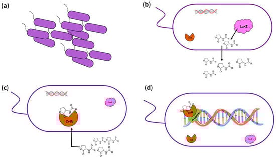
Figure 1.
Quorum sensing mechanism in Chromobacterium violaceum. (a) The high-density population of C. violaceum. (b) The synthesis of N-HSL molecules from LuxI synthase and their diffusion in the environment. (c) Diffusion of N-HSL molecules back into the cell and their binding to the CviR receptor. (d) Binding of the CviR-HSL complex to the promoter region, leading to the activation of QS-regulated genes.
QS controls lytic activity via exoproteases, chitinases, and virulence factors such as type VI secretion system [20,38]. Furthermore, QS regulates type II (TIISS) and type III (TIIISS) secretion systems, swarming motility, lipases, flagellar proteins, collagenase, elastase, and cyanide production [32,33]. In addition, QS also regulates resistance to a few antimicrobials, including bactobolin, for which QS-controlled resistance is carried out via an efflux pump [43]. Another important activity discovered in C. violaceum is biofilm formation, which is responsible for virulence via resistance to antibiotics, phagocytosis, and disinfectants. It has been established that in biofilms, bacteria communicate through diffusible AIs [38]. The secretion of the previously mentioned virulence factors, in combination with the formation of biofilms, are important for initiating infection in host cells and therefore in developing antimicrobial resistance [33,44].
3.2. Pigment Production
Violacein, a bisindole derivative, is biosynthesized via the condensation of two molecules of L-tryptophan by the products of the vioABCDE operon in response to QS [31,45]. It is a bioactive secondary metabolite with a putative function as a respiratory pigment, although it is not essential for bacterial survival and growth. Its role in the regulation of tryptophan synthesis has also been demonstrated [32]. The pigment violacein has biocidal activity against different kingdoms (bacteria, fungi, viruses, nematodes, etc.) during the microbial stationary phase of growth when cell density is high and nutrients are limited. Hence, its production could be considered part of a competitive strategy to extend the duration of the life of the microbial colony [20]. Additionally, it shows a synergistic antimicrobial effect with different antibiotics against pathogenic bacteria. Furthermore, violacein can be used as a bio-dye because of its good color tone and long-lasting stability [45].
It is important to note that different strains of C. violaceum use HSLs of different lengths. For instance, in C. violaceum ATCC 31532, violacein synthesis is activated only by short-acyl chain AHLs (C4–C8) and is inhibited by long-chain AHLs (C10–C14). In this case, the length of the acyl chain plays a key role in the binding of the complex with the RNA polymerase [20]. On the other hand, the strain C. violaceum ATCC 12472 uses C10-HSL as a QS signaling molecule, while longer HSLs, such as C12–C14 HSLs, prevent the receptor from binding to DNA [46,47].
The violacein operon is negatively regulated by a new repressor protein, VioS, and positively by the CviI/R system. VioS does not regulate the CviI/R system. Shortly, at high cell densities, the CviR protein binds AHLs and activates the expression of the vioA promoter while at the same time, the vioA promoter is suppressed by the expression of VioS, so violacein is not produced. The colonies of the wild-type C. violaceum ATCC 31532 are pale. A vioS mutant that lacks this repression at the vioA promoter forms visible violet colonies [48].
C. violaceum is one of the most commonly used bacterial species in QS research and in studying the potential QSI activities of natural substances; more precisely, C. violaceum ATCC 12472 and C. violaceum CV026 are frequently used [2,49,50]. C. violaceum is widely used in finding new ways to disrupt the QS system. Violacein production is easily detected and quantified and is thus used for screening potential QSI molecules. The disruption of QS can decrease the secretion of virulence factors without killing the bacteria or inhibiting their growth [51]. This allows for a reduction in the selective pressure on the pathogen, averting the development of resistance. QSIs can be used as alternatives to conventional antibiotics [32]. The synthesis of this visible and quantifiable pigment provides a simple way to search for potential QSIs and provides the prospect of developing new biosensor strains. A similar application finds the biosensor strain CV026, which is mini-T5-mutant defective in AHL synthase because it lacks cviI and thus requires the addition of exogenous AHL signal molecules for violacein production [52,53]. Such mutants find applications in the detection of bacterial AHLs molecules in any environment [32]. The mutant strain CV026 synthesizes violacein only in response to exogenously added 3-oxo-C6-HSL and C4-C8 AHLs [46,54,55]. Furthermore, the fact that violacein production is QS-dependent makes it a suitable marker for detecting and estimating the potential of new QSIs extracted from plants [40].
4. Plant Inhibitors: A New Way to Control Bacterial Communication
One of the most impressive processes in microbiology is the ability of bacteria to communicate with each other via signal molecules [56]. This type of bacterial communication coordinates the accumulation and responses to small molecules called AIs [7,8,57,58]. The process known as QS allows the bacterial community to coordinate gene expression, leading to the activation of specific phenotypes within the population. The most common processes under QS control, which are used by bacteria as survival strategies, are bioluminescence, biofilm formation and dispersal, the expression of virulence factors, motility, pigment synthesis, sporulation, conjugation, symbiosis, and antibiotic production [5,6,7,8].
During the antibiotic century, the revolution of better human health was a good scenario. Unfortunately, this development also led to an increase in bacterial resistance. It is now necessary to discover new targets for inhibiting microbial pathogenicity without stimulating microbial resistance [7]. One of the most novel anti-virulence strategies is to interrupt the cascade of the QS system [59,60]. Each step of the QS signaling cascade could be a good target, resulting in the inhibition of pathogenicity [61]. Some of the most attractive biomolecules, that could be used for this purpose are natural QSIs [7,59]. Similar inhibitors that can mediate bacterial QS have been found in different marine algae, fungi, corals, tunicates, and cyanobacteria [62,63,64,65], as well as bacterial [7,66,67] and mammalian cells [68]. Many of these inhibitors have been isolated from plant cells [69,70,71,72,73].
Keeping this in mind, our major interest is focused on QSIs isolated from plants, including their medicinal and anti-QS properties with respect to C. violaceum.
The plant kingdom is one of the most populated, with species and families whose metabolite products have broad biological activities. The antimicrobial activities of different plant extracts [12,13,74,75,76], essential oils [22,77], fractions, and their constituents are well known, but their efficacies against QS systems are poorly understood. Over the last few years, it has been found that plant extracts can act as inhibitors of QS pathways. These active metabolites can be extracted from different parts of plant tissues such as the roots, stems, leaves, bark, fruits, flowers, seeds, and green pods [78,79,80,81]. The major groups of these compounds can be identified as QSIs, including cyclic compounds, phenolic derivatives, nitrogen cyclics, furanones, lactones, cinnamaldehydes, alkaloids, phenolics, saponins, tannins, and terpenoids [46,82]. Their functionalities are different as they can inhibit bioluminescence, fluorescence, biofilm formation, and pigment production, block enzyme activity, and inhibit a variety of signaling pathways [7,12,13]. These abilities depend on their chemical structures and stabilities. In order to interfere with signal acceptance, QSIs must be competitive and non-competitive molecules that prevent the binding of a signal to its receptor. It is essential to note that for competitive molecules to bind to a receptor, they must have structural similarity with the original signal molecules. Non-competitive binding molecules will bind to a site different from the signal-binding site on the receptor. Several scenarios have been known using plant molecules or metabolites as QSIs: (a) homologically masking the QS signal and disrupting bacterial communication; (b) interfering with different enzymes; (c) preventing the accumulation of signals; (d) blocking the main receptors [22,46].
Quorum-Sensing Inhibitory Potential of Plants
In the environment, plants are constantly exposed to a wide range of stress conditions. These stress factors affecting plants are temperature changes, nutrient deficiencies, drought, salinity, UV radiation, a lack of oxygen, pesticides, pollutants, and anthropogenic activities. Apart from environmental stress, some species such as bacteria, fungi, viruses, nematodes, and insects can cause distress. Plants have been facing the majority of their attackers for more than millions of years. Living with their natural enemies in reciprocal evolutionary interactions, they have been learning and developing mechanisms to resist stress and attacks. For this reason, plants reveal that they each have an “immune system” comparable to those of animals, wherein they biosynthesize active compounds and secondary metabolites as protection against infections or in response to pathogen attacks. Aside from improving defenses against both biotic and abiotic stresses, most secondary metabolites have therapeutic activities, including anticancer, antioxidant, antidiabetic, immunosuppressive, antifungal, anti-inflammatory, antimalarial, anti-oomycete, antibacterial, anti-fever, anti-diabetic, insecticidal, anti-biofilm and antiviral activities [9,10,12,13,76].
Lately, one of the most interesting QSI applications is their use in blocking the signaling molecules produced by bacteria to consequently obstruct the bacterial virulence factors by disrupting QS systems. For this reason, the bacterial QS system is an excellent target for novel QSIs. Scientific evidence has shown that the identification of the binding conformations of QSIs onto the binding sites of main proteins via molecular docking analyses provides new information about their antagonistic characteristics [83]. QSIs have been reported in many plants, including medicinal plants such as Syzygium cumini, Pimenta dioica, Psidium guajava, Medicago truncatula, Lotus corniculatus, Pisum sativum, Moringa oleifera, Vernonia blumeoides, Tecoma capensis, and many others [7,82,84]. Their acetone, methanol, and water extracts have been proven to possess quorum-sensing inhibitory activity against C. violaceum.
Our review represents summarized information on plant QSIs, comprehensively studied in C. violaceum. C. violaceum is Gram-negative bacteria that is easily cultivated on laboratory media like Blood agar, MacConkey agar, and Nutrient agar. It produces smooth violet colonies whose color comes from a violet antioxidant pigment known as violacein. The increased interest of research communities in C. violaceum is related to its phenotypic characteristics: violacein production, elastase production, biofilm formation, and cyanide production controlled by the QS system through the use of signal molecules—AHLs.
Many years ago, plants were studied for their medicinal values (as digestives, diuretics, expectorants, and sedatives), and for their antioxidant and antimicrobial activities, which further developed the basis of modern phytotherapy. The main interests in their biological functions and modes of action for regulating bacterial communication have escalated in recent years. The structural variety and complexities of natural products provide them with a wide range of mechanisms of action [85]. Plant metabolites and compounds disrupt QS in three ways: (1) by inhibiting LuxI synthase function, (2) by degrading the signaling molecules, and lastly, (3) by disrupting the signaling process by targeting the LuxR receptor (Figure 2) [2,50]. Some plants, such as M. truncatula, Oryzia sativa (rice), Solanum lycopersicum (tomato), and Glycine max (soybean), can produce substances that have the ability to mimic AHL activity [52]. Different types of berries (wild blueberry, cranberry, strawberry, raspberry, and blackberry) and grape possess QSI activities as they inhibit signaling in C. violaceum and reduce swarming motility in P. aerugonosa PA01 and E. coli [54,86].
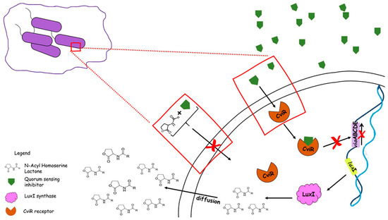
Figure 2.
Inhibition of violacein production in Chromobacterium violaceum by QSIs.
Studies include tests on crude extracts or ethanol, methanol, acetone, ethyl acetate, dichloromethane, hexane, or water extracts, essential oils, and phytochemicals, whether partially purified, enriched, or pure fractions. All these plant products could suppress the production of the pigment violacein, biofilm formation, motility, and microbial activity in C. violaceum (Table 1).

Table 1.
List of plant extracts with anti-quorum sensing activities in Chromobacterium violaceum.
Koh and Tham [87] screened ten Chinese medicinal plants, including Prunus armeniaca, Prunella vulgaris, Nelumbo nucifera, Panax notoginseng (root and flower), Punica granatum, Areca catechu, and Imperata cylindrical, to evaluate their QS activities. Seven of the extracts inhibited QS in the bioreporter strain C. violaceum CV026 and reduced swarming activity in P. aeruginosa PA01, both of which are QS-regulated functions. Part of the tested compounds had the potential to suppress violacein synthesis, and six of them formed clear zones, indicating antimicrobial activity. These results could be compared to other aqueous extracts from Ananas comosus, Musa paradiciaca, Manilkara zapota, Ocimum sanctum, Camellia sinensis L., Nymphaea tetragona, and Quercus cortex, whose active components were responsible only for inhibiting the synthesis of the pigment violacein in C. violaceum CV026 and ATCC 12472 and decreasing pyocyanin synthesis, elastase production, and biofilm formation in P. aeruinosa. Part of the active metabolites from the Q. cortex also influenced QS-regulated traits in Vibrio spp. [58,91,112]. Important observations were made about methanol extracts from herbal plants like Pisum sativum, Trigonella foenum graecum, Myristica cinnamomea, Kigelia africana, Melicope lunuankenda, Cuminum cyminum, and Costus speciosus, which proved to be inhibitors of violacein production. Additionally, M. lunuankenda reduced bioluminescence in E. coli (pSB401) and inhibited pyocyanin synthesis and the expression of lecA::lux in P. aeruginosa PA01, and the M. cinnamomea extract influenced pyocyanin production and biofilm formation in P. aeruginosa [58,88,90,92,102,104]. Bioscreening of ethanol extracts from Egypt’s ornamental and medicinal plants and those collected from Jordan, such as Adhatoda vasica, Bauhinia purpurea L., Lantana camara L., Myoporum laetum, Piper longum L., Taraxacum officinale, Laurus nobilis L., Populus alba L., Populus nigra L., Lavandula angustifolia, Rosmarinus officinalis L., Sonchus oleraceus L., Tecoma capensis Thunb. Lindl., and Jasminum sambac Ait., revealed anti-microbial activities against C. violaceum [79,96]. In contrast, ethanol extracts from Cinnamomum zeylanicum, Ocimum basilicum, and Scutellaria baicalensis Georgi demonstrated violacein inhibition in C. violaceum CV12472 and QS inhibition in C. violaceum CV026, as well as the inhibitory modulation of swarming motility in P. aeruginosa PA01 [99]. Similar results with ethanol extracts obtained from Acer monspessulanum subsp. Monspessulanum were reported by Ceylan et al. [98]. The authors determined violacein inhibition in C. violaceum CV12472 and CV026, as well as the anti-QS activity of ethanol extracts. Fatima also used the same bioreporter strains to detect the QS regulatory roles of ethanol seed extracts from the leguminous plants Pisum sativum and Trigonella foenum graecum [87]. Eight fractions, including phenolic (gallic acid, ellagic acid, epicatechin, and rutin), from the green pods of Acacia nilotica have been studied for their capacity to inhibit pigment production in C. violaceum 12472 as two of them can be classified as QSIs with the potential to regulate violacein production without influencing bacterial growth. Other phenolic plant extracts from Rubus rosaefolius also have shown similar effects on pigmentation and biofilm formation [80,106]. Polyphenolic extracts from Rosa rugosa have been the focus of Zhang et al.’s research [110] due totheir anti-biofilm and QS inhibitory potentials as inhibitors of violacein synthesis and swarming motility, as well as biofilm formation in E. coli K-12 and P. aeruginosa PA01. The authors proved high reductions in pigment without changes in microbial growth. Indian medicinal plants, flowers seeds, barks, and fruits from Punica granatum, Syzygium cumini, Embelia ribes, Phyllanthus emblica, Terminalia bellirica, Terminalia chebula, Punica granatum, Mangifera indica, Acacia arabica, Terminalia arjuna, Thespesia populnea, and Casuarina equisetifolia, were screened for the anti-QS activity in which tannin-rich extracts and punicalagin influence QS mechanisms by decreasing violacein synthesis. Shukla and Bhathena [109] qualify this phenomenon in the presence of tannin extracts at subinhibitory concentrations [106,111].
The ethyl acetate fractions and eugenol of Syzygium cumini L. and Pimenta dioica L. displayed significant anti-QS activities by inhibiting pigment production in C. violaceum [94,97,100]. Extracts from different plants such as Rhizophora annamalayana (bark), Astilbe rivularis, Fragaria nubicola, Osbeckia nepalensis, Dionysia revolute, Eucalyptus camaldulensis, green tea, Amomum tsaoko, Punica granatum, and Saraca asoca bark (stem) were found to possess QS activities, but most of them were active against violet pigmentation in Chromobacterium. Moreover, these extracts exhibited inhibitory potential against many virulence factors in P. aeruginosa PA01, including pyocyanin, elastase, exoprotease, swimming motility, and rhamnolipid production. Green tea was particularly active against S. marcesens with respect, to protease activity and swimming [58,91,100,103,105,113].
Essential oils (EOs) are natural compounds produced by aromatic plant species that are stored in various plant organs, e.g., flowers, leaves, wood, roots, rhizomes, fruit seedling, and seeds. They are secondary metabolites from plant sources and are characterized by natural multicomponent systems composed mainly of terpenes (monoterpenes, sesquiterpenes, and diterpenes) and oxygenated compounds, which are mainly phenols, alcohols, aldehydes, ketones, esters, oxides, and hydrocarbons. Essential oils and their constituents are important for biomedical and pharmaceutical purposes due to their bactericidal, virucidal, fungicidal, analgesic, sedative, anti-inflammatory, spasmolytic, and local anesthetic properties [114,115].
Among plant products, essential oils are most popular for their widespread use in ethnomedicine. EOs, isolated from three species of the genus Piper growing in Colombia, Piper bredemeyer, Piper bogotense, and Piper brachypodon, interfered with the pigment production and proved minor effects against bacterial growth in C. violaceum CV026 as well [93]. Likewise, four EOs prepared from Cinnamomum verum, Origanum majorana, Thymus vulgaris, and Eugenia caryophyllata were evaluated as QSIs in which the disruption of pigmentation production occurred with a lower percentage only for marjoram oil. However, these EOs have significant anti-bacterial, anti-QS, and anti-biofilm activities against almost all of the 44 MDR-tested bacterial strains [101]. Many scientists reported different EOs manifesting the inhibition of violacein production, identified in Elletaria cardamomum, Eucalyptus radiate, Origanum vulgare, Melaleuca alternifolia, and Mentha suaveolens. The EOs from M. alternifolia were also able to inhibit swarming motility in P. aeruginosa PA01 and biofilm formation in S. aureus MRSA [24,107,108]. Interestingly, among some EOs, like limonene from Citrus lemon, terpinene-4-ol, pinene from Juniperus communis, and tea tree oil from Melaleuca alternifolia, which were identified as QSIs for the purple pigment in C. violaceum, only cis-cis-p-menthenolide from Mentha suaveolens altered the biofilm matrix during biofilm formation [107,108,116].
Plants produce molecules that are structurally similar to AHLs and can thus bind to LuxR receptors via competitive inhibition and block QS (Table 2). For instance, furanones have the ability to inhibit QS by competitively binding to LuxR receptors, promoting their degradation. On the other hand, when the concentration of AHLs increases, the inhibition process decreases [2,25,52].
In recent years, it was found that the compound malabaricone C, extracted from Myristica cinnamomea, does not structurally mimic AHL but successfully inhibited both lasR and rhlR QS systems in P. aeruginosa PA01 and the CviR receptor in C. violaceum [52,82]. The flavonoid naringenin restricted the synthesis of QS molecules like N-(3-oxododecanoyl), lactone-1-homoserine (3-oxo-C12-HSL), acyl homoserine lactone, and N-butanoyl-1-homoserine lactone (C4-HSL) [2,117]. Quercetin, another type of flavonoid, acted as a competitive inhibitor toward the receptors, thus inhibiting QS phenotypes such as biofilm formation, violacein synthesis, motility, etc. [53,82]. The monoterpene carvacrol reduced the expression of the cviI gene, resulting in the inhibition of biofilm formation, violacein production, and chitinase activity in C. violaceum ATCC 12 472 [82,118], as well as the production of pyocyanin in P. aeruginosa. Moreover, monoterpenoids can bind with LuxR-type receptors and disrupt QS [53]. In another study, it was demonstrated that the natural diterpene compound phytol bound to CviR receptors with high affinity, effectively reducing QS-regulated processes (e.g., cell aggregation, biofilm formation, and alkaline protease activity) [47]. Two types of metabolites from G. hypoleucum DC, apigenin and luteolin, downregulated some of the genes for violacein synthesis: vioB, vioC, and vioD [119]. The compound vanillin from Vanilla planifolia Andrews inhibited violacein synthesis in C. violaceum because it inhibits the synthesis of short (C4) and long (C8) AHLs. Different data confirm that curcumin was able to block LuxI-type synthases, reduced the expression of receptor genes, and additionally reduced the synthesis of violacein in C. violaceum ATCC 12472. Another mode of action of curcumin is that it could significantly reduce the activity of genes for the type III secretion system and cyclic diguanylate (c-di-GMP) [53,85]. The phytochemical eugenol reduced violacein synthesis and the production of 3-oxo-C12-HSL and C4-HSL. Sesquiterpene lactones are another type of phytochemical isolated from plants. Six lactones from the families of goyazensolide and isogoyazensolide inhibited the production of AHLs [120].
Table 2.
List of pure plant compounds with anti-quorum sensing activities and their mechanisms of action in Chromobacterium violaceum.
Table 2.
List of pure plant compounds with anti-quorum sensing activities and their mechanisms of action in Chromobacterium violaceum.
| Plant | Compound | Strain | Mechanism of Action or Effect | Ref. |
|---|---|---|---|---|
| Combretum albiflorum | Catechin | C. violaceum CV026 | Inhibition of violacein production | [121] |
| Rosa rugosa | Epigallocatechin gallate Epicatechin | C. violaceum CV026 | Reduction in violacein production | [110] |
| Vernonia blumeoides | Sesquiterpene lactone | C. violaceum CV026 C. violaceum VIR07 C. violaceum ATCC 12472 C. violaceum ATCC 31532 | Antagonist effect against CviR | [2] |
| Drimys winteri | Cinnamolide Valdiviolide | C. violaceum ATCC 12472 | Inhibition of QS and violacein reduction | [122] |
| Polydora serratuloides | Sesquiterpene lactone (13-acetoxy 1(4β),5(6)βdiepoxy-8α-(senecioyloxy) 3-oxo-1,7(11)-germacradiene-12,6-olide 1) | C. violaceum ATCC 12472 | Inhibition of QS mediators | [123] |
| Allium sativum | P-Coumaric acid | C. violaceum 5999 and wt 494 | Inhibition of biofilm formation and the expression of bacterial virulence factor; antagonizes the activity of LuxR, ahyR, and TraR receptors | [2] |
| Caffeine (1,3,7-trimethylxanthine) | C. violaceum CV026 | Inhibition of violacein production; inhibition of CviI synthase | [25] | |
| Isothiocyanates | C. violaceum CV12472 | Modulation of AHL activity and synthesis | [25] | |
| N, N-disubstituted biguanides | C. violaceum ATCC 12472 | Reduces the synthesis of violacein; inhibition of the transcription factor CviR | [25] | |
| Psidium guajava L. | Quercetin (2-(3,4-dihydroxyphenyl)-3,5,7-trihydroxy-4H-chromen-4-one) | C. violaceum ATCC 31532 | Inhibition of violacein synthesis; binds to transcription factor CviR | [53] |
| Gnaphalium hypoleucum DC | Apigenin and luteolin | C. violaceum ATCC 12472 | Effects on violacein pigment biosynthesis, biofilm formation, and motility; downregulation of the vioB, vioC, and vioD genes | [119] |
| Quercetin 4′-O-β-D-glucopyranoside | C. violaceum ATCC 12472 C. violaceum CV026 | Reduction in violacein synthesis, biofilm formation, EPS production, motility, and alginate production; inhibition of the C6-AHL communication molecule | [120] | |
| Myristica Cinnamomea | Malabaricone C | C. violaceum CV026 | Inhibition of violacein production | [54] |
| Bitter orange | Naringin | C. violaceum (CECT 494) | Inhibition of the production of violacein | [2] |
| Vanilla planifolia Andrews | Vanillin (4-hydroxy-3 methoxybenzaldehyde) | C. violaceum CV026 | Reduced violacein production | [20] |
| Amphypterygium adstringens | Anacardic acids mixture | C. violaceum ATCC 12472 | Inhibition of violacein production | [20] |
| Syzygium aromaticum | Eugenol | C. violaceum CV026 | Dose-dependent inhibitory effect on violacein synthesis | [20] |
| Syzygium cumini | Malvidin | C. violaceum CV026 (CECT 5999) C. violaceum MTCC2656 | Inhibition of violacein production; reduction in biofilm biomass | [82] |
| Origanum vulgare | Carvacrol | C. violaceum ATCC 12472 | Reductions in biofilm formation, violacein production, and chitinase activity; reduces the expression of CviI | [82] |
| Coumarin (2H-chromen-2-one) | C. violaceum ATCC 12472 C. violaceum CV026 | Inhibition of violacein biosynthesis | [53] | |
| Cinnamic acid derivatives | C. violaceum ATCC 12472 | Reduces the production of virulence factors—violacein, hemolysin, chitinase, and biofilm formation; downregulation of some QS-related metabolites (ethanolamine and L-methionine); decreases the expression of cviI and cviR genes; inhibition of the C10-HSL synthesis | [124] | |
| Methyl gallate | C. violaceum ATCC 12472 C. violaceum ATCC 31532 C. Violaceum CV026 | Suppression of the synthesis and activity of AHL | [125] | |
| Phytol | C. violaceum ATCC 12472 C. Violaceum ATCC 31532 | Reducing QS-regulated traits—biofilm formation, cell aggregation, and alkaline protease activity; binds to CviR | [47] | |
| Thymol | C. violaceum ATCC 12472 | Inhibition of violacein synthesis, biofilm formation, and EPS production; binds to CviR | [126] |
5. Conclusions
In this review, we try to emphasize and summarize the information on natural QSIs, their functionalities, and their main inhibitory roles in C. violaceum’s QS system. We emphasize some critical points that show the effectiveness of such small molecules in broad biological activities, especially in mediating QS processes in Gram-negative bacteria. The new era of QSIs provides a sufficient motive to help scientists battle bacterial resistance by discovering new strategies related to isolating and synthesizing natural products or their analogs. In conclusion, this highlight on QSIs and their importance in bacterial combat will help us identify a variety of them as targets for the development of new antimicrobials. This will be the subject of future investigations.
Author Contributions
Literature search, writing, and image design: P.D.D. and T.D.; design of this review, drafting, and critical revision: T.P.-K. All authors have read and agreed to the published version of the manuscript.
Funding
This work was supported by the National Science Fund at the Ministry of Education and Science, Bulgaria, approved by Research Grant KP-06-H41/8; 30 November 2020.
Institutional Review Board Statement
Not applicable.
Informed Consent Statement
Not applicable.
Data Availability Statement
Not applicable.
Acknowledgments
The authors thank Stoyanka R. Stoitsova for the critical reading of the draft and review.
Conflicts of Interest
The authors declare no conflict of interest.
Abbreviations
| QS | Quorum sensing |
| QSIs | Quorum-sensing inhibitors |
| AI | Autoinducer |
| AHLs | Acyl-homoserine lactones |
References
- Kumar, P.; Sharma, S.; Tripathi, V.N. Quorum sensing in bacteria of rice rhizospheres from Chhattisgarh, India. Bioinformation 2023, 19, 199–205. [Google Scholar] [CrossRef]
- Bouyahya, A.; Dakka, N.; Et-Touys, A.; Abrini, J.; Bakri, Y. Medicinal plant products targeting quorum sensing for combating bacterial infections. Asian Pac. J. Trop. Med. 2017, 10, 729–743. [Google Scholar] [CrossRef] [PubMed]
- Lu, L.; Hu, W.; Tian, Z.; Yuan, D.; Yi, G.; Zhou, Y.; Cheng, Q.; Zhu, J.; Li, M. Developing natural products as potential anti-biofilm agents. Chin. Med. 2019, 14, 11. [Google Scholar] [CrossRef]
- Rasmussen, T.B.; Manefield, M.; Andersen, J.B.; Eberl, L.; Anthoni, U.; Christophersen, C.; Steinberg, P.; Kjelleberg, S.; Givskov, M. How Delisea pulchra furanones affect quorum sensing and swarming motility in Serratia liquefaciens MG1. Microbiology 2000, 146, 3237–3244. [Google Scholar] [CrossRef] [PubMed]
- Parker, C.T.; Sperandio, V. Cell-to-cell signalling during pathogenesis. Cell. Microbiol. 2009, 11, 363–369. [Google Scholar] [CrossRef]
- Rutherford, S.T.; Bassler, B.L. Bacterial Quorum Sensing: Its Role in Virulence and Possibilities for Its Control. Cold Spring Harb. Perspect. Med. 2012, 2, a012427. [Google Scholar] [CrossRef]
- Kalia, V.C. Quorum sensing inhibitors: An overview. Biotechnol. Adv. 2013, 31, 224–245. [Google Scholar] [CrossRef]
- Yi, L.; Dong, X.; Grenier, D.; Wang, K.; Wang, Y. Research progress of bacterial quorum sensing receptors: Classification, structure, function and characteristics. Sci. Total Environ. 2021, 763, 143031. [Google Scholar] [CrossRef]
- Stoitsova, S.R.; Paunova-Krasteva, T.S.; Borisova, D.B. Modulation of Biofilm Growth by Sub-Inhibitory Amounts of Antibacterial Substances. In Microbial Biofilms—Importance and Applications; Dhanasekaran, D., Thajuddin, N., Eds.; InTech: Rijeka, Croatia, 2016; ISBN 978-953-51-2436-8. [Google Scholar] [CrossRef]
- Trendafilova, A.; Ivanova, V.; Rangelov, M.; Todorova, M.; Ozek, G.; Yur, S.; Ozek, T.; Aneva, I.; Veleva, R.; Moskova-Doumanova, V.; et al. Caffeoylquinic Acids, Cytotoxic, Antioxidant, Acetylcholinesterase and Tyrosinase Enzyme Inhibitory Activities of Six Inula Species from Bulgaria. Chem. Biodivers. 2020, 17, e2000051. [Google Scholar] [CrossRef]
- Stoitsova, S.; Paunova-Krasteva, T.; Dimitrova, P.D.; Damyanova, T. The concept for the antivirulence therapeutics approach as alternative to antibiotics: Hope or still a fiction? Biotechnol. Biotechnol. Equip. 2022, 36, 697–705. [Google Scholar] [CrossRef]
- Hemdan, B.A.; Mostafa, A.; Elbatanony, M.M.; El-Feky, A.M.; Paunova-Krasteva, T.; Stoitsova, S.; El-Liethy, M.A.; El-Taweel, G.E.; Abu Mraheil, M. Bioactive Azadirachta indica and Melia azedarach leaves extracts with anti-SARS-CoV-2 and antibacterial activities. PLoS ONE 2023, 18, e0282729. [Google Scholar] [CrossRef]
- Paunova-Krasteva, T.; Hemdan, B.A.; Dimitrova, P.D.; Damyanova, T.; El-Feky, A.M.; Elbatanony, M.M.; Stoitsova, S.; El-Liethy, M.A.; El-Taweel, G.E.; El Nahrawy, A.M. Hybrid Chitosan/CaO-Based Nanocomposites Doped with Plant Extracts from Azadirachta indica and Melia azedarach: Evaluation of Antibacterial and Antibiofilm Activities. BioNanoScience 2023, 13, 88–102. [Google Scholar] [CrossRef]
- Nealson, K.H.; Platt, T.; Hastings, J.W. Cellular Control of the Synthesis and Activity of the Bacterial Luminescent System. J. Bacteriol. 1970, 104, 313–322. [Google Scholar] [CrossRef]
- Nealson, K.H.; Hastings, J.W. Bacterial Bioluminescence: Its Control and Ecological Significance. Microbiol. Rev. 1979, 43, 496–518. [Google Scholar] [CrossRef] [PubMed]
- Papenfort, K.; Bassler, B.L. Quorum sensing signal–response systems in Gram-negative bacteria. Nat. Rev. Microbiol. 2016, 14, 576–588. [Google Scholar] [CrossRef] [PubMed]
- Miller, M.B.; Bassler, B.L. Quorum Sensing in Bacteria. Annu. Rev. Microbiol. 2001, 55, 165–199. [Google Scholar] [CrossRef]
- Abisado, R.G.; Benomar, S.; Klaus, J.R.; Dandekar, A.A.; Chandler, J.R. Bacterial Quorum Sensing and Microbial Community Interactions. MBio 2018, 9, e02331-17. [Google Scholar] [CrossRef]
- Chugani, S.; Greenberg, E.P. An evolving perspective on the Pseudomonas aeruginosa orphan quorum sensing regulator QscR. Front. Cell. Infect. Microbiol. 2014, 4, 152. [Google Scholar] [CrossRef]
- Durán, N.; Justo, G.Z.; Durán, M.; Brocchi, M.; Cordi, L.; Tasic, L.; Castro, G.R.; Nakazato, G. Advances in Chromobacterium violaceum and properties of violacein-Its main secondary metabolite: A review. Biotechnol. Adv. 2016, 34, 1030–1045. [Google Scholar] [CrossRef]
- Rajamanikandan, S.; Srinivasan, P. Exploring the selectivity of auto-inducer complex with LuxR using molecular docking, mutational studies and molecular dynamics simulations. J. Mol. Struct. 2017, 1131, 281–293. [Google Scholar] [CrossRef]
- Vadakkan, K.; Choudhury, A.A.; Gunasekaran, R.; Hemapriya, J.; Vijayanand, S. Quorum sensing intervened bacterial signaling: Pursuit of its cognizance and repression. J. Genet. Eng. Biotechnol. 2018, 16, 239–252. [Google Scholar] [CrossRef]
- Pena, R.T.; Blasco, L.; Ambroa, A.; González-Pedrajo, B.; Fernández-García, L.; López, M.; Bleriot, I.; Bou, G.; García-Contreras, R.; Wood, T.K.; et al. Relationship Between Quorum Sensing and Secretion Systems. Front. Microbiol. 2019, 10, 1100. [Google Scholar] [CrossRef]
- Saeki, E.K.; Kobayashi, R.K.T.; Nakazato, G. Quorum sensing system: Target to control the spread of bacterial infections. Microb. Pathog. 2020, 142, 104068. [Google Scholar] [CrossRef] [PubMed]
- Haque, S.; Ahmad, F.; Dar, S.A.; Jawed, A.; Mandal, R.K.; Wahid, M.; Lohani, M.; Khan, S.; Singh, V.; Akhter, N. Developments in strategies for Quorum Sensing virulence factor inhibition to combat bacterial drug resistance. Microb. Pathog. 2018, 121, 293–302. [Google Scholar] [CrossRef] [PubMed]
- Majdura, J.; Jankiewicz, U.; Gałązka, A.; Orzechowski, S. The Role of Quorum Sensing Molecules in Bacterial–Plant Interactions. Metabolites 2023, 13, 114. [Google Scholar] [CrossRef]
- Waters, C.M.; Bassler, B.L. Quorum sensing: Cell-to-Cell Communication in Bacteria. Annu. Rev. Cell Dev. Biol. 2005, 21, 319–346. [Google Scholar] [CrossRef] [PubMed]
- De Keersmaecker, S.C.J.; Sonck, K.; Vanderleyden, J. Let LuxS speak up in AI-2 signaling. Trends Microbiol. 2006, 14, 114–119. [Google Scholar] [CrossRef]
- Stephens, K.; Bentley, W.E. Quorum Sensing from Two Engineers’ Perspectives. Isr. J. Chem. 2023, 63, e202200083. [Google Scholar] [CrossRef]
- Ha, C.; Park, S.J.; Im, S.J.; Park, S.J.; Lee, J.H. Interspecies signaling through QscR, a quorum receptor of Pseudomonas aeruginosa. Mol. Cells 2012, 33, 53–59. [Google Scholar] [CrossRef]
- Stauff, D.L.; Bassler, B.L. Quorum Sensing in Chromobacterium violaceum: DNA Recognition and Gene Regulation by the CviR Receptor. J. Bacteriol. 2011, 193, 3871–3878. [Google Scholar] [CrossRef]
- Kothari, V.; Sharma, S.; Padia, D. Recent research advances on Chromobacterium violaceum. Asian Pac. J. Trop. Med. 2017, 10, 744–752. [Google Scholar] [CrossRef] [PubMed]
- Venkatramanan, M.; Sankar Ganesh, P.; Senthil, R.; Akshay, J.; Veera Ravi, A.; Langeswaran, K.; Vadivelu, J.; Nagarajan, S.; Rajendran, K.; Shankar, E.M. Inhibition of Quorum Sensing and Biofilm Formation in Chromobacterium violaceum by Fruit Extracts of Passiflora edulis. ACS Omega 2020, 5, 25605–25616. [Google Scholar] [CrossRef] [PubMed]
- Lorenz, N.; Shin, J.Y.; Jung, K. Activity, Abundance, and Localization of Quorum Sensing Receptors in Vibrio harveyi. Front. Microbiol. 2017, 8, 634. [Google Scholar] [CrossRef] [PubMed]
- Chen, J.; Lu, Y.; Ye, X.; Emam, M.; Zhang, H.; Wang, H. Current advances in Vibrio harveyi quorum sensing as drug discovery targets. Eur. J. Med. Chem. 2020, 207, 112741. [Google Scholar] [CrossRef] [PubMed]
- Watve, S.; Barrasso, K.; Jung, S.A.; Davis, K.J.; Hawver, L.A.; Khataokar, A.; Palaganas, R.G.; Neiditch, M.B.; Perez, L.J.; Ng, W.-L. Parallel quorum-sensing system in Vibrio cholerae prevents signal interference inside the host. PLoS Pathog. 2020, 16, e1008313. [Google Scholar] [CrossRef] [PubMed]
- Brazilian National Genome Project Consortium. The complete genome sequence of Chromobacterium violaceum reveals remarkable and exploitable bacterial adaptability. Proc. Natl. Acad. Sci. USA 2003, 100, 11660–11665. [Google Scholar] [CrossRef] [PubMed]
- Kamaeva, A.A.; Vasilchenko, A.S.; Deryabin, D.G. Atomic Force Microscopy Reveals a Morphological Differentiation of Chromobacterium violaceum Cells Associated with Biofilm Development and Directed by N-Hexanoyl-L-Homoserine Lactone. PLoS ONE 2014, 9, e103741. [Google Scholar] [CrossRef]
- Kaniyarakkal, V.; Orvankundil, S.; Lalitha, S.K.; Thazhethekandi, R.; Thottathil, J. Chromobacterium violaceum Septicaemia and Urinary Tract Infection: Case Reports from a Tertiary Care Hospital in South India. Case Rep. Infect. Dis. 2016, 2016, 6795743. [Google Scholar] [CrossRef]
- de Oca-Mejía, M.M.; Castillo-Juárez, I.; Martínez-Vázquez, M.; Soto-Hernandez, M.; García-Contreras, R. Influence of quorum sensing in multiple phenotypes of the bacterial pathogen Chromobacterium violaceum. Pathog. Dis. 2015, 73, 1–4. [Google Scholar] [CrossRef]
- Durán, M.; Faljoni-Alario, A.; Durán, N. Chromobacterium violaceum and its important metabolites—Review. Folia Microbiol. 2010, 55, 535–547. [Google Scholar] [CrossRef]
- Batista, J.H.; da Silva Neto, J.F. Chromobacterium violaceum Pathogenicity: Updates and Insights from Genome Sequencing of Novel Chromobacterium Species. Front. Microbiol. 2017, 10, 2213. [Google Scholar] [CrossRef]
- Evans, K.C.; Benomar, S.; Camuy-Vélez, L.A.; Nasseri, E.B.; Wang, X.; Neuenswander, B.; Chandler, J.R. Quorum-sensing control of antibiotic resistance stabilizes cooperation in Chromobacterium violaceum. ISME J. 2018, 12, 1263–1272. [Google Scholar] [CrossRef]
- Cheng, W.J.; Zhou, J.W.; Zhang, P.P.; Luo, H.Z.; Tang, S.; Li, J.J.; Deng, S.M.; Jia, A.Q. Quorum sensing inhibition and tobramycin acceleration in Chromobacterium violaceum by two natural cinnamic acid derivatives. Appl. Microbiol. Biotechnol. 2020, 104, 5025–5037. [Google Scholar] [CrossRef] [PubMed]
- Gohil, N.; Bhattacharjee, G.; Gayke, M.; Narode, H.; Alzahrani, K.J.; Singh, V. Enhanced production of violacein by Chromobacterium violaceum using agro-industrial waste soybean meal. J. Appl. Microbiol. 2022, 132, 1121–1133. [Google Scholar] [CrossRef] [PubMed]
- Grandclément, C.; Tannières, M.; Moréra, S.; Dessaux, Y.; Faure, D. Quorum quenching: Role in nature and applied developments. FEMS Microbiol. Rev. 2016, 40, 86–116. [Google Scholar] [CrossRef] [PubMed]
- Zhu, X.; Chen, W.J.; Bhatt, K.; Zhou, Z.; Huang, Y.; Zhang, L.H.; Chen, S.; Wang, J. Innovative microbial disease biocontrol strategies mediated by quorum quenching and their multifaceted applications: A review. Front. Plant Sci. 2023, 13, 1063393. [Google Scholar] [CrossRef]
- Devescovi, G.; Kojic, M.; Covaceuszach, S.; Cámara, M.; Williams, P.; Bertani, I.; Subramoni, S.; Venturi, V. Negative Regulation of Violacein Biosynthesis in Chromobacterium violaceum. Front. Microbiol. 2017, 8, 349. [Google Scholar] [CrossRef]
- Mookherjee, A.; Singh, S.; Maiti, M.K. Quorum sensing inhibitors: Can endophytes be prospective sources? Arch. Microbiol. 2018, 200, 355–369. [Google Scholar] [CrossRef]
- Snoussi, M.; Noumi, E.; Punchappady-Devasya, R.; Trabelsi, N.; Kanekar, S.; Nazzaro, F.; Fratianni, F.; Flamini, G.; De Feo, V.; Al-Sieni, A. Antioxidant properties and anti-quorum sensing potential of Carum copticum essential oil and phenolics against Chromobacterium violaceum. J. Food Sci. Technol. 2018, 55, 2824–2832. [Google Scholar] [CrossRef] [PubMed]
- Shi, W.P.; Zeng, H.; Wan, C.X.; Zhou, Z.B. Amicoumacins from a desert bacterium: Quorum sensing inhibitor against Chromobacterium violaceum. Nat. Prod. Res. 2021, 35, 5508–5512. [Google Scholar] [CrossRef] [PubMed]
- Koh, C.L.; Sam, C.K.; Yin, W.F.; Tan, L.; Krishnan, T.; Chong, Y.M.; Chan, K.G. Plant-Derived Natural Products as Sources of Anti-Quorum Sensing Compounds. Sensors 2013, 13, 6217–6228. [Google Scholar] [CrossRef] [PubMed]
- Deryabin, D.; Galadzhieva, A.; Kosyan, D.; Duskaev, G. Plant-Derived Inhibitors of AHL-Mediated Quorum Sensing in Bacteria: Modes of Action. Int. J. Mol. Sci. 2019, 20, 5588. [Google Scholar] [CrossRef] [PubMed]
- Truchado, P.; Larrosa, M.; Castro-Ibáñez, I.; Allende, A. Plant food extracts and phytochemicals: Their role as Quorum Sensing Inhibitors. Trends Food Sci. Technol. 2015, 43, 189–204. [Google Scholar] [CrossRef]
- Joo, H.S.; Deyrup, S.T.; Shim, S.H. Endophyte-produced antimicrobials: A review of potential lead compounds with a focus on quorum-sensing disruptors. Phytochem. Rev. 2021, 20, 543–568. [Google Scholar] [CrossRef]
- Bassler, B.L.; Losick, R. Bacterially Speaking. Cell 2006, 125, 237–246. [Google Scholar] [CrossRef] [PubMed]
- Bassler, B.L. How bacteria talk to each other: Regulation of gene expression by quorum sensing. Curr. Opin. Microbiol. 1999, 2, 582–587. [Google Scholar] [CrossRef]
- Yang, M.; Meng, F.; Gu, W.; Li, F.; Tao, Y.; Zhang, Z.; Zhang, F.; Yang, X.; Li, J.; Yu, J. Effects of Natural Products on Bacterial Communication and Network-Quorum Sensing. BioMed Res. Int. 2020, 2020, 8638103. [Google Scholar] [CrossRef]
- Rasmussen, T.B.; Givskov, M. Quorum sensing inhibitors: A bargain of effects. Microbiology 2006, 152, 895–904. [Google Scholar] [CrossRef]
- Fleitas Martínez, O.; Rigueiras, P.O.; da Silva Pires, Á.; Porto, W.F.; Silva, O.N.; de la Fuente-Nunez, C.; Franco, O.L. Interference with Quorum-Sensing Signal Biosynthesis as a Promising Therapeutic Strategy Against Multidrug-Resistant Pathogens. Front. Cell Infect. Microbiol. 2019, 8, 444. [Google Scholar] [CrossRef]
- Rasko, D.A.; Sperandio, V. Anti-virulence strategies to combat bacteria-mediated disease. Nat. Rev. Drug Discov. 2010, 9, 117–128. [Google Scholar] [CrossRef]
- Gao, M.; Teplitski, M.; Robinson, J.B.; Bauer, W.D. Production of Substances by Medicago truncatula that Affect Bacterial Quorum Sensing. Mol. Plant-Microbe Interact. 2003, 16, 827–834. [Google Scholar] [CrossRef]
- Hentzer, M.; Givskov, M. Pharmacological inhibition of quorum sensing for the treatment of chronic bacterial infections. J. Clin. Investig. 2003, 112, 1300–1307. [Google Scholar] [CrossRef] [PubMed]
- Frisvad, J.C.; Andersen, B.; Thrane, U. The use of secondary metabolite profiling in chemotaxonomy of filamentous fungi. Mycol. Res. 2008, 112, 231–240. [Google Scholar] [CrossRef] [PubMed]
- Dobretsov, S.; Teplitski, M.; Bayer, M.; Gunasekera, S.; Proksch, P.; Paul, V.J. Inhibition of marine biofouling by bacterial quorum sensing inhibitors. Biofouling 2011, 27, 893–905. [Google Scholar] [CrossRef]
- Wahjudi, M.; Papaioannou, E.; Hendrawati, O.; van Assen, A.H.G.; van Merkerk, R.; Cool, R.H.; Poelarends, G.J.; Quax, W.J. PA0305 of Pseudomonas aeruginosa is a quorum quenching acylhomoserine lactone acylase belonging to the Ntn hydrolase superfamily. Microbiology 2011, 157, 2042–2055. [Google Scholar] [CrossRef]
- Terwagne, M.; Mirabella, A.; Lemaire, J.; Deschamps, C.; De Bolle, X.; Letesson, J.J. Quorum Sensing and Self-Quorum Quenching in the Intracellular Pathogen Brucellamelitensis. PLoS ONE 2013, 8, e82514. [Google Scholar] [CrossRef] [PubMed]
- Teiber, J.F.; Horke, S.; Haines, D.C.; Chowdhary, P.K.; Xiao, J.; Kramer, G.L.; Haley, R.W.; Draganov, D.I. Dominant Role of Paraoxonases in Inactivation of the Pseudomonas aeruginosa Quorum-Sensing Signal N-(3-Oxododecanoyl)-l-Homoserine Lactone. Infect. Immun. 2008, 76, 2512–2519. [Google Scholar] [CrossRef]
- Martinelli, D.; Grossmann, G.; Séquin, U.; Brandl, H.; Bachofen, R. Effects of natural and chemically synthesized furanones on quorum sensing in Chromobacterium violaceum. BMC Microbiol. 2004, 4, 25. [Google Scholar] [CrossRef] [PubMed]
- Amaya, S.; Pereira, J.A.; Borkosky, S.A.; Valdez, J.C.; Bardón, A.; Arena, M.E. Inhibition of quorum sensing in Pseudomonas aeruginosa by sesquiterpene lactones. Phytomedicine. 2012, 19, 1173–1177. [Google Scholar] [CrossRef]
- Martín-Rodríguez, A.J.; Ticona, J.C.; Jiménez, I.A.; Flores, N.; Fernández, J.J.; Bazzocchi, I.L. Flavonoids from Piper delineatum modulate quorum-sensing-regulated phenotypes in Vibrio harveyi. Phytochemistry 2015, 117, 98–106. [Google Scholar] [CrossRef]
- Majik, M.S.; Gawas, U.B.; Mandrekar, V.K. Next generation quorum sensing inhibitors: Accounts on structure activity relationship studies and biological activities. Bioorg. Med. Chem. 2020, 28, 115728. [Google Scholar] [CrossRef]
- Ahmed, S.O.; Zedan, H.H.; Ibrahim, Y.M. Quorum sensing inhibitory effect of bergamot oil and aspidosperma extract against Chromobacterium violaceum and Pseudomonas aeruginosa. Arch. Microbiol. 2021, 203, 4663–4675. [Google Scholar] [CrossRef]
- Kazemian, H.; Ghafourian, S.; Heidari, H.; Amiri, P.; Yamchi, J.K.; Shavalipour, A.; Houri, H.; Maleki, A.; Sadeghifard, N. Antibacterial, anti-swarming and anti-biofilm formation activities of Chamaemelum nobile against Pseudomonas aeruginosa. Rev. Soc. Bras. Med. Trop. 2015, 48, 432–436. [Google Scholar] [CrossRef]
- Bacha, K.; Tariku, Y.; Gebreyesus, F.; Zerihun, S.; Mohammed, A.; Weiland-Bräuer, N.; Schmitz, R.A.; Mulat, M. Antimicrobial and anti-Quorum Sensing activities of selected medicinal plants of Ethiopia: Implication for development of potent antimicrobial agents. BMC Microbiol. 2016, 16, 139. [Google Scholar] [CrossRef] [PubMed]
- Nikolova, I.; Paunova-Krasteva, T.; Petrova, Z.; Grozdanov, P.; Nikolova, N.; Tsonev, G.; Triantafyllidis, A.; Andreev, S.; Trepechova, M.; Milkova, V.; et al. Bulgarian Medicinal Extracts as Natural Inhibitors with Antiviral and Antibacterial Activity. Plants 2022, 11, 1666. [Google Scholar] [CrossRef] [PubMed]
- Olivero-Verbel, J.; Barreto-Maya, A.; Bertel-Sevilla, A.; Stashenko, E.E. Composition, anti-quorum sensing and antimicrobial activity of essential oils from Lippia alba. Braz. J. Microbiol. 2014, 45, 759–767. [Google Scholar] [CrossRef]
- Choo, J.H.; Rukayadi, Y.; Hwang, J.K. Inhibition of bacterial quorum sensing by vanilla extract. Lett. Appl. Microbiol. 2006, 42, 637–641. [Google Scholar] [CrossRef]
- Al-Hussaini, R.; Mahasneh, A. Microbial Growth and Quorum Sensing Antagonist Activities of Herbal Plants Extracts. Molecules 2009, 14, 3425–3435. [Google Scholar] [CrossRef]
- Singh, B.N.; Singh, B.R.; Singh, R.L.; Prakash, D.; Sarma, B.K.; Singh, H.B. Antioxidant and anti-quorum sensing activities of green pod of Acacia nilotica L. Food Chem. Toxicol. 2009, 47, 778–786. [Google Scholar] [CrossRef]
- Singh, B.N.; Singh, B.R.; Singh, R.L.; Prakash, D.; Dhakarey, R.; Upadhyay, G.; Singh, H.B. Oxidative DNA damage protective activity, antioxidant and anti-quorum sensing potentials of Moringa oleifera. Food Chem. Toxicol. 2009, 47, 1109–1116. [Google Scholar] [CrossRef] [PubMed]
- Asfour, H. Anti-quorum sensing natural compounds. J. Microsc. Ultrastruct. 2018, 6, 1. [Google Scholar] [CrossRef] [PubMed]
- Chen, G.; Swem, L.R.; Swem, D.L.; Stauff, D.L.; O’Loughlin, C.T.; Jeffrey, P.D.; Bassler, B.L.; Hughson, F.M. A Strategy for Antagonizing Quorum Sensing. Mol. Cell 2011, 42, 199–209. [Google Scholar] [CrossRef]
- Aliyu, A.B.; Koorbanally, N.A.; Moodley, B.; Singh, P.; Chenia, H.Y. Quorum sensing inhibitory potential and molecular docking studies of sesquiterpene lactones from Vernonia blumeoides. Phytochemistry 2016, 126, 23–33. [Google Scholar] [CrossRef]
- Peña-González, M.C.; Muñoz-Cázares, N.; Peña-Rodríguez, L.M. Natural Inhibitors of Quorum-Sensing Factors: A Novel Strategy to Control Pathogenic Bacteria. Rev. Bras. Farmacogn. 2020, 30, 743–755. [Google Scholar] [CrossRef]
- Takó, M.; Kerekes, E.B.; Zambrano, C.; Kotogán, A.; Papp, T.; Krisch, J.; Vágvölgyi, C. Plant Phenolics and Phenolic-Enriched Extracts as Antimicrobial Agents against Food-Contaminating Microorganisms. Antioxidants 2020, 9, 165. [Google Scholar] [CrossRef] [PubMed]
- Koh, K.H.; Tham, F.Y. Screening of traditional Chinese medicinal plants for quorum-sensing inhibitors activity. J. Microbiol. Immunol. Infect. 2011, 44, 144–148. [Google Scholar] [CrossRef]
- Fatima, Q.; Zahin, M.; Khan, M.S.A.; Ahmad, I. Modulation of quorum sensing controlled behaviour of bacteria by growing seedling, seed and seedling extracts of leguminous plants. Indian. J. Microbiol. 2010, 50, 238–242. [Google Scholar] [CrossRef]
- Song, C.; Ma, H.; Zhao, Q.; Song, S.; Jia, Z. Inhibition of Quorum Sensing Activity by Ethanol Extract of Scutellaria baicalensis Georgi. J. Plant Pathol. Microbiol. 2012, 7, 1–4. [Google Scholar] [CrossRef]
- Chong, Y.M.; Yin, W.F.; Ho, C.Y.; Mustafa, M.R.; Hadi, A.H.A.; Awang, K.; Narrima, P.; Koh, C.L.; Appleton, D.R.; Chan, K.G. Malabaricone C from Myristica cinnamomea Exhibits Anti-Quorum Sensing Activity. J. Nat. Prod. 2011, 74, 2261–2264. [Google Scholar] [CrossRef]
- Musthafa, K.S.; Ravi, A.V.; Annapoorani, A.; Packiavathy, I.S.V.; Pandian, S.K. Evaluation of Anti-Quorum-Sensing Activity of Edible Plants and Fruits through Inhibition of the N-Acyl-Homoserine Lactone System in Chromobacterium violaceum and Pseudomonas aeruginosa. Chemotherapy 2010, 56, 333–339. [Google Scholar] [CrossRef]
- Chenia, H. Anti-Quorum Sensing Potential of Crude Kigelia africana Fruit Extracts. Sensors 2013, 13, 2802–2817. [Google Scholar] [CrossRef]
- Olivero, V.; Jesús, T.; Pájaro, C.; Nerlis, P.; Stashenko, E. Antiquorum sensing activity of essential oils isolated from different species of the genus Piper. Vitae 2011, 18, 77–82. [Google Scholar] [CrossRef]
- Zhou, L.; Zheng, H.; Tang, Y.; Yu, W.; Gong, Q. Eugenol inhibits quorum sensing at sub-inhibitory concentrations. Biotechnol. Lett. 2013, 35, 631–637. [Google Scholar] [CrossRef]
- Musthafa, K.S.; Sahu, S.K.; Ravi, A.V.; Kathiresan, K. Anti-quorum sensing potential of the mangrove Rhizophora annamalayana. World J. Microbiol. Biotechnol. 2013, 29, 1851–1858. [Google Scholar] [CrossRef] [PubMed]
- Zaki, A.A. Assessment of Anti-Quorum Sensing Activity for Some Ornamental and Medicinal Plants Native to Egypt. Sci. Pharm. 2013, 81, 251–258. [Google Scholar] [CrossRef] [PubMed]
- Vasavi, H.S.; Arun, A.B.; Rekha, P.D. Inhibition of quorum sensing in Chromobacterium violaceum by Syzygium cumini L. and Pimenta dioica L. Asian Pac. J. Trop. Biomed. 2013, 3, 954–959. [Google Scholar] [CrossRef]
- Ceylan, O.; Sahin, M.; Akdamar, G. Antioxidant and Anti-quorum Sensing Potential of Acer monspessulanum subsp. monspessulanum Extracts. Planta Med. 2016, 82, 1335–1340. [Google Scholar] [CrossRef] [PubMed]
- Tamfu, A.N.; Kucukaydin, S.; Ceylan, O.; Sarac, N.; Duru, M.E. Phenolic Composition, Enzyme Inhibitory and Anti-quorum Sensing Activities of Cinnamon (Cinnamomum zeylanicum Blume) and Basil (Ocimum basilicum Linn). Chem. Afr. 2021, 4, 759–767. [Google Scholar] [CrossRef]
- Moradi, F.; Hadi, N.; Bazargani, A. Evaluation of quorum-sensing inhibitory effects of extracts of three traditional medicine plants with known antibacterial properties. New Microbes New Infect. 2020, 38, 100769. [Google Scholar] [CrossRef]
- Alibi, S.; Ben Selma, W.; Ramos-Vivas, J.; Smach, M.A.; Touati, R.; Boukadida, J.; Navas, J.; Ben Mansour, H. Anti-oxidant, antibacterial, anti-biofilm, and anti-quorum sensing activities of four essential oils against multidrug-resistant bacterial clinical isolates. Curr. Res. Transl. Med. 2020, 68, 59–66. [Google Scholar] [CrossRef]
- Li, Y.; Xiao, P.; Wang, Y.; Hao, Y. Mechanisms and Control Measures of Mature Biofilm Resistance to Antimicrobial Agents in the Clinical Context. ACS Omega 2020, 5, 22684–22690. [Google Scholar] [CrossRef] [PubMed]
- Qais, F.A.; Khan, M.S.; Ahmad, I. Broad-spectrum quorum sensing and biofilm inhibition by green tea against gram-negative pathogenic bacteria: Deciphering the role of phytocompounds through molecular modelling. Microb. Pathog. 2019, 126, 379–392. [Google Scholar] [CrossRef] [PubMed]
- Ibrahim, S.R.M.; Asfour, H.Z.; Elshali, K.Z.; Shaaban, M.I.A.; Al-Attas, A.A.M.; Mohamed, G.A.A. Antimicrobial, antiquorum sensing, and antiproliferative activities of sesquiterpenes from Costus speciosus rhizomes. Pak. J. Pharm. Sci. 2019, 32, 109–115. [Google Scholar]
- Rahman, M.R.T.; Lou, Z.; Yu, F.; Wang, P.; Wang, H. Anti-quorum sensing and anti-biofilm activity of Amomum tsaoko (Amommum tsao-ko Crevost et Lemarie) on foodborne pathogens. Saudi J. Biol. Sci. 2017, 24, 324–330. [Google Scholar] [CrossRef] [PubMed]
- Yang, Q.; Wang, L.; Gao, J.; Liu, X.; Feng, Y.; Wu, Q.; Baloch, A.B.; Cui, L.; Xia, X. Tannin-Rich Fraction from Pomegranate Rind Inhibits Quorum Sensing in Chromobacterium violaceum and Biofilm Formation in Escherichia coli. Foodborne Pathog. Dis. 2016, 13, 28–35. [Google Scholar] [CrossRef]
- Poli, J.P.; Guinoiseau, E.; de Rocca Serra, D.; Sutour, S.; Paoli, M.; Tomi, F.; Quilichini, Y.; Berti, L.; Lorenzi, V. Anti-Quorum Sensing Activity of 12 Essential Oils on Chromobacterium violaceum and Specific Action of cis-cis-p-Menthenolide from Corsican Mentha suaveolens ssp. Insularis. Molecules 2018, 23, 2125. [Google Scholar] [CrossRef]
- Noumi, E.; Merghni, A.; Alreshidi, M.M.; Haddad, O.; Akmadar, G.; De Martino, L.; Mastouri, M.; Ceylan, O.; Snoussi, M.; Al-sieni, A.; et al. Chromobacterium violaceum and Pseudomonas aeruginosa PAO1: Models for Evaluating Anti-Quorum Sensing Activity of Melaleuca alternifolia Essential Oil and Its Main Component Terpinen-4-ol. Molecules 2018, 23, 2672. [Google Scholar] [CrossRef]
- Shukla, V.; Bhathena, Z. Broad Spectrum Anti-Quorum Sensing Activity of Tannin-Rich Crude Extracts of Indian Medicinal Plants. Scientifica. 2016, 2016, 5823013. [Google Scholar] [CrossRef]
- Zhang, J.; Rui, X.; Wang, L.; Guan, Y.; Sun, X.; Dong, M. Polyphenolic extract from Rosa rugosa tea inhibits bacterial quorum sensing and biofilm formation. Food Control 2014, 42, 125–131. [Google Scholar] [CrossRef]
- Li, G.; Yan, C.; Xu, Y.; Feng, Y.; Wu, Q.; Lv, X.; Yang, B.; Wang, X.; Xia, X. Punicalagin Inhibits Salmonella Virulence Factors and Has Anti-Quorum-Sensing Potential. Appl. Environ. Microbiol. 2014, 80, 6204–6211. [Google Scholar] [CrossRef]
- Deryabin, D.; Tolmacheva, A. Antibacterial and Anti-Quorum Sensing Molecular Composition Derived from Quercus cortex (Oak bark) Extract. Molecules. 2015, 20, 17093–17108. [Google Scholar] [CrossRef]
- Paliya, B.S.; Mathew, J.; Singh, B.N. Evaluation of Anti-quorum Sensing Potential of Saraca asoca (Family Caesalpiniaceae) against Chromobacterium violaceum and Pseudomonas aeruginosa PA01. J. Pharm. Res. Int. 2021, 33, 71–82. [Google Scholar] [CrossRef]
- Ma, L.; Yao, L. Antiviral Effects of Plant-Derived Essential Oils and Their Components: An Updated Review. Molecules 2020, 25, 2627. [Google Scholar] [CrossRef] [PubMed]
- Reyes-Jurado, F.; Navarro-Cruz, A.R.; Ochoa-Velasco, C.E.; Palou, E.; López-Malo, A.; Ávila-Sosa, R. Essential oils in vapor phase as alternative antimicrobials: A review. Crit. Rev. Food Sci. Nutr. 2020, 60, 1641–1650. [Google Scholar] [CrossRef] [PubMed]
- Li, J.; Zhao, X. Effects of quorum sensing on the biofilm formation and viable but non-culturable state. Food Res. Int. 2020, 137, 109742. [Google Scholar] [CrossRef]
- Bhattacharya, S.P.; Karmakar, S.; Acharya, K.; Bhattacharya, A. Quorum sensing inhibition and antibiofilm action of triterpenoids: An updated insight. Fitoterapia 2023, 167, 105508. [Google Scholar] [CrossRef] [PubMed]
- Ghosh, S.; Lahiri, D.; Nag, M.; Dey, A.; Pandit, S.; Sarkar, T.; Pati, S.; Abdul Kari, Z.; Ishak, A.R.; Edinur, H.A.; et al. Phytocompound Mediated Blockage of Quorum Sensing Cascade in ESKAPE Pathogens. Antibiotics 2022, 11, 61. [Google Scholar] [CrossRef]
- Li, Y.L.; Chu, Z.Y.; Liu, G.M.; Yang, S.Q.; Zeng, H. The Derived Components of Gnaphalium hypoleucum DC. Reduce Quorum Sensing of Chromobacterium violaceum. Molecules 2022, 27, 4881. [Google Scholar] [CrossRef]
- Bouyahya, A.; Chamkhi, I.; Balahbib, A.; Rebezov, M.; Shariati, M.A.; Wilairatana, P.; Mubarak, M.S.; Benali, T.; El Omari, N. Mechanisms, Anti-Quorum-Sensing Actions, and Clinical Trials of Medicinal Plant Bioactive Compounds against Bacteria: A Comprehensive Review. Molecules 2022, 27, 1484. [Google Scholar] [CrossRef] [PubMed]
- Vandeputte, O.M.; Kiendrebeogo, M.; Rajaonson, S.; Diallo, B.; Mol, A.; El Jaziri, M.; Baucher, M. Identification of Catechin as One of the Flavonoids from Combretum albiflorum Bark Extract That Reduces the Production of Quorum-Sensing-Controlled Virulence Factors in Pseudomonas aeruginosa PAO1. Appl. Environ. Microbiol. 2010, 76, 243–253. [Google Scholar] [CrossRef]
- Cárcamo, G.; Silva, M.; Becerra, J.; Urrutia, H.; Sossa, K.; Paz, C. Inhibition of quorum sensing by drimane lactones from chilean flora. J. Chil. Chem. Soc. 2014, 59, 2622–2624. [Google Scholar] [CrossRef]
- Aliyu, A.B.; Koorbanally, N.A.; Moodley, B.; Chenia, H.Y. Sesquiterpene lactones from Polydora serratuloides and their quorum sensing inhibitory activity. Nat. Prod. Res. 2021, 35, 4517–4523. [Google Scholar] [CrossRef] [PubMed]
- Wang, H.; Chu, W.; Ye, C.; Gaeta, B.; Tao, H.; Wang, M.; Qiu, Z. Chlorogenic acid attenuates virulence factors and pathogenicity of Pseudomonas aeruginosa by regulating quorum sensing. Appl. Microbiol. Biotechnol. 2019, 103, 903–915. [Google Scholar] [CrossRef] [PubMed]
- Rayan, M.; Abu Lafi, S.; Falah, M.; Kacergius, T.; Kirkliauskiene, A.; Gabe, V.; Rayan, A. Alkyl Gallates as Potential Antibiofilm Agents: A Review. Molecules 2023, 28, 1751. [Google Scholar] [CrossRef]
- Saptami, K.L.; Arokia, D.; Chandrasekaran, J.; Rekha, P.D. Competitive interaction of thymol with cviR inhibits quorum sensing and associated biofilm formation in Chromobacterium violaceum. Int. Microbiol. 2022, 25, 629–638. [Google Scholar] [CrossRef]


Disclaimer/Publisher’s Note: The statements, opinions and data contained in all publications are solely those of the individual author(s) and contributor(s) and not of MDPI and/or the editor(s). MDPI and/or the editor(s) disclaim responsibility for any injury to people or property resulting from any ideas, methods, instructions or products referred to in the content. |
© 2023 by the authors. Licensee MDPI, Basel, Switzerland. This article is an open access article distributed under the terms and conditions of the Creative Commons Attribution (CC BY) license (https://creativecommons.org/licenses/by/4.0/).