Article

Ameliorating Effect of Combined Cinnamon and Ginger Oils against the Neurotoxicity of Nicotine Administration on the Prefrontal Cortex of Adult Albino Rats: Immunohistochemical and Ultrastructural Study

by
Medhat Taha
1,2,*,
Mohie Mahmoud Ibrahim
1,
Mamdouh Eldesoqui
1,
Mohamed A. M. Iesa
3,
Tourki A. S. Baokbah
4 and
Hany M. A. Sonpol
1,5
1
Department of Anatomy and Embryology, Faculty of Medicine, Mansoura University, Mansoura 35516, Egypt
2
Anatomy Department, Al-Qunfudah Medical College, University of Umm AL-Qura, Makka, Saudi Arabia
3
Physiology Department, Al-Qunfudah Medical College, University of Umm AL-Qura, Makka, Saudi Arabia
4
Department of Medical Emergency Services, Al-Qunfudah Health Sciences College, University of Umm AL-Qura, Makka, Saudi Arabia
5
Anatomy Unit, Basic Medical Sciences Department, College of Medicine, University of Bisha, Bisha 67714, Saudi Arabia
*
Author to whom correspondence should be addressed.
Sci. Pharm. 2021, 89(3), 41; https://doi.org/10.3390/scipharm89030041
Submission received: 13 July 2021 / Revised: 22 August 2021 / Accepted: 26 August 2021 / Published: 1 September 2021

Abstract

:
Background: Nicotine is the active alkaloid in cigarettes. It was reported that tobacco smoking has many hazards; one of these hazards is the effect on the cognitive function of the prefrontal cortex. The aim of our study is to investigate the antioxidant effects of ginger, cinnamon oils, and their combination on morphological changes in the prefrontal cortex that were induced by nicotine. Materials and methods: Fifty adult male albino rats were divided into five groups: group I (control group), group II (nicotine), group III (nicotine + cinnamon), group IV (nicotine + ginger), and group V (nicotine + cinnamon + ginger). The coronal sections from the anterior part of the rat brain at the site of prefrontal cortex were examined by light microscope for (H&E and immunohistochemical staining with TNF-α and GFAP), while the ultrastructure morphology was examined by transmission electron microscopy. Levels of the oxidative stress markers (MDA, GSH) in the rats’ brain tissue homogenate were biochemically assessed. Results: Compared to the control group, the rats that were treated with nicotine (group II) showed a significant oxidative stress in the form of marked elevation of MDA and decrease in GSH, apoptotic changes especially in the pyramidal cells in the form of neuronal cell degeneration and pyknosis, and an elevation in the inflammatory marker TNF-α and GFAP expressions. These changes were observed to a lesser degree in rat group (III) and group (IV), while there was a marked improvement achieved by the combined usage of cinnamon and ginger oils, together compared to the nicotine group. Conclusions: Ginger and cinnamon are powerful antioxidants which ameliorate the degenerative and oxidative effects produced by nicotine on a rat’s prefrontal cortex.

1. Introduction

Smoking cigarettes has many hazards; however, one of its great complications is that the consumption of tobacco increases more and more [1,2]. Nicotine is the major parasympathomimetic alkaloid substance in cigarettes [3,4], and nicotine is present in the roots and leaves of nightshade family of plants [5]. After smoking cigarettes, nicotine can be found inside the brain after 7 seconds with each inhaled cigarette containing 10 mg nicotine [6]; with every 10 puffs of a cigarette, from 0.5 to 3 mg of nicotine are absorbed [7]. The level of nicotine in the blood reduced 2–3 h after cigarette smoking [8]. There are many systems affected after nicotine exposure including the nervous system [9], and the effects on the nervous system have many forms, e.g., oxidative stress, nerve growth factor deprivation, and glutamate induced neurotoxicity [10]. The authors of [11] studied the morphological changes that occur in the prefrontal cortex of adult male albino rats.
The prefrontal cortex is the anterior part of rat cerebral hemisphere which has cognitive and memory function [12]. Chronic exposure to nicotine through cigarette smoking has many hazardous substances which induces oxidative stress in the prefrontal cortex [13]. The authors of [14] stated that the chronic use of electron cigarette affects the prefrontal cortex function in the form of impaired cognitive and memory functions.
Ginger (Zingiber officinale) is one of the widely used herbs that was proved to have many beneficial metabolic actions such as hypoglycaemic, insulinotropic, and hypolipidemic effects either in rats [15] and in human [16] also antitumor [17] and antioxidant effects [18]. The ginger antioxidant effect is due to its gingerols, shogaols, and some phenolic ketone derivatives which play an important role in the treatment of reactive oxygen species induced injury in the CNS [19].
Cinnamomum cassia also is a powerful antioxidant herb that can be used in treatment of diabetes, ischemia, cancers, and inflammatory diseases [20]. Its anti-inflammatory, antioxidant [21], and antitumor [22] properties are due to its contents of cinnamaldehyde and cinnamic acid. The research conducted by [23] proved that the usage of cinnamon oil has a neuroprotective property against Alzheimer’s and Parkinson’s diseases.
In the literature, there are no previous studies that investigated the antioxidant power of combined herbs on the prefrontal cortex of chronic nicotine-exposed rats. In our study, we tried to examine the amelioration of the neurotoxicity induced by nicotine smoking in rats by the neuroprotective and the antioxidant effects of cinnamon and ginger oils.

2. Materials and Methods

2.1. Experimental Animals

Fifty adult male albino rats, aged 12–17 weeks, 200–250 g weight, purchased from the animal house of the faculty of pharmacy, Mansoura University. Rats had free access to diet and water. Rats were housed in separate 5 cages in Mansoura Experimental Research Center (MERC), under standard animal experimental research circumstances with ad libitum access to food and water, temperature 18 °C and 12 h light/dark cycles. All the procedures of our experiment were carried out according to the regulation prepared by experimental committee for animal research in Mansoura University.

2.2. Study Design

Rats were randomly subdivided into 5 equal groups (10 rats each) as follows:
Group I, control group (10 rats): Normal rats that received 2 mL/kg/bw normal saline, twice per day.
Group II, nicotine group (10 rats): In this group, rats received nicotine (0.5 mg/kg/bw purchased from Sigma-Aldrich, USA) dissolved in normal saline (2 mL/kg/bw) given by orogastric tube, taken at 6:00 a.m. and 6:00 p.m., to make the concentration of the nicotine in plasma steady for 4 weeks. This dose made the concentration of nicotine in the plasma equal to the nicotine in 20 cigarettes smoked by human/day [3].
Group III, nicotine + cinnamon group (10 rats): Rats of this group received the same dose of nicotine as group II and cinnamon oil in a dose of 400 mg/kg/bw (purchased from Cap-pharm Company for extracting natural oils, herbs, and cosmetics, Cairo, Egypt) via gastric gavage once daily for 4 weeks, according to previous study conducted by [24].
Group IV, nicotine + ginger group (10 rats): Rats of this group received nicotine in same dose as group II plus ginger oil (50 mg/kg/bw) (obtained from Sigma Aldrich Company) via gastric gavage once daily for 4 weeks [25].
Group V, nicotine + cinnamon + ginger (10 rats): Rats of this group treated with nicotine in same dose as group II and cinnamon and ginger oils as same as group II and group VI, respectively.

2.3. Biochemical Analysis

After 4 weeks, the rats were deeply anesthetized by using of ketamine (90 mg/kg i.p.). After that, the brain was extracted from the rat’s skull. Parts of the prefrontal cortex of the brain were cut and homogenized in 5–10 mL cold buffer and centrifugated at 4000 r.p.m for 10 min at 4 °C. The supernatant was separated and used for the assessment of the MDA and GSH levels, and other parts were used for histopathological studies.

2.4. Histopathological Examination of the Brain Tissues by H&E

The extracted cerebral hemispheres were fixed in 10% neutral buffered formalin after that immersed in block of paraffin, and several coronal sections (5 μm) were cut. The sections were deparaffinized and prepared to be stained with Harris hematoxylin and eosin.

2.5. Immunohistochemical Examination for TNF Alpha and GFAP

Prefrontal cortex paraffin sections was deparaffinized by usage of xylene, after that rehydrated by graded ethanol (100%, 95%, and 70%), and the sections were incubated with primary antibody of TNF-α (mouse monoclonal antibody with dilution 1:1000, ab259411, Abcam, Waltham, USA) and GFAP (mouse monoclonal antibody with dilution 1:300, ab68428, Abcam, USA), which was kept at 4 °C overnight. After that, it was rinsed with PBS and incubated with secondary antibody. Next, there was the amplification of immunostaining by adding horseradish peroxidase conjugated IHC kits, after that the visualization of secondary antibody sites with 3,3diaminobenzidine (Dako, REALTM DAB + Chromogen) gave a brown color for antigen sites, then was constrained with hematoxylin, and, after that, was dehydrated with alcohol, cleared with xylene. Positive area staining in brain tissue (region of interest, ROI) (calculated by taken the average values from ten fields at 10 × magnification) for each prefrontal cortex area by using ImageJ software).

2.6. Electron Microscopic Examination of the Prefrontal Cortex

Prefrontal cortex specimen was prepared for examination with an electron microscope immersed in 2.5% gluteraldyhyde in phosphate buffer. After that, the specimen was washed with phosphate buffer and fixed in osmium tetroxide 1% and then dehydrated with ascending ethanol scales (100%, 95%, and 70%) and embedded in ebox resin capsules, staining it with toluidine blue and examining it by light microscope to localize the selected area. Then, we cut ultrathin sections with a diamond knife on grids of copper and staining it with uranyl acetate then lead citrate [26]. Lastly, we examine the grids and photographed it with JEOL-JEM-100 SX electron microscope, Japan at 80 kilo vol (Jeol Ltd., Tokyo, Japan) at electron microscope unit of Faculty of Agriculture, Mansoura University.

2.7. Statistical Analysis

Statistical assessment was performed using GraphPad Prism-6, GraphPad Software, San Diego, California. The significant differences between groups were evaluated by one-way ANOVA using the Duncan test as a post hoc. Results are expressed as mean ± SEM. All values at p < 0.05 were considered statistically significant.

3. Results

3.1. Oxidative Stress Markers (MDA, GSH) Level in Nicotine-Exposed Rat’s Prefrontal Cortex with Combined Treatment with Ginger and Cinnamon Oils

The MDA level significantly increased in the prefrontal cortex of nicotine-exposed rats in relation to the control group; in nicotine-exposed rat groups treated with cinnamon and ginger oils separately, there was a mild to moderate decrease in the level of MDA compared to nicotine group, which is more evident with ginger administration then cinnamon; however, there is a marked decrease in the MDA level in the combined (cinnamon + ginger) treated group compared to the nicotine group, (p ˂ 0.05) (Table 1) (Figure 1A). In the same context, the levels of the GSH significantly decreased in the nicotine-exposed prefrontal cortex compared to control rats, but the treatment with combined herbs significantly increases the level in relation to the nicotine group (p ˂ 0.05) (Table 2) (Figure 1B). In addition, there is an increase in GSH by the treatment of cinnamon and ginger separately but less significant compared to nicotine group.

3.2. Morphological Changes in Prefrontal Cortex under Combined Treatment of Ginger and Cinnamon Oils

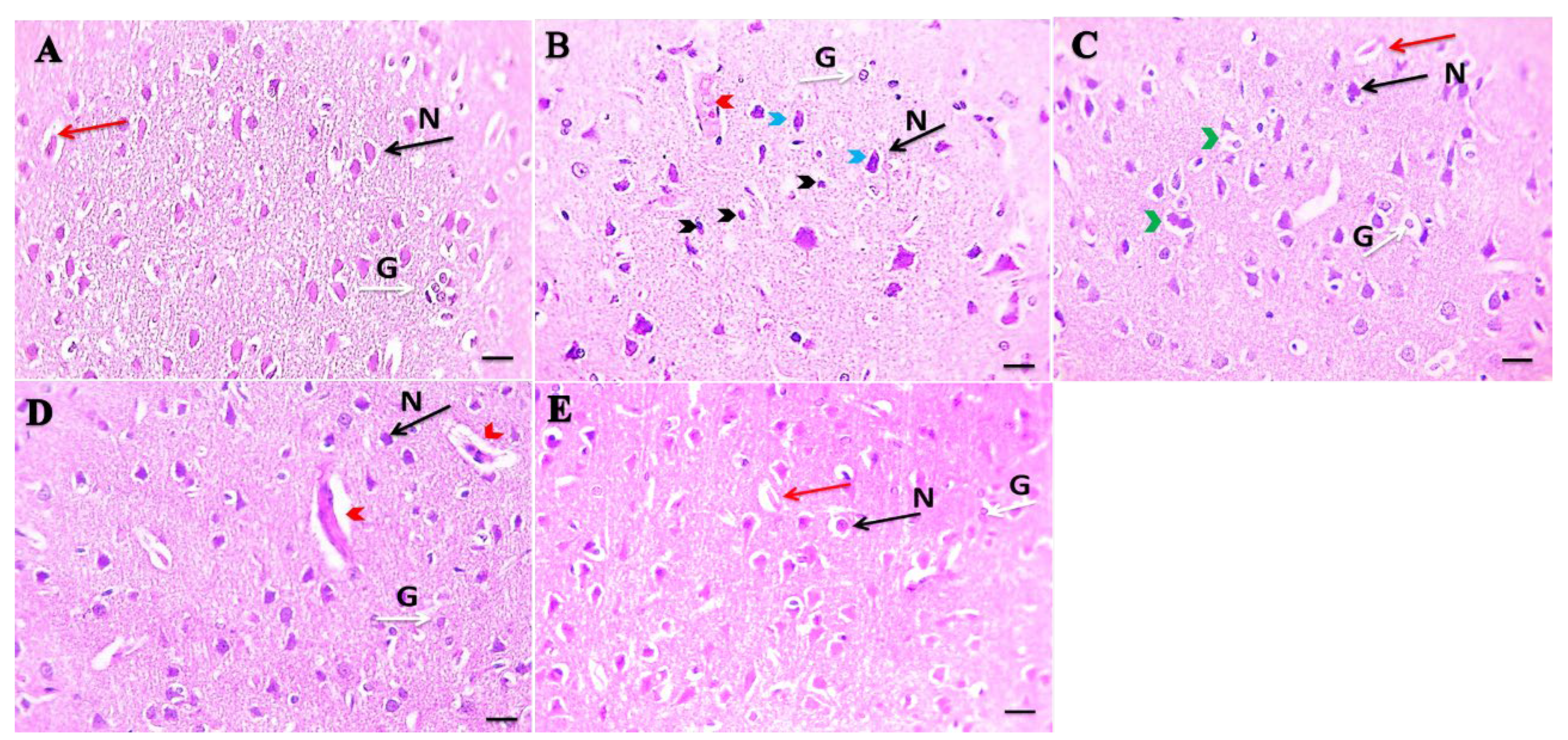
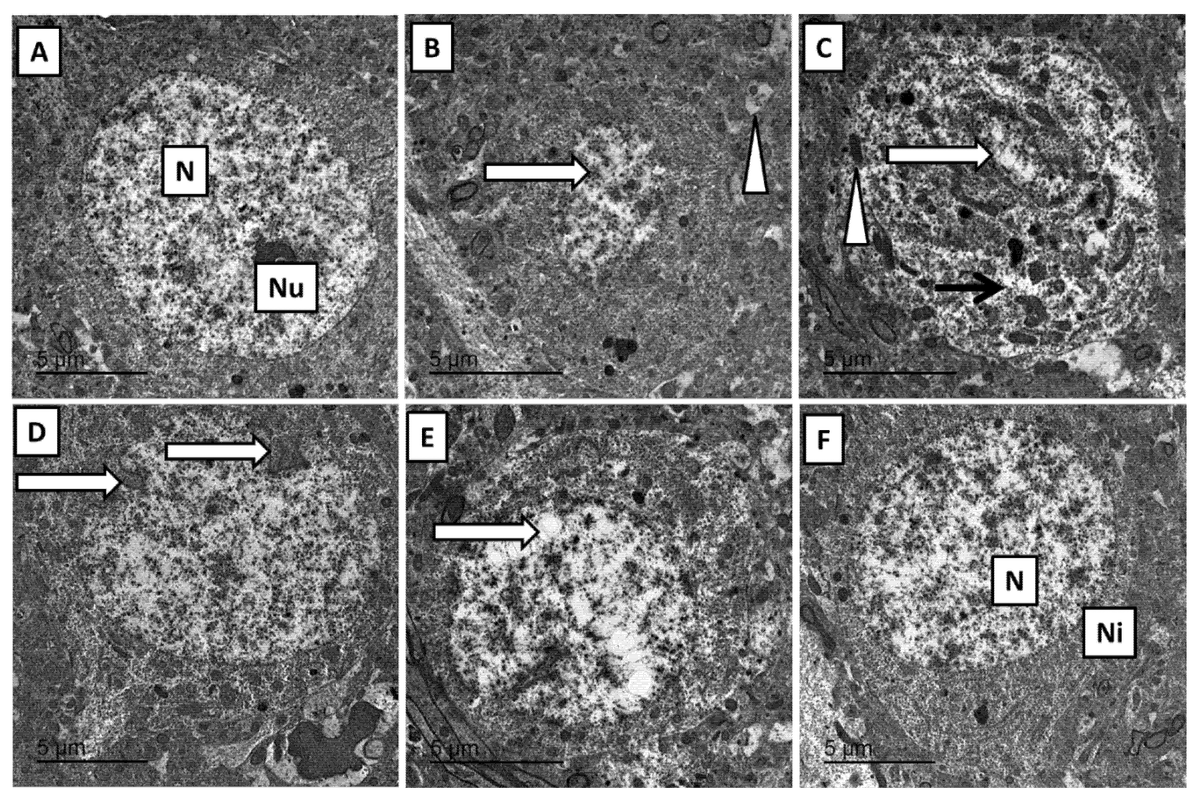
By examining the control group rats’ prefrontal cortex under a light microscope, we found normal pyramidal neurons, glial cells and blood vessels (Figure 2A) with normal neuronal structure, large euchromatic nucleus, and prominent nucleolus beside normal cytoplasmic organelles under electron microscope (Figure 3A), by the exposure of rats to nicotine in the prefrontal cortex showed marked damage in the form of pyramidal cells shrinkage and apoptosis with congestion of blood vessels (Figure 2B,C) with marked ultrastructural changes in the form of pyknosis of neuronal nucleus with shrunken irregularity of mitochondria, few mitochondrial swollen, and disintegration of nissl granules (Figure 3B,C). This degenerative change in the nicotine group was greatly improved by treatment with the combination of ginger and cinnamon oil in the form of a near-normal appearance of pyramidal neurons, glial cells and blood vessels (Figure 2F) with normal ultrastructural appearance of neuron with euchromatic nucleus, numerous free ribosomes (nissl granules) and normal cytoplasmic organelles (Figure 3F); however, the prefrontal cortex of cinnamon and ginger groups also shows some degenerative changes in the form of pericellular oedema and congestion of blood vessels (Figure 2D,E) with mild ultrastructure change in the form of reduction in nuclear size with intranuclear vacuoles and chromatin clumping and nuclear membrane wrinkling (Figure 3D,E).

3.3. Effects of Combined Cinnamon and Ginger Oils on Proinflammatory Cytokines Marker (TNF-α) in Brain Tissues

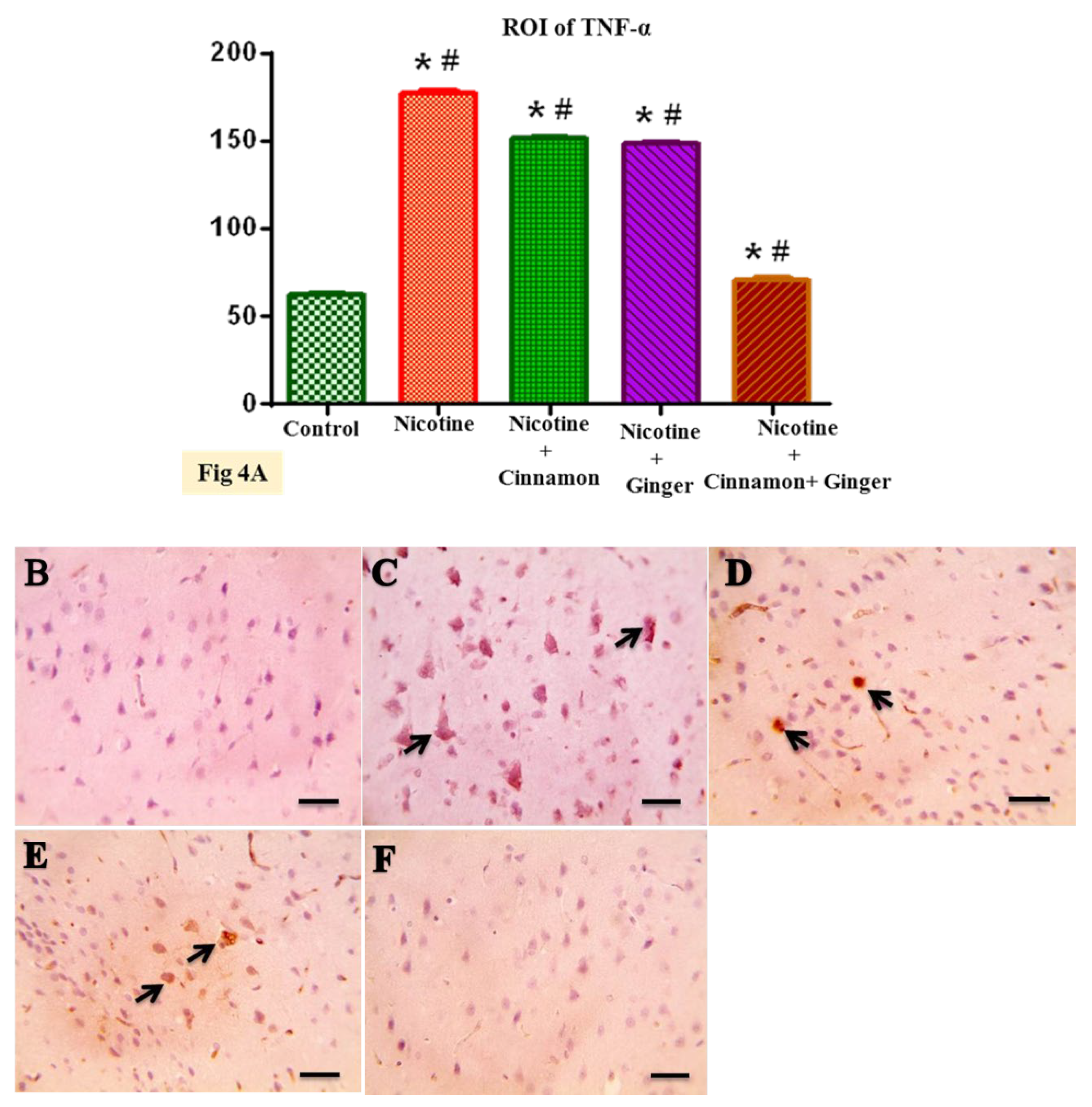
By examining the prefrontal cortex with TNF-α immunostaining, we found that control rats show a normal negative cytoplasmic expression (Figure 4B) which significantly increased in nicotine-exposed prefrontal cortex (Figure 4C) in comparison to the control group (p ≤ 0.05) by image analysis (Figure 4A). This increase in the immunoexpression of TNF-α markedly decreased with treatment by combination of cinnamon and ginger oils (Figure 4F) which was significant in the nicotine group (p ≤ 0.01); thus, combined treatment greatly improved the inflammation that occur in prefrontal cortex of chronic nicotine-exposed rats, and at the same time, TNF-α immunoexpression in cinnamon- and ginger-treated groups separately moderately decreased (Figure 4D,E) but was not significant in the diabetic group (Figure 4A). By these results, we confirmed that the combination of cinnamon and ginger oils have a more powerful anti-inflammatory effect on nicotine-exposed prefrontal cortex than when we use cinnamon and ginger oils separately.

3.4. Combined Effect of Cinnamon and Ginger Oils on Marker of Astrogliosis GFAP in Brain Tissues

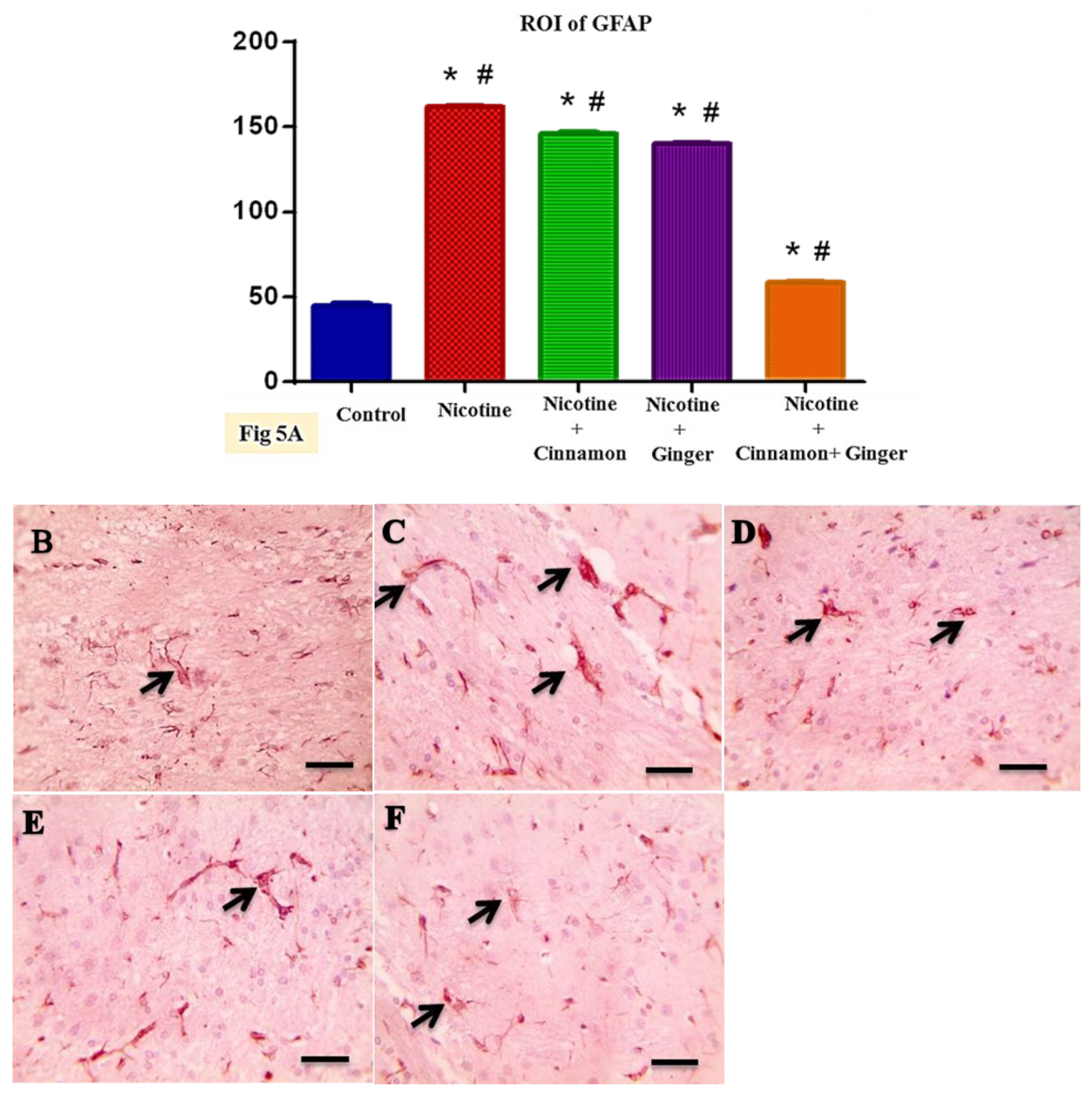
In our study, we found that the immunostaining of the prefrontal cortex with GFAP show a mild positive reaction in control group (Figure 5B); while in nicotine group, there are strong astrocytic positive reactions (Figure 5C), which are significant when compared to control group by region of interest (p ≤ 0.01) (Figure 5A). This indicates that chronic nicotine exposure is a good stimulant for glial astrocytes producing brain gliosis affecting the planning and cognitive function of the prefrontal cortex. This increase in expression of GFAP produced by nicotine greatly improved by the combined treatment with ginger and cinnamon oil (Figure 5F), which is significant in nicotine-exposed brain tissues (p ≤ 0.01) (Figure 5A). In addition, the expression of GFAP is moderately decreased in the prefrontal cortex of cinnamon and ginger oils groups (Figure 5D,E) but less significantly in relation to the nicotine group (Figure 5A). From these results, we confirm that combined treatment as a powerful antioxidant improves astrogliosis which results in chronic nicotine exposure and affecting on the brain function.

4. Discussion

By studying the combined effect of cinnamon and ginger oil on the prefrontal cortex which was exposed to nicotine, we found that (A) chronic nicotine administration disturbs the prefrontal cortex neuronal morphology, which is associated with increased neuronal oxidative stress, inflammation, and neuronal gliosis markers. (B) Treatment with combined ginger and cinnamon oils significantly improves the previous markers through its antioxidative stress propriety.
Several studies were performed on the chronic effect of nicotine on prefrontal cortex, and because of its effect on learning and memory processes, there are many controversies. The authors of [25,27,28,29,30,31] found a great improving effect of nicotine on learning and memory impairment, while [32,33,34] did not find any negative effect. This difference between researchers may be due to dose, time of treatment, and the change in methods of exposure and animal strains used.
The present study targeted the prefrontal cortex, because the research conducted by [30] found that the major destructive effect of chronic nicotine exposure on the brain is on the dopaminergic system, which forms major part of prefrontal cortex. These findings were confirmed by the study performed by [11], which found that the chronic nicotine administration change the neuronal morphology of CA1 region in male albino rats, and [35] also found that the chronic use of electronic cigarette smoking increased the number of necrotic cells in the prefrontal cortex; the authors of [36] have found that chronic nicotine exposure decreases nitric oxide, which leads to sever vasoconstriction and thus decreases the brain blood supply with its glucose and oxygen, causing a disturbance of the metabolic function and energy of the brain leading to necrosis of nerve cells.
Our study confirmed that chronic nicotine exposure greatly affects the oxidative stress scavenging system in the form of increasing the MAD level, which is an indicator of lipid peroxidation and also marked a decrease in the level of GSH. This finding is in agreement with [37] who found that chronic exposure to nicotine stimulates brain lipid peroxidation leading to the elevation in the level of MDA, which leads to damage of the lipids of the brain. In addition, the study conducted by [38] gives the explanation of decreasing the level of GSH due to its consumption in nicotine detoxification mechanism. Our study is the first study to prove that the beneficial synergetic effect of cinnamon and ginger oils on oxidative stress in the prefrontal cortex, which was chronically exposed to nicotine, may be due to the combined its polyphenols content. This is consistent with [39] who confirmed the beneficial antioxidant effect of ginger separately and also with [40] who studied the antioxidant effect of ginger on acetaminophen overdose, but in our study, we found that the combination of cinnamon and ginger has significant improvement in oxidative stress markers, compared to cinnamon and ginger when used separately.
The results of the present study confirm the destructive effect of nicotine on the morphology of the prefrontal cortex neuron in the form of shrinkage and apoptosis in pyramidal neurons with congestion of the blood vessels by light microscope. Under an electron microscope, we found pyknosis of the neuron nucleus, with swelling in the mitochondria and destruction of nissl granules. These results are in agreement with [29], who found that chronic exposure of the medial frontal cortex to nicotine causes pyknosis in the pyramidal cells in the form of pyknosis of nuclei, vacuolation of cytoplasm and condensation of nuclear chromatin with indentation of nuclear membrane. In our study, treatment of nicotine-exposed rats with combined cinnamon and ginger oils greatly improved the brain morphology compared to separate treatment with cinnamon and ginger oils. This may be due to its synergistic antioxidant effect, which improves the inflammation and acetyl choline expression in the prefrontal cortex, as is consistent with [18,28], who found that the usage of ginger oil greatly improves the histomorphological changes and apoptosis produced by diabetes-induced oxidative stress on the prefrontal cortex due to its powerful antioxidant property. In addition, the authors of [41] study the synergetic effect of ginger and cinnamon together on spermatogenesis in diabetic rats due to its powerful antioxidants effect.
In addition, in the present study, the immunoexpression of proinflammatory cytokines, especially tumor necrosis factor TNFα, increased in nicotine-exposed rats. This is in agreement with [42], who found that the acute exposure of rats’ prefrontal cortex to nicotine in the form of 30 days of exposure to four cigarettes/day increases the expression of TNFα. This explained that the oxidative stress produced by chronic nicotine exposure produces neuroinflammation; this expression of TNFα was significantly improved by combined treatment with cinnamon oil and ginger oil, as is consistent with [43], who found that treatment with ginger improves TNFα expression in diabetic rats’ prefrontal cortex. The author explained this by the anti-inflammatory property of ginger, which inhibits diabetes reactive oxygen species production. In addition, [44] found that phenol, which is cinnamon antioxidant metabolite, crosses the blood–brain barrier blocking the activation of inflammatory cascades and optimizing the viability of serotonin neuronal cells.
Our results show strongly significant GFAP expression in the astroglial cells of the prefrontal cortex of nicotine-exposed rats. These findings are in line with [45], who stated that intraperitoneal injection of nicotine producing marked the increase in GFAP expression in rat’s cerebral cortex. The authors gives the explanation for it by stating that glial cells have a large number of nicotinic acetyl choline receptors, which is stimulated by chronic exposure to nicotine, leading to overproduction of GFAP and reactive gliosis. In addition, [46] found that reactive oxygen species have a role in regulation, triggering the astrogliosis. Combined treatment of rats with both cinnamon and ginger oil decreases expression of GFAP, which is in agreement with previous studies conducted by [47,48], which proved that the treatment of diabetic rats with ginger improves oxidative stress-induced reactive gliosis. This suggests that ginger prevents reactive gliosis possibly by reducing the damaging effects of reactive oxygen species in the central nervous system.

5. Conclusions

The administration of ginger and cinnamon oil showed synergistic effects, as compared to each single herb, in improving the effect of nicotine on the prefrontal cortex through its antioxidant anti-inflammatory and neuroprotection. We suggest that the usage of ginger and cinnamon with other polyphenol-containing herbs such as onion to improve nicotine-induced oxidative stress to maximize the antioxidant defense.

Author Contributions

Conceptualization, M.T.; methodology, M.T. and H.M.A.S.; software, H.M.A.S.; validation, M.T., M.M.I. and M.E.; formal analysis, H.M.A.S. and M.A.M.I.; investigation, M.M.I. and M.E.; resources, M.T. and M.M.I.; writing—original draft preparation, M.T. and T.A.S.B.; writing—review and editing, M.T. and M.E.; visualization, M.T.; supervision, M.M.I. and M.A.M.I.; project administration, M.T., M.A.M.I. and T.A.S.B. All authors have read and agreed to the published version of the manuscript.

Funding

This research received no external funding.

Institutional Review Board Statement

Not applicable.

Informed Consent Statement

Not applicable.

Data Availability Statement

Not applicable.

Acknowledgments

Authors would like to thanks to the staff of the Pathology Department, University of Mansoura, Egypt.

Conflicts of Interest

The authors declare no conflict of interest.

References

  1. Noor, K.C.; Mawaddatul, U.S.; Bahrudin, M. Effect of Electronic Cigarette on Brain Prefrontal Cortex of Male wistar Rats. J. Med. Res. 2020, 6, 98–102. [Google Scholar] [CrossRef]
  2. Center for Disease Control. State specific prevalence of current cigarettes smoking among adults and their proportion of adults who work in a smoke-free environment—United States, 1999. Morb. Mortal. Wkly. Rep. 2000, 200, 978–982. [Google Scholar]
  3. Benowitz, N.L. Pharmacology of nicotine: Addiction, smoking-induced disease, and therapeutics. Ann. Rev. Pharmacol. Toxicol. 2009, 49, 57–71. [Google Scholar] [CrossRef] [Green Version]
  4. Djordjevic, M.V.; Branneman, K.D.; Hoffman, D. Identification and analysis of a nicotine derived N. nitrosamine acid and other nitrosamine acids in tobacco. Carcinogenesis 1989, 10, 1725–1731. [Google Scholar] [CrossRef]
  5. Abel, E.L. Prenatal effects of alcohol on growth: A brief overview. Fed. Proc. 1985, 44, 2318–2322. [Google Scholar]
  6. Jha, P.; Chaloupka, F.J.; Moore, J.; Gajalakshmi, V.; Gupta, P.C.; Peck, R.; Asma, S.; Zatonski, W. Tobacco Addiction, Disease Control Properties in Developing Countries, 2nd ed.; The International Bank for Reconstruction and Development/the World Bank: Washington, DC, USA, 2006; Chapter 46. [Google Scholar]
  7. Farsalinos, K.E.; Spyrou, A.; Stefopoulos, C.; Tsimopoulou, K.; Kourkoveli, P.; Tsiapras, D.; Kyrzopoulos, S.; Poulas, K.; Voudris, V. Nicotine absorption from electronic cigarette use: Comparison between experienced consumers (vapers) and naïve users (smokers). Sci. Rep. 2015, 17, 11269. [Google Scholar] [CrossRef]
  8. Hecht, S.S. Tobacco carcinogens, their biomarkers and tobacco induced cancer. Nat. Rev. Cancer 2007, 3, 733–744. [Google Scholar] [CrossRef]
  9. Hellenbrand, W.; Seidler, A.; Robra, B.P. Smoking and Parkinson’s disease: A case-control study in Germany. Int. J. Epidemiol. 1997, 26, 328–339. [Google Scholar] [CrossRef] [PubMed] [Green Version]
  10. Grandinetti, A.; Morens, D.M.; Reed, D. Prospective study of cigarette smoking and the risk of developing idiopathic Parkinson’s disease. Am. J. Epidemiol. 1994, 139, 1129–1138. [Google Scholar] [CrossRef]
  11. Riljak, V.M.; Milotová, M.K.; Jandová, K.; Pokorný, J.; Langmeier, M. Morphological changes in the hippocampus following nicotine and kainic acid administration. Physiol. Res. 2007, 56, 641–649. [Google Scholar]
  12. Valentine, G.; Sofuoglu, M. Cognitive Effects of Nicotine: Recent Progress. Curr. Neuropharmacol. 2018, 16, 403–414. [Google Scholar] [CrossRef] [PubMed]
  13. Akkoc, R.F.; Ogeturk, M. The Prefrontal Cortex: A Basic Embryological, Histological, Anatomical, and Functional Guideline. J. Hum. Anat. Physiol. 2017, 1, 4. [Google Scholar]
  14. Qasim, H.; Karim, Z.A.; Rivera, J.O.; Khasawneh, F.T.; Alshbool, F.Z. Impact of Electronic Cigarettes on the Cardiovascular System. J. Am. Heart Assoc. 2017, 6, 1–14. [Google Scholar] [CrossRef]
  15. Shanmugam, K.R.; Ramakrishna, C.H.; Mallikarjuna, K.; Reddy, K.S. Protective effect of ginger against alcohol-induced renal damage and antioxidant enzymes in male albino rats. Indian J. Exp. Biol. 2010, 48, 143–149. [Google Scholar] [PubMed]
  16. Huang, C.N.; Horng, J.S.; Yin, M.C. Antioxidative and antiglycative effects of six organo sulfur compounds in low-density lipoprotein and plasma. J. Agric. Food Chem. 2004, 52, 3674–3678. [Google Scholar] [CrossRef]
  17. Habib, S.H.; Makpol, S.; Hamid, N.A.A.; Das, S.; Ngah, W.Z.W.; Yusof, Y.A.M. Ginger extract (Zingiber officinale) has anti-cancer and anti-inflammatory effects on ethionine-induced hepatoma rats. Clinics 2008, 63, 807–813. [Google Scholar] [CrossRef] [Green Version]
  18. Shanmugam, K.R.; Mallikarjuna, K.; Kesireddy, N.; Reddy, K.S. Neu-roprotective effect of ginger on anti-oxidant enzymes in streptozotocin-induced diabetic rats. Food Chem. Toxicol. 2011, 49, 893–897. [Google Scholar] [CrossRef]
  19. Tsakanova, G.V.; Ayvazyan, V.A.; Boyajyan, A.S.; Arakelova, E.A.; Grigoryan, G.S.; Guevorkyan, A.A.; Mamikonyan, A.A. A comparative study of antioxidant system and intensity of lipid peroxidation in type 2 diabetes mellitus and ischemic stroke aggravated and not aggravated by type 2 diabetes mellitus. Bull. Exp. Biol. Med. 2011, 151, 564–566. [Google Scholar] [CrossRef]
  20. Medagama, A.B. The glycaemic outcomes of Cinnamon, a review of the experimental evidence and clinical trials. Nutr. J. 2015, 14, 1–12. [Google Scholar] [CrossRef] [Green Version]
  21. Rao, P.V.; Gan, S.H. Cinnamon: A multifaceted medicinal plant. Evid.-Based Complement. Altern. Med. 2014, 2014, 1–12. [Google Scholar] [CrossRef] [PubMed] [Green Version]
  22. Dutta, A.; Chakraborty, A. Cinnamon in Anticancer Armamentarium: A Molecular Approach. J. Toxicol. 2018, 2018, 8978731. [Google Scholar] [CrossRef] [PubMed] [Green Version]
  23. Yulug, B.; Kilic, E.; Altunay, S.; Ersavas, C.; Orhan, C.; Dalay, A.; Mehmet, T.; Nurhan, S.; Vijaya, J.; Sahin, K. Cinnamon polyphenol extract exerts neuroprotective activity in traumatic brain injury through modulation of Nfr2 and cytokine expression. CNS Neurol. Disord.-Drug Targets 2018, 17, 439–447. [Google Scholar] [CrossRef]
  24. Sohrabi, R.; Pazgoohan, N.; Seresht, H.R.; Amin, B. Repeated systemic administration of the cinnamon essential oil possesses anti-anxiety and anti-depressant activities in mice Iran. Iran. J. Basic Med. Sci. 2017, 20, 708–714. [Google Scholar] [CrossRef]
  25. Masuda, Y.; Kikuzaki, H.; Hisamoto, M.; Nakatani, N. Antioxidant properties of gingerol related compounds from ginger. Biofactors 2004, 21, 293–306. [Google Scholar] [CrossRef] [PubMed]
  26. Reynolds, E.S. The use of lead citrate of high pH as an electron opaque stain in electron microscopy. J. Cell Biol. 1963, 17, 208. [Google Scholar] [CrossRef] [Green Version]
  27. Levin, E.D.; Rezvani, A. Nicotinic interactions with antipsychotic drugs, models of schizophrenia and impacts on cognitive function. Biochem. Pharmacol. 2007, 74, 1182–1191. [Google Scholar] [CrossRef] [PubMed] [Green Version]
  28. Li, Y.; Tran, V.H.; Duke, C.C.; Roufogalis, B.D. Preventive and protective properties of Zingiber officinale (ginger) in diabetes mellitus, diabetic complications, and associated lipid and other metabolic disorders: A brief review. Evid.-Based Complement. Altern. Med. 2012, 2012, 516870. [Google Scholar] [CrossRef] [Green Version]
  29. Mahmoud, F.Y.; Abou Elghait, A.T.; Abdel Aziz, H.A.; Mohamed, H. Effect of chronic nicotine administration on the anterior cingulate cortex (area 24a) of adult albino rats: A histological and immunohistochemical study. Egypt. J. Histol. 2013, 36, 149–163. [Google Scholar] [CrossRef]
  30. Mansour, A.; Fox, C.A.; Burke, S.; Meng, F.; Thompson, R.C.; Akil, H.; Watson, S.J. Mu, delta, and kappa opioid receptor mRNA expression in the rat CNS: An in situ hybridization study. J. Comp. Neurol. 1994, 350, 412–438. [Google Scholar] [CrossRef]
  31. Matta, S.G.; Elberger, A.J. Combined exposure to nicotine and ethanol throughout full gestation results in enhanced acquisition of nicotine self-administration in young adult rat offspring. Psychopharmacology 2007, 193, 199–213. [Google Scholar] [CrossRef]
  32. Dunnet, S.B.; Martel, F.L. Proactive interference effects on short-term memory in rats, Basic parameters and drug effects. Behav. Neurosci. 1990, 104, 655–665. [Google Scholar] [CrossRef]
  33. Heisham, S.J.; Kleykamp, B.A.; Singleton, E. Meta-analysis of the acute effects of nicotine and smoking on human performance. Psychopharmacology 2010, 210, 453–469. [Google Scholar] [CrossRef] [Green Version]
  34. Spilich, G.J.; June, L.; Renner, J. Cigarette smoking and cognitive performance. Br. J. Addict. 1992, 87, 1313–1326. [Google Scholar] [CrossRef]
  35. Mishra, A.; Chaturvedi, P.; Datta, S.; Sinukumar, S.; Joshi, P.; Garg, A. Harmful effect of nicotine. Indian J. Med. Paediatr. Oncol. 2015, 36, 24–31. [Google Scholar] [CrossRef] [PubMed] [Green Version]
  36. Toda, N.; Okamura, T. Cigarette smoking impairs nitric oxide- mediated cerebral blood flow increase: Implication for Alzheimer’s disease. J. Pharmacol. Sci. 2016, 131, 223–232. [Google Scholar] [CrossRef] [Green Version]
  37. Budzynska, A.; Boguszewska-Czubara, M.; Kruk-Slomka, J.; Biala, G. Mephedrone and nicotine: Oxidative stress and behavioral interactions in animal models. Neurochem. Res. 2015, 40, 1083–1093. [Google Scholar] [CrossRef] [PubMed] [Green Version]
  38. Mons, U.; Muscat, J.E.; Modesto, J.; Richie, J.P., Jr.; Brenner, H. Effect of smoking reduction and cessation on the plasma levels of the oxidative stress biomarker glutathione—Post-hoc analysis of data from a smoking cessation trial. Free Radic. Biol. Med. 2016, 91, 172–177. [Google Scholar] [CrossRef] [PubMed] [Green Version]
  39. Zarei, M.; Uppin, V.; Acharya, P.; Talahalli, R. Ginger and turmeric lipid-soluble attenuate heated oil-induced oxidative stress in the brain via the upregulation of NRF2 and improve cognitive function in rats. Metab. Brain Dis. 2021, 36, 225–238. [Google Scholar] [CrossRef] [PubMed]
  40. Ashafaq, M.; Hussain, S.; Alshahrani, S.; Madkhali, O.; Siddiqui, R.; Khuwaja, G.; Alam, M.I.; Islam, F. Role of cinnamon oil against acetaminophen overdose induced neurological aberrations through brain stress and cytokine upregulation in rat brain. Drug Chem. Toxicol. 2020, 5, 1–8. [Google Scholar] [CrossRef] [PubMed]
  41. Khaki, A.; Khaki, A.A.; Hajhosseini, L.; Golzar, F.S.; Ainehchi, N. The anti-oxidant effects of ginger and cinnamon on spermatogenesis dys-function of diabetes rats. Afr. J. Tradit. Complement. Altern. Med. 2014, 11, 1–8. [Google Scholar] [CrossRef] [Green Version]
  42. Khanna, A.; Guo, M.; Mehra, M.; Royal, W., III. Inflammation and oxidative stress induced by cigarette smoke in Lewis rat brains. J. Neuroimmunol. 2013, 254, 69–75. [Google Scholar] [CrossRef] [Green Version]
  43. El-Akabawy, G.; El-Kholy, W. Neuroprotective effect of ginger in the brain of streptozotocin-induced diabetic rats. Ann. Anat. 2014, 196, 119–128. [Google Scholar] [CrossRef]
  44. Lee, S.C.; Wang, S.Y.; Li, C.C.; Liu, C.T. Anti-inflammatory effect of cinnamaldehyde and linalool from the leaf essential oil of Cinnamomum osmophloeum Kanehira in endotoxin-induced mice. J. Food Drug Anal. 2018, 26, 211–220. [Google Scholar] [CrossRef] [PubMed] [Green Version]
  45. Takarada, T.; Nakamichi, N.; Kawagoe, H.; Ogura, M.; Fukumori, R.; Nakazato, R.; Fujikawa, K.; Kou, M.; Yoneda, Y. Possible neuroprotective property of nicotinic acetylcholine receptors in association with predominant upregulation of glial cell line derived neurotrophic factor in astrocytes. J. Neurosci. Res. 2012, 90, 2074–2085. [Google Scholar] [CrossRef]
  46. Sofroniew, M.V.; Vinters, H.V. Astrocytes: Biology and pathology. Acta Neuropathol. 2010, 119, 7–35. [Google Scholar] [CrossRef] [PubMed] [Green Version]
  47. Baydas, G.; Reiter, R.J.; Yasar, A.; Tuzcu, M.; Akdemir, I.; Nedzvetskii, V.S. Melatonin reduces glial reactivity in the hippocampus, cortex, and cerebellum of streptozotocin-induced diabetic rats. Free Radic. Biol. Med. 2003, 35, 797–804. [Google Scholar] [CrossRef]
  48. Duarte, J.M.; Agostinho, P.M.; Carvalho, R.A.; Cunha, R.A. Caffeine consumption prevents diabetes-induced memory impairment and synaptotoxicity in the hippocampus of NONcZNO10/LTJ mice. PLoS ONE 2012, 7, e21899. [Google Scholar] [CrossRef] [Green Version]
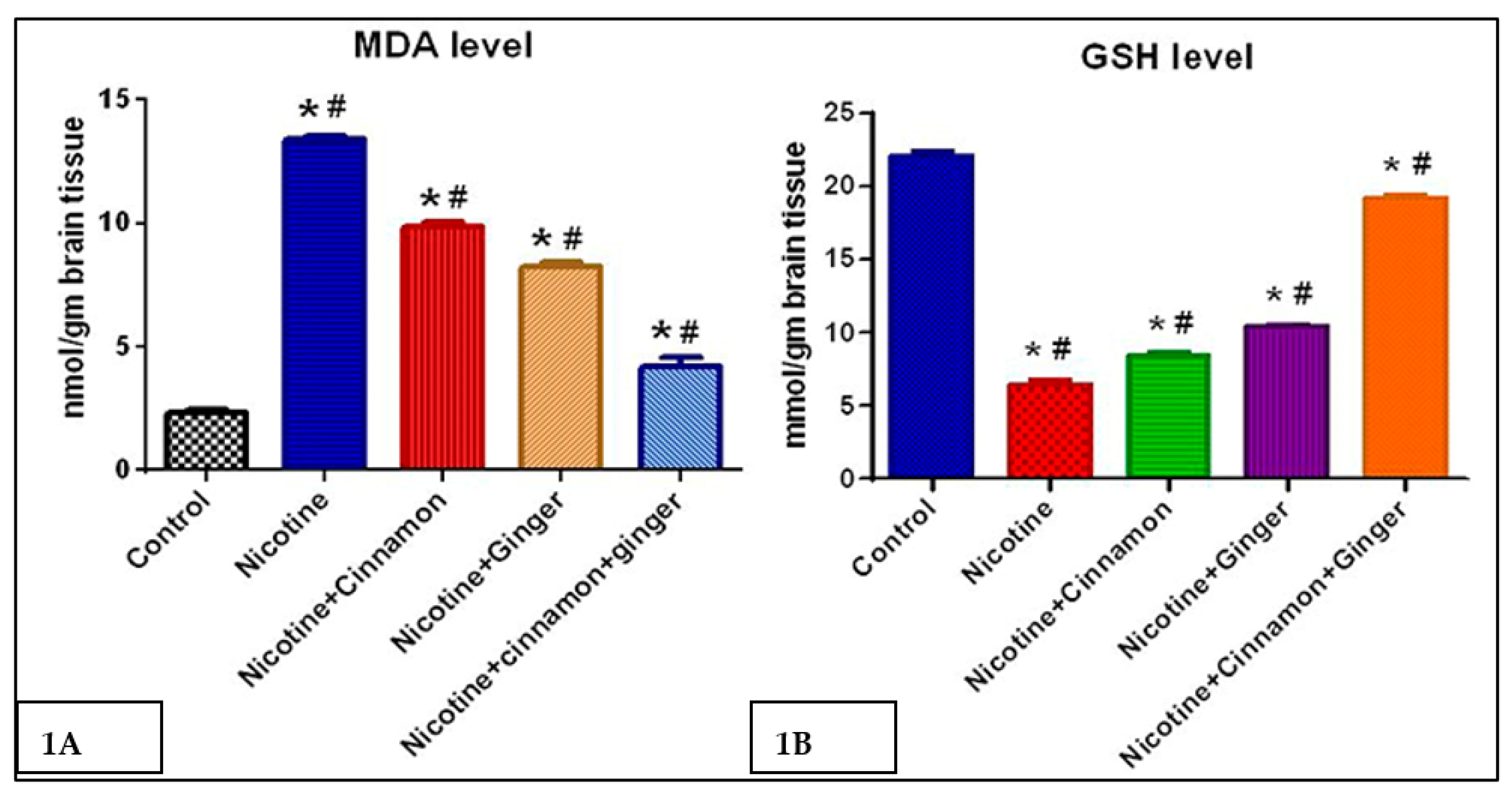
Figure 1. A, B: Brain oxidative stress markers (1A) MDA concentration (nmol/g brain tissue), (1B) glutathione (GSH) (mmol/brain tissue). * significantly different from the control group; # significantly different from other group p value = 0.05.
Scipharm 89 00041 g001
Figure 2. Histopathological examination of the prefrontal cortex of brain stained with H&E. (A) Prefrontal cortex specimen from control group showing normal pyramidal neurons (N) (black arrow), glial cells (G) (white arrow) and blood vessels (red arrow), (B) prefrontal cortex specimen from nicotine group showing marked damage characterized by shrinkage (blue arrowheads), apoptosis (black arrowheads) in pyramidal neurons with congested blood vessels (red arrowhead), (C) prefrontal cortex specimen from nicotine + cinnamon oil group showing mild damage characterized by pericellular edema (green arrowheads), (D) prefrontal cortex specimen from nicotine + ginger oil group showing mild damage characterized by congested blood vessels and perivascular edema (red arrowheads), and (E) prefrontal cortex specimen from the nicotine+cinnamon oil+ginger oil group showed an improved histological appearance of pyramidal neurons (N) (black arrow), glial cells (G) (white arrow), and blood vessels (red arrow). H&E X: 400 bar 50.
Scipharm 89 00041 g002
Figure 3. Representative TEM micrograph of brain ultrathin section from different treatment. (A) Control brain showing normal neuronal structure with a large euchromatic nucleus and prominent nucleolus beside normal cytoplasmic organelles. (B) Nicotine-exposed brain showing pyknosis of neuronal nucleus (thick arrow) with few mitochondrial swollen (arrowhead). (C) Nicotine-exposed brain showing necrotic neuron with severe, more pyknotic nucleus, disintegration of nissl granules (thin arrow), and shrunken irregularity of mitochondria (arrowhead). (D) Ginger-oil-exposed brain showing mild ultrastructure change in neuronal nucleus with chromatin clumping and nuclear membrane wrinkling (thick arrow). (E) Cinnamon-oil-exposed brain showing mild reduction in nuclear size with intranuclear vacuoles (thick arrow). (F) Combined group showing normal ultrastructural appearance of neuron with euchromatic nucleus (N), numerous free ribosomes (nissl granules) (Ni), and normal cytoplasmic organelles. Scale bar  =  5 μm.
Scipharm 89 00041 g003
Figure 4. The expression of TNF-α by immunostaining in different groups. (A) Graphs of ROI of the expression of TNF-α in different groups. (B) Brain specimens showing normal expression of TNF-α neurons of prefrontal cortex (control group), (C) marked strong positive reaction of TNF-α in prefrontal cortex neurons (black arrows) (nicotine group), (D,E) moderate expression of TNF-α in prefrontal cortex neurons (black arrows) (nicotine+ cinnamon oil and nicotine + ginger groups), and (F) negative expression of TNF-α in prefrontal cortex neurons (nicotine+ cinnamon oil +ginger oil group). * Significantly different from the control group; # significantly different from other group p value = 0.05. X: 400 bar 50.
Scipharm 89 00041 g004
Figure 5. The expression of GFAP by immunostaining in different groups. (A) Graphs of ROI of the expression of GFAP in different groups. (B) Brain specimens showing mild expression of GFAP in astroglial cells of prefrontal cortex (black arrow) (control group). (C) Marked strong positive brownish reaction of GFAP in prefrontal cortex astroglial cells (black arrows) (nicotine group). (D,E) Moderate expression of GFAP in prefrontal cortex glial cells (black arrows) (nicotine + cinnamon oil and nicotine + ginger groups), and (F) mild expression of GFAP in prefrontal cortex astroglial cells (nicotine + cinnamon oil + ginger oil group). * Significantly different from the control group; # significantly different from other group p value = 0.05. X: 400 bar 50.
Scipharm 89 00041 g005
Table 1. MDA level in nmol/gm in the brain tissue of the different groups.
GroupControlNicotineNicotine + CinnamonNicotine + GingerNicotine + Cinnamon + Ginger
Mean ± SEM2.317 ± 0.06513.381 ± 0.060 *#9.850 ± 0.076 *#8.250 ± 0.067 *#4.217 ± 0.135 *#
* Significantly different from the control group; # significantly different from other group p value = 0.05.
Table 2. GSH level in mmol/gm in the brain tissue of the different groups.
GroupControlNicotineNicotine + CinnamonNicotine + GingerNicotine + Cinnamon + Ginger
Mean ± SEM22.13 ± 0.1256.533 ± 0.108 *#8.421 ± 0.091 *#10.431 ± 0.061 *#19.28 ± 0.070 *#
* Significantly different from the control group; # significantly different from other group p value = 0.05.
Publisher’s Note: MDPI stays neutral with regard to jurisdictional claims in published maps and institutional affiliations.