Article

Prevalence, Virulence, and Antibiotics Gene Profiles in Lactococcus garvieae Isolated from Cows with Clinical Mastitis in China

1
College of Animal Science & Veterinary Medicine, Shenyang Agricultural University, Shenyang 110866, China
2
College of Veterinary Medicine, Nanjing Agricultural University, Nanjing 210095, China
*
Authors to whom correspondence should be addressed.
Microorganisms 2023, 11(2), 379; https://doi.org/10.3390/microorganisms11020379
Submission received: 26 December 2022 / Revised: 27 January 2023 / Accepted: 30 January 2023 / Published: 2 February 2023
(This article belongs to the Special Issue Domestic Animals and Wildlife Zoonotic Microorganisms)

Abstract

:
Lactococcus garvieae (L. garvieae) is a pathogenic gram-positive, catalase-negative (GPCN) bacterium that causes bovine mastitis. A total of 49 L. garvieae isolates were identified from 1441 clinical mastitis (CM) samples. The pathogenic effects of L. garvieae were studied with two infection models: bovine mammary epithelial cells cultured in vitro and murine mammary infections in vivo. The overall farm prevalence was 15.5% (13/84 farms in 9/19 provinces) and sample prevalence was 3.40% (49/1441). Post-treatment somatic cell count (SCC) post L. garvieae infection was significantly higher than the other GPCN pathogens isolated, and the bacteriological cure fraction was 41.94% (13/31) after intramammary antibiotic treatment. All L. garvieae isolates were resistant to rifaximin, 12.24% of isolates were resistant to cephalexin, and 10.20% (5/49) were multidrug-resistant (MDR). The most prevalent virulence genes were Hemolysin 1 (hly1)(100%), Hemolysin 2 (hly2) (97.96%), NADH oxidase (NADHO) (100%), Superoxide dismutase (SOD) (100%), Adhesin Pav (Pav) (100%), Adhesin PsaA (PsaA) (100%), Enolase (eno) (100%), Adhesin cluster 1(AC1) (100%), Adhesin cluster 2 (AC2) (100%), and several exopolysaccharides. L. garvieae rapidly adhered to bovine mammary epithelial cells, resulting in an elevated lactate dehydrogenase release. Edema and congestion were observed in challenged murine mammary glands and bacteria were consistently isolated at 12, 24, 48, 72, and 120 h after infection. We concluded that L. garvieae had good adaptive ability in the bovine and murine mammary cells and tissue. Given the resistance profile, penicillin and ampicillin are potential treatments for CM cases caused by L. garvieae.

1. Introduction

Mastitis, an inflammatory process of the mammary gland, is the most common bacterial disease [1] and one of the most costly diseases of dairy cattle [2]. Lactococcus species have been known to be associated with mastitis since as early as 1932 [3]. However, GPCN streptococci or streptococci-like bacteria including Streptococcus, Enterococcus, Aerococcus, and Lactococcus are phenotypically and biochemically alike. This means the identification of Lactococcus species has been inaccurate and unreliable in many studies and diagnostic laboratories [4]. The incidence of Lactococcus species identified on-farm may have been historically underreported or was phenotypically identified as Streptococcus uberis (S. uberis) or Streptococcus spp. [5]. Thus, little is known about the clinical importance of this genus as a mastitis pathogen, and awareness and focus have only increased in recent years.
Based on phenotypic similarities, Lactococcus species were initially assigned to the genus Streptococcus, and a new genus was assigned to Lactococcus in 1985 [6]. L. garvieae was known as an emergent disease affecting many fish species and it is considered a potential zoonotic microorganism [7]. This is because it is known to cause several opportunistic human infections, such as endocarditis [8], diverticulitis [9], peritonitis [9], infective spondylodiscitis [10], liver abscess [11], urinary infection [12], subdural hematoma [13], sepsis [14,15,16], bacteremia [9], and late onset periprosthetic infection of the hip [17]. More recently, L. garvieae has been reported in sepsis cases in human patients receiving platelet concentrates from the same donor, one day after transfusion [16]. This highlights the importance of surveillance of this emerging pathogen for humans, especially those who have predisposing health conditions [10]. L. garvieae has also been identified in other animal species (e.g., pigs with pneumonia [18], canine tonsils, sheep, goats, cats, horses, camels, turtles, snakes, and crocodiles [19]); it has also been found in environmental sources, water [20], sand [21], soil [22], and in foodstuffs including radish, broccoli [23], fennel, celery, broccoli, zucchini, wheat flour, and bran [24].
In cattle, L. garvieae was first identified from a mastitis case in 1983 [25], and it was then found in buffalo and cows in Spain [20,26]. Reports from a variety of different countries include clinical and subclinical mastitis cases, such as in dairy cows in Belgium [3], in raw cow’s milk [27,28], cheese made from cow’s milk [29,30], fermented cow’s milk [31], and in beef [32]. In 2016, a Lactococcus genus mastitis outbreak was reported in the USA [33].
Hemolysis-, adhesion-, and immune-evasion-related and other putative virulence genes of L. garvieae have been described and detected, and may play an important role in bacterial virulence [34]. Hemolysins are proteins and lipids that cause cell membrane damage during infection. Many bacterial pathogens are capable of expressing different adhesins, and different adhesins are expressed at different stages during infection, and their synergistic effect plays an important role in the pathogenicity of pathogenic bacteria [35]. LPxTG is a surface protein that covalently binds peptidoglycans isolated from many gram-positive bacteria including L. garvieae, and has an adhesive effect [36]. The formation of a capsule improved the resistance of pathogenic bacteria to phagocytosis in fish-derived L. garvieae [37]. The polysaccharide capsule of L. garvieae has been widely described as a major virulence factor involved mainly in the evasion of the host’s immune response [7]. NADHO and SOD are enzymes that aid in the survival of pathogens in aerobic environments; the bacteria protect themselves by producing these enzymes to avoid being killed [34]. Different pathogenic L. garvieae isolates have different phenotypes; thus, it is important to investigate the putative virulence genes of L. garvieae strains [34].
Multidrug resistant pathogens are defined as being resistant to three or more classes of antimicrobial agents [38]. The widespread use of antibiotics in agriculture, such as for the treatment and prevention of infections, has led to the selection of drug-resistant and multidrug-resistant bacteria [39]. Monitoring multidrug-resistance in pathogenic bacteria is important because drug-resistant bacterial infections reduce treatment options, increase health care costs, and may lead to increased morbidity and mortality [40].
CM samples (n = 1441) were taken from commercial Chinese dairy farms and 49 isolates were ultimately identified as L. garvieae. This was the first time the emerging mastitis pathogen has been detected in Chinese CM samples. There is little research on the characterization of L. garvieae from dairy cattle mastitis. Therefore, the objectives of this study were to determine the epidemiology, SCC resolution and bacteriological cure fraction, antimicrobial resistance profile, detection of putative virulence genes, and pathogenic effects of L. garvieae isolated from CM samples with MAC-T cell and murine mammary infection models.

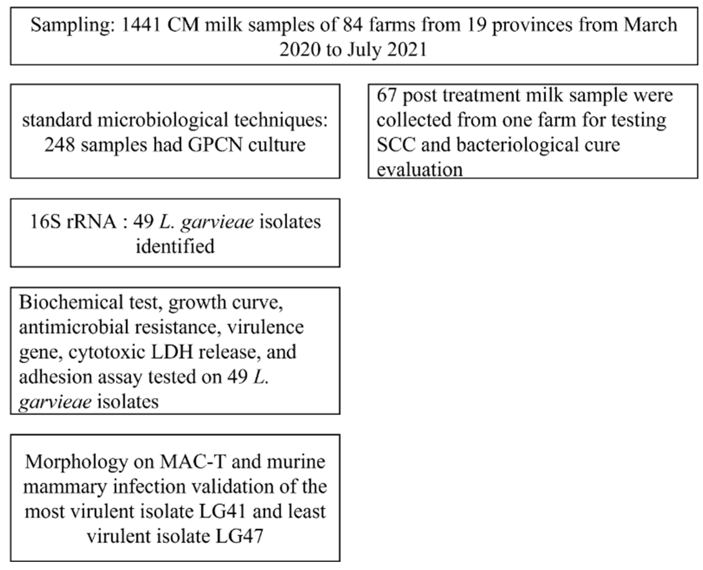
2. Materials and Methods

2.1. Statement of Ethics

The present study was conducted according to the ethical guidelines of Shenyang Agricultural University. Prior to commencement, the animal study was reviewed and approved by the Laboratory Animal Management Committee of Shenyang Agricultural University (protocol: 2021060102).

2.2. Dairy Farm Information and Clinical Mastitis Sample Collection

CM milk samples (n = 1441) were obtained from 84 Chinese commercial dairy farms from 19 different provinces. Abnormal milk (e.g., clots, flake, and watery milk) was identified by farm personnel as CM samples. CM samples were aseptically collected from individual quarters by the authors of this study or trained on-farm personnel. The CM samples for each farm were collected within a 7 day time span. The samples were quickly frozen (−20 °C) overnight and then shipped to Shenyang Agricultural University in Shenyang, China for further identification. The CM sample collection period was from March 2020 to July 2021.

2.3. Bacterial Culture of L. garvieae

Sheep blood agar and brain heart infusion (BHI) agar were used to isolate and purify the colony of the GPCN cocci growing on sheep blood agar from 1441 CM milk samples. Then, BHI broth was used to proliferate bacteria from the colonies that grew on the agar. Gram staining and scanning electron microscopy (SEM) were carried out in order to observe the morphology of L. garvieae.

2.4. 16 S rDNA Sequencing Identification and Biochemical Testing

Bacterial DNA was extracted from 248 isolates using a bacterial DNA extraction kit (Sangon Biotech (Shanghai) Co., Ltd., Shanghai, China) according to the manufacturer’s instructions. The extracted DNA was used as a PCR template for amplification; GPCN streptococci-like isolates were determined by 16S rDNA sequencing [41], where primer p27f (5′-AGAGTTTGATCCTGGCTCAG-3′) and primer 1492r (5′-TACGGCTACCTTGTTACGACTT-3′) were used to amplify a 1460-bp product of the 16S rDNA gene. The PCR cycling conditions included an initial denaturation step at 95 °C for 3 min, followed by 35 cycles at 95 °C for 15 s, 55 °C for 15 s, and 72 °C for 1 min, with a final extension step at 72 °C for 5 min. The PCR products were subjected to sequencing (Sanger sequencing by Sangon Biotech (Shanghai) Co., Ltd., Shanghai, China) after verification on 1.2% agarose gel. The 16S rDNA sequences were compared with sequences deposited in the nucleotide database of the National Center for Biotechnology Information. Identification was deemed reliable if the values for sequence similarities were ≥99%. The biochemical reacting kit (Qingdao Hi-Tech Industrial Park Haibo Biotechnology Co., Ltd., Qingdao, China) was used for biochemical testing. A total of 11 reagents (ribose, sucrose, lactose, liquid gelatin, sorbitol, maltose, esculin, galactose, VP, trehalose, and glucose) were fermented with isolates, following the manufacturer’s instructions. Briefly, isolates were seeded in the tubes that were subsequently cultured in an incubator at 37 °C for the required time; some of the reagents needed further operations and the colors were compared with negative tubes.

2.5. Post-Treatment Milk Sample Collection for SCC and Bacteriological Cure Evaluation

The post-intramammary-antibiotic-treatment milk samples were collected from the farm with the highest prevalence of L. garvieae. The dairy farm, located in northwest China, had an average of 6400 milking cows during the study period. Cows were fed a TMR and housed in freestall barns with sand bedding. Lactating cows were milked thrice daily in two rotary parlors. Milk samples were aseptically collected from the same individual quarter first identified as CM 17 ± 3 d after an extended therapy of 5 d of antimicrobial intramammary treatment (Ubrolexin, Boehringer Ingelheim, Ingelheim am Rhein, Germany) instead of the standard 2 d regimen, and with anti-inflammatory treatment (meloxicam, Boehringer Ingelheim, Germany) for bacteriological cure evaluation. A bacteriological cure was defined as being L. garvieae culture-positive in the clinical mastitis sample, and culture-negative in the post-treatment milk sample. After collection, the quarter milk sample was shaken several times to ensure good mixing and then tested for SCC with the DeLaval DCC instrument (Delaval (Tianjian) Co., Ltd., Tianjin, China).

2.6. Growth Curve of Lactococcus garvieae

The growth curves of one isolate of L. garvieae (LG41), one isolate of L. lactis, one strain of Staphylococcus aureus (S. aureus), and one isolate of Enterococcus faecalis (E. faecalis) were assessed simultaneously. The S. aureus was ATCC 29213, and the remaining isolates from CM milk samples from the same dairy farm had the highest L. garvieae prevalence. The mediums were prepared according to the manufacturer’s instructions. For each isolate, 30 μL of the bacterial solution was added to 3 mL BHI in 5 mL sterile tubes for each different isolate, and they were placed on a constant temperature shaker (37 °C, 220 rpm). At 0, 0.5, 1, 1.5, 2, 2.5, 3, 3.5, 4, 6, 8, 10, 12, 14, 16, 18, 20, 22, and 24 h, the optical density (OD) of each bacterial suspension was determined using 3 tubes per isolate at 600 nm in a UV spectrophotometer (Shimadzu Corporation, Kyoto, Japan). Each experiment was performed in triplicate.

2.7. Antimicrobial Resistance Determination

The minimal inhibitory concentrations (MIC) for penicillin (β-lactams), cephalexin (β-lactams), ampicillin (β-lactams), ceftiofur (β-lactams), cefquinome (β-lactams), lincomycin (lincosamide class), oxytetracycline (Tetracycline class), marbofloxacin (Quinolone class), rifaximin (Rifamycin class), and vancomycin (Glycopeptides class) (Shanghai Yuanye Biotechnology Co., Ltd., Shanghai, China) were determined against 49 L. garvieae isolates using micro-broth dilution assays, following the Clinical Laboratory and Standards Institute guidelines [42]. All antimicrobial agents were used in concentrations ranging from 0.03 to 16μg/mL. S. aureus ATCC 29,213 was used as a quality control strain. Antimicrobial resistance was defined by combining intermediate and resistant categories into a single category. MDR was defined as resistance to ≥3 classes of antimicrobial agents [2]. Each experiment was performed in triplicate.

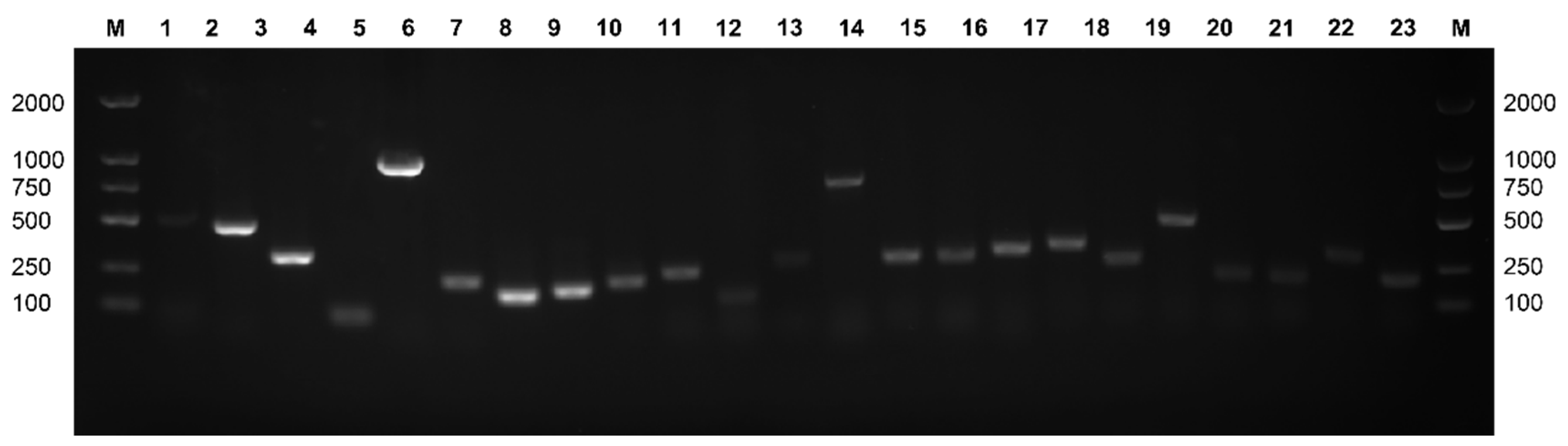
2.8. Virulence Gene Detection

The following potential virulence genes were detected by PCR, with primers listed in Table 1, for 49 L. garvieae isolates: hly1 [34], hly2 [34], Hemolysin 3 (hly3) [34], NADHO [34], SOD [34], Phosphoglucomutase (pgm) [34], Pav [34], PsaA [34], eno [34], containing surface proteins-1, -2, -3, -4 (LP1, LP2, LP3, LP4) [34], AC1 [34], AC2 [34], Adhesin (Adh) [34], capsule gene cluster (1020-F, 1323-R) [34], capsule gene cluster (851-F, 1399-R) [34], capsule gene cluster (6329-F, 7175-R) [34], capsule gene cluster (5358-F, 6007-R) [34], conserved hypothetical protein (CHP) [34], exopolysaccharide R, X, A, B, C, D, and L (epsRXABCDL) [34], oligosaccharide repeat unit polymerase (ORUP) [37], rhamnosyltransferase (RIF) [37], and 30S rRNA gene [37]. The reaction mixtures (25 μL) consisted of 12.5 μL of Green Teq Mix, 1 μL of template DNA, 1 μL of each primer, and 9.5 μL of ultra-pure distilled water. Initial denaturation at 95 °C for 5 min was followed by 34 cycles of amplification at 95 °C for 15 s, annealing at 52 °C for 30 s (all primers), extension at 72 °C for 60 s, and a final extension step at 72 °C for 5 min. The PCR products were analyzed on 1.2% agarose gel. For the potential virulence genes, we confirmed that if an amplicon of the same size as that reported was observed, the isolate contained the target gene [34,37].

2.9. Cell Cultures

Bovine mammary alveolar cell T (MAC-T) (Shanghai Baiye Biotechnology Center, Shanghai, China) was prepared as previously described and cultured in Dulbecco’s Modified Eagle’s Medium (DMEM) supplemented with 10% fetal bovine serum (FBS) and 1% penicillin and streptomycin (Sigma Aldrich, St. Louis, MO, USA), at 37 °C with 5% CO2.

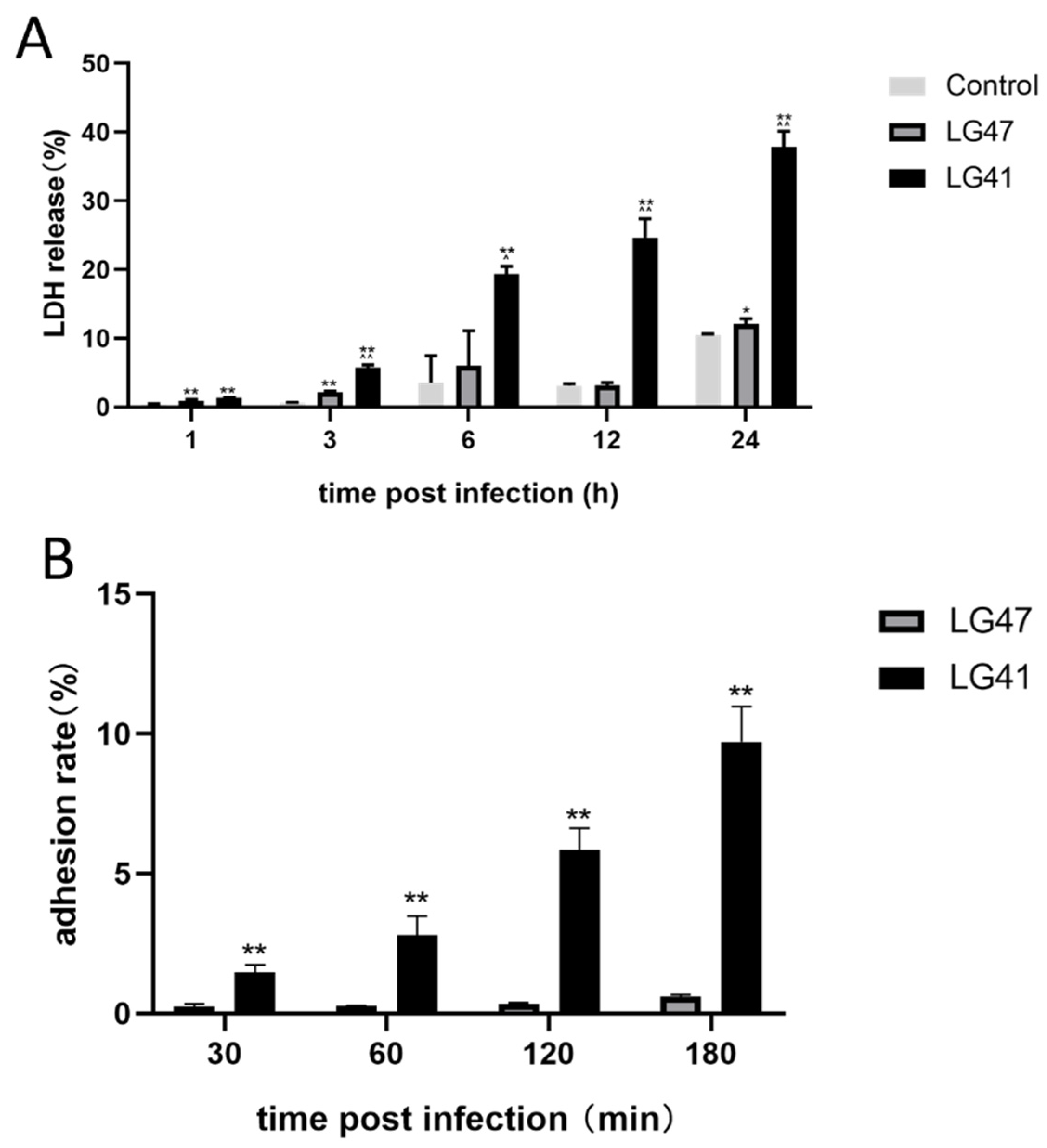
2.10. Cytotoxic Lactate Dehydrogenase (LDH) Release Assay

LDH release was used to identify the most and least cytotoxic isolates among the 49 isolates for further pathogenicity studies, as well as to study the cytotoxic effects of L. garvieae on bovine mammary alveolar cell T (MAC-T) (Shanghai Baiye Biotechnology Center, Shanghai, China). This was assessed using an LDH assay kit (Beyotime Biotechnology, Beijing, China). Cells were cultured at 37 °C with 5% CO2 in 96-well plates (Corning Inc., Corning, NY, USA) and confluent growth (approximately 80% full) was achieved, then challenged with different L. garvieae isolates (n = 49) at a multiplicity of infection (MOI, ratio of L. garvieae to cells) of 5:1 for 12 h. The most cytotoxic isolate should have the highest LDH release, and the least cytotoxic should have the lowest LDH release; thus, the most and least cytotoxic isolates were selected for mouse mastitis model experiments. Then, MAC-T cells were cultured again at 37 °C with 5% CO2 in 96-well plates and challenged with the most and least cytotoxic isolates with an MOI of 5:1 at 1, 3, 6, 12, and 24 h. Uninfected cells were cultured as the control group. After incubation, 200 μL of the supernatant was collected from each well and transferred to a centrifuge tube and centrifuged (8000× g, 5 min at 4 °C). Then, 120 μL of the supernatant was transferred to a new 96-well polystyrene plate and 60 μL of reaction mixture was added to each well. The reaction mixture was then incubated in the dark on a rotating shaker (150 rpm) at room temperature for 30 min. The absorbance was read at 490 nm (QuantStudio3, Thermo Fisher, Waltham, MA, USA). Each experiment was performed in triplicate.

2.11. Adhesion Assay

To assess the adhesion capacity of L. garvieae to MAC-T, the bacterial adhesion of two L. garvieae isolates (LG41 and LG47) was slightly modified as described [2]. The MAC-T were cultured in 6-well plates (Corning Inc.) and confluent growth (approximately 80% full) was achieved in an antibiotic-free medium, followed by infection with L. garvieae at an MOI of 50:1 for 30 min, 1, 2, and 3 h, and cultured at 37 °C and 5% CO2. After incubation, cells were washed twice with sterile PBS (pH 7.4) to remove unbound bacteria. Adhered bacteria were released by adding 1 mL of PBS and 1 mL of 1% triton X-100 (0.5% vol/vol) to lyse cells. Both the bacterial suspension (1 mL) and cells in the control group were treated with 1 mL triton X-100. The cell lysates and treated bacterial supernatant of the infected group and bacterial supernatant of the control group were diluted using a 10-fold serial method, cultured on SBA, incubated at 37 °C for 24 h, and CFU counts were determined. The adhesion fraction was determined as follows:
Bacterial   adhesion   fraction = bacterial   CFU   count   of   Cell   lysates   of   infected   group ( CFU / mL ) bacterial   CFU   count   of   control   group   ( CFU / mL )   × 100 %
The adhesion assays were repeated 3 times, in triplicate for each test.

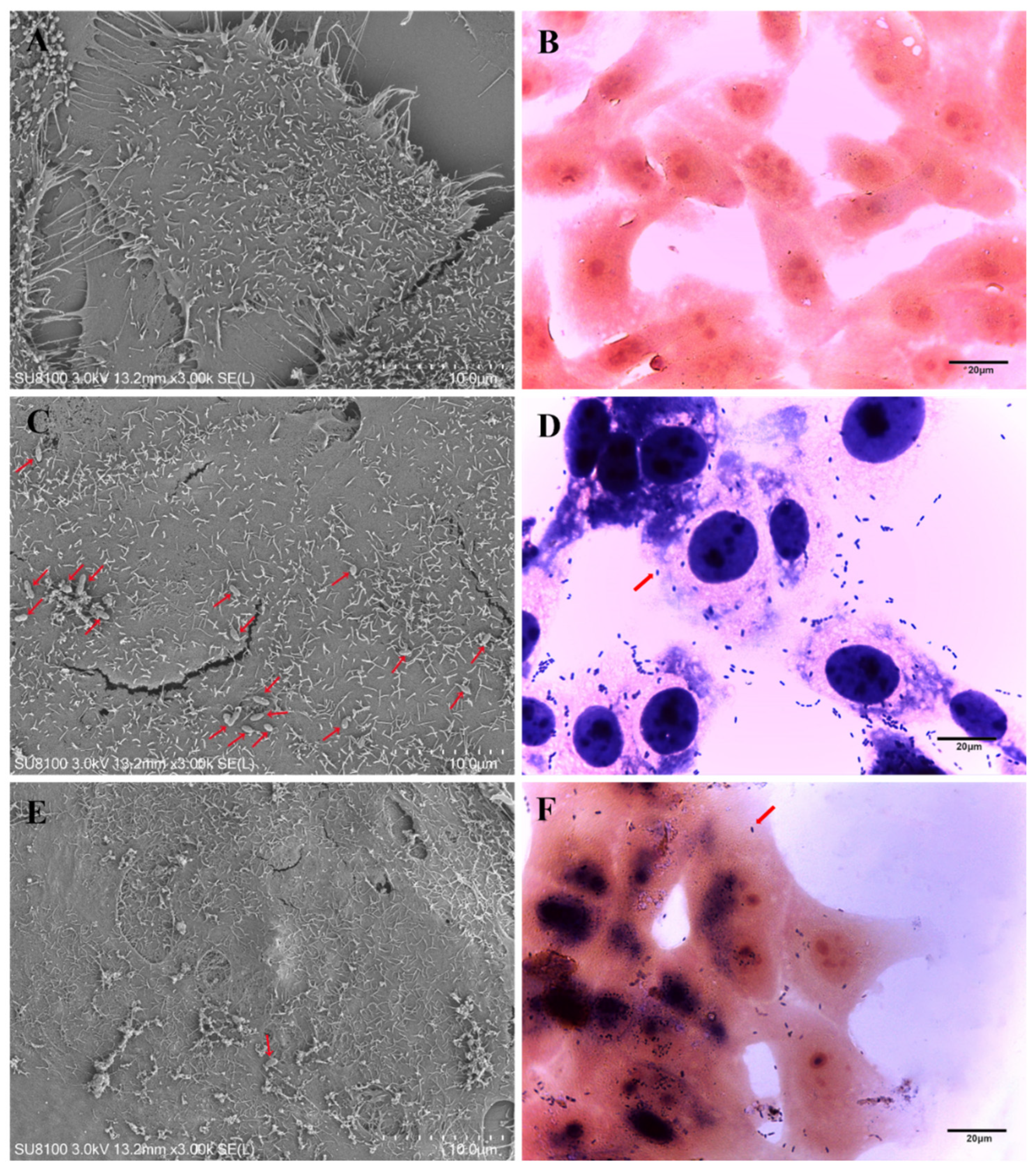
2.12. Morphology of Lactococcus garvieae on MAC-T

Gram staining and SEM of cell slides were carried out in order to observe the morphology of the co-culture of MAC-T and the two L. garvieae isolates (LG41 and LG47). The MAC-T were cultured in 6-well plates (Corning Inc.) for 2 days and grown to confluence in an antibiotic-free medium, followed by infection with L. garvieae (MOI 50:1) for 24 h. After incubation, cells were washed twice with sterile PBS (pH 7.4) to remove unbound bacteria. Gram staining was performed with stained with hematoxylin-eosin. After co-culturing for 24 h, an electron microscope fixing solution was added rapidly to fix the sample at room temperature for 2 h. Then, the samples were rinsed 3 times with 0.1 M phosphate buffer Pb (pH 7.4) for 15 min each time, and the samples were fixed with 1% osmic acid with 0.1 M phosphate buffer Pb (pH 7.4) at room temperature in the dark for 1–2 h. Afterwards, they were rinsed 3 times with 0.1 M phosphate buffer Pb (pH 7.4) for 15 min each time. After that, 30%–50%–70%–80%–90%–95%–100%–100% ethanol was injected into the tissue for 15 min each time. The final process of dehydration involved adding isoamyl acetate for 15 min; then, the sample was put into the critical point dryer for drying. The dried sample was placed on the sample table of the ion sputtering instrument and sprayed with gold for approximately 30 s. Finally, a scanning electron microscope was used to observe and collect pictures.

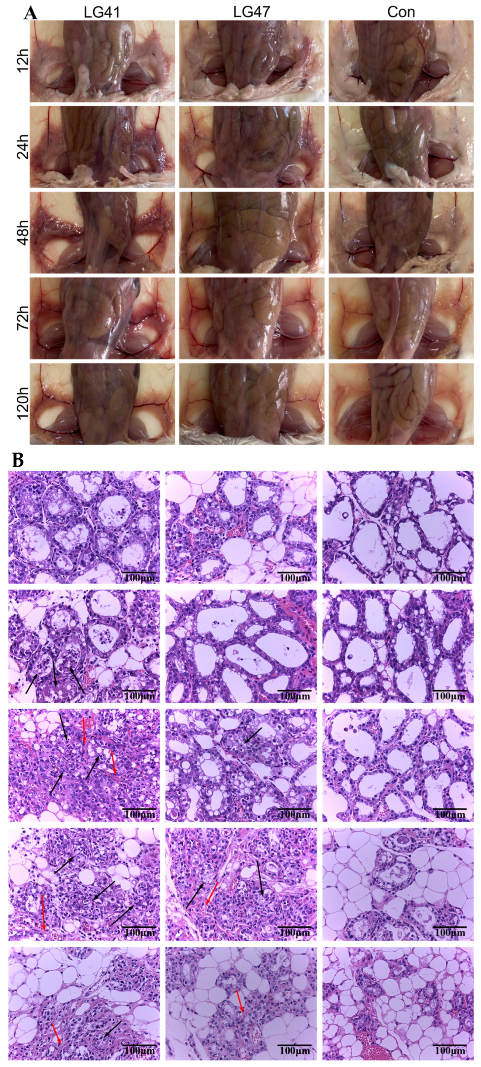
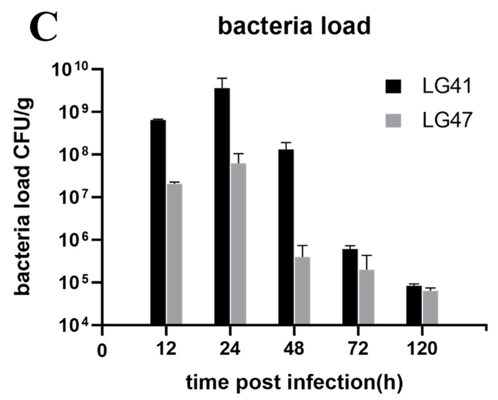
2.13. Lactococcus garvieae Experimental Infection in a Mouse Mammary Gland

The pathogenic effect of two L. garvieae isolates (LG41 and LG47) during intramammary infection was determined using 6–8 week old female specific-pathogen-free BALB/c mice (Liaoning Changsheng Biotechnology Co., Ltd., Benxi, China) [43]. Pregnant (19 d of gestation) mice were kept in germ-free isolators and fed ad libitum in a controlled environment with light and dark cycles (12 h light and 12 h darkness). On the third day after parturition, mice were anesthetized by intramuscular injection of 50 mg/kg Zoletil 50 (Virbac, Carros, France). The fourth pair of mammary glands ducts were exposed by cutting the teat tip and 50 μL of bacterial suspension (5 × 10⁷ CFU) was slowly injected using a small-gauge blunt-tipped needle (Guangdong Xiapute Technology Co., Ltd., Yangjiang, China). Three groups (n = 25 per group) of mice were allocated as 2 challenge groups (LG41 and LG47, respectively) and 1 negative control group (sterile PBS). The pups were removed 1 h before intramammary inoculation. The sedated mice were euthanized with cervical dislocation. The skin was fixed using pins before photographing the mammary glands. The bacterial load in the mammary glands at 12, 24, 48, 72, and 120 h after challenge (5 mice per time point) was measured as described [43]. Briefly, mammary gland tissue (0.1 g) was separated into a sterile Petri dish under a germ-free environment. After homogenization, 50 μL of supernatant was spread on sheep blood plates (multiple dilutions). The numbers of viable colonies were expressed as CFU/g. Mammary gland tissue was fixed with 5% paraformaldehyde, and embedded, sectioned, and stained with hematoxylin-eosin. Histological evaluation was performed to assess tissue necrosis, polymorphonuclear neutrophilic granulocyte inflammation (i.e., neutrophilic inflammation), and lymphocytic inflammation, as described [43]. The flow diagram showing the sample collection and identification for enrollment in the in vivo and in vitro study are shown in Figure 1.

2.14. Statistical Analyses

SPSS 22.0 (SPSS Corporation, Chicago, IL, USA) was used to analyze the data. One-way analysis of variance (ANOVA) was used to compare post-treatment SCC between L. garvieae and L. lactis, or other pathogen infected CM milk samples, LDH release, invasion and adhesion fractions between the treatment groups, and the Duncan test was used to determine the difference. If p < 0.05, the difference was considered to be statistically significant.

3. Results

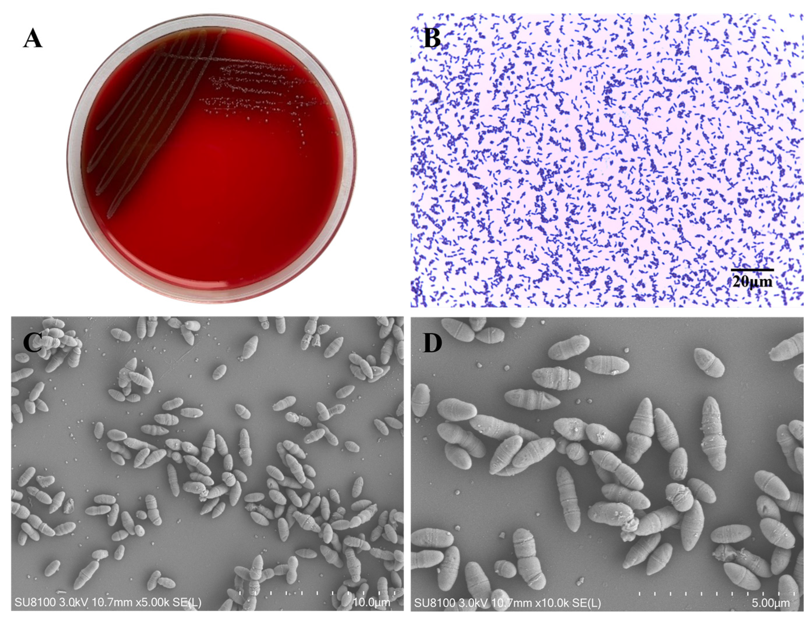
3.1. Morphological Characteristics of Lactococcus garvieae

L. garvieae formed round, medium-sized (approximately 1–2 mm in diameter) colonies on sheep blood TSA plates, with smooth edges, moist surfaces, and α light green hemolysis around the colony. The colony morphology and hemolysis was very similar to streptococcus after incubation at 37 °C for 24 h (Figure 2A). The bacteria stained gram-positive, and, microscopically, the bacterial body appeared spherical or ellipsoidal. The arrangement shape was either two to three bacteria lined up in short chains, or a single bacterium was present (Figure 2B). The results of the scanning electron microscopy showed that the bacteria were ellipsoidal in shape and approximately 1.5–2 μm in diameter. (Figure 2C,D).

3.2. Identification of Suspected Isolates by 16S rDNA Sequence

After the initial isolation and identification of the 1441 milk samples, GPCN cocci isolates were subjected to 16S rDNA gene sequencing. A total of 248 GPCN cocci isolates (all suspected) were sequenced with 16S rDNA gene fragment amplicons, and the three bacteria with the highest percentage were L. garvieae (19.76%), L. lactis (16.53%), and Streptococcus agalactiae (13.71%). The details are shown in Table 2. Positive samples of L. garvieae were isolated from nine provinces. A total of 49 strains of L. garvieae were isolated and identified on 13 farms, with a sample detection frequency of 3.40% (49/1441). Between farms, there was a positive detection frequency of 16.25% (13/84). A farm in Ningxia had the highest detection frequency, with 31 strains of bacteria isolated from 149 clinical mastitis milk samples, and a detection frequency of 20.81%. The distribution of L. garvieae isolated from different Chinese commercial dairy farms are listed in Supplementary Materials in Table S1.

3.3. Biochemical Testing of Lactococcus garvieae

Biochemical testing of 49 L. garvieae isolates showed all negative results of substrate fermentation assays for ribose, sucrose, liquid gelatin, and sorbitol; 95.92% (47/49) positive results for lactose, maltose, trehalose, and glucose; 81.63% (40/49) positive results for galactose; and 79.59% (39/49) positive results for hesperidin. The VP test showed 77.55% (38/49) positive results, see Table 3. Detailed biochemical testing results are included in the Supplementary Materials.

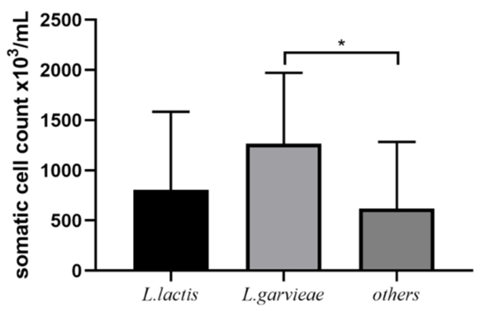
3.4. Post-Treatment SCC and Bacteriological Cure

Post-treatment SCC of L. garvieae (31 isolates), L. lactis (7 isolates), and other bacteria (7 L. lactis isolates, 2 Aerococcus viridans isolates, 1 Enterococcus faecium isolate, and 1 S. uberis isolate) are shown in Figure 3. Post-treatment SCC of L. garvieae infections was not significantly different to L. lactis but was significantly different from other bacterial isolates. The bacteriological cure fraction was 41.94% (13/31) for L. garvieae, 71.43% (5/7) for L. lactis, and 54.55% (6/11) for other bacteria (detailed bacteriological cure data not shown). There was no significant difference in the bacterial cure percentage, which is not surprising given the low numbers in the L. lactis and other groups. All 31 cows identified as being infected with mastitis caused by L. garvieae were classified as mild CM cases (abnormal milk only); however, eight had a recurrence within 30 days after initial diagnosis. The recurrence rate was 25.81% (8/31), while the recurrence rate of other pathogens was 9.10% (1/11).

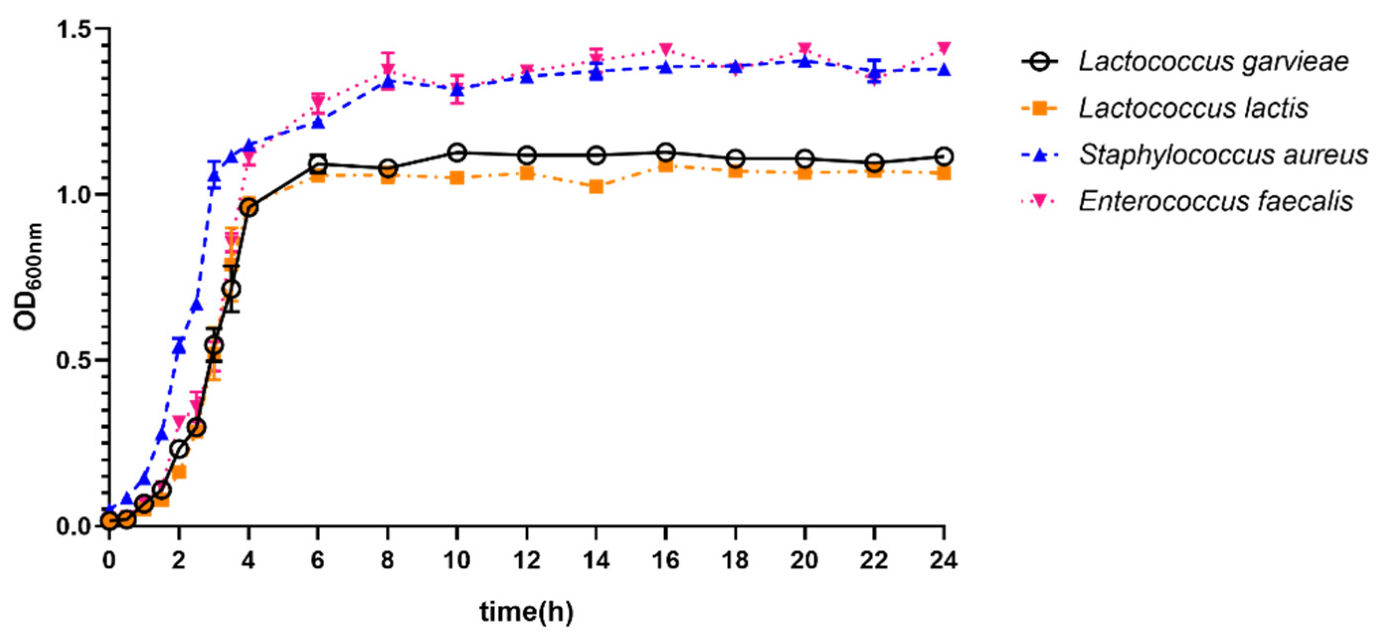
3.5. Growth Ability of Lactococcus garvieae

Growth curves of L. garvieae, L. lactis, S. aureus, and E. faecalis isolates cultured in BHI broth are shown in Figure 4. For L. garvieae, the bacterial growth curve consisted of a lag phase (~2 h), a log phase (~4 h), and ultimately, a stationary phase. Based on subjective observations, the growth curve of L. garvieae had a similar lag phase to other isolates. Additionally, the OD600nm value of L. garvieae isolates reached as high as ~1.0, whereas it was almost the same for L. lactis and up to ~1.3 for S. aureus and E. faecalis isolates.

3.6. Antimicrobial Resistance Profiles of Lactococcus garvieae

All 49 L. garvieae isolates were susceptible to penicillin, ampicillin, ceftiofur, and cefquinome among the β-lactam antibiotics, but 12.24% were resistant to cephalexin. All L. garvieae isolates were resistant to lincomycin and rifaximin, and 73.47% of isolates were resistant to oxytetracycline. All L. garvieae isolates were sensitive to marbofloxacin and vancomycin, see Table 4. As L. garvieae is intrinsically resistant to clindamycin [44], it was excluded from the drug-resistant (DR) or multidrug-resistant (MDR) statistics in this paper. In summary, 89.20% (44/49) of L. garvieae were DR and 10.20% (5/49) were MDR.

3.7. Detection of Virulence Genes in Lactococcus garvieae

Hemolysis-, adhesion-, and immune-evasion-related and other putative virulence genes of L. garvieae were examined. In total, two hemolysis genes (hly1 and hly2), five adhesion-related genes (Pav, PsaA, AC1, AC2, and LP3), nine immune-evasion-related genes (Capsule gene cluster (6329-F, 7175-R), capsule gene cluster (1020-F, 1323-R), EpsA, EpsB, EpsC, EpsD, EpsL, EpsR, and EpsX), and seven other putative virulence genes (NADHO, SOD, CHP, RIF, pgm, eno, and 30S gene) were detected. Of the five MDR L. garvieae isolates, hly2, Eps, and RIF were detected, while none of CGC-related genes were detected. Putative genes detected from the CM case are shown in Figure 5, and detailed putative virulence gene detect results are listed in Table 5.

3.8. Pathogenic Effects of Lactococcus garvieae on MAC-T

LDH release 12 h after infection among different isolates indicated that LG41 was the most cytotoxic, while LG47 was the least cytotoxic. Therefore, these two isolates were selected. At 1, 3, 6, 12, and 24 h after infection, LDH release of LG41 was higher than in the control group (p < 0.01). At 1, 3, and 24 h after infection, LDH release of LG47 was higher than in the control group (p < 0.05). At 3, 6, 12, and 24 h after infection, LDH release of LG41 was higher than LG47 (p < 0.01, Figure 6A). The LG41 isolate adhered to MAC-T at a higher frequency compared with LG47 at the different time points (p < 0.01, Figure 6B).

3.9. Morphology of Lactococcus garvieae on MAC-T

The cells in the control group were closely attached to the round coverslip, the cell morphology was paving stone-like, the surface of the cell membrane was covered with rich microvilli, the cells were arranged in an orderly manner, and there were elongated and rich pseudopods scattered in the cells, which was conducive to cell sticking (Figure 7A,B). After 24 h of LG41 challenge, the microvilli on the cell surface were broken, a large number of bacteria (red arrow) adhered to the cell surface, and there was damage to the cell surface where bacteria were attached. This phenomenon may be a manifestation of endocytosis (Figure 7C,D). After 24 h of LG47 challenge, there was a much smaller number of bacteria (red arrow) adhered to the cell surface than the LG41 group (Figure 7E,F).

3.10. Inflammation of Murine Mammary Gland Infected by Lactococcus garvieae

Edema and hyperemia were evident in murine mammary glands at 12 h after infection with L. garvieae, with more profound pathological changes seen over time (24, 48, and 72 h after infection; Figure 8A). Histological characteristics of the mammary glands infected with L. garvieae were observed (Figure 8B). The structure of the gland alveoli was destroyed, the walls of the gland alveoli was thicker, and infiltrating inflammatory cells (mainly neutrophils) were observed in the gland alveoli and interstitium of the infected mammary gland at 24, 48, 72, and 120 h post infection. At 48 h post infection, the LG41 group showed interstitial tissue hyperplasia. Acute inflammation resolved at 120 h after infection, leaving connective tissue to fill the gap after epithelial death. No evidence of inflammation in mice from the control group (uninfected) was observed.
Bacteria were isolated from the mammary glands of mice challenged with L. garvieae, whereas no bacteria were isolated from the non-infected control group. The bacterial load (mean value) was 3.80 × 10⁸ CFU/g at 12 h after infection, but rapidly increased to 2.10 × 10⁹ CFU/g at 24 h after infection and started to drop to 7.55 × 10⁷ CFU/g at 48 h, 4.31 × 10⁵CFU/g at 72 h, and 7.49 × 10⁴ CFU/g at 120 h (Figure 8C).

4. Discussion

This is the first report of L. garvieae associated with bovine CM cases from multiple farms. L. garvieae is an emerging pathogen that has been confirmed with molecular testing methods such as PCR [5], RAPD, REP-PCR, MLRT, and MALDI-TOF [8]. The strain (98/4289) isolated from water was genetically more closely related to that from bovines than fish-oriented strains. This raises the possibility some environmental L. garvieae strains having evolved from mammals, and this may be involved in the epidemiology of fish lactococcosis [20]. This highlights the importance of implementing screening for L. garvieae as an emerging zoonotic bacterium. Humans, particularly those who have an anatomically or physiologically altered gastrointestinal tract or coexisting local predisposing health problems, are considered to be at-risk individuals. Some human cases have been associated with consuming raw seafood [8]. Consuming raw milk, therefore, has the potential for serious morbidity and mortality [10].
In the present study, the farms suffering from L. garvieae infection were using sand bedding. Sand bedding can be a reservoir of L. garvieae strains and be a potential vehicle for their dissemination in dairy farms. Contaminated sand bedding could also transfer infection between cows [21]. When L. garvieae was initially identified as causing bovine mastitis outbreaks on farms, it is possible the Lactococcus strain already existed and changes in the environment selectively favored the strain responsible for the outbreak. Alternatively, a new Lactococcus strain could have been introduced. Lactococcus has been found in samples from mastitic and normal milk, the bulk tank, and sand bedding. The relative abundance of the Lactococcus genus would be higher in the microbiome of mastitic samples, compared with milk samples from healthy animals [33].
The results of the MIC tests performed in this study agree with other studies that found L. garvieae to be resistant to clindamycin [8,47]. All isolates were sensitive to penicillin, ampicillin, ceftiofur, and cefquinome. However, 12.24% of isolates were resistant to cephalexin; this may be slightly biased in that the majority of isolates were from one farm that also had the most cytotoxic isolate. The IMM antibiotic tube used by the farm at the time of the study was a combination of cefalexin and kanamycin (Ubrolexin). Using a breakpoint of 16/1.6, as used by Sorge et al. (2021), 14.9% of the L. garvieae isolates were resistant to the cefalexin-kanamycin combination. In contrast, L. garvieae had a 5.3% resistance rate to marbofloxacin [48], but in this study, all isolates were sensitive; furthermore, all isolates were resistant to rifaximin. Rifaximin has frequently been used as an antibiotic in dry cow intramammary tubes on some Chinese dairy farms. It concerns us that, if a Lactococcus IMI outbreak occurred, rifaximin might not cure the subclinical infection in dry cows. Therefore, during the next lactation, these infected cows might be more likely to have a flare-up of a clinical case of Lactococcus mastitis. These cows would also be more likely to have a high individual SCC. The results of the MIC tests performed in this study also agree with other studies that L. garvieae from fish-derived [49] and from bovine-milk-derived [50] sources is resistance to tetracycline, and tetS genes were detected from all isolates. Of the 31 milk-derived L. garvieae isolates studied by Walther in 2008, 45.2% were resistant to tetracycline [50]. In comparison, the resistant fraction was 73.47% in this study. The MDR fraction of L. garvieae in this study was 10.20%, which was lower than other mastitic-milk-derived pathogenic bacteria reported in China, including 33% for S. aureus, 56% for non-aureus staphylococci, and 21% for Streptococcus species [51].
Previous reports have agreed that SCC will decline with time when a bacteriological cure is achieved, and this measure is a practical and reliable indicator of treatment success [1]. Lactococcus genus showed a lower bacteriological cure fraction and slower individual SCC resolution than Streptococcus dysaglactiae or S. uberis [52]. The bacteriological cure fraction of L. garvieae was comparable to that of S. aureus, ranging from 38.8% to 52% [53]. The bacteriological cure fraction of other GPCN bacteria isolated in this study (L. lactis, Aerococcus viridans, Enterococcus faecium, and S. uberis) were higher, suggesting that L. garvieae may be comparable to the major pathogenic bacteria in terms of the bacteriological cure fraction. On the other hand, the bacteriological cure fraction of L. lactis was significantly higher, suggesting that L. lactis might be less pathogenic than other pathogens.
Extended therapy with ceftiofur was reported to provide a greater probability of bacteriological cure for gram-positive pathogens, both in clinical and subclinical cases [54,55]. Based on these results from previous studies and the MIC result of our study, we inferred that a low bacteriological cure might be improved by extended therapy. Control measures could also include vaccines, autovaccines, bacteriophages, and antiserum [7]. In bovines, an effective dry cow antibiotic tube could also be a good control method [56].
Furthermore, hly-1 was detected in all isolates, and hly-2 was detected in 97.96% of isolates. This was demonstrated by α-hemolysis of the bacterial colonies, as shown in Figure 2. The three adhesion genes (PavA, eno, and PsaA) were detected in all isolates [34]. Pgm is a metabolic enzyme conferring resistance to peptide antimicrobials and was detected in the most cytotoxic isolate; however, it was not detected in the least cytotoxic isolate. In this study, we investigated the presence of four LPxTG genes, and the LP3 gene was detected in 22.45% of the isolates; however, the LP1, LP2, and LP4 genes were not detected in any isolates. CGC was detected from fish-derived L. garvieae, and four different primers were used to detect CGC [37]; however, none of the isolates from CM cases had all four target stripes, which suggests the similarity between fish and bovines is not high. Seven genes (epsRXABCD) were conserved in the exopolysaccharide (EPS) biosynthetic gene cluster of 49 L. garvieae isolates. Similarly, these data suggest that capsule gene clusters may have spread wildly in Lactococcus spp. as genomic islands. The seven genes also appeared to encode enzymes involved in the polysaccharide structure of the capsule. It has been known that having only a few virulence genes is not enough to cause a pathogenic state, and an appropriate combination of virulence genes must be obtained to cause disease in a particular host species. The most cytotoxic isolate had more virulent genes identified than the least cytotoxic isolate. We inferred the difference in phenotypic virulence might be related to genotypic virulence, but further studies need to be carried out to confirm this.
In vivo challenge models in mice with bovine mastitis pathogens have been successfully used to assess bacterial infection and tissue damage [43]. In the current study, L. garvieae infections stimulated the inflammatory response of the murine mammary gland, which was manifested by the general appearance of concentrated inflammatory cell infiltration, progressive mammary alveolar damage, and the concentration of bacteria in the tissue. L. garvieae multiplied rapidly, leading to the migration of inflammatory cells to the mammary tissue, resulting in edema and congestion [2]. In addition to rapid growth in vitro and a high bacterial count, L. garvieae then declined but was not cleared. Infection with L. garvieae in the murine model indicated that the organism is well adapted to proliferation in the mammary gland and to cause tissue damage. The in vitro study (MAC-T) also supported the bacteria having good adaptive ability in bovine mammary cells.

5. Conclusions

This was the first time the zoonotic pathogen L. garvieae was isolated in CM milk samples from large dairy farms in China (prevalence of 3.40%). All L. garvieae isolates were susceptible to penicillin, ampicillin, cephalexin, cefquinome, ceftiofur, marbofloxacin, and vancomycin. L. garvieae had high resistance to lincomycin, oxytetracycline, and rifaximin, and 12.24% of isolates were resistant to cephalexin, with 10.20% (5/49) being multidrug-resistant (MDR). This suggests bacterial clearance may be decreased during the dry period after the application of dry cow antibiotic preparations and that extended therapy may result in better bacteriological cures in CM cases. The study demonstrated the adhesive ability of L. garvieae in MAC-T and how it can cause cell damage both in vitro and in vivo in the murine model of intramammary infection. The findings of this study help to explain the high prevalence, tissue-damaging nature, and antimicrobial resistance of L. garvieae as an emerging mastitis pathogen.

Supplementary Materials

The following supporting information can be downloaded at: https://www.mdpi.com/article/10.3390/microorganisms11020379/s1, Table S1: 1441 milk samples collected from 84 herds in 5 regions of China; Table S2: Biochemical results of 49 L. garvieae isolates; Table S3: Putative Virulence Gene Detection results of 49 L. garvieae isolates.

Author Contributions

Conceptualization, X.X. and Z.P.; Data curation, X.X.; Formal analysis, X.X.; Funding acquisition, J.D., T.W. and L.L.; Investigation, X.X., L.Y. and F.W.; Methodology, X.X. and Z.P.; Project administration, X.X. and T.W.; Resources, X.X. and Z.P.; Software, X.X.; Supervision, T.W.; Validation, X.X., Z.P. and Y.Y.; Visualization, X.X.; Writing—original draft, X.X.; Writing—review and editing, T.W. All authors have read and agreed to the published version of the manuscript.

Funding

This research was funded by the National Natural Science Foundation of China (grant number 31972639), the Liaoning Provincial joint fund for innovation capability improvement (grant number 2021-NLTS-11-05), and the Youth Project of Liaoning Provincial Education Department Scientific Research Program (grant number LSNQN201908).

Data Availability Statement

All of the relevant data are provided in the form of regular figures, tables, and Supplementary Materials Files.

Acknowledgments

We appreciate the help and technical assistance received from Bo Han from China Agricultural University, and we are grateful to the 84 dairy farms for allowing us to take samples from the cattle with mastitis and for all the help they provided.

Conflicts of Interest

The authors declare that no competing interests exist.

References

  1. Ruegg, P.L. What Is Success? A Narrative Review of Research Evaluating Outcomes of Antibiotics Used for Treatment of Clinical Mastitis. Front. Vet. Sci. 2021, 8, 639641. [Google Scholar] [CrossRef] [PubMed]
  2. Chen, P.; Qiu, Y.; Liu, G.; Li, X.; Cheng, J.; Liu, K.; Qu, W.; Zhu, C.; Kastelic, J.P.; Han, B.; et al. Characterization of Streptococcus lutetiensis isolated from clinical mastitis of dairy cows. J. Dairy Sci. 2021, 104, 702–714. [Google Scholar] [CrossRef] [PubMed]
  3. Devriese, L.A.; Hommez, J.; Laevens, H.; Pot, B.; Vandamme, P.; Haesebrouck, F. Identification of aesculin-hydrolyzing streptococci, lactococci, aerococci and enterococci from subclinical intramammary infections in dairy cows. Vet. Microbiol. 1999, 70, 87–94. [Google Scholar] [CrossRef]
  4. Goyache, J.; Vela, A.I.; Gibello, A.; Blanco, M.M.; Briones, V.; González, S.; Téllez, S.; Ballesteros, C.; Domínguez, L.; Fernández-Garayzábal, J.F. Lactococcus lactis subsp. lactis infection in waterfowl: First confirmation in animals. Emerg. Infect. Dis. 2001, 7, 884–886. [Google Scholar] [CrossRef]
  5. Werner, B.; Moroni, P.; Gioia, G.; Lavin-Alconero, L.; Yousaf, A.; Charter, M.E.; Carter, B.M.; Bennett, J.; Nydam, D.V.; Welcome, F.; et al. Short communication: Genotypic and phenotypic identification of environmental streptococci and association of Lactococcus lactis ssp. lactis with intramammary infections among different dairy farms. J. Dairy Sci. 2014, 97, 6964–6969. [Google Scholar] [CrossRef] [PubMed]
  6. Schleifer, K. Transfer of Streptococcus lactis and related streptococci to the genus Lactococcus gen. nov. Syst. Appl. Microbiol. 1985, 6, 183–195. [Google Scholar] [CrossRef]
  7. Meyburgh, C.M.; Bragg, R.R.; Boucher, C.E. Lactococcus garvieae: An emerging bacterial pathogen of fish. Dis. Aquat. Org. 2017, 123, 67–79. [Google Scholar] [CrossRef]
  8. Malek, A.; De la Hoz, A.; Gomez-Villegas, S.I.; Nowbakht, C.; Arias, C.A. Lactococcus garvieae, an unusual pathogen in infective endocarditis: Case report and review of the literature. BMC Infect. Dis. 2019, 19, 301. [Google Scholar] [CrossRef]
  9. Wang, C.Y.C.; Shie, H.S.; Chen, S.C.; Huang, J.P.; Hsieh, I.C.; Wen, M.S.; Lin, F.C.; Wu, D. Lactococcus garvieae infections in humans: Possible association with aquaculture outbreaks. Int. J. Clin. Pract. 2007, 61, 68–73. [Google Scholar] [CrossRef]
  10. Chan, J.F.W.; Woo, P.C.Y.; Teng, J.L.L.; Lau, S.K.P.; Leung, S.S.M.; Tam, F.C.C.; Yuen, K.Y. Primary infective spondylodiscitis caused by Lactococcus garvieae and a review of human L. garvieae infections. Infection 2011, 39, 259–264. [Google Scholar] [CrossRef] [Green Version]
  11. Mofredj, A.; Baraka, D.; Kloeti, G.; Dumont, J.L. Lactococcus garvieae septicemia with liver abscess in an immunosuppressed patient. Am. J. Med. 2000, 109, 513–514. [Google Scholar] [CrossRef]
  12. Gibello, A.; Galán-Sánchez, F.; Blanco, M.M.; Rodríguez-Iglesias, M.; Domínguez, L.; Fernández-Garayzábal, J.F. The zoonotic potential of Lactococcus garvieae: An overview on microbiology, epidemiology, virulence factors and relationship with its presence in foods. Res. Vet. Sci. 2016, 109, 59–70. [Google Scholar] [CrossRef] [PubMed]
  13. Rasmussen, M.; Björk Werner, J.; Dolk, M.; Christensson, B. Lactococcus garvieae endocarditis presenting with subdural haematoma. BMC Cardiovasc. Disord. 2014, 14, 13. [Google Scholar] [CrossRef] [PubMed]
  14. Nakamura, S.; Nakai, K.; Sakata, M.; Nagaoka, Y.; Yoshida, K.; Katsumata, U.; Chiba, T.; Matsubayashi, K. Recipient sepsis caused by Lactococcus garvieae contamination of platelets from a donor with colon cancer. Vox Sang. 2019, 114, 182–184. [Google Scholar] [CrossRef]
  15. Sahu, K.K.; Sherif, A.A.; Syed, M.P.; Rajendran, A.; Mishra, A.K.; Davaro, R. A rare cause of sepsis: Lactococcus garvieae. QJM Int. J. Med. 2019, 112, 447–448. [Google Scholar] [CrossRef] [PubMed]
  16. Colagrossi, L.; Costabile, V.; Scutari, R.; Agosta, M.; Onori, M.; Mancinelli, L.; Lucignano, B.; Muda, A.O.; Baldo, G.D.; Mastronuzzi, A.; et al. Evidence of pediatric sepsis caused by a drug resistant contaminated platelet concentrate. Emerg. Microbes Infect. 2022, 11, 1325–1334. [Google Scholar] [CrossRef] [PubMed]
  17. Westberg, M.; Brekke, H.; Hermansen, N.O.; Flatøy, B. Late onset periprosthetic infection of the hip caused by the fish pathogen in a patient not associated with fish exposure. J. Bone Jt. Infect. 2020, 5, 106–109. [Google Scholar] [CrossRef] [PubMed]
  18. Tejedor, J.L.; Vela, A.I.; Gibello, A.; Casamayor, A.; Domínguez, L.; Fernández-Garayzábal, J.F. A genetic comparison of pig, cow and trout isolates of Lactococcus garvieae by PFGE analysis. Lett. Appl. Microbiol. 2011, 53, 614–619. [Google Scholar] [CrossRef] [PubMed]
  19. Thiry, D.; Billen, F.; Boyen, F.; Duprez, J.-N.; Quenault, H.; Touzain, F.; Blanchard, Y.; Clercx, C.; Mainil, J.G. Genomic relatedness of a canine Lactococcus garvieae to human, animal and environmental isolates. Res. Vet. Sci. 2021, 137, 170–173. [Google Scholar] [CrossRef]
  20. Vela, A.I.; Vázquez, J.; Gibello, A.; Blanco, M.M.; Moreno, M.A.; Liébana, P.; Albendea, C.; Alcalá, B.; Mendez, A.; Domínguez, L.; et al. Phenotypic and genetic characterization of Lactococcus garvieae isolated in Spain from lactococcosis outbreaks and comparison with isolates of other countries and sources. J. Clin. Microbiol. 2000, 38, 3791–3795. [Google Scholar] [CrossRef] [Green Version]
  21. Eraclio, G.; Ricci, G.; Moroni, P.; Santisteban, C.; Plumed-Ferrer, C.; Bennett, J.; Fortina, M.G. Sand bedding as a reservoir for Lactococcus garvieae dissemination in dairy farms. Can. J. Microbiol. 2019, 65, 84–89. [Google Scholar] [CrossRef]
  22. Altın, G.; Nikerel, E.; Şahin, F. Draft Genome Sequence of Magnesium-Dissolving A1, Isolated from Soil. Genome Announc. 2017, 5, e00386-17. [Google Scholar] [CrossRef] [PubMed]
  23. Kawanishi, M.; Yoshida, T.; Kijima, M.; Yagyu, K.; Nakai, T.; Okada, S.; Endo, A.; Murakami, M.; Suzuki, S.; Morita, H. Characterization of Lactococcus garvieae isolated from radish and broccoli sprouts that exhibited a KG+ phenotype, lack of virulence and absence of a capsule. Lett. Appl. Microbiol. 2007, 44, 481–487. [Google Scholar] [CrossRef] [PubMed]
  24. Ferrario, C.; Ricci, G.; Borgo, F.; Rollando, A.; Fortina, M.G. Genetic investigation within Lactococcus garvieae revealed two genomic lineages. FEMS Microbiol. Lett. 2012, 332, 153–161. [Google Scholar] [CrossRef] [PubMed]
  25. Collins, M.D.; Farrow, J.A.; Phillips, B.A.; Kandler, O. Streptococcus garvieae sp. nov. and Streptococcus plantarum sp. nov. Microbiology 1983, 129, 3427–3431. [Google Scholar] [CrossRef]
  26. Teixeira, L.M.; Merquior, V.L.; Vianni, M.C.; Carvalho, M.G.; Fracalanzza, S.E.; Steigerwalt, A.G.; Brenner, D.J.; Facklam, R.R. Phenotypic and genotypic characterization of atypical Lactococcus garvieae strains isolated from water buffalos with subclinical mastitis and confirmation of L. garvieae as a senior subjective synonym of Enterococcus seriolicida. Int. J. Syst. Evol. Microbiol. 1996, 46, 664–668. [Google Scholar] [CrossRef]
  27. Klijn, N.; Weerkamp, A.H.; de Vos, W.M. Detection and characterization of lactose-utilizing Lactococcus spp. in natural ecosystems. Appl. Environ. Microbiol. 1995, 61, 788–792. [Google Scholar] [CrossRef]
  28. Lafarge, V.; Ogier, J.-C.; Girard, V.; Maladen, V.; Leveau, J.-Y.; Gruss, A.; Delacroix-Buchet, A. Raw cow milk bacterial population shifts attributable to refrigeration. Appl. Environ. Microbiol. 2004, 70, 5644–5650. [Google Scholar] [CrossRef]
  29. Abdelfatah, E.N.; Mahboub, H.H.H. Studies on the effect of Lactococcus garvieae of dairy origin on both cheese and Nile tilapia (O. niloticus). Int. J. Vet. Sci. Med. 2018, 6, 201–207. [Google Scholar] [CrossRef]
  30. Alegría, A.; Alvarez-Martín, P.; Sacristán, N.; Fernández, E.; Delgado, S.; Mayo, B. Diversity and evolution of the microbial populations during manufacture and ripening of Casín, a traditional Spanish, starter-free cheese made from cow’s milk. Int. J. Food Microbiol. 2009, 136, 44–51. [Google Scholar] [CrossRef] [Green Version]
  31. Akabanda, F.; Owusu-Kwarteng, J.; Tano-Debrah, K.; Parkouda, C.; Jespersen, L. The Use of Lactic Acid Bacteria Starter Culture in the Production of Nunu, a Spontaneously Fermented Milk Product in Ghana. Int. J. Food Sci. 2014, 2014, 721067. [Google Scholar] [CrossRef]
  32. Ricci, G.; Ferrario, C.; Borgo, F.; Eraclio, G.; Fortina, M.G. Genome Sequences of Two Lactococcus garvieae Strains Isolated from Meat. Genome Announc. 2013, 1, e00018-12. [Google Scholar] [CrossRef] [PubMed]
  33. Rodrigues, M.X.; Lima, S.F.; Higgins, C.H.; Canniatti-Brazaca, S.G.; Bicalho, R.C. The Lactococcus genus as a potential emerging mastitis pathogen group: A report on an outbreak investigation. J. Dairy Sci. 2016, 99, 9864–9874. [Google Scholar] [CrossRef] [PubMed]
  34. Ture, M.; Altinok, I. Detection of putative virulence genes of Lactococcus garvieae. Dis. Aquat. Org. 2016, 119, 59–66. [Google Scholar] [CrossRef] [PubMed]
  35. Klemm, P.; Schembri, M.A. Bacterial adhesins: Function and structure. Int. J. Med Microbiol. 2000, 290, 27–35. [Google Scholar] [CrossRef]
  36. Miyauchi, E.; Toh, H.; Nakano, A.; Tanabe, S.; Morita, H. Comparative Genomic Analysis of Lactococcus garvieae Strains Isolated from Different Sources Reveals Candidate Virulence Genes. Int. J. Microbiol. 2012, 2012, 728276. [Google Scholar] [CrossRef]
  37. Morita, H.; Toh, H.; Oshima, K.; Yoshizaki, M.; Kawanishi, M.; Nakaya, K.; Suzuki, T.; Miyauchi, E.; Ishii, Y.; Tanabe, S.; et al. Complete genome sequence and comparative analysis of the fish pathogen Lactococcus garvieae. PLoS ONE 2011, 6, e23184. [Google Scholar] [CrossRef]
  38. Schwarz, S.; Silley, P.; Simjee, S.; Woodford, N.; van Duijkeren, E.; Johnson, A.P.; Gaastra, W. Assessing the antimicrobial susceptibility of bacteria obtained from animals. Vet. Microbiol. 2010, 141, 601–604. [Google Scholar] [CrossRef]
  39. Burgos, J.M.; Ellington, B.A.; Varela, M.F. Presence of multidrug-resistant enteric bacteria in dairy farm topsoil. J. Dairy Sci. 2005, 88, 1391–1398. [Google Scholar] [CrossRef] [PubMed]
  40. Oliveira, L.; Langoni, H.; Hulland, C.; Ruegg, P.L. Minimum inhibitory concentrations of Staphylococcus aureus recovered from clinical and subclinical cases of bovine mastitis. J. Dairy Sci. 2012, 95, 1913–1920. [Google Scholar] [CrossRef] [Green Version]
  41. Su, F.-J.; Chen, M.-M. Protective Efficacy of Novel Oral Biofilm Vaccines against Infection in Mullet. Vaccines 2021, 9, 844. [Google Scholar] [CrossRef] [PubMed]
  42. CLSI. Performance Standards for Antimicrobial Disk and Dilution Susceptibility Tests for Bacteria Isolated From Animals, 5th ed.; CLSI document; VET01SEd05E; CLSI: Wayne, PA, USA, 2020. [Google Scholar]
  43. Liu, G.; Yin, J.; Han, B.; Barkema, H.W.; Shahid, M.; De Buck, J.; Cobo, E.R.; Kastelic, J.P.; Gao, J. Adherent/invasive capacities of bovine-associated Aerococcus viridans contribute to pathogenesis of acute mastitis in a murine model. Vet. Microbiol. 2019, 230, 202–211. [Google Scholar] [CrossRef] [PubMed]
  44. Elliott, J.A.; Facklam, R.R. Antimicrobial susceptibilities of Lactococcus lactis and Lactococcus garvieae and a proposed method to discriminate between them. J. Clin. Microbiol. 1996, 34, 1296–1298. [Google Scholar] [CrossRef] [PubMed]
  45. Mi, K.; Li, M.; Sun, L.; Hou, Y.; Zhou, K.; Hao, H.; Pan, Y.; Liu, Z.; Xie, C.; Huang, L. Determination of Susceptibility Breakpoint for Cefquinome against in Pigs. Antibiotics 2021, 10, 958. [Google Scholar] [CrossRef] [PubMed]
  46. Colangeli, R.; Jedrey, H.; Kim, S.; Connell, R.; Ma, S.; Chippada Venkata, U.D.; Chakravorty, S.; Gupta, A.; Sizemore, E.E.; Diem, L.; et al. Bacterial Factors That Predict Relapse after Tuberculosis Therapy. N. Engl. J. Med. 2018, 379, 823–833. [Google Scholar] [CrossRef]
  47. Villani, F.; Aponte, M.; Blaiotta, G.; Mauriello, G.; Pepe, O.; Moschetti, G. Detection and characterization of a bacteriocin, garviecin L1-5, produced by Lactococcus garvieae isolated from raw cow’s milk. J. Appl. Microbiol. 2001, 90, 430–439. [Google Scholar] [CrossRef]
  48. Sorge, U.S.; Huber-Schlenstedt, R.; Schierling, K. In vitro antimicrobial resistance profiles of Streptococcus uberis, Lactococcus spp., and Enterococcus spp. from quarter milk samples of cows between 2015 and 2019 in Southern Germany. J. Dairy Sci. 2021, 104, 5998–6012. [Google Scholar] [CrossRef]
  49. Kawanishi, M.; Kojima, A.; Ishihara, K.; Esaki, H.; Kijima, M.; Takahashi, T.; Suzuki, S.; Tamura, Y. Drug resistance and pulsed-field gel electrophoresis patterns of Lactococcus garvieae isolates from cultured Seriola (yellowtail, amberjack and kingfish) in Japan. Lett. Appl. Microbiol. 2005, 40, 322–328. [Google Scholar] [CrossRef]
  50. Walther, C.; Rossano, A.; Thomann, A.; Perreten, V. Antibiotic resistance in Lactococcus species from bovine milk: Presence of a mutated multidrug transporter mdt(A) gene in susceptible Lactococcus garvieae strains. Vet. Microbiol. 2008, 131, 348–357. [Google Scholar] [CrossRef]
  51. Cheng, J.; Qu, W.; Barkema, H.W.; Nobrega, D.B.; Gao, J.; Liu, G.; De Buck, J.; Kastelic, J.P.; Sun, H.; Han, B. Antimicrobial resistance profiles of 5 common bovine mastitis pathogens in large Chinese dairy herds. J. Dairy Sci. 2019, 102, 2416–2426. [Google Scholar] [CrossRef] [Green Version]
  52. Scillieri Smith, J.C.; Moroni, P.; Santisteban, C.G.; Rauch, B.J.; Ospina, P.A.; Nydam, D.V. Distribution of Lactococcus spp. in New York State dairy farms and the association of somatic cell count resolution and bacteriological cure in clinical mastitis samples. J. Dairy Sci. 2020, 103, 1785–1794. [Google Scholar] [CrossRef]
  53. Sol, J.; Sampimon, O.C.; Barkema, H.W.; Schukken, Y.H. Factors associated with cure after therapy of clinical mastitis caused by Staphylococcus aureus. J. Dairy Sci. 2000, 83, 278–284. [Google Scholar] [CrossRef]
  54. Oliver, S.P.; Gillespie, B.E.; Headrick, S.J.; Moorehead, H.; Lunn, P.; Dowlen, H.H.; Johnson, D.L.; Lamar, K.C.; Chester, S.T.; Moseley, W.M. Efficacy of extended ceftiofur intramammary therapy for treatment of subclinical mastitis in lactating dairy cows. J. Dairy Sci. 2004, 87, 2393–2400. [Google Scholar] [CrossRef] [PubMed]
  55. Truchetti, G.; Bouchard, E.; Descôteaux, L.; Scholl, D.; Roy, J.-P. Efficacy of extended intramammary ceftiofur therapy against mild to moderate clinical mastitis in Holstein dairy cows: A randomized clinical trial. Can. J. Vet. Res. 2014, 78, 31–37. [Google Scholar] [PubMed]
  56. McMullen, C.K.; Sargeant, J.M.; Kelton, D.F.; Churchill, K.J.; Cousins, K.S.; Winder, C.B. Modifiable management practices to improve udder health in dairy cattle during the dry period and early lactation: A scoping review. J. Dairy Sci. 2021, 104, 10143–10157. [Google Scholar] [CrossRef] [PubMed]
Figure 1. Flow diagram from sample collection and identification in the in vivo and in vitro study.
Microorganisms 11 00379 g001
Figure 2. Morphological characteristics of L. garvieae. (A) Pinpoint colonies with α-hemolysis were observed on trypticase soy agar with 5% sheep blood; (B) Gram-positive cocci were observed under an optical microscope; and (C,D) SEM of L. garvieae.
Microorganisms 11 00379 g002
Figure 3. Post-treatment SCC of L. lactis vs. L. garvieae or others (includes L. lactis). * indicates a significant difference between different groups (p < 0.05 by ANOVA test).
Microorganisms 11 00379 g003
Figure 4. Growth curves of L. garvieae, L. lactis, S. aureus, and E. faecalis isolates. Data were mean ± SD of OD600nm values of four isolates. The curves were plotted using GraphPad prism 8.
Microorganisms 11 00379 g004
Figure 5. Identification of different virulence genes amongst L. garvieae isolates. M: 100 bp DNA ladder. Lanes 1−23: hly1, hly 2, NADHO, SOD, pgm, Pav, PsaA, eno, LP3, AC1, AC2, CGC (1020-F, 1323-R), CGC (6329-F, 7175-R), CHP, EpsA, EpsB, EpsC, EpsD, EpsL, EpsR, EpsX, RIF, and 30S rRNA gene.
Microorganisms 11 00379 g005
Figure 6. In vitro pathogenic effects of L. garvieae on bovine mammary epithelial cells (MAC-T). (A) Effects of L. garvieae isolates (LG41 and LG47) on lactate dehydrogenase release of MAC-T. Data are mean ± SD of three independent experiments. * indicates a significant difference between different treatment groups (p < 0.05 by ANOVA test), and ** indicates a very significant difference between different treatment groups (p < 0.01 by ANOVA test). (B) Adhesion of L. garvieae (up to 3 h after infection) into MAC-T. ** indicates a very significant difference between different treatment groups (p < 0.01 by ANOVA test). The bar charts were calculated and plotted using GraphPad prism 8.
Microorganisms 11 00379 g006
Figure 7. Image of morphology and structure of MAC-T observed by SEM, and the red arrow is bacteria. (A) Control group, (C) LG41 infection after 24 h, and (E) LG47 infection after 24 h. Gram staining image of morphology and structure of MAC-T; (B) control group, (D) LG41 infection after 24 h, and (F) LG47 infection after 24 h.
Microorganisms 11 00379 g007
Figure 8. Pathological changes, histology, and bacterial load after mammary gland inoculation with L. garvieae in mice. (A) Pathological changes, including edema and hyperemia, in mammary glands of mice, challenged with two L. garvieae isolates (LG41 and LG47) and the control group. (B) Histological evaluation of mammary tissue (hematoxylin–eosin staining). L. garvieae provoked acute mastitis, with an increasing number of inflammatory cells (mainly neutrophils) infiltrated into the gland alveoli and interstitium of the mammary gland (black arrow) after infection, and connective tissue filled in the vacancy caused by inflammation after epithelial cell apoptosis (red arrow). CON = control. (C) Bacterial burden in mammary glands of mice (up to 120 h after inoculation). Each time point represents three mice in each of the five groups. There was no difference between L. garvieae isolates by ANOVA test. The bar charts were calculated and plotted using GraphPad prism 8.
Microorganisms 11 00379 g008aMicroorganisms 11 00379 g008b
Table 1. Primers of PCR.
Target GeneAbbreviation of Target GenePrimer NamePrimer Sequence (5′ to 3′)Product Size
Hemolysin 1hly1H1 FCCTCCTCCGACTAGGAACCA521
H1 RGAAAAGCCAGCTTCTCGTGC
Hemolysin 2hly2H2 FTCTCGTGCACACCGATGAAA492
H2 RTGAACTTCGGCTTCTGCGAT
Hemolysin 3hly3H3 FAACGCGAGAACAGGCAAAAC291
H3 RCCCACGTCGAGAGCATAGAC
NADH oxidaseNADHONADHO FTGCGATGGGTTCAAGACCAA331
NADHO RGCCTTTAAAAGCCTCGGCAG
Superoxide dismutaseSODSOD FGCAGCGATTGAAAAACACCCA80
SOD RTCTTCTGGCAAACGGTCCAA
PhosphoglucomutasepgmPG FAAGTTTACGGCGAAGACGGT997
PG RTTTTCTGGTGCATTGGCACG
Adhesin PavpavAP FCCTGTCGGGCGCTTTTATTG232
AP RTCCCGGAAGAAGAGTACGGT
Adhesin PsaAPsaAAPSA FGTTGCAACAGCTGGACACAG180
APSA RATACGGTTGAGTTGGGCTGG
EnolaseenoE FCAAGAGCGATCATTGCACGG201
E RCATTCGGACGCGGTATGGTA
LPxTG-1LP1LP1-FGTGAACGTGGAGCTTCCAGA878
LP1-RCCACTCACATGGGGGAGTTC
LPxTG-2LP2LP2 FGCCAGTGAGAGAACCGTTGA767
LP2 RCAGGTTCAAGTGCAACTGCC
LPxTG-3LP3LP3 FTTAAGCACAACGGCAACAGC231
LP3 RCACGCGAAATGATGGTGCAT
LPxTG-4LP4LP4-FGGGAGCACCGGATTCACTTT928
LP4-RACAAAGCCGCAGACCTTACA
Adhesin cluster 1AC1AC1 FTTGGGCACATCAGACTGGAC264
AC1 RAGCATCATCAGCTGCCAAGT
Adhesin cluster 2AC2AC2 FCTGCGAGTGGCATCTCCATT160
AC2 RTCAACACTGCGACCTTCTGT
AdhesinAFAF FCAGCCAGCACCAGGTTATGA358
AF RCTCCTGCGTTGACATGGACT
capsule gene cluster ACGC A1020-FACCTTCACTTGCATTCATAGGGT304
1323-RTTGTCCCAGAGGGTTCTCCT
capsule gene cluster BCGC B851-FTAGGAGGTGTTCCTGGGAGG549
1399-RTGTCCCACTCCTACTGTCGT
capsule gene cluster CCGC C6329-FAAAAACGGAGGGCAACAAGC785
7175-RCACTTGTACAGGCCACTGGT
capsule gene cluster DCGC D5358-FTGGAGGGTATTGCCTACCGA650
6007-RCCACAGCAGCTTCTTCACCT
conserved hypotherical proteinCHPCHP FCTGCTGATCAAGTCCAAGC303
CHP RGAGAAACGACCTTAGCTCCA
exopolysaccharide AEpsAEpsA FTTATAGCCTCCCCAGTTTACAC299
EpsA RTTTAGCAGTCTCGTCTGCAATC
exopolysaccharide BEpsBEpsB FCGCAAGTGCTAATCTAGCTG317
EpsB RAGAGAGGCGGAGTATCAATC
exopolysaccharide CEpsCEpsC FTAACAACTATCACTGCGACTCC343
EpsC RTCAGGGTTCTCAATGATTCCAC
exopolysaccharide DEpsDEpsD FTTTCTTATTGCGGCTGCATTGC270
EpsD RCTCATCAATTGAGTGTCGTCTG
exopolysaccharide LEpsLEpsL FACCAATCGTACAGATCAACG473
EpsL RCTTGAGCCACCACTATCAAG
exopolysaccharide REpsREpsR FTTTTACCACCGGCTAAAGGAAC211
EpsR RTTGCAGAACTGTCATTAGGCTC
exopolysaccharide XEpsXEpsX FTATTGAAGCAACAGCCTCACTG198
EpsX RTTTTTGTCTGGGTAACTAGCCC
rhamnosyltransferaseRITRIT FTTGATGGTAAATCCTGATGG307
RIT RGAACAAACCGACCTACAACA
30S rRNA gene30s rRNA gene30s FTACGAACACCGTATCCTTGAC207
30s RTTGTGTTGGTTCGATGATGTCG
Table 2. Identification of suspected GPCN isolates by 16S rDNA sequence.
GPCN CocciNumber of IsolatesPercent of Isolates a (%)
Lactococcus garvieae4919.76%
Lactococcus lactis4116.53%
Streptococcus agalactiae3413.71%
Streptococcus dysgalactiae2610.48%
Streptococcus uberis2510.08%
Streptococcus lutetiensis208.06%
Aerococcus viridans197.66%
Enterococcus faecium156.05%
Enterococcus faecalis135.24%
Trueperella pyogenes62.42%
total248100.00%
a = the number of specific pathogens/total number of all GPCN bacteria.
Table 3. Biochemical results of 49 L. garvieae isolates.
RiboseSucroseLactoseLiquid GelatinSorbitolMaltoseEsculinVPGalactose TrehaloseGlucose
Number of positives004700473938404747
Percentage positive0.00%0.00%95.92%0.00%0.00%95.92%79.59%77.55%81.63%95.92%95.92%
Table 4. Number of isolates at each MIC value of antimicrobial agents against L. garvieae.
MIC (μg/mL)
AntimicrobialBreakpoint>161684210.50.250.120.060.03Resistance RateMIC50 (μg/mL)MIC90 (μg/mL)
Penicillin16 a 83551 0.00%0.250.5
Cephalexin16 b 6281311 12.24%816
Ampicillin8 a 517197 0.00%0.120.5
Ceftiofur2 c 12121 31 0.00%0.51
Cefquinome1 d 1315291 0.00%0.120.25
Lincomycin4 e 409 100.00%1616
Oxytetracycline8 f 12420841 73.47%416
Marbofloxacin8 g 43114 0.00%0.50.5
Rifaximin1 h49 100.00%≥16≥16
Vancomycin32 i 232141 0.00%0.250.25
a = CLSI resistance breakpoint for enterococci [42]; b = CLSI resistance breakpoint of cephalothin for Streptococcus spp. [42]; c = CLSI resistance breakpoint for cattle mastitis [42]; d = breakpoint of cefquinome for S. suis [45]; e = CLSI resistance breakpoint of clindamycin for Streptococcus spp. [42]; f = CLSI breakpoints of tetracycline used for Streptococcus spp. [42]; g = CLSI resistance breakpoint for Streptococcus spp. [42]; h = Resistance breakpoint of rifampin [46]; i = CLSI breakpoints used in humans [42].
Table 5. Putative Virulence Gene Detection results of 49 L. garvieae isolates.
Genes Number of PositivesPercentage Positive
hly149100.00%
hly24897.96%
hly300.00%
NADHO49100.00%
SOD49100.00%
pgm1530.61%
Pav49100.00%
PsaA49100.00%
eno49100.00%
LP100.00%
LP200.00%
LP31122.45%
LP400.00%
AC149100.00%
AC249100.00%
Adh00.00%
1020-F, 1323-R24.08%
851-F, 1399-R00.00%
6329-F, 7175-R2448.98%
5358-F, 6007-R00.00%
CHP4489.80%
EpsA49100.00%
EpsB4693.88%
EpsC49100.00%
EpsD3571.43%
EpsL4285.71%
EspR49100.00%
EspX49100.00%
ORUP00.00%
RIF2959.18%
30S gene49100.00%
Disclaimer/Publisher’s Note: The statements, opinions and data contained in all publications are solely those of the individual author(s) and contributor(s) and not of MDPI and/or the editor(s). MDPI and/or the editor(s) disclaim responsibility for any injury to people or property resulting from any ideas, methods, instructions or products referred to in the content.

