Article

Prognostic Factors, Survival Analyses and the Risk of Second Primary Cancer: A Population-Based Study on Burkitt Lymphoma/Leukemia

1
Pharmaceutical Sciences Postgraduate Program, Federal University of Paraná, Curitiba 80210-170, PR, Brazil
2
Hematology Service, University Hospital Professor Polydoro Ernani de São Thiago, Federal University of Santa Catarina, Florianópolis 88036-800, SC, Brazil
3
Department of Clinical Analyses, Federal University of Paraná, Curitiba 80210-170, PR, Brazil
4
Department of Pharmacy, Federal University of Paraná, Curitiba 80210-170, PR, Brazil
*
Author to whom correspondence should be addressed.
Diseases 2021, 9(2), 43; https://doi.org/10.3390/diseases9020043
Submission received: 15 May 2021 / Revised: 3 June 2021 / Accepted: 12 June 2021 / Published: 15 June 2021
(This article belongs to the Section Oncology)

Abstract

:
Burkitt lymphoma/leukemia (BL/L) is an aggressive oncohematological disease. This study evaluated the population-based prognosis and survival on BL/L as well as if BL/L behaved as a risk factor for the development of second primary cancers (SPCs) and if other first tumors behaved as risk factors for the occurrence of BL/L as an SPC. A retrospective cohort using the Surveillance, Epidemiology and End Results (SEER) Program (2008–2016) was performed. Kaplan–Meier, time-dependent covariate Cox regression and Poisson regression models were conducted. Overall, 3094 patients were included (median, 45 years; IQR, 22–62). The estimated overall survival was 65.4 months (95% CI, 63.6–67.3). Significantly more deaths occurred for older patients, black race, disease at an advanced stage, patients without chemotherapy/surgery and patients who underwent radiotherapy. Hodgkin lymphomas (nodal) (RR, 7.6 (3.9–15.0; p < 0.001)), Kaposi sarcomas (34.0 (16.8–68.9; p < 0.001)), liver tumors (3.4 (1.2–9.3; p = 0.020)) and trachea, mediastinum and other respiratory cancers (15.8 (2.2–113.9; p = 0.006)) behaved as risk factors for the occurrence of BL/L as an SPC. BL/L was a risk factor for the occurrence of SPCs as acute myeloid leukemias (4.6 (2.1–10.4; p < 0.001)), Hodgkin lymphomas (extranodal) (74.3 (10.0–549.8; p < 0.001)) and Kaposi sarcomas (35.1 (12.1–101.4; p < 0.001)). These results may assist the development of diagnostic and clinical recommendations for BL/L.

1. Introduction

Burkitt lymphoma (BL) is an aggressive B-cell non-Hodgkin lymphoma (NHL) with an extremely short doubling time that often affects the extranodal sites or manifests as acute leukemia (Burkitt leukemia: BLK) [1]. The diagnosis of this malignancy is based on clinical, morphological, immunophenotypic and genetic features, yet the disease’s classification may eventually change given new research findings. Many classification systems have been proposed [2] with the World Health Organization Classification of Tumors of Haematopoietic and Lymphoid Tissues (WHOc) being the most currently used, with previous publications in 2001 [3], 2008 [4] and 2016 [1,5]. The BL cases classified as an atypical morphological variant in WHOc 2001 were called ‘Burkitt-like lymphoma’ [3]. Subsequently, in WHOc 2008, these cases were reallocated into the category ‘B-cell lymphoma, unclassifiable, with features intermediate between diffuse large B-cell lymphoma and Burkitt lymphoma’ (DLBCL/BL) [4] and in WHOc 2016 they were reclassified to the category ‘high-grade B-cell lymphoma’, which usually has a more aggressive clinical course and a worse prognosis than BL [1,5].
Three epidemiological clinical variants of BL exist. Endemic BL (eBL) occurs in equatorial Africa and malaria-endemic regions, being the most common childhood malignancy (5–10 cases per 100,000 inhabitants). Sporadic BL (sBL) occurs worldwide, mainly in children and young adults (2–3 cases per 1,000,000 inhabitants) and immunodeficiency-associated BL (iBL) is usually related to HIV-positive individuals (6 cases per 1000 AIDS cases) [1,6,7]. sBL patients from high-income countries (e.g., with support for intensive chemotherapy) have higher survival rates (70% to over 90%) [8,9,10,11], which is not the scenario in low-income countries, probably due to their limited therapeutic resources. These cases usually require chemotherapy protocol simplification, which may lead to lower survival rates (around 60%) [12,13].
Cross-sectional and cohort studies are important tools for identifying the distribution of a specific disease in a population to explain possible factors related to its cause and diseases outcomes and to understand the interference of social and clinical factors in the disease risk [14]. Previous epidemiological studies [15,16] on Burkitt lymphoma/leukemia (BL/L) have investigated population-based prognostic and risk factors [17,18], which include older age, black race and advanced staging. To the best of the authors’ knowledge, this is the first study in the literature evaluating patients who developed BL/L as second primary cancer (SPC) as well as other SPCs after this hematological malignancy. These data could contribute towards the early detection of other malignancies and promote tailored treatments [17,18,19]. Additionally, few registries or databases have been previously evaluated, with a small number of variables being assessed. One of the most recognized sources of information on cancer incidence and survival in the United States of America (USA) is the Surveillance, Epidemiology and End Results (SEER) Program of the National Cancer Institute, which currently covers approximately 35% of the USA population through its population-based cancer registries [20].
Thus, in addition to evaluating the prognosis and survival of BL/L patients, the main aim of this study was to evaluate the primary cancers that behaved as risk factors to the development of BL/L as an SPC as well as the risk of BL/L providing the occurrence of other SPCs in a time period from 2008 to 2016 in the USA using the SEER Program database.

2. Materials and Methods

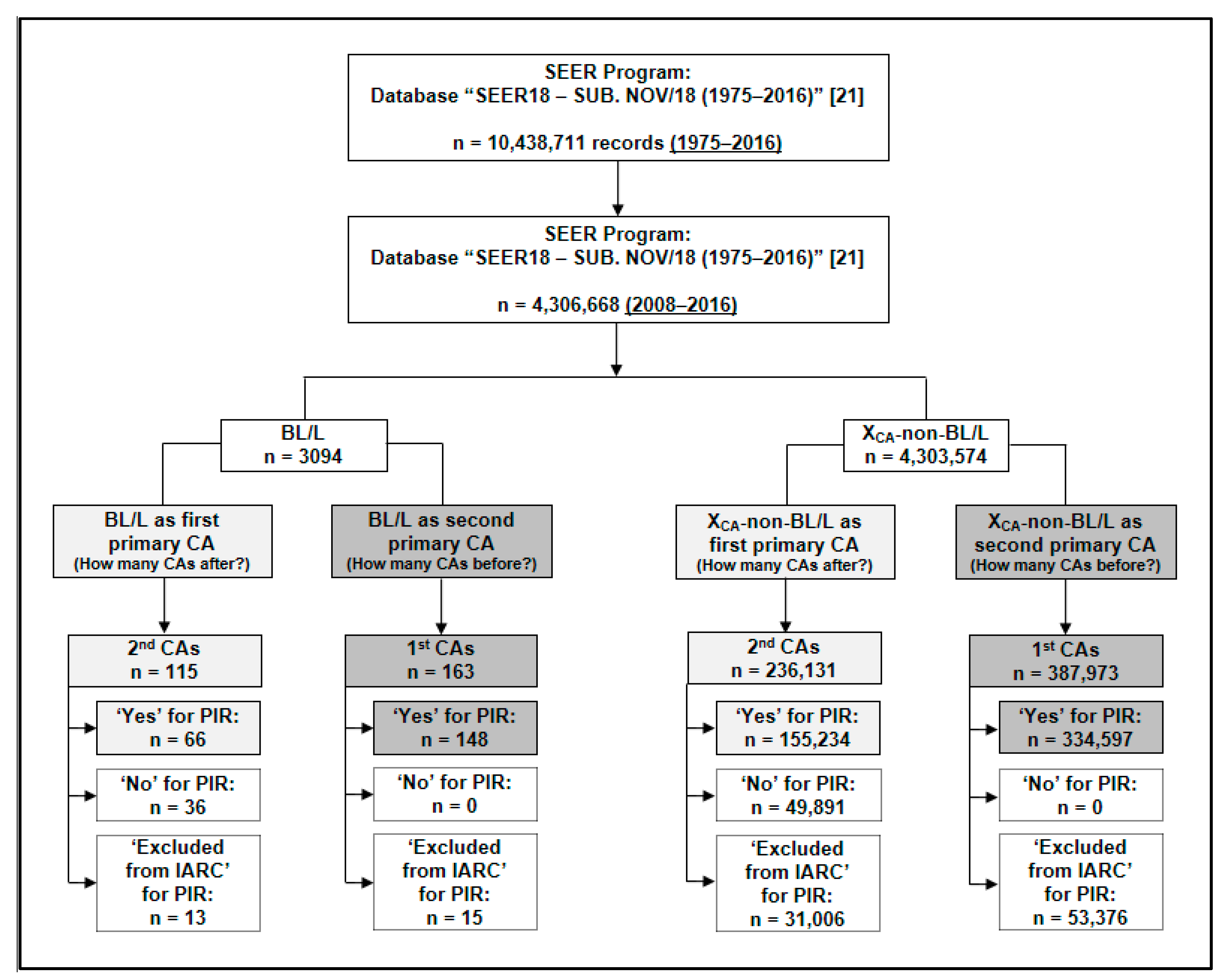
2.1. Study Design, Data Source and Cohort Selection

This was an observational, analytical and retrospective cohort study. Data were collected from the SEER Program database (submitted data in November 2018, with information available in April 2019) [21]. The incidence (rates crude) was estimated using the SEER Program database [22], which considered the Katrina/Rita population adjustment. SEER*Stat v.8.3.5 [23] was used to collect the data and incidence calculation. Patients diagnosed with BL/L between 2008 and 2016 according to the WHOc 2008 (or later version) were included in the descriptive, prognostic factors and survival analyses (n = 3094). For risk factors analyses, patients with any cancer other than BL/L in this same date restriction were also included (n = 4,303,574). Specifically for the incidence calculation, the total population used by SEER*Stat was of 786,813,193 and the displayed rate was cases per 1,000,000.

2.2. Variables

The variables were extracted as follows: age at diagnosis, Ann Arbor staging, chemotherapy, histology/behavior (International Classification of Diseases for Oncology, 3rd edition: ICD-O-3), origin, primary site, primary cancer by international rules (PIR), race, radiotherapy, record number, sex, site (ICD-O-3/WHO 2008), surgery of primary site, survival (months), vital status and year of diagnosis. The definition of the variables according to the specific document from the SEER Program [24] is available in the Supplementary Materials, Data S1. Four age ranges were defined by the authors (0–19, 20–39, 40–59 and ≥ 60 years old). The variables of the primary site, surgery of the primary site and radiotherapy were regrouped according to their similarities and the number of events (Supplementary Materials, Data S2).

2.3. Statistical Analyses

All statistical analyses were performed using SPSS® v.20 software (IBM®, Chicago, IL, USA). The risk relative (RR) calculation of the bivariate analysis was performed using BioEstat® v.5.3 software (Pará, Brazil; Maryland, IL, USA). Values of p < 0.05 were considered statistically significant for all analyses and point estimates are shown along with the 95% confidence interval (CI).

2.3.1. Descriptive Data

Descriptive statistical analyses were performed. The Kolmogorov–Smirnov test showed the non-normal distribution of the majority of the continuous variables, which were reported as the median, interquartile ranges (IQR—25%; 75%) and minimum and maximum values (range). The normally-distributed continuous variables were reported as the mean, standard deviation (SD) and standard error (SE). The categorical variables were reported as their absolute and relative frequency.

2.3.2. Prognostic Factors and Survival Analyses

The Kaplan–Meier method was performed to estimate the survival of the patients. The outcome was the overall survival (OS) (dependent variable), represented through the ‘vital status’ variable. The mean OS survival time (in months) was estimated by the interval between the date of the BL/L diagnosis until the date of the patients’ death (due to this disease or other causes) or right censorship (patients who remained alive with or without BL/L until the end of the follow-up). The statistical comparison of the Kaplan–Meier curves at different moments of the study follow-up was assessed through Breslow (generalized Wilcoxon), Tarone–Ware and log rank (Mantel–Cox) tests.
A time-dependent covariate Cox regression model (with bivariate and multivariate analyses) was used to investigate the prognostic factors associated with the OS. This model type was selected due to the lack of proportionality observed in the survival curves (e.g., crossing) from the Kaplan–Meier graphs [25]. The size of the effects of the prognostic factors was quantified through the hazard ratio (HR). The Enter method was applied as a statistical model.

2.3.3. Risk Factor Analyses for the Development of Second Primary Cancers (SPCs)

The variable ‘record number’ was chosen for the risk factor analysis. This variable sequentially numbers the tumors of a person, which has its ordered values based on the date of diagnosis and then the sequence number [24]. Only cancer numbers 1 (first cancer) and 2 (second cancer) were chosen for the ‘cases’ (BL/L) and ‘controls’ (any cancer other than BL/L) to avoid the influence of other cancers that occurred in different orders in the lifetime of a patient.
The International Agency for Research on Cancer (IARC) defines primary cancer as the one that originates in a primary site or tissue and is not an extension nor a recurrence nor a metastasis [26]. In our analysis, aiming only to evaluate primary cancers, the SEER variable called ‘primary cancer by international rules’ (PIR), which was created using IARC multiple primary rules, was used. This variable contains the category ‘excluded from IARC’, which excluded from the IARC multiple primary algorithm due to behavior. As the PIR variable did not include benign tumors or non-bladder in situ tumors in the algorithm, the ‘excluded from IARC’ category was not included in our analyses. In the SEER Research Data Record Description document, there is a note informing that for bladder tumors only, all in situ are converted to invasive tumors [24].
We evaluated which first primary cancers acted as risk factors for BL/L occurrence as an SPC (‘cases’, n = 148; ‘controls’, n = 334,597) and also if BL/L (as a first primary cancer) behaved as a risk factor for the occurrence of other SPCs (‘cases’, n = 66; ‘controls’, n = 155,234). An SPC is defined as a new primary cancer that occurs in a person who has had cancer in the past and it may occur months or years after the original (primary) cancer was diagnosed and treated [27].
A bivariate analysis was performed through an RR calculation. Cancers with a p-value up to 0.25 were selected as independent variables for an interrelated primary cancer risk for BL/L and were included in the Poisson regression model. For a first primary cancer, the dependent variable was the presence or absence of BL/L as an SPC. On the other hand, for the risk of any other SPCs other than BL/L, the dependent variable was the presence or absence of BL/L as a first cancer. The independent variables were tested in different combinations to obtain the final model, which was built with the type of main effects and Wald statistics.

3. Results

3.1. Population Characteristics

A total of n = 3094 patients were diagnosed with BL/L in the USA from 2008 to 2016 (mean 343.8 ± 29.9 new cases per year). The estimated incidence of the disease was 3.9 cases per 1,000,000 inhabitants (SE, 0.1; 95% CI, 3.8–4.1) [22]. The median survival time was 19 months (IQR, 4.25–57.0; range, 0–107). During this follow-up, 1,907 (61.6%) patients remained alive. The median age at diagnosis was 45.0 years (IQR, 22.0–62.0; range, 0–97) with two peaks of BL/L incidence: 5–9 years (n = 231; 7.5%) and 45–49 years (n = 240; 7.8%) (see Supplementary Materials, Data S3 and Figures S1 and S2). According to the ICD-O-3 and the WHOc 2008, 2685 (86.8%) patients had BL (ICD-O-3: 9687/3), of whom 1849 (59.8%) had a nodal presentation and 836 (27.0%) had an extranodal presentation. Overall, 409 (13.2%) patients were classified as having BLK (ICD-O-3: 9826/3). Other characteristics are shown in Table 1.

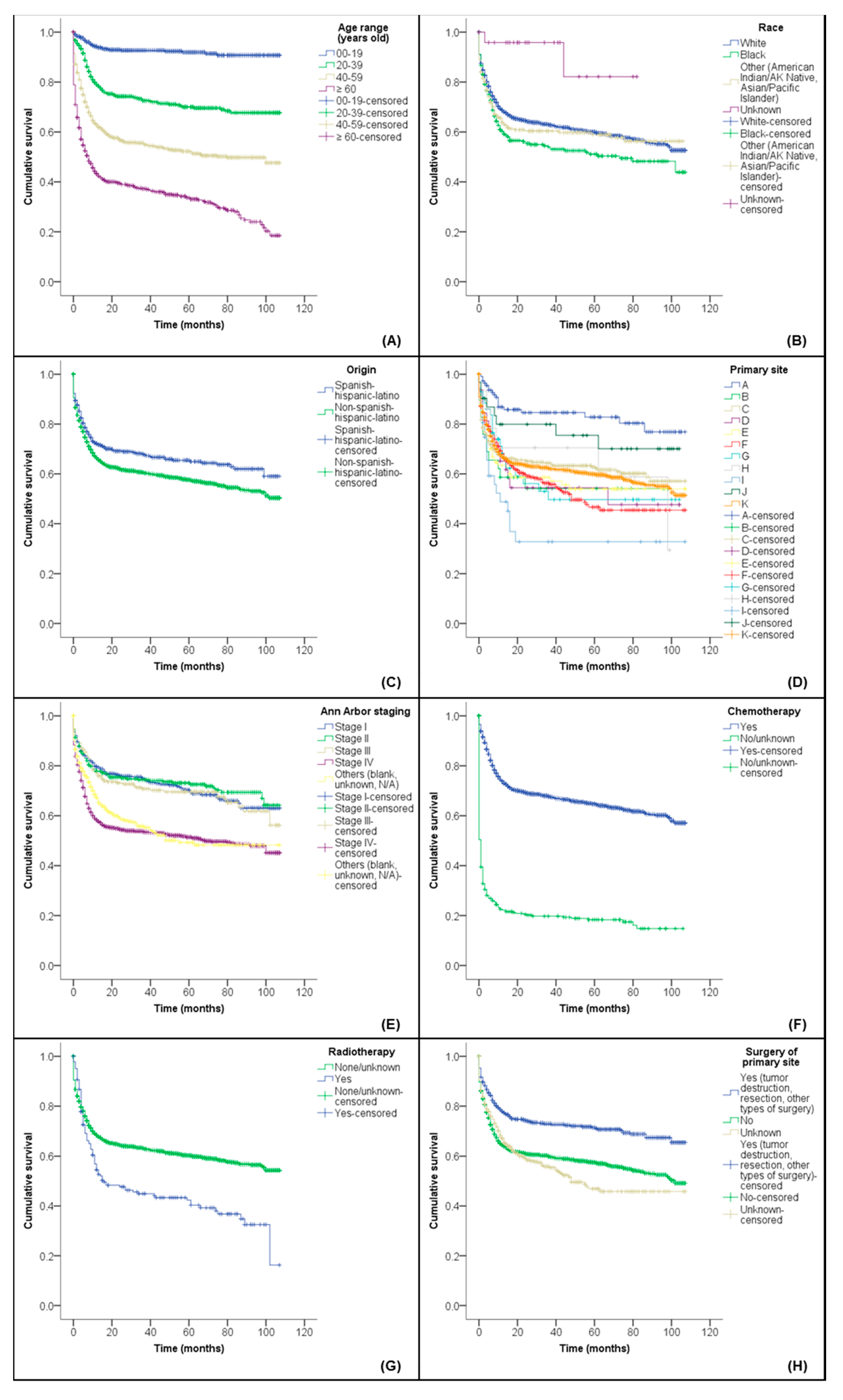
3.2. Influence of Prognostic Factors on the Overall Survival (OS)

A total of 10 out of 3094 patients were considered as missing by the SEER*Stat software. Thus, a survival analysis was performed with 3084 patients. In the 108 months of follow-up (2008 to 2016), a total of 1177 deaths (38.2%) for any cause occurred. The mean OS was 65.4 months (SE ± 0.9; 95% CI, 63.6–67.3). Figure 1 shows Kaplan–Meier graphs for the significant variables of the OS. See Supplementary Materials, Data S4 for the complete results of the Kaplan–Meier analysis (Table S2).
The time-dependent Cox model regression (OS bivariate analysis) showed the following factors associated with a worse prognosis for BL/L: population ageing, black race, disease in stage IV by Ann Arbor staging, no chemotherapy and no surgery on the primary site. No radiotherapy was associated with a good prognosis. Similar results were obtained with the multivariate analysis that additionally showed males associated with a worse OS prognosis when compared with females. In addition, the multivariate analysis showed that two primary sites (respiratory and cardiac systems and those parts related to the central nervous system: CNS) behaved as a worse prognosis for BL/L. All values of OS outcome (bivariate and multivariate analyses) are shown in Table 2.

3.3. Assessment of the Development Risk of Second Primary Cancers (SPCs)

Figure 2 shows the risk factor analysis flowchart. In the bivariate analysis, the first primary cancers that occurred before BL/L and behaved as risk factors for the BL/L occurrence as an SPC were anus, anal canal and anorectum tumors (RR, 4.2; 95% CI, 1.4–12.9; p = 0.017); Hodgkin lymphomas (HLs) (nodal) (RR, 9.9; 95% CI, 5.4–18.0; p < 0.001); Kaposi sarcomas (RR, 44.8; 95% CI, 23.6–85.0; p < 0.001); liver cancers (RR, 4.4; 95% CI, 1.7–11.5; p = 0.004) and trachea, mediastinum and other respiratory tumors (RR, 20.6; 95% CI, 2.9–146.2; p = 0.021). The median time until diagnosis of BL/L as an SPC was of five years (IQR, 2–9; range, 0–28).
BL/L behaved as a risk factor for the occurrence of the following SPCs: acute myeloid leukemia (AML) (RR, 9.9; 95% CI, 5.1–18.9; p < 0.001); anus, anal canal and anorectum tumors (RR, 6.4; 95% CI, 1.6–25.1; p = 0.017); HLs (extranodal) (RR, 168.0; 95% CI, 22.4–1259.4; p < 0.001) and Kaposi sarcomas (RR, 76.5; 95% CI, 29.1–201.0; p < 0.001). On the other hand, there was a decrease in the incidence of lung and bronchus cancer as an SPC (RR, 0.3; 95% CI, 0.1–0.8; p = 0.005). The median time until the presentation of other SPCs was of two years (IQR, 1–4; range, 0–8).
Lastly, the cancers that presented a p-value up to 0.25 in the bivariate analysis (see Supplementary Materials, Data S5) were tested for the risk cancers and were included in the Poisson regression model (see final model in Table 3).

4. Discussion

To our knowledge, this is the first population-based study that selected BL/L patients diagnosed with the most up-to-date WHOc [1,4,5], evaluated the prognostic factors associated with the OS and assessed what primary cancers were risk factors for the occurrence of BL/L as an SPC as well as if BL/L was a risk factor for the development of other SPCs. We found that BL/L patients had a mean OS of 65 months and a few factors as older age, advanced staging, no chemotherapy and radiotherapy were associated with a worse prognosis. Furthermore, we found that the main hematological cancers, Kaposi sarcomas and gastrointestinal tract (GIT) tumors were cancers with the risk of occurring before (BL/L as an SPC) or after (any cancer other than BL/L as an SPC) BL/L. Other population-based studies also using the SEER Program reported the occurrence of further malignancies after a previous cancer including prostate [28], breast [29] and chronic myeloid leukemia [30].
The international classification of BL was revised in 2008 and 2016. The cases initially classified as an atypical morphological variant of BL (‘Burkitt-like lymphoma’) in WHOc 2001 [3] were reallocated to the category DLBCL/BL in WHOc 2008 [4] and subsequently reclassified in WHOc 2016 as ‘high-grade B-cell lymphoma’. The latter may contain MYC and BCL2 and/or BCL6 rearrangements (“double-hit” or “triple-hit” lymphomas) and they usually have an aggressive clinical course and a worse prognosis than BL [1,5]. In our study, we restricted the date of the patient’s diagnosis (2008–2016) to prevent a misclassification of BL cases.

4.1. Prognostic Factors and Overall Survival (OS)

Our results showed that a few prognostic factors negatively impacted on the OS of BL/L patients. Aging was one of them. In the past decades, the significant increase in somatic mutations in animal and human cells damaged the genetic material of patients, resulting in a greater mortality from cancers at older ages including BL/L [31]. Studies showed that BL/L patients diagnosed <19 years old have a better prognosis (OS 5 years, 87%) than patients >60 years old or with an advanced tumor (OS 5 years, around 33%) [16,32].
Blacks were also associated with a worse prognosis for OS leading to a greater risk of dying from BL/L, which may be explained due to socioeconomic features. Wang et al. (2008) studied elderly patients with NHL and highlighted that African-Americans presented a lower educational level, income and socioeconomic status than Caucasian patients. Consequently, the proportion of patients on chemotherapy was lower among blacks and a poorer socioeconomic status was significantly associated with a higher risk of mortality [33]. However, a few studies have shown that a poorer prognosis regardless of the socioeconomic status would involve a host susceptibility factor, as observed in eBL in Africa [15].
Almost half of the patients in our study presented stage IV BL. Liu et al. (2019) showed the difficulty in finding BL cases at earlier stages (I and II) [34]. In our study, almost 60% of BL patients had a nodal presentation. A Chinese study reported sBL patients with a greater disease involvement at extranodal sites [35] while a Spanish study found lymph nodes to be the most frequently involved areas [36]. Similar epidemiological studies demonstrated that a nodal presentation in BL is commonplace in the American population [15,34]. These differences may have occurred due to the influence of the patient’s race, ethnicity, economic conditions and origin [37].
The bivariate analysis of the time-dependent covariate Cox regression showed the involvement of certain primary sites at diagnosis by BL/L as being statistically associated with a worse prognosis. The GIT was the second most frequently involved extranodal site as a primary diagnostic site while skin, breasts, the CNS and glands had less than 5% involvement each. sBL has an important abdominal involvement (affecting up to 5% of GIT NHL cases in the North American population) followed by the head and neck, nose, oropharynx and tonsils [7,38].
Patients not treated with chemotherapy or those who underwent radiotherapy had higher risks of death. Chemotherapy is the standard BL treatment [32]. Prophylactic CNS irradiation appears effective in acute lymphocytic leukemia and BL but is associated with a high toxicity, causing secondary malignant neoplasms and neuropsychological sequelae [39]. Surgery is not usually performed for this disease except when disease complications (e.g., intestinal obstruction) occur [32].

4.2. Risk of Second Primary Cancers (SPCs)

NHL comprises of a heterogeneous group of hematological diseases varying according to their biological characteristics. Each NHL subtype may have a different treatment, prognosis, clinical response and survival. Consequently, the risk for developing other cancers may also differ between these subtypes [1,17,40]. In both the bivariate and Poisson regression analyses, we found that anus, anal canal and anorectum tumors, HLs (nodal), Kaposi sarcomas, liver cancers and trachea, mediastinum and other respiratory cancers behaved as risk factors for the occurrence of BL/L as an SPC. One hypothesis is that these cancers involve sites that usually are affected as nodal or extranodal primary sites of BL [7]. On the other hand, BL/L behaved as a risk factor for the development of other SPCs such as AMLs, HLs (extranodal) and Kaposi sarcomas in the Poisson regression analysis. In addition, anus, anal canal and anorectum tumors presented a significant association with BL/L in the bivariate analysis.
There was an overall SPC risk 25% higher in patients who had a first NHL compared with the reference population. However, confounding factors such as age, sex, staging, follow-up time, number of chemotherapy agents and radiotherapy sessions might influence this outcome [17]. In addition, hormonal status and environmental and genetic factors may modify the risk of developing a second cancer [41] and should be considered during data analyses. AML occurrence as an SPC may be a late complication of cytotoxic therapy called ‘therapy-related myeloid neoplasm’ (t-MN). Alkylating agents (e.g., cyclophosphamide) and topoisomerase II inhibitors (e.g., etoposide) are chemotherapy agents that cause t-MN [19]. Kaposi sarcomas and BL/L have a high rate of incidence in HIV/AIDS individuals. Despite the change in pattern of secondary cancers after a Kaposi sarcoma in the era of antiretroviral therapy, NHL remains one of the most common [42,43].
We found that lung and bronchus cancers could decrease the occurrence of BL/L as an SPC as well as BL/L as a first primary cancer in the patient’s life decreased the incidence of these tumors and breast cancer as SPCs. These results should be interpreted with caution especially because our analysis did not consider the confounding factors. However, a recent study that investigated SPCs in breast cancer showed that the standardized incidence ratios in patients with chemotherapy were higher in all malignancies except lymphoma, myeloma and chronic lymphocytic leukemia [44].

4.3. Limitations

Our study has a few limitations. First, we cannot be absolutely sure that all BL/L patients in our study were diagnosed based on WHOc 2008 (or a later version) because this information was not available. With the date cutoff applied in the sample selection (2008–2016), we could infer that the most recent version of the WHOc may have been used. Second, the search date cutoff restricted the risk factor analysis to cancers that occurred after BL/L (that is, any cancer other than BL/L as an SPC). Third, the radiotherapy and chemotherapy variables were described according to the SEER*Stat as ‘No/Unknown’. The lack of descriptive information on these variables hampered further analyses. Fourth, information regarding aspects of treatment (e.g., drug name, radiation site), disease (e.g., relapse) and comorbidities (e.g., HIV) was not available; thus, other analyses including the prognosis of patients according to the type of treatment, evaluation of malignancies in patients with HIV (e.g., Kaposi sarcoma) or the distinction between the epidemiological clinical variants of BL could not be performed. Lastly, although our risk factor analysis presented several characteristics of a case-control study, factor and patient characteristics were not paired.

5. Conclusions

In conclusion, population ageing, black race, advanced staging, no chemotherapy, radiotherapy and no surgery were associated with a worse prognosis in BL/L, significantly decreasing the OS. First primary cancers that behaved as risk factors for the development of BL/L as an SPC were aleukemic, subleukemic and NOS tumors; anus, anal canal and anorectum tumors; HLs (nodal); Kaposi sarcomas; liver cancers and trachea, mediastinum and other respiratory organs cancers. On the other hand, lung and bronchus cancers decreased the incidence. At the same time, BL/L acted as a risk factor for the occurrence of other SPCs such as AML, HL (extranodal) and Kaposi sarcomas. On the other hand, a decreased incidence was observed for breast cancer and lung/bronchus tumors as SPCs. These results may assist the development of diagnostic and clinical recommendations in this field and guide the conduction of further studies on risk factors for hematological malignancies.

Supplementary Materials

The following are available online at https://www.mdpi.com/article/10.3390/diseases9020043/s1, containing the items: Data S1. Definition of the variables (with Table S1); Data S2. Variables with their groups regrouped (with Data S2.1, Data S2.2 and Data S2.3); Data S3. Population characteristics of BL/L patients (with Figures S1 and S2); Data S4. Kaplan–Meier results (with Table S2); and Data S5. Bivariate analysis of risk factors of second primary cancer on BL/L (with Table S3).

Author Contributions

Conceptualization: A.M.D.R., F.S.T., G.S.-D., J.A.G.D.M., L.L., R.P. Data curation: A.M.D.R., M.M.F., L.L. Formal analysis: A.M.D.R., M.M.F., A.F.C., V.L.F., L.P.L., L.L. Methodology: A.M.D.R., F.S.T., G.S.-D., J.A.G.D.M., L.L., R.P. Project administration: L.L., R.P. Visualization: A.M.D.R., M.M.F. Writing—original draft: A.M.D.R., F.S.T., M.M.F., A.F.C., V.L.F., L.P.L. Writing—review and editing: A.M.D.R., F.S.T., M.M.F., A.F.C., V.L.F., L.P.L., G.S.-D., J.A.G.D.M., L.L., R.P. All authors have read and agreed to the published version of the manuscript.

Funding

This study was financed in part by the Coordenação de Aperfeiçoamento de Pessoal de Nível Superior, Brasil (CAPES), Finance Code 001.

Institutional Review Board Statement

Ethical review and approval were waived for this study because the SEER Program provides secondary and open databases.

Informed Consent Statement

Patient consent was waived because the SEER Program provides secondary and open databases.

Data Availability Statement

The data used in this study are available to the public in the references listed [21,22,23,24].

Acknowledgments

None.

Conflicts of Interest

The authors declare no conflict of interest.

References

  1. Swerdlow, S.H.; Campo, E.; Harris, N.L.; Jaffe, E.S.; Pileri, S.A.; Stein, H.; Thiele, J. WHO Classification of Tumors of Haematopoietic and Lymphoid Tissues, 4th ed.; International Agency for Research on Cancer (IARC): Lyon, France, 2017; pp. 330–341. [Google Scholar]
  2. PathologyOutlines.com. Lymphoma & Related Disorders-General-WHO Classification-B Cell. Available online: https://www.pathologyoutlines.com/topic/lymphomabcellwho.html (accessed on 5 March 2021).
  3. Jaffe, E.S.; Harris, N.L.; Stein, H.; Vardiman, J.W. WHO Classification of Tumours-Pathology and Genetics of Tumours of Haematopoietic and Lymphoid Tissues, 3rd ed.; International Agency for Research on Cancer (IARC): Lyon, France, 2001; pp. 181–184. [Google Scholar]
  4. Swerdlow, S.H.; Campo, E.; Harris, N.L.; Jaffe, E.S.; Pileri, S.A.; Stein, H.; Thiele, J.; Vardiman, J.W. WHO Classification of Tumours of Haematopoietic and Lymphoid Tissues, 4th ed.; International Agency for Research on Cancer (IARC): Lyon, France, 2008; pp. 262–266. [Google Scholar]
  5. Swerdlow, S.H.; Campo, E.; Pileri, S.A.; Harris, N.L.; Stein, H.; Siebert, R.; Advani, R.; Ghielmini, M.; Salles, G.A.; Zelenetz, A.D.; et al. The 2016 revision of the World Health Organization classification of lymphoid neoplasms. Blood 2016, 127, 2375–2390. [Google Scholar] [CrossRef] [Green Version]
  6. Dunleavy, K.; Little, R.F.; Wilson, W.H. Update on Burkitt Lymphoma. Hematol. Oncol. Clin. N. Am. 2016, 30, 1333–1343. [Google Scholar] [CrossRef] [PubMed]
  7. Molyneux, E.M.; Rochford, R.; Griffin, B.; Newton, R.; Jackson, G.; Menon, G.; Harrison, C.J.; Israels, T.; Bailey, S. Burkitt’s lymphoma. Lancet 2012, 379, 1234–1244. [Google Scholar] [CrossRef] [Green Version]
  8. Patte, C.; Auperin, A.; Gerrard, M.; Michon, J.; Pinkerton, R.; Sposto, R.; Weston, C.; Raphael, M.; Perkins, S.L.; McCarthy, K.; et al. Results of the randomized international FAB/LMB96 trial for intermediate risk B-cell non-Hodgkin lymphoma in children and adolescents: It is possible to reduce treatment for the early responding patients. Blood 2007, 109, 2773–2780. [Google Scholar] [CrossRef] [Green Version]
  9. Gerrard, M.; Cairo, M.S.; Weston, C.; Auperin, A.; Pinkerton, R.; Lambilliote, A.; Sposto, R.; McCarthy, K.; Lacombe, M.J.; Perkins, S.L.; et al. Excellent survival following two courses of COPAD chemotherapy in children and adolescents with resected localized B-cell non-Hodgkin’s lymphoma: Results of the FAB/LMB 96 international study. Br. J. Haematol. 2008, 141, 840–847. [Google Scholar] [CrossRef] [PubMed]
  10. Jacobson, C.; LaCasce, A. How I treat Burkitt lymphoma in adults. Blood 2014, 124, 2913–2920. [Google Scholar] [CrossRef] [Green Version]
  11. Armitage, J.O.; Gascoyne, R.D.; Lunning, M.A.; Cavalli, F. Non-Hodgkin lymphoma. Lancet 2017, 390, 298–310. [Google Scholar] [CrossRef]
  12. Hesseling, P.B.; Molyneux, E.; Tchintseme, F.; Welbeck, J.; McCormick, P.; Pritchard-Jones, K.; Wagner, H.P. Treating Burkitt’s lymphoma in Malawi, Cameroon, and Ghana. Lancet Oncol. 2008, 9, 512–513. [Google Scholar] [CrossRef]
  13. Depani, S.; Banda, K.; Bailey, S.; Israels, T.; Chagaluka, G.; Molyneux, E. Outcome is unchanged by adding vincristine upfront to the Malawi 28-day protocol for endemic Burkitt lymphoma. Pediatr. Blood Cancer 2015, 62, 1929–1934. [Google Scholar] [CrossRef] [PubMed]
  14. Nature Communications Editors. Epidemiology is a science of high importance. Nat. Commun. 2018, 9, 1703. [Google Scholar] [CrossRef] [Green Version]
  15. Castillo, J.J.; Winer, E.S.; Olszewski, A.J. Population-based prognostic factors for survival in patients with Burkitt lymphoma: An analysis from the Surveillance, Epidemiology, and End Results database. Cancer 2013, 119, 3672–3679. [Google Scholar] [CrossRef] [PubMed]
  16. Costa, L.J.; Xavier, A.C.; Wahlquist, A.E.; Hill, E.G. Trends in survival of patients with Burkitt lymphoma/leukemia in the USA: An analysis of 3691 cases. Blood 2013, 121, 4861–4866. [Google Scholar] [CrossRef] [PubMed] [Green Version]
  17. Rossi, C.; Jegu, J.; Mounier, M.; Dandoit, M.; Colonna, M.; Daubisse-Marliac, L.; Tretarre, B.; Ganry, O.; Guizard, A.V.; Bara, S.; et al. Risk assessment of second primary cancer according to histological subtype of non-Hodgkin lymphoma. Leuk. Lymphoma 2015, 56, 2876–2882. [Google Scholar] [CrossRef] [PubMed]
  18. Pirani, M.; Marcheselli, R.; Marcheselli, L.; Bari, A.; Federico, M.; Sacchi, S. Risk for second malignancies in non-Hodgkin’s lymphoma survivors: A meta-analysis. Ann. Oncol. 2011, 22, 1845–1858. [Google Scholar] [CrossRef] [PubMed]
  19. McNerney, M.E.; Godley, L.A.; Le Beau, M.M. Therapy-related myeloid neoplasms: When genetics and environment collide. Nat. Rev. Cancer 2017, 17, 513–527. [Google Scholar] [CrossRef]
  20. Surveillance, Epidemiology, and End Results (SEER) Program. National Cancer Institute. Overview of the SEER Program. Available online: https://seer.cancer.gov/about/overview.html (accessed on 4 August 2019).
  21. Surveillance, Epidemiology, and End Results (SEER) Program (www.seer.cancer.gov) SEER*Stat Database: Incidence-SEER 18 Regs Custom Data (with Additional Treatment Fields), Nov 2018 Sub (1975–2016 Varying)-Linked To County Attributes-Total U.S., 1969-2017 Counties, National Cancer Institute, DCCPS, Surveillance Research Program, Released April 2019, Based on the November 2018 Submission. Available online: https://seer.cancer.gov/seerstat/ (accessed on 29 April 2019).
  22. Surveillance, Epidemiology, and End Results (SEER) Program (www.seer.cancer.gov) SEER*Stat Database: Incidence-SEER 18 Regs Custom Data (with Additional Treatment Fields), Nov 2018 Sub (2000–2016) <Katrina/Rita Population Adjustment>-Linked to County Attributes-Total U.S., 1969–2017 Counties, National Cancer Institute, DCCPS, Surveillance Research Program, Released April 2019, Based on the November 2018 Submission. Available online: https://seer.cancer.gov/seerstat/ (accessed on 15 November 2019).
  23. Surveillance, Epidemiology, and End Results (SEER) Program. National Cancer Institute. SEER*Stat Software-Latest Release: Version 8.3.5-6 March 2018. Available online: https://seer.cancer.gov/seerstat/ (accessed on 19 February 2019).
  24. Surveillance, Epidemiology, and End Results (SEER) Program. National Cancer Institute. Documentation for the ASCII Text Data Files. 2019. Available online: https://seer.cancer.gov/data-software/documentation/seerstat/nov2018/TextData.FileDescription.pdf (accessed on 22 May 2019).
  25. Schemper, M.; Wakounig, S.; Heinze, G. The estimation of average hazard ratios by weighted Cox regression. Stat. Med. 2009, 28, 2473–2489. [Google Scholar] [CrossRef]
  26. International Agency for Research on Cancer (IARC), World Health Organization (WHO), International Association of Cancer Registries (IACR), European Network of Cancer Registrier (ENCR). International Rules for Multiple Primary Cancers (ICD-O Third Edition). Internal Report No. 2004/02. Available online: http://www.iacr.com.fr/images/doc/MPrules_july2004.pdf (accessed on 5 March 2021).
  27. National Cancer Institute (NCI). NIH. NCI Dictionary of Cancer Terms. Available online: https://www.cancer.gov/publications/dictionaries/cancer-terms (accessed on 29 October 2019).
  28. Mazzone, E.; Mistretta, F.A.; Knipper, S.; Palumbo, C.; Tian, Z.; Pecoraro, A.; Preisser, F.; Gallina, A.; Shariat, S.F.; Saad, F.; et al. Long-term incidence of secondary bladder and rectal cancer in patients treated with brachytherapy for localized prostate cancer: A large-scale population-based analysis. BJU Int. 2019, 124, 1006–1013. [Google Scholar] [CrossRef]
  29. Malmgren, J.A.; Calip, G.S.; Pyott, S.M.; Atwood, M.K.; Kaplan, H.G. Therapy-related myelodysplastic syndrome following primary breast cancer. Leuk. Res. 2016, 47, 178–184. [Google Scholar] [CrossRef]
  30. Jamy, O.; Sarmad, R.; Costa, L. Risk and outcomes of second malignant neoplasms in chronic myeloid leukemia survivors. Leuk. Res. 2019, 82, 1–6. [Google Scholar] [CrossRef]
  31. DePinho, R.A. The age of cancer. Nature 2000, 408, 248–254. [Google Scholar] [CrossRef]
  32. Kalisz, K.; Alessandrino, F.; Beck, R.; Smith, D.; Kikano, E.; Ramaiya, N.H.; Tirumani, S.H. An update on Burkitt lymphoma: A review of pathogenesis and multimodality imaging assessment of disease presentation, treatment response, and recurrence. Insights Imaging 2019, 10, 56. [Google Scholar] [CrossRef] [PubMed]
  33. Wang, M.; Burau, K.D.; Fang, S.; Wang, H.; Du, X.L. Ethnic variations in diagnosis, treatment, socioeconomic status, and survival in a large population-based cohort of elderly patients with non-Hodgkin lymphoma. Cancer 2008, 113, 3231–3241. [Google Scholar] [CrossRef]
  34. Liu, Z.L.; Liu, P.P.; Bi, X.W.; Lei, D.X.; Wang, Y.; Li, Z.M.; Jiang, W.Q.; Xia, Y. Trends in survival of patients with stage I/II Burkitt lymphoma in the United States: A SEER database analysis. Cancer Med. 2019, 8, 874–881. [Google Scholar] [CrossRef] [PubMed]
  35. Ye, Z.; Xiao, Y.; Shi, H.; Ke, Z.; Liu, Y.; Liang, Y.; Han, A. Sporadic Burkitt lymphoma in southern China: 12 years’ experience in a single institution in Guangzhou. J. Clin. Pathol. 2011, 64, 1132–1135. [Google Scholar] [CrossRef]
  36. Carrillo-Cruz, E.; Marin-Oyaga, V.A.; Sole Rodriguez, M.; Borrego-Dorado, I.; de la Cruz Vicente, F.; Quiroga Cantero, E.; Manzanares Perez, M.; Capote, F.J.; Ramirez Sanchez, M.J.; Espigado Tocino, I.; et al. Role of 18F-FDG-PET/CT in the management of Burkitt lymphoma. Eur. J. Haematol. 2015, 94, 23–30. [Google Scholar] [CrossRef] [PubMed]
  37. Schwartz, K.L.; Crossley-May, H.; Vigneau, F.D.; Brown, K.; Banerjee, M. Race, socioeconomic status and stage at diagnosis for five common malignancies. Cancer Causes Control 2003, 14, 761–766. [Google Scholar] [CrossRef]
  38. Howell, J.M.; Auer-Grzesiak, I.; Zhang, J.; Andrews, C.N.; Stewart, D.; Urbanski, S.J. Increasing incidence rates, distribution and histological characteristics of primary gastrointestinal non-Hodgkin lymphoma in a North American population. Can. J. Gastroenterol. 2012, 26, 452–456. [Google Scholar] [CrossRef]
  39. Hill, Q.A.; Owen, R.G. CNS prophylaxis in lymphoma: Who to target and what therapy to use. Blood Rev. 2006, 20, 319–332. [Google Scholar] [CrossRef]
  40. Han, X.; Kilfoy, B.; Zheng, T.; Holford, T.R.; Zhu, C.; Zhu, Y.; Zhang, Y. Lymphoma survival patterns by WHO subtype in the United States, 1973-2003. Cancer Causes Control 2008, 19, 841–858. [Google Scholar] [CrossRef]
  41. Allan, J.M.; Travis, L.B. Mechanisms of therapy-related carcinogenesis. Nat. Rev. Cancer 2005, 5, 943–955. [Google Scholar] [CrossRef]
  42. Mukhtar, F.; Ilozumba, M.; Utuama, O.; Cimenler, O. Change in Pattern of Secondary Cancers After Kaposi Sarcoma in the Era of Antiretroviral Therapy. JAMA Oncol. 2018, 4, 48–53. [Google Scholar] [CrossRef] [PubMed]
  43. Jiamsakul, A.; Polizzotto, M.; Wen-Wei Ku, S.; Tanuma, J.; Hui, E.; Chaiwarith, R.; Kiertiburanakul, S.; Avihingasanon, A.; Yunihastuti, E.; Kumarasamy, N.; et al. Brief Report: Malignancies in Adults Living With HIV in Asia. J. Acquir. Immune Defic. Syndr. 2019, 80, 301–307. [Google Scholar] [CrossRef] [PubMed]
  44. Wei, J.L.; Jiang, Y.Z.; Shao, Z.M. Survival and chemotherapy-related risk of second primary malignancy in breast cancer patients: A SEER-based study. Int. J. Clin. Oncol. 2019, 24, 934–940. [Google Scholar] [CrossRef] [PubMed]
Figure 1. Cumulative survival probability from BL/L according to (A) age range, (B) race, (C) origin, (D) primary site, (E) Ann Arbor staging, (F) chemotherapy, (G) radiotherapy and (H) surgery of the primary site. Abbreviations: BL/L, Burkitt lymphoma/leukemia; CNS, central nervous system; GIT, gastrointestinal tract; N/A, not applicable. Notes: all variables were significant (p-value < 0.05) for all three statistical tests (Breslow (Generalized Wilcoxon), Tarone–Ware and log rank (Mantel–Cox)) applied in the analyses. The captions for the ‘primary site’ variable groups (Figure 1D) are as follows: A, lip, oral cavity and pharynx; B, unknown primary site and other and ill-defined sites; C, GIT and attachments; D, respiratory system and cardiac system and attachments; E, skin, bones, joints, articular cartilage and other tissues/sites; F, hematopoietic and reticuloendothelial systems; G, breast and female/male genital organs; H, urinary system; I, eye, adnexa, brain and other parts of the CNS; J, glands (thyroid and adrenal); K, lymph nodes.
Diseases 09 00043 g001
Figure 2. Data collection flowchart for the risk analysis of primary cancers before and after BL/L with BL/L behaving as an SPC and any cancer other than BL/L behaving as an SPC, respectively. Abbreviations: BL/L, Burkitt lymphoma/leukemia; CA, cancer; IARC, International Agency for Research on Cancer; PIR, primary cancer by international rules; SEER, Surveillance, Epidemiology and End Results; SPC, second primary cancer; XCA-non-BL/L, any cancer other than Burkitt lymphoma/leukemia.
Diseases 09 00043 g002
Table 1. Sociodemographic, clinical and treatment characteristics of patients with BL/L.
Characteristicsn (%)
(Total: n = 3094)
Sociodemographic characteristics
Age range (years old)
00–19702 (22.7)
20–39633 (20.5)
40–59887 (28.7)
≥60872 (28.2)
Sex
Male2255 (72.9)
Female839 (27.1)
Race
White2458 (79.4)
Black326 (10.5)
Other (American Indian/Alaska Native, Asian/Pacific Islander)284 (9.2)
Unknown26 (0.8)
Origin
Non-Spanish-Hispanic-Latino2451 (79.2)
Spanish-Hispanic-Latino643 (20.8)
Clinical characteristics
Primary site 1
Lymph nodes1798 (58.1)
Hematopoietic and reticuloendothelial systems467 (15.1)
GIT and attachments433 (14.0)
Lip, oral cavity and pharynx112 (3.6)
Skin, bones, joints, articular cartilage and other tissues/sites98 (3.2)
Breast and female/male genital organs43 (1.4)
Eye, adnexa, brain and other parts of the CNS36 (1.2)
Respiratory system and cardiac system and attachments32 (1.0)
Glands (thyroid and adrenal)31 (1.0)
Urinary system15 (0.5)
Unknown primary site and other and ill-defined sites29 (0.9)
Ann Arbor staging 2
Stage I448 (14.5)
Stage II373 (12.1)
Stage III270 (8.7)
Stage IV1248 (40.3)
Others (Blank, Unknown, N/A)755 (24.4)
Treatment characteristics
Chemotherapy
No/Unknown389 (12.6)
Yes2705 (87.4)
Radiotherapy 1
No/Unknown2912 (94.1)
Yes182 (5.9)
Surgery of primary site 1
No1973 (63.8)
Yes (tumor destruction, resection, other types of surgery)614 (19.8)
Unknown507 (16.4)
Abbreviations: BL/L, Burkitt lymphoma/leukemia; CNS, central nervous system; GIT, gastrointestinal tract; N/A, not applicable. Notes: 1 Variable regrouped according to the similarities and number of events in each one of the groups (Supplementary Materials, Data S2). 2 Cases diagnosed until the year 2015. The background color was used to highlight the main group characteristics, while the bold highlighted the titles and subtitles.
Table 2. Bivariate and multivariate analyses by a time-dependent covariate Cox regression model.
CharacteristicsOverall Survival
Bivariate AnalysisMultivariate AnalysisDeaths (%)
HR95% CIp-Value *HR95% CIp-Value *
Sociodemographic characteristics
Age range (years old)
00–191.0--1.0--7.0
20–393.92.9–5.4<0.0013.52.5–4.8<0.00126.2
40–597.75.6–10.4<0.0016.14.5–8.4<0.00145.2
≥6014.010.2–19.3<0.00110.57.5–14.5<0.00164.9
Sex
Male1.0--1.0--37.1
Female1.10.9–1.20.3900.90.8–1.00.02840.9
Race
White1.0--1.0--37.4
Black1.41.1–1.60.0011.61.3–1.9<0.00145.8
Other 11.10.9–1.30.4631.10.9–1.30.50838.9
Unknown0.20.1–0.80.0250.20.1–0.80.0247.7
Origin
Spanish-Hispanic-Latino1.0--1.0--31.1
Non-Spanish-Hispanic-Latino1.21.0–1.40.0141.00.8–1.10.59240.0
Clinical characteristics
Histology/Behavior (ICD-O-3)
9687/3: Burkitt lymphoma1.0--1.0--38.0
9826/3: Burkitt cell leukemia1.11.0–1.40.1100.80.5–1.30.34639.1
Primary site 2
Sites in lip, oral cavity and pharynx1.0--1.0--17.0
GIT and attachments2.41.5–3.8<0.0011.50.9–2.40.10235.6
Respiratory system and cardiac system (with attachments)3.11.6–6.20.0012.11.1–4.20.02846.9
Skin, bones, joints, articular cartilage and other tissues/sites2.71.6–4.7<0.0011.60.9–2.70.09642.9
Hematopoietic and reticuloendothelial systems3.01.9–4.8<0.0010.80.2–3.10.69941.3
Breast and female/male genital organs2.51.3–4.70.0051.50.8–2.80.22444.2
Urinary system2.10.8–5.20.1231.10.4–2.70.89940.0
Eye, adnexa, brain and other parts of the CNS4.72.5–8.7<0.0012.21.2–4.20.01558.3
Glands (thyroid and adrenal)1.10.5–2.60.7890.80.4–1.90.64225.8
Lymph nodes2.41.6–3.9<0.0011.40.9–2.30.12738.4
Unknown primary site and other and ill-defined sites3.21.6–6.50.0010.50.1–2.30.38244.8
Ann Arbor staging 3
Stage I1.0--1.0--29.8
Stage II0.90.7–1.20.6641.10.8–1.40.56427.3
Stage III1.10.8–1.40.5921.20.9–1.60.14031.5
Stage IV1.91.5–2.3<0.0011.81.5–2.2<0.00148.0
Others (Blank, Unknown, N/A)1.71.4–2.1<0.0011.51.1–2.00.00434.6
Treatment characteristics
Chemotherapy
Yes1.0--1.0--32.5
No/Unknown4.94.3–5.7<0.0014.03.5–4.6<0.00178.4
Radiotherapy 2
Yes1.0--1.0--58.2
No/Unknown0.70.6–0.8<0.0010.70.6–0.90.00136.9
Surgery of primary site 2
Yes (tumor destruction, resection, other types of surgery)1.0--1.0--27.2
No1.61.3–1.9<0.0011.41.1–1.60.00140.7
Unknown1.81.5–2.2<0.0013.51.0–12.80.05841.8
Abbreviations: CI, confidence interval; CNS, central nervous system; GIT, gastrointestinal tract; ICD-O-3, International Classification of Diseases for Oncology, 3rd edition; HR, hazard ratio; N/A, not applicable. Notes: * p-value from the enter statistic test. 1 American Indian/AK Native, Asian/Pacific Islander. 2 Variable regrouped according to the similarities and number of events in each one of the groups (see Supplementary Materials, Data S2). 3 Cases diagnosed until 2015. The background color was used to highlight the main group characteristics, while the bold highlighted the titles, subtitles and significant p-values.
Table 3. Poisson regression final model of interrelated cancer risk on BL/L.
CancersFirst Primary CancerSecond Primary Cancer
BL/L-2nd (%)XCA-Non-BL/L-2nd (%)RR (95% CI)p-Value *BL/L-1st (%)XCA-Non-BL/L-1st (%)RR (95% CI)p-Value *
Aleukemic, subleukemic and NOS1 (0.7)184 (0.1)9.5 (1.3–68.3)0.026----
AML----8 (12.1)1909 (1.2)4.6 (2.1–10.4)<0.001
Anus, anal canal and anorectum3 (2.0)1615 (0.5)3.2 (1.0–10.4)0.0482 (3.0)736 (0.5)3.0 (0.7–12.8)0.134
Breast14 (9.5)43,177 (12.9)0.6 (0.3–1.0)0.0591 (1.5)9899 (6.4)0.1 (0.0–0.8)0.032
HL—extranodal----1 (1.5)14 (0.01)74.3 (10.0–549.8)<0.001
HL—nodal10 (6.8)2288 (0.7)7.6 (3.9–15.0)<0.001----
Kaposi sarcoma9 (6.1)454 (0.1)34.0 (16.8–68.9)<0.0014 (6.1)123 (0.1)35.1 (12.1–101.4)<0.001
Liver4 (2.7)2079 (0.6)3.4 (1.2–9.3)0.020----
Lung and bronchus4 (2.7)20,642 (6.2)0.3 (0.1–0.9)0.0373 (4.5)26,604 (17.1)0.1 (0.0–0.4)0.001
Melanoma of the skin4 (2.7)15,905 (4.8)0.4 (0.2–1.2)0.113----
Trachea, mediastinum and other respiratory organs1 (0.7)110 (0.03)15.8 (2.2–113.9)0.006----
Abbreviations: AML, acute myeloid leukemia; BL/L, Burkitt lymphoma/leukemia; CI, confidence interval; HL, Hodgkin lymphoma; NOS, not otherwise specified; RR, relative risk; XCA-non-BL/L, any cancer other than BL/L. Notes: * p-value from the Wald statistic test. The bold was used to highlight the significant p-values.
Publisher’s Note: MDPI stays neutral with regard to jurisdictional claims in published maps and institutional affiliations.

