Review

The Role of Obesity in the Regulation of Immunosuppressive Cell Infiltration and Immunosurveillance in Cancers

1
Bond Life Sciences Center, University of Missouri, Columbia, MO 65212, USA
2
State Key Laboratory of Quality Research in Chinese Medicine, Macau University of Science and Technology, Taipa, Macau 999078, China
3
Department of Surgery, University of Connecticut Health Center, Farmington, CT 06030, USA
*
Author to whom correspondence should be addressed.
Diseases 2025, 13(8), 271; https://doi.org/10.3390/diseases13080271
Submission received: 14 July 2025 / Revised: 16 August 2025 / Accepted: 20 August 2025 / Published: 21 August 2025

Abstract

Cancer is a leading cause of death worldwide, causing about 10 million deaths annually. Obesity contributes to cancer progression by inducing chronic inflammation, immunosuppressive microenvironment, metabolic dysfunction, and therapeutic resistance. Accumulating evidence shows that obesity can advance the infiltration of immunosuppressive cells and ameliorate the function and cytotoxicity of tumor-killing cells such as natural killer cells, natural killer T cells, macrophages, and CD8 T cells in cancer patients, resulting in cancer progression. Understanding the molecular signaling pathways involved in obesity-induced immunosuppression and cancer cell proliferation enables us to screen new biomarkers for cancer early diagnosis and improve anti-tumor therapeutic efficacy in obese patients with cancer. In this review, we first review the molecular mechanisms by which obesity induces the immunosuppressive landscape in the tumor microenvironment and some key obesity-associated factors causing immunotherapeutic suppression and metabolic dysfunction. Then, the application of natural products in the treatment of obesity and obesity-associated cancers is summarized. In addition, we discuss the contradictory functions of obesity in cancer risk and treatment outcome. The potent roles of precision medicine and artificial intelligence in the management of obesity-related cancers are highlighted.

1. Introduction

In adults (aged ≥20 years), obesity is defined by a body mass index (BMI) ≥ 30 kg/m2. Among youth (aged 2 to 19 years), obesity is characterized by a BMI above the 95th percentile [1,2]. The World Obesity Federation has predicted that about 51% of the global population, or more than four million people, will be overweight or obese by 2035, which poses a huge economic impact [3]. Obesity is a major risk factor contributing to many chronic diseases, such as cardiovascular diseases (CVD) [4], metabolic dysfunction-associated steatotic liver disease (MASLD) [5], type 2 diabetes (T2D), and different types of cancers [6]. Many factors can cause obesity, including genetics, lifestyle, physical activity, aging, medical conditions, socioeconomic factors, and environmental factors [7,8]. Among these factors, excessive calorie intake and a sedentary lifestyle are the key drivers for obesity.
Cancer causes about 10 million deaths globally each year [9]. According to the American Cancer Society, the number of global cancer cases was predicted to increase to 35 million by 2025. The key risk factors leading to cancer include smoking, high BMI (overweight or obese), and infection [10]. Obesity can impact systemic alteration of metabolites, inflammation, hormone secretion, insulin sensitivity, and expression of cytokines and chemokines [11,12,13,14]. These factors can create a microenvironment to advance tumor initiation, progression, and invasion [15]. In addition, clinical studies show that obesity is closely associated with the development [16,17], prognosis [18,19], and treatment [20,21,22] of different types of cancers (Table 1). On the contrary, obesity has been reported to be negatively associated with the development of non-small cell lung cancer (NSCLC). The risk of NSCLC decreased in participants gradually across the BMI trajectory from a normal BMI at baseline to overweight or from overweight to obesity [23]. Another study also shows that obesity did not influence the management and treatment of breast cancer in the elderly population [24]. Thus, further investigation is required to test how obesity impacts tumor development and therapy.
To better understand the underlying mechanisms by which obesity can significantly impact cancer initiation and progression, we focus on investigating the role of obesity in regulating infiltration of immunosuppressive cells and immunosurveillance in cancer. The following aspects are discussed to elucidate how obesity contributes to cancer progression and resistance to immunotherapy, including obesity-induced chronic inflammation, immunosuppressive microenvironment, metabolic dysfunction, and immunotherapeutic resistance.

2. Obesity-Associated Factors in Inflammation, Energy Metabolism, and Cancer

Many different adipokines, cytokines, and chemokines are secreted by adipose tissues in obese subjects, which play an essential role in systemic inflammation and sugar and lipid metabolism. In this section, we review their roles in inflammation, energy metabolism, immune responses, and cancers.

2.1. Adipokines

Adipose tissue serves as a vital repository of fat. Excessive accumulation of adipose tissue causes obesity. Many adipokines, such as adiponectin and leptin, are secreted by adipocytes [25]. In addition to adipocytes, other cells in the adipose tissue, such as preadipocytes and infiltrating immune cells, can also contribute to the production of different adipokines and inflammation-associated cytokines [26]. These secreted factors can regulate insulin resistance, glucose and lipid metabolism, appetite, weight gain, energy intake and expenditure, inflammation, and immune responses [27,28]. Therefore, they play a significant role in health and diseases, including obesity-related cancers [29,30].
Adiponectin is mainly produced by fat cells to regulate glucose and lipid metabolism. It can enhance insulin sensitivity by binding to its receptors, such as adiponectin receptor 1 (AdipoR1) and AdipoR2, leading to the activation of downstream AMP-activated protein kinase (AMPK) and peroxisome proliferator-activated receptor alpha (PPARα) signaling pathways to maintain metabolic homeostasis [31]. However, patients with obesity have a decreased level of adiponectin, causing insulin resistance or reduced insulin sensitivity [32]. Adiponectin can impact cancer cell growth by regulating the cell cycle and apoptosis in different cancers [33], such as cervical cancer, breast cancer, and endometrial cancer. Therefore, it can also be applied to predict a better prognostic outcome in cancer patients [34].
Leptin secreted by adipose tissues can activate the signaling pathways of Janus kinase 2 (JAK2)/signal transducer and activator of transcription 3 (STAT3), extracellular signal-regulated kinases 1 and 2 (ERK1/2), c-Jun (transcription factor Jun), and AKT (protein kinase B) to promote tumor progression and migration [35,36]. For example, adipocyte-derived leptin and IL-6 can promote breast cancer cell metastasis by increasing the expression of lysyl hydroxylase (an enzyme in collagen synthesis) in tumor cells [37]. In obesity, overexpression of leptin increases cancer development risk, poor prognosis, and decreases the efficacy of immunotherapy against cancers [38,39]. The underlying mechanism is caused by the elevated inflammation and angiogenesis, cancer cell proliferation, and chemoresistance [40]. Although leptin plays an important role in inflammation and cancer development, it has a beneficial effect in enhancing the efficacy of cancer immunotherapy. Patients with obesity had better responses to immunotherapy in different cancers, such as melanoma and NSCLC, which was associated with an elevated leptin level [41,42,43]. This paradox highlights the complicated role of leptin in cancer therapy.
The level of visfatin is shown to be increased in obese subjects. Visfatin secreted from adipose tissues can activate phosphoinositide 3-kinase (PI3K)/AKT and ERK signaling pathways to promote tumor cell invasion and proliferation [44].
Resistin plays an important role in obesity-related carcinogenesis. It contributes to the growth and stemness of tumor cells in breast cancer through the signal transducer and activator of transcription 3 (STAT3) signaling pathway. Resistin can promote cancer cell proliferation through nuclear factor kappa B (NF-κB) and PI3K/AKT signaling pathways [45,46]. In addition, a high level of serum resistin has been reported to be correlated with poor overall survival of patients with breast cancer, which is caused by the activation of Toll-like receptor 4 (TLR4)/NF-κB/STAT3 signaling pathway [47,48]. Resistin is also considered as a prognostic marker for basal triple-negative breast cancer, which can induce tumor cell migration by activating the mitogen-activated protein kinase (MAPK) signaling pathway in breast cancer cells [49].
The expression of other adipokines, such as apelin and chemerin, is also elevated during obesity and has been reported to be associated with cancer cell survival, angiogenesis, and metastasis [50,51].

2.2. Cytokines

The proinflammatory cytokines secreted by adipose tissue-infiltrated macrophages and other immune cells, as well as the elevated production of free fatty acids (FFAs), can form a tumor cell-preferable microenvironment to increase the risk of cancer development and progression [52]. In obesity, adipose tissue chronic inflammation promotes the secretion of proinflammatory cytokines, such as tumor necrosis factor alpha (TNF-α), interleukin (IL)-1β, IL-6, IL-17, and interferon (IFN)-γ, which prolong the extension of chronic inflammation in adipose tissues [53]. Those secreted proinflammatory cytokines can promote cancer cell survival, proliferation, aggregation, and metastasis. An increased level of those cytokines has been detected to be associated with a poor prognostic outcome of patients with cancers and therapeutic resistance in cancer treatment [54,55]. Their involved signaling pathways contribute to both obesity and cancer. A study shows that an elevated level of T helper type 17 (Th17)-related cytokines, such as IL-17a, TNF-α, and IL-6, has a synergistic effect on the activation of STAT3/NF-kB signaling pathway, to promote colorectal cancer cell proliferation [56]. Upregulation of IL-1β has been found in both obesity and the tumor microenvironment. IL-1β stimulates the secretion of IL-6 by activating NF-kB signaling pathway, which promotes the progression and invasiveness of breast cancer [57]. Obesity aggregates tumor development and metastasis in ovarian cancer by upregulating IL-6 expression to boost the recruitment of myeloid-derived suppressor cells (MDSCs), causing overexpression of immune suppression-associated genes and cancer cell immune evasion [58].
An alteration of anti-inflammatory cytokines also occurs in both obesity and cancer. IL-15 plays an important role in the regulation of obesity-related metabolic diseases by inducing chronic adipose tissue inflammation [59]. IL-15 knockout mice have less diet-induced weight gain and accumulation of lipids in both visceral and subcutaneous tissues compared to control mice [59]. However, another study shows that a reduced expression of IL-15 is correlated with an increased body weight and adiposity in mice and humans [60]. Further studies also show that IL-15-mediated weight loss is independent of lymphocytes [60]. IL-15 confers anti-cancer function by stimulating immune response CD8+ T cells and natural killer (NK) cells against cancer cells. Thus, a decreased level of IL-15 in obesity may reduce the anti-tumor immune response. An upregulation of adipose tissue-associated IL-2 expression in overweight or obese individuals contributes to an increased expression of metabolic parameters, such as C-reactive protein and triglyceride levels, which increases the risk of the development of insulin resistance. Adipose tissue-associated IL-2 is also associated with overexpression of a variety of inflammatory markers, such as IL-8, IL-12a, IL-1β, C-C motif chemokine ligand 5 (CCL5), CCL15, TLR2, and an inflammatory macrophage marker (CD11c), inducing inflammation and insulin resistance [61].

2.3. Chemokines

Chemokines play a pivotal role in the recruitment or attraction of immune cells, such as monocytes and macrophages, which leads to the augmentation of inflammation and immune cell infiltration in adipose tissues [62,63]. Similarly, in the tumor microenvironment, chemokines contribute to the alteration of immune cell profiles by regulating immune cell infiltration [64]. Moreover, chemokines are responsible for promoting tumor angiogenesis, tumor cell invasion, and migration. Adipose tissue-derived CCL5 can aggravate the immune response of proinflammatory monocytic myeloid-derived suppressor cells (M-MDSCs) (CD11b+Ly6GLy6Chi) and promote proinflammatory M1 macrophage polarization. Therefore, CCL5 can exacerbate tissue inflammation and decrease insulin sensitivity in obese mice [65]. Cancer-associated fibroblasts-derived CCL5 is responsible for the progression of hepatocellular carcinoma (HCC) and cancer cell metastasis. The cancer cell metastasis is mediated by hypoxia-inducible factor 1α (HIF1α)/zinc finger E-box binding homeobox 1 (ZEB1) axis, which is involved in the process of epithelial–mesenchymal transition (EMT) [66]. In triple-negative breast cancer, the CCL5-CCR5 axis plays an essential role in the infiltration of immunosuppressive myeloid cells and neutrophils. Bone marrow-derived CCL5 regulates tumor-associated macrophage differentiation to promote tumorigenesis [67]. In an obese mouse model of pancreatic cancer, depletion of tumor-derived C-X-C motif chemokine ligand 5 (CXCL5) enhances the efficacy of anti-programmed cell death protein 1 (PD-1) immunotherapy, which is mediated by the upregulation of CD8+ T cell tumor infiltration [68]. CXCL8 derived from cancer-associated adipocytes shows a synergistic effect on anti-tumor immune response by inducing CD4+ T cell and CD8+ T cell infiltration and upregulating CD274 expression [69].
Other chemokines include but not limited to CCL2 (MCP-1), CCL3 (MIP-1α), CCL4 (MIP-1β), CCL19, CXCL1, CXCL8 or IL-8, CXCL10, and CXCL12 also play an important role in obesity and cancer immunotherapy [70,71]. In summary, obesity-associated changes in chemokine expression can regulate inflammation and immune responses to influence cancer initiation and progression.

3. Obesity-Associated Immunosuppressive Microenvironment

Obesity can induce an immunosuppressive microenvironment in the tumor. A recent study demonstrates that obesity can induce the expression of PD-1 on tumor-associated macrophages, which is mediated by the activation of mechanistic target of rapamycin (mTOR) and MYC (MYC proto-oncogene, bHLH transcription factor). This process causes the dysfunction of microphages, featured by an increased level of oxidative phosphorylation, elevated mitochondrial mass, impaired phagocytosis, and a decreased expression level of major histocompatibility complex (MHC)-II [72]. Therefore, obesity-associated alteration of PD-1 expression and macrophage dysfunction suppresses anti-tumor immune response or induces an immunosuppressive microenvironment. In contrast, treatment targeting PD-1 expression on macrophages can enhance the anti-tumor function of T cells [72]. Another study shows that high-fat diet-induced obesity can promote cancer growth in a mouse model of breast cancer induced by the implantation of cancer cell line E0771. Obesity weakened anti-tumor immune responses by reducing the infiltration of CD8+ T cells and the ratio of M1/M2 macrophages.
The recruitment and infiltration of immunosuppressive cells dysregulate the phenotypes and cytotoxicity of tumor-killing cells, such as NK cells, natural killer T (NKT) cells, macrophages, and CD8+ T cells in many cancers, which advances cancer progression. For example, in obese subjects with breast cancer, the level of regulatory T cells (Tregs) in visceral adipose tissue is increased, which causes tumor cell proliferation [73]. These studies highlight the importance of immune profile alteration in obesity and its impact on tumor growth. In this section, several examples are discussed to show how obesity can regulate the anti-tumor immune response in the tumor microenvironment.

3.1. CD8 T Cells

Obesity alters amino acid metabolism, resulting in a decreased uptake of glutamine. A study reveals that a reduced level of glutamine can impact the activity of solute carrier family 7 member 5 (SLC7A5) and phosphorylation of S6 (known as mTOR downstream signaling) [74]. This alteration decreases CD8+ T cell proliferation and induces CD8+ T cell dysfunction (Figure 1). Obesity-induced metabolic alteration also causes reduced expression of Ki-67 (antigen Kiel 67), IFN-γ, and phosphorylation of S6 (pS6), resulting in a decreased level of kynurenine uptake and a reduced CD8+ T cell activation, consequently inducing an immunosuppressive condition. In addition to the decreased level of CD8+ T cells, obesity also induces decreased expression levels of CXCR3, CD49d, CXCL9, and CXCL10. The reduced expression of CXCL9/10-CXCR3 ameliorates the number of tumor-infiltrating lymphocytes (TILs). Moreover, obesity suppresses the expression of IFN-γ, IFN-β, TNF, and granzyme B (GzmB) in CD8 T cells. Another study also reveals that obesity induces a decreased expression level of PD-1, lymphocyte-activation gene 3 (Lag3), and T cell immunoglobulin and mucin domain-3 (Tim3) in tumor-infiltrating CD8+ T cells, resulting in CD8+ T cell dysfunction [75].
However, clinical studies show that obesity has divergent effects in cancer immunotherapy. The contradictory role of obesity in cancer risk and immunotherapy outcome is shown in patients with renal cancer [76]. For example, obese patients with renal cell carcinoma (RCC) had shorter progression-free survival (PFS) with anti-PD-1 therapy compared to non-obese patients with RCC. The underlying mechanism is associated with the levels of immune infiltration of PD-1highCD8 T cells and proinflammatory cytokine IL-1β [77]. In contrast, other studies find that overweight or obese RCC patients had longer PFS, better overall survival outcomes, and lower time-to-treatment failure after receiving anti-PD-1/PD-L1 treatments compared to control non-obese groups [78]. Thus, the role of obesity in impacting anti-tumor immunotherapy outcome is complex, and the underlying biological mechanisms need to be further investigated.
Moreover, for overweight and obese individuals with cancer, many confounders are potential contributors that influence the immune landscape and the subsequent therapeutic outcomes. For instance, the accuracy of adjusting dosages (commonly based on body weight) of chemotherapeutic drugs could be one of the challenges. In such conditions, many factors need to be taken into consideration, such as cytotoxicity and drug absorption, distribution, metabolism, and excretion. The interplay of molecular and signaling pathways between obesity and cancer can also impact the therapeutic efficacy of drugs. However, to what extent of the dosage is calculated based on bodyweight in obese individuals might influence the drug treatment outcome is unclear [79]. The impact of obesity on cancer therapy and immune cell function in the tumor microenvironment needs to be further investigated.

3.2. Macrophages, MDSCs, and NKT Cells

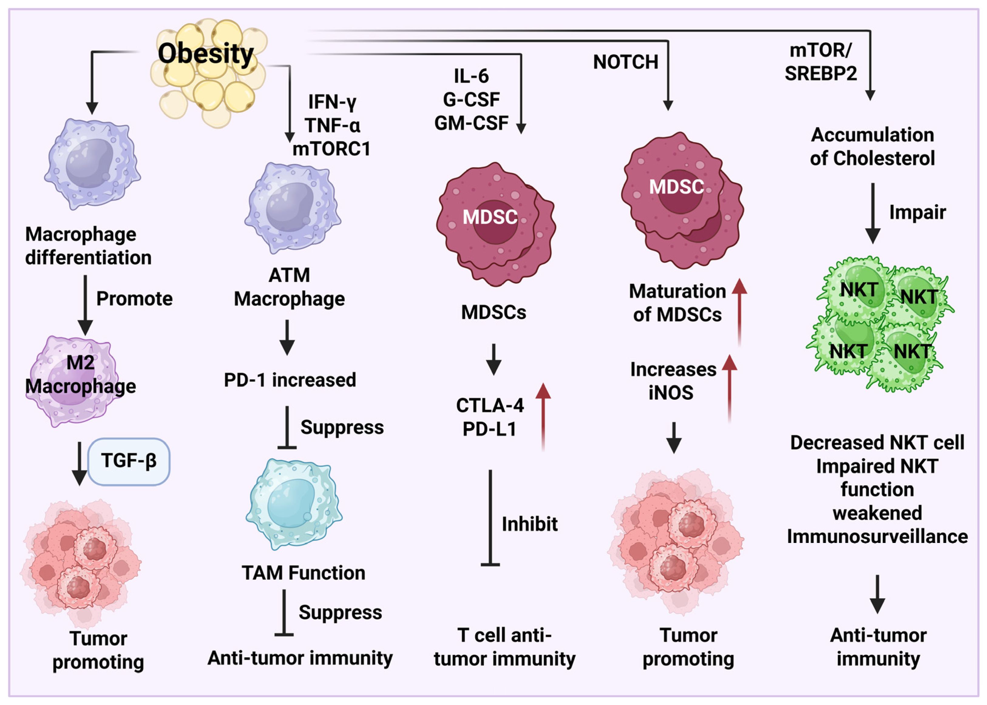
The expression of transforming growth factor-β (TGF-β) in M2-like macrophages during obesity can promote tumor cell proliferation and induce an immunosuppressive microenvironment (Figure 2). One study demonstrates that adipose tissue macrophages (ATMs) have elevated expression levels of PD-1, which is mediated by the production of IFN-γ, TNF-α, and mTORC1. However, PD-1 expression can decrease the functionality of ATMs to suppress their anti-tumor function [80]. A novel macrophage subtype, lipid-associated macrophages (LAMs), also known as CD9+ macrophages, has been identified to play an important role in tissue metabolic homeostasis. The expansion of LAMs is found in the adipose tissues in obese mouse models, which confers an anti-inflammatory effect. ATMs maintain tissue homeostasis by reducing serum insulin, blood cholesterol levels, and glucose intolerance. The underlying mechanism of LAM function is driven by a lipid receptor, Trem2 (triggering receptor expressed on myeloid cells 2) [81].
Obesity can also promote tumor growth and inhibit T cell antitumor function through the modulation of MDSCs. Obesity induces elevated expression levels of IL-6, granulocyte colony-stimulating factor (G-CSF), and granulocyte-macrophage colony-stimulating factor, which increase the expression of cytotoxic T-lymphocyte-associated protein 4 (CTLA-4) and programmed death-ligand 1 (PD-L1) to suppress T cell cytotoxicity [82]. Additionally, obesity promotes the maturation of MDSCs and increases the expression of inducible nitric oxide synthase (iNOS) through neurogenic locus notch homolog protein (NOTCH) signaling pathway (Figure 2), leading to a tumor-promoting effect [83]. Using a diet-induced obesity mouse model with breast cancer, researchers demonstrate that an elevated expression level of intratumor CXCL1 can induce the accumulation of CXCR2-expressing G-MDSC into the tumor microenvironment. However, immunosuppressive cell infiltration causes CD8+ T cell apoptosis and immunotherapy resistance [84].
In a diet and chemical (azoxymethane)-induced obesity-associated colon cancer model, NK cell number and functions are decreased when compared to NK cell number and functions in the non-obese tumor model. Meanwhile, the severity of tumor progression is also higher in the obese group compared to that in the normal weight group [85]. Obese individuals have fewer circulating NK cells and decreased NK cell functions, including decreased cytotoxicity, reduced secretion of perforin and granzyme, and weakened metabolic activity. The dysfunction of NK cells increases the proliferation of malignant tumor cells and promotes tumor development [86,87,88].
Obesity reshapes the metabolic profile in the tumor microenvironment to induce tumor immunosuppression. For instance, a study reveals that the accumulation of cholesterol in obesity can reduce NKT cell number and impair their function to reduce their immunosurveillance in HCC [89]. Moreover, obesity can regulate the mutation of oncogenes, such as Kirsten rat sarcoma virus (KRAS), epidermal growth factor receptor (EGFR), SET domain-containing 2 protein (SETD2), and BRCA1-associated protein 1 (BAP1), which also increases the risk of cancer development [90].
Obesity-associated gut dysbiosis can disrupt gut homeostasis and induce an imbalance of gut microbial species, which might result in an expansion of tumor-promoting pathogenic bacteria and a decrease in tumor-preventive bacteria. For example, treatment with a high-fat diet can increase colorectal tumorigenesis in mice by increasing levels of tumor-promoting bacteria Alistipes sp. Marseille-P5997 and Alistipes sp. 5CPEGH and a decreased amount of probiotic strain Parabacteroides distasonis [91]. In addition, gut microbiota dysbiosis can damage the tight junctions of epithelial cells in the gut, causing leakage of microbial components and metabolites into the circulating system and systemic inflammation. It also induces abnormal metabolism and dysregulation of energy metabolism to cause immune evasion and tumorigenesis in cancers, such as breast cancer [92].
Obesity regulates breast cancer progression and therapy. In mice with obesity-related breast cancer, anti-PD-1 treatment significantly suppressed tumor progression by increasing macrophage M1 polarization and the populations of dendritic cells and cytotoxic CD8 T cells. The alteration of immune cell profiles was also shown in breast cancer patients with anti-PD-1 therapy. Additionally, the abundances of some gut microbiota, such as Bifidobacterium and Lactobacillus, were closely associated with the efficacy of immune checkpoint blockade (ICB) therapy [93]. In obese mice with breast cancer, anti-PD-1 treatment uniquely increased the abundances of gut microbiota [93], such as Odoribacter, Adlercreutzia, and Mogibacteriaceae.
A meta-analysis study was performed to dissect the role of obesity or BMI on the survival outcomes of NSCLC patients with the treatment of immune checkpoint inhibitors. The results revealed that overweight and obese patients had prolonged or increased overall survival (OS) and PFS compared to patients with normal weight. It is commonly known that overweight and obesity can increase the risk of cancer development; however, obese patients had better survival outcomes compared to lean individuals in this meta-analysis study [94]. It is known as a phenomenon of obesity paradox. The results suggest there are complex roles of obesity in some cancers.

4. Important Signaling Pathways Involved in Obesity-Related Cancer Development and Therapy

There are some key signaling pathways involved in obesity-related cancer development and immunosuppression. Here, we list some examples to discuss their functions.

4.1. JNK, IKK/NF-κB, and STAT3 Signaling Pathways

An elevated level of FFAs in obesity can activate some inflammatory kinases, such as c-Jun N-terminal kinases (JNK), inhibitory-κB kinase (IKK)/NF-κB, and STAT3 in liver and adipose tissues. Their activation can induce insulin resistance, inflammation, tumor cell proliferation and metastasis, and immune suppression [95,96,97]. The activation of JNK expression in macrophages is necessary for M1 macrophage polarization (proinflammatory phenotype) and in the process of obesity-induced insulin resistance [98,99]. Activation of JNK promotes the serine phosphorylation of insulin receptor substrate 1 (IRS-1), which disrupts the insulin receptor signaling pathway to influence glucose uptake, eventually resulting in insulin resistance [100].
The IKK/NF-κB signaling pathway can be activated to increase the production of cytokines (e.g., TNF-α, IL-6, and IL-1β) to promote inflammation and insulin resistance. The activation of STAT3 stimulates the secretion of IL-6 and activates JNK, mTOR, and protein kinase C (PKC) signaling pathways, which aggravate insulin resistance [101].
In cancer, JNK is well known for its role in regulating tumorigenesis. The activation of JNK is inhibited by NF-κB expression, resulting in cancer cell survival. The JNK signaling pathway also promotes an immunosuppressive tumor microenvironment in triple-negative breast cancer. Activation of IKK/NF-κB promotes the release of proinflammatory cytokines to aggravate EMT, angiogenesis, and immune suppression in tumors [102]. Activation of STAT3 and NF-κB can directly promote tumor cell survival, proliferation, metastasis, EMT, angiogenesis, and immune suppression. Moreover, obesity-induced expression of lipocalin 2 also contributes to the activation of NF-κB and STAT3 to facilitate M1 macrophage polarization, exacerbating the insulin resistance and inflammation [103].

4.2. PI3K/AKT/mTOR Signaling Pathway

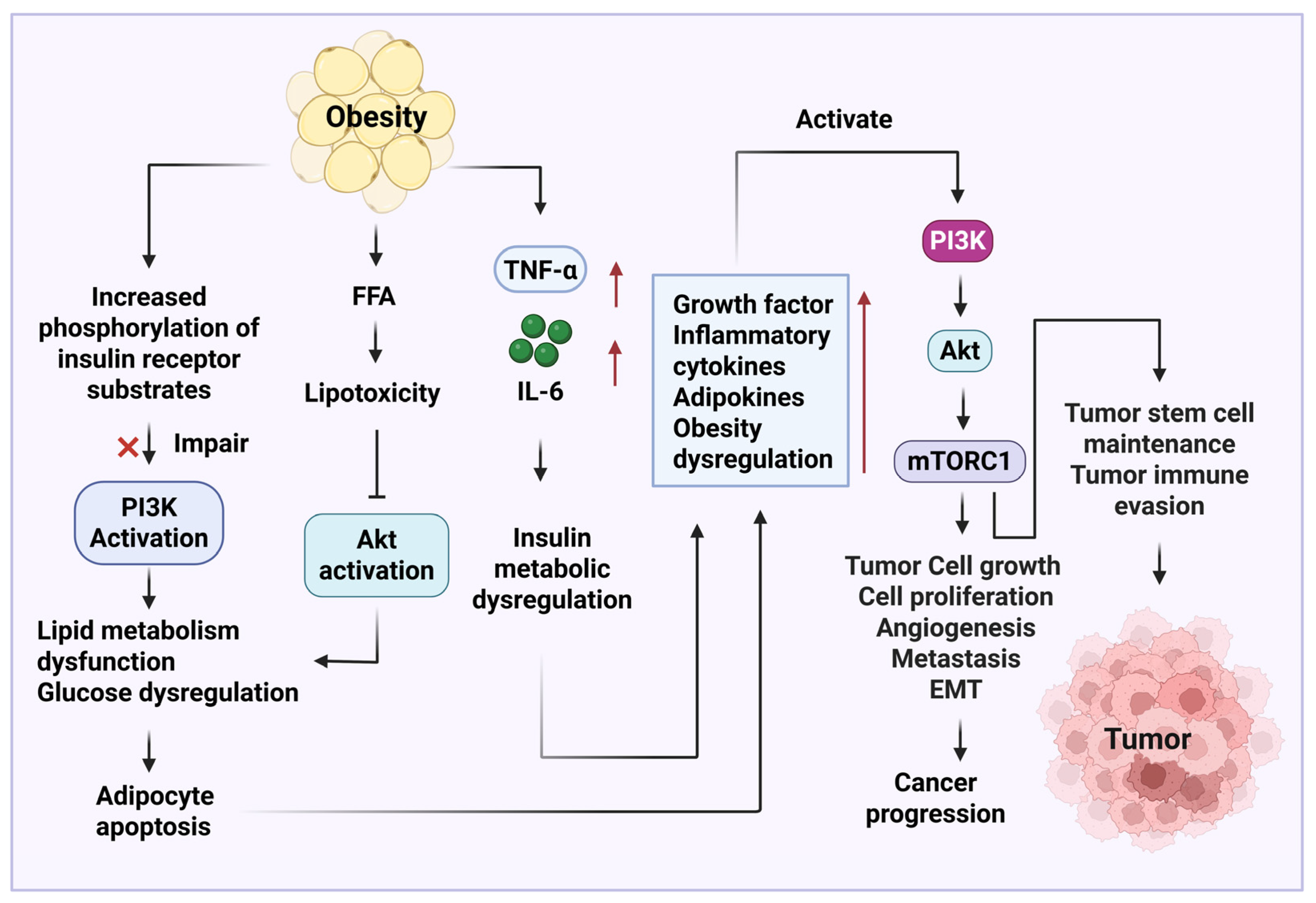
Obesity can increase serine phosphorylation of insulin receptor substrates to reduce PI3K activation, which results in a dysregulation of glucose and lipid metabolism [104]. This process led to adipocyte apoptosis. Additionally, obesity induces an increased expression of inflammatory cytokines, such as IL-6 and TNF-α, to promote insulin resistance and metabolic dysfunction [105]. Obesity-induced elevated levels of FFAs can aggravate lipotoxicity, consequently inhibiting the activation of AKT downstream signaling pathways [106]. This process induces glucose and lipid metabolism dysregulation and adipocyte apoptosis. The activation of PI3K/AKT/mTOR signaling pathway in obesity induces hepatic steatosis and accumulation of visceral fat, further promoting liver and systemic inflammation [107]. In cancer, obesity-induced expression of growth factors, inflammatory cytokines (e.g., IL-6 and TNF-α), and adipokines (e.g., leptin) can induce dysregulation PI3K/AKT/mTOR signaling pathway, to induce cancer progression and predict a poor prognosis. Alteration of the PI3K/AKT/mTOR signaling pathway also contributes to tumor cell survival, proliferation, angiogenesis, metastasis, EMT, and chemotherapy resistance (Figure 3). In addition, the PI3K/AKT/mTOR signaling pathway is critical for maintaining the survival of tumor stem cells and causing tumor evasion [108,109]. Finally, obesity-induced hyperinsulinemia and inflammation impact the PI3K-targeted immune therapy [110].

4.3. Wnt/β-Catenin Signaling Pathway

In obesity, the Wnt/β-catenin signaling pathway (Figure 4) plays a key role in promoting liver steatosis, adipose tissue inflammation, and dysregulation of lipid metabolism and glucose homeostasis [111,112,113]. In cancers, it also contributes to liver steatosis and inhibition of adipogenesis. Activation of Wnt/β-catenin promotes the M2 macrophage polarization to enhance tumor cell propagation and resistance to T cell cytotoxicity [114,115]. In NSCLC, the Wnt/β-catenin signaling pathway is responsible for immune escape and resistance to immunotherapies, such as anti-PD-1/PD-L1 antibody and anti-CTLA-4 antibody [116]. Wnt/β-catenin can also regulate dendritic cell recruitment in a murine melanoma model by inducing the activation of transcription factor 3 to suppress CCL4 expression [117].

4.4. NOTCH Signaling Pathway

In obesity, the NOTCH signaling pathway is activated in preadipocytes, which is responsible for weight gain and disruption of glucose homeostasis, and decreases insulin sensitivity in white adipose tissues. The elevated level of FFAs in liver adipose tissues during obesity can activate the NOTCH signaling pathway, to increase insulin resistance (IR), production of reactive oxygen species (ROS), and lipophagy. NOTCH promotes mTORC1 activation to subsequently increase the production of triglycerides (TGs), causing an increase in the production of VLDL and lipophagy [118].
In cancer, activation of the NOTCH signaling pathway increases the production of cytokine IL-1β and chemokine CCL2, which induce inflammation and recruitment of tumor-associated macrophages. The NOTCH signaling pathway also modulates TGF-β and CXCL5 secretion to promote the infiltration of tumor-associated neutrophils, and reprograms metabolic processes, such as switching glycolytic processes, to induce tumor growth. The NOTCH signaling pathway is also involved in tumor stem cell renewal, EMT, and tumor angiogenesis [119].
NOTCH signaling pathway mediates M1 and M2 polarization of tumor-associated macrophages to display anti-tumor and tumor-promoting functions, respectively [120]. NOTCH can increase anti-tumor immune response by regulating CD8+ effector T cells, and cause tumor-promoting function by modulating PD-1-induced exhaustion of CD8+ T cells [121]. This pathway impacts CD4+ T cell function and differentiation in cancers. Type 1 T helper cell (Th1) plays an anti-tumor role, whereas Th2 and regulatory T cells (Treg) play the function of tumor-promoting immunity [122].

4.5. HIF-1α Signaling Pathway

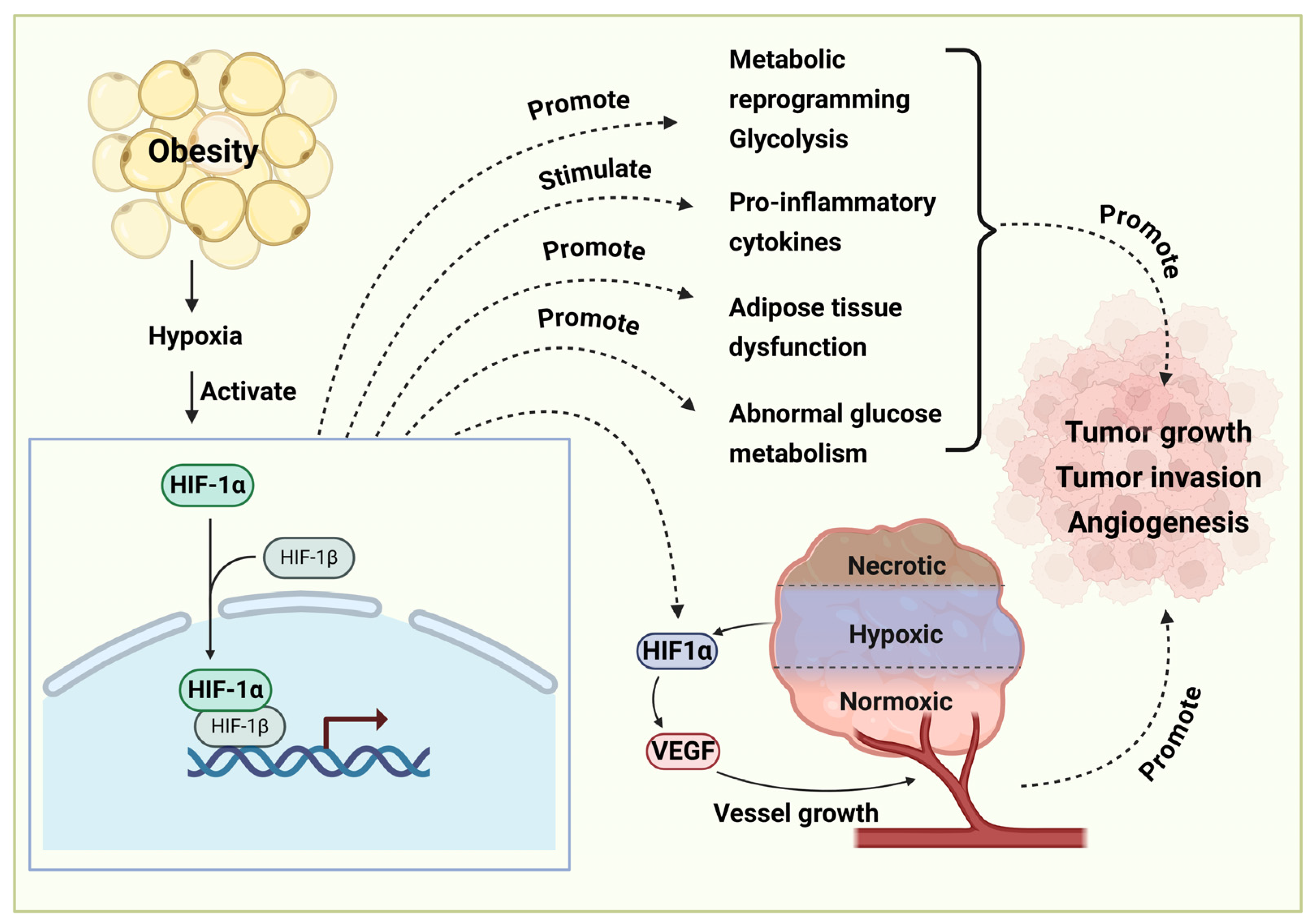
Obesity-induced hypoxia in adipose tissues activates the expression of hypoxia-inducible factor 1 alpha (HIF-1α), which can stimulate the release of proinflammatory cytokines to induce inflammation (Figure 5). The activation of HIF-1α impairs adipocyte function and metabolism, causing an energy metabolism imbalance and insulin resistance [123]. The activation of HIF-1α also promotes angiogenesis to promote cancer cell survival, proliferation, and metastasis [124,125]. HIF-1α activation is also involved in metabolic reprogramming, such as increasing glycolysis, which promotes tumor invasion and angiogenesis [126].
Obesity-associated signaling pathways, such as Wnt/β-catenin, PI3K/AKT/mTOR, NOTCH, and JAK/STAT3 signaling pathways, are involved in immunotherapy resistance. In addition, they are implicated in the maintenance and survival of cancer stem cells. Targeting cancer stem cells can be a promising strategy to cope with immunotherapy resistance and manage cancer relapses post-immunotherapy. Integration of multiple approaches in investigating obesity-associated cancers is beneficial to facilitate the identification of novel biomarkers and therapeutic targets for cancer treatment [127]. In addition, spatial technologies and multi-omics such as transcriptomics, metabolomics, proteomics, genomics, and epigenomics can advance our understanding of obesity-related factors in cancer development and therapy [128].

5. Natural Products in Obesity-Associated Cancer Therapy

As a chronic disease caused by multiple factors, obesity predisposes individuals to various cancers. Natural medicines such as phytomedicines demonstrate irreplaceable advantages in the treatment of obesity and cancer due to their multi-component and multi-target characteristics. These medicines can not only reshape anti-tumor immune responses by ameliorating or reducing immunosuppression, metabolic disorders, and secretion of obesity-associated inflammatory factors in the tumor microenvironment, but also can break the malignant progression chain from obesity to cancer via regulating the balance of the immune system and energy metabolism. The findings in the functions of natural products provide new therapeutic strategies for tumor prevention and treatment, and open new avenues for deciphering the mechanisms of developing multi-target therapies in metabolic disorders, including cancers.

5.1. Ginsenosides

Ginsenosides, a complex of steroid saponins, are extracted from ginseng, the root of the plant Panax. Ginseng extract can treat obesity-associated type 2 diabetes [129]. Many experiments have demonstrated that treatment with ginseng extract induces weight loss in mice, rats, and humans [130,131,132,133,134,135,136]. Ginsenoside Rg3, an active component of ginsenosides, can inhibit oxidative stress and the formation of advanced glycation end products (AGEs) [137]. Amino acid derivatives of ginsenosides can also suppress the digestion and absorption of carbohydrates in the gastrointestinal tract, thereby reducing blood sugar levels [138]. Furthermore, ginsenosides can enhance the binding ability and cytotoxicity of NK cells to tumor cells, thus displaying the potential in treating diseases driven by NK cell dysfunction and chronic inflammation. For example, ginsenoside Rh2 inhibited the growth and metastasis of postmenopausal breast cancer cells [139].

5.2. Artemisinin

Artemisinin, extracted from Artemisia annua (sweet wormwood), has garnered significant attention for its role in the treatment of obesity and obesity-related metabolic diseases. In obese individuals, the expression of inflammatory cytokine TNF-α is elevated, which is a key marker for systemic inflammation. Artemisinin exerts an immunosuppressive function by suppressing B cell and pathogenic T cell activation and increasing regulatory T cell expansion [140]. Artemisinin and its derivatives can also suppress endoplasmic reticulum stress to prevent obesity progression [140]. They can upregulate the expression levels of uncoupling protein 1 (UCP1), peroxisome proliferator-activated receptor gamma coactivator 1-α (PGC1α), and PR domain containing 16 (PRDM16), to promote adipocyte browning or enhance UCP1 expression in the inguinal adipose tissues of C57BL/6J mice to increase thermogenesis, thereby reducing bodyweight [141].
Furthermore, the anti-cancer mechanism of artemisinin is closely related to its molecular properties. The endoperoxide bridge in its structure interacts with intracellular iron ions or heme groups, generating cytotoxic effects [142]. Increased intracellular irons in tumor cells can significantly boost the cytotoxicity of artemisinin [143]. Due to their increased demand for iron, cancer cells exhibit higher rates of iron metabolism and increase expression levels of transferrin receptors compared to normal cells [144], which increases the cytotoxicity of artemisinin to tumor cells.

5.3. Paclitaxel

Paclitaxel is an active monomer applied to treat various cancers. For example, it can inhibit the proliferation of endometrial cancer (EC) cell lines, induce EC cells to undergo apoptosis, enhance cellular stress responses, and cause cell cycle arrest in the G1 phase [145]. Obesity is a high-risk factor causing the development of EC. Studies show that paclitaxel combined with weight loss management (e.g., intermittent energy restriction and administration of low-fat diets) can reverse the obesity-induced abnormal elevation of serum insulin, leptin, and inflammatory factors, and reduce tumor incidence and weight, thereby reversing the cancer-promoting effect of obesity in mouse EC models [146].
Obesity is a contributing factor in the development and diagnosis of ovarian cancer [147]. Epithelial ovarian cancer (EOC) is the gynecological malignancy with the highest mortality rate, and the primary target organ for local metastasis is the omentum, which is rich in adipocytes. Research has found that adipocytes and omentum-derived stromal cells can enhance ovarian cancer resistance to paclitaxel [148], a mechanism related to fatty acid metabolism-associated stemness. Omental adipocytes can drive the EMT of EOC cells to promote their invasion and resistance to chemotherapy [149].

5.4. Hesperidin

Hesperidin is a flavanone glycoside extracted from citrus peels, and it possesses anti-obesity and anti-cancer activities by reducing cholesterol levels and blood pressure [150]. Regarding its anti-obesity functions, hesperidin exhibits multi-dimensional therapeutic properties. It can stimulate the release of appetite-regulating cholecystokinin (CCK) from intestinal endocrine STC-1 cells, thereby exerting an anti-obesity effect through appetite suppression [151]. It also inhibits the expression of several key targets involved in adipogenesis, including CCAAT enhancer-binding protein β (C/EBPβ), sterol regulatory element binding protein 1c (SREBP1c), peroxisome proliferator-activated receptor γ (PPAR-γ), and perilipin [152]. In addition, hesperidin can suppress fat accumulation by inhibiting the expression of stearoyl-CoA desaturase [153].
Hesperidin can enhance the production of ROS in tumor cells. It induces apoptosis by upregulating caspase family members and activating the mitochondrial apoptosis pathway [154] and inhibiting the cell proliferation marker [155]. Hesperidin can also arrest the cancer cell cycle in the G0/G1 and G2/M phases and regulate angiogenesis to promote cancer growth [156].

5.5. Quercetin

Quercetin is a flavonol found in many plants. In improving obesity and related metabolic syndrome, it can promote adiponectin secretion from adipocytes via a PPAR-independent pathway [157]. Furthermore, oral administration of quercetin can enrich intestinal Lactobacillus, modulate gut microbiota structure, to consequently promote the production of non-12α-hydroxylated bile acids in serum. In brown fat and browning of white fat, bile acids can interact with their receptor Takeda G protein-coupled receptor 5 (TGR5) to increase energy metabolism, thereby alleviating metabolic dysfunction in obese models [158].
In direct anti-cancer action, quercetin exerts inhibitory effects through multiple targets [159]. It can cause cell cycle arrest by several mechanisms, such as inhibiting the promoter activity of cyclin B1, suppressing the MAPK/ERK1/2 signaling pathway, and activating the transcription factor p53. Simultaneously, quercetin promotes apoptosis through mechanisms, including activation of TGF-β (transforming growth factor-beta) signaling pathway, and inhibition of pro-survival signaling pathways like PI3K/AKT/mTOR, Wnt/β-catenin, NOTCH, sonic hedgehog protein (SHH), and JAK/STAT, thereby impeding DNA repair [160]. Additionally, quercetin can inhibit tumor angiogenesis by regulating the vascular endothelial growth factor (VEGF) and its receptor axis to stimulate tumor blood vessel formation, consequently inhibiting tumorigenesis and development [160].

5.6. Celastrol

Celastrol, a natural active ingredient extracted from the root bark of Tripterygium wilfordii, possesses multi-dimensional anti-obesity effects by acting on lipid metabolism, inflammatory responses, energy metabolism, and gut microbiota modulation. Specifically, it can reduce macrophage infiltration and inflammatory cytokine production in the liver and adipose tissues, alleviating metabolic disorders in obese mice by inhibiting TLR3/NLRP3 (NLR family pyrin domain containing 3) inflammasome activation [161]. Additionally, it can directly induce cell apoptosis of preadipocytes by interacting with RAB7 (Ras-related protein 7) and VAMP7 (vesicle-associated membrane protein 7) to inhibit preadipocyte autophagy [162]. Celastrol can disrupt the link between endoplasmic reticulum stress (ERS) signaling pathways and downstream inflammation and lipid metabolism [163]. Thus, it inhibits ERS, inflammation, and adipogenesis, and promotes hepatic lipolysis to achieve effective weight loss.
In direct antitumor action, Celastrol can induce tumor cell apoptosis by downregulating the anti-apoptotic protein B-cell lymphoma 2 (Bcl-2) and upregulating the pro-apoptotic protein BCL2-associated X (Bax) [164]. It can also inhibit cell viability via the AMPK-mediated PLK-2 (Polo-like kinase 2, a serine/threonine-protein kinase) pathway to inhibit the growth of the human breast cancer cell line MCF-7 [165]. In addition, Celastrol displays anti-gastric cancer effects by targeting the antioxidant enzyme peroxiredoxin-2 in tumor cells to overexpress ROS and induce cancer apoptosis [166,167].

5.7. Curcumin

Curcumin, a phenolic active ingredient extracted from the rhizomes of Curcuma plants, shows significant potential in colorectal cancer treatment. Curcumin can inhibit the binding of NF-κB to IKK and the activation of the NF-κB-DNA complex, ultimately downregulating the gene expression of NF-κB-mediated adhesion molecules, such as VCAM-1 (vascular-cell adhesion molecule 1), ICAM-1 (intercellular adhesion molecule 1), and ELAM-1 (endothelial-leukocyte adhesion molecule 1), thereby inhibiting tumor metastasis [168]. However, the clinical translation of curcumin is limited by its low bioavailability [169,170].

5.8. Ursolic Acid

Ursolic acid is a triterpenoid component widely distributed in plants, with preventive and therapeutic effects against cancer. As demonstrated in the introductions of various drugs above, cancer and obesity share multiple molecular targets and signaling pathways, and the induction of both is closely linked to inflammation. Experimental studies show that ursolic acid can inhibit cancer cell growth. For instance, accumulating data show that it can inhibit cell proliferation and induce cancer cell apoptosis of many tumors, such as liver cancer (e.g., HepG2) [171], colorectal cancer (e.g., HT-29) [172], and breast cancer (e.g., MCF-7) [173].
Using natural products for managing metabolic disorders such as obesity and cancer is one of the treatment strategies. However, there are some challenges and translation barriers when using natural products as a therapeutic intervention. For example, complex formulation, diverse bioactivity, manufacturing procedure, and evaluation of therapeutic efficacy need to be well understood and analyzed to increase their targeted effects and reduce potential side effects. In addition, the delivery method for targeted therapy of bioactive natural compounds should also be considered for their application.

6. Conclusions

Obesity is a multifactorial chronic disease, with excessive fat accumulation in adipose tissues. Adipose tissues secrete a variety of adipokines, cytokines, and chemokines to alter immune cell responses and cause chronic metabolic disorders. In cancer patients, obesity contributes to the development of an immunosuppressive microenvironment by promoting cancer survival, proliferation, and metastasis and inducing immune cell exhaustion and dysfunction. Several key signaling pathways, such as PI3K/AKT/mTOR, NOTCH, and HIF-1α signaling pathways, are involved in obesity-associated cancer development and progression. Natural product medicines have multiple targets and functions, and display promising therapeutic potential to treat obesity-associated cancer and metabolic disorders.
There are many challenges and barriers in the management of obesity-related cancers. Complex biological processes influence the development and progression of obesity and cancer. Factors impacting the efficacy of cancer immunotherapy in patients with overweight/obesity are complicated. Pre-clinical studies and clinical studies have shown that obesity increases inflammation and cancer progression in most cases. Controversially, obesity also demonstrates a beneficial effect in improving anti-tumor immunotherapeutic efficacy in some cancers, such as renal cancer. This “obesity paradox” phenomenon increases the complex roles of obesity in different cancers. Understanding the mechanism of obesity impacting different cancers helps develop obesity-targeted cancer therapy. The evidence from clinical trials provides translational guidance in the treatment of overweight and obese patients with cancer. For example, regarding day-to-day clinical oncology management, proper interventions such as weight loss, dietary intervention, and personalized nutritional plans can be applied to facilitate weight control and improve the quality of life. Sufficient exercise and physical activity are helpful to maintain body structure and reduce fat accumulation. Precision medicine and personalized treatment will also be helpful in this situation. The roles of obesity in cancer development risk and therapy in patients can be evaluated by its roles in the recruitment of different immune cells, including cytotoxic CD8 T cells and immunosuppressive myeloid cells. Therefore, both pre-clinical and clinical research studies are required in the future to precisely dissect the underlying cellular and molecular mechanisms of obesity-related cancer progression or suppression in cell and animal models and clinical studies. In addition, new therapeutic targets or diagnostic biomarkers are needed to better manage and treat obesity-associated cancers.
As artificial intelligence (AI) rapidly becomes a transformative approach in healthcare, AI-driven approaches can be harnessed to improve the daily healthcare of patients, increase innovative research findings, and accelerate the process of personalized treatment [174,175]. Big databases from clinical samples in patients with obesity-related cancers and health controls worldwide, and information on treatment and diagnosis, pave the way for exploring novel diagnostic markers and evaluating treatment efficacy. Machine learning and AI-driven approaches are useful tools and methods to predict the risk of obesity in cancer development and progression, and evaluate the treatment efficacy [176]. They have been applied to facilitate the discovery of novel biomarkers and therapeutic targets. The methodology for targeted delivery can be further advanced based on the findings of machine learning and AI technologies. In drug development, such as screening molecules, toxicity prediction, and bioactivity assessment, AI-driven approaches have provided promising results in accelerating the drug discovery process and the translation from pre-clinical to clinical practice [177]. Furthermore, the AI-driven approach could assist clinical decision-making, and AI-driven technology could be applied for developing portable health tracking devices for daily healthcare monitoring.

Author Contributions

Conceptualization, C.Z., J.L. and M.Y.; investigation, C.Z., K.Z., J.L. and M.Y.; writing—original draft preparation, C.Z., K.Z., J.L. and M.Y.; writing—review and editing, C.Z., J.L. and M.Y. All authors have read and agreed to the published version of the manuscript.

Funding

This study received no external funding.

Institutional Review Board Statement

Not applicable.

Informed Consent Statement

Not applicable.

Data Availability Statement

All the data supporting the reported findings can be found in this study.

Conflicts of Interest

The authors declare no conflicts of interest.

Abbreviations

The following abbreviations are used in this manuscript:
Acs-2acyl-CoA synthetase
AGEsAdvanced glycation end products
AKTProtein kinase B
AMPK5′-adenosine monophosphate (AMP)-activated protein kinase
ATMsAdipose tissue macrophages
BAP1BRCA1-associated protein 1
BaxBCL2-associated X
Bcl-2B-cell lymphoma 2
BMIBody mass index
BRCA1Breast cancer susceptibility gene 1
C/EBPβCCAAT enhancer-binding protein β
CCKCholecystokinin
CCLChemokine (C-C motif) ligand
CCRC-C chemokine receptor
CD8Cluster of differentiation 8
c-JunJun proto-oncogene
CRPC-reactive protein
CTLA-4Cytotoxic T-lymphocyte-associated protein 4
CVDCardiovascular diseases
CXCLChemokine (C-X-C motif) ligand
CXCRChemokine receptors
DTCDifferentiated thyroid cancer
ECEndometrial cancer
EGFREpidermal growth factor receptor
ELAM-1Endothelial-leukocyte adhesion molecule 1
EMTEpithelial–mesenchymal transition
EOCEpithelial ovarian cancer
ERK1/2Extracellular signal-related kinases 1 and 2
ERSEndoplasmic reticulum stress
FAT-6Stearoyl-CoA desaturase
FFAFree fatty acid
G-CSFGranulocyte colony-stimulating factor
GM-CSFGranulocyte-macrophage colony-stimulating factor
G-MDSCGranulocytic myeloid-derived suppressor cells
GzmBGranzyme B
HCCHepatocellular carcinoma
HDLHigh-density lipoprotein
HepG2Liver cancer cells
HIF1αHypoxia-inducible factor 1 alpha
ICAM-1Intercellular adhesion molecule 1
IFN-γInterferon gamma
IKKInhibitory-κB kinase
IL-6Interleukin 6
iNOSInducible nitric oxide synthase
IRInsulin resistance
IRS-1Insulin receptor substrate 1
JAK2Janus kinase 2
JNKc-Jun N-terminal kinases
Kat-13-ketoacyl-CoA thiolase 1
Ki-67Marker of proliferation Kiel 67
KRASKirsten rat sarcoma virus
Lag3Lymphocyte activation gene 3 protein
LAMsLipid-associated macrophages
LDLinear dichroism
LDLLow-density lipoprotein
MAPKMitogen-activated protein kinase
MASLDMetabolic dysfunction-associated steatotic liver disease
MCF-7Michigan cancer foundation-7 cancer cell line
MCP-1Monocyte chemoattractant protein-1
MDSCMyeloid-derived suppressor cell
Mdt-15Mediator subunit MDT-15
MHC-IIMajor histocompatibility complex class II molecules
MIP-1αMacrophage inflammatory protein (MIP)-1α
MIP-1βMacrophage inflammatory protein (MIP)-1β
M-MDSCsMonocytic myeloid-derived suppressor cells
mTORMammalian target of rapamycin
mTORCMammalian target of rapamycin complex
MYCMyelocytomatosis oncogene
NF-κBNuclear factor kappa-light-chain-enhancer of activated B cells
NK cellNatural killer cell
NKT cellNatural killer T cell
NLRP3NLR family pyrin domain containing 3
NOTCHNeurogenic locus notch homolog protein
NSCLCNon-small cell lung cancer
p53Tumor protein p53
PCNAProliferating cell nuclear antigen
PD-1Programmed cell death protein 1
PD-L1Programmed cell death ligand 1
PGC1αPeroxisome proliferator-activated receptor gamma coactivator 1-α
PI3KPhosphoinositide 3-kinases
PKCProtein kinase C
PLK-2Polo-like kinase 2
pod-2Gene encoding acetyl-CoA carboxylase
PPARαPeroxisome proliferator-activated receptor-alpha
PPAR-γPeroxisome proliferator-activated receptor γ
PRDM16PR domain containing 16
RAB7Ras-related protein
ROSReactive oxygen species
S6Ribosomal protein S6
SETD2SET domain-containing 2 protein
SHHSonic hedgehog protein
SLC7A5Solute carrier family 7 member 5
SREBP1cSterol regulatory element binding protein 1c
SREBP2Sterol regulatory element-binding protein 2
STAT3Signal transducer and activator of transcription 3
STC-1 cellsIntestinal secretin tumor cell line
T2DType 2 diabetes
TGF-βTransforming growth factor-β
TGR5Takeda G protein-coupled receptor 5
TGsTriglycerides
Th1Type 1 T helper cell
Th17T helper type 17
TILsTumor-infiltrating lymphocytes
Tim3T-cell immunoglobulin and mucin domain 3
TLR4Toll-like receptor 4
TNF-αTumor necrosis factor alpha
TregRegulatory T cell
Trem2Triggering receptor expressed on myeloid cells 2
UCP1Uncoupling protein 1
VAMP7Vesicle-associated membrane protein 7
VATVisceral adipose tissue
VCAM-1Vascular-cell adhesion molecule-1
VEGFVascular endothelial growth factor
WntWingless-type MMTV integration site family
ZEB1Zinc finger E-box binding homeobox 1

References

  1. Hales, C.M.; Fryar, C.D.; Carroll, M.D.; Freedman, D.S.; Ogden, C.L. Trends in Obesity and Severe Obesity Prevalence in US Youth and Adults by Sex and Age, 2007–2008 to 2015–2016. JAMA 2018, 319, 1723–1725. [Google Scholar] [CrossRef]
  2. Tsigos, C.; Hainer, V.; Basdevant, A.; Finer, N.; Fried, M.; Mathus-Vliegen, E.; Micic, D.; Maislos, M.; Roman, G.; Schutz, Y.; et al. Management of obesity in adults: European clinical practice guidelines. Obes. Facts 2008, 1, 106–116. [Google Scholar] [CrossRef] [PubMed]
  3. World Obesity Federation. Economic Impact of Overweight and Obesity to Surpass $4 Trillion by 2035. Available online: https://www.worldobesity.org/ (accessed on 9 July 2025).
  4. Lopez-Jimenez, F.; Almahmeed, W.; Bays, H.; Cuevas, A.; Di Angelantonio, E.; le Roux, C.W.; Sattar, N.; Sun, M.C.; Wittert, G.; Pinto, F.J.; et al. Obesity and cardiovascular disease: Mechanistic insights and management strategies. A joint position paper by the World Heart Federation and World Obesity Federation. Eur. J. Prev. Cardiol. 2022, 29, 2218–2237. [Google Scholar] [CrossRef] [PubMed]
  5. Julián, M.T.; Arteaga, I.; Torán-Monserrat, P.; Pera, G.; Pérez-Montes de Oca, A.; Ruiz-Rojano, I.; Casademunt-Gras, E.; Chacón, C.; Alonso, N. The Link between Abdominal Obesity Indices and the Progression of Liver Fibrosis: Insights from a Population-Based Study. Nutrients 2024, 16, 1586. [Google Scholar] [CrossRef] [PubMed]
  6. Llévenes, P.; Chen, A.; Lawton, M.; Rondón-Ortiz, A.N.; Qiu, Y.; Seen, M.; Monti, S.; Denis, G.V. Plasma exosomes in insulin resistant obesity exacerbate progression of triple negative breast cancer. BMC Cancer 2025, 25, 1089. [Google Scholar] [CrossRef]
  7. Williams, E.P.; Mesidor, M.; Winters, K.; Dubbert, P.M.; Wyatt, S.B. Overweight and Obesity: Prevalence, Consequences, and Causes of a Growing Public Health Problem. Curr. Obes. Rep. 2015, 4, 363–370. [Google Scholar] [CrossRef]
  8. Yang, M.; Liu, S.; Zhang, C. The Related Metabolic Diseases and Treatments of Obesity. Healthcare 2022, 10, 1616. [Google Scholar] [CrossRef]
  9. Singh, D.; Dhiman, V.K.; Pandey, M.; Dhiman, V.K.; Sharma, A.; Pandey, H.; Verma, S.K.; Pandey, R. Personalized medicine: An alternative for cancer treatment. Cancer Treat. Res. Commun. 2024, 42, 100860. [Google Scholar] [CrossRef]
  10. Bray, F.; Laversanne, M.; Sung, H.; Ferlay, J.; Siegel, R.L.; Soerjomataram, I.; Jemal, A. Global cancer statistics 2022: GLOBOCAN estimates of incidence and mortality worldwide for 36 cancers in 185 countries. CA Cancer J. Clin. 2024, 74, 229–263. [Google Scholar] [CrossRef]
  11. Yoshimoto, S.; Loo, T.M.; Atarashi, K.; Kanda, H.; Sato, S.; Oyadomari, S.; Iwakura, Y.; Oshima, K.; Morita, H.; Hattori, M.; et al. Obesity-induced gut microbial metabolite promotes liver cancer through senescence secretome. Nature 2013, 499, 97–101. [Google Scholar] [CrossRef]
  12. Zhao, C.; Zhang, T.; Xue, S.T.; Zhang, P.; Wang, F.; Li, Y.; Liu, Y.; Zhao, L.; Wu, J.; Yan, Y.; et al. Adipocyte-derived glutathione promotes obesity-related breast cancer by regulating the SCARB2-ARF1-mTORC1 complex. Cell Metab. 2025, 37, 692–707.e9. [Google Scholar] [CrossRef]
  13. Baek, A.E.; Yu, Y.A.; He, S.; Wardell, S.E.; Chang, C.Y.; Kwon, S.; Pillai, R.V.; McDowell, H.B.; Thompson, J.W.; Dubois, L.G.; et al. The cholesterol metabolite 27 hydroxycholesterol facilitates breast cancer metastasis through its actions on immune cells. Nat. Commun. 2017, 8, 864. [Google Scholar] [CrossRef]
  14. Zhang, C.; Liu, S.; Yang, M. Hepatocellular Carcinoma and Obesity, Type 2 Diabetes Mellitus, Cardiovascular Disease: Causing Factors, Molecular Links, and Treatment Options. Front. Endocrinol. 2021, 12, 808526. [Google Scholar] [CrossRef]
  15. Hopkins, B.D.; Goncalves, M.D.; Cantley, L.C. Obesity and Cancer Mechanisms: Cancer Metabolism. J. Clin. Oncol. 2016, 34, 4277–4283. [Google Scholar] [CrossRef]
  16. Oberman, B.; Khaku, A.; Camacho, F.; Goldenberg, D. Relationship between obesity, diabetes and the risk of thyroid cancer. Am. J. Otolaryngol. 2015, 36, 535–541. [Google Scholar] [CrossRef] [PubMed]
  17. Terry, P.D.; Miller, A.B.; Rohan, T.E. Obesity and colorectal cancer risk in women. Gut 2002, 51, 191–194. [Google Scholar] [CrossRef] [PubMed]
  18. Stock, W.; Luger, S.M.; Advani, A.S.; Yin, J.; Harvey, R.C.; Mullighan, C.G.; Willman, C.L.; Fulton, N.; Laumann, K.M.; Malnassy, G.; et al. A pediatric regimen for older adolescents and young adults with acute lymphoblastic leukemia: Results of CALGB 10403. Blood 2019, 133, 1548–1559. [Google Scholar] [CrossRef] [PubMed]
  19. Shaukat, A.; Dostal, A.; Menk, J.; Church, T.R. BMI Is a Risk Factor for Colorectal Cancer Mortality. Dig. Dis. Sci. 2017, 62, 2511–2517. [Google Scholar] [CrossRef]
  20. Dieli-Conwright, C.M.; Courneya, K.S.; Demark-Wahnefried, W.; Sami, N.; Lee, K.; Sweeney, F.C.; Stewart, C.; Buchanan, T.A.; Spicer, D.; Tripathy, D.; et al. Aerobic and resistance exercise improves physical fitness, bone health, and quality of life in overweight and obese breast cancer survivors: A randomized controlled trial. Breast Cancer Res. 2018, 20, 124. [Google Scholar] [CrossRef]
  21. Bechtel, M.D.; Michel, C.; Srinivasan, P.; Chalise, P.; Parker, W.P.; Mirza, M.; Thrasher, B.; Gibbs, H.D.; DiGiovanni, J.; Hamilton-Reeves, J. Impact of Weight Management on Obesity-Driven Biomarkers of Prostate Cancer Progression. J. Urol. 2024, 211, 552–562. [Google Scholar] [CrossRef]
  22. Mita, M.M.; Mita, A.C.; Carver, B.J.; Shanahan, J.M.; Mayes, B.A.; Dufour, P.J.; Browning, D.; Anderson-Villaluz, A.; Petersen, J.S.; Turnquist, D.J.; et al. A Phase 1 Safety Study of Evexomostat (SDX-7320) in Patients with Late-Stage Cancer: An Antiangiogenic, Insulin-Sensitizing Drug Conjugate Targeting METAP2. Cancer Res. Commun. 2025, 5, 1008–1017. [Google Scholar] [CrossRef] [PubMed]
  23. You, D.; Wang, D.; Wu, Y.; Chen, X.; Shao, F.; Wei, Y.; Zhang, R.; Lange, T.; Ma, H.; Xu, H.; et al. Associations of genetic risk, BMI trajectories, and the risk of non-small cell lung cancer: A population-based cohort study. BMC Med. 2022, 20, 203. [Google Scholar] [CrossRef] [PubMed]
  24. Tangalakis, L.L.; Cortina, C.S.; Son, J.D.; Poirier, J.; Madrigrano, A. Obesity Does Not Influence Management of Advanced Breast Cancer in the Elderly. Clin. Breast Cancer 2019, 19, 197–199. [Google Scholar] [CrossRef] [PubMed]
  25. Tilg, H.; Ianiro, G.; Gasbarrini, A.; Adolph, T.E. Adipokines: Masterminds of metabolic inflammation. Nat. Rev. Immunol. 2025, 25, 250–265. [Google Scholar] [CrossRef] [PubMed]
  26. Michailidou, Z.; Gomez-Salazar, M.; Alexaki, V.I. Innate Immune Cells in the Adipose Tissue in Health and Metabolic Disease. J. Innate Immun. 2022, 14, 4–30. [Google Scholar] [CrossRef]
  27. Beghini, M.; Metz, M.; Baumgartner, C.; Wolf, P.; Bastian, M.; Hackl, M.; Baumgartner-Parzer, S.; Marculescu, R.; Krebs, M.; Harreiter, J.; et al. Leptin acutely increases hepatic triglyceride secretion in patients with lipodystrophy. Metabolism 2025, 169, 156261. [Google Scholar] [CrossRef]
  28. Wang, X.; Zhang, S.; Li, Z. Adipokines in glucose and lipid metabolism. Adipocyte 2023, 12, 2202976. [Google Scholar] [CrossRef]
  29. He, S.; Ryu, J.; Liu, J.; Luo, H.; Lv, Y.; Langlais, P.R.; Wen, J.; Dong, F.; Sun, Z.; Xia, W.; et al. LRG1 is an adipokine that mediates obesity-induced hepatosteatosis and insulin resistance. J. Clin. Investig. 2021, 131, e148545. [Google Scholar] [CrossRef]
  30. Chen, D.; Zhang, Y.; Yidilisi, A.; Xu, Y.; Dong, Q.; Jiang, J. Causal Associations Between Circulating Adipokines and Cardiovascular Disease: A Mendelian Randomization Study. J. Clin. Endocrinol. Metab. 2022, 107, e2572–e2580. [Google Scholar] [CrossRef]
  31. Kadowaki, T.; Yamauchi, T.; Kubota, N.; Hara, K.; Ueki, K.; Tobe, K. Adiponectin and adiponectin receptors in insulin resistance, diabetes, and the metabolic syndrome. J. Clin. Investig. 2006, 116, 1784–1792. [Google Scholar] [CrossRef]
  32. Liu, W.; Zhou, X.; Li, Y.; Zhang, S.; Cai, X.; Zhang, R.; Gong, S.; Han, X.; Ji, L. Serum leptin, resistin, and adiponectin levels in obese and non-obese patients with newly diagnosed type 2 diabetes mellitus: A population-based study. Medicine 2020, 99, e19052. [Google Scholar] [CrossRef]
  33. Gelsomino, L.; Naimo, G.D.; Catalano, S.; Mauro, L.; Andò, S. The Emerging Role of Adiponectin in Female Malignancies. Int. J. Mol. Sci. 2019, 20, 2127. [Google Scholar] [CrossRef]
  34. Lohmann, A.E.; Soldera, S.V.; Pimentel, I.; Ribnikar, D.; Ennis, M.; Amir, E.; Goodwin, P.J. Association of Obesity With Breast Cancer Outcome in Relation to Cancer Subtypes: A Meta-Analysis. JNCI J. Natl. Cancer Inst. 2021, 113, 1465–1475. [Google Scholar] [CrossRef] [PubMed]
  35. Hu, J.J.; Zhang, Q.Y.; Yang, Z.C. The correlation between obesity and the occurrence and development of breast cancer. Eur. J. Med. Res. 2025, 30, 419. [Google Scholar] [CrossRef] [PubMed]
  36. Wang, J.; Yang, F.; Chen, Y.; Xing, Y.; Huang, J.; Cao, J.; Xiong, J.; Liu, Y.; Zhao, Q.; Luo, M.; et al. A positive feedback loop of OTUD1 and c-Jun driven by leptin expedites stemness maintenance in ovarian cancer. Oncogene 2025, 44, 1731–1745. [Google Scholar] [CrossRef] [PubMed]
  37. He, J.Y.; Wei, X.H.; Li, S.J.; Liu, Y.; Hu, H.L.; Li, Z.Z.; Kuang, X.H.; Wang, L.; Shi, X.; Yuan, S.T.; et al. Adipocyte-derived IL-6 and leptin promote breast Cancer metastasis via upregulation of Lysyl Hydroxylase-2 expression. Cell Commun. Signal. 2018, 16, 100. [Google Scholar] [CrossRef]
  38. Kumar, R.; Mal, K.; Razaq, M.K.; Magsi, M.; Memon, M.K.; Memon, S.; Afroz, M.N.; Siddiqui, H.F.; Rizwan, A. Association of Leptin With Obesity and Insulin Resistance. Cureus 2020, 12, e12178. [Google Scholar] [CrossRef]
  39. Caruso, A.; Gelsomino, L.; Panza, S.; Accattatis, F.M.; Naimo, G.D.; Barone, I.; Giordano, C.; Catalano, S.; Andò, S. Leptin: A Heavyweight Player in Obesity-Related Cancers. Biomolecules 2023, 13, 1084. [Google Scholar] [CrossRef]
  40. Bocian-Jastrzębska, A.; Malczewska-Herman, A.; Kos-Kudła, B. Role of Leptin and Adiponectin in Carcinogenesis. Cancers 2023, 15, 4250. [Google Scholar] [CrossRef]
  41. Jiménez-Cortegana, C.; López-Saavedra, A.; Sánchez-Jiménez, F.; Pérez-Pérez, A.; Castiñeiras, J.; Virizuela-Echaburu, J.A.; de la Cruz-Merino, L.; Sánchez-Margalet, V. Leptin, Both Bad and Good Actor in Cancer. Biomolecules 2021, 11, 913. [Google Scholar] [CrossRef]
  42. Murphy, W.J.; Longo, D.L. The Surprisingly Positive Association Between Obesity and Cancer Immunotherapy Efficacy. JAMA 2019, 321, 1247–1248. [Google Scholar] [CrossRef] [PubMed]
  43. Frąk, M.; Grenda, A.; Krawczyk, P.; Kuźnar-Kamińska, B.; Pazdrowski, P.; Kędra, K.; Chmielewska, I.; Milanowski, J. The influence of nutritional status, lipid profile, leptin concentration and polymorphism of genes encoding leptin and neuropeptide Y on the effectiveness of immunotherapy in advanced NSCLC patients. BMC Cancer 2024, 24, 937. [Google Scholar] [CrossRef] [PubMed]
  44. Miethe, C.; Torres, L.; Zamora, M.; Price, R.S. Inhibition of PI3K/Akt and ERK signaling decreases visfatin-induced invasion in liver cancer cells. Horm. Mol. Biol. Clin. Investig. 2021, 42, 357–366. [Google Scholar] [CrossRef] [PubMed]
  45. Deshmukh, S.K.; Srivastava, S.K.; Bhardwaj, A.; Singh, A.P.; Tyagi, N.; Marimuthu, S.; Dyess, D.L.; Dal Zotto, V.; Carter, J.E.; Singh, S. Resistin and interleukin-6 exhibit racially-disparate expression in breast cancer patients, display molecular association and promote growth and aggressiveness of tumor cells through STAT3 activation. Oncotarget 2015, 6, 11231–11241. [Google Scholar] [CrossRef]
  46. Zhang, J.; Lu, E.; Deng, L.; Zhu, Y.; Lu, X.; Li, X.; Li, F.; Yan, Y.; Han, J.Y.; Li, Y.; et al. Immunological roles for resistin and related adipokines in obesity-associated tumors. Int. Immunopharmacol. 2024, 142, 112911. [Google Scholar] [CrossRef]
  47. Deshmukh, S.K.; Srivastava, S.K.; Zubair, H.; Bhardwaj, A.; Tyagi, N.; Al-Ghadhban, A.; Singh, A.P.; Dyess, D.L.; Carter, J.E.; Singh, S. Resistin potentiates chemoresistance and stemness of breast cancer cells: Implications for racially disparate therapeutic outcomes. Cancer Lett. 2017, 396, 21–29. [Google Scholar] [CrossRef]
  48. Wang, C.H.; Wang, P.J.; Hsieh, Y.C.; Lo, S.; Lee, Y.C.; Chen, Y.C.; Tsai, C.H.; Chiu, W.C.; Chu-Sung Hu, S.; Lu, C.W.; et al. Resistin facilitates breast cancer progression via TLR4-mediated induction of mesenchymal phenotypes and stemness properties. Oncogene 2018, 37, 589–600. [Google Scholar] [CrossRef]
  49. Cuachirria-Espinoza, R.L.; García-Miranda, A.; Hernández-Barragán, R.; Nava-Tapia, D.A.; Olea-Flores, M.; Navarro-Tito, N. Analysis of the relationship between resistin with prognosis, cell migration, and p38 and ERK1/2 activation in breast cancer. Biochimie 2025, 229, 19–29. [Google Scholar] [CrossRef]
  50. Yu, S.; Zhang, Y.; Li, M.Z.; Xu, H.; Wang, Q.; Song, J.; Lin, P.; Zhang, L.; Liu, Q.; Huang, Q.X.; et al. Chemerin and apelin are positively correlated with inflammation in obese type 2 diabetic patients. Chin. Med. J. 2012, 125, 3440–3444. [Google Scholar]
  51. Sun, X.; Guo, Y. Chemerin Enhances Migration and Invasion of OC Cells via CMKLR1/RhoA/ROCK-Mediated EMT. Int. J. Endocrinol. 2024, 2024, 7957018. [Google Scholar] [CrossRef]
  52. Hu, X.; Jia, F.; Li, L.; Chen, W.; Zhang, L.; Pan, J.; Zhu, S.; Wang, Z.; Huang, J. Single-Cell and Single-Nuclei transcriptomics profiling reveals dynamic cellular features in tumor-related adipose microenvironment of breast cancer patients with high BMI. Transl. Oncol. 2025, 57, 102408. [Google Scholar] [CrossRef] [PubMed]
  53. Zhang, X.; Gao, L.; Meng, H.; Zhang, A.; Liang, Y.; Lu, J. Obesity alters immunopathology in cancers and inflammatory diseases. Obes. Rev. 2023, 24, e13638. [Google Scholar] [CrossRef] [PubMed]
  54. Habanjar, O.; Bingula, R.; Decombat, C.; Diab-Assaf, M.; Caldefie-Chezet, F.; Delort, L. Crosstalk of Inflammatory Cytokines within the Breast Tumor Microenvironment. Int. J. Mol. Sci. 2023, 24, 4002. [Google Scholar] [CrossRef] [PubMed]
  55. Kulkarni, A.; Bowers, L.W. The role of immune dysfunction in obesity-associated cancer risk, progression, and metastasis. Cell. Mol. Life Sci. 2021, 78, 3423–3442. [Google Scholar] [CrossRef]
  56. De Simone, V.; Franzè, E.; Ronchetti, G.; Colantoni, A.; Fantini, M.C.; Di Fusco, D.; Sica, G.S.; Sileri, P.; MacDonald, T.T.; Pallone, F.; et al. Th17-type cytokines, IL-6 and TNF-α synergistically activate STAT3 and NF-kB to promote colorectal cancer cell growth. Oncogene 2015, 34, 3493–3503. [Google Scholar] [CrossRef]
  57. Oh, K.; Lee, O.Y.; Park, Y.; Seo, M.W.; Lee, D.S. IL-1β induces IL-6 production and increases invasiveness and estrogen-independent growth in a TG2-dependent manner in human breast cancer cells. BMC Cancer 2016, 16, 724. [Google Scholar] [CrossRef]
  58. Yang, Q.; Yu, B.; Kang, J.; Li, A.; Sun, J. Obesity Promotes Tumor Immune Evasion in Ovarian Cancer Through Increased Production of Myeloid-Derived Suppressor Cells via IL-6. Cancer Manag. Res. 2021, 13, 7355–7363. [Google Scholar] [CrossRef]
  59. Lacraz, G.; Rakotoarivelo, V.; Labbé, S.M.; Vernier, M.; Noll, C.; Mayhue, M.; Stankova, J.; Schwertani, A.; Grenier, G.; Carpentier, A.; et al. Deficiency of Interleukin-15 Confers Resistance to Obesity by Diminishing Inflammation and Enhancing the Thermogenic Function of Adipose Tissues. PLoS ONE 2016, 11, e0162995. [Google Scholar] [CrossRef]
  60. Barra, N.G.; Chew, M.V.; Reid, S.; Ashkar, A.A. Interleukin-15 treatment induces weight loss independent of lymphocytes. PLoS ONE 2012, 7, e39553. [Google Scholar] [CrossRef]
  61. Kochumon, S.; Al Madhoun, A.; Al-Rashed, F.; Thomas, R.; Sindhu, S.; Al-Ozairi, E.; Al-Mulla, F.; Ahmad, R. Elevated adipose tissue associated IL-2 expression in obesity correlates with metabolic inflammation and insulin resistance. Sci. Rep. 2020, 10, 16364. [Google Scholar] [CrossRef]
  62. Ullah, A.; Ud Din, A.; Ding, W.; Shi, Z.; Pervaz, S.; Shen, B. A narrative review: CXC chemokines influence immune surveillance in obesity and obesity-related diseases: Type 2 diabetes and nonalcoholic fatty liver disease. Rev. Endocr. Metab. Disord. 2023, 24, 611–631. [Google Scholar] [CrossRef] [PubMed]
  63. Gallerand, A.; Stunault, M.I.; Merlin, J.; Luehmann, H.P.; Sultan, D.H.; Firulyova, M.M.; Magnone, V.; Khedher, N.; Jalil, A.; Dolfi, B.; et al. Brown adipose tissue monocytes support tissue expansion. Nat. Commun. 2021, 12, 5255. [Google Scholar] [CrossRef] [PubMed]
  64. Mukherjee, A.; Bilecz, A.J.; Lengyel, E. The adipocyte microenvironment and cancer. Cancer Metastasis Rev. 2022, 41, 575–587. [Google Scholar] [CrossRef] [PubMed]
  65. Chan, P.C.; Lu, C.H.; Chien, H.C.; Tian, Y.F.; Hsieh, P.S. Adipose Tissue-Derived CCL5 Enhances Local Pro-Inflammatory Monocytic MDSCs Accumulation and Inflammation via CCR5 Receptor in High-Fat Diet-Fed Mice. Int. J. Mol. Sci. 2022, 23, 14226. [Google Scholar] [CrossRef]
  66. Xu, H.; Zhao, J.; Li, J.; Zhu, Z.; Cui, Z.; Liu, R.; Lu, R.; Yao, Z.; Xu, Q. Cancer associated fibroblast-derived CCL5 promotes hepatocellular carcinoma metastasis through activating HIF1α/ZEB1 axis. Cell Death Dis. 2022, 13, 478. [Google Scholar] [CrossRef]
  67. Ban, Y.; Mai, J.; Li, X.; Mitchell-Flack, M.; Zhang, T.; Zhang, L.; Chouchane, L.; Ferrari, M.; Shen, H.; Ma, X. Targeting Autocrine CCL5-CCR5 Axis Reprograms Immunosuppressive Myeloid Cells and Reinvigorates Antitumor Immunity. Cancer Res. 2017, 77, 2857–2868. [Google Scholar] [CrossRef]
  68. Walsh, R.M.; Ambrose, J.; Jack, J.L.; Eades, A.E.; Bye, B.A.; Tannus Ruckert, M.; Messaggio, F.; Olou, A.A.; Chalise, P.; Pei, D.; et al. Depletion of tumor-derived CXCL5 improves T cell infiltration and anti-PD-1 therapy response in an obese model of pancreatic cancer. J. Immunother. Cancer 2025, 13, e010057. [Google Scholar] [CrossRef]
  69. Huang, R.; Wang, Z.; Hong, J.; Wu, J.; Huang, O.; He, J.; Chen, W.; Li, Y.; Chen, X.; Shen, K. Targeting cancer-associated adipocyte-derived CXCL8 inhibits triple-negative breast cancer progression and enhances the efficacy of anti-PD-1 immunotherapy. Cell Death Dis. 2023, 14, 703. [Google Scholar] [CrossRef]
  70. He, W.; Wang, H.; Yang, G.; Zhu, L.; Liu, X. The Role of Chemokines in Obesity and Exercise-Induced Weight Loss. Biomolecules 2024, 14, 1121. [Google Scholar] [CrossRef]
  71. Li, H.; Wu, M.; Zhao, X. Role of chemokine systems in cancer and inflammatory diseases. MedComm 2022, 3, e147. [Google Scholar] [CrossRef]
  72. Bader, J.E.; Wolf, M.M.; Lupica-Tondo, G.L.; Madden, M.Z.; Reinfeld, B.I.; Arner, E.N.; Hathaway, E.S.; Steiner, K.K.; Needle, G.A.; Hatem, Z.; et al. Obesity induces PD-1 on macrophages to suppress anti-tumour immunity. Nature 2024, 630, 968–975. [Google Scholar] [CrossRef] [PubMed]
  73. Núñez-Ruiz, A.; Sánchez-Brena, F.; López-Pacheco, C.; Acevedo-Domínguez, N.A.; Soldevila, G. Obesity modulates the immune macroenvironment associated with breast cancer development. PLoS ONE 2022, 17, e0266827. [Google Scholar] [CrossRef] [PubMed]
  74. Dyck, L.; Prendeville, H.; Raverdeau, M.; Wilk, M.M.; Loftus, R.M.; Douglas, A.; McCormack, J.; Moran, B.; Wilkinson, M.; Mills, E.L.; et al. Suppressive effects of the obese tumor microenvironment on CD8 T cell infiltration and effector function. J. Exp. Med. 2022, 219. [Google Scholar] [CrossRef] [PubMed]
  75. Piening, A.; Ebert, E.; Gottlieb, C.; Khojandi, N.; Kuehm, L.M.; Hoft, S.G.; Pyles, K.D.; McCommis, K.S.; DiPaolo, R.J.; Ferris, S.T.; et al. Obesity-related T cell dysfunction impairs immunosurveillance and increases cancer risk. Nat. Commun. 2024, 15, 2835. [Google Scholar] [CrossRef]
  76. Farag, K.I.; Makkouk, A.; Norian, L.A. Re-Evaluating the Effects of Obesity on Cancer Immunotherapy Outcomes in Renal Cancer: What Do We Really Know? Front. Immunol. 2021, 12, 668494. [Google Scholar] [CrossRef]
  77. Boi, S.K.; Orlandella, R.M.; Gibson, J.T.; Turbitt, W.J.; Wald, G.; Thomas, L.; Buchta Rosean, C.; Norris, K.E.; Bing, M.; Bertrand, L.; et al. Obesity diminishes response to PD-1-based immunotherapies in renal cancer. J. Immunother. Cancer 2020, 8, e000725. [Google Scholar] [CrossRef]
  78. Cortellini, A.; Bersanelli, M.; Buti, S.; Cannita, K.; Santini, D.; Perrone, F.; Giusti, R.; Tiseo, M.; Michiara, M.; Di Marino, P.; et al. A multicenter study of body mass index in cancer patients treated with anti-PD-1/PD-L1 immune checkpoint inhibitors: When overweight becomes favorable. J. Immunother. Cancer 2019, 7, 57. [Google Scholar] [CrossRef]
  79. Donnelly, D.; Bajaj, S.; Yu, J.; Hsu, M.; Balar, A.; Pavlick, A.; Weber, J.; Osman, I.; Zhong, J. The complex relationship between body mass index and response to immune checkpoint inhibition in metastatic melanoma patients. J. Immunother. Cancer 2019, 7, 222. [Google Scholar] [CrossRef]
  80. Singh, A.; Mayengbam, S.S.; Yaduvanshi, H.; Wani, M.R.; Bhat, M.K. Obesity Programs Macrophages to Support Cancer Progression. Cancer Res. 2022, 82, 4303–4312. [Google Scholar] [CrossRef]
  81. Jaitin, D.A.; Adlung, L.; Thaiss, C.A.; Weiner, A.; Li, B.; Descamps, H.; Lundgren, P.; Bleriot, C.; Liu, Z.; Deczkowska, A.; et al. Lipid-Associated Macrophages Control Metabolic Homeostasis in a Trem2-Dependent Manner. Cell 2019, 178, 686–698.e14. [Google Scholar] [CrossRef]
  82. Sanchez-Pino, M.D.; Gilmore, L.A.; Ochoa, A.C.; Brown, J.C. Obesity-Associated Myeloid Immunosuppressive Cells, Key Players in Cancer Risk and Response to Immunotherapy. Obesity 2021, 29, 944–953. [Google Scholar] [CrossRef]
  83. Peng, J.; Hu, Q.; Chen, X.; Wang, C.; Zhang, J.; Ren, X.; Wang, Y.; Tao, X.; Li, H.; Song, M.; et al. Diet-induced obesity accelerates oral carcinogenesis by recruitment and functional enhancement of myeloid-derived suppressor cells. Cell Death Dis. 2021, 12, 946. [Google Scholar] [CrossRef]
  84. Gibson, J.T.; Orlandella, R.M.; Turbitt, W.J.; Behring, M.; Manne, U.; Sorge, R.E.; Norian, L.A. Obesity-Associated Myeloid-Derived Suppressor Cells Promote Apoptosis of Tumor-Infiltrating CD8 T Cells and Immunotherapy Resistance in Breast Cancer. Front. Immunol. 2020, 11, 590794. [Google Scholar] [CrossRef]
  85. Bähr, I.; Goritz, V.; Doberstein, H.; Hiller, G.G.; Rosenstock, P.; Jahn, J.; Pörtner, O.; Berreis, T.; Mueller, T.; Spielmann, J.; et al. Diet-Induced Obesity Is Associated with an Impaired NK Cell Function and an Increased Colon Cancer Incidence. J. Nutr. Metab. 2017, 2017, 4297025. [Google Scholar] [CrossRef] [PubMed]
  86. Lynch, L.A.; O’Connell, J.M.; Kwasnik, A.K.; Cawood, T.J.; O’Farrelly, C.; O’Shea, D.B. Are natural killer cells protecting the metabolically healthy obese patient? Obesity 2009, 17, 601–605. [Google Scholar] [CrossRef] [PubMed]
  87. Bähr, I.; Spielmann, J.; Quandt, D.; Kielstein, H. Obesity-Associated Alterations of Natural Killer Cells and Immunosurveillance of Cancer. Front. Immunol. 2020, 11, 245. [Google Scholar] [CrossRef] [PubMed]
  88. Michelet, X.; Dyck, L.; Hogan, A.; Loftus, R.M.; Duquette, D.; Wei, K.; Beyaz, S.; Tavakkoli, A.; Foley, C.; Donnelly, R.; et al. Metabolic reprogramming of natural killer cells in obesity limits antitumor responses. Nat. Immunol. 2018, 19, 1330–1340. [Google Scholar] [CrossRef]
  89. Tang, W.; Zhou, J.; Yang, W.; Feng, Y.; Wu, H.; Mok, M.T.S.; Zhang, L.; Liang, Z.; Liu, X.; Xiong, Z.; et al. Aberrant cholesterol metabolic signaling impairs antitumor immunosurveillance through natural killer T cell dysfunction in obese liver. Cell. Mol. Immunol. 2022, 19, 834–847. [Google Scholar] [CrossRef]
  90. Tang, C.; Castillon, V.J.; Waters, M.; Fong, C.; Park, T.; Boscenco, S.; Kim, S.; Pekala, K.; Carrot-Zhang, J.; Hakimi, A.A.; et al. Obesity-dependent selection of driver mutations in cancer. Nat. Genet. 2024, 56, 2318–2321. [Google Scholar] [CrossRef]
  91. Yang, J.; Wei, H.; Zhou, Y.; Szeto, C.H.; Li, C.; Lin, Y.; Coker, O.O.; Lau, H.C.H.; Chan, A.W.H.; Sung, J.J.Y.; et al. High-Fat Diet Promotes Colorectal Tumorigenesis Through Modulating Gut Microbiota and Metabolites. Gastroenterology 2022, 162, 135–149.e2. [Google Scholar] [CrossRef]
  92. Parida, S.; Siddharth, S.; Gatla, H.R.; Wu, S.; Wang, G.; Gabrielson, K.; Sears, C.L.; Ladle, B.H.; Sharma, D. Gut colonization with an obesity-associated enteropathogenic microbe modulates the premetastatic niches to promote breast cancer lung and liver metastasis. Front. Immunol. 2023, 14, 1194931. [Google Scholar] [CrossRef]
  93. Pingili, A.K.; Chaib, M.; Sipe, L.M.; Miller, E.J.; Teng, B.; Sharma, R.; Yarbro, J.R.; Asemota, S.; Al Abdallah, Q.; Mims, T.S.; et al. Immune checkpoint blockade reprograms systemic immune landscape and tumor microenvironment in obesity-associated breast cancer. Cell Rep. 2021, 35, 109285. [Google Scholar] [CrossRef]
  94. Zhang, T.; Li, S.; Chang, J.; Qin, Y.; Li, C. Impact of BMI on the survival outcomes of non-small cell lung cancer patients treated with immune checkpoint inhibitors: A meta-analysis. BMC Cancer 2023, 23, 1023. [Google Scholar] [CrossRef]
  95. Rehman, K.; Akash, M.S. Mechanisms of inflammatory responses and development of insulin resistance: How are they interlinked? J. Biomed. Sci. 2016, 23, 87. [Google Scholar] [CrossRef] [PubMed]
  96. Jayaraman, S.; Devarajan, N.; Rajagopal, P.; Babu, S.; Ganesan, S.K.; Veeraraghavan, V.P.; Palanisamy, C.P.; Cui, B.; Periyasamy, V.; Chandrasekar, K. β-Sitosterol Circumvents Obesity Induced Inflammation and Insulin Resistance by down-Regulating IKKβ/NF-κB and JNK Signaling Pathway in Adipocytes of Type 2 Diabetic Rats. Molecules 2021, 26, 2101. [Google Scholar] [CrossRef] [PubMed]
  97. Zhang, C.Y.; Liu, S.; Yang, M. Antioxidant and anti-inflammatory agents in chronic liver diseases: Molecular mechanisms and therapy. World J. Hepatol. 2023, 15, 180–200. [Google Scholar] [CrossRef] [PubMed]
  98. Engin, A.B. Message Transmission Between Adipocyte and Macrophage in Obesity. Adv. Exp. Med. Biol. 2024, 1460, 273–295. [Google Scholar] [CrossRef]
  99. Pan, D.; Li, G.; Jiang, C.; Hu, J.; Hu, X. Regulatory mechanisms of macrophage polarization in adipose tissue. Front. Immunol. 2023, 14, 1149366. [Google Scholar] [CrossRef]
  100. Saraswat, N.; Wal, P.; Pal, R.S.; Wal, A.; Pal, Y.; Pharmacophore, D. Current review on IRS-1, JNK, NF-ΚB & m-TOR pathways in Insulin Resistance. Pharmacophores 2020, 11, 1–14. [Google Scholar]
  101. Zhao, M.; Cheng, Y.; Wang, X.; Cui, X.; Cheng, X.; Fu, Q.; Song, Y.; Yu, P.; Liu, Y.; Yu, Y. Hydrogen Sulfide Attenuates High-Fat Diet-Induced Obesity: Involvement of mTOR/IKK/NF-κB Signaling Pathway. Mol. Neurobiol. 2022, 59, 6903–6917. [Google Scholar] [CrossRef]
  102. Ahmad, A.; Biersack, B.; Li, Y.; Kong, D.; Bao, B.; Schobert, R.; Padhye, S.B.; Sarkar, F.H. Targeted regulation of PI3K/Akt/mTOR/NF-κB signaling by indole compounds and their derivatives: Mechanistic details and biological implications for cancer therapy. Anticancer. Agents Med. Chem. 2013, 13, 1002–1013. [Google Scholar] [CrossRef]
  103. Jin, W. Role of JAK/STAT3 Signaling in the Regulation of Metastasis, the Transition of Cancer Stem Cells, and Chemoresistance of Cancer by Epithelial-Mesenchymal Transition. Cells 2020, 9, 217. [Google Scholar] [CrossRef]
  104. Pande, M.; Bondy, M.L.; Do, K.A.; Sahin, A.A.; Ying, J.; Mills, G.B.; Thompson, P.A.; Brewster, A.M. Association between germline single nucleotide polymorphisms in the PI3K-AKT-mTOR pathway, obesity, and breast cancer disease-free survival. Breast Cancer Res. Treat. 2014, 147, 381–387. [Google Scholar] [CrossRef]
  105. Chen, J. Multiple signal pathways in obesity-associated cancer. Obes. Rev. 2011, 12, 1063–1070. [Google Scholar] [CrossRef]
  106. Ning, H.; Sun, Z.; Liu, Y.; Liu, L.; Hao, L.; Ye, Y.; Feng, R.; Li, J.; Li, Y.; Chu, X.; et al. Insulin Protects Hepatic Lipotoxicity by Regulating ER Stress through the PI3K/Akt/p53 Involved Pathway Independently of Autophagy Inhibition. Nutrients 2016, 8, 227. [Google Scholar] [CrossRef]
  107. He, Y.; Wang, H.; Lin, S.; Chen, T.; Chang, D.; Sun, Y.; Wang, C.; Liu, Y.; Lu, Y.; Song, J.; et al. Advanced effect of curcumin and resveratrol on mitigating hepatic steatosis in metabolic associated fatty liver disease via the PI3K/AKT/mTOR and HIF-1/VEGF cascade. Biomed. Pharmacother. 2023, 165, 115279. [Google Scholar] [CrossRef] [PubMed]
  108. Glaviano, A.; Foo, A.S.C.; Lam, H.Y.; Yap, K.C.H.; Jacot, W.; Jones, R.H.; Eng, H.; Nair, M.G.; Makvandi, P.; Geoerger, B.; et al. PI3K/AKT/mTOR signaling transduction pathway and targeted therapies in cancer. Mol. Cancer 2023, 22, 138. [Google Scholar] [CrossRef] [PubMed]
  109. Meng, Y.; Si, Y.; Guo, T.; Zhao, W.; Zhang, L.; Wang, Y.; Wang, L.; Sun, K.; Feng, S. Ethoxychelerythrine as a potential therapeutic strategy targets PI3K/AKT/mTOR induced mitochondrial apoptosis in the treatment of colorectal cancer. Sci. Rep. 2025, 15, 6642. [Google Scholar] [CrossRef] [PubMed]
  110. Zhang, A.M.Y.; Wellberg, E.A.; Kopp, J.L.; Johnson, J.D. Hyperinsulinemia in Obesity, Inflammation, and Cancer. Diabetes Metab. J. 2021, 45, 285–311. [Google Scholar] [CrossRef]
  111. Wang, K.; Zhang, R.; Lehwald, N.; Tao, G.Z.; Liu, B.; Liu, B.; Koh, Y.; Sylvester, K.G. Wnt/β-catenin signaling activation promotes lipogenesis in the steatotic liver via physical mTOR interaction. Front. Endocrinol. 2023, 14, 1289004. [Google Scholar] [CrossRef]
  112. Yu, Y.; Mo, H.; Zhuo, H.; Yu, C.; Liu, Y. High Fat Diet Induces Kidney Injury via Stimulating Wnt/β-Catenin Signaling. Front. Med. 2022, 9, 851618. [Google Scholar] [CrossRef]
  113. Das, B.; Das, M.; Kalita, A.; Baro, M.R. The role of Wnt pathway in obesity induced inflammation and diabetes: A review. J. Diabetes Metab. Disord. 2021, 20, 1871–1882. [Google Scholar] [CrossRef]
  114. Tsukanov, V.V.; Tonkikh, J.L.; Kasparov, E.V.; Vasyutin, A.V. Inhibition of M2 tumor-associated macrophages polarization by modulating the Wnt/β-catenin pathway as a possible liver cancer therapy method. World J. Gastroenterol. 2024, 30, 4399–4403. [Google Scholar] [CrossRef]
  115. Mortezaee, K. WNT/β-catenin regulatory roles on PD-(L)1 and immunotherapy responses. Clin. Exp. Med. 2024, 24, 15. [Google Scholar] [CrossRef]
  116. Muto, S.; Enta, A.; Maruya, Y.; Inomata, S.; Yamaguchi, H.; Mine, H.; Takagi, H.; Ozaki, Y.; Watanabe, M.; Inoue, T.; et al. Wnt/β-Catenin Signaling and Resistance to Immune Checkpoint Inhibitors: From Non-Small-Cell Lung Cancer to Other Cancers. Biomedicines 2023, 11, 190. [Google Scholar] [CrossRef] [PubMed]
  117. Spranger, S.; Bao, R.; Gajewski, T.F. Melanoma-intrinsic β-catenin signalling prevents anti-tumour immunity. Nature 2015, 523, 231–235. [Google Scholar] [CrossRef] [PubMed]
  118. Zhou, B.; Lin, W.; Long, Y.; Yang, Y.; Zhang, H.; Wu, K.; Chu, Q. Notch signaling pathway: Architecture, disease, and therapeutics. Signal Transduct. Target. Ther. 2022, 7, 95. [Google Scholar] [CrossRef] [PubMed]
  119. Shi, Q.; Xue, C.; Zeng, Y.; Yuan, X.; Chu, Q.; Jiang, S.; Wang, J.; Zhang, Y.; Zhu, D.; Li, L. Notch signaling pathway in cancer: From mechanistic insights to targeted therapies. Signal Transduct. Target. Ther. 2024, 9, 128. [Google Scholar] [CrossRef]
  120. Li, X.; Yan, X.; Wang, Y.; Kaur, B.; Han, H.; Yu, J. The Notch signaling pathway: A potential target for cancer immunotherapy. J. Hematol. Oncol. 2023, 16, 45. [Google Scholar] [CrossRef]
  121. Pu, Q.; Yu, L.; Liu, X.; Yan, H.; Xie, Y.; Cai, X.; Wu, Y.; Du, J.; Yang, Z. Prognostic value of CD8(+)T cells related genes and exhaustion regulation of Notch signaling pathway in hepatocellular carcinoma. Front. Immunol. 2024, 15, 1375864. [Google Scholar] [CrossRef]
  122. Jiang, N.; Hu, Y.; Wang, M.; Zhao, Z.; Li, M. The Notch Signaling Pathway Contributes to Angiogenesis and Tumor Immunity in Breast Cancer. Breast Cancer Targets Ther. 2022, 14, 291–309. [Google Scholar] [CrossRef] [PubMed]
  123. He, Q.; Gao, Z.; Yin, J.; Zhang, J.; Yun, Z.; Ye, J. Regulation of HIF-1α activity in adipose tissue by obesity-associated factors: Adipogenesis, insulin, and hypoxia. Am. J. Physiol.-Endocrinol. Metab. 2011, 300, E877–E885. [Google Scholar] [CrossRef] [PubMed]
  124. Haroon, M.; Kang, S.C. Kaempferol promotes apoptosis and inhibits proliferation and migration by suppressing HIF-1α/VEGF and Wnt/β-catenin activation under hypoxic condition in colon cancer. Appl. Biol. Chem. 2025, 68, 19. [Google Scholar] [CrossRef]
  125. Teleb, E.K.; Mehanna, R.A.; Assem, N.M.; Houssen, M.E. Antitumor effects of dauricine on sorafenib-treated human lung cancer cell lines via modulation of HIF-1α signaling pathways. Med. Oncol. 2025, 42, 157. [Google Scholar] [CrossRef]
  126. Aird, R.; Wills, J.; Roby, K.F.; Bénézech, C.; Stimson, R.H.; Wabitsch, M.; Pollard, J.W.; Finch, A.; Michailidou, Z. Hypoxia-driven metabolic reprogramming of adipocytes fuels cancer cell proliferation. Front. Endocrinol. 2022, 13, 989523. [Google Scholar] [CrossRef]
  127. Zhang, S.; Yang, R.; Ouyang, Y.; Shen, Y.; Hu, L.; Xu, C. Cancer stem cells: A target for overcoming therapeutic resistance and relapse. Cancer Biol. Med. 2023, 20, 985–1020. [Google Scholar] [CrossRef]
  128. Liu, X.; Peng, T.; Xu, M.; Lin, S.; Hu, B.; Chu, T.; Liu, B.; Xu, Y.; Ding, W.; Li, L.; et al. Spatial multi-omics: Deciphering technological landscape of integration of multi-omics and its applications. J. Hematol. Oncol. 2024, 17, 72. [Google Scholar] [CrossRef]
  129. Zhang, L.; Virgous, C.; Si, H. Ginseng and obesity: Observations and understanding in cultured cells, animals and humans. J. Nutr. Biochem. 2017, 44, 1–10. [Google Scholar] [CrossRef]
  130. Dey, L.; Xie, J.T.; Wang, A.; Wu, J.; Maleckar, S.A.; Yuan, C.S. Anti-hyperglycemic effects of ginseng: Comparison between root and berry. Phytomedicine 2003, 10, 600–605. [Google Scholar] [CrossRef]
  131. Lee, Y.S.; Cha, B.Y.; Yamaguchi, K.; Choi, S.S.; Yonezawa, T.; Teruya, T.; Nagai, K.; Woo, J.T. Effects of Korean white ginseng extracts on obesity in high-fat diet-induced obese mice. Cytotechnology 2010, 62, 367–376. [Google Scholar] [CrossRef]
  132. Song, M.Y.; Kim, B.S.; Kim, H. Influence of Panax ginseng on obesity and gut microbiota in obese middle-aged Korean women. J. Ginseng Res. 2014, 38, 106–115. [Google Scholar] [CrossRef]
  133. Wu, Y.; Yu, Y.; Szabo, A.; Han, M.; Huang, X.F. Central inflammation and leptin resistance are attenuated by ginsenoside Rb1 treatment in obese mice fed a high-fat diet. PLoS ONE 2014, 9, e92618. [Google Scholar] [CrossRef]
  134. Xiong, Y.; Shen, L.; Liu, K.J.; Tso, P.; Xiong, Y.; Wang, G.; Woods, S.C.; Liu, M. Antiobesity and antihyperglycemic effects of ginsenoside Rb1 in rats. Diabetes 2010, 59, 2505–2512. [Google Scholar] [CrossRef] [PubMed]
  135. Xie, J.T.; Wang, C.Z.; Ni, M.; Wu, J.A.; Mehendale, S.R.; Aung, H.H.; Foo, A.; Yuan, C.S. American ginseng berry juice intake reduces blood glucose and body weight in ob/ob mice. J. Food Sci. 2007, 72, S590–S594. [Google Scholar] [CrossRef] [PubMed]
  136. Xie, J.T.; Zhou, Y.P.; Dey, L.; Attele, A.S.; Wu, J.A.; Gu, M.; Polonsky, K.S.; Yuan, C.S. Ginseng berry reduces blood glucose and body weight in db/db mice. Phytomedicine 2002, 9, 254–258. [Google Scholar] [CrossRef] [PubMed]
  137. Lee, H.; Kong, G.; Tran, Q.; Kim, C.; Park, J.; Park, J. Relationship Between Ginsenoside Rg3 and Metabolic Syndrome. Front. Pharmacol. 2020, 11, 130. [Google Scholar] [CrossRef]
  138. Ha, K.S.; Jo, S.H.; Kang, B.H.; Apostolidis, E.; Lee, M.S.; Jang, H.D.; Kwon, Y.I. In vitro and in vivo antihyperglycemic effect of 2 amadori rearrangement compounds, arginyl-fructose and arginyl-fructosyl-glucose. J. Food Sci. 2011, 76, H188–H193. [Google Scholar] [CrossRef]
  139. Yang, C.; Qian, C.; Zheng, W.; Dong, G.; Zhang, S.; Wang, F.; Wei, Z.; Xu, Y.; Wang, A.; Zhao, Y.; et al. Ginsenoside Rh2 enhances immune surveillance of natural killer (NK) cells via inhibition of ERp5 in breast cancer. Phytomedicine 2024, 123, 155180. [Google Scholar] [CrossRef]
  140. Shen, S.; Liao, Q.; Lyu, M.; Wong, Y.K.; Zhang, X.; Zhou, J.; Ma, N.; Wang, J. The potential of artemisinins as anti-obesity agents via modulating the immune system. Pharmacol. Ther. 2020, 216, 107696. [Google Scholar] [CrossRef]
  141. Lu, P.; Zhang, F.C.; Qian, S.W.; Li, X.; Cui, Z.M.; Dang, Y.J.; Tang, Q.Q. Artemisinin derivatives prevent obesity by inducing browning of WAT and enhancing BAT function. Cell Res. 2016, 26, 1169–1172. [Google Scholar] [CrossRef]
  142. Sarder, A.; Pokharel, Y.R. Synthetic derivatives of artemisinin and cancer. Int. J. Med. Biomed. Sci. 2016, 1, 12–16. [Google Scholar] [CrossRef]
  143. Konstat-Korzenny, E.; Ascencio-Aragón, J.A.; Niezen-Lugo, S.; Vázquez-López, R. Artemisinin and Its Synthetic Derivatives as a Possible Therapy for Cancer. Med. Sci. 2018, 6, 19. [Google Scholar] [CrossRef] [PubMed]
  144. Ward, P.S.; Thompson, C.B. Metabolic reprogramming: A cancer hallmark even warburg did not anticipate. Cancer Cell 2012, 21, 297–308. [Google Scholar] [CrossRef] [PubMed]
  145. Zhao, X.; Kong, W.; Tucker, K.; Staley, A.; Fan, Y.; Sun, W.; Yin, Y.; Huang, Y.; Fang, Z.; Wang, J.; et al. SPR064, a pro-drug of paclitaxel, has anti-tumorigenic effects in endometrial cancer cell lines and mouse models. Am. J. Transl. Res. 2020, 12, 4264–4276. [Google Scholar]
  146. Zhao, Z.; Wang, J.; Kong, W.; Fang, Z.; Coleman, M.F.; Milne, G.L.; Burkett, W.C.; Newton, M.A.; Lara, O.; Lee, D.; et al. Intermittent energy restriction inhibits tumor growth and enhances paclitaxel response in a transgenic mouse model of endometrial cancer. Gynecol. Oncol. 2024, 186, 126–136. [Google Scholar] [CrossRef]
  147. Nagle, C.M.; Dixon, S.C.; Jensen, A.; Kjaer, S.K.; Modugno, F.; deFazio, A.; Fereday, S.; Hung, J.; Johnatty, S.E.; Fasching, P.A.; et al. Obesity and survival among women with ovarian cancer: Results from the Ovarian Cancer Association Consortium. Br. J. Cancer 2015, 113, 817–826. [Google Scholar] [CrossRef]
  148. Nowicka, A.; Marini, F.C.; Solley, T.N.; Elizondo, P.B.; Zhang, Y.; Sharp, H.J.; Broaddus, R.; Kolonin, M.; Mok, S.C.; Thompson, M.S.; et al. Human omental-derived adipose stem cells increase ovarian cancer proliferation, migration, and chemoresistance. PLoS ONE 2013, 8, e81859. [Google Scholar] [CrossRef]
  149. Williams, M.E.; Howard, D.; Donnelly, C.; Izadi, F.; Parra, J.G.; Pugh, M.; Edwards, K.; Lutchman-Sigh, K.; Jones, S.; Margarit, L.; et al. Adipocyte derived exosomes promote cell invasion and challenge paclitaxel efficacy in ovarian cancer. Cell Commun. Signal. 2024, 22, 443. [Google Scholar] [CrossRef]
  150. Jeon, H.J.; Seo, M.J.; Choi, H.S.; Lee, O.H.; Lee, B.Y. Gelidium elegans, an edible red seaweed, and hesperidin inhibit lipid accumulation and production of reactive oxygen species and reactive nitrogen species in 3T3-L1 and RAW264.7 cells. Phytother. Res. 2014, 28, 1701–1709. [Google Scholar] [CrossRef]
  151. Kim, H.Y.; Park, M.; Kim, K.; Lee, Y.M.; Rhyu, M.R. Hesperetin Stimulates Cholecystokinin Secretion in Enteroendocrine STC-1 Cells. Biomol. Ther. 2013, 21, 121–125. [Google Scholar] [CrossRef]
  152. Gómez-Zorita, S.; Lasa, A.; Abendaño, N.; Fernández-Quintela, A.; Mosqueda-Solís, A.; Garcia-Sobreviela, M.P.; Arbonés-Mainar, J.M.; Portillo, M.P. Phenolic compounds apigenin, hesperidin and kaempferol reduce in vitro lipid accumulation in human adipocytes. J. Transl. Med. 2017, 15, 237. [Google Scholar] [CrossRef] [PubMed]
  153. Xiong, H.; Wang, J.; Ran, Q.; Lou, G.; Peng, C.; Gan, Q.; Hu, J.; Sun, J.; Yao, R.; Huang, Q. Hesperidin: A Therapeutic Agent For Obesity. Drug Des. Devel Ther. 2019, 13, 3855–3866. [Google Scholar] [CrossRef] [PubMed]
  154. Roohbakhsh, A.; Parhiz, H.; Soltani, F.; Rezaee, R.; Iranshahi, M. Molecular mechanisms behind the biological effects of hesperidin and hesperetin for the prevention of cancer and cardiovascular diseases. Life Sci. 2015, 124, 64–74. [Google Scholar] [CrossRef] [PubMed]
  155. Jeong, S.A.; Yang, C.; Song, J.; Song, G.; Jeong, W.; Lim, W. Hesperidin Suppresses the Proliferation of Prostate Cancer Cells by Inducing Oxidative Stress and Disrupting Ca(2+) Homeostasis. Antioxidants 2022, 11, 1633. [Google Scholar] [CrossRef]
  156. Pandey, P.; Khan, F. A mechanistic review of the anticancer potential of hesperidin, a natural flavonoid from citrus fruits. Nutr. Res. 2021, 92, 21–31. [Google Scholar] [CrossRef]
  157. Wein, S.; Behm, N.; Petersen, R.K.; Kristiansen, K.; Wolffram, S. Quercetin enhances adiponectin secretion by a PPAR-gamma independent mechanism. Eur. J. Pharm. Sci. 2010, 41, 16–22. [Google Scholar] [CrossRef]
  158. Zhu, X.; Dai, X.; Zhao, L.; Li, J.; Zhu, Y.; He, W.; Guan, X.; Wu, T.; Liu, L.; Song, H.; et al. Quercetin activates energy expenditure to combat metabolic syndrome through modulating gut microbiota-bile acids crosstalk in mice. Gut Microbes 2024, 16, 2390136. [Google Scholar] [CrossRef]
  159. Sharma, A.; Kashyap, D.; Sak, K.; Tuli, H.S.; Sharma, A.K. Therapeutic charm of quercetin and its derivatives: A review of research and patents. Pharm. Pat. Anal. 2018, 7, 15–32. [Google Scholar] [CrossRef]
  160. Farooqi, A.A.; Jabeen, S.; Attar, R.; Yaylim, I.; Xu, B. Quercetin-mediated regulation of signal transduction cascades and microRNAs: Natural weapon against cancer. J. Cell. Biochem. 2018, 119, 9664–9674. [Google Scholar] [CrossRef]
  161. Yang, X.; Wu, F.; Li, L.; Lynch, E.C.; Xie, L.; Zhao, Y.; Fang, K.; Li, J.; Luo, J.; Xu, L.; et al. Celastrol alleviates metabolic disturbance in high-fat diet-induced obese mice through increasing energy expenditure by ameliorating metabolic inflammation. Phytother. Res. 2021, 35, 297–310. [Google Scholar] [CrossRef]
  162. Liu, C.; Li, N.; Peng, M.; Huang, K.; Fan, D.; Zhao, Z.; Huang, X.; Liu, Y.; Chen, S.; Li, Z. Celastrol directly binds with VAMP7 and RAB7 to inhibit autophagy and induce apoptosis in preadipocytes. Front. Pharmacol. 2023, 14, 1094584. [Google Scholar] [CrossRef] [PubMed]
  163. Luo, D.; Fan, N.; Zhang, X.; Ngo, F.Y.; Zhao, J.; Zhao, W.; Huang, M.; Li, D.; Wang, Y.; Rong, J. Covalent inhibition of endoplasmic reticulum chaperone GRP78 disconnects the transduction of ER stress signals to inflammation and lipid accumulation in diet-induced obese mice. eLife 2022, 11, e72182. [Google Scholar] [CrossRef] [PubMed]
  164. Yang, H.S.; Kim, J.Y.; Lee, J.H.; Lee, B.W.; Park, K.H.; Shim, K.H.; Lee, M.K.; Seo, K.I. Celastrol isolated from Tripterygium regelii induces apoptosis through both caspase-dependent and -independent pathways in human breast cancer cells. Food Chem. Toxicol. 2011, 49, 527–532. [Google Scholar] [CrossRef] [PubMed]
  165. Kim, J.H.; Lee, J.O.; Lee, S.K.; Kim, N.; You, G.Y.; Moon, J.W.; Sha, J.; Kim, S.J.; Park, S.H.; Kim, H.S. Celastrol suppresses breast cancer MCF-7 cell viability via the AMP-activated protein kinase (AMPK)-induced p53-polo like kinase 2 (PLK-2) pathway. Cell. Signal. 2013, 25, 805–813. [Google Scholar] [CrossRef]
  166. Guo, D.; Zhang, W.; Yang, H.; Bi, J.; Xie, Y.; Cheng, B.; Wang, Y.; Chen, S. Celastrol Induces Necroptosis and Ameliorates Inflammation via Targeting Biglycan in Human Gastric Carcinoma. Int. J. Mol. Sci. 2019, 20, 5716. [Google Scholar] [CrossRef]
  167. Chen, X.; Zhao, Y.; Luo, W.; Chen, S.; Lin, F.; Zhang, X.; Fan, S.; Shen, X.; Wang, Y.; Liang, G. Celastrol induces ROS-mediated apoptosis via directly targeting peroxiredoxin-2 in gastric cancer cells. Theranostics 2020, 10, 10290–10308. [Google Scholar] [CrossRef]
  168. Karthika, C.; Hari, B.; Mano, V.; Radhakrishnan, A.; Janani, S.K.; Akter, R.; Kaushik, D.; Rahman, M.H. Curcumin as a great contributor for the treatment and mitigation of colorectal cancer. Exp. Gerontol. 2021, 152, 111438. [Google Scholar] [CrossRef]
  169. Idoudi, S.; Bedhiafi, T.; Hijji, Y.M.; Billa, N. Curcumin and Derivatives in Nanoformulations with Therapeutic Potential on Colorectal Cancer. AAPS PharmSciTech 2022, 23, 115. [Google Scholar] [CrossRef]
  170. Kumar, A.; Singam, A.; Swaminathan, G.; Killi, N.; Tangudu, N.K.; Jose, J.; Gundloori Vn, R.; Dinesh Kumar, L. Combinatorial therapy using RNAi and curcumin nano-architectures regresses tumors in breast and colon cancer models. Nanoscale 2022, 14, 492–505. [Google Scholar] [CrossRef]
  171. Yan, S.-l.; Huang, C.-y.; Wu, S.-t.; Yin, M.-c. Oleanolic acid and ursolic acid induce apoptosis in four human liver cancer cell lines. Toxicol. Vitr. 2010, 24, 842–848. [Google Scholar] [CrossRef]
  172. Lin, J.; Chen, Y.; Wei, L.; Shen, A.; Sferra, T.J.; Hong, Z.; Peng, J. Ursolic acid promotes colorectal cancer cell apoptosis and inhibits cell proliferation via modulation of multiple signaling pathways. Int. J. Oncol. 2013, 43, 1235–1243. [Google Scholar] [CrossRef]
  173. Lewinska, A.; Adamczyk-Grochala, J.; Kwasniewicz, E.; Deregowska, A.; Wnuk, M. Ursolic acid-mediated changes in glycolytic pathway promote cytotoxic autophagy and apoptosis in phenotypically different breast cancer cells. Apoptosis 2017, 22, 800–815. [Google Scholar] [CrossRef]
  174. Li, X.; Asemi, A. A Novel AI-Driven Expert System for Obesity Diagnosis and Personalised Treatment. CAAI Trans. Intell. Technol. 2025. Early View. [Google Scholar]
  175. Liu, J.; Liu, Z.; Liu, C.; Sun, H.; Li, X.; Yang, Y. Integrating artificial intelligence in the diagnosis and management of metabolic syndrome: A comprehensive review. Diabetes/Metab. Res. Rev. 2025, 41, e70039. [Google Scholar] [CrossRef]
  176. Zhang, B.; Shi, H.; Wang, H. Machine Learning and AI in Cancer Prognosis, Prediction, and Treatment Selection: A Critical Approach. J. Multidiscip. Heal. 2023, 16, 1779–1791. [Google Scholar] [CrossRef]
  177. Gangwal, A.; Lavecchia, A. Artificial intelligence in anti-obesity drug discovery: Unlocking next-generation therapeutics. Drug Discov. Today 2025, 30, 104333. [Google Scholar] [CrossRef]
Figure 1. Obesity induces an immunosuppressive microenvironment by regulating CD8+ T cell function. Obesity induces the alteration of amino acid metabolism, resulting in a decreased expression level of glutamine and solute carrier family 7 member 5 (SLC7A5). Consequently, it triggers the activation of the mTOR signaling pathway and leads to a decreased proliferation of CD8+ T cells. It also causes the dysfunction of CD8+ T cells. Obesity induces decreased levels of tumor-infiltrating lymphocytes (TILs) mediated by downregulating CXCR3, CD49d, CXCL9, and CXCL10 expression. Obesity reduces the secretion of IFN-γ, IFN-β, and TNF, resulting in less production of granzyme B. This process contributes to the compromised level of TILs and decreased PD-1 expression. Therefore, the anti-tumor immunity of CD8+ T cells is impaired. Obesity causes reduced expression of PD-1, Tim3, and Lag3 on CD8+ T cells in melanoma cancer, weakening anti-tumor immunity. The cartoons in this figure were prepared using Biorender (https://biorender.com).
Diseases 13 00271 g001
Figure 2. Obesity induces an immunosuppressive microenvironment by regulating the infiltration and function of macrophages, MDSC, and NKT cells. Obesity promotes tumor growth and proliferation by regulating the expression of transforming growth factor-β (TGF-β) and M2 macrophage polarization. Obesity promotes elevated expressions of cytotoxic T-lymphocyte-associated protein 4 (CTLA-4) and programmed death-ligand 1 (PD-L1) through the recruitment of myeloid-derived suppressor cells (MDSCs), suppressing T cell anti-tumor immunity. Activation of the NOTCH signaling pathway in obesity promotes the maturation of MDSCs and the upregulation of inducible nitric oxide synthase (iNOS), contributing to tumor progression. The activation of mechanistic target of rapamycin (mTOR)/sterol regulatory element-binding protein 2 (SREBP2) signaling induces the accumulation of cholesterol, resulting in a low level of NKT cell number and immunosurveillance dysfunction. Consequently, it weakens the anti-tumor immunity of NKT cells. The cartoons in this figure were prepared using Biorender (https://biorender.com).
Diseases 13 00271 g002
Figure 3. Obesity promotes tumor growth through the activation of phosphoinositide 3-kinase (PI3K)/protein kinase B(AKT)/mammalian target of rapamycin (mTOR) signaling pathway. Obesity-induced deactivation of PI3K and activation of the AKT signaling pathway contribute to adipocyte apoptosis. Obesity-induced secretion of inflammatory cytokines, such as IL-6 and TNF-α, promotes insulin metabolic dysregulation. Both adipocyte apoptosis and insulin metabolic dysfunction further exacerbate the elevated levels of growth factor, inflammation, and adipokines, resulting in the activation of PI3K/AKT/mTOR to cause cancer progression. The cartoons in this figure were prepared using Biorender (https://biorender.com).
Diseases 13 00271 g003
Figure 4. Obesity promotes tumor growth through activation of the Wnt/β-catenin signaling pathway. Obesity-induced inflammation and disruption of lipid metabolism and glucose homeostasis activate the Wnt signaling pathway, causing the failure of ubiquitin-mediated proteolysis of β-catenin. This β-catenin could then be involved in promoting the gene transcription processes. This process promotes tumor cell survival, cell propagation, EMT, and metastasis. The activation of the Wnt/β-catenin signaling pathway also promotes M2 macrophage polarization and resistance to T cell cytotoxicity. The cartoons in this figure were prepared using Biorender (https://biorender.com).
Diseases 13 00271 g004
Figure 5. Obesity promotes tumor growth through the activation of the HIF-1α signaling pathway. Obesity-induced hypoxia can activate the expression of hypoxia-inducible factor 1 alpha (HIF-1α) to promote gene transcription. Subsequently, it promotes tumor development via reprogramming metabolic processes (e.g., increasing glycolysis), inflammation, adipose tissue dysfunction, and abnormal glucose metabolism. Activation of the HIF-1α signaling pathway promotes tumor vessel growth by upregulating vascular endothelial growth factor expression. The cartoons in this figure were prepared using Biorender (https://biorender.com).
Diseases 13 00271 g005
Table 1. Obesity impacts cancer development, treatment, and prognosis.
CancersClinical TrialsInterventionFunctionsReferences
Differentiated thyroid cancerRandomized controlled trialDevelopmentPatients with obesity have an increased risk of developing differentiated thyroid cancer (DTC).[16]
Colorectal cancerCanadian National Breast Screening StudyDevelopmentObesity (BMI ≥ 30 kg/m2) was associated with an approximately 2-fold increased risk of colorectal cancer among women who were premenopausal at baseline.[17]
Acute lymphoblastic leukemiaNCT00558519 *PrognosisPretreatment obesity (BMI ≥ 30 kg/m2) was significantly associated with worse overall survival rates of patients.[18]
Colorectal cancerRandomized controlled TrialPrognosisA higher BMI increased the risk of colorectal cancer mortality.[19]
Breast cancerNCT01140282TreatmentA 16-week aerobic and resistance exercise improved physical fitness and quality of life in ethnically diverse overweight or obese breast cancer survivors.[20]
Prostate cancerNCT03261271TreatmentA weight-loss intervention reduced obesity-related biomarkers of prostate cancer progression, such as insulin, cholesterol component, leptin/adiponectin ratio, visceral adipose tissue, C-reactive peptide, plasminogen-activator-inhibitor-1, and T cell count, as well as the quality of life of cancer patients.[21]
Advanced refractory or late-stage solid tumorsNCT02743637TreatmentTreatment with evexomostat (SDX-7320), a novel antiangiogenic and antimetastatic drug with insulin-sensitizing and anti-obesity properties, decreased plasma levels of low-density lipoprotein (LDL) cholesterol and leptin and insulin resistance, but increased plasma levels of adiponectin and high-density lipoprotein (HDL) in patients.[22]
* Case number ID in the weblink of https://clinicaltrials.gov/.
Disclaimer/Publisher’s Note: The statements, opinions and data contained in all publications are solely those of the individual author(s) and contributor(s) and not of MDPI and/or the editor(s). MDPI and/or the editor(s) disclaim responsibility for any injury to people or property resulting from any ideas, methods, instructions or products referred to in the content.