Article

Vaginal Microbiota in Short Cervix Pregnancy: Secondary Analysis of Pessary vs. Progesterone Trial

by
Antonio G. Amorim Filho
1,*,
Roberta C. R. Martins
2,
Lucas A. M. Franco
2,
Juliana V. C. Marinelli
1,
Stela V. Peres
1,
Rossana P. V. Francisco
1 and
Mário H. B. Carvalho
1
1
Disciplina de Obstetricia, Departamento de Obstetricia e Ginecologia, Faculdade de Medicina, Universidade de Sao Paulo, Sao Paulo 05403-000, Brazil
2
Instituto de Medicina Tropical, Faculdade de Medicina, Universidade de Sao Paulo, Sao Paulo 05403-000, Brazil
*
Author to whom correspondence should be addressed.
Diseases 2025, 13(10), 338; https://doi.org/10.3390/diseases13100338
Submission received: 3 August 2025 / Revised: 19 September 2025 / Accepted: 28 September 2025 / Published: 14 October 2025
(This article belongs to the Special Issue Microbiota in Human Disease)

Abstract

Background/Objectives: Preterm birth (PTB) is a leading cause of neonatal mortality, particularly in women with a short cervix. Vaginal dysbiosis has been associated with increased PTB risk. Progesterone (PR) and Arabin pessary (PE) are commonly used for PTB prevention, but their impact on vaginal microbiome composition is unclear. This study aimed to compare the effects of these interventions on the vaginal microbiome in women at risk of PTB. Methods: In a secondary analysis of a randomized trial at Hospital das Clínicas, Universidade de São Paulo, 203 women with singleton pregnancies and cervical length ≤ 25 mm at the second trimester were assigned to daily vaginal PR (200 mg) or PE. Vaginal swabs from 44 participants (n = 22 per group) were collected at baseline and 4 weeks post-treatment and analyzed via 16S rRNA gene sequencing. Results: From 88 samples analyzed, 80 showed a low-diversity, Lactobacillus-dominated microbiota, 42 classified into Lactobacillus iners-dominated community state type (CST-III), and 38 presented other Lactobacillus species dominance (termed CST-I/II/V). The remaining eight samples presented non-Lactobacillus dominance (CST-IV). Comparing the two groups, no significant changes in CST were observed between sampling timepoints (PE group, p = 0.368; PR group, p = 0.223). Similarly, Shannon alpha diversity did not change (PE group, p = 0.62; PR group, p = 0.30), and Bray–Curtis dissimilarity also did not change after treatment (p = 0.96, before; p = 0.87, after treatment). Conclusions: Arabin pessary and vaginal progesterone maintain vaginal microbiome stability in women at high PTB risk, supporting the microbiological safety of both interventions.

Graphical Abstract

1. Introduction

Preterm birth (PTB), defined as birth before 37 weeks of gestation, is a primary public health concern owing to its significant contribution to infant morbidity and mortality. In Brazil, the incidence of PTB is approximately 11.5%, accounting for at least three-quarters of infant mortality and often leading to long-lasting sequelae, such as cerebral palsy and learning disability, in survivors [1]. Despite the complex interplay of risk factors and causes, spontaneous PTB (sPTB), which occurs without any medical indications, is associated with bacterial colonization of the amniotic cavity via an ascending vaginal infection. Bacterial vaginosis (BV) is one such risk factor [2]. The precise mechanism remains unclear, but it is believed that bacterial vaginosis (BV) promotes the release of inflammatory mediators, such as cytokines and prostaglandins, which contribute to cervical ripening and the onset of uterine contractions. In addition, vaginal dysbiosis, such as BV, may compromise the protective barrier provided by the normal microbiota, making the environment more susceptible to infection and preterm birth (PTB) [2]. Therefore, investigating vaginal dysbiosis is crucial for understanding the pathophysiology of spontaneous preterm birth (sPTB). For example, Carvalho et al. [3] analyzed a cohort of 611 pregnancies at the same institution as in the present study and found a significantly higher incidence of sPTB in women with BV than in healthy women (9.7% vs. 3.2%, p = 0.008). Another important risk factor is the presence of a short cervix (i.e., ≤25 mm), typically identified via transvaginal ultrasound (TVUS) screening during the second trimester of gestation [4]. Daily vaginal progesterone (PR) tablets are a well-established therapy for preventing sPTB in pregnant women with short cervical length. Although not fully understood, the protective effects of progesterone are thought to result from both the inhibition of uterine contractions and its anti-inflammatory properties [5]. On the other hand, the Arabin pessary (PE), a flexible silicone ring placed within the vagina, has a purely mechanical effect, supporting the cervix and maintaining its integrity. It has been proposed as an alternative treatment, and it is being investigated in clinical trials; however, results remain inconsistent [6]. Conde-Agudelo et al. [7], in a recent meta-analysis involving 12 studies (4687 women), concluded that current evidence does not definitely support the use of a cervical PE to prevent sPTB in pregnant women with a short cervix. Thus, further research is warranted, particularly in pregnancies at a higher risk of sPTB (such as in women with a short cervix [i.e., ≤25 mm], a history of sPTB, or women with a very short cervix [i.e., <10 mm]). Despite this uncertainty, the use of a PE for sPTB prevention is routine in many settings may be a less expensive alternative for the Brazilian public health system. It is generally well tolerated but can sometimes be associated with worsened or concerning vaginal discharge [6,8]. Cervical cerclage, a surgical procedure that closes the cervix with stitches, is reserved for patients at risk of cervical insufficiency [9].
Microbiome studies employing 16S rRNA gene sequencing techniques have shown that the vaginal ecosystem in asymptomatic, reproductive-age women typically falls into five compositions, based on the abundance of microbial taxa, also termed basic community state types (CSTs). Four CSTs are characterized by a low-diversity, Lactobacillus-dominated microbiota, distinguished by the dominant Lactobacillus species: Lactobacillus crispatus (CST-I), Lactobacillus gasseri (CST-II), Lactobacillus iners (CST-III), and Lactobacillus jenseni (CST-V). In contrast, the fifth CST (CST-IV) is more diverse and lacks Lactobacillus dominance, often featuring BV-associated anaerobes [10]. The distribution of these CSTs varies considerably by ethnicity and physiological condition [11]. For instance, during uncomplicated pregnancies, the vaginal microbiome typically becomes more stable, less diverse, and dominated by Lactobacillus species [12,13,14,15], which are believed to confer protection against infectious microorganisms [16]. By contrast, studies [17,18,19,20,21,22] have shown that sPTB is associated with vaginal microbiome dysbiosis, characterized by loss of stability, increased diversity, and reduced Lactobacillus content. Of note, CST-IV profiles (i.e., low in Lactobacillus content) are more prevalent in ethnic groups at higher risk for sPTB, including Hispanic and African American women [15]. Current evidence supports an association between low-Lactobacillus CSTs and sPTB risk, as well as the protective effect of L. crispatus CST-I [23,24]. L. crispatus maintains a stable and healthy vaginal environment through various mechanisms, including the production of D-lactic acid and L-lactic acid (D-La and L-La, respectively) isomers, which reduce vaginal pH and inhibit microbial proliferation. D-La, in particular, may reduce inflammation in the vaginal mucosa, contributing to its stability. By contrast, L. iners-dominated communities (CST-III) primarily produce L-La, which lacks anti-inflammatory properties, thereby making the environment prone to dysbiosis and potential adverse effects [25].
In this study, we aimed to investigate—using microbiome profiling of vaginal secretions from pregnant women at high risk of sPTB who were treated with a PE or PR—whether the treatment itself could drive dysbiosis associated with sPTB, thereby posing further threats to these pregnancies. This association has been evaluated in only a few studies [19,26]; therefore, we believe this information will be valuable in helping clinicians choose an appropriate treatment for sPTB. A preprint of the present study has previously been published [27].

2. Materials and Methods

2.1. Study Design and Participants

This study was part of an ongoing single center randomized trial comparing the efficacy of PE and PR treatment for sPTB prevention in women with singleton pregnancies and short cervical lengths who were diagnosed via TVUS during the second trimester (NCT02511574, clinicaltrials.gov, Bethesda, MD, USA). The trial was conducted at the prenatal care clinic of Hospital das Clinicas, Universidade de São Paulo (São Paulo, Brazil). Sample size was calculated based on the rate of sPTB before 34 weeks and the local prevalence of a short cervix during the second trimester [4]. By using a logistic regression model, we estimated a 20% reduction in the sPTB rate with PE or PR with a type 1 error (α) of 5% and a statistical power (1 − β) of 80% [4]. Two-hundred and three participants were accordingly randomized to receive 200 mg of vaginal PR daily or a 65/25/35 mm Arabin PE (Dr. Arabin GmbH and Co., Witten, Germany). The inclusion criteria were singleton pregnancies at 20–24 weeks of gestation; a cervical length of ≤25 mm, assessed with TVUS; intact membranes; no history of cervical insufficiency; no painful uterine contractions; and no major fetal abnormalities. Only women with singleton pregnancies were included because no consensus has been reached on the effect of PE or PR on twin pregnancies. PR treatment was discontinued, or the PE was removed at ≤37 weeks in cases of sPTB.
As an exploratory sub-analysis of the RCT aimed at investigating whether the treatment was associated with vaginal microbiome dysbiosis, a cohort of 44 individuals (22 from each treatment group) was selected via convenience sampling for microbiome profiling. The microbiome composition was determined using 16S rRNA gene amplicon sequencing [10] at two timepoints: before treatment at randomization (T0) and after 4 weeks of treatment (T1) at the first subsequent patient visit (T1), as per protocol. Previous studies in which sampling was conducted at similar timepoints have shown that vaginal CSTs remain relatively stable between the second and third trimesters in uncomplicated pregnancies [14,19]. Notably, in the study by Kindinger et al. [19], involving a cohort of pregnant women at high risk of sPTB treated with vaginal progesterone, a significant difference in CST composition was observed at 28 weeks between those who delivered before and after 34 weeks. Therefore, the four-week sampling interval appeared adequate for detecting treatment-induced changes in CSTs. To ensure our methods produced results consistent with the expected microbiome composition in normal pregnancies, vaginal samples from four pregnant women with similar baseline characteristics but a normal cervical length (i.e., >25 mm) were initially profiled (Figure A1).
For baseline data analysis, categorical variables were compared using the chi-square or Fisher’s exact test when frequencies of <5% were observed. Continuous variables were compared using the Mann–Whitney U test or Student’s t-test, depending on whether the data were distributed non-normally or normally, respectively. Statistical analyses were performed using the SPSS software package (v24.0; IBM Corp., Armonk, NY, USA). A significance level of 95% was considered for all analyses. The baseline characteristics of the participants were similar for the two study groups (Table 1). The study was conducted in accordance with the Declaration of Helsinki and approved by the Ethics Committee for Analysis of Research Projects (CAPPesq) of the Hospital das Clinicas, Universidade de São Paulo (São Paulo, Brazil; approval number: CAAE 20611813.0.0000.0068; approval date: 27 November 2013).

2.2. Sample Collection, DNA Extraction, Library Preparation, and DNA Sequencing

Vaginal specimens were collected during routine prenatal care visits via speculum examination. Samples were obtained using a standard plastic brush, dispersed in 2 mL of sterile 0.9% saline, and immediately stored at −80 °C for subsequent analysis. A second sample was collected simultaneously in an appropriate transfer medium for Gram staining and Nugent scoring [28]. Bacterial DNA was extracted from the thawed samples in a clean, sterile environment by using a PowerSoil kit (MoBio Laboratories, Carlsbad, CA, USA), based on the manufacturer’s recommendations.
The V4 region of the 16S rRNA gene was amplified via polymerase chain reaction (PCR), using primers F515 (5′-CACGGTCGKCGGCGCCATT-3′) and R806 (5′-GGACTACHVGGGTWTCTAAT-3′) [29]. Sequencing adapters and barcodes were added to the primers (full primer sequences are summarized in Table A1). PCR amplification was conducted with the Platinum PCR SuperMix High Fidelity kit (Thermo Fisher, Waltham, MA, USA) under the following cycling conditions: 94 °C for 3 min, followed by 30 cycles of denaturation at 94 °C for 30 s, annealing at 58 °C for 30 s, and extension at 68 °C for 1 min. Amplicons were purified and quantified, as previously described [30]. Templates were prepared using the Ion Chef System (Thermo Fisher Scientific, Waltham, MA, USA), and DNA sequencing was performed using the Ion Personal Genome Machine (Thermo Fisher Scientific) with 318 semiconductor chips, based on the manufacturer’s recommendations.

2.3. Microbiome Profiling of the Vaginal Samples

Microbiome analysis was conducted using Quantitative Insights into Microbial Ecology (QIIME 2; version 2020.2; Caporaso Lab at Northern Arizona University, Flagstaff, AZ, USA). Raw sequences were demultiplexed, denoised, and truncated at 240 bp, using a minimum Phred score of 33 to generate amplicon sequence variants (ASVs) by using the DADA2 algorithm [31]. The average number of ASVs per sample was 232.965 (range: 55.588–544.234). ASVs were taxonomically classified into operational taxonomic units (OTUs) with a minimum of 97% similarity, and a phylogenetic tree was constructed using Greengenes 13_8 (https://greengenes2.ucsd.edu/#, UCSD, San Diego, CA, USA) as the reference. The data were subsequently summarized in a feature table containing the relative abundance of OTUs at each taxonomic rank.
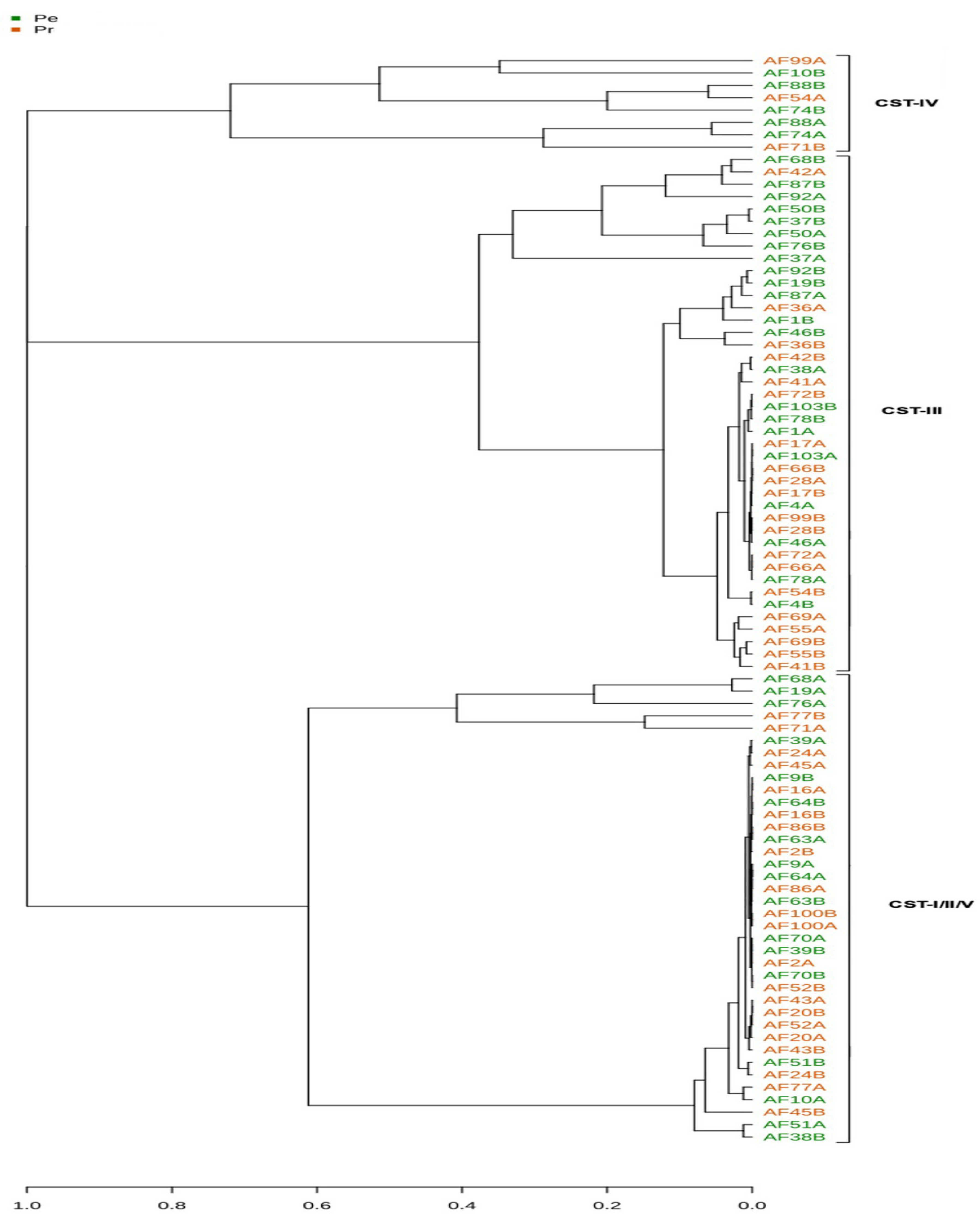
Microbial communities were clustered into CSTs by constructing a dendrogram with Microbiome Analyst (https://www.microbiomeanalyst.ca), using the complete linkage algorithm and Bray–Curtis dissimilarity as distance metrics (Figure A2). Given the limited species-level resolution of the V4 16S rRNA sequencing strategy, it was not possible to differentiate CST-I (L. crispatus), CST-II (L. gasseri), and CST-V (L. jensenii) with confidence. These were therefore grouped together as CST-I/II/V (i.e., non-L. iners-dominant communities). Importantly, CST-III (L. iners dominance) and CST-IV (non-Lactobacillus-dominant communities) remained clearly distinguishable, allowing for meaningful clinical interpretation.

2.4. Statistical Analyses of Microbiomes

The CST profiles of each treatment group were compared between T0 and T1 using the McNemar–Bowker test. Rarefied samples were analyzed using QIIME2 to determine alpha (species richness and Shannon index) and beta diversity indices (i.e., Bray–Curtis dissimilarity). Alpha diversity was compared between the PE and PR groups at both timepoints, using the Kruskal–Wallis test (performed with the R package v4.2.0), following the syntax provided by Chen et al. [32]. Beta diversity comparisons were conducted using permutational multivariate analysis of variance–pairwise analysis, with visualizations generated through Principal Coordinates Analysis plot by using EMPeror (Microbiome Analysis and Visualization Lab, University of California, San Diego, San Diego, CA, USA) [33]. All statistical analyses were conducted at a significance level of 95%. Differential taxonomic abundance was determined using analysis of the composition of microbiomes (ANCOM) within QIIME2.

3. Results

The mean age of patients was 30.1 ± 7.2 years in the PE group and 28.1 ± 6.0 years in the PR group. Most participants were White (68.2% and 72.7%, respectively), and 15.9% reported a history of sPTB. In the current pregnancy, sPTB occurred in 9.1% of patients in the PE group and 30.0% in the PR group. While this difference may seem clinically relevant, it did not reach statistical significance. For reference, the original RCT reported sPTB rates of 32.4% and 29.7% in the PE and PR groups, respectively (p = 0.68). Overall, baseline characteristics were consistent with those of the RCT, with no significant differences between groups. Nonetheless, the small sample size of the present study should be acknowledged as a limitation (Table 1).
As shown in Figure 1 and consistent with expected trends during pregnancy, most analyzed samples exhibited low diversity and were Lactobacillus-dominated (n = 80). Of these, 42 samples were dominated by L. iners (i.e., CST-III), whereas 38 samples were dominated by other Lactobacillus species (i.e., CST-I/II/V). The remaining eight samples had higher diversity, no Lactobacillus dominance, and increased abundance of anaerobes, including BV-associated Gardnerella (i.e., CST-IV), which indicated a state of dysbiosis. The observed CST distribution was consistent with that of previous findings in a larger, similar Brazilian population in which L. iners dominance was also common [34].
Most samples in both groups had normal Nugent scores (Table 1). An altered score (i.e., >3), while not frequently observed, was associated with highly diverse communities, as expected (Figure 1).
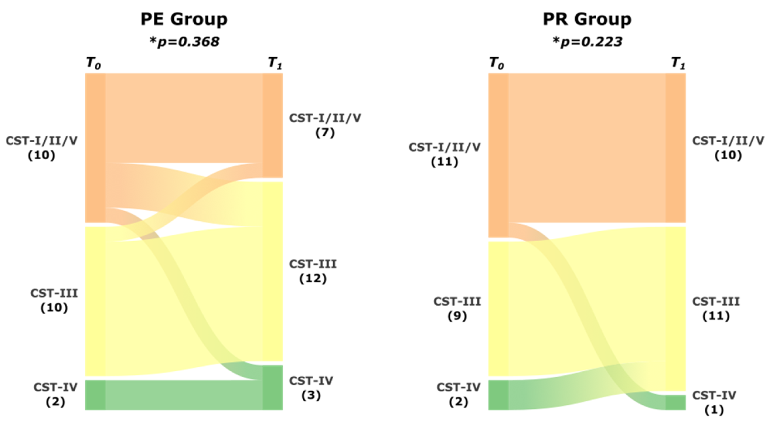
With regard to community stability, some CST changes were observed between the two sampling timepoints, with two samples (AF10 and AF71) shifting toward the potentially dysbiotic CST-IV. Notably, sample AF10 was collected from a patient in the PE group who delivered at 25 weeks. In contrast, sample AF71 was obtained from the PR group, and the patient delivered at term. Yet, no significant difference in the CST profile was observed between T0 and T1 in the PE group (p = 0.368) or PR group (p = 0.223) (Figure 2). Thus, treatment with a PE or PR did not significantly alter the CST distribution. L. iners dominance similarly did not increase with either treatment.
A comparison of individual sample species diversity, or alpha diversity, was then performed by using two methods: richness (i.e., observed), which accounts solely for the number of observed species in the community, and the Shannon index, which accounts for the richness and the evenness of species distribution. Comparisons between T0 and T1 within each group (Figure 3A,B) and between each group at T0 and T1 (Figure 3C,D) revealed no significant differences. This finding suggested that both groups were equally diverse and stable during the observation period.
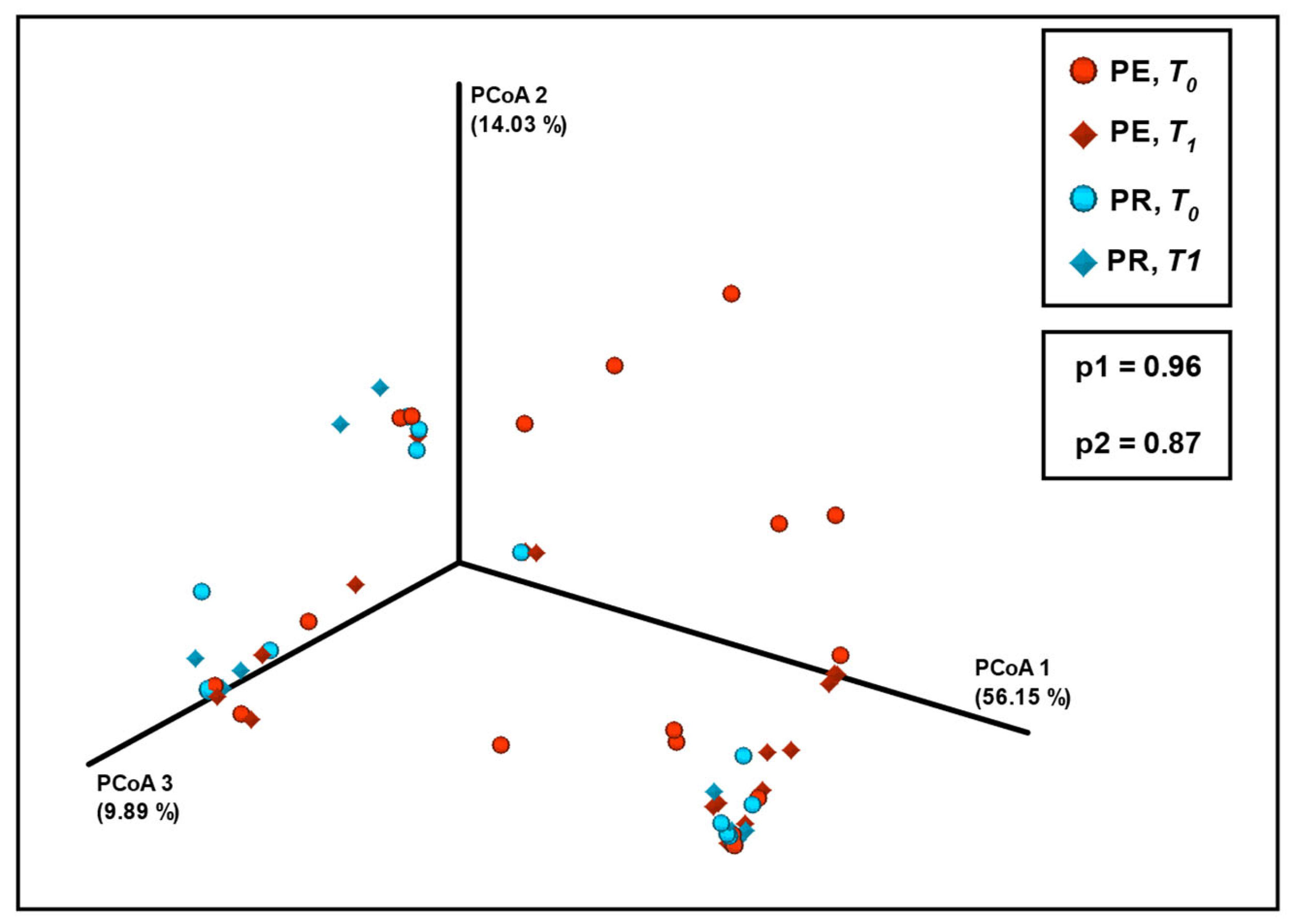
No significant differences in species composition dissimilarity or in beta diversity were observed between the sampling timepoints in the PE or PR group (Figure 4) because no clusters related to the sampling timepoint were observed in the ordination plot. The Bray–Curtis dissimilarity method was chosen because of its quantitative nature, making it more suitable for the analysis of communities dominated by a specific species such as the vaginal microbiome. This method was also preferred for CST clustering in the study population (Figure A2). Additional diversity analyses were performed using other methods. A complete dataset can be found at https://drive.google.com/drive/folders/12uEFU2X0AuTpqToRhMOuoMJnSOYYTvpp?usp=drive (accessed 29 September 2025).
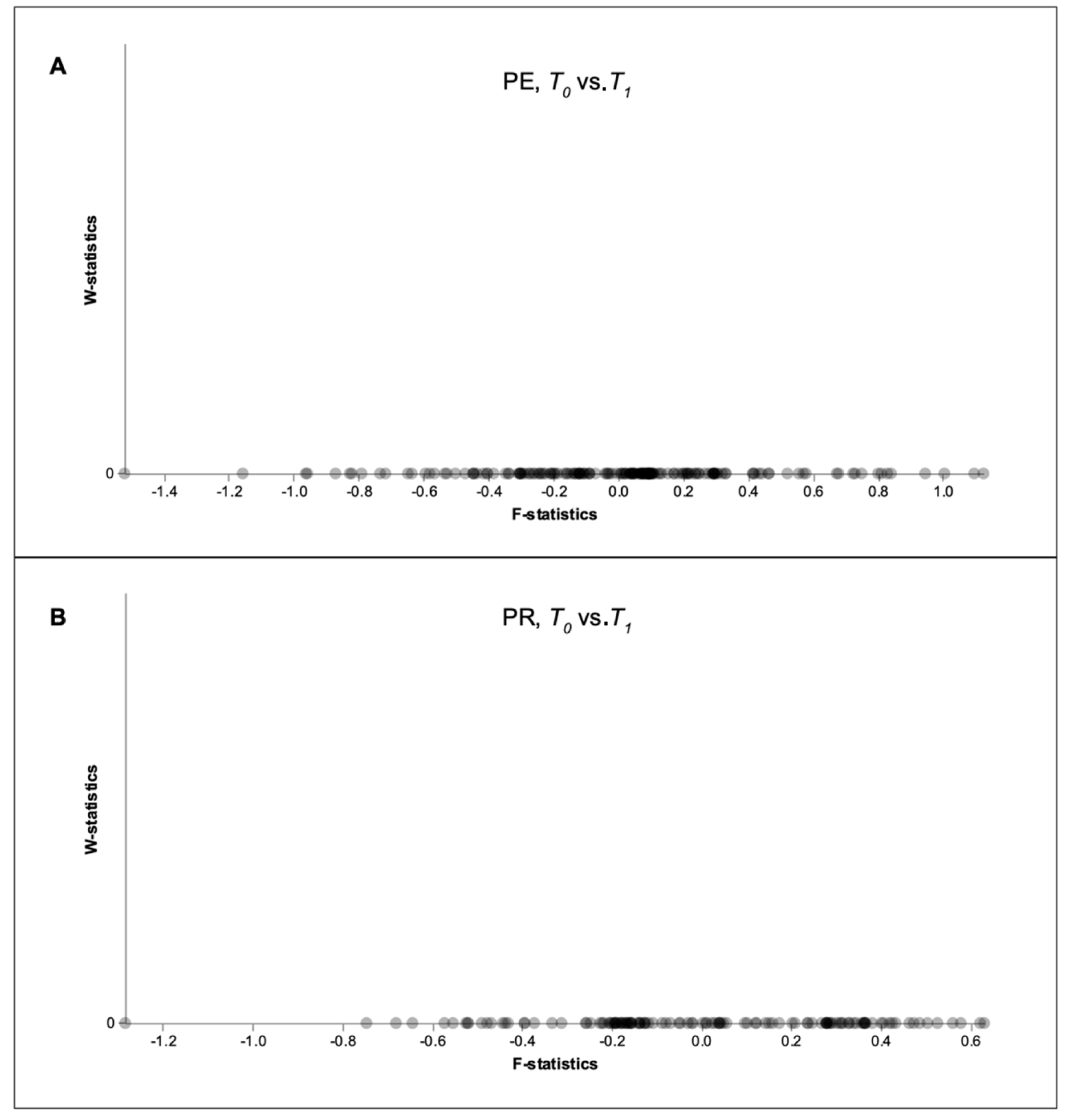
Finally, differential abundance analysis using ANCOM, which accounts for the compositional nature of microbiomes, was performed. It allows for the inference of the absolute abundance from the relative abundance and for the correction of sampling bias. This analysis was conducted to further explore possible differences in taxa abundance between treatment groups and sampling timepoints, including dominant features such as Lactobacillus species and Gardnerella. Accordingly, ANCOM did not detect any differences in taxa abundances between T0 and T1 in either the PE or PR group (Figure A3). Although the small sample size and the known limitations of ANCOM must be considered, these findings suggest that neither treatment was associated with significant changes in the abundance of individual taxa.

4. Discussion

In this study, we compared the vaginal microbiome after PE or PR treatment in women at high risk for sPTB. We found that, regardless of the treatment method, the vaginal microbiome remained stable in terms of bacterial diversity and CST composition, similar to patterns observed in normal pregnancies [12,13,14,15]. Vaginal PR is the standard treatment for sPTB prevention in women with a short cervix. It is generally regarded as safe. Moreover, by promoting proliferation of Lactobacillus and maintenance of an acidic pH, PR helps in keeping a safe vaginal environment [16]. By contrast, a PE functions as a foreign body and can potentially increase the susceptibility of the vaginal environment to colonization by infectious microorganisms and consequently increase the risk of sPTB. Indeed, PE is frequently associated with abundant and malodorous vaginal discharge. However, in our study, neither treatment appeared to drive vaginal dysbiosis more than the other, indicating that both treatments are equivalent from the perspective of the vaginal microbiota. Considering the mechanisms of action, microbiome stability was expected for PR, but comes as a surprise for PE, as no correlation with the characteristics of vaginal contents was observed.
Our findings are consistent with those of two previous studies addressing this issue. Kindinger et al. [19] observed no PR-associated alterations in the vaginal microbiome in a predominantly Caucasian English population. Vargas et al. [26] studied a mixed-ancestry European cohort and similarly found that patients treated with cerclage (another vaginal foreign body) had higher vaginal microbiome diversity and a reduced Lactobacillus content; however, women treated with a PE did not exhibit such microbiome modifications. A possible explanation for this difference could be related to the component materials of the devices. Maybe the silicone used to make PE renders it less prone to pathogen proliferation.
Considerable efforts have recently been expended to identify sPTB-related signatures in the vaginal microbiome. Growing evidence suggests that sPTB may be associated with vaginal ecosystem instability [35]. Microbiome-based sPTB prediction models have been developed using large publicly available datasets. However, the compositional complexity and heterogeneity of the molecular and statistical techniques employed across studies have made drawing definite conclusions difficult [36]. Lactobacillus dominance, CST type, and the presence of specific microorganisms such as Gardnerella are clinically key factors to consider when evaluating sPTB risk.
An argument is that the microbial composition associated with a short cervix may already reflect a dysbiotic state, predisposing patients to sPTB, even before treatment begins. For instance, Witkin et al. [34] analyzed the data of a larger Brazilian population (n = 340), with most (90%) presenting with a normal-sized cervix. They found the CST distribution was similar to that demonstrated in our present study. An interesting finding is that the authors identified CST-III as a potential risk factor for cervical shortening, possibly because of biochemical modifications related to L-La secretion by L. iners, resulting in a proinflammatory state. Kindinger et al. [19] similarly suggested that CST-III may represent an intermediate dysbiotic state. Therefore, CST-III dominance in our population may not only reflect a high risk of sPTB but also be a causative factor in cervical shortening. This observation warrants further investigation. Screening pregnant women for CST-III may be clinically relevant and contribute to sPTB prevention in the future. Additionally, adjunctive strategies aimed at modulating the vaginal microbiota toward a more eubiotic (i.e., healthy) composition may improve sPTB prevention when used with PE or PR treatment.
A key strength of our study lies in its focus on vaginal microbiome stability in a Brazilian population at risk of sPTB. This patient group has not been extensively studied in this context. Moreover, to the best of our knowledge, the present study is the first in which PE and PR treatments were compared. In previous studies, each of these treatments was independently compared against a no-treatment control (e.g., cervical length > 25 mm).
Nevertheless, this study has several limitations. The relatively small sample size (n = 22 per group), although comparable to those reported by Kindinger et al. [19] (n = 25, PR group) and Vargas et al. [26] (n = 26, PE group), limits the statistical power to detect microbiome alterations. For instance, in a post hoc analysis, the power to detect CST shifts towards dysbiosis was only 25%, assuming a fixed sample of 22, the lowest observed shift rate (9%), and a one-tailed hypothesis (calculated with the software G*Power 3.1.9.7.). Consequently, establishing definitive conclusions about the association between treatment-induced vaginal dysbiosis and sPTB remains challenging. Another limitation of our study relates to the sequencing strategy employed, which intrinsically restricts taxonomic resolution at the species level. This limitation precluded accurate discrimination among CST-I (L. crispatus), CST-II (L. gasseri), and CST-V (L. jensenii). Consequently, these subtypes were aggregated and analyzed as CST-I/II/V (i.e., non-L. iners-dominated profiles). Although this approach was methodologically necessary, it may have underestimated the actual heterogeneity within Lactobacillus-dominated CSTs and potentially obscured species-specific associations with clinical outcomes. For instance, previous evidence suggests that L. crispatus may confer stronger protection compared with other Lactobacillus species. Further analyses of the remaining 159 samples from the original trial, together with larger prospective cohorts employing higher species-level resolution, will be critical to validate our observations and to determine their potential clinical implications.

5. Conclusions

Vaginal PR tablets and Arabin PEs are widely prescribed to prevent sPTB in at-risk women with a short cervix, identified during the second trimester. Our findings indicate that these treatments do not significantly alter the vaginal microbiome; thus, patients and clinicians can be reassured about the safety of these methods with regard to infectious complications. This knowledge contributes to improving the quality of care provided to patients at risk of sPTB.

Author Contributions

Conceptualization, A.G.A.F., J.V.C.M., R.P.V.F. and M.H.B.C.; methodology, A.G.A.F., L.A.M.F., R.C.R.M. and M.H.B.C.; validation, A.G.A.F., L.A.M.F. and R.C.R.M.; formal analysis, A.G.A.F., S.V.P. and R.C.R.M.; investigation, A.G.A.F., L.A.M.F. and R.C.R.M.; resources, R.P.V.F.; data curation, A.G.A.F., J.V.C.M. and M.H.B.C.; writing—original draft preparation, A.G.A.F. and M.H.B.C.; writing—review and editing, A.G.A.F. and M.H.B.C.; visualization, A.G.A.F.; supervision, M.H.B.C.; project administration, M.H.B.C.; funding acquisition, R.P.V.F. All authors have read and agreed to the published version of the manuscript.

Funding

This research received no external funding.

Institutional Review Board Statement

The study was conducted in accordance with the Declaration of Helsinki and approved by the Ethics Committee for Analysis of Research Projects (CAPPesq) of the Hospital das Clinicas, Universidade de São Paulo (São Paulo, Brazil; approval number: CAAE 20611813.0.0000.0068; approval date: 27 November 2013).

Informed Consent Statement

Informed consent was obtained from all subjects (or their legal guardians) involved in this study. Signed consent forms are kept in our department for further reference.

Data Availability Statement

The original data presented in this study are openly available in Google Drive at https://drive.google.com/drive/folders/12uEFU2X0AuTpqToRhMOuoMJnSOYYTvpp?usp=drive (accessed 29 September 2025).

Acknowledgments

The authors wish to express their gratitude to Mark Carrington for reviewing the paper and to Esther C. Sabino, Mara Hoshida, and Priscila G. Amaral for their invaluable contributions to this research.

Conflicts of Interest

The authors declare no conflicts of interest.

Abbreviations

The following abbreviations are used in this manuscript:
ASVAmplicon sequence variant
BVBacterial vaginosis
CSTCommunity state type
D-LaD-lactic acid
L-LaL-lactic acid
OTUOperational taxonomic unit
PCRPolymerase chain reaction
PEPessary
PRProgesterone
PTBPreterm birth
sPTBSpontaneous preterm birth
TVUSTransvaginal ultrasound
QIIMEQuantitative Insights into Microbial Ecology

Appendix A

Appendix A.1

Figure A1. Initial analysis of vaginal microbiome composition, including a control group with normal-sized cervices. The figure shows the percentage abundance of the 10 most abundant taxa identified in the control, pessary, and progesterone groups.
Diseases 13 00338 g0a1

Appendix A.2

Table A1. V4 16S rRNA gene primer fusion sequences. The sequences of DNA oligonucleotides used for PCR amplification of the V4 region of the rDNA gene. PCR: polymerase chain reaction; DNA: deoxyribonucleic acid; rDNA: ribosomal DNA.
PRIMERCOMPLETE SEQUENCEADAPTERBARCODE16S-V4
R806_trP1_revCCTCTCTATGGGCAGTCGGTGATGGACTACHVGGGTWTCTAAT
F515_16S(for)-BC-01CCATCTCATCCCTGCGTGTCTCCGACTCAGCTAAGGTAACGATGTGCCAGCMGCCGCGGTAACCATCTCATCCCTGCGTGTCTCCGACTCACTAAGGTAACGATGTGCCAGCMGCCGCGGTAA
F515_16S(for)-BC-02CCATCTCATCCCTGCGTGTCTCCGACTCAGTAAGGAGAACGATGTGCCAGCMGCCGCGGTAACCATCTCATCCCTGCGTGTCTCCGACTCAGTAAGGAGAACGATGTGCCAGCMGCCGCGGTAA
F515_16S(for)-BC-03CCATCTCATCCCTGCGTGTCTCCGACTCAGAAGAGGATTCGATGTGCCAGCMGCCGCGGTAACCATCTCATCCCTGCGTGTCTCCGACTCAGAAGAGGATTCGATGTGCCAGCMGCCGCGGTAA
F515_16S(for)-BC-04CCATCTCATCCCTGCGTGTCTCCGACTCAGTACCAAGATCGATGTGCCAGCMGCCGCGGTAACCATCTCATCCCTGCGTGTCTCCGACTCAGTACCAAGATCGATGTGCCAGCMGCCGCGGTAA
F515_16S(for)-BC-05CCATCTCATCCCTGCGTGTCTCCGACTCAGCAGAAGGAACGATGTGCCAGCMGCCGCGGTAACCATCTCATCCCTGCGTGTCTCCGACTCAGCAGAAGGAACGATGTGCCAGCMGCCGCGGTAA
F515_16S(for)-BC-06CCATCTCATCCCTGCGTGTCTCCGACTCAGCTGCAAGTTCGATGTGCCAGCMGCCGCGGTAACCATCTCATCCCTGCGTGTCTCCGACTCAGCTGCAAGTTCGATGTGCCAGCMGCCGCGGTAA
F515_16S(for)-BC-07CCATCTCATCCCTGCGTGTCTCCGACTCAGTTCGTGATTCGATGTGCCAGCMGCCGCGGTAACCATCTCATCCCTGCGTGTCTCCGACTCAGTTCGTGATTCGATGTGCCAGCMGCCGCGGTAA
F515_16S(for)-BC-08CCATCTCATCCCTGCGTGTCTCCGACTCAGTTCCGATAACGATGTGCCAGCMGCCGCGGTAACCATCTCATCCCTGCGTGTCTCCGACTCAGTTCCGATAACGATGTGCCAGCMGCCGCGGTAA
F515_16S(for)-BC-09CCATCTCATCCCTGCGTGTCTCCGACTCAGTGAGCGGAACGATGTGCCAGCMGCCGCGGTAACCATCTCATCCCTGCGTGTCTCCGACTCAGTGAGCGGAACGATGTGCCAGCMGCCGCGGTAA
F515_16S(for)-BC-10CCATCTCATCCCTGCGTGTCTCCGACTCAGCTGACCGAACGATGTGCCAGCMGCCGCGGTAACCATCTCATCCCTGCGTGTCTCCGACTCAGCTGACCGAACGATGTGCCAGCMGCCGCGGTAA
F515_16S(for)-BC-11CCATCTCATCCCTGCGTGTCTCCGACTCAGTCCTCGAATCGATGTGCCAGCMGCCGCGGTAACCATCTCATCCCTGCGTGTCTCCGACTCAGTCCTCGAATCGATGTGCCAGCMGCCGCGGTAA
F515_16S(for)-BC-12CCATCTCATCCCTGCGTGTCTCCGACTCAGTAGGTGGTTCGATGTGCCAGCMGCCGCGGTAACCATCTCATCCCTGCGTGTCTCCGACTCAGTAGGTGGTTCGATGTGCCAGCMGCCGCGGTAA
F515_16S(for)-BC-13CCATCTCATCCCTGCGTGTCTCCGACTCAGTCTAACGGACGATGTGCCAGCMGCCGCGGTAACCATCTCATCCCTGCGTGTCTCCGACTCAGTCTAACGGACGATGTGCCAGCMGCCGCGGTAA
F515_16S(for)-BC-14CCATCTCATCCCTGCGTGTCTCCGACTCAGTTGGAGTGTCGATGTGCCAGCMGCCGCGGTAACCATCTCATCCCTGCGTGTCTCCGACTCAGTTGGAGTGTCGATGTGCCAGCMGCCGCGGTAA
F515_16S(for)-BC-15CCATCTCATCCCTGCGTGTCTCCGACTCAGTCTAGAGGTCGATGTGCCAGCMGCCGCGGTAACCATCTCATCCCTGCGTGTCTCCGACTCAGTCTAGAGGTCGATGTGCCAGCMGCCGCGGTAA
F515_16S(for)-BC-16CCATCTCATCCCTGCGTGTCTCCGACTCAGTCTGGATGACGATGTGCCAGCMGCCGCGGTAACCATCTCATCCCTGCGTGTCTCCGACTCAGTCTGGATGACGATGTGCCAGCMGCCGCGGTAA
F515_16S(for)-BC-17CCATCTCATCCCTGCGTGTCTCCGACTCAGTCTATTCGTCGATGTGCCAGCMGCCGCGGTAACCATCTCATCCCTGCGTGTCTCCGACTCAGTCTATTCGTCGATGTGCCAGCMGCCGCGGTAA
F515_16S(for)-BC-18CCATCTCATCCCTGCGTGTCTCCGACTCAGAGGCAATTGCGATGTGCCAGCMGCCGCGGTAACCATCTCATCCCTGCGTGTCTCCGACTCAGAGGCAATTGCGATGTGCCAGCMGCCGCGGTAA
F515_16S(for)-BC-19CCATCTCATCCCTGCGTGTCTCCGACTCAGTTAGTCGGACGATGTGCCAGCMGCCGCGGTAACCATCTCATCCCTGCGTGTCTCCGACTCAGTTAGTCGGACGATGTGCCAGCMGCCGCGGTAA
F515_16S(for)-BC-20CCATCTCATCCCTGCGTGTCTCCGACTCAGCAGATCCATCGATGTGCCAGCMGCCGCGGTAACCATCTCATCCCTGCGTGTCTCCGACTCAGCAGATCCATCGATGTGCCAGCMGCCGCGGTAA
F515_16S(for)-BC-21CCATCTCATCCCTGCGTGTCTCCGACTCAGTCGCAATTACGATGTGCCAGCMGCCGCGGTAACCATCTCATCCCTGCGTGTCTCCGACTCAGTCGCAATTACGATGTGCCAGCMGCCGCGGTAA
F515_16S(for)-BC-22CCATCTCATCCCTGCGTGTCTCCGACTCAGTTCGAGACGCGATGTGCCAGCMGCCGCGGTAACCATCTCATCCCTGCGTGTCTCCGACTCAGTTCGAGACGCGATGTGCCAGCMGCCGCGGTAA
F515_16S(for)-BC-23CCATCTCATCCCTGCGTGTCTCCGACTCAGTGCCACGAACGATGTGCCAGCMGCCGCGGTAACCATCTCATCCCTGCGTGTCTCCGACTCAGTGCCACGAACGATGTGCCAGCMGCCGCGGTAA
F515_16S(for)-BC-24CCATCTCATCCCTGCGTGTCTCCGACTCAGAACCTCATTCGATGTGCCAGCMGCCGCGGTAACCATCTCATCCCTGCGTGTCTCCGACTCAGAACCTCATTCGATGTGCCAGCMGCCGCGGTAA
F515_16S(for)-BC-25CCATCTCATCCCTGCGTGTCTCCGACTCAGCCTGAGATACGATGTGCCAGCMGCCGCGGTAACCATCTCATCCCTGCGTGTCTCCGACTCAGCCTGAGATACGATGTGCCAGCMGCCGCGGTAA
F515_16S(for)-BC-26CCATCTCATCCCTGCGTGTCTCCGACTCAGTTACAACCTCGATGTGCCAGCMGCCGCGGTAACCATCTCATCCCTGCGTGTCTCCGACTCAGTTACAACCTCGATGTGCCAGCMGCCGCGGTAA
F515_16S(for)-BC-27CCATCTCATCCCTGCGTGTCTCCGACTCAGAACCATCCGCGATGTGCCAGCMGCCGCGGTAACCATCTCATCCCTGCGTGTCTCCGACTCAGAACCATCCGCGATGTGCCAGCMGCCGCGGTAA
F515_16S(for)-BC-28CCATCTCATCCCTGCGTGTCTCCGACTCAGATCCGGAATCGATGTGCCAGCMGCCGCGGTAACCATCTCATCCCTGCGTGTCTCCGACTCAGATCCGGAATCGATGTGCCAGCMGCCGCGGTAA
F515_16S(for)-BC-29CCATCTCATCCCTGCGTGTCTCCGACTCAGTCGACCACTCGATGTGCCAGCMGCCGCGGTAACCATCTCATCCCTGCGTGTCTCCGACTCAGTCGACCACTCGATGTGCCAGCMGCCGCGGTAA
F515_16S(for)-BC-30CCATCTCATCCCTGCGTGTCTCCGACTCAGCGAGGTTATCGATGTGCCAGCMGCCGCGGTAACCATCTCATCCCTGCGTGTCTCCGACTCAGCGAGGTTATCGATGTGCCAGCMGCCGCGGTAA
F515_16S(for)-BC-31CCATCTCATCCCTGCGTGTCTCCGACTCAGTCCAAGCTGCGATGTGCCAGCMGCCGCGGTAACCATCTCATCCCTGCGTGTCTCCGACTCAGTCCAAGCTGCGATGTGCCAGCMGCCGCGGTAA
F515_16S(for)-BC-32CCATCTCATCCCTGCGTGTCTCCGACTCAGTCTTACACACGATGTGCCAGCMGCCGCGGTAACCATCTCATCCCTGCGTGTCTCCGACTCAGTCTTACACACGATGTGCCAGCMGCCGCGGTAA

Appendix A.3

Figure A2. CST clustering of samples in the study population. Using the complete linkage algorithm and Bray–Curtis dissimilarity as distance metrics, three CST clusters could be identified in the study population. Pe, pessary group; Pr, progesterone group; CST, community state type.
Diseases 13 00338 g0a2

Appendix A.4

Figure A3. Differential taxa abundance in the vaginal microbiome between the two sampling timepoints in the PE and PR groups (ANCOM). The F-statistics are represented on the x-axis of the volcano plots, and the W-statistics on the y-axis. The dots correspond to the identified taxa, including the dominant Lactobacillus. No taxa with significant W values were identified (i.e., W values close to zero). (A) Comparison results between T0 and T1 in the PE group. (B) Comparison results between T0 and T1 in the PR group. T0: before treatment; T1: after treatment; ANCOM: analysis of compositions of microbiomes; PR: progesterone; PE: pessary.
Diseases 13 00338 g0a3

References

  1. Leal, M.D.; Esteves-Pereira, A.P.; Nakamura-Pereira, M.; Torres, J.A.; Theme-Filha, M.; Domingues, R.M.S.M.; Dias, M.A.B.; Moreira, M.E.; Gama, S.G. Prevalence and risk factors related to preterm birth in Brazil. Reprod. Health 2016, 13 (Suppl. S3), 127. [Google Scholar] [CrossRef]
  2. Romero, R.; Gomez-Lopez, N.; Winters, A.D.; Jung, E.; Shaman, M.; Bieda, J.; Panaitescu, B.; Pacora, P.; Erez, O.; Greenberg, J.M.; et al. Evidence that intra-amniotic infections are often the result of an ascending invasion—A molecular microbiological study. J. Perinat. Med. 2019, 47, 915–931. [Google Scholar] [CrossRef]
  3. Carvalho, M.H.B.D.; Bittar, R.E.; Maganha, P.P.D.A.; Pereira, S.V.; Zugaib, M. Associação da vaginose bacteriana com o parto prematuro espontâneo [The association between bacterial vaginosis and premature birth]. Rev. Bras. Gynecol. Obstet. 2001, 23, 529–533. [Google Scholar]
  4. de Carvalho, M.H.B.; Bittar, R.E.; Brizot, M.; Bicudo, C.; Zugaib, M. Prediction of preterm delivery in the second trimester. Obstet. Gynecol. 2005, 105, 532–536. [Google Scholar] [CrossRef] [PubMed]
  5. Romero, R.; Nicolaides, K.H.; Conde-Agudelo, A.; O’Brien, J.M.; Cetingoz, E.; Da Fonseca, E.; Creasy, G.W.; Hassan, S.S. Vaginal progesterone decreases preterm birth ≤34 weeks of gestation in women with a singleton pregnancy and a short cervix: An updated meta-analysis including data from the OPPTIMUM study. Ultrasound Obstet. Gynecol. 2016, 48, 308–317. [Google Scholar] [CrossRef] [PubMed]
  6. Saccone, G.; Ciardulli, A.; Xodo, S.; Dugoff, L.; Ludmir, J.; Pagani, G.; Visentin, S.; Gizzo, S.; Volpe, N.; Maruotti, G.M.; et al. Cervical pessary for preventing preterm birth in singleton pregnancies with short cervical length: A systematic review and meta-analysis. J. Ultrasound Med. 2017, 36, 1535–1543. [Google Scholar] [CrossRef]
  7. Conde-Agudelo, A.; Romero, R.; Nicolaides, K.H. Cervical pessary to prevent preterm birth in asymptomatic high-risk women: A systematic review and meta-analysis. Am. J. Obstet. Gynecol. 2020, 223, 42–65.e2. [Google Scholar] [CrossRef]
  8. Pacagnella, R.C.; Silva, T.V.; Cecatti, J.G.; Passini, R., Jr.; Fanton, T.F.; Borovac-Pinheiro, A.; Pereira, C.M.; Fernandes, K.G.; França, M.S.; Li, W.; et al. Pessary plus progesterone to prevent preterm birth in women with short cervixes: A randomized controlled trial. Obstet. Gynecol. 2022, 139, 41–51. [Google Scholar] [CrossRef]
  9. ACOG. Practice Bulletin No.142: Cerclage for the management of cervical insufficiency. Obstet. Gynecol. 2014, 123, 372–379. [Google Scholar] [CrossRef]
  10. Ravel, J.; Gajer, P.; Abdo, Z.; Schneider, G.M.; Koenig, S.S.K.; McCulle, S.L.; Karlebach, S.; Gorle, R.; Russell, J.; Tacket, C.O.; et al. Vaginal microbiome of reproductive-age women. Proc. Natl. Acad. Sci. USA 2011, 108 (Suppl. S1), 4680–4687. [Google Scholar] [CrossRef]
  11. Gajer, P.; Brotman, R.M.; Bai, G.; Sakamoto, J.; Schütte, U.M.E.; Zhong, X.; Koenig, S.S.K.; Fu, L.; Ma, Z.S.; Zhou, X.; et al. Temporal dynamics of the human vaginal microbiota. Sci. Transl. Med. 2012, 4, 132ra52. [Google Scholar] [CrossRef]
  12. Romero, R.; Hassan, S.S.; Gajer, P.; Tarca, A.L.; Fadrosh, D.W.; Nikita, L.; Galuppi, M.; Lamont, R.F.; Chaemsaithong, P.; Miranda, J.; et al. The composition and stability of the vaginal microbiota of normal pregnant women is different from that of non-pregnant women. Microbiome 2014, 2, 4. [Google Scholar] [CrossRef] [PubMed]
  13. DiGiulio, D.B.; Callahan, B.J.; McMurdie, P.J.; Costello, E.K.; Lyell, D.J.; Robaczewska, A.; Sun, C.L.; Goltsman, D.S.A.; Wong, R.J.; Shaw, G.; et al. Temporal and spatial variation of the human microbiota during pregnancy. Proc. Natl. Acad. Sci. USA 2015, 112, 11060–11065. [Google Scholar] [CrossRef] [PubMed]
  14. MacIntyre, D.A.; Chandiramani, M.; Lee, Y.S.; Kindinger, L.; Smith, A.; Angelopoulos, N.; Lehne, B.; Arulkumaran, S.; Brown, R.; Teoh, T.G.; et al. The vaginal microbiome during pregnancy and the postpartum period in a European population. Sci. Rep. 2015, 5, 8988. [Google Scholar] [CrossRef] [PubMed]
  15. Serrano, M.G.; Parikh, H.I.; Brooks, J.P.; Edwards, D.J.; Arodz, T.J.; Edupuganti, L.; Huang, B.; Girerd, P.H.; Bokhari, Y.A.; Bradley, S.P.; et al. Racioethnic diversity in the dynamics of the vaginal microbiome during pregnancy. Nat. Med. 2019, 25, 1001–1011. [Google Scholar] [CrossRef]
  16. Witkin, S.S.; Linhares, I.M. Why do lactobacilli dominate the human vaginal microbiota? BJOG 2017, 124, 606–611. [Google Scholar] [CrossRef]
  17. Callahan, B.J.; DiGiulio, D.B.; Goltsman, D.S.A.; Sun, C.L.; Costello, E.K.; Jeganathan, P.; Biggio, J.R.; Wong, R.J.; Druzin, M.L.; Shaw, G.M.; et al. Replication and refinement of a vaginal microbial signature of preterm birth in two racially distinct cohorts of US women. Proc. Natl. Acad. Sci. USA 2017, 114, 9966–9971. [Google Scholar] [CrossRef]
  18. Stout, M.J.; Zhou, Y.; Wylie, K.M.; Tarr, P.I.; Macones, G.A.; Tuuli, M.G. Early pregnancy vaginal microbiome trends and preterm birth. Am. J. Obstet. Gynecol. 2017, 217, 356.e1–356.e18. [Google Scholar] [CrossRef]
  19. Kindinger, L.M.; Bennett, P.R.; Lee, Y.S.; Marchesi, J.R.; Smith, A.; Cacciatore, S.; Holmes, E.; Nicholson, J.K.; Teoh, T.G.; MacIntyre, D.A. The interaction between vaginal microbiota, cervical length, and vaginal progesterone treatment for preterm birth risk. Microbiome 2017, 5, 6. [Google Scholar] [CrossRef]
  20. Freitas, A.C.; Bocking, A.; Hill, J.E.; Money, D.M.; VOGUE Research Group. Increased richness and diversity of the vaginal microbiota and spontaneous preterm birth. Microbiome 2018, 6, 117. [Google Scholar] [CrossRef]
  21. Fettweis, J.M.; Serrano, M.G.; Brooks, J.P.; Edwards, D.J.; Girerd, P.H.; Parikh, H.I.; Huang, B.; Arodz, T.J.; Edupuganti, L.; Glascock, A.L.; et al. The vaginal microbiome and preterm birth. Nat. Med. 2019, 25, 1012–1021. [Google Scholar] [CrossRef]
  22. Feehily, C.; Crosby, D.; Walsh, C.J.; Lawton, E.M.; Higgins, S.; McAuliffe, F.M.; Cotter, P.D. Shotgun sequencing of the vaginal microbiome reveals both a species and functional potential signature of preterm birth. NPJ Biofilms Microbiomes 2020, 6, 50. [Google Scholar] [CrossRef]
  23. Gudnadottir, U.; Debelius, J.W.; Du, J.; Hugerth, L.W.; Danielsson, H.; Schuppe-Koistinen, I.; Fransson, E.; Brusselaers, N. The vaginal microbiome and the risk of preterm birth: A systematic review and network meta-analysis. Sci. Rep. 2022, 12, 7926. [Google Scholar] [CrossRef]
  24. Baud, A.; Hillion, K.H.; Plainvert, C.; Tessier, V.; Tazi, A.; Mandelbrot, L.; Poyart, C.; Kennedy, S.P. Microbial diversity in the vaginal microbiota and its link to pregnancy outcomes. Sci. Rep. 2023, 13, 9061. [Google Scholar] [CrossRef]
  25. France, M.; Alizadeh, M.; Brown, S.; Ma, B.; Ravel, J. Towards a deeper understanding of the vaginal microbiota. Nat. Microbiol. 2022, 7, 367–378. [Google Scholar] [CrossRef] [PubMed]
  26. Vargas, M.; Yañez, F.; Elias, A.; Bernabeu, A.; Goya, M.; Xie, Z.; Farrás, A.; Sánchez, O.; Soler, Z.; Blasquez, C.; et al. Cervical pessary and cerclage placement for preterm birth prevention and cervicovaginal microbiome changes. Acta Obstet. Gynecol. Scand. 2022, 101, 1403–1413. [Google Scholar] [CrossRef] [PubMed]
  27. Amorim Filho, A.G.; Martins, R.C.R.; Franco, L.A.M.; Marinelli, J.V.; Peres, S.V.; Francisco, R.P.; Carvalho, M.H. Effect of the Arabin Pessary and Natural Progesterone on the Vaginal Microbiome. Res. Sq. 2023; preprint (version 1). [Google Scholar] [CrossRef]
  28. Nugent, R.P.; Krohn, M.A.; Hillier, S.L. Reliability of diagnosing bacterial vaginosis is improved by a standardized method of Gram stain interpretation. J. Clin. Microbiol. 1991, 29, 297–301. [Google Scholar] [CrossRef] [PubMed]
  29. Caporaso, J.G.; Lauber, C.L.; Walters, W.A.; Berg-Lyons, D.; Lozupone, C.A.; Turnbaugh, P.J.; Fierer, N.; Knight, R. Global patterns of 16S rRNA diversity at a depth of millions of sequences per sample. Proc. Natl. Acad. Sci. USA 2011, 108 (Suppl. S1), 4516–4522. [Google Scholar] [CrossRef]
  30. Ribeiro, R.M.; Souza-Basqueira, M.; Oliveira, L.C.; Salles, F.C.; Pereira, N.B.; Sabino, E.C. An alternative storage method for characterization of the intestinal microbiota through next generation sequencing. Rev. Inst. Med. Trop. Sao Paulo 2018, 60, e77. [Google Scholar] [CrossRef]
  31. Callahan, B.J.; McMurdie, P.J.; Rosen, M.J.; Han, A.W.; Johnson, A.J.A.; Holmes, S.P. DADA2: High-resolution sample inference from Illumina amplicon data. Nat. Methods 2016, 13, 581–583. [Google Scholar] [CrossRef]
  32. Chen, Y.; Lin, H.; Cole, M.; Morris, A.; Martinson, J.; Mckay, H.; Mimiaga, M.; Margolick, J.; Fitch, A.; Methe, B.; et al. Signature changes in gut microbiome are associated with increased susceptibility to HIV-1 infection in MSM. Microbiome 2021, 9, 237. [Google Scholar] [CrossRef]
  33. Vázquez-Baeza, Y.; Pirrung, M.; Gonzalez, A.; Knight, R. EMPeror: A tool for visualizing high-throughput microbial community data. GigaScience 2013, 2, 16. [Google Scholar] [CrossRef]
  34. Witkin, S.S.; Moron, A.F.; Ridenhour, B.J.; Minis, E.; Hatanaka, A.; Sarmento, S.G.P.; Franca, M.S.; Carvalho, F.H.C.; Hamamoto, T.K.; Mattar, R.; et al. Vaginal biomarkers that predict cervical length and dominant bacteria in the vaginal microbiomes of pregnant women. mBio 2019, 10, e02242-19. [Google Scholar] [CrossRef]
  35. Dominguez-Bello, M.G. Gestational shaping of the maternal vaginal microbiome. Nat. Med. 2019, 25, 882–883. [Google Scholar] [CrossRef]
  36. Golob, J.L.; Oskotsky, T.T.; Tang, A.S.; Roldan, A.; Chung, V.; Ha, C.W.Y.; Wong, R.J.; Flynn, K.J.; Parraga-Leo, A.; Wibrand, C.; et al. Microbiome preterm birth DREAM challenge: Crowdsourcing machine learning approaches to advance preterm birth research. Cell Rep. Med. 2024, 5, 101350. [Google Scholar] [CrossRef] [PubMed]
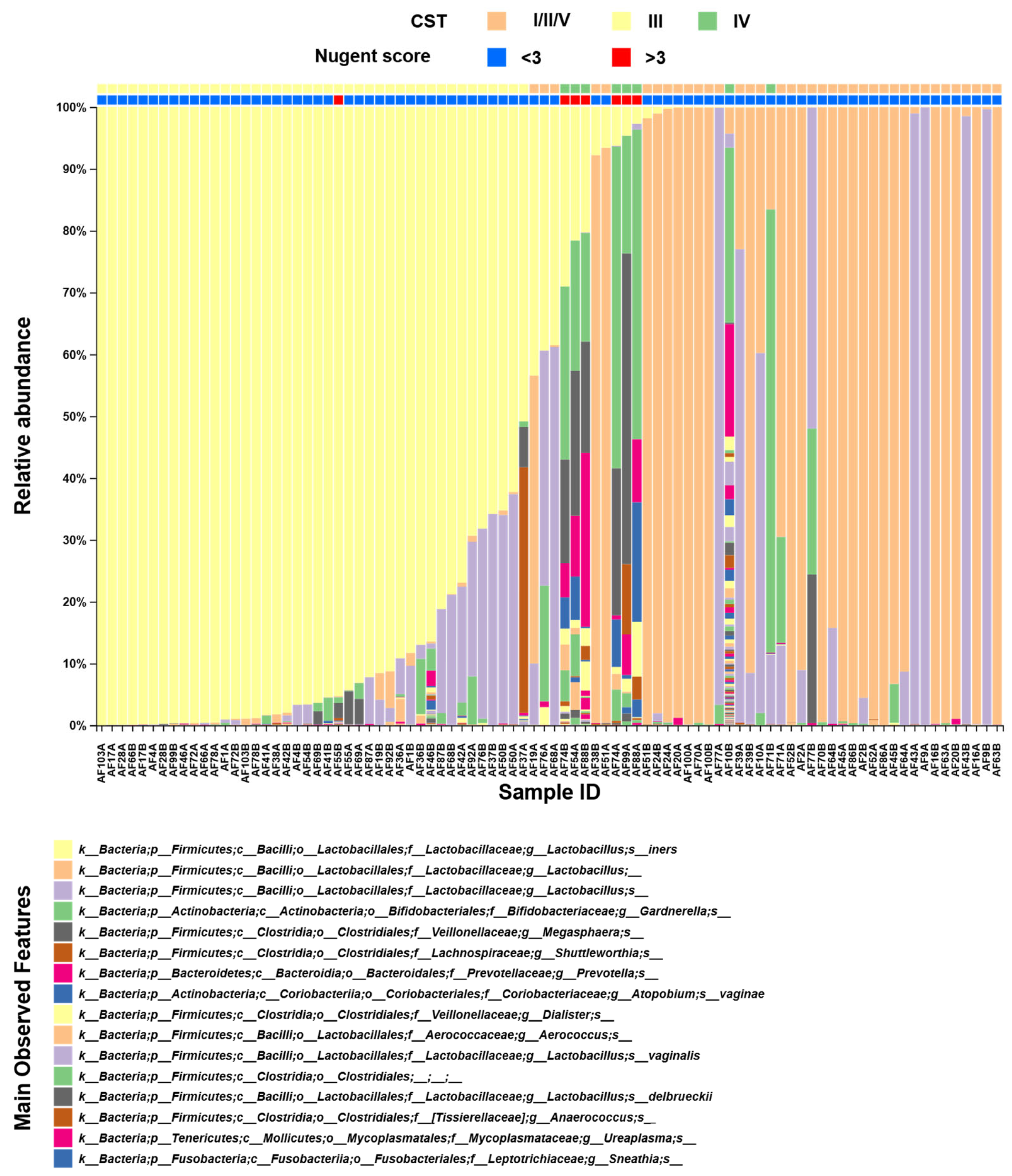
Figure 1. Composition of the vaginal microbiome. The taxa bar plot shows the main features identified, as shown in the bottom panel. Samples were ordinated based on L. iners abundance. The letters A and B in each sample identification (ID) denote timepoint T0 (i.e., before treatment) and timepoint T1 (i.e., after treatment), respectively. Community state types and Nugent scores are indicated by the bars at the top. CST: community state type; L. iners: Lactobacillus iners.
Diseases 13 00338 g001
Figure 2. Microbiome stability after treatment for a short cervix. The alluvial diagram shows the comparison of the distribution of the CST before treatment (T0) and after treatment (T1) in the PE and PR groups. * Based on the McNemar–Bowker test. PE: pessary; PR: progesterone; CST: community state type.
Diseases 13 00338 g002
Figure 3. Vaginal microbiome alpha diversity at the genus level. The upper two panels show the comparison of alpha diversity between T0 and T1 in the PE group (A) and PR group (B). The lower two panels show the comparison between the PE and PR groups at T0 (C) and T1 (D). In the boxes, the p-values are based on the Kruskal–Wallis test. Observed: richness; Shannon: Shannon’s diversity index; PE: pessary; PR: progesterone; T0: before treatment; T1: after treatment.
Diseases 13 00338 g003
Figure 4. The PCoA-ordinated plot of vaginal microbiome beta diversity, using the Bray–Curtis dissimilarity method at the genus level. The red dots indicate the PE group at T0 (i.e., before treatment); the red diamonds indicate the PE group at T1 (i.e., after treatment). The blue dots indicate the PR group at T0, and the blue diamonds indicate the PR group at T1. Bottom box presents the p-values, based on the PERMANOVA–pairwise test comparing T0 and T1. p1: PE group; p2: PR group. PCoA: principal coordinate analysis; PE: pessary; PR: progesterone; PERMANOVA: permutational multivariate analysis of variance.
Diseases 13 00338 g004
Table 1. Baseline characteristics of the study population.
CharacteristicPE (n = 22)PR (n = 22)p
Demographics
Age, y, mean ± SD (range)30.1 ± 7.2 (15–42)28.1 ± 6.0 (17–37)0.315 1
Ethnicity
White, n15 (68.2%)16 (72.7%)0.741 2
Black/mixed, n7 (31.8%)6 (27.3%)
BMI (kg/m2), mean ± SD (range)26.5 ± 4.3 (18.6–36.1)27.7 ± 5.1 (20.5–37.7)0.453 3
Obstetric history
Nulliparous, n13 (59.1%)8 (36.4%)0.131 2
Miscarriage, n7 (31.8%)8 (36.4%)0.750 2
Preterm birth, n4 (18.2%)3 (13.6%)1.000 4
Sample collection
Mean GA at T0 ± SD (range), wk22.3 ± 1.1 (20.6–23.9)22.9 ± 0.8 (21.6–24.9)0.107 3
Mean cervical length at T0 ± SD (range), mm16.0 ± 6.0 (5.0–24.0)17.0 ± 5.0 (7.0–23.0)0.494 3
Nugent score > 3, n4 (18.2%)3 (13.6%)1.000 4
Pregnancy outcome
Mean GA at birth ± SD (range), wk37.4 ± 4.0 (25.4–40.3)37.4 ± 3.4 (25.9–40.7)0.677 3
Spontaneous preterm birth, n2 (9.1%)6 (30.0%)0.123 4
PE: pessary group; PR: progesterone group; BMI: body mass index; GA: gestational age; SD: standard deviation. 1 Based on Student’s t-test. 2 Based on the chi-square test. 3 Based on the Mann–Whitney U test. 4 Based on Fisher’s exact test.
Disclaimer/Publisher’s Note: The statements, opinions and data contained in all publications are solely those of the individual author(s) and contributor(s) and not of MDPI and/or the editor(s). MDPI and/or the editor(s) disclaim responsibility for any injury to people or property resulting from any ideas, methods, instructions or products referred to in the content.