
Epidemiological Characteristics of Hospitalized Patients with Moderate versus Severe COVID-19 Infection: A Retrospective Cohort Single Centre Study

 , , , ,
Abstract
:1. Introduction
2. Methods
2.1. Study Setting
2.2. Study Design and Population
2.3. Data Collection and Analysis
2.4. Stastical Methods
2.5. Ethical Approval
3. Results
3.1. Description of the Characteristics of Hospitalized COVID-19 Patients (N = 1002)
3.2. Relationship of the Potential Factors for the COVID-19 Patients with Disease Severity (N = 1002)
3.3. Identifying Potential Risk Factors for Severity among Hospitalized COVID-19 Patients
4. Discussion
5. Strengths and Limitations
6. Conclusions
Author Contributions
Funding
Institutional Review Board Statement
Informed Consent Statement
Data Availability Statement
Conflicts of Interest
References
- WHO COVID-19 Dashboard. Available online: https://covid19.who.int/?adgroupsurvey={adgroupsurvey}&gclid=CjwKCAjw9aiIBhA1EiwAJ_GTSnCYhzmUZLYFXSiPFfmpjX89RdB5Xv-1LORycQso81Wu5VTT8Mk81hoCZLgQAvD_BwE (accessed on 23 March 2021).
- Khamis, F.; Memish, Z.; Al Bahrani, M.; Al Dowaiki, S.; Pandak, N.; Al Bolushi, Z.; Al Salmi, I.; Al-Zakwani, I. Prevalence and predictors of in-hospital mortality of patients hospitalized with COVID-19 infection. J. Infect. Public Health 2021, 14, 759–765. [Google Scholar] [CrossRef]
- Khamis, F.; Al Rashidi, B.; Al-Zakwani, I.; Al Wahaibi, A.H.; Al Awaidy, S.T. Epidemiology of COVID-19 infection in Oman: Analysis of the first 1304 cases. Oman Med. J. 2020, 35, e145. [Google Scholar] [CrossRef] [PubMed]
- Khamis, F.; Al-Zakwani, I.; Al Naamani, H.; Al Lawati, S.; Pandak, N.; Omar, M.B.; Al Bahrani, M.; AL Bulushi, Z.; Al Khalili, H.; Al Salmi, I.; et al. Clinical characteristics and outcomes of the first 63 adult patients hospitalized with COVID-19: An experience from Oman. J. Infect. Public Health 2020, 13, 906–913. [Google Scholar] [CrossRef]
- Wu, J.T.; Leung, K.; Bushman, M.; Kishore, N.; Niehus, R.; de Salazar, P.M.; Cowling, B.J.; Lipsitch, M.; Leung, G.M. Estimating clinical severity of COVID-19 from the transmission dynamics in Wuhan, China. Nat. Med. 2020, 26, 506–510. [Google Scholar] [CrossRef] [PubMed] [Green Version]
- Li, J.; He, X.; Yuan, Y.; Zhang, W.; Li, X.; Zhang, Y.; Li, S.; Guan, C.; Gao, Z.; Dong, G. Meta-analysis investigating the relationship between clinical features, outcomes, and severity of severe acute respiratory syndrome coronavirus 2 (SARS-CoV-2) pneumonia. Am. J. Infect. Control. 2021, 49, 82–89. [Google Scholar] [CrossRef]
- Yang, J.; Zheng, Y.; Gou, X.; Pu, K.; Chen, Z.; Guo, Q.; Ji, R.; Wang, H.; Wang, Y.; Zhou, Y. Prevalence of comorbidities and its effects in patients infected with SARS-CoV-2: A systematic review and meta-analysis. Int. J. Infect. Dis. 2020, 94, 91–95. [Google Scholar] [CrossRef] [PubMed]
- Russell, C.D.; Parajuli, A.; Gale, H.J.; Bulteel, N.S.; Schuetz, P.; de Jager, C.P.; Loonen, A.J.; Merekoulias, G.I.; Baillie, J.K. The utility of peripheral blood leucocyte ratios as biomarkers in infectious diseases: A systematic review and meta-analysis. J. Infect. 2019, 78, 339–348. [Google Scholar] [CrossRef] [Green Version]
- World Population Review. Available online: https://worldpopulationreview.com/countries/oman-population (accessed on 15 May 2021).
- McFee, R. COVID-19 Laboratory Testing/CDC Guidelines. Dis. Mon. 2020, 66, 101067. [Google Scholar] [CrossRef]
- Vaughan, L.; Veruttipong, D.; Shaw, J.G.; Levy, N.; Edwards, L.; Winget, M. Relationship of socio-demographics, comorbidities, symptoms and healthcare access with early COVID-19 presentation and disease severity. BMC Infect. Dis. 2021, 21, 1–10. [Google Scholar] [CrossRef]
- Huang, C.; Wang, Y.; Li, X.; Ren, L.; Zhao, J.; Hu, Y.; Zhang, L.; Fan, G.; Xu, J.; Gu, X.; et al. Clinical features of patients infected with 2019 novel coronavirus in Wuhan, China. Lancet 2020, 395, 497–506. [Google Scholar] [CrossRef] [Green Version]
- Zhu, N.; Zhang, D.; Wang, W.; Li, X.; Yang, B.; Song, J.; Zhao, X.; Huang, B.; Shi, W.; Lu, R.; et al. A novel coronavirus from patients with pneumonia in China. N. Engl. J. Med. 2020, 382, 727–733. [Google Scholar] [CrossRef] [PubMed]
- Wang, W.; Tang, J.; Wei, F. Updated understanding of the outbreak of 2019 novel coronavirus (2019-nCoV) in Wuhan, China. J. Medl. Virol. 2020, 92, 441–447. [Google Scholar] [CrossRef] [PubMed] [Green Version]
- Porcheddu, R.; Serra, C.; Kelvin, D.; Kelvin, N.; Rubino, S. Similarity in case fatality rates (CFR) of COVID-19/SARS-CoV-2 in Italy and China. J. Infect. Dev. Ctries. 2020, 14, 125–128. [Google Scholar] [CrossRef] [PubMed]
- Shi, S.; Qin, M.; Shen, B.; Cai, Y.; Liu, T.; Yang, F.; Gong, W.; Liu, X.; Liang, J.; Zhao, Q.; et al. Association of Cardiac Injury with Mortality in hos-pitalized patients with COVID-19 in Wuhan, China. JAMA Cardiol. 2020, 382, 727–733. [Google Scholar]
- Zhou, F.; Yu, T.; Du, R.; Fan, G.; Liu, Y.; Liu, Z.; Xiang, J.; Wang, Y.; Song, B.; Gu, X.; et al. Clinical course and risk factors for mortality of adult inpatients with COVID-19 in Wuhan, China: A retrospective cohort study. Lancet 2020, 395, 1054–1062. [Google Scholar] [CrossRef]
- Wingert, A.; Pillay, J.; Gates, M.; Guitard, S.; Rahman, S.; Beck, A.; Vandermeer, B.; Hartling, L. Risk factors for severity of COVID-19: A rapid review to inform vaccine prioritisation in Canada. BMJ Open 2021, 11, e044684. [Google Scholar] [CrossRef] [PubMed]
- Azar, K.M.J.; Shen, Z.; Romanelli, R.J.; Lockhart, S.H.; Smits, K.; Robinson, S.; Brown, S.; Pressman, A.R. Disparities in Outcomes among COVID-19 Patients in a Large Health Care System in California. Health Aff. 2020, 39, 1253–1262. [Google Scholar] [CrossRef]
- Bhargava, A.; Fukushima, E.A.; Levine, M.; Zhao, W.; Tanveer, F.; Szpunar, S.M.; Saravolatz, L. Predictors for Severe COVID-19 Infection. Clin. Infect. Dis. 2020, 71, 1962–1968. [Google Scholar] [CrossRef]
- Petrilli, C.M.; Jones, S.A.; Yang, J.; Rajagopalan, H.; O’Donnell, L.; Chernyak, Y. Factors associated with hospital admission and critical illness among 5279 people with coronavirus disease 2019 in New York City: Prospective cohort study. BMJ 2020, 369, m1966. [Google Scholar] [CrossRef]
- Giacomelli, A.; Ridolfo, A.L.; Milazzo, L.; Oreni, L.; Bernacchia, D.; Siano, M.; Bonazzetti, C.; Covizzi, A.; Schiuma, M.; Passerini, M.; et al. 30-day mortality in patients hospitalized with COVID-19 during the first wave of the Italian epidemic: A prospective cohort study. Pharmacol. Res. 2020, 158, 104931. [Google Scholar] [CrossRef]
- Dehingia, N.; Raj, A. Sex differences in COVID-19 case fatality: Do we know enough? Lancet Glob. Health 2021, 9, e14–e15. [Google Scholar] [CrossRef]
- Peckham, H.; de Gruijter, N.M.; Raine, C.; Radziszewska, A.; Ciurtin, C.; Wedderburn, L.R.; Rosser, E.C.; Webb, K.; Deakin, C.T. Male sex identified by global COVID-19 meta-analysis as a risk factor for death and ITU admission. Nat. Commun. 2020, 11, 1–10. [Google Scholar] [CrossRef]
- Guan, W.-J.; Liang, W.-H.; Zhao, Y.; Liang, H.-R.; Chen, Z.-S.; Li, Y.-M.; Liu, X.-Q.; Chen, R.-C.; Tang, C.-L.; Wang, T.; et al. Comorbidity and its impact on 1590 patients with COVID-19 in China: A nationwide analysis. Eur. Respir. J. 2020, 55, 2000547. [Google Scholar] [CrossRef] [Green Version]
- Ya’Qoub, L.; Elgendy, I.Y.; Pepine, C.J. Sex and Gender Differences in COVID-19: More to be learned! Am. Hear. J. Plus: Cardiol. Res. Pr. 2021, 3, 100011. [Google Scholar] [CrossRef] [PubMed]
- Bektas, A.; Schurman, S.; Sen, R.; Ferrucci, L. Human T cell immunosenescence and inflammation in aging. J. Leukoc. Biol. 2017, 102, 977–988. [Google Scholar] [CrossRef] [PubMed] [Green Version]
- Chung, H.Y.; Ki, W.C.; Lee, E.K.; Chung, K.W.; Chung, S.; Lee, B.; Seo, A.Y.; Chung, J.H.; Jung, Y.S.; Im, E.; et al. Redefining Chronic Inflammation in Aging and Age-Related Diseases: Proposal of the Senoinflammation Concept. Aging Dis. 2019, 10, 367–382. [Google Scholar] [CrossRef] [PubMed] [Green Version]
- Salam, N.; Rane, S.; Das, R.; Faulkner, M.; Gund, R.; Kandpal, U.; Lewis, V.; Mattoo, H.; Prabhu, S.; Ranganathan, V.; et al. T cell ageing: Effects of age on development, survival & function. Indian J. Med. Res. 2013, 138, 595–608. [Google Scholar]
- Dhochak, N.; Singhal, T.; Kabra, S.K.; Lodha, R. Pathophysiology of COVID-19: Why Children Fare Better than Adults? Indian J. Pediatr. 2020, 87, 537–546. [Google Scholar] [CrossRef] [PubMed]
- Cummings, M.J.; Baldwin, M.R.; Abrams, D.; Jacobson, S.D.; Meyer, B.J.; Balough, E.M.; Aaron, J.G.; Claassen, J.; Rabbani, L.E.; Hastie, J.; et al. Epidemiology, clinical course, and outcomes of critically ill adults with COVID-19 in New York City: A prospective cohort study. Lancet 2020, 395, 1763–1770. [Google Scholar] [CrossRef]
- Richardson, S.; Hirsch, J.S.; Narasimhan, M.; Crawford, J.M.; McGinn, T.; Davidson, K.W.; Northwell COVID-19 Research Consortium. Presenting Characteristics, Comorbidities, and Outcomes Among 5700 Patients Hospitalized with COVID-19 in the New York City Area. JAMA 2020, 323, 2052–2059. [Google Scholar] [CrossRef]
- Ayelign, B.; Negash, M.; Genetu, M.; Wondmagegn, T.; Shibabaw, T. Immunological Impacts of Diabetes on the Susceptibility of Mycobacterium tuberculosis. J. Immunol. Res. 2019, 2019, 16196532. [Google Scholar] [CrossRef] [PubMed]
- Casqueiro, J.; Casqueiro, J.; Alves, C. Infections in patients with diabetes mellitus: A review of pathogenesis. Ind. J. Endocrinol. Metab. 2012, 16, S27–S36. [Google Scholar]
- Geerlings, S.E.; Hoepelman, A.I. Immune dysfunction in patients with diabetes mellitus (DM). FEMS Immunol. Med. Microbiol. 1999, 26, 259–265. [Google Scholar] [CrossRef] [PubMed]
- Vardakas, K.Z.; Siempos, I.I.; Falagas, M.E. Diabetes mellitus as a risk factor for nosocomial pneumonia and associated mortality. Diabet. Med. 2007, 24, 1168–1171. [Google Scholar] [CrossRef]
- Yadav, D.K.; Singh, A.; Zhang, Q.; Bai, X.; Zhang, W.; Yadav, R.K.; Singh, A.; Zhiwei, L.; Adhikari, V.P.; Liang, T. Involvement of liver in COVID-19: Systematic review and meta-analysis. Gut 2021, 70, 807–809. [Google Scholar] [CrossRef]
- Ponti, G.; Maccaferri, M.; Ruini, C.; Tomasi, A.; Ozben, T. Biomarkers associated with COVID-19 disease progression. Crit. Rev. Clin. Lab. Sci. 2020, 57, 389–399. [Google Scholar] [CrossRef]
- Ni, W.; Yang, X.; Yang, D.; Bao, J.; Li, R.; Xiao, Y.; Hou, C.; Wang, H.; Liu, J.; Yang, D.; et al. Role of angiotensin-converting enzyme 2 (ACE2) in COVID-19. Crit. Care 2020, 24, 1–10. [Google Scholar] [CrossRef]
- Chang, M.C.; Park, Y.-K.; Kim, B.-O.; Park, D. Risk factors for disease progression in COVID-19 patients. BMC Infect. Dis. 2020, 20, 445. [Google Scholar] [CrossRef]
- Zhao, C.; Bai, Y.; Wang, C.; Zhong, Y.; Lu, N.; Tian, L.; Cai, F.; Jin, R. Risk factors related to the severity of COVID-19 in Wuhan. Int. J. Med. Sci. 2021, 18, 120–127. [Google Scholar] [CrossRef]
- Geng, M.-J.; Wang, L.-P.; Ren, X.; Yu, J.-X.; Chang, Z.-R.; Zheng, C.-J.; An, Z.-J.; Li, Y.; Yang, X.-K.; Zhao, H.-T.; et al. Risk factors for developing severe COVID-19 in China: An analysis of disease surveillance data. Infect. Dis. Poverty 2021, 10, 48. [Google Scholar] [CrossRef]
- Jalaber, C.; Lapotre, T.; Morcet-Delattre, T.; Ribet, F.; Jouneau, S.; Lederlin, M. Chest CT in COVID-19 pneumonia: A review of current knowledge. Diagn. Interv. Imaging 2020, 101, 431–437. [Google Scholar] [CrossRef] [PubMed]
- Bradley, P.; Frost, F.; Tharmaratnam, K.; Wootton, D.G. Utility of established prognostic scores in COVID-19 hospital admissions: Multicentre prospective evaluation of CURB-65, NEWS2 and qSOFA. BMJ Open Respir. Res. 2020, 7, e000729. [Google Scholar] [CrossRef] [PubMed]
- Oostdam, A.S.H.-V.; Castañeda-Delgado, J.E.; Oropeza-Valdez, J.J.; Borrego, J.C.; Monárrez-Espino, J.; Zheng, J.; Mandal, R.; Zhang, L.; Soto-Guzmán, E.; Fernández-Ruiz, J.C.; et al. Immunometabolic signatures predict risk of progression to sepsis in COVID-19. PLoS ONE 2021, 16, e0256784. [Google Scholar] [CrossRef]
- Wu, C.; Chen, X.; Cai, Y.; Xia, J.; Zhou, X.; Xu, S.; Huang, H.; Zhang, L.; Zhou, X.; Du, C.; et al. Risk Factors Associated With Acute Respiratory Distress Syndrome and Death in Patients With Coronavirus Disease 2019 Pneumonia in Wuhan, China. JAMA Intern. Med. 2020, 180, 934–943. [Google Scholar] [CrossRef] [Green Version]
- Gibson, P.G.; Qin, L.; Puah, S.H. COVID -19 acute respiratory distress syndrome (ARDS): Clinical features and differences from typical pre- COVID -19 ARDS. Med. J. Aust. 2020, 213, 54–56.e1. [Google Scholar] [CrossRef]
- Xu, Z.; Shi, L.; Wang, Y.; Zhang, J.; Huang, L.; Zhang, C.; Liu, S.; Zhao, P.; Liu, H.; Zhu, L.; et al. Pathological findings of COVID-19 associated with acute respiratory distress syndrome. Lancet Respir. Med. 2020, 8, 420–422. [Google Scholar] [CrossRef]
- Mo, P.; Xing, Y.; Xiao, Y.; Deng, L.; Zhao, Q.; Wang, H.; Xiong, Y.; Cheng, Z.; Gao, S.; Liang, K.; et al. Clinical Characteristics of Refractory Coronavirus Disease 2019 in Wuhan, China. Clin. Infect. Dis. 2021, 73, e4208–e4213. [Google Scholar] [CrossRef] [Green Version]
- Lacap, E.M.; Varghese, A.; Khamis, F.; Al Bahrani, M.; Al Naamani, H.; Kolamban, S.; Al Dowaiki, S.; Al Shuaily, H.S. Development and Validation of CoV19-OM Intensive Care Unit Score: An Early ICU Identification for Laboratory-Confirmed SARS-CoV-2 Patients from a Retrospective Cohort Study in Oman. Int. J. Infect. Dis. 2021, 23. [Google Scholar] [CrossRef]
- Akirov, A.; Gorshtein, A.; Shraga-Slutzky, I.; Shimon, I. Calcium levels on admission and before discharge are associated with mortality risk in hospitalized patients. Endocrine 2017, 57, 344–351. [Google Scholar] [CrossRef]
- Cheungpasitporn, W.; Thongprayoon, C.; Mao, M.A.; Kittanamongkolchai, W.; Sakhuja, A.; Erickson, S.B. Impact of admission serum calcium levels on mortality in hospitalized patients. Endocr. Res. 2017, 43, 116–123. [Google Scholar] [CrossRef]
- Nathan, L.; Lai, A.L.; Millet, J.K.; Straus, M.R.; Freed, J.H.; Whittaker, G.R.; Daniel, S. Calcium Ions Directly Interact with the Ebola Virus Fusion Peptide to Promote Structure–Function Changes That Enhance Infection. ACS Infect. Dis. 2020, 6, 250–260. [Google Scholar] [CrossRef] [PubMed]
- Straus, M.R.; Tang, T.; Lai, A.L.; Flegel, A.; Bidon, M.; Freed, J.H.; Daniel, S.; Whittaker, G.R. Ca2+ Ions Promote Fusion of Middle East Respiratory Syndrome Coronavirus with Host Cells and Increase Infectivity. J. Virol. 2020, 94, e00426-20. [Google Scholar] [CrossRef] [PubMed]
- Osman, W.; Al Fahdi, F.; Al Salmi, I.; Al Khalili, H.; Gokhale, A.; Khamis, F. Serum Calcium and Vitamin D levels: Correlation with severity of COVID-19 in hospitalized patients in Royal Hospital, Oman. Int. J. Infect. Dis. 2021, 107, 153–163. [Google Scholar] [CrossRef]
- Di Filippo, L.; Doga, M.; Frara, S.; Giustina, A. Hypocalcemia in COVID-19: Prevalence, clinical significance and therapeutic implications. Rev. Endocr. Metab. Disord. 2021, 1–10. [Google Scholar] [CrossRef] [PubMed]
- Ali, N. Elevated level of C-reactive protein may be an early marker to predict risk for severity of COVID-19. J. Med Virol. 2020, 92, 2409–2411. [Google Scholar] [CrossRef] [PubMed]
- Garcia, P.D.W.; Aguirre-Bermeo, H.; Buehler, P.K.; Alfaro-Farias, M.; Yuen, B.; David, S.; Tschoellitsch, T.; Wengenmayer, T.; Korsos, A.; Fogagnolo, A.; et al. Implications of early respiratory support strategies on disease progression in critical COVID-19: A matched subanalysis of the prospective RISC-19-ICU cohort. Crit. Care 2021, 25, 1–12. [Google Scholar] [CrossRef]
- Hur, K.; Price, C.P.E.; Gray, E.L. Factors associated with intubation and prolonged intubation in hospitalized patients with COVID-19. JAMA Otolaryngol. Head Neck Surg. 2020, 163, 170–178. [Google Scholar] [CrossRef]
- Kumar, A.; Zarychanski, R.; Pinto, R.; Cook, D.J.; Marshall, J.; Lacroix, J.; Stelfox, T.; Bagshaw, S.; Choong, K.; Lamontagne, F.; et al. Critically Ill Patients With 2009 Influenza A(H1N1) Infection in Canada. JAMA 2009, 302, 1872–1879. [Google Scholar] [CrossRef] [Green Version]
- Li, Q.; Li, W.; Jin, Y.; Xu, W.; Huang, C.; Li, L.; Huang, Y.; Fu, Q.; Chen, L. Efficacy Evaluation of Early, Low-Dose, Short-Term Corticosteroids in Adults Hospitalized with Non-Severe COVID-19 Pneumonia: A Retrospective Cohort Study. Infect. Dis. Ther. 2020, 9, 823–836. [Google Scholar] [CrossRef]
- The RECOVERY Collaborative Group Dexamethasone in Hospitalized Patients with Covid-19. N. Engl. J. Med. 2021, 384, 693–704. [CrossRef]
- van Paassen, J.; Vos, J.S.; Hoekstra, E.M.; Neumann, K.M.I.; Boot, P.C.; Arbous, S.M. Corticosteroid use in COVID-19 patients: A systematic review and meta-analysis on clinical outcomes. Crit. Care 2020, 24, 696. [Google Scholar] [CrossRef] [PubMed]
- Hyun, J.H.; Kim, M.H.; Sohn, Y.; Cho, Y.; Baek, Y.J.; Kim, J.H.; Ahn, J.Y.; Choi, J.Y.; Yeom, J.S.; Ahn, M.Y.; et al. Effects of early corticosteroid use in patients with severe coronavirus disease. BMC Infect. Dis. 2021, 21, 506. [Google Scholar] [CrossRef]
- Ho, K.S.; Narasimhan, B.; Difabrizio, L.; Rogers, L.; Bose, S.; Li, L.; Chen, R.; Sheehan, J.; El-Halabi, M.A.; Sarosky, K.; et al. Impact of corticosteroids in hospitalised COVID-19 patients. BMJ Open Respir. Res. 2021, 8, e000766. [Google Scholar] [CrossRef] [PubMed]
- Sanders, J.M.; Monogue, M.L.; Jodlowski, T.Z.; Cutrell, J.B. Pharmacologic Treatments for Coronavirus Disease 2019 (COVID-19). JAMA 2020, 382, 1824–1836. [Google Scholar] [CrossRef]
- Bhimra, A.; Morgan, R.; Shumaker, A.; Lavergne, V. COVID-19 Guideline, Part 1: Treatment and Management. 2021. Available online: https://www.idsociety.org/practice-guideline/covid-19-guideline-treatment-and-management/ (accessed on 16 April 2021).
- Pasin, L.; Navalesi, P.; Zangrillo, A.; Kuzovlev, A.; Likhvantsev, V.; Hajjar, L.A.; Fresilli, S.; Lacerda, M.V.G.; Landoni, G. Corticosteroids for Patients With Coronavirus Disease 2019 (COVID-19) With Different Disease Severity: A Meta-Analysis of Randomized Clinical Trials. J. Cardiothorac. Vasc. Anesth. 2021, 35, 578–584. [Google Scholar] [CrossRef] [PubMed]
- Ma, S.; Xu, C.; Liu, S.; Sun, X.; Li, R.; Mao, M.; Feng, S.; Wang, X. Efficacy and safety of systematic corticosteroids among severe COVID-19 patients: A systematic review and meta-analysis of randomized controlled trials. Signal Transduct. Target. Ther. 2021, 6, 83. [Google Scholar] [CrossRef]
- Balcells, M.E.; Rojas, L.; Le Corre, N.; Martínez-Valdebenito, C.; Ceballos, M.E.; Ferrés, M.; Chang, M.; Vizcaya, C.; Mondaca, S.; Huete, A. Early versus deferred anti-SARS-CoV-2 convalescent plasma in patients admitted for COVID-19: A randomized phase II clinical trial. PLoS Med. 2021, 18, e1003415. [Google Scholar] [CrossRef]
- Libster, R.; Marc, G.P.; Wappner, D.; Coviello, S.; Bianchi, A.; Braem, V.; Esteban, I.; Caballero, M.T.; Wood, C.; Berrueta, M.; et al. Early High-Titer Plasma Therapy to Prevent Severe Covid-19 in Older Adults. N. Engl. J. Med. 2021, 384, 610–618. [Google Scholar] [CrossRef]
- Thompson, M.A.; Henderson, J.P.; Shah, P.K.; Rubinstein, S.M.; Joyner, M.J.; Choueiri, T.K.; Flora, D.B.; Griffiths, E.A.; Gulati, A.P.; Hwang, C.; et al. Association of Convalescent Plasma Therapy with Survival in Patients with Hematologic Cancers and COVID-19. JAMA Oncol. 2021, 7, 1167–1175. [Google Scholar] [CrossRef]
- Katz, L.M. (A Little) Clarity on Convalescent Plasma for COVID-19. N. Engl. J. Med. 2021, 384, 666–668. [Google Scholar] [CrossRef]
- Janiaud, P.; Axfors, C.; Schmitt, A.M.; Gloy, V.; Ebrahimi, F.; Hepprich, M.; Smith, E.R.; Haber, N.A.; Khanna, N.; Moher, D.; et al. Association of Convalescent Plasma Treatment with Clinical Outcomes in Patients With COVID-19. JAMA 2021, 325, 1185–1195. [Google Scholar] [CrossRef] [PubMed]
- Klassen, S.A.; Senefeld, J.W.; Senese, K.A.; Johnson, P.W.; Wiggins, C.C.; Baker, S.E.; van Helmond, N.; Bruno, K.A.; Pirofski, L.-A.; Shoham, S.; et al. Convalescent Plasma Therapy for COVID-19: A Graphical Mosaic of the Worldwide Evidence. Front. Med. 2021, 8, 684151. [Google Scholar] [CrossRef] [PubMed]
- Rejeki, M.S.; Sarnadi, N.; Wihastuti, R.; Fazharyasti, V.; Samin, W.Y.; Yudhaputri, F.A.; Johar, E.; Nurainy, N.; Bachtiar, N.S.; Muljono, D.H. Convalescent plasma therapy in patients with moderate-to-severe COVID-19: A study from Indonesia for clinical research in low- and middle-income countries. EClinicalMedicine 2021, 36, 100931. [Google Scholar] [CrossRef]
- Li, L.; Zhang, W.; Hu, Y.; Tong, X.; Zheng, S.; Yang, J. Effect of Convalescent Plasma Therapy on Time to Clinical Improvement in Patients with Severe and Life-threatening COVID-19: A Randomized Clinical Trial. JAMA 2020, 324, 460–470. [Google Scholar] [CrossRef] [PubMed]
- Casadevall, A.; Pirofski, L.A. The convalescent sera option for containing COVID-19. J. Clin. Investig. 2020, 130, 1545–1548. [Google Scholar] [CrossRef] [Green Version]
- Mair-Jenkins, J.; Saavedra-Campos, M.; Baillie, J.K.; Cleary, P.; Khaw, F.; Lim, W.S.; Makki, S.; Rooney, K.D.; Nguyen-Van-Tam, J.S.; Beck, C.R. The effectiveness of conva- lescent plasma and hyperimmune immuno-globulin for the treatment of severe acute respiratory infections of viral etiology: A systematic review and exploratory me-ta-analysis. J. Infect. Dis. 2015, 211, 80–90. [Google Scholar] [CrossRef] [PubMed] [Green Version]
- Szakó, L.; Farkas, N.; Kiss, S.; Váncsa, S.; Zádori, N.; Vörhendi, N.; Erőss, B.; Hegyi, P.; Alizadeh, H. Convalescent plasma therapy for COVID-19 patients: A protocol of a prospective meta-analysis of randomized controlled trials. Trials 2021, 22, 112. [Google Scholar] [CrossRef]
- Focosi, D.; Anderson, A.O.; Tang, J.W.; Tuccori, M. Convalescent Plasma Therapy for COVID-19: State of the Art. Clin. Microbiol. Rev. 2020, 33, e00072-20. [Google Scholar] [CrossRef]
- Simonovich, V.A.; Pratx, L.D.B.; Scibona, P.; Beruto, M.V.; Vallone, M.G.; Vázquez, C.; Savoy, N.; Giunta, D.H.; Pérez, L.G.; Sánchez, M.D.L.; et al. A Randomized Trial of Convalescent Plasma in Covid-19 Severe Pneumonia. N. Engl. J. Med. 2021, 384, 619–629. [Google Scholar] [CrossRef]
- Agarwal, A.; Mukherjee, A.; Kumar, G.; Chatterjee, P.; Bhatnagar, T.; Malhotra, P. Convalescent plasma in the management of moderate covid-19 in adults in India: Open label phase II multicentre randomised controlled trial (PLACID Trial). BMJ 2020, 371, m3939. [Google Scholar] [CrossRef]
- Joyner, M.J.; Carter, R.E.; Senefeld, J.W.; Klassen, S.A.; Mills, J.R.; Johnson, P.W.; Theel, E.S.; Wiggins, C.C.; Bruno, K.A.; Klompas, A.M.; et al. Convalescent Plasma Antibody Levels and the Risk of Death from Covid-19. N. Engl. J. Med. 2021, 384, 1015–1027. [Google Scholar] [CrossRef] [PubMed]
- Salazar, E.; Christensen, P.A.; Graviss, E.A.; Nguyen, D.T.; Castillo, B.; Chen, J.; Lopez, B.V.; Eagar, T.N.; Yi, X.; Zhao, P.; et al. Treatment of Coronavirus Disease 2019 Patients with Convalescent Plasma Reveals a Signal of Significantly Decreased Mortality. Am. J. Pathol. 2020, 190, 2290–2303. [Google Scholar] [CrossRef] [PubMed]
- Salama, C.; Han, J.; Yau, L.; Reiss, W.G.; Kramer, B.; Neidhart, J.D.; Criner, G.J.; Kaplan-Lewis, E.; Baden, R.; Pandit, L.; et al. Tocilizumab in Patients Hospitalized with Covid-19 Pneumonia. N. Engl. J. Med. 2021, 384, 20–30. [Google Scholar] [CrossRef] [PubMed]
- RECOVERY Collaborative Group. Tocilizumab in patients admitted to hospital with COVID-19 (RECOVERY): A randomised, controlled, open-label, platform trial. Lancet 2021, 397, 1637–1645. [Google Scholar]


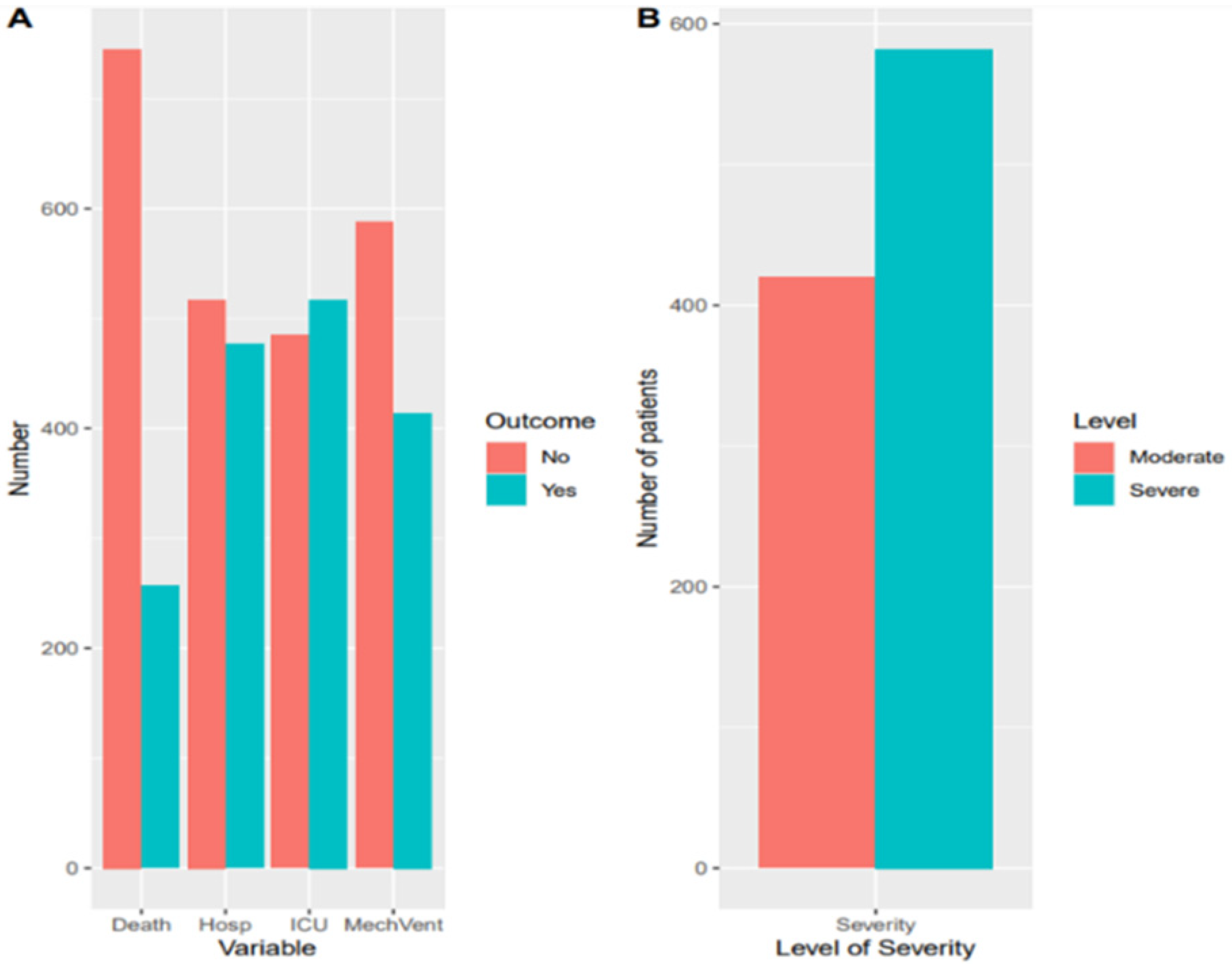
| Factors | Measurement | N | Percent (%) |
|---|---|---|---|
| Severity Outcome | |||
| Died | Yes | 257 | 25.7 |
| Hospitalized at least 9 days | Yes | 477 | 47.6 |
| Admitted to ICU | Yes | 517 | 51.6 |
| Required mechanical ventilation | Yes | 414 | 41.3 |
| Demographics | |||
| Age * | Mean (SD) | 54.13 (16.09) | |
| Gender | Male | 355 | 35.4 |
| Female | 646 | 64.5 | |
| Nationality | Others | 231 | 23.1 |
| Omani | 770 | 76.8 | |
| Comorbidities | |||
| Diabetes Mellitus | No | 503 | 50.2 |
| Yes | 499 | 49.8 | |
| Hypertension | No | 488 | 48.7 |
| Yes | 514 | 51.3 | |
| Dyslipidemia | No | 786 | 78.4 |
| Yes | 216 | 21.6 | |
| Respiratory disease | No | 886 | 88.4 |
| Yes | 116 | 11.6 | |
| Cardiac disease | No | 793 | 79.1 |
| Yes | 209 | 20.9 | |
| Liver disease | No | 959 | 95.7 |
| Yes | 43 | 4.3 | |
| Clinical Factors | |||
| Shortness of breath | No | 162 | 16.2 |
| Yes | 836 | 83.4 | |
| Bilateral lung consolidation | No | 155 | 15.5 |
| Yes | 847 | 84.5 | |
| Sepsis | No | 828 | 82.6 |
| Yes | 174 | 17.4 | |
| Acute respiratory distress syndrome | No | 577 | 57.6 |
| Yes | 425 | 42.4 | |
| Non-invasive ventilation requirement | No | 722 | 72.1 |
| Yes | 280 | 27.9 | |
| Laboratory Parameters | |||
| Total white cell count, (109/L) * | Mean (SD) | 7.7 (4.7) | |
| Hemoglobin A1c,(mmol/L) * | Mean (SD) | 1.2 (1.1) | |
| C-reactive protein(mg/L) * | Mean (SD) | 106.5 (82.1) | |
| Lactate dehydrogenase (mmol/L) * | Mean (SD) | 470.65 (536.5) | |
| Ferritin, (ng/mL) * | Mean (SD) | 1283.45 (2410.2) | |
| Alanine aminotransferase, (U/L) * | Mean (SD) | 65.15 (167.1) | |
| D-dimer, (ng/mL) * | Mean (SD) | 4.45 (13.3) | |
| Corrected calcium, (mmol/L) * | Mean (SD) | 2.15 (0.2) | |
| Troponin (pg/mL) * | Mean (SD) | 159.1 (938.2) | |
| Vitamin D (IU) * | Mean (SD) | 69.4 (36.9) | |
| Estimated glomerular filtration rate (mL/min/1.73 m2) | No | 689 | 68.8 |
| Yes | 311 | 31.0 | |
| Therapeutic Interventions | |||
| Plasmapheresis | No | 940 | 93.8 |
| Yes | 62 | 6.2 | |
| Convalescent plasma | No | 609 | 60.8 |
| Yes | 393 | 39.2 | |
| Tocilizumab | No | 677 | 67.6 |
| Yes | 325 | 32.4 | |
| Steroids | No | 353 | 35.2 |
| Yes | 649 | 64.8 | |
| Factor | Measurement | Severity Level | p-Value | |
|---|---|---|---|---|
| Moderate N = 163 | Severe N = 839 | |||
| Demographic Factors | ||||
| Age | Mean (SD) | 51.5 (15.8) | 56.0 (16.0) | <0.001 |
| Gender | Male | 250 (59.5) | 396 (68.2) | 0.006 |
| Female | 170 (40.5) | 185 (31.8) | ||
| Nationality | Omani citizen | 353 (84.0) | 417 (71.8) | <0.001 |
| Others | 67 (16.0) | 164 (28.2) | ||
| Comorbidities | ||||
| Diabetes Mellitus | No | 242 (57.6) | 261 (44.8) | <0.001 |
| Yes | 178 (42.4) | 321 (55.2) | ||
| Hypertension | No | 216 (51.4) | 272 (46.7) | 0.161 |
| Yes | 204 (48.6) | 310 (53.3) | ||
| Dyslipidemia | No | 340 (81.0) | 446 (76.6) | 0.118 |
| Yes | 80 (19.0) | 136 (23.4) | ||
| Respiratory disease | No | 378 (90.0) | 508 (87.3) | 0.220 |
| Yes | 42 (10.0) | 74 (12.7) | ||
| Cardiac disease | No | 344 (81.9) | 449 (77.1) | 0.080 |
| Yes | 76 (18.1) | 133 (22.9) | ||
| Liver disease | No | 407 (96.9) | 552 (94.8) | 0.153 |
| Yes | 13 (3.1) | 30 (5.2) | ||
| Clinical Factors | ||||
| Shortness of breath | No | 126 (30.1) | 36 (6.2) | <0.001 |
| Yes | 292 (69.9) | 544 (93.8) | ||
| Bilateral lung consolidation | No | 123 (29.3) | 32 (5.5) | <0.001 |
| Yes | 297 (70.7) | 550 (94.5) | ||
| Sepsis | No | 405 (96.4) | 423 (72.7) | <0.001 |
| Yes | 15 (3.6) | 159 (27.3) | ||
| Acute respiratory distress syndrome | No | 408 (97.1) | 169 (29.0) | <0.001 |
| Yes | 12 (2.9) | 413 (71.0) | ||
| Non-invasive ventilation requirement | No | 406 (96.7) | 316 (54.3) | <0.001 |
| Yes | 14 (3.3) | 266 (45.7) | ||
| Laboratory Parameters | ||||
| Total white cell count, (109/L) | Mean (SD) | 6.2 (3.6) | 9.1 (5.9) | <0.001 |
| Hemoglobin A1C, (mmol/L) | Mean (SD) | 1.2 (0.7) | 1.2 (1.4) | 0.207 |
| C-reactive protein(mg/L) | Mean (SD) | 81.8 (70.9) | 129.1 (91.7) | <0.001 |
| Lactate dehydrogenase, (mmol/L) | Mean (SD) | 403.0 (302.5) | 534.1 (463.8) | <0.002 |
| Ferritin, (ng/mL) | Mean (SD) | 1205.0(1857.3) | 1391.9 (3020.1) | 0.477 |
| Alanine aminotransferase, (U/L) | Mean (SD) | 60.0 (81.7) | 71.7 (252.1) | 0.576 |
| D-dimer,(ng/mL) | Mean (SD) | 2.5 (9.9) | 6.4 (16.9) | 0.013 |
| Corrected calcium, (mmol/L) | Mean (SD) | 2.1 (0.1) | 2.2 (0.2) | <0.001 |
| Troponin, (pg/mL) | Mean (SD) | 64.6 (224.7) | 227.9 (1532.5) | 0.370 |
| Vitamin D, (IU) | Mean (SD) | 66.5 (36.3) | 72.0 (37.6) | 0.382 |
| Estimated glomerular filtration rate (mL/min/1.73 m2) | No | 287 (68.5) | 402 (69.2) | 0.869 |
| Yes | 132 (31.5) | 179 (30.8) | ||
| Therapeutic Interventions | ||||
| Plasmapheresis | No | 418 (99.5) | 522 (89.7) | <0.001 |
| Yes | 2 (0.5) | 60 (10.3) | ||
| Convalescent plasma | No | 131 (80.4) | 478 (57.0) | <0.001 |
| Yes | 32 (19.6) | 361 (43.0) | ||
| Tocilizumab | No | 154 (94.5) | 523 (62.3) | <0.001 |
| Yes | 9 (5.5) | 316 (37.7) | ||
| Steroids | No | 128 (78.5) | 225 (26.8) | <0.001 |
| Yes | 35 (21.5) | 614 (73.2) | ||
| Factor | Measure | Severity Level | Unadjusted OR (95% CI, P) | Adjusted OR (95% CI, P) | |
|---|---|---|---|---|---|
| Moderate (N = 163) | Severe (N = 839) | ||||
| Demographics | |||||
| Age | Mean (SD) | 51.5 (15.8) | 56.0 (16.0) | 1.02 (1.01–1.03, p < 0.001) | 1.00 (0.98–1.02, p = 0.961) |
| Gender | Female | 170 (47.9) | 185 (52.1) | - | - |
| Male | 250 (38.7) | 396 (61.3) | 1.46 (1.12–1.89, p = 0.005) | 1.46 (0.80–2.70, p = 0.225) | |
| Nationality | Others | 67 (29.0) | 164 (71.0) | - | - |
| Omani | 353 (45.8) | 417 (54.2) | 0.48 (0.35–0.66, p < 0.001) | 0.43 (0.22–0.83, p = 0.012) | |
| Comorbidities | |||||
| Diabetes Mellitus | No | 242 (57.6) | 261 (44.8) | - | - |
| Yes | 178 (42.4) | 321 (55.2) | 1.67 (1.30–2.16, p < 0.001) | 0.99 (0.55–1.78, p = 0.986) | |
| Clinical Factors | |||||
| Shortness of breath | No | 126 (30.1) | 36 (6.2) | - | - |
| Yes | 292 (69.9) | 544 (93.8) | 6.52 (4.43–9.82, p < 0.001) | 1.20 (0.47–3.33, p = 0.711) | |
| Bilateral lung infiltrates | No | 123 (29.3) | 32 (5.5) | - | - |
| Yes | 297 (70.7) | 550 (94.5) | 7.12 (4.76–10.92, p < 0.001) | 2.38 (0.83–7.45, p = 0.118) | |
| Sepsis | No | 405 (96.4) | 423 (72.7) | - | - |
| Yes | 15 (3.6) | 159 (27.3) | 10.15 (6.07–18.25, p < 0.001) | 3.76 (1.35–11.20, p = 0.013) | |
| Acute respiratory distress syndrome | No | 408 (97.1) | 169 (29.0) | - | - |
| Yes | 12 (2.9) | 413 (71.0) | 83.09 (47.53–159.85, p < 0.001) | 27.05 (11.03–82.00, p < 0.001) | |
| Non-invasive ventilation requirement | No | 406 (96.7) | 316 (54.3) | - | - |
| Yes | 14 (3.3) | 266 (45.7) | 24.41 (14.50–44.57, p < 0.001) | 7.09 (2.80–20.77, p < 0.001) | |
| Laboratory Parameters | |||||
| Total white cell count, (109/L) | Mean (SD) | 6.2 (3.6) | 9.1 (5.9) | 1.14 (1.11–1.18, p < 0.001) | 1.02 (0.97–1.08, p = 0.355) |
| C-reactive protein (mg/L) | Mean (SD) | 81.8 (70.9) | 129.1 (91.7) | 1.01 (1.00–1.01, p < 0.001) | 1.00 (1.00–1.01, p = 0.045) |
| Lactate dehydrogenase, (mmol/L) | Mean (SD) | 403.0 (302.5) | 534.1 (463.8) | 1.00 (1.00–1.00, p < 0.001) | 1.00 (1.00–1.00, p = 0.162) |
| D-dimer, (ng/mL) | Mean (SD) | 2.5 (9.9) | 6.4 (16.9) | 1.04 (1.02–1.06, p = 0.001) | 1.00 (0.98–1.03, p = 0.781) |
| Corrected calcium, (mmol/L) | Mean (SD) | 2.1 (0.1) | 2.2 (0.2) | 1.06 (0.55–2.05, p = 0.859) | 2.05 (0.45–9.58, p = 0.352) |
| Therapeutic Interventions | |||||
| Plasmapheresis | No | 418 (99.5) | 522 (89.7) | - | - |
| Yes | 2 (0.5) | 60 (10.3) | 24.02 (7.45–147.04, p < 0.001) | 2.56 (0.55–19.47, p = 0.283) | |
| Convalescent plasma | No | 131 (80.4) | 478 (57.0) | - | - |
| Yes | 32 (19.6) | 361 (43.0) | 5.28 (3.94–7.13, p < 0.001) | 1.18 (0.63–2.17, p = 0.608) | |
| Tocilizumab | No | 154 (94.5) | 523 (62.3) | - | - |
| Yes | 9 (5.5) | 316 (37.7) | 6.83 (4.90–9.68, p < 0.001) | 1.15 (0.59–2.23, p = 0.680) | |
| Steroids | No | 128 (78.5) | 225 (26.8) | - | - |
| Yes | 35 (21.5) | 614 (73.2) | 6.28 (4.73–8.38, p < 0.001) | 3.28 (1.81–6.07, p < 0.001) | |
Publisher’s Note: MDPI stays neutral with regard to jurisdictional claims in published maps and institutional affiliations. |
© 2021 by the authors. Licensee MDPI, Basel, Switzerland. This article is an open access article distributed under the terms and conditions of the Creative Commons Attribution (CC BY) license (https://creativecommons.org/licenses/by/4.0/).
Share and Cite
Khamis, F.; Al Awaidy, S.; Shaaibi, M.A.; Shukeili, M.A.; Chhetri, S.; Balushi, A.A.; Sulaimi, S.A.; Balushi, A.A.; Wesonga, R. Epidemiological Characteristics of Hospitalized Patients with Moderate versus Severe COVID-19 Infection: A Retrospective Cohort Single Centre Study. Diseases 2022, 10, 1. https://doi.org/10.3390/diseases10010001
Khamis F, Al Awaidy S, Shaaibi MA, Shukeili MA, Chhetri S, Balushi AA, Sulaimi SA, Balushi AA, Wesonga R. Epidemiological Characteristics of Hospitalized Patients with Moderate versus Severe COVID-19 Infection: A Retrospective Cohort Single Centre Study. Diseases. 2022; 10(1):1. https://doi.org/10.3390/diseases10010001
Chicago/Turabian StyleKhamis, Faryal, Salah Al Awaidy, Muna Al Shaaibi, Mubarak Al Shukeili, Shabnam Chhetri, Afra Al Balushi, Sumaiya Al Sulaimi, Amal Al Balushi, and Ronald Wesonga. 2022. "Epidemiological Characteristics of Hospitalized Patients with Moderate versus Severe COVID-19 Infection: A Retrospective Cohort Single Centre Study" Diseases 10, no. 1: 1. https://doi.org/10.3390/diseases10010001
APA StyleKhamis, F., Al Awaidy, S., Shaaibi, M. A., Shukeili, M. A., Chhetri, S., Balushi, A. A., Sulaimi, S. A., Balushi, A. A., & Wesonga, R. (2022). Epidemiological Characteristics of Hospitalized Patients with Moderate versus Severe COVID-19 Infection: A Retrospective Cohort Single Centre Study. Diseases, 10(1), 1. https://doi.org/10.3390/diseases10010001