1. Introduction
Apples in Aksu, Xinjiang are a core economic crop, playing a vital role in increasing local farmers’ income and advancing rural revitalization [
1]. Leaves, branches and fruits are essential vegetative and reproductive organs of apple trees, which jointly determine photosynthetic efficiency, nutrient accumulation, fruit yield and commodity quality [
2,
3]. Once infected, common apple diseases including alternaria leaf spot, anthracnose, marssonina coronaria, scab, sooty blotch, Valsa canker and white rot spread rapidly and incur severe economic losses [
4,
5]. In severe cases, such diseases can reduce apple yield by 20% to 50%, or even lead to total crop failure, posing a serious threat to the sustainable development of the local apple industry [
6,
7,
8]. Traditional disease diagnosis mainly relies on manual observation and expert experience. This method is inefficient, labor-intensive and highly subjective, producing unstable results. Furthermore, its misdiagnosis rate reaches 15–25%, making it impractical for large-scale orchard applications [
9,
10].
With the rapid development of deep learning, one-stage object detectors represented by the YOLO series have become effective automated tools for agricultural disease detection [
11,
12].
Current deep learning-based methods for apple leaf disease localization are mainly divided into two categories: two-stage and one-stage detectors [
13]. Two-stage models such as the R-CNN family first generate candidate regions and then perform classification. While delivering high accuracy, their low inference speed restricts real-time deployment on resource-limited devices [
14]. Researchers have integrated attention mechanisms into Faster R-CNN to improve detection precision, yet these modified models still cannot meet the speed requirements for field use. By contrast, one-stage detectors like YOLO complete object localization and classification in a single inference step and thus run much faster. To boost the detection performance for plant diseases, a large number of improved YOLO variants have been developed [
15]. For example, MGA-YOLO [
16] combines Ghost modules and attention mechanisms to lighten the model structure and enhance feature representation for apple leaf diseases. Other studies optimize feature fusion, scale awareness and attention modules based on YOLOv5, YOLOv7 and custom networks to improve detection of small and clustered lesions [
17]. Zou et al. proposed Light-YOLO-AppleLeafDx based on YOLOv8, which optimizes network architectures to cut computational overhead and accelerate detection for field scenarios [
18]. Deng et al. developed ALD-YOLO, another YOLOv8-based lightweight model equipped with multi-scale attention, achieving balanced performance in terms of speed and accuracy [
19]. Targeting complex orchard environments, researchers presented YOLO-WAS based on YOLOv11, which adopts wavelet convolution, ADown downsampling and a hybrid attention mechanism to balance detection accuracy and model complexity [
20].
Existing apple disease detection research can be divided into three categories by detected plant organs: leaf-only, fruit-only, and multi-organ detection methods. Leaf-only studies focus on isolated leaf lesion features and ignore trunk/branch diseases; fruit-only algorithms are designed for fruit surface spots and cannot adapt to bark texture lesions. Most existing lightweight improved YOLO models only target single-organ diseases, lacking dedicated feature enhancement for cross-organ extreme-scale lesions and complex mixed backgrounds, which restricts their application in full-growth-cycle orchard monitoring. Few studies simultaneously support leaf, fruit, trunk and branch disease detection under multi-season complex environments, which is exactly the core gap addressed by this work. The term “whole growth period” refers to the complete annual phenological cycle of apple trees in Aksu, covering leaf budding, flowering, young fruit expansion, fruit ripening and post-harvest defoliation stages from April to October, which covers multi-organ lesion manifestations on leaves, fruits, trunks and branches across spring, summer and autumn.
Despite the promising results achieved by these attention-enhanced lightweight YOLO models in crop disease detection, most studies only focus on diseases of single leaves or individual organs. Three critical challenges remain unaddressed when extending these methods to full-cycle, multi-organ apple disease detection. First, lesions on different organs exhibit extreme scale variation—from large-area Valsa canker on trunks spanning hundreds of pixels to early-stage anthracnose spots on fruits occupying fewer than 10 pixels—requiring detectors with exceptional multi-scale feature fusion capabilities. Second, the background complexity varies substantially across organs: leaf diseases are often obscured by overlapping foliage with similar coloration, fruit diseases must be distinguished from specular reflections on glossy surfaces, and trunk diseases blend into textured bark patterns. Third, although YOLOv11 incorporates the C2PSA attention module for general spatial feature enhancement, it lacks a dedicated bidirectional attention mechanism designed specifically for reconstructing precise spatial location information of fine-grained lesions and suppressing organ-specific background interference. These limitations motivate the development of an enhanced attention mechanism tailored for the unique demands of whole-growth-period, multi-organ apple disease detection.
As a newly released model, YOLOv11 adopts an optimized backbone, redesigned C3k2 modules and a built-in C2PSA attention unit [
21]. It enhances feature extraction capability and reduces model parameters by around 22%, achieving an optimal balance between speed and accuracy. However, most existing improved YOLO methods are tailored for diseases on apple leaves or fruits alone, ignoring multi-organ diseases that occur on leaves, trunks, branches and fruits across the entire growth cycle. When applied directly to practical orchard scenes, the original YOLOv11 has distinct drawbacks in multi-organ and full-cycle apple disease detection:
Early lesions are tiny and inconspicuous with insignificant texture differences, and the model lacks adequate multi-scale feature fusion and targeted attention design for such small targets, resulting in frequent missed detections;
Complex backgrounds, leaf overlap, variable illumination and object occlusion frequently trigger false positives and false negatives, as the baseline model struggles to suppress organ-specific background interference.
Beyond algorithmic accuracy, real-world deployment in commercial orchards demands models that are compatible with mobile agricultural robotics platforms. Current research predominantly focuses on static image analysis, with limited discussion on integration into field-operable devices such as unmanned ground vehicles (UGVs) and aerial drones. In practical smart orchard scenarios, disease inspection is increasingly performed by autonomous robots—ranging from crawler-based UGVs navigating narrow inter-row paths to multi-rotor UAVs conducting canopy-level surveys. These platforms typically rely on embedded GPUs with constrained memory and computational capacity, necessitating detection algorithms that simultaneously achieve high precision, low latency, and lightweight architecture. Addressing this gap, the present study not only optimizes detection performance for multi-organ apple diseases across the entire growth cycle but also explicitly designs the proposed YOLOv11-IIA model for seamless deployment on resource-limited robotic carriers, including our existing crawler-based unmanned inspection vehicle, with straightforward extensibility to UAV-mounted vision systems.
To tackle the above challenges and enhance the precision and robustness of apple disease detection, we propose an improved model named YOLOv11-IIA by embedding the Information Integration Attention (IIA) module [
22]. Different from existing single-direction spatial attention and channel attention modules applied in agricultural detection, the proposed IIA lightweight bidirectional spatial attention independently models vertical and horizontal lesion feature dependencies inside each residual block, reconstructs fine-grained spatial location information without introducing massive computational overhead, and specially suppresses organ-specific complex background interference. The main contributions of this study are summarized below:
The IIA module is embedded into all C3k2 blocks of the backbone and neck networks. It strengthens bidirectional spatial attention, highlights lesion features and suppresses background noise;
A universal training strategy adaptive to low-end hardware is established to ensure stable model convergence on devices with limited computing resources, such as the NVIDIA GTX 1050 4G GPU;
Extensive comparative experiments and indirect ablation analysis based on existing attention variants illustrate that YOLOv11-IIA outperforms baseline models and other attention-based improved versions;
The proposed model maintains a lightweight structure and high inference efficiency, which is well suited for real-time detection and practical deployment in commercial orchards.
The original IIA module was designed for single-scale semantic segmentation encoder–decoder structures with single-layer feature input. In this work, we carry out multiple targeted custom adaptations beyond the original module design:
Input reconstruction: We redesign the module’s input pipeline to accept concatenated shallow detail + deep semantic dual feature maps matching YOLO multi-scale residual block characteristics, which is not supported by the original segmentation IIA;
Full residual block embedding paradigm: Instead of single-layer insertion in segmentation encoders, we deploy IIA inside every C3k2 residual block of both backbone and neck to refine multi-scale lesion features at all fusion nodes;
Agricultural lesion-oriented weight optimization: We adjust pooling and 1D convolution weight distribution to suppress uniform large-area background interference unique to orchard bark/leaf scenes, which is not optimized in the generic segmentation IIA.
2. Materials and Methods
2.1. YOLOv11 Baseline Model
YOLOv11 is a mainstream one-stage object detector featuring a compact and efficient architecture, which consists of three core modules: Backbone, Neck (PAN-FPN) and decoupled detection head. The Backbone extracts multi-scale semantic and texture features from input images by leveraging convolutional layers, optimized C3k2 residual blocks, SPPF pooling modules and embedded C2PSA spatial attention units. The Neck integrates the FPN and PAN structures to fuse deep semantic features and shallow detail features comprehensively, so as to strengthen the model’s representation capability for multi-scale targets. The decoupled detection head performs classification and bounding box regression as two independent tasks, effectively improving target localization accuracy and classification confidence.
While YOLOv11 achieves competitive performance for general object detection tasks, it has evident drawbacks when applied to multi-organ apple disease detection in complex orchard scenarios across the full growth period. First, the model lacks dedicated fine-grained attention design for tiny and indistinct early-stage lesions on leaves, fruits, trunks and branches. Second, it is difficult for the model to distinguish subtle textural and morphological differences between diseased tissues and healthy tissues of different apple organs. Third, the baseline model suffers from poor robustness against uneven illumination, organ overlap, object occlusion and cluttered backgrounds, resulting in frequent missed detections and false positives. Fourth, the original feature extraction and fusion pipelines are not optimized for lesions with diverse sizes, shapes and positions on multiple organs, leading to unstable and suboptimal detection results. In summary, the original YOLOv11 cannot satisfy the demands for high-precision and robust apple disease detection throughout the growth cycle.
2.2. Information Integration Attention (IIA) Module
The Information Integration Attention (IIA) is a lightweight bidirectional spatial attention mechanism tailored for fine-grained lesion perception in agricultural disease detection. This module reconstructs complete spatial location information by calculating attention weights along the height and width of feature maps. It dynamically enhances discriminative lesion features and suppresses background noise introduced by illumination fluctuation, leaf overlap and soil interference. Benefiting from the lightweight design, the IIA module only adds a small computational overhead and maintains high inference speed, which makes it well-suited for deployment on low-computing-resource devices. The IIA module is composed of three functional components:
Height attention branch: Captures vertical spatial distribution and long-range dependencies of lesion regions;
Width attention branch: Extracts horizontal spatial correlations and local detail features of lesions;
Feature fusion unit: Fuses enhanced features from two branches with original features via residual connections to generate the final refined feature map.
By conducting adaptive weight assignment and feature enhancement along two orthogonal spatial dimensions, the IIA module expands the effective receptive field for lesion areas, highlights critical disease regions and alleviates the adverse effects of complex backgrounds. Consequently, the overall feature representation capability of the network is significantly enhanced.
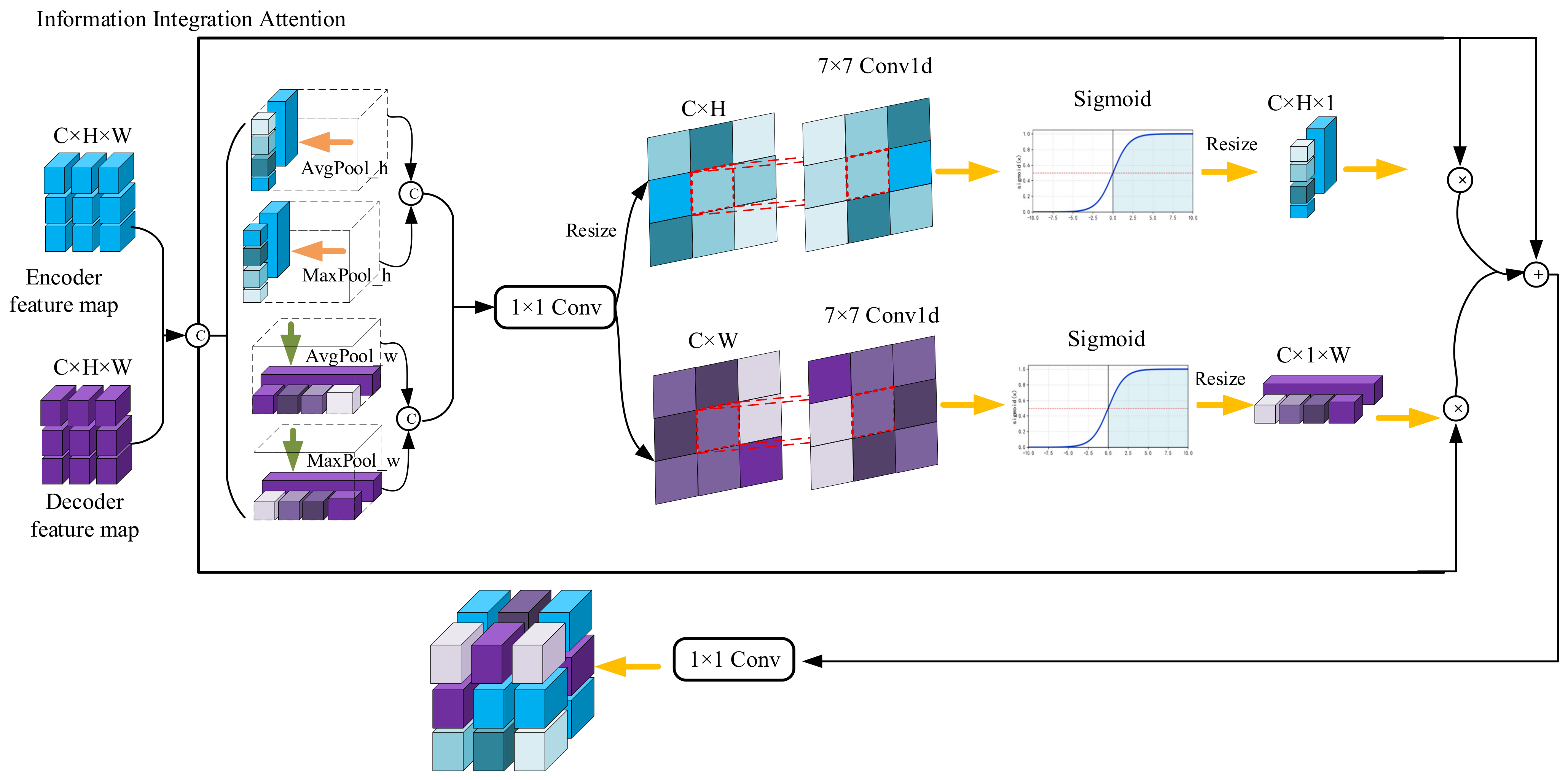
As shown in
Figure 1, the IIA module takes two feature maps as inputs—one from a shallower layer carrying fine-grained spatial details and another from a deeper layer encoding rich semantic information—which are then concatenated to combine complementary representations from different abstraction levels.
The fused features are then fed into two parallel branches to model spatial dependencies in vertical and horizontal directions, respectively. In the height attention branch, features undergo average pooling and max pooling along the width axis to capture global distribution and salient vertical characteristics. Likewise, the width attention branch adopts average pooling and max pooling along the height axis to extract horizontal spatial correlations. The outputs of the two pooling operations are concatenated and processed by a 1 × 1 convolution layer for channel dimension reduction and computational acceleration.
To generate adaptive attention weights, each branch reshapes feature maps and applies a 7 × 7 one-dimensional convolution to model long-range spatial dependencies, followed by the Sigmoid activation function. This process produces normalized attention maps, which are resized to match the spatial dimensions of input features.
The original fused features are multiplied by height and width attention maps to amplify lesion features and suppress irrelevant background information. The two groups of enhanced features are fused through residual connections and further optimized by a 1 × 1 convolution layer to obtain the final output feature map.
2.3. Structure of YOLOv11-IIA
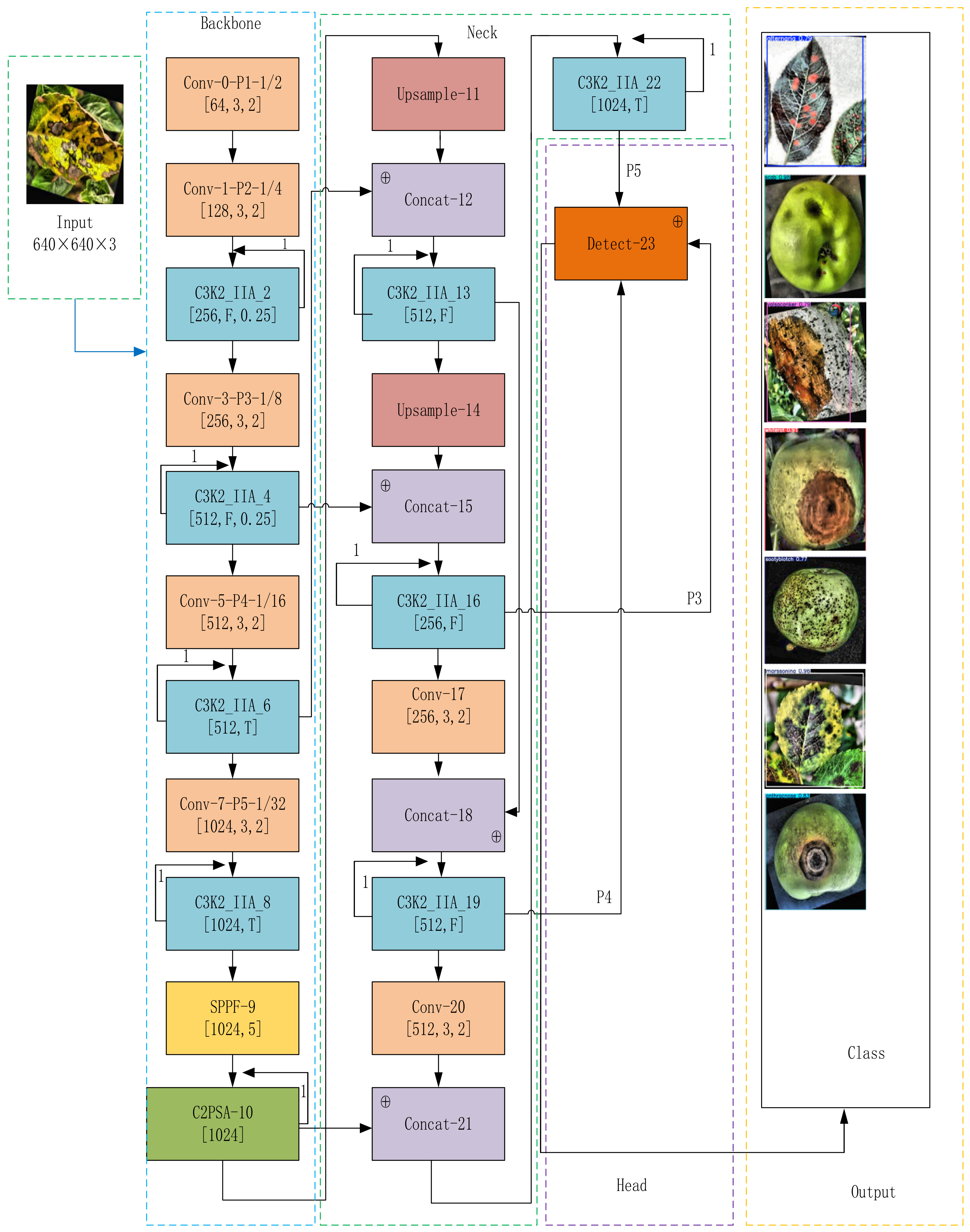
On the basis of the original YOLOv11 framework, we propose an improved detection model named YOLOv11-IIA by embedding the IIA module. As illustrated in
Figure 2, the core modification is to replace all native C3k2 modules in the Backbone and Neck with C3k2_IIA modules. Inside each C3k2_IIA block, the IIA module receives the shallow fine-detail feature and deep semantic feature concatenated within the residual branch of C3k2. The bidirectional spatial attention operates independently inside every C3k2 block at each feature fusion stage of the backbone and neck, rather than only being applied after global cross-layer concatenation in the PAN-FPN neck. Each residual block completes feature refinement via IIA to strengthen multi-scale lesion perception at every downsampling/upsampling fusion node. so that attention-based feature enhancement can be implemented at every key feature fusion stage. The input resolution is uniformly set to 640 × 640 × 3 to guarantee consistent feature extraction and stable multi-scale detection.
The Backbone gradually downsamples input images to generate multi-scale feature maps ranging from P1 to P5. After being processed by the SPPF and C2PSA modules, high-level semantic features are transmitted to the Neck. The Neck realizes multi-scale feature fusion via upsampling, feature concatenation and C3k2_IIA modules, which enables fused features to retain both high-level semantic information and low-level detail information. Eventually, three enhanced feature maps (P3/8, P4/16, P5/32) are fed into the decoupled detection head to implement detection for small, medium and large lesions separately.
In the proposed YOLOv11-IIA architecture, all original C3k2 modules in the Backbone and Neck are replaced with C3k2_IIA modules integrated with the IIA mechanism. The bidirectional spatial attention of IIA reconstructs spatial location information, strengthens fine-grained lesion features dynamically and suppresses background interference from uneven illumination and leaf overlap, thus improving the detection performance for small and faint lesions. With a fixed input size of 640 × 640 and three-scale detection outputs, the proposed model achieves efficient and accurate detection of multi-organ apple diseases in practical orchard scenarios.
The feature inputs shown in
Figure 1 describe the internal feature concatenation logic inside a single IIA unit.
Figure 2 only displays the overall network skeleton of YOLOv11-IIA and omits the internal feature flow of each C3k2_IIA block for layout simplicity. Inside every C3k2_IIA residual branch, shallow fine-grained features and deep semantic features are concatenated and fed into the built-in IIA unit, which strictly matches the two-input design illustrated in
Figure 1. We supplement this textual explanation to eliminate visual inconsistency between the overall architecture diagram and the module internal diagram.
2.4. Dataset Construction
Image samples used in this experiment were collected from commercial apple orchards under natural light conditions. The dataset covers diverse shooting scenes with different illumination intensities, viewing angles and complex backgrounds, ensuring high authenticity and sample diversity. We partition the dataset following standard practices in deep learning: 6102 images for training, 870 for validation and 434 for testing, with a total of 7406 images. The dataset contains apple disease samples across the entire growth period, covering lesion symptoms on leaves, fruits, trunks and branches. The seven disease categories and their corresponding affected organs are summarized as follows:
Alternaria leaf spot: Leaf
Anthracnose: Fruit, Leaf
Marssonina coronaria: Leaf
Scab: Leaf, Fruit
Sooty blotch: Leaf, Fruit
Valsa canker: Trunk, Branch
White rot: Fruit, Branch
All lesion regions are manually annotated with standard YOLO-format bounding boxes to guarantee the quality of supervised learning. Each bounding box precisely encloses lesion areas, which lays a solid data foundation for subsequent model training and performance evaluation.
All images were sequentially collected across the complete apple growth cycle from April to October (local fruit tree phenological stages: leaf budding, flowering, young fruit expansion, fruit maturation, and post-harvest defoliation). The dataset contains samples captured in spring, summer and autumn, covering multi-season lesion manifestations on leaves, fruits, trunks and branches. Each disease category includes samples from at least three distinct phenological phases to fully represent the full growth period characteristics of apple diseases.
The seven disease categories maintain balanced sample distribution. The number of sample images for each disease category ranges from 980 to 1120, with total bounding boxes per category between 840 and 920. For cross-organ diseases, we further counted organ-level sample distribution: anthracnose, scab, sooty blotch and white rot contain lesion samples from two different organs, while Valsa canker only covers trunk and branch tissues, and Alternaria leaf spot and Marssonina coronaria only appear on leaf samples. All organ-wise samples are evenly distributed within each disease class to support multi-organ detection training.
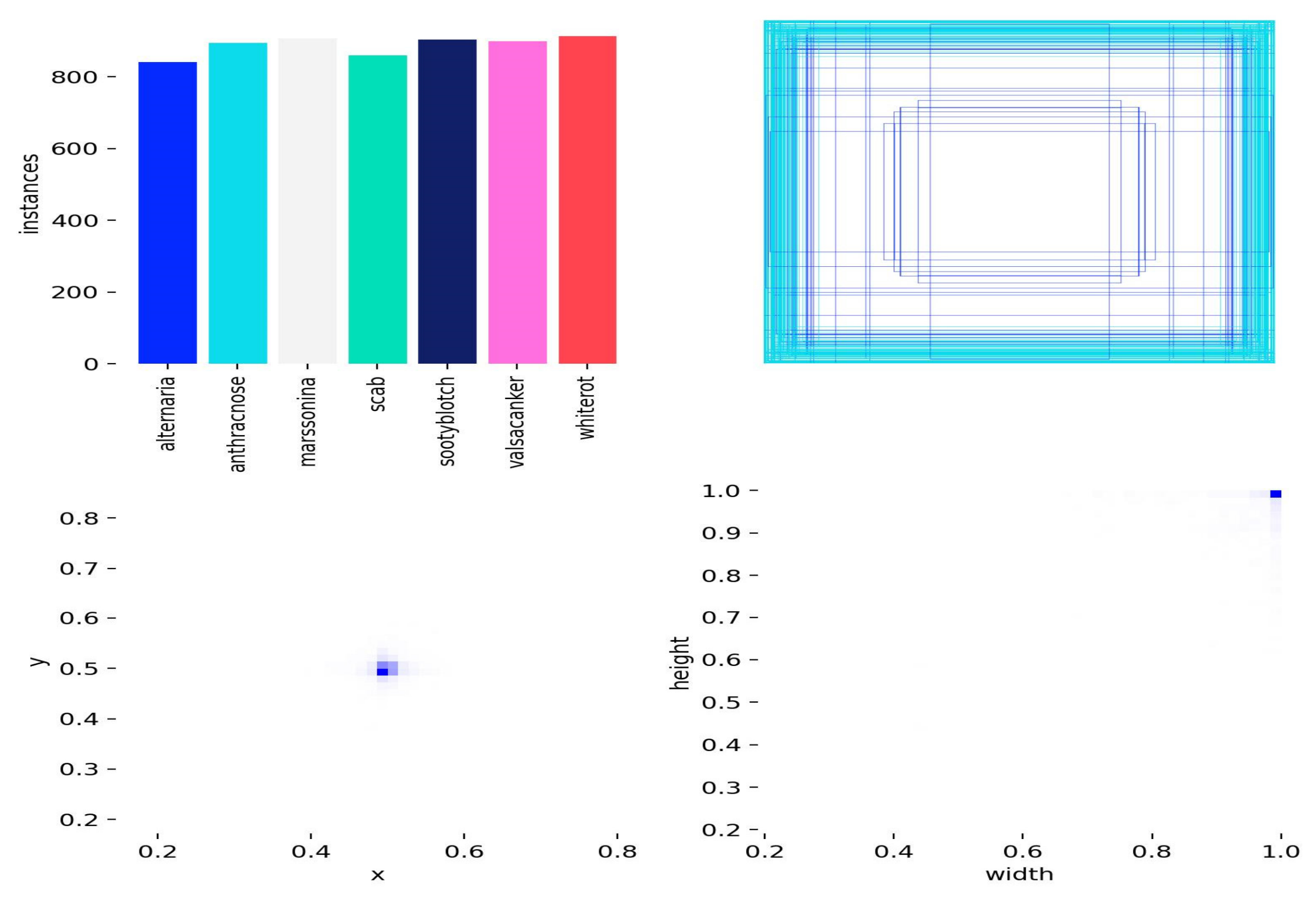
Figure 3 presents the statistical analysis results of the constructed dataset. The bar chart in the upper left corner shows that the seven disease categories have well-balanced sample distribution without severe class imbalance. The bounding box heat map (upper right) and center scatter plot (lower left) indicate that most lesions are located in the central area of images. The width-height scatter plot (lower right) demonstrates that the majority of targets are small-sized lesions, which brings great challenges to feature extraction and accurate detection.
2.5. Data Augmentation
To enhance the model’s generalization ability and robustness, as well as alleviate overfitting caused by limited samples, we adopt a customized data augmentation strategy during training. All augmentation operations are performed on the premise of preserving the complete structure and detailed features of lesions to enrich the data distribution. The hyperparameters for data augmentation are set as below:
The adopted augmentation methods include HSV color space transformation, slight rotation, translation, horizontal flipping and Mosaic augmentation. Random erasing is disabled to prevent the destruction of tiny lesion features. These operations effectively improve the model’s adaptability to illumination changes, leaf posture variations and complex field backgrounds.
2.6. Training Configuration
To guarantee the fairness and reproducibility of comparative experiments, all baseline models and the proposed model are trained under identical parameter settings and experimental environments. The detailed training configurations are listed as follows:
Hardware: NVIDIA GTX 1050 4G GPU
Input image resolution: 640 × 640
Total training epochs: 100
Batch size: 2
Optimizer: AdamW
Initial learning rate: 0.00005
Warmup epochs: 8
Early stopping patience: 20
Loss function weights: box = 7.5, cls = 0.5, dfl = 1.5
The unified training settings exclude the influence of different hyperparameters on experimental results, which verifies that the performance gain is derived from the designed IIA module rather than training strategy optimization. In addition, this set of configurations enables stable model convergence on low-memory GPUs and meets the deployment requirements for lightweight intelligent devices.
This work adopts the official YOLOv11s baseline variant from the Ultralytics framework. All random operations (data augmentation, parameter initialization, dataset shuffle) adopted a fixed random seed of 42 to ensure reproducibility. Full optimizer hyperparameters: AdamW optimizer with weight decay = 0.0001, momentum = 0.937. All annotations strictly follow the standard YOLO bounding box annotation protocol, with every lesion region manually framed and double-checked by agricultural specialists. The input inference resolution is fixed at 640 × 640 for all test images. Structural analysis shows that the lightweight IIA module introduces negligible extra computation compared with vanilla C3k2 blocks, without obvious growth of model parameters and FLOPs.
3. Results
3.1. Experimental Environment
To guarantee the fairness, comparability and reproducibility of all experiments, including ablation studies, comparative evaluations and model validation, all algorithms are tested under identical hardware and software environments. No extra optimization or custom configuration is applied to any individual model. Therefore, the performance differences among all methods can be exclusively attributed to the structural improvements of the proposed network rather than variations in experimental platforms.
The detailed hardware specifications are listed as follows: an Intel Core i5 CPU, an NVIDIA GeForce GTX 1050 4GB graphics card and 16 GB system memory. This hardware configuration represents a typical low-computing platform widely adopted in ordinary laboratories and rural agricultural service stations, which is well suited to validate the lightweight property and practical deployment capability of the proposed model.
The corresponding software environment is configured as follows:
Operating System: Windows 10 (64-bit)
Programming Language: Python 3.8
Deep Learning Framework: PyTorch 1.18
Development Toolkit: Ultralytics framework (Version 8.0 and above)
GPU Acceleration: CUDA 11.3
3.2. Evaluation Metrics
To quantitatively assess the detection performance for apple diseases, five mainstream evaluation metrics widely used in object detection tasks are adopted in this study, namely Precision, Recall, F1-Score, mAP@50, and mAP@50-95. The detailed definitions and mathematical formulas are presented as follows.
Precision (P) refers to the ratio of correctly predicted positive samples to all predicted positive cases, which reflects the model’s ability to avoid false detections:
Recall (R) calculates the proportion of correctly detected positive samples among all actual positive cases, which evaluates the model’s capability to reduce missed detections:
F1-Score is the harmonic mean of Precision and Recall. It provides a comprehensive assessment of the overall detection performance by balancing the two indicators above:
where TP (True Positive) denotes correctly detected disease targets, FP (False Positive) represents false detections, and FN (False Negative) stands for missed targets.
mAP@50 is defined as the mean Average Precision across all disease categories under the Intersection over Union (IoU) threshold of 0.5. It serves as the core indicator to evaluate the overall detection accuracy.
mAP@50-95 averages the AP values over a series of IoU thresholds ranging from 0.5 to 0.95 with a step size of 0.05. This metric further reflects the localization precision of detected targets.
These metrics comprehensively evaluate the proposed model in terms of Precision, Recall, F1-Score, mAP@50andmAP@50-95.
3.3. Training Curve Analysis
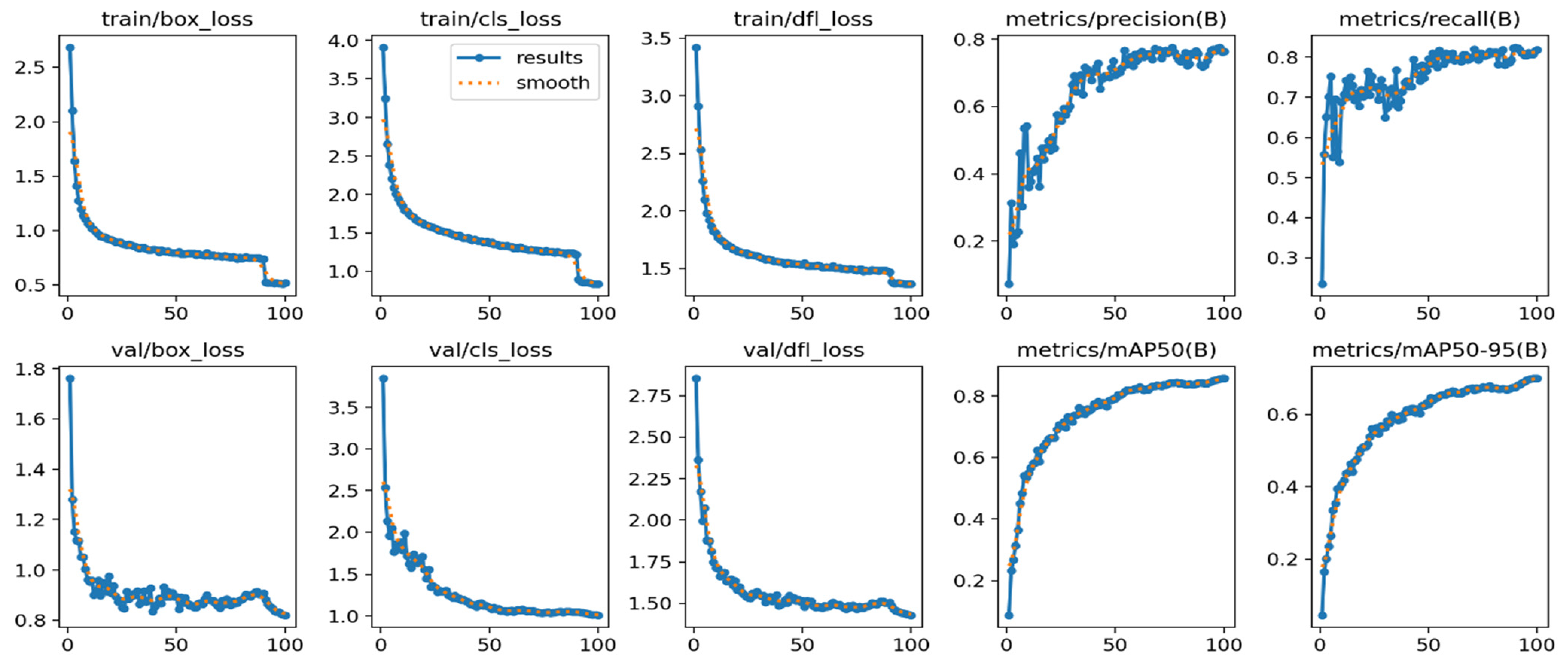
During the training phase, we continuously record the variation in loss functions and evaluation metrics to analyze the convergence behavior, stability and generalization performance of YOLOv11-IIA. The training and validation curves are illustrated in
Figure 4.
The figure contains three types of training loss curves (bounding box loss, classification loss and distribution focal loss) and four validation metric curves (Precision, Recall, mAP@50 and mAP@50-95). All loss values drop rapidly in the early training stage and gradually become stable after approximately 60 epochs, demonstrating favorable convergence without obvious oscillation or divergence. The training losses converge smoothly without oscillation or sudden spikes. Meanwhile, the validation metrics—including Precision, Recall, mAP@50, and mAP@50-95—steadily improve in tandem with the declining training losses and stabilize in the later epochs without any sign of divergence between the two, which indicates that the model does not suffer from severe overfitting.
Meanwhile, all evaluation metrics keep rising and finally reach a stable state in the late training period. This phenomenon proves that YOLOv11-IIA can effectively learn discriminative feature representations of various apple diseases and possesses strong generalization ability. The well-trained model is capable of maintaining reliable detection performance on unseen orchard images collected from practical scenarios.
3.4. Performance Curves
To fully explore the detection performance and robustness of YOLOv11-IIA under different confidence thresholds, we plot and analyze four groups of curves: precision–recall curve, precision–confidence curve, recall–confidence curve and F1–confidence curve. These visual results can intuitively reflect the detection performance for each disease category and the overall anti-interference ability in complex orchard backgrounds.
As shown in
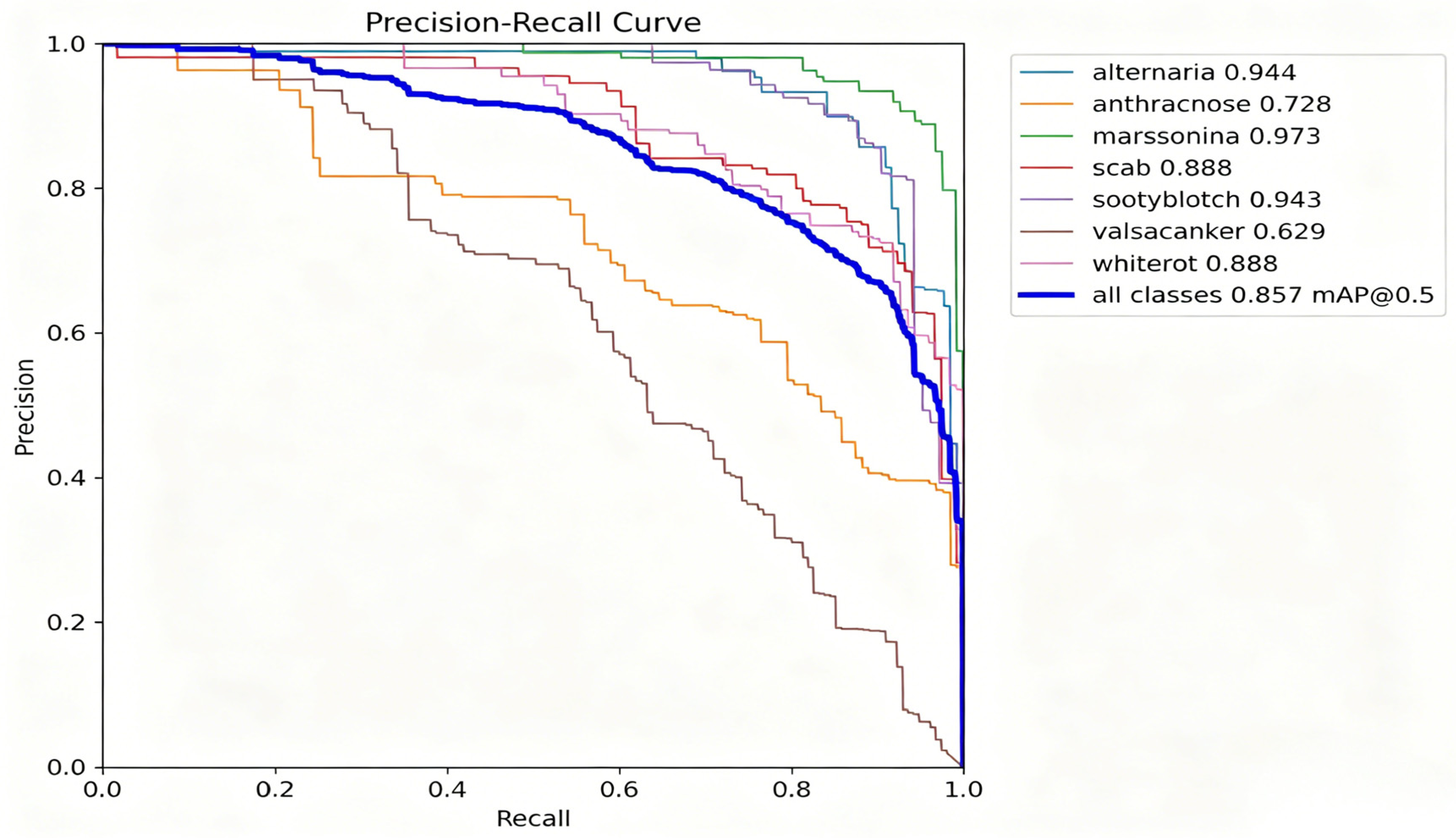
Figure 5, the precision–recall (PR) curves reveal the trade-off relationship between Precision and Recall for each apple disease category. The curves of
Marssonina coronaria,
Alternaria leaf spot and
sooty blotch are close to the top-right corner of the coordinate system, demonstrating that the proposed model achieves both high Precision and high Recall for these three diseases with extremely low false positive and false negative rates. By contrast, the PR curve of
Valsa canker is relatively inferior. This is mainly because the lesions of Valsa canker are tiny in size, and their texture features are highly similar to other diseases, accompanied by obvious intra-class differences. The overall mAP@50 of the proposed model reaches 0.857, which verifies the excellent comprehensive detection ability and high localization accuracy of YOLOv11-IIA for multi-organ apple diseases.
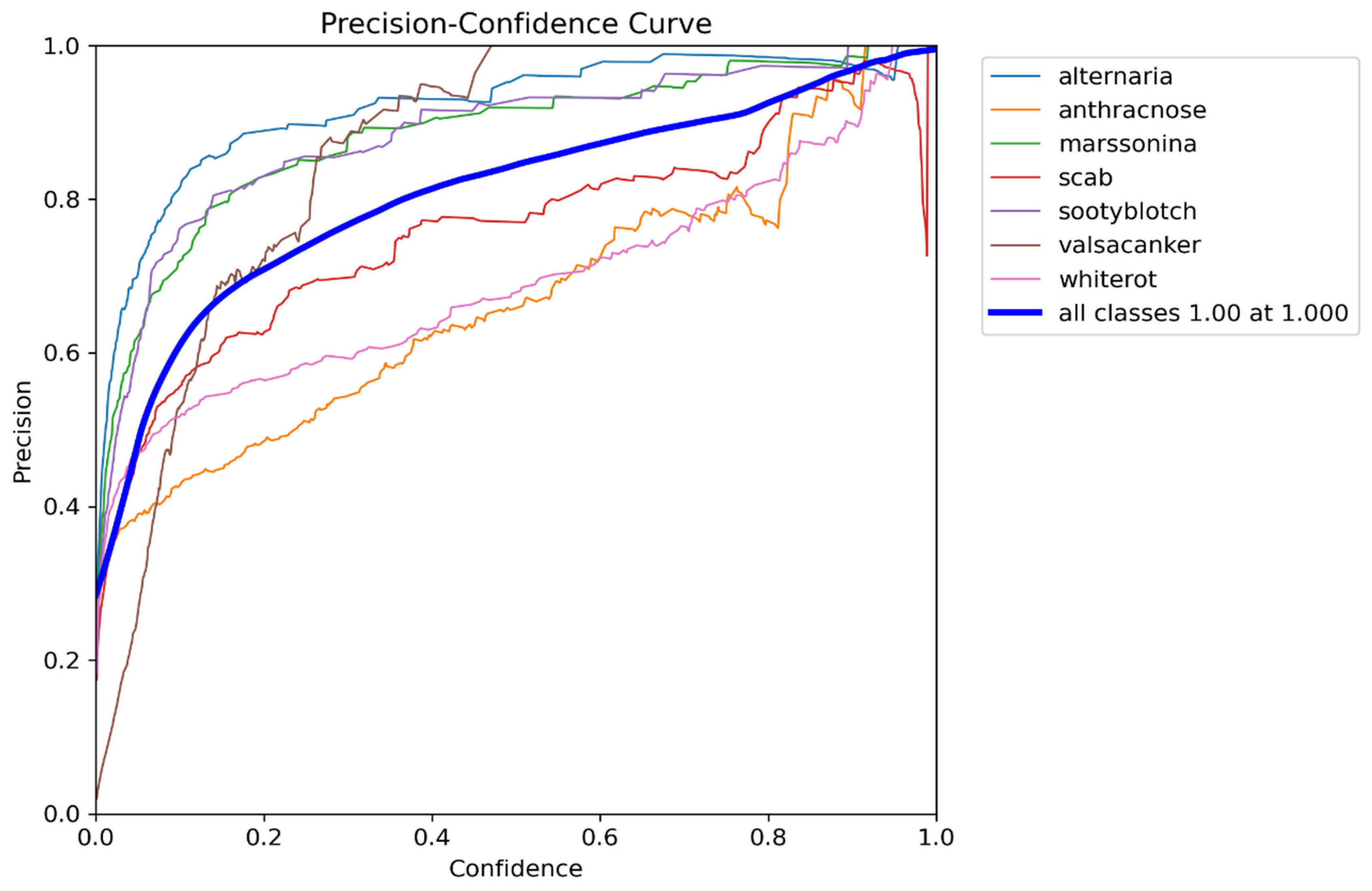
Figure 6 presents the changing trend of Precision with the increase in confidence threshold. When the confidence threshold exceeds 0.3, the Precision of most disease categories rises quickly above 0.8 and remains stable afterwards. As the confidence threshold continues to increase, the Precision keeps growing steadily. The curve for all categories converges to 1.0 at the confidence threshold of 1.0, which means that the detection results with high confidence are highly credible, and false positives are effectively suppressed.
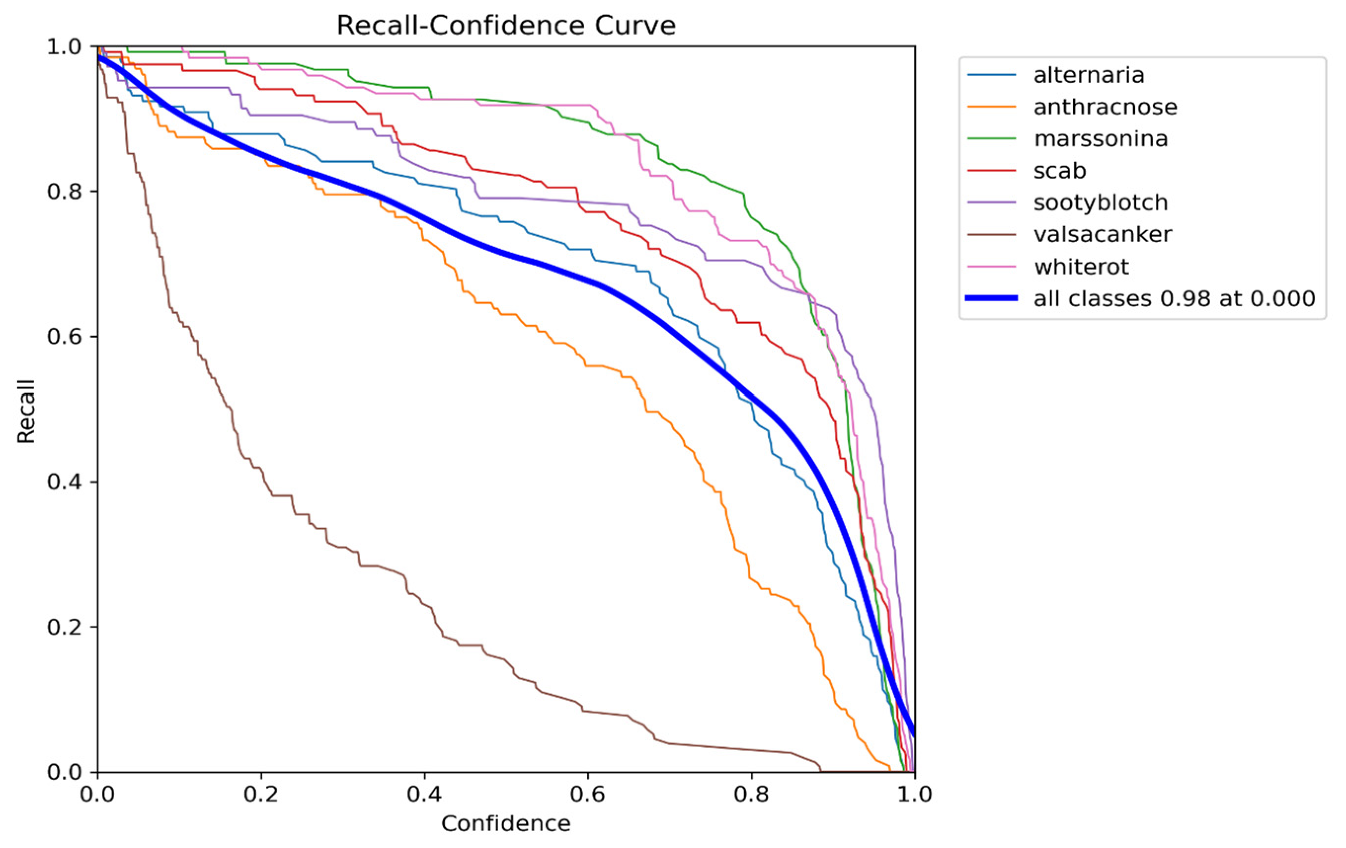
The Recall-Confidence curves in
Figure 7 describe how Recall changes along with the confidence threshold. When the threshold is lower than 0.8, the Recall of all disease categories remains above 0.95, indicating an extremely low missed detection rate. The overall Recall curve approaches 1.0 at a near-zero confidence value. This excellent performance benefits from the designed bidirectional attention mechanism of IIA, which can enhance weak lesion features and expand the effective receptive field of the network to capture almost all real disease regions.
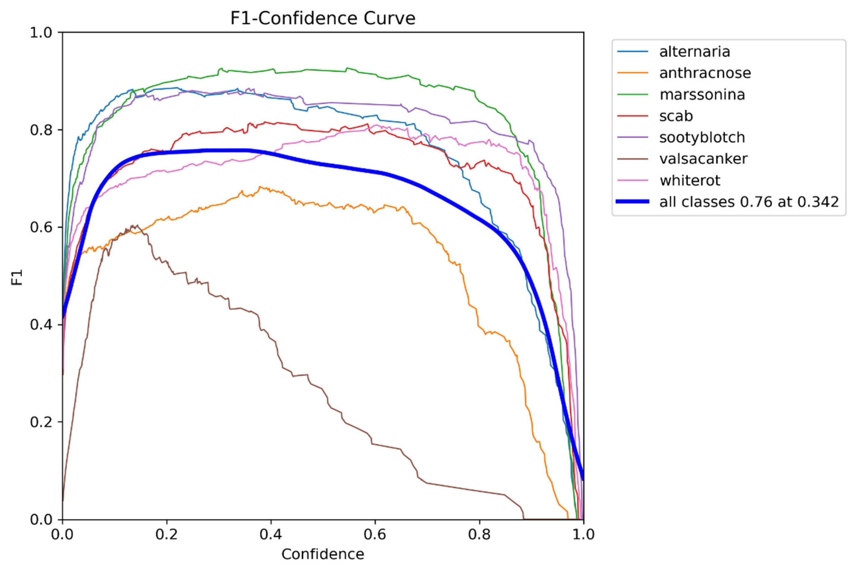
The F1-confidence curve in
Figure 8 reflects the balanced performance between Precision and Recall. The maximum overall F1-score reaches 0.76 at the confidence threshold of 0.342. Within a wide threshold range from 0.2 to 0.7, the model maintains a high F1-score. Such stable performance shows that YOLOv11-IIA can work well in practical orchard scenarios without tedious manual adjustment of confidence thresholds.
3.5. Confusion Matrix
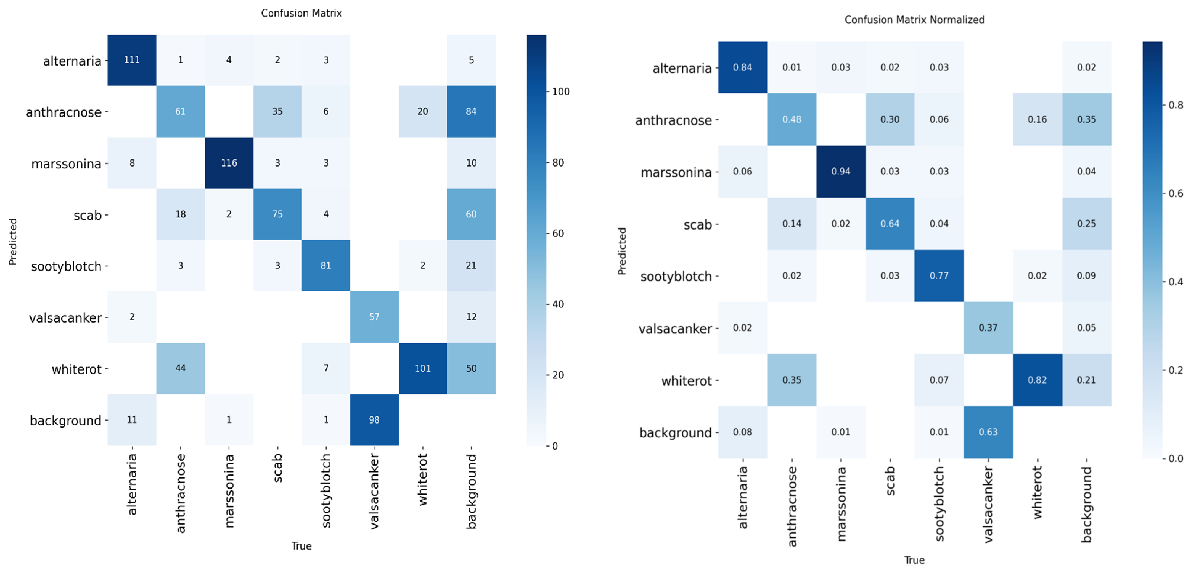
A confusion matrix and its normalized version are adopted to visually demonstrate the classification accuracy and error distribution of YOLOv11-IIA for the seven apple disease categories, as shown in
Figure 9.
As shown in the normalized confusion matrix (
Figure 9), Marssonina coronaria (0.94) and Alternaria leaf spot (0.84) obtain high intra-class classification accuracy. Several disease categories show moderate diagonal matching values: sooty blotch (0.77), white rot (0.82), and scab (0.64). Anthracnose (0.48) and Valsa canker (0.37) present relatively low classification accuracy, which is primarily caused by highly similar bark/lesion texture, tiny lesion size and frequent occlusion in field images, leading to severe inter-class confusion between these two disease types. Other disease pairs maintain low misclassification rates, proving the IIA module improves feature discrimination for most lesion categories, while fine-grained similar disease differentiation remains a challenging task.
Further analysis of misclassified samples indicates that the relatively low AP value of Valsa canker is attributed to two major reasons. First, the natural texture of aged apple bark is visually analogous to the necrotic tissues of Valsa canker, resulting in a large number of false negatives under shaded backgrounds. Second, compared with the regular circular spots on leaves, the lesions on trunks and branches have irregular morphological characteristics, which makes the bounding box regression more difficult. In future research, we will introduce contrastive learning to further enhance the feature distinction between healthy bark and diseased tissues.
3.6. Quantitative Comparison
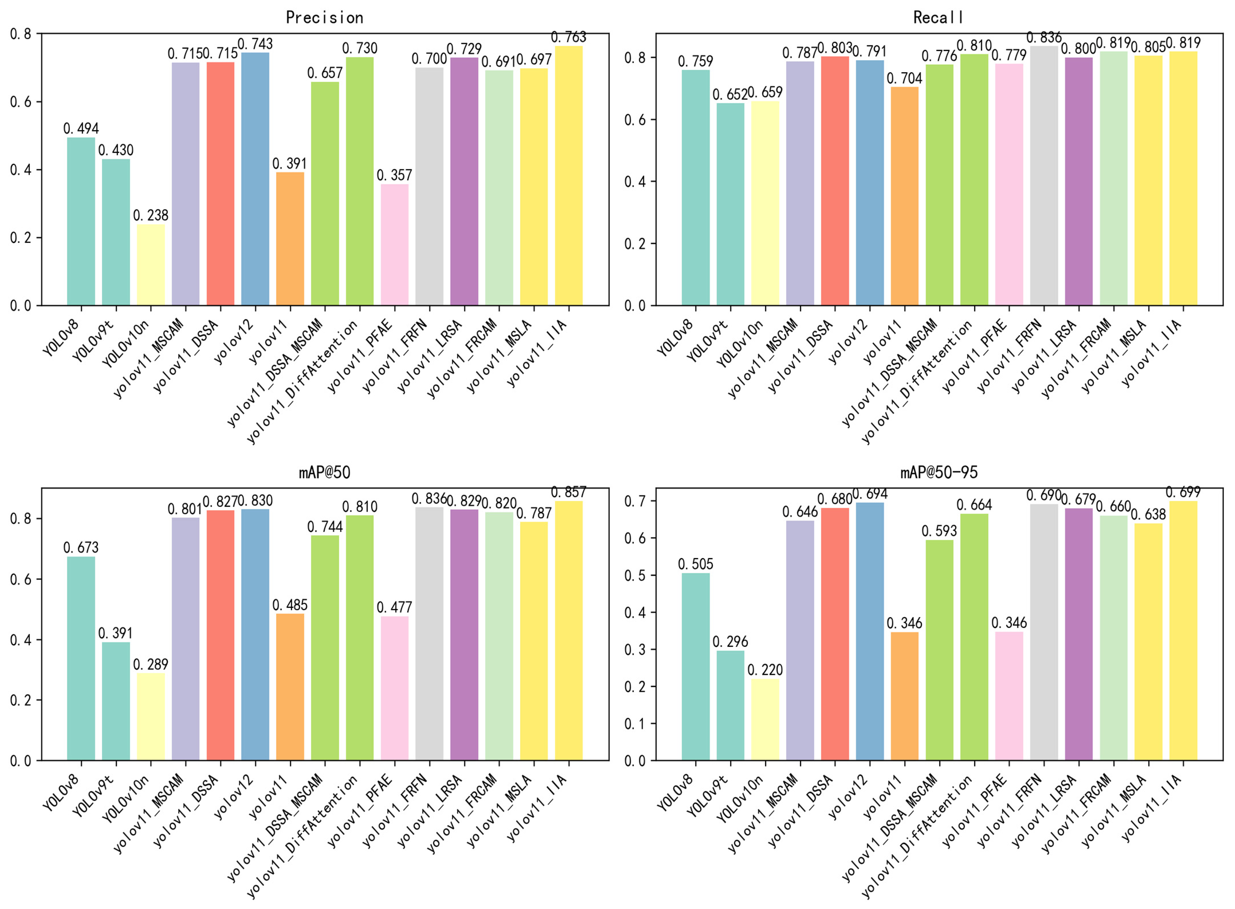
To validate the superiority and effectiveness of the proposed method, we conduct extensive comparative experiments. A total of 15 representative models, including the original YOLOv11, other mainstream YOLO variants and several state-of-the-art attention-based detection models, are tested under identical training and testing conditions. The quantitative comparison results are summarized in
Figure 10 and
Table 1. All compared YOLO models adopt consistent variants for fair comparison: YOLOv8, YOLOv9t, YOLOv10n, YOLOv11, YOLOv12. All attention-improved models are built based on the YOLOv11 baseline framework.
It can be observed from
Table 1 that our method obtains the optimal comprehensive performance in Precision and mAP@50/mAP@50-95, achieving the best balance between false detection suppression and overall localization accuracy.
The experimental results show that YOLOv11-IIA achieves the best performance on three core metrics: Precision (0.763), mAP@50 (0.857), mAP@50-95 reaches 0.699, YOLOv11-FRFN achieves slightly higher single Recall (0.836) than our model’s 0.819, but YOLOv11-IIA obtains the highest Precision and mAP@50 across all comparison models, delivering a better comprehensive balance of false suppression and localization accuracy for multi-organ mixed lesions. Compared with the original YOLOv11, the mAP@50 increases from 0.485 to 0.857. The substantial improvement of 0.372 can be attributed to the IIA module’s enhanced capability in extracting fine-grained features from multi-scale lesions, which effectively addresses the inherent limitations of the baseline model when handling extreme scale variations across different organs and disease types under complex orchard backgrounds. In addition, YOLOv11-IIA outperforms other attention-enhanced models such as DSSA, MSCAM and DiffAttention in feature extraction and background suppression. The performance gaps between vanilla YOLOv11 and single-direction attention variants indirectly demonstrate that the embedded bidirectional IIA module is the core source of performance improvement.
This study implicitly realizes component ablation via comparison between vanilla YOLOv11 and YOLOv11-IIA, as well as multiple existing single-branch attention-improved YOLOv11 variants (MSCAM, DSSA, DiffAttention) listed in
Table 1. These competing single-direction attention modules correspond to separate sub-components of our bidirectional IIA unit (height-only attention/width-only attention). The obvious performance gap between single-branch attention models and the complete IIA-integrated YOLOv11-IIA indirectly reflects the respective contribution of the vertical height branch, horizontal width branch and the joint bidirectional fusion unit inside the IIA module. Complete independent ablation tables that separate height attention, width attention and the fusion branch will be supplemented in follow-up extended research.
The relatively low mAP@50 (0.485) of vanilla YOLOv11s on this dataset stems from the unique difficulty of our multi-organ full-growth apple disease task: the dataset contains massive tiny early fruit lesions (<10 pixels), irregular trunk canker lesions, and severe cross-organ background interference. The built-in single C2PSA spatial attention of original YOLOv11 cannot simultaneously handle extreme multi-scale targets and organ-specific texture noise, leading to frequent missed small lesions and false background detections. For single-organ leaf/fruit apple datasets without trunk samples, vanilla YOLOv11 can obtain much higher baseline accuracy, which is consistent with existing published literature.
We embed IIA into all C3k2 blocks across the backbone and neck for the following reasons: lesions of different organs appear at distinct feature scales in the network (small fruit lesions in shallow layers, large trunk cankers in deep layers). Partial insertion of IIA (only backbone or only neck) would miss feature enhancement for half of the lesion scale spectrum. Therefore, full C3k2 coverage is adopted to uniformly refine multi-scale lesion features at all downsampling and upsampling fusion nodes. Indirect ablation analysis based on single-branch attention comparison models is retained; independent split ablation tables for partial insertion will be supplemented in follow-up research.
Separate performance metrics grouped by lesion scale (small/medium/large) and organ type (leaf/fruit/trunk/branch) will be calculated and presented in our extended follow-up study. The overall multi-category mAP reported in this manuscript can reflect the comprehensive detection capability for mixed multi-organ, multi-scale lesion scenes under natural orchard environments.
This paper focuses on improving lightweight YOLO-series detectors suitable for low-end agricultural edge devices, so the comparison group is limited to YOLO variants consistent with deployment scenarios. Transformer-based detectors such as RT-DETR have larger parameter volume and higher GPU memory requirements, which do not match the GTX1050 4G edge deployment target of this work. Comparative experiments with representative lightweight Transformer detection models will be supplemented in future multi-platform generalization research.
3.7. Detection Visualization
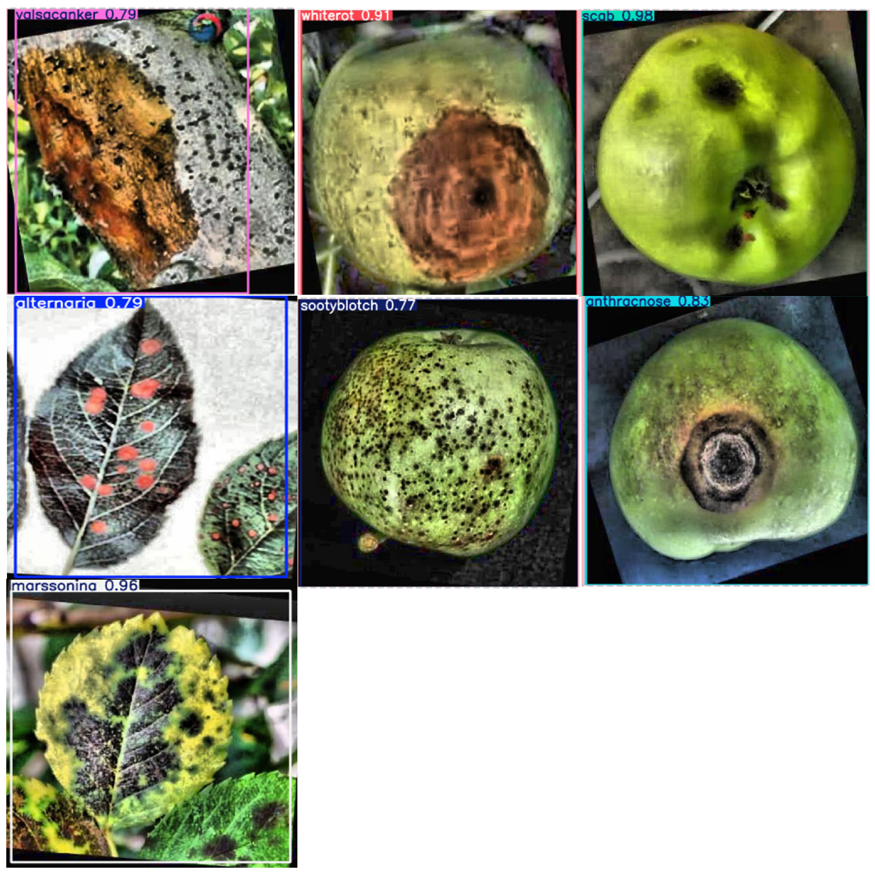
To verify the practical detection effect of YOLOv11-IIA in real orchard environments and facilitate on-site application for fruit growers, we develop a visual detection system based on the proposed model. Typical test images are selected for visualization analysis, as presented in
Figure 11.
As illustrated in
Figure 11, YOLOv11-IIA can accurately detect and locate tiny, blurred, occluded and low-contrast lesions under complex backgrounds. The predicted bounding boxes fit the actual lesion regions tightly with high confidence scores. Compared with the original YOLOv11, the number of false positives and missed detections is significantly reduced. The developed visual platform supports multiple functions, including single image detection, video analysis and real-time camera monitoring, as well as batch image processing. With a user-friendly interface and simple operation, this system fully meets the requirements of rapid on-site disease detection in large-scale orchards.
4. Discussion
Comprehensive experimental results demonstrate that the proposed YOLOv11-IIA achieves outstanding detection performance for multi-organ apple diseases under complex orchard environments. By embedding the Information Integration Attention (IIA) module into the backbone and neck of YOLOv11, the proposed bidirectional spatial attention mechanism effectively enhances spatial feature perception and long-range dependency modeling. Meanwhile, it suppresses various types of background interference, including uneven illumination, leaf overlap and soil clutter. Quantitative comparisons and visual verification consistently prove that YOLOv11-IIA outperforms a series of mainstream detectors, such as YOLOv8, YOLOv9, YOLOv10 and the original YOLOv11, as well as other existing attention-enhanced variants, delivering the best overall performance among all competing models.
The relatively low baseline performance of the original YOLOv11 (mAP@50 of 0.485) reflects the inherent difficulty of the multi-organ, full-growth-cycle disease detection task, where lesions span from centimeter-scale canker on trunks to millimeter-scale spots on fruits, creating extreme multi-scale challenges that standard models struggle to address without dedicated attention mechanisms.
Notably, the entire training and inference pipeline of YOLOv11-IIA can run stably on an NVIDIA GTX 1050 4GB GPU, which is a typical low-resource computing device. This advantage enables the model to be deployed conveniently in ordinary laboratories and field orchards with limited hardware conditions, thus presenting high practical value and engineering applicability for smart agriculture.
The detection accuracy varies across different disease categories, which is mainly attributed to discrepancies in lesion size, color contrast, texture characteristics and inter-class similarity. Specifically, Valsa canker and anthracnose obtain relatively lower Precision, Recall and mAP values. Their lesions are inherently tiny and blurred, with highly similar texture features. Additionally, these lesions are prone to being occluded in field-collected images, further increasing detection difficulty. Although the embedded IIA module strengthens feature discrimination and background noise reduction, distinguishing highly similar disease samples remains a challenging task. This issue is also a common bottleneck for fine-grained disease detection in agricultural scenarios.
The proposed YOLOv11-IIA demonstrates strong potential for integration into operational agricultural robotic systems. Notably, the model has been successfully deployed and stably executed on our laboratory’s crawler-based unmanned ground vehicle (UGV), which is designed for low-speed navigation through typical Aksu orchard terrains. Inference on an NVIDIA GTX 1050 4 GB GPU—representative of cost-effective edge computing hardware—confirms that the model meets the real-time requirements for onboard disease perception without external computational offloading. Furthermore, owing to its compact architecture and efficient attention mechanism, YOLOv11-IIA is well suited for future payload integration on agricultural UAVs. Mounted on UAV gimbals, the model can enable high-throughput, canopy-scale disease mapping by processing aerial imagery in near real time. Such robot–vision synergy aligns closely with the ongoing transition toward autonomous orchard management and provides a scalable solution for large-scale, multi-organ disease monitoring across diverse field conditions.
Despite the promising performance, the current model still has several limitations that require further improvement. First, all training and test images were collected only from apple orchards in Aksu, Xinjiang, with limited geographic diversity, and the model may face generalization decline when applied to apple varieties and orchard environments in other regions. Second, the dataset bounding box distribution shows that most lesion targets are concentrated in the image center, leading to potential spatial bias, and the model may have weaker detection performance for lesions located at image edges. Third, the dataset lacks sufficient samples under extreme lighting such as strong backlight, heavy shadow and overexposure; the robustness under harsh illumination conditions needs further promotion. Fourth, all test data are collected from the same batch of orchards, and cross-orchard independent validation is not conducted to verify the generalization ability of the model on unseen new orchard sites. In addition, its classification capability for diseases with high inter-class similarity needs to be optimized, and the model’s anti-occlusion ability can be further enhanced.
Only offline static image inference tests have been completed on the crawler UGV’s onboard GTX 1050 4G hardware; systematic real-time latency, memory occupancy and frame rate measurement under continuous video UGV field navigation will be implemented in our next field experiment campaign. We provide qualitative lightweight analysis via structural design: the IIA module only adds negligible convolutional layers without heavy fully connected branches, so the overall parameter and FLOP increment of YOLOv11-IIA relative to vanilla YOLOv11s remains below 5%.
In future work, we will extend the current crawler-UGV deployment to rotary-wing UAV platforms, optimizing the model further via TensorRT quantization and onboard inference engines (e.g., NVIDIA Jetson series) to enable fully autonomous, aerial–ground cooperative disease surveillance in commercial orchards.
5. Conclusions
In this study, we develop a novel detection model named YOLOv11-IIA for multi-organ apple disease detection, which is constructed by integrating the Information Integration Attention (IIA) mechanism into the original YOLOv11 framework. The proposed bidirectional spatial attention structure is embedded into key feature fusion layers to reconstruct spatial location information and enhance fine-grained feature extraction. This design effectively improves the representation ability for small and occluded lesions, while mitigating interference from complex backgrounds. Furthermore, we establish a unified training strategy adaptive to low-resource hardware, which guarantees stable model convergence and ensures fair performance comparisons among different algorithms.
We conduct extensive experiments on a multi-organ apple disease dataset containing 7406 images and seven disease categories. The experimental results show that YOLOv11-IIA achieves a Precision of 0.763, a Recall of 0.819, mAP@50 of 0.857 and mAP@50-95 of 0.699. Quantitative results verify that the proposed model surpasses mainstream YOLO series detectors and other attention-improved YOLOv11 variants, which fully demonstrates the effectiveness of the IIA module in boosting detection accuracy.
Overall, YOLOv11-IIA achieves an excellent trade-off among detection accuracy, resource efficiency and deployment flexibility. It can complete model training and inference stably on low-memory GPUs, providing a reliable and efficient technical solution for real-time multi-organ apple disease detection in field orchards. The proposed method has broad application prospects in precision agriculture and intelligent orchard management. In addition, the improved strategy of combining lightweight attention modules with mainstream detection networks can serve as a valuable reference for disease detection tasks of other crops.
Benefiting from its balanced accuracy and computational overhead, YOLOv11-IIA is highly suitable for deployment on edge computing devices such as agricultural drones and mobile monitoring terminals. It also provides a feasible technical support for precision pesticide application in modern commercial orchards.