
Secure Retrieval of Brain Tumor Images Using Perceptual Encryption in Cloud-Assisted Scenario †


Abstract
1. Introduction
2. Related Works
3. Methods
3.1. Preliminaries
- Squared Sine Logistic Map. The squared sine logistic map (SSLM) proposed in [24] is defined as a combination of two simple dynamic chaotic maps, namely the logistic chaotic map and sine chaotic map. The logistic map parameterized by the control variable is as follows:
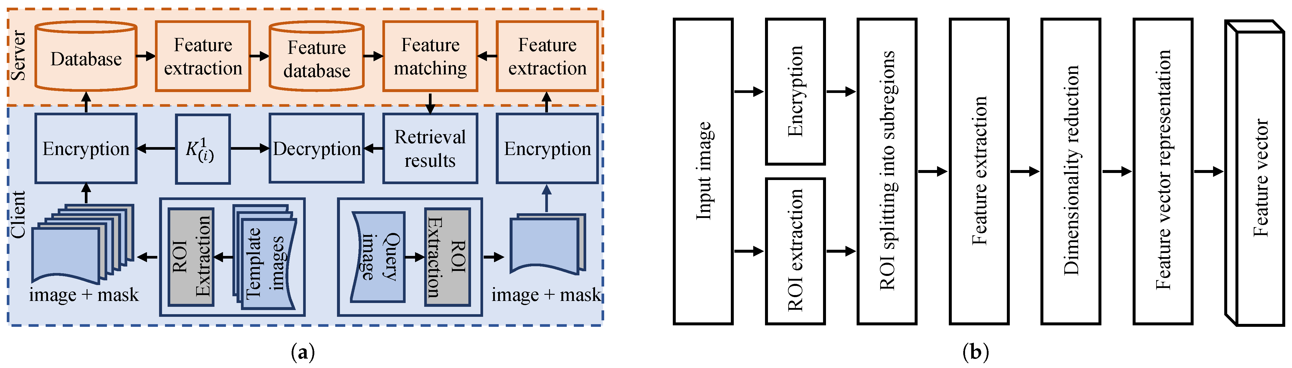
3.2. Proposed Secure Medical Image Retrieval System
3.2.1. Proposed Perceptual Encryption Algorithm
- Key generation. To generate a secret key , we iterate the SSLM in Equation (3) times to obtain three random sequences , and , each of a size N. The first M iterations are discarded to eliminate the transient effect, and N is the number of pixels in an image. The control parameter of the SSLM is set to for sequence , for sequence and for sequence . The initial value of the first sequence is set at random to , while it is set to for the second sequence and for the third sequence. These three sequences are preprocessed as and in Equation (4) to generate the secret key in Equation (5):
- Encryption. A cipher image vector of a plain image vector can be obtained as follows:where is the number of intensity levels and is the previously encrypted pixel value. When encrypting the first pixel value , the value is set as the last pixel value of the plain image.
- Decryption. The proposed encryption method is a symmetric key algorithm, and its corresponding decryption process to recover the original image is the inverse of Equation (6), given by
3.2.2. Image Retrieval System
- Feature extraction. The first step is to segment images by differentiating the ROI (such as the tumor region) from the background. This segmentation aids the feature extraction function in representing an image in a high-dimensional feature space by taking the disease characteristics into account. Also, we use an augmented tumor region as the ROI to identify the tumor-surrounding tissues because they can provide important information for the identification of brain tumor types, as highlighted in [3]. Second, we implement the method proposed in [22] that considers the intensity distribution and spatial information to further divide the ROI into subregions. To define the image’s local features, we extract raw image patches from each subregion and apply principal component analysis (PCA) to reduce their dimensions. To give these feature vectors a single vector representation, we apply a Fisher kernel in the next step.
- Feature vector representation. For feature vector representation, we apply a Fisher kernel [23], which is defined as follows:where is the Fisher information matrix of , expressed as
- Feature matching. Let be the feature vector for a query image q. The goal of a feature matching function is to return the template image , wherewith being a distance metric. This can be posed as an optimization problem where the objective is to minimize the distance between similar images while maximizing it between dissimilar images. This objective can be defined as follows [3]:where
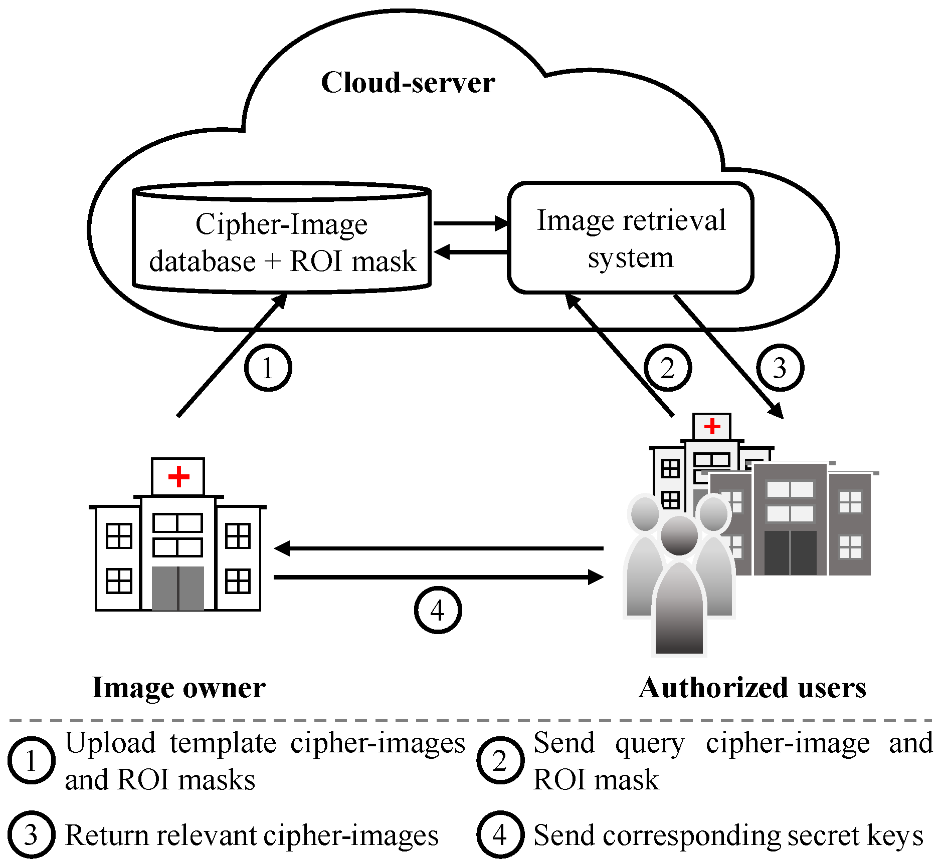
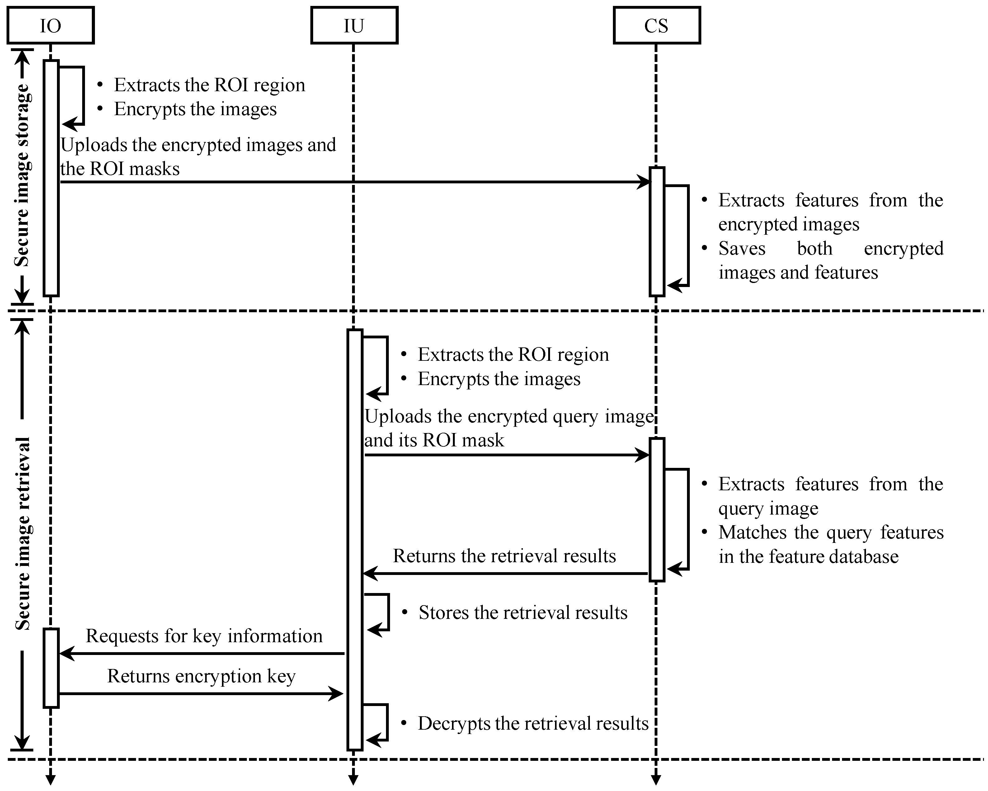
3.3. System Model
3.4. Threat Model
4. Simulation Results
4.1. Retrieval Performance Analysis
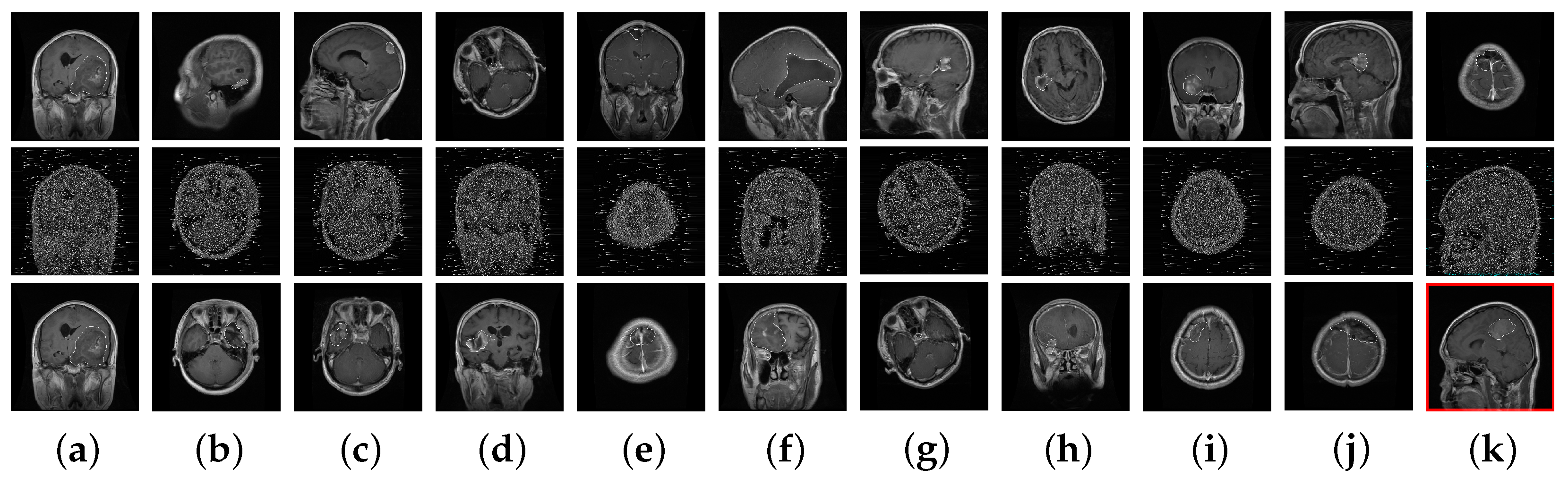
- Dataset. Although different medical image modalities, such as Positron Emission Tomography (PET), Computed Tomography (CT), and Magnetic Resonance Imaging (MRI) scans can be used to create images of the brain, MRI remains the primary diagnostic modality for brain tumors because it is highly sensitive in visualizing soft tissues within the brain compared with other modalities. Therefore, we used an MRI dataset in our experiments. It is worth mentioning that our proposed secure retrieval scheme can be readily implemented for other types of image modalities. In simulations, we evaluated the performance of different brain tumor retrieval schemes on a public brain tumor MRI dataset available from [3]. This dataset contains 3064 T1-weighted, contrast-enhanced images in size from 233 patients with three kinds of brain tumors: meningioma (708 slices), glioma (1426 slices), and pituitary (930 slices). The images in the dataset were split into five subsets for fivefold cross-validation. Among these subsets, one was used as query images, while the rest of them were used as template images. Also, it was ensured that slices from the same patient did not appear simultaneously in the template and query images. Figure 4 shows an example image from the dataset for each brain tumor type in plain and encrypted form. The cipher images were obtained by encrypting the plain images using our proposed PE algorithm. In the cipher images, pixel values which were substituted during encryption appear as white noise for the key value in Equation (6).Figure 4. Example images from the dataset for each brain tumor type. (a–c) Plain-images and (d–f) their corresponding cipher images obtained from our proposed encryption technique. The tumor region (ROI) in each image is enlarged and shown below it. This information is visually unrecognizable in the cipher images.Figure 4. Example images from the dataset for each brain tumor type. (a–c) Plain-images and (d–f) their corresponding cipher images obtained from our proposed encryption technique. The tumor region (ROI) in each image is enlarged and shown below it. This information is visually unrecognizable in the cipher images.
![Electronics 14 01759 g004]()
- Retrieval performance evaluation metric. The precision metric is one of the most commonly used metrics to evaluate an image retrieval system’s performance. It measures the retrieval performance in terms of how many retrieved images are relevant. Let and denote the sets of similar and retrieved images, respectively. The precision (P) is defined as follows:

- Retrieval results. To analyze the efficiency of proposed technique, we implemented the different PE techniques proposed in [25,26,27] and analyzed their impact on the image retrieval system. For all techniques, analyses were carried out on the same dataset with fivefold cross-validation. Following [3], throughout our experiments, image dilation with a disk-shaped structuring element with a radius of 24 was used to augment the ROI region, and the size of the raw image patches was set to be nine for the local features. Also, for all encryption techniques, we considered encrypting all images with the same and different keys.
4.2. Encryption Efficiency Analysis
- Key space analysis. In general, the key space of an encryption algorithm should be larger than to resist brute-force attacks. The secret key of our proposed scheme is generated using three random sequences by varying the control parameter of the SSLM. Considering a precision of , the key space size can be determined to be , which is larger than the recommended value to resist a brute-force attack.
- Comparison with existing PE techniques. Similar to the proposed technique, the conventional PE techniques proposed in [25,26] modify only half of the pixel values. Their encryption function is an identity map for , while a cipher pixel value was computed to be in [25] and in [26] for . Consequently, each pixel is independently encrypted in them, which may make them vulnerable to select plaintext attacks. On the other hand, a PE technique was proposed in [27] where each pixel substitution is dependent on all previously encrypted pixels. Their encryption function is closely related to ours, given in Equation (6), with a difference in that the term computes as , as opposed to our identity map. This can achieve better security efficiency but at the cost of the downstream application performance, as given in Table 1.
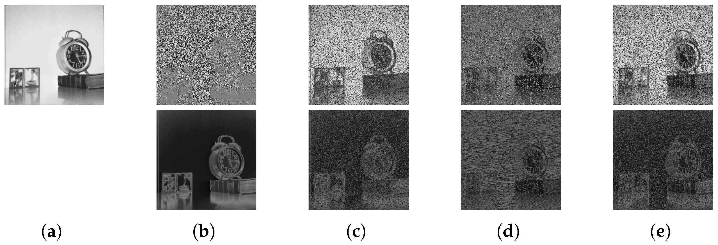
- Robustness against ciphertext-only attacks. For this analysis, we considered a threat model where an adversary (for example, a semi-honest cloud service owner) performed a ciphertext-only attack (COA) to decrypt a cipher image without access to the secret key. A popular example of such COAs against PE was proposed in [28], where the goal was to recover partial information from a cipher image to identify its contents. This attack works by guessing the leading bit of each pixel value with respect to the neighboring pixels, and hence it is also named the “leading bit attack”. Figure 6 shows an example image (https://sipi.usc.edu/database/, accessed on 30 October 2024) recovered by performing a leading bit attack on the cipher images of different PE techniques. Here, the example is of a natural image, as it is easy to understand visually meaningful information in such images. It can be observed that the image recovered from the proposed cipher image did not reveal any information. On the contrary, a PE technique that simply inverts pixel values in an image is more susceptible to such an attack; that is, its contents can be recovered with high visibility, as shown in Figure 6b.
- Time complexity analysis. To analyze the computational overhead of the proposed PE scheme on the client side, Table 3 compares its execution time with those of the schemes proposed in [25,26,27]. For this analysis, we chose 2124 images from our dataset, which were uniformly distributed among the three tumor types and each image was 512 × 512 in size. The time was reported in seconds as a mean across all of the images. It can be observed from Table 3 that a PE technique such as the one proposed in [25,26], which processes each pixel independently, had a shorter encryption time, as it is highly parallelizable. However, this can also lead to certain security vulnerabilities, such as data reconstruction attacks. On the other hand, in proposed method, only half of the current pixel processing is dependent on the previously encrypted pixel values to deliver a desirable level of security in an acceptable execution time.
4.3. Discussions
5. Conclusions
Author Contributions
Funding
Institutional Review Board Statement
Informed Consent Statement
Data Availability Statement
Conflicts of Interest
References
- Ahmad, I.; Uzzal, M.S.; Shin, S. Secure Retrieval of Brain Tumor Images using Perceptual Encryption. In Proceedings of the 2025 IEEE International Conference on Big Data and Smart Computing (BigComp), Kota Kinabalu, Malaysia, 9–12 February 2025; pp. 73–74. [Google Scholar] [CrossRef]
- Tabatabaei, Z.; Wang, Y.; Colomer, A.; Oliver Moll, J.; Zhao, Z.; Naranjo, V. WWFedCBMIR: World-Wide Federated Content-Based Medical Image Retrieval. Bioengineering 2023, 10, 1144. [Google Scholar] [CrossRef] [PubMed]
- Cheng, J.; Yang, W.; Huang, M.; Huang, W.; Jiang, J.; Zhou, Y.; Yang, R.; Zhao, J.; Feng, Y.; Feng, Q.; et al. Retrieval of Brain Tumors by Adaptive Spatial Pooling and Fisher Vector Representation. PLoS ONE 2016, 11, e0157112. [Google Scholar] [CrossRef] [PubMed]
- Silva, W.; Gonçalves, T.; Härmä, K.; Schröder, E.; Obmann, V.C.; Barroso, M.C.; Poellinger, A.; Reyes, M.; Cardoso, J.S. Computer-Aided Diagnosis Through Medical Image Retrieval in Radiology. Sci. Rep. 2022, 12, 20732. [Google Scholar] [CrossRef]
- Owais, M.; Arsalan, M.; Choi, J.; Park, K.R. Effective Diagnosis and Treatment through Content-Based Medical Image Retrieval (CBMIR) by Using Artificial Intelligence. J. Clin. Med. 2019, 8, 462. [Google Scholar] [CrossRef] [PubMed]
- Sharma, S.; Aggarwal, A. A New Approach for Effective Retrieval of Medical Images: A Step towards Computer-Assisted Diagnosis. J. Imaging 2024, 10, 210. [Google Scholar] [CrossRef] [PubMed]
- Zhu, Y.; Yin, X.; Liew, A.W.C.; Tian, H. Privacy-Preserving in Medical Image Analysis: A Review of Methods and Applications. arXiv 2024, arXiv:2412.03924. [Google Scholar]
- Bellafqira, R.; Coatrieux, G.; Bouslimi, D.; Quellec, G. Secure Content Based Image Retrieval in Medical Databases. In Proceedings of the Journées RITS, Dourdan, France, 25–27 March 2015. [Google Scholar]
- Zhang, C.; Li, J.; Wang, S.; Wang, Z. An Encrypted Medical Image Retrieval Algorithm Based on DWT-DCT Frequency Domain. In Proceedings of the 2017 IEEE 15th International Conference on Software Engineering Research, Management and Applications (SERA), London, UK, 7–9 June 2017; pp. 135–141. [Google Scholar] [CrossRef]
- Zhu, D.; Zhu, H.; Wang, X.; Lu, R.; Feng, D. An Accurate and Privacy-Preserving Retrieval Scheme Over Outsourced Medical Images. IEEE Trans. Serv. Comput. 2023, 16, 913–926. [Google Scholar] [CrossRef]
- Li, D.; Wu, Y.; Lü, Q.; Zhang, K.; Wang, Z.; Wu, J. PMIR: An Efficient Privacy-Preserving Medical Images Search in Cloud-Assisted Scenario. Springer Neural Comput. Appl. 2024, 36, 1477–1493. [Google Scholar] [CrossRef]
- Li, D.; Lü, Q.; Liao, X.; Xiang, T.; Wu, J.; Le, J. AVPMIR: Adaptive Verifiable Privacy-Preserving Medical Image Retrieval. IEEE Trans. Dependable Secur. Comput. 2024, 21, 4637–4651. [Google Scholar] [CrossRef]
- Duan, Y.; Li, Y.; Lu, L.; Ding, Y. A Faster Outsourced Medical Image Retrieval Scheme with Privacy Preservation. J. Syst. Archit. 2022, 122, 102356. [Google Scholar] [CrossRef]
- Rajan, A.A.; V, V.; Raikwar, M.; Balaraman, R. SMedIR: Secure Medical Image Retrieval Framework with ConvNeXt-based Indexing and Searchable Encryption in the Cloud. Springer J. Cloud Comput. 2024, 13, 139. [Google Scholar] [CrossRef]
- Feng, Q.; Li, P.; Lu, Z.; Zhou, Z.; Wu, Y.; Weng, J.; Huang, F. DHAN: Encrypted JPEG Image Retrieval via DCT Histograms-Based Attention Networks. Appl. Soft Comput. 2023, 133, 109935. [Google Scholar] [CrossRef]
- Guo, C.; Jia, J.; Choo, K.K.R.; Jie, Y. Privacy-Preserving Image Search (PPIS): Secure Classification and Searching Using Convolutional Neural Network Over Large-Scale Encrypted Medical Images. Comput. Secur. 2020, 99, 102021. [Google Scholar] [CrossRef]
- Cai, G.; Wei, X.; Li, Y. Privacy-Preserving CNN Feature Extraction and Retrieval Over Medical Images. Int. J. Intell. Syst. 2022, 37, 9267–9289. [Google Scholar] [CrossRef]
- Shen, M.; Deng, Y.; Zhu, L.; Du, X.; Guizani, N. Privacy-Preserving Image Retrieval for Medical IoT Systems: A Blockchain-Based Approach. IEEE Netw. 2019, 33, 27–33. [Google Scholar] [CrossRef]
- Ahmad, I.; Kim, J.; Shin, S. A Searchable Encryption Technique for Secure Color Image Retrieval. In Proceedings of the 2024 15th International Conference on Information and Communication Technology Convergence (ICTC), Jeju Island, Republic of Korea, 16–18 October 2024; pp. 36–41. [Google Scholar] [CrossRef]
- Iida, K.; Kiya, H. Privacy-Preserving Content-Based Image Retrieval Using Compressible Encrypted Images. IEEE Access 2020, 8, 200038–200050. [Google Scholar] [CrossRef]
- Uzzal, S.M.; Ahmad, I.; Shin, S. Perceptual Encryption-Based Privacy-Preserving Image Retrieval Application. In Proceedings of the 10th International Conference on Next Generation Computing (ICNGC), Angeles City, Philippines, 17–18 December 2024. (in press). [Google Scholar]
- Fan, B.; Wu, F.; Hu, Z. Aggregating Gradient Distributions into Intensity Orders: A Novel Local Image Descriptor. In Proceedings of the CVPR 2011, Colorado Springs, CO, USA, 20–25 June 2011; pp. 2377–2384. [Google Scholar] [CrossRef]
- Perronnin, F.; Liu, Y.; Sánchez, J.; Poirier, H. Large-Scale Image Retrieval with Compressed Fisher Vectors. In Proceedings of the IEEE Computer Society Conference on Computer Vision and Pattern Recognition, San Francisco, CA, USA, 13–18 June 2010; pp. 3384–3391. [Google Scholar] [CrossRef]
- de Carvalho, R.E.; Leonel, E.D. Squared Sine Logistic Map. Phys. A Stat. Mech. Its Appl. 2016, 463, 37–44. [Google Scholar] [CrossRef]
- Sirichotedumrong, W.; Kinoshita, Y.; Kiya, H. Pixel-Based Image Encryption without Key Management for Privacy-Preserving Deep Neural Networks. IEEE Access 2019, 7, 177844–177855. [Google Scholar] [CrossRef]
- Ahmad, I.; Shin, S. A Pixel-Based Encryption Method for Privacy-Preserving Deep Learning Models. arXiv 2022, arXiv:2203.16780. [Google Scholar]
- Ahmad, I.; Shin, S. Learnable Pixel-Based Encryption for Privacy-Preserving Image Classification. In Proceedings of the Symposium of the Korean Institute of communications and Information Sciences (KICS), Gyeongju, Republic of Korea, 20–22 November 2024; pp. 252–253. [Google Scholar]
- Chang, A.H.; Case, B.M. Attacks on Image Encryption Schemes for Privacy-Preserving Deep Neural Networks. arXiv 2020, arXiv:2004.13263. [Google Scholar]


| D | Methods | mAP (%) | Prec@5 (%) | Prec@10 (%) | |
|---|---|---|---|---|---|
| 16 | Non-secure | [3] | 89.89 ± 1.00 | 87.16 ± 1.52 | 87.16 ± 1.45 |
| Secure | [25] | 81.82 ± 1.39 | 80.26 ± 1.18 | 80.39 ± 1.30 | |
| [26] | 86.66 ± 0.80 | 83.79 ± 1.13 | 83.95 ± 1.03 | ||
| [27] | 81.65 ± 0.91 | 77.51 ± 1.55 | 77.50 ± 1.32 | ||
| Ours | 87.06 ± 0.99 | 83.89 ± 1.53 | 83.84 ± 1.43 | ||
| Secure | [25] | 74.57 ± 1.46 | 72.57 ± 2.38 | 72.79 ± 2.15 | |
| [26] | 80.17 ± 2.66 | 75.98 ± 3.31 | 75.96 ± 3.39 | ||
| [27] | 77.96 ± 2.16 | 72.61 ± 3.26 | 72.64 ± 3.18 | ||
| Ours | 83.52 ± 1.29 | 80.05 ± 2.49 | 80.19 ± 2.45 | ||
| 32 | Non-secure | [3] | 92.49 ± 0.89 | 90.14 ± 0.96 | 90.10 ± 0.96 |
| Secure | [25] | 84.86 ± 0.91 | 82.37 ± 0.56 | 82.40 ± 0.55 | |
| [26] | 88.84 ± 0.74 | 85.99 ± 1.31 | 85.88 ± 1.22 | ||
| [27] | 86.00 ± 1.08 | 81.47 ± 1.76 | 81.51 ± 1.67 | ||
| Ours | 89.44 ± 1.33 | 86.40 ± 2.00 | 86.43 ± 1.99 | ||
| Secure | [25] | 77.55 ± 1.64 | 74.16 ± 2.36 | 74.06 ± 2.73 | |
| [26] | 83.42 ± 2.43 | 78.38 ± 3.70 | 78.47 ± 3.58 | ||
| [27] | 80.38 ± 1.55 | 74.25 ± 2.35 | 74.30 ± 2.26 | ||
| Ours | 86.32 ± 1.23 | 82.15 ± 2.14 | 82.22 ± 2.04 | ||
| 64 | Non-secure | [3] | 94.02 ± 0.88 | 91.75 ± 1.17 | 91.80 ± 1.18 |
| Secure | [25] | 86.51 ± 1.16 | 83.44 ± 0.85 | 83.32 ± 0.93 | |
| [26] | 90.72 ± 1.08 | 87.93 ± 1.63 | 87.97 ± 1.63 | ||
| [27] | 88.80 ± 1.50 | 85.03 ± 1.78 | 85.05 ± 1.77 | ||
| Ours | 91.41 ± 1.34 | 88.38 ± 1.54 | 88.44 ± 1.56 | ||
| Secure | [25] | 79.71 ± 2.19 | 74.69 ± 2.74 | 74.63 ± 2.73 | |
| [26] | 85.48 ± 2.23 | 81.21 ± 3.01 | 81.15 ± 3.02 | ||
| [27] | 84.26 ± 1.94 | 79.09 ± 2.84 | 79.11 ± 2.84 | ||
| Ours | 88.56 ± 1.82 | 85.22 ± 2.19 | 85.17 ± 2.23 | ||
| D | Methods | Type | mAP (%) | Prec@5 (%) | Prec@10 (%) |
|---|---|---|---|---|---|
| 16 | Non-secure ([3]) | T1 | 82.75 | 84.60 | 84.11 |
| T2 | 94.04 | 89.16 | 89.41 | ||
| T3 | 88.88 | 86.00 | 86.00 | ||
| 5.65 | 2.34 | 2.69 | |||
| Secure ([26]) | T1 | 76.52 ∣ 70.42 | 79.66 ∣ 72.07 | 79.47 ∣ 72.10 | |
| T2 | 92.75 ∣ 88.38 | 86.05 ∣ 79.55 | 86.63 ∣ 79.97 | ||
| T3 | 85.39 ∣ 75.43 | 83.92 ∣ 73.86 | 83.73 ∣ 73.11 | ||
| 8.13 ∣ 9.27 | 3.25 ∣ 3.91 | 3.60 ∣ 4.28 | |||
| Secure (Ours) | T1 | 76.05 ∣ 73.36 | 79.24 ∣ 75.31 | 78.59 ∣ 74.98 | |
| T2 | 92.90 ∣ 91.00 | 86.21 ∣ 83.58 | 86.58 ∣ 84.11 | ||
| T3 | 86.85 ∣ 80.38 | 84.36 ∣ 78.85 | 84.09 ∣ 78.78 | ||
| 8.54 ∣ 8.88 | 3.61 ∣ 4.15 | 4.09 ∣ 4.59 | |||
| 32 | Non-secure ([3]) | T1 | 82.75 | 84.60 | 84.11 |
| T2 | 94.04 | 89.16 | 89.41 | ||
| T3 | 88.88 | 86.00 | 86.00 | ||
| 5.65 | 2.34 | 2.69 | |||
| Secure ([26]) | T1 | 76.52 ∣ 70.42 | 79.66 ∣ 72.07 | 79.47 ∣ 72.10 | |
| T2 | 92.75 ∣ 88.38 | 86.05 ∣ 79.55 | 86.63 ∣ 79.97 | ||
| T3 | 85.39 ∣ 75.43 | 83.92 ∣ 73.86 | 83.73 ∣ 73.11 | ||
| 8.13 ∣ 9.27 | 3.25 ∣ 3.91 | 3.60 ∣ 4.28 | |||
| Secure (Ours) | T1 | 76.05 ∣ 73.36 | 79.24 ∣ 75.31 | 78.59 ∣ 74.98 | |
| T2 | 92.90 ∣ 91.00 | 86.21 ∣ 83.58 | 86.58 ∣ 84.11 | ||
| T3 | 86.85 ∣ 80.38 | 84.36 ∣ 78.85 | 84.09 ∣ 78.78 | ||
| 8.54 ∣ 8.88 | 3.61 ∣ 4.15 | 4.09 ∣ 4.59 | |||
| 64 | Non-secure ([3]) | T1 | 88.06 | 86.05 | 86.05 |
| T2 | 97.14 | 94.91 | 95.01 | ||
| T3 | 93.75 | 91.24 | 91.25 | ||
| 4.59 | 4.45 | 4.50 | |||
| Secure ([26]) | T1 | 82.29 ∣ 75.89 | 80.55 ∣ 72.89 | 80.61 ∣ 72.98 | |
| T2 | 95.48 ∣ 92.34 | 91.60 ∣ 86.77 | 91.71 ∣ 86.92 | ||
| T3 | 90.21 ∣ 82.72 | 88.30 ∣ 79.47 | 88.24 ∣ 79.02 | ||
| 6.64 ∣ 8.26 | 5.67 ∣ 6.94 | 5.68 ∣ 6.99 | |||
| Secure (Ours) | T1 | 83.53 ∣ 80.22 | 81.91 ∣ 77.90 | 81.60 ∣ 77.71 | |
| T2 | 95.39 ∣ 94.49 | 91.59 ∣ 90.65 | 91.85 ∣ 90.79 | ||
| T3 | 91.74 ∣ 86.51 | 85.22 ∣ 83.38 | 89.01 ∣ 83.14 | ||
| 6.07 ∣ 7.15 | 5.01 ∣ 6.40 | 5.29 ∣ 6.57 |
Disclaimer/Publisher’s Note: The statements, opinions and data contained in all publications are solely those of the individual author(s) and contributor(s) and not of MDPI and/or the editor(s). MDPI and/or the editor(s) disclaim responsibility for any injury to people or property resulting from any ideas, methods, instructions or products referred to in the content. |
© 2025 by the authors. Licensee MDPI, Basel, Switzerland. This article is an open access article distributed under the terms and conditions of the Creative Commons Attribution (CC BY) license (https://creativecommons.org/licenses/by/4.0/).
Share and Cite
Ahmad, I.; Uzzal, M.S.; Shin, S. Secure Retrieval of Brain Tumor Images Using Perceptual Encryption in Cloud-Assisted Scenario. Electronics 2025, 14, 1759. https://doi.org/10.3390/electronics14091759
Ahmad I, Uzzal MS, Shin S. Secure Retrieval of Brain Tumor Images Using Perceptual Encryption in Cloud-Assisted Scenario. Electronics. 2025; 14(9):1759. https://doi.org/10.3390/electronics14091759
Chicago/Turabian StyleAhmad, Ijaz, Md Shahriar Uzzal, and Seokjoo Shin. 2025. "Secure Retrieval of Brain Tumor Images Using Perceptual Encryption in Cloud-Assisted Scenario" Electronics 14, no. 9: 1759. https://doi.org/10.3390/electronics14091759
APA StyleAhmad, I., Uzzal, M. S., & Shin, S. (2025). Secure Retrieval of Brain Tumor Images Using Perceptual Encryption in Cloud-Assisted Scenario. Electronics, 14(9), 1759. https://doi.org/10.3390/electronics14091759