Article

Cu2O Nanowire Chemiresistors for Detection of Organophosphorus CWA Simulants

1
Department of Physics and Measurements, Faculty of Chemical Engineering, University of Chemistry and Technology Prague (UCT), 166 28 Prague, Czech Republic
2
Department of Mathematics, Informatics and Cybernetics, Faculty of Chemical Engineering, University of Chemistry and Technology Prague (UCT), 166 28 Prague, Czech Republic
*
Author to whom correspondence should be addressed.
Electronics 2025, 14(17), 3478; https://doi.org/10.3390/electronics14173478
Submission received: 1 August 2025 / Revised: 26 August 2025 / Accepted: 29 August 2025 / Published: 31 August 2025

Abstract

Rapid on-site detection of chemical warfare agents (CWAs) is vital for security and environmental monitoring. In this work, copper(I) oxide (Cu2O) nanowire (NW) chemiresistors were investigated as gas sensors for low-concentration organophosphorus chemical warfare agent (CWA) simulants. The NWs were hydrothermally synthesized and deposited onto microheater platforms, enabling them to operate at elevated working temperatures. Their sensing performance was tested against a range of vapor-phase simulants, including dimethyl methylphosphonate (DMMP), triethyl phosphate (TEP), diethyl ethylphosphonate (DEEP), diphenyl phosphoryl chloride (DPPCl), parathion, diethyl phosphite (DEP), diethyl adipate (DEA), and cyanogen chloride (ClCN). Fully oxidized P(V) simulants (DMMP, DEEP, TEP) produced modest, predominantly reversible responses (~3–6% RR). On the contrary, DPPCl and DEP induced the strongest relative responses (RR −94.67% and >200%, respectively), accompanied by irreversible surface modification as revealed by SEM and EDS. ClCN produced a substantial but reversible negative response (RR −9.5%), consistent with transient oxidative interactions. Surface poisoning was confirmed after exposure to DEP and DPPCl, which left phosphorus or chlorine residues on the Cu2O surface. These results highlight both the promise and limitations of Cu2O NW chemiresistors for selective CWA detection.

1. Introduction

Ensuring public safety and environmental stewardship demands rapid, on-site detection of chemical warfare agents (CWAs). Conventional CWA detection techniques (e.g., ion mobility spectrometry, flame photometry, infrared/Raman spectroscopy, gas chromatography–mass spectrometry) offer both high sensitivity and selectivity but require complex, costly, and bulky instrumentation [1,2,3]. This has driven interest in compact chemical sensors, particularly chemiresistors based on semiconducting materials, which transduce chemical interactions into measurable electrical signals [1]. Beyond chemiresistors, colorimetric/electrical array approaches, most notably polydiacetylene (PDA) platforms that report vapor exposure via RGB analysis and impedance spectroscopy, have also recently been explored for CWA surrogates [4]. Metal-oxide semiconductor (MOS) chemiresistors are a dominant class of such sensors, leveraging conductivity changes in a metal-oxide film upon gas adsorption [1]. Numerous MOS sensor studies have targeted nerve agents and blister agents via safer simulant chemicals [5]. In particular, dimethyl methylphosphonate (DMMP) is widely used as a simulant for the nerve agent sarin [6], and its surface reaction mechanism on oxide sensors has been extensively studied [7,8,9]. Metal oxide nanowire sensors have demonstrated the ability to detect chemical warfare simulants, such as DMMP, at sub-ppm concentrations, highlighting their potential for sensitive real-time detection of CWAs [10]. Complementary system-level developments include low-power “electronic-nose” arrays and hybrid MOX + PID payloads for mobile platforms (e.g., UAVs), which use multichannel signals and machine learning to classify CWA surrogates and interferents, increasing the potential of sensor arrays for field detection [11].
Numerous reports have refined MOS-based and hybrid approaches for CWA/simulant detection, including topical reviews on chemoresistive metal-oxide nanosystems [6], MXene, and other emerging nanoarchitectures for CWA sensing [12], and updates dedicated to MOx chemical/electrochemical sensors [13]. Parallel efforts have demonstrated array-based concepts of “electronic nose” with room-temperature or micro-heated MEMS sensors and machine-learning classification of CWA surrogates and toxic industrial chemicals [14,15]. Together, these studies underscore trends toward lower-power operation, selectivity obtained by processing multidimensional array signals, and data-driven analytics, also addressing challenges such as poisoning/reversibility and field-relevant validation.
Although numerous recent studies report highly sensitive detection of parathion and related organophosphates using nanostructured materials and electrochemical techniques, these methods are typically designed for analytes in liquid-phase or solution-based matrices, such as groundwater, food, or soil extracts [16,17,18]. Incorporating modifiers such as reduced graphene oxide or combining Cu2O with electrochemical or spectroscopic transduction methods (e.g., SERS) can enhance selectivity and sensitivity for specific CWAs [18,19].
Organophosphorus CWAs (G- and V-agents) and their simulants typically act as reducing gases on MOS sensors: at elevated operating temperatures (ca. 300–400 °C) they oxidize on the sensor’s surface, consuming adsorbed oxygen and releasing electrons [20,21,22]. In contrast to solution-phase detection, which benefits from analyte preconcentration and stable liquid interfaces, gas-phase sensing, particularly of volatile nerve agent simulants, requires robust surface interactions, fast kinetics, and ambient stability under fluctuating humidity and temperature. For n-type oxide (e.g., SnO2, ZnO) chemiresistors, this interaction causes a resistance decrease, whereas for p-type semiconductors, it leads to a decrease in hole concentration and thus an increase in resistance [23]. A known challenge is that organophosphates often produce persistent surface species (e.g., phosphates) upon oxidation, causing sensor poisoning that manifests itself as slow or incomplete recovery and long-term sensitivity loss [1]. For example, on the oxide surfaces, DMMP oxidizes to methylphosphonic acid, which can remain adsorbed and block active sites [24]. This effect has been observed on various materials (e.g., SnO2, ZnO, Mn3O4, etc.), limiting sensor reusability [25]. Thus, while MOS chemiresistors are promising for CWA detection due to their simplicity and low cost, improving their sensitivity and mitigating poisoning effects are key research goals [1].
Copper oxides have emerged as interesting p-type semiconductor sensing materials. Copper(I) oxide (Cu2O) and Copper(II) oxide (CuO) are both p-type with band gaps of ~2.0 eV and ~1.2 eV, respectively [26]. They have been studied for various applications (e.g., catalysis [27], photovoltaics [28], etc.). Especially in gas sensors, the specific oxide phase and stoichiometry can significantly influence the device performance. Recent near-ambient-pressure XPS studies on Cu2O/CuO nanowires (NWs) revealed that under oxidizing conditions, Cu+ in Cu2O can be partially oxidized to Cu2+, whereas introducing a reducing atmosphere can restore some Cu+, indicating the formation of dynamic Cu2O/CuO core–shell heterostructures during sensing [29]. Such in situ changes in the copper oxidation state and the presence of p–p′ junctions at Cu2O/CuO interfaces may alter charge transport and surface chemistry, thereby affecting gas response. In situ NAP-XPS indicates a Cu2O(core)–CuO(shell) architecture: devices with a Cu2O-rich core and an ultrathin CuO surface shell (≈1 nm) show stable, repeatable p-type transduction, whereas a thicker CuO shell (≈5–8 nm) leads to anomalous behavior and drift [29]. In line with this, DMMP decomposes on CuO at room temperature, so increasing the CuO surface fraction raises reactivity but also the probability of residue accumulation [30]. Notably, CuO-based sensors have shown responses to organophosphorus compounds in prior studies [31]. However, comparatively few works have explored Cu2O NW networks as active layers for CWA simulant detection. Compared to their thin-film counterparts, nanowire-based devices exhibit higher sensitivity to nerve agent simulants such as DMMP due to their distinct single-crystalline surfaces and surface-dominated conduction mechanisms [10].
This paper presents a study of chemiresistive sensors based on Cu2O nanowire (NW) networks for detecting selected chemical warfare agent (CWA) simulants and relevant toxic industrial chemicals (Table 1). We focus on several representative organophosphorus simulants: dimethyl methylphosphonate (DMMP) as a sarin analog; triethyl phosphate (TEP) and diethyl ethylphosphonate (DEEP) as G-agent simulants (e.g., soman); and diphenyl phosphoryl chloride (DPPCl) and diethyl phosphite (DEP) as tabun-like compounds, based on their structural similarity to multiple G-agents, including sarin and tabun, as indicated by their Euclidean Distance (ED) and Tanimoto Coefficient (TC) values [32]. Parathion is included as a low-volatility organophosphorothioate representative, often discussed as a VX simulant, while diethyl adipate (DEA), a non-phosphorus compound, is used as a structural analog of the blister agent sulfur mustard. In addition, cyanogen chloride (ClCN), a blood agent, was tested for its relevance as a high-volatility and highly reactive oxidizer. The study evaluates sensor response patterns, investigates the underlying sensing mechanisms (including the role of Cu2O vs. CuO surface composition), and addresses limitations such as sensor poisoning. Electrical response measurements are complemented by surface analysis techniques such as scanning electron microscopy (SEM) and energy-dispersive X-ray spectroscopy (EDS). The findings provide insights into the applicability of Cu2O/CuO-based sensors for CWA monitoring and suggest future directions for improving selectivity and stability.

2. Materials and Methods

2.1. Synthesis and Sensor Fabrication

Copper(I) oxide nanowires were used as the sensing material in this work. Cu2O NWs were prepared via a wet-chemical hydrothermal method following Hozák et al. [29]. Cu(CH3COO)2 (1.6 mmol, 290 mg) was dissolved in 64 mL of deionized water and mixed with 80 mL of 0.04 M o-phenetidine; after ~2 min, 16 mL of 0.05 M acetic acid was added. The mixture was kept at 90 °C for 10 h and cooled to room temperature. The precipitated NWs were filtered and dispersed in 80 mL of N-methyl-2-pyrrolidone (50 °C, 24 h) to remove poly(o-phenetidine), then filtered and washed with methanol. For sensor fabrication, the Cu2O NW suspension was drop-casted onto a sensor substrate consisting of a ceramic sensor platform KBS4 (TESLA BLATNÁ a.s., Blatná, Czech Republic), which is a double-sided ceramic substrate equipped with platinum interdigitated electrodes (IDE, 40 μm line/40 μm gap) on the front side and a platinum microheater on the reverse side. The as-deposited NW layer was dried in an oven at 100 °C to evaporate the solvent and promote adhesion. This process produced a highly porous layer of Cu2O nanowires bridging the electrode gaps.

2.2. Characterization

The morphology and structure of the Cu2O NW layer were analyzed using a MIRA 3 field emission scanning electron microscope (FE-SEM; TESCAN, Brno, Czech Republic) operated at an accelerating voltage of 15 kV. SEM images were captured at magnifications up to 100,000×, providing detailed visualization of the nanowire network structure. The elemental composition of the active sensing layer was investigated using energy-dispersive X-ray spectroscopy (EDS) with a Quantax 200 system and XFlash 6 detector (Bruker Nano GmbH, Berlin, Germany). Measurements were performed with an estimated interaction depth of 1.2–1.8 μm and a lateral resolution of ~1 μm. EDS measurements were acquired on multiple measurement areas of the Cu2O active layer before and after exposure and are reported as atomic % (mean ± SD across measurement areas). The complete area-by-area EDS values, additional high-magnification SEM/EDS of the Cu2O layer, and overview SEM/EDS of the entire sensor platform are provided in the Supplementary Materials.

2.3. Gas Exposure Setup

Analyte concentration generation and sensor response measurements were performed using a dynamic flow system (Figure 1). Known volumes of each liquid simulant were injected into Tedlar™ gas sampling bags (10 L volume) pre-filled with a measured amount of synthetic air. A volume of 5–10 µL of liquid DMMP, DPPCl, DEEP, DEP, DEA, TEP, or parathion was injected via gas-tight syringe into a 10 L air-filled bag, yielding target analyte concentrations on the order of 60–120 ppm (Table 1). Low-volatility compounds, such as parathion and DPPCl, required injecting an excess amount and were tested at their saturated vapor pressure in air, which was determined to be 49 ppb for DPPCl and 8.9 ppb for parathion at laboratory temperature (Table 1). For the gaseous analyte ClCN (boiling point 13 °C, at ambient pressure), a 1 mL volume of the gas was injected into 10 L of air to achieve 120 ppm.
The Cu2O NW sensor device was placed in a glass flow chamber that was kept at 45 °C to avoid any condensation. The sensor’s integrated Pt microheater was powered to maintain the active layer at an operating temperature of 320 °C. This temperature was chosen based on our previous experience with Cu2O NW sensors and commonly reported operating windows (150–350 °C) [33,34] to ensure a rapid, measurable response while minimizing thermal degradation of the sensor and its baseline drift. The temperature was monitored via the resistance of the Pt heater element integrated in the sensor platform. During the testing, either synthetic air or the sample was continuously passed through the chamber at 300 mL·min−1 using a pump. A three-way valve system allowed switching between a reference and the sample, both pulled at the same flow rate. Each sensor was first stabilized in clean air (baseline) and then exposed to the sample for approximately 15 min, followed by purging with synthetic air for recovery. Multiple exposure cycles (typically 2–4) were performed for each analyte on a given sensor to check repeatability, except in cases where the first exposure caused permanent sensor changes. Sensor resistance and sensor platform temperature were recorded using an Agilent 34970A data acquisition unit.

2.4. Simulants’ Similarity to CWAs

Table 2 presents the Euclidean Distance (ED) and Tanimoto Coefficient (TC) values used to evaluate the structural similarity of the tested simulants to key chemical warfare agents (CWAs), specifically sarin (GB), soman (GD), tabun (GA), VX, and the blister agent distilled mustard (HD). These molecular similarity metrics were extracted from the cheminformatics study by Lavoie et al. [32].
The ED is a descriptor-based similarity measure, with values ranging from 0 to 1, where smaller values indicate greater structural similarity to the target CWA. In contrast, the TC is a fingerprint-based metric, with values ranging from 0 to 1, where 1 indicates identical structures and, conversely, values closer to 0 indicate low similarity. Therefore, lower ED and higher TC values both suggest closer molecular resemblance.
In this table, DMMP and DEEP show good structural similarity to G-series agents (e.g., sarin and soman), with low ED values and high TC scores, supporting their use as sarin and soman simulants, respectively. DEP also shows moderate resemblance to G-agents. DPPCl (diphenyl phosphoryl chloride) exhibits moderate similarity to sarin (ED = 0.541, TC = 0.500) and tabun (ED 0.511, TC = 0.245), which may still support its tentative use as a tabun-like simulant, although this selection appears more pragmatic than structurally justified. Notably, DEA (diethyl adipate) does not structurally resemble nerve agents but shows a moderate ED of 0.560 with HD, consistent with its role as a blister agent analog in this study. DMMP, TEP, and DEEP are standard G-agent simulants because they are fully oxidized P(V) phosphonate esters that retain the key P=O functionality and offer a “workable” vapor pressure and safer handling. Simulant selection used in this study hence balances structural similarity with safety/availability. Notably, fully oxidized P(V) simulants are typically less reactive on MOS surfaces than species bearing labile P–Cl or P–H bonds—consistent with our modest DMMP/DEEP/TEP responses versus the strong effects from DEP and DPPCl.

2.5. Data Analysis

The primary sensor signal was the measured electrical resistance across the entire sensor device, which includes contributions from the Cu2O nanowire (NW) layer, as well as contact interfaces and the underlying substrate. However, the dominant resistance change was attributed to the NW layer’s interaction with analytes. For these p-type sensors, a positive response was defined as an increase in resistance upon analyte exposure, expressed as a percentage of the baseline value and often called Relative Response ( R R ( c ) ) [35,36,37,38].
R R ( c ) = R a i r R a n a l y t e R a i r · 100 %
To quantify the response/recovery dynamics, the resistance transients were fitted with exponential kinetic models (Equation (2)).
R ( t ) = A · ( 1 e t τ ) + y 0
A first-order (mono-exponential) adsorption/desorption model was applied consistently across all analytes to fit the sensor response and recovery curves. From these fits, key kinetic parameters were extracted, including the Relative Response (RR, %), response and recovery time constants (τ), and time to reach 10% and 90% of the final signal (τ10% and τ90%).

3. Results and Discussion

3.1. Scanning Electron Microscopy

The pristine Cu2O nanowires (Figure 2a) exhibited a clean, entangled morphology with smooth surfaces and high uniformity across the electrode area. Upon exposure to DEP, the surface of the nanowires was roughened, with visible deposition of nanostructured particulates (Figure 2b), indicating the accumulation of phosphorus-containing species. In contrast, nanowires exposed to DPPCl displayed more pronounced morphological changes (Figure 2c), with the presence of irregular, dense deposits consistent with chlorine-rich and organic residues. These visual changes, confirmed by EDS analysis, suggest strong surface adsorption or reaction of analyte vapors with the nanowire material, potentially contributing to sensor poisoning and incomplete signal recovery observed in resistance response measurements.
The elemental composition of the Cu2O nanowire surfaces after exposure to various simulants was evaluated using energy-dispersive X-ray spectroscopy (EDS), and the results are summarized in Table 3. Notably, only exposure to DEP and DPPCl resulted in measurable accumulation of new elements on the surface. After exposure to DEP, phosphorus was detected at a significant level (12.31 ± 0.42 at. %), alongside a marked increase in oxygen content (53.86 ± 3.26 at. %), suggesting the formation of phosphate species. For DPPCl, the surface showed a high chlorine content (40.79 ± 1.07 at. %), indicating decomposition products containing Cl, likely due to surface hydrolysis or substitution reactions. In contrast, samples exposed to DMMP, DEEP, parathion, ClCN, and DEA showed no detectable changes in elemental composition within the semi-quantitative limits of EDS, indicating that these analytes did not leave persistent residues under the test conditions. In the pristine state, the elevated carbon signal is attributed to adventitious hydrocarbons and synthesis-related organics on the high-surface-area nanowires.

3.2. Sensor Response Overview

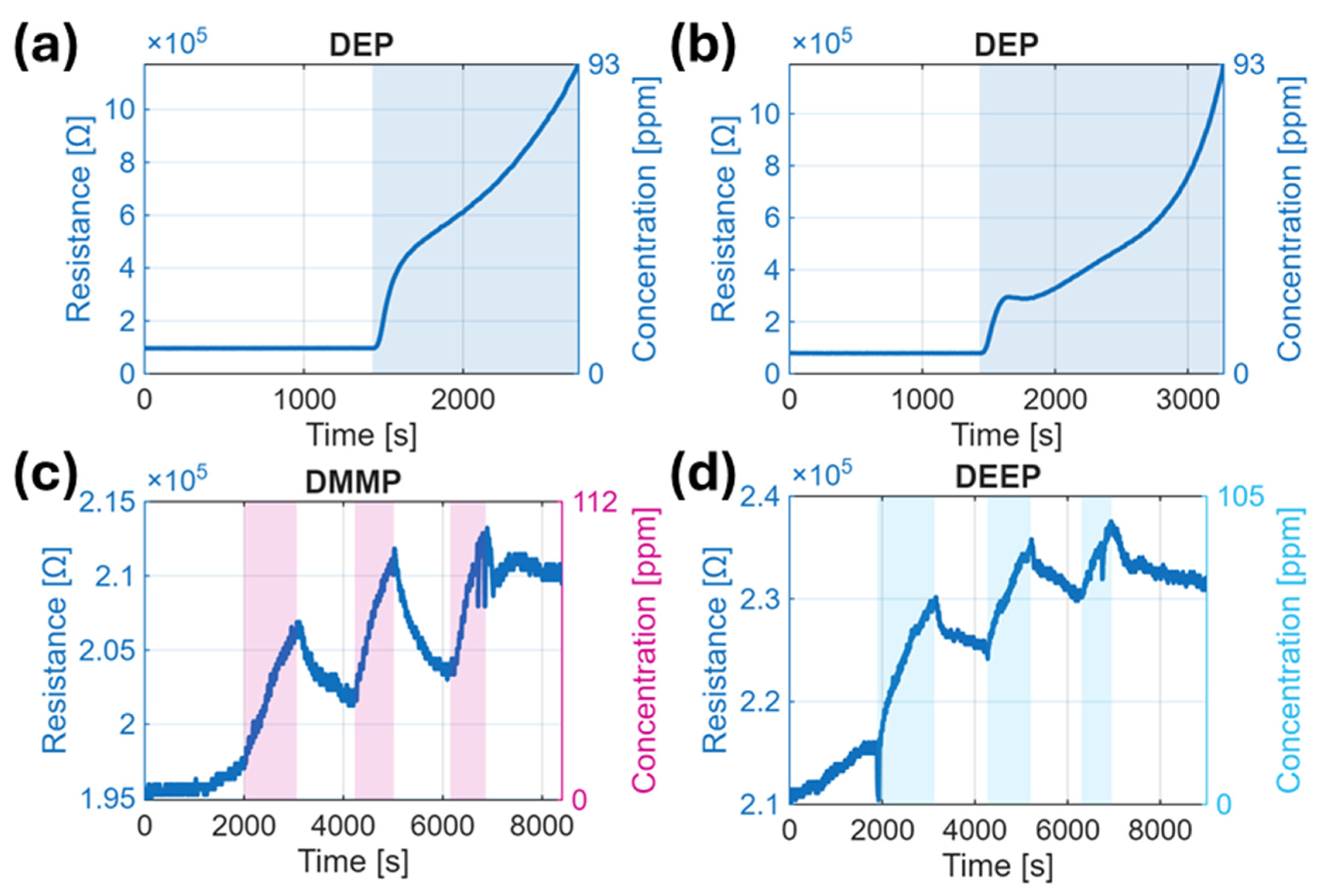
The Cu2O nanowire chemiresistors exhibited measurable responses to most tested CWA simulants; however, the magnitude, direction, and reversibility of the responses varied significantly depending on the analyte (Table 4, Figure 3, Figure 4 and Figure 5). Representative resistance–time curves illustrate these differences (Figure 3, Figure 4 and Figure 5). We performed all of these tests at 320 °C to ensure direct comparability, although the optimal setpoint may differ by analyte. For organophosphorus compounds such as DMMP, TEP, DEEP, and for DEA, a non-phosphorus compound, the sensors displayed a positive chemiresistor response, i.e., an increase in resistance upon analyte introduction, consistent with the p-type nature of Cu2O and the electron-donating behavior of these vapors. In contrast, exposure to DPPCl and ClCN induced a pronounced decrease in resistance, which may reflect stronger electron-withdrawing interactions or surface redox processes that temporarily increase hole concentration. While the overall sensing mechanism still involves surface adsorption and charge modulation, the response polarity highlights differing electronic effects of the analytes on the Cu2O surface. These directional differences in signal (positive vs. negative ΔR) suggest different types of charge transfer mechanisms and may enable analyte discrimination. The resistance increase occurs because molecules such as DMMP, DEA, DEP, and DEEP may react with adsorbed oxygen on the sensor, releasing electrons that neutralize holes in the p-type semiconductor and thereby decreasing hole conduction. The Cu2O nanowire sensors also exhibited minimal cross-response to interfering gases such as NO2, which is promising for field-relevant selectivity, as some atmospheric pollutants and industrial gases are unlikely to trigger false alarms.
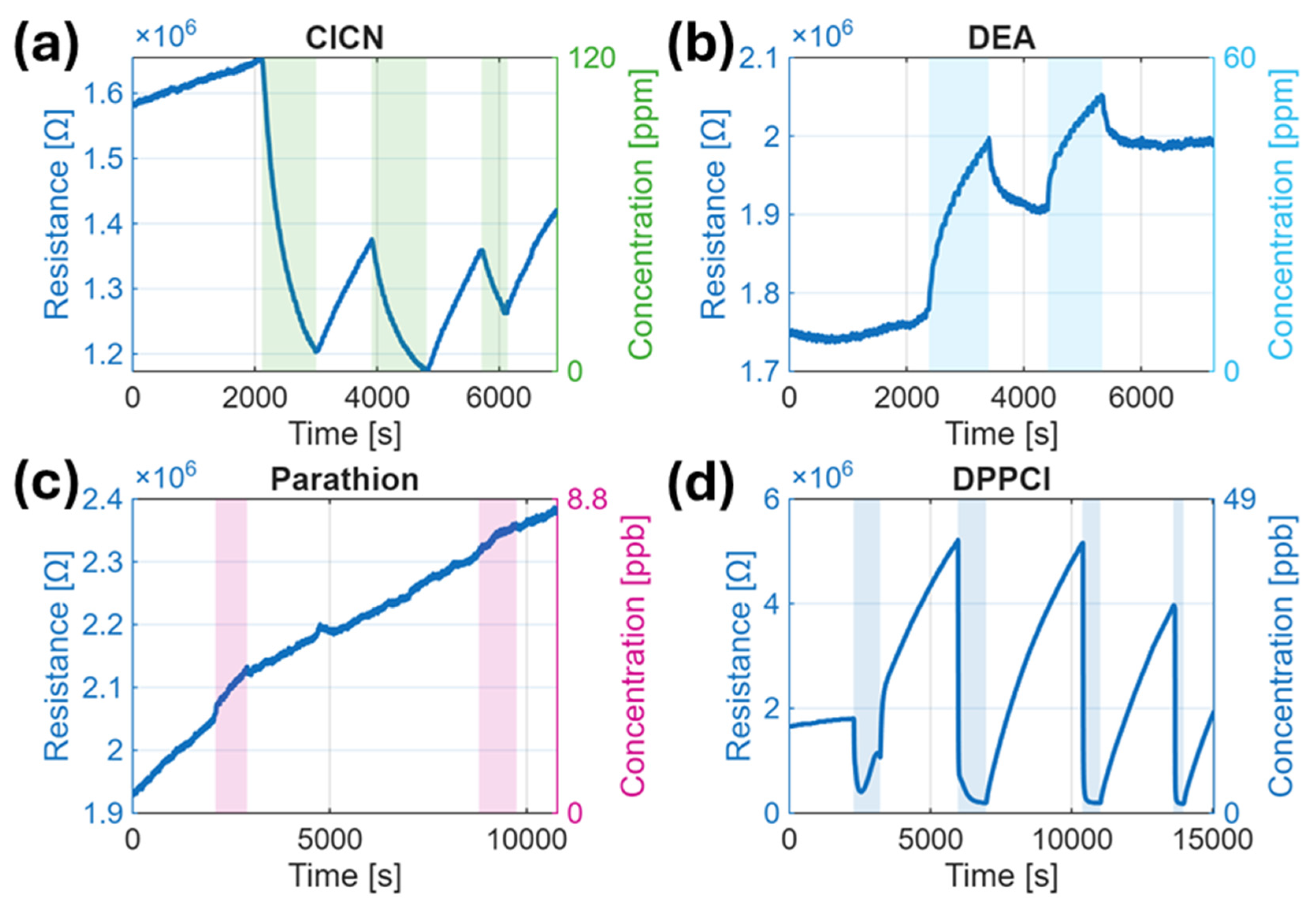
Among the tested analytes, DEP and DPPCl induced strong and irreversible sensor responses due to surface modification. DEP caused an increase in resistance exceeding 200% RR, while DPPCl triggered a sharp decrease of nearly −95%, suggesting opposite interaction pathways. These non-reversible behaviors indicate persistent surface reactions and sensor poisoning. As detailed in the SEM (Figure 2) and EDS analysis (Table 3), DEP exposure was associated with phosphorus-containing residues, whereas DPPCl led to chlorine-rich surface products. Together, these results confirm that strong responses to these analytes correspond to chemical modifications that compromise long-term sensor performance (Figure 3, Figure 4 and Figure 5). In contrast, ClCN produced a substantial but reversible resistance decrease (RR −9.5%) and did not leave detectable residue on the sensor by EDS (Table 3). This suggests that although ClCN interacted with the surface—likely through physisorption or transient oxidative reactions—it did not form stable surface-bound products and thus caused no permanent modification of the active layer. This behavior differs notably from that of other chlorine-containing oxidizers such as Cl2, which have been reported to induce irreversible changes and incomplete recovery in CuO-based sensors [39].
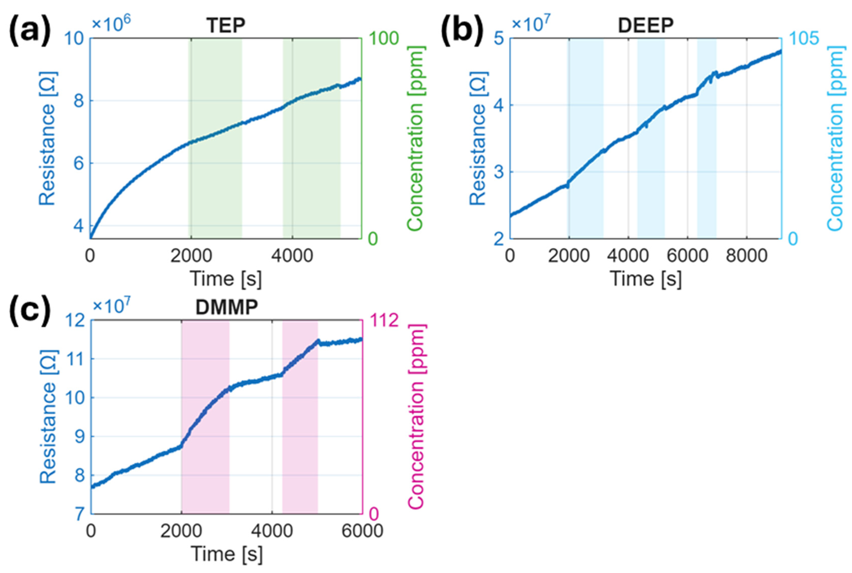
DEA, despite lacking phosphorus, produced a measurable and reproducible positive response (RR 9.9%), suggesting some interaction with the Cu2O surface. Parathion, tested at a much lower concentration (8.9 ppb), yielded a weak and inconsistent response, indicating limited surface reactivity under these conditions. In contrast, DMMP, DEEP, and TEP—fully oxidized P(V) compounds—exhibited only modest increases in resistance of ~3–6% RR (Figure 4); note that these measurements were acquired after the previous DPPCl exposure (Figure 5) and therefore represent lower-bound responses due to partial surface poisoning. Their low reactivity likely stems from their chemical stability, requiring higher activation energy or catalytic assistance for surface decomposition.
The observed sensing behavior of the Cu2O nanowire sensor towards DMMP can be understood in the context of surface adsorption and reaction mechanisms on p-type copper oxide. When DMMP molecules reach the sensor surface, they can donate electrons upon adsorption and undergo partial oxidation on the oxide surface. In the case of Cu2O (with a CuO surface layer), the adsorbed oxygen species (O/O2−) on the surface likely oxidize the DMMP, producing intermediates such as formaldehyde, or phosphate species while releasing electrons [9]. Those released electrons recombine with holes in the p-type Cu2O, thereby decreasing the hole concentration and increasing the sensor’s resistance. This mechanism is consistent with the observed response trend for DMMP in our measurements (Figure 3), where the resistance increased by ~3.4–5.8% upon exposure to 112 ppm (Figure 4).
It has been shown by XPS analysis [9] that DMMP can decompose on Cu2O even at room temperature, resulting in the deposition of phosphate species on the surface. DFT calculations indicated that surface lattice oxygen atoms, rather than oxygen vacancies, play a key role in DMMP decomposition on CuO, forming bonds with DMMP fragments even at room temperature [30]. Such chemisorbed phosphorus-containing species withdraw electrons from the Cu2O surface or also block active sites, further contributing to a decrease in hole charge carriers’ concentration (i.e., an increase in resistance). Operating the sensor at moderate heating was found to improve the response, which can be attributed to enhanced surface reaction kinetics and desorption of products. At higher temperature, DMMP adsorbates react more readily with Cu2O lattice oxygen or also adsorbed oxygen, leading to a larger effective change in the charge carrier density. However, prolonged exposure to air at elevated temperatures can also slowly oxidize a larger fraction of the Cu2O core into CuO. Since CuO is also p-type but with different conductivity and surface chemistry, an increasing CuO fraction might alter the sensor’s baseline and sensitivity over time [29]. Other metal oxide sensors, such as Al-doped ZnO nanoparticle sensors, achieved a response of ~20% for 10 ppm DMMP at 350 °C [40]. Additionally, DFT calculations [41] and NAP-XPS analyses of nanostructured MOS sensors, including MoO2 [7] and MoO3 [8], have been applied to study DMMP detection mechanisms. Such studies suggest that oxygen lattice atoms in the oxide play a larger role in decomposition than oxygen vacancies, which may be a direction for further Cu-based material design.
A remaining challenge is the complete baseline recovery after DMMP exposure; some residual adsorption or slow desorption was observed, which is a common issue for DMMP sensors because phosphorus species can bind strongly to metal oxide surfaces [9]. Possible solutions include integrating a UV light or pulsed heating cycles to help desorb or decompose the strongly bound residues, a strategy that has been effective in other sensor systems for organophosphates [1]. While the sensitivity of the present Cu2O sensor is sufficient for tens of ppm of DMMP, further improvements would be necessary to detect the sub-ppm concentrations required for early warning of nerve agent exposure. The relatively modest RR and slow recovery for DMMP suggest room for improvement (Figure 4). Strategies such as doping the Cu2O with catalytic metals to promote DMMP oxidation, or forming heterojunctions, such as in the case of the heterojunction of CuO nanoparticles/ZnO flowers [42] with n-type oxides (to enhance charge modulation), could be explored to amplify the sensor response [1]. Additionally, arraying multiple sensors with complementary sensing materials could allow selective pattern recognition of different CWA simulants [1].
However, the amplitude of the resistance change differed significantly across analytes: highly reactive or high-affinity compounds showed large responses, whereas others produced only modest changes. Notably, the strongest sensor responses were observed for DEP, DPPCl, and ClCN (Table 4). For example, exposure to 93 ppm of DEP vapor caused a relative response exceeding 200% for both tested sensors (Figure 3). Interestingly, despite its very low vapor concentration of 49 ppb, DPPCl induced a pronounced decrease in resistance, with an RR of nearly 95% (Figure 4d), indicating strong interaction and possible redox surface processes. Each DPPCl exposure produced a pronounced resistance drop (negative RR) followed by a strong baseline drift; the same sequence repeats over successive exposures, indicating incomplete recovery and cumulative, non-first-order behavior. Additionally, 120 ppm of ClCN yielded a substantial resistance decrease, with an RR of −9.5% (Figure 4a).
In contrast, the more volatile nerve-agent simulants DMMP (112 ppm) and DEEP (105 ppm) showed slower, smaller resistance increases with RRs of 3.4% and 4.2%, respectively (Figure 4, Figure 5). TEP (100 ppm) yielded a weak and barely distinguishable positive response (0.2%), suggesting only limited interaction with the sensor surface (Figure 5).
Parathion, an organophosphorothioate tested at 8.9 ppb (saturated vapor), showed a small response (~1.8% RR), likely due to its low vapor pressure and possible decomposition. The non-phosphorus analog diethyl adipate (DEA, 60 ppm) caused only a minor resistance increase (RR ~1–2%) (Figure 4b).
The observed response hierarchy reflects both the chemical reactivity and the distinct interaction mechanisms of the simulants. While both DEP and DPPCl induced strong responses, they did so in opposite directions—DEP caused a sharp resistance increase (Figure 3), whereas DPPCl triggered a pronounced resistance decrease (Figure 4d, Table 4). DEP, a phosphorus(III) compound, likely undergoes surface oxidation to phosphate, releasing electrons that neutralize holes in the p-type Cu2O and increase resistance. In contrast, DPPCl, containing a reactive P–Cl bond, may donate electrons or modify surface states in a way that increases carrier concentration or forms chlorine-containing surface residues, leading to decreased resistance. These contrasting responses suggest fundamentally different surface reactions and charge transfer processes for the two analytes.
In contrast, DMMP, DEEP, and TEP are stable, “fully oxidized” esters lacking bonds that can be easily dissociated, explaining their lower and slower responses. Their interaction likely involves weak physisorption or slow oxidation, i.e., processes insufficient to strongly modulate the sensor’s conductivity within short exposure times.
Parathion’s poor reproducibility may originate from its aromatic nitro group or P=S moiety, which could degrade or react incompletely on the surface. Overall, the Cu2O NW sensor was more sensitive to simulants capable of strong chemisorption or catalytic oxidation on Cu-containing surfaces, consistent with typical p-type MOS behavior.

3.3. Response Kinetics and Reproducibility

The transient response of DPPCl showed an immediate resistance drop within ~10 s (τ90%), followed by a slower recovery taking longer than 128 min (Table 4). This response suggests an initial rapid adsorption process, likely dominated by physisorption or fast surface interactions, followed by a much slower recovery phase attributed to chemisorption or accumulation of non-volatile reaction chlorine-containing residues or possible organic residues. ClCN produced a slower reversible signal change with response time over 1100 s, indicating adsorption–desorption dynamics without evident poisoning. ClCN’s behavior is consistent with its small molecular size and pronounced reactivity, which may lead to rapid oxidation on Cu2O surfaces or halogenation reactions. Previous studies have shown that copper oxide can promote halogenation processes and form copper chlorides upon interaction with chlorine-containing compounds such as HCl or Cl2 [43]. Although specific interaction mechanisms between ClCN and CuO/Cu2O are not fully elucidated, analogies can be drawn from the known chlorination and dechlorination reactions facilitated by copper oxides during pyrolysis or disinfection byproduct formation [43].
DEP exhibited an instantaneous and irreversible response, driving the resistance beyond the measurable range after a single exposure (~93 ppm). Due to this sensor failure, no kinetic parameters could be extracted, but the extreme response indicates strong surface interaction and permanent modification (Figure 4).
Parathion, tested at its saturated vapor (8.9 ppb), showed a slow, drifting resistance increase with poor reproducibility, likely reflecting weak adsorption and partial decomposition to surface-bound byproducts. Its response was small but detectable.
These kinetic differences reflect the underlying molecular structure and reactivity of the simulants. Highly reactive compounds like DPPCl and DEP rapidly chemisorb and alter the sensor surface, whereas stable organophosphates like DMMP show slower, reversible adsorption–desorption cycles.
In summary, the Cu2O NW sensor exhibited the strongest and fastest responses to DEP, DPPCl, and ClCN, and the weakest to DEA and parathion (Table 4). Because analyte concentrations differed between the individual experiments (and the vapor pressures of the simulated warfare agents also cover several orders of magnitude), relative responses (RR) and time constants τ(s) are not directly comparable across species. Kinetic descriptors (τ10%, τ90%) may still aid with the analyte discrimination, but only under matched concentrations or with dose-normalized analysis. These findings underscore the potential of Cu2O NW sensors, especially when used in arrays or coupled with machine learning classifiers, for selective identification of chemical warfare agent simulants.
The repeatability of the sensor signal varied significantly across analytes. For ClCN and DPPCl, when sensors remained functional, consecutive exposures produced similar response magnitudes, indicating a relatively stable sensor behavior for those. For example, the response to 120 ppm ClCN remained consistent across two successive runs.
The relatively low DMMP sensitivity of the p-type Cu2O sensors is consistent with literature reports, which show that n-type semiconductors typically provide stronger responses to organophosphonate vapors. This may be attributed to differences in surface chemistry; n-type oxides generally have more electron-rich adsorption sites and exhibit higher oxidative catalytic activity [1].
Moreover, the operating temperature of 320 °C, although elevated, may not be optimal for complete oxidative decomposition of organophosphates like DMMP; thus, e.g., many SnO2-based sensors operate effectively in the 350–500 °C range [1]. Future optimization of working temperature and doping/catalysts on Cu2O could improve responses to less reactive simulants.

3.4. Surface Analysis and Sensor Stoichiometry Effects

A crucial finding of this study is that exposure to certain organophosphorus analytes led to irreversible changes in the Cu2O nanowire (NW) sensors, providing evidence of sensor poisoning. After the very first exposure to ~93 ppm of diethyl phosphite (DEP), the resistance of two sensors increased beyond the upper limit of the measurement range, indicating immediate, irreversible surface modification. SEM imaging of a DEP-exposed sensor (Figure 2b) shows a conformal deposit on the nanowires, and EDS spectra confirmed the high amount of phosphorus (12.31 at. %) in the active layer. We conclude that DEP underwent surface oxidation, forming a non-volatile phosphorus-containing species that coated the nanowires. This passivating layer is consistent with the observed permanent resistance increase and loss of sensor functionality.
DPPCl produced a similar poisoning effect, but over a longer timescale. A sensor repeatedly exposed to 49 ppb of DPPCl initially showed a strong and relative response (>90% RR), but after multiple exposures, its baseline resistance gradually increased, and the sensor lost responsiveness to both DPPCl and subsequent analytes. SEM images after DPPCl testing (Figure 2c) showed a coated nanowire surface, and EDS confirmed the presence of chlorine residues (40.79 at. %) (Table 3). This is consistent with decomposition to chlorine-containing residues and classical reactions of phosphoryl chlorides with CuO yielding copper chlorides and phosphate derivatives [44], which modify conductivity and block active sites, leading to gradual deactivation of the sensor. Accordingly, DPPCl exposure inhibits subsequent detection of organophosphonates such as DEEP and DMMP (Figure 3, Figure 4).
By contrast, exposure to 120 ppm of cyanogen chloride (ClCN) produced a substantial but reversible negative response and no EDS-detectable residue, indicating no persistent modification of the active layer.
At 320 °C in air, the Cu2O NWs likely form Cu2O(core)–CuO(shell) heterostructures [45]. The role of Cu2O/CuO stoichiometry is also critical to interpreting these effects. Consistent with prior NAP-XPS results, a thin CuO shell on a Cu2O core favors a stable p-type response, whereas CuO-enriched surfaces are more reactive and more prone to residue buildup with organophosphorus species [29,30]. While a thin CuO shell can enhance reactivity, CuO enrichment increases susceptibility to non-volatile residue accumulation. Mildly reducing analytes (e.g., DMMP) may locally reduce CuO to Cu2O, partially offsetting the expected resistance increase and contributing to modest, slower responses [42].
In summary, controlling the Cu+/Cu2+ ratio is essential for sensor performance and longevity; practical approaches include pre-annealing/controlled oxidation, selective doping/heterostructures, and regeneration protocols (e.g., pulsed heating or UV) to limit residual accumulation and restore activity [1].

3.5. Selectivity Considerations

Although our tests covered a range of organophosphorus compounds, the field deployment also requires discrimination of CWAs from diverse background chemicals. The Cu2O nanowire (NW) sensor alone does not exhibit high selectivity toward individual organophosphorus species; however, clear trends emerged based on oxidation state and chemical structure. The sensor responded most strongly to phosphites (e.g., DEP) and chlorophosphates (e.g., DPPCl), both of which possess labile bonds (such as P–H and P–Cl) susceptible to oxidation on Cu2O. ED/TC similarity metrics indicate DPPCl is closer to G-series agents (e.g., sarin, soman). In contrast, fully oxidized P(V) phosphonates (DMMP, DEEP) produced modest responses, consistent with lower surface reactivity and greater chemical stability.
These observations suggest that Cu2O NW sensors are particularly sensitive to reactive organophosphorus compounds with easily oxidizable moieties, rather than being highly selective for specific CWA analogs such as tabun or sarin. In practice, the deployment of Cu2O NW sensors within multi-sensor arrays—for example, combined with n-type MOS or catalytically modified platforms—is preferred to enhance the target analyte’s coverage and to promote the discrimination between them. The measurable response to DEA (a non-phosphorus ester) indicates that ester functionality/volatility and electron-donating capacity also contribute to signal formation, i.e., not solely the presence of phosphorus in the molecule.
Regarding potential interferents, humidity effects were not evaluated (dry chamber, ~45 °C, synthetic air). Future work should include controlled RH cycling to assess both hydrolysis-assisted signal enhancement and site blocking by water under field-relevant conditions.

4. Conclusions

We investigated Cu2O nanowire (NW) chemiresistors for the detection of several CWA simulants and found clear strengths alongside important limitations. We observed the strongest responses for DPPCl (sub-ppm), DEP (93 ppm), and ClCN (120 ppm), while fully oxidized P(V) phosphonates (DMMP, DEEP) produced modest steady-state signals (~3–6%). We also measured a notable positive response to the non-phosphorus analog diethyl adipate (DEA), indicating that the observed selectivity cannot be solely attributed to the presence of phosphorus. Instead, it suggests that other molecular features, such as ester functionality or surface affinity, also play a role in sensor–analyte interactions and should be considered when evaluating sensor specificity.
Consistent with the electrical irreversibility, SEM/EDS analyses indicate that chlorine- (DPPCl) and phosphorus-containing (DEP) residues remain on the Cu2O active layer, supporting site blocking and/or permanent modification.
Irreversible surface poisoning by DEP and DPPCl degraded performance and, in some cases, led to sensor failure. Addressing the durability and chemical specificity of the sensors will require catalytic doping, protective overcoats, and a special regeneration procedure. While this study focused on pristine Cu2O NWs, future research should investigate the impact of surface modifications to enhance sensitivity, selectivity, and long-term stability.
Additionally, controlling the Cu+/Cu2+ surface stoichiometry and engineering Cu2O/CuO core–shell structures may allow tuning of reactivity and selectivity.
Overall, we see Cu2O NW chemiresistors as promising components of multi-sensor arrays or confirmatory roles (particularly valuable for reactive phosphites and chlorinated species), but improving responses to stable P(V) simulants and mitigating poisoning remain key priorities for practical deployment.

Supplementary Materials

The following supporting information can be downloaded at: https://doi.org/10.5281/zenodo.16633819.

Author Contributions

Conceptualization, L.F. and M.V.; methodology, J.O., L.F., J.K. (Jaromír Kukal), and M.V.; software, J.M., M.H., and J.K. (Jaromír Kukal); validation, J.M., J.O., M.H., and J.K. (Jaromír Kukal); formal analysis, L.F. and M.V.; investigation, J.M., J.O., and J.K. (Jan Kejzlar); resources, M.V.; data curation, J.M. and J.O.; writing—original draft preparation, J.O.; writing—review and editing, J.M., J.O., L.F., J.K. (Jan Kejzlar), M.H., and M.V.; visualization, J.M. and J.O.; supervision, L.F. and M.V.; project administration, M.V.; funding acquisition, M.V. All authors have read and agreed to the published version of the manuscript.

Funding

This work has been funded by a grant from the Programme Johannes Amos Comenius under the Ministry of Education, Youth and Sports of the Czech Republic SENDISO project No. CZ.02.01.01/00/22_008/0004596. As set out in the Legal Act, beneficiaries must ensure that the open access to the published version or the final peer-reviewed manuscript accepted for publication is provided immediately after the date of publication via a trusted repository under the latest available version of the Creative Commons Attribution International Public Licence (CC BY) or a licence with equivalent rights. For long-text formats, CC BY-NC, CC BY-ND, CC BY-NC-ND or equivalent licenses could be applied.

Data Availability Statement

The raw data supporting the conclusions of this article are openly available on Zenodo at https://doi.org/10.5281/zenodo.16633819. Additional datasets generated and analyzed during the current study are contained within the article and its Supplementary Materials. Further information is available from the corresponding author upon reasonable request.

Conflicts of Interest

The authors declare no conflicts of interest.

Abbreviations

The following abbreviations are used in this manuscript:
CWAChemical Warfare Agent
Cu2OCopper(I) oxide
CuOCopper(II) oxide
DMMPDimethyl Methylphosphonate
DEPDiethyl Phosphite
DPPClDiphenyl Phosphoryl Chloride
DEEPDiethyl Ethylphosphonate
TEPTriethyl Phosphate
DEADiethyl Adipate
EDEuclidean Distance
GC-MSGas Chromatography–Mass Spectrometry
HDDistilled Mustard
ClCNCyanogen Chloride
MOSMetal Oxide Semiconductor
SEMScanning Electron Microscopy
EDSEnergy-Dispersive X-ray Spectroscopy
XPSX-ray Photoelectron Spectroscopy
NAP-XPSNear-Ambient Pressure XPS
DFTDensity Functional Theory
RRRelative Response
PtPlatinum
TCTanimoto Coefficient

References

  1. Witkiewicz, Z.; Jasek, K.; Grabka, M. Semiconductor gas sensors for detecting chemical warfare agents and their simulants. Sensors 2023, 23, 3272. [Google Scholar] [CrossRef]
  2. Diauudin, F.N.; Rashid, J.I.A.; Knight, V.F.; Yunus, W.M.Z.W.; Ong, K.K.; Kasim, N.A.M.; Halim, N.A.; Noor, S.A.M. A review of current advances in the detection of organophosphorus chemical warfare agents based biosensor approaches. Sens. BioSens. Res. 2019, 26, 100305. [Google Scholar] [CrossRef]
  3. Sferopoulos, R. A Review of Chemical Warfare Agent (CWA) Detector Technologies and Commercial-Off-The-Shelf Items. 2009. Available online: https://apps.dtic.mil/sti/tr/pdf/ADA502856.pdf (accessed on 29 July 2025).
  4. White, S.; Miller, P.; Sorge, M.; Curtis, C.; Hare, A.; Whiting, J.; Sammon, J.; Corbin, W.C. Colorimetric/Electrical Sensing of Chemical Warfare Agent Surrogates with Polydiacetylenes. In Proceedings of the 2024 IEEE SENSORS, Kobe, Japan, 20–23 October 2024. [Google Scholar] [CrossRef]
  5. Kanu, A.B.; Haigh, P.E.; Hill, H.H. Surface detection of chemical warfare agent simulants and degradation products. Anal. Chim. Acta 2005, 553, 148–159. [Google Scholar] [CrossRef]
  6. Barreca, D.; Maccato, C.; Gasparotto, A. Metal Oxide Nanosystems As Chemoresistive Gas Sensors for Chemical Warfare Agents: A Focused Review. Adv. Mater. Interfaces 2022, 9, 2102525. [Google Scholar] [CrossRef]
  7. Head, A.R.; Tsyshevsky, R.; Trotochaud, L.; Yu, Y.; Karslıoǧlu, O.; Eichhorn, B.; Kuklja, M.M.; Bluhm, H. Dimethyl methylphosphonate adsorption and decomposition on MoO2 as studied by ambient pressure x-ray photoelectron spectroscopy and DFT calculations. J. Phys. Condens. Matter 2018, 30, 134005. [Google Scholar] [CrossRef]
  8. Head, A.R.; Tsyshevsky, R.V.; Trotochaud, L.; Yu, Y.; Kyhl, L.; Karslıoǧlu, O.; Kuklja, M.M.; Bluhm, H. Adsorption of Dimethyl Methylphosphonate on MoO3: The Role of Oxygen Vacancies. J. Phys. Chem. C 2016, 120, 29077–29088. [Google Scholar] [CrossRef]
  9. Trotochaud, L.; Head, A.R.; Büchner, C.; Yu, Y.; Karslıoğlu, O.; Tsyshevsky, R.; Holdren, S.; Eichhorn, B.; Kuklja, M.M.; Bluhm, H. Room temperature decomposition of dimethyl methylphosphonate on cuprous oxide yields atomic phosphorus. Surf. Sci. 2019, 680, 75–87. [Google Scholar] [CrossRef]
  10. Ponzoni, A.; Baratto, C.; Bianchi, S.; Comini, E.; Ferroni, M.; Pardo, M.; Vezzoli, M.; Vomiero, A.; Faglia, G.; Sberveglieri, G. Metal Oxide Nanowire and Thin-Film-Based Gas Sensors for Chemical Warfare Simulants Detection. IEEE Sens. J. 2008, 8, 735–742. [Google Scholar] [CrossRef]
  11. Fumian, F.; Chierici, A.; Bianchelli, M.; Martellucci, L.; Rossi, R.; Malizia, A.; Gaudio, P.; D’eRrico, F.; Di Giovanni, D. Development and performance testing of a miniaturized multi-sensor system combining MOX and PID for potential UAV application in TIC, VOC and CWA dispersion scenarios. Eur. Phys. J. Plus 2021, 136, 913. [Google Scholar] [CrossRef]
  12. Panigrahi, P.; Pal, Y.; Kaewmaraya, T.; Bae, H.; Nasiri, N.; Hussain, T. Molybdenum Carbide MXenes as Efficient Nanosensors toward Selected Chemical Warfare Agents. ACS Appl. Nano Mater. 2023, 6, 8404–8415. [Google Scholar] [CrossRef]
  13. Devi, K.S.S.; Anantharamakrishnan, A.; Krishnan, U.M. Expanding Horizons of Metal Oxide-based Chemical and Electrochemical Sensors. Electroanalysis 2021, 33, 1979–1996. [Google Scholar] [CrossRef]
  14. Xu, M.; Hu, X.; Zhang, H.; Miao, T.; Ma, L.; Liang, J.; Zhu, Y.; Zhu, H.; Cheng, Z.; Sun, X. Detection and Pattern Recognition of Chemical Warfare Agents by MOS-Based MEMS Gas Sensor Array. Sensors 2025, 25, 2633. [Google Scholar] [CrossRef]
  15. Xu, M.; Zhong, Y.; Hu, X.; Tao, Y.; Shen, Q.; Zhang, S.; Zhang, P.; Zhang, H.; Cheng, Z.; Sun, X. Detection of CWA Simulants by Electronic Nose Based on Low-Powered MEMS Gas Sensor Array. IEEE Sens. J. 2025, 25, 30361–30369. [Google Scholar] [CrossRef]
  16. Dhull, V. Fabrication of AChE/SnO2-cMWCNTs/Cu Nanocomposite-Based Sensor Electrode for Detection of Methyl Parathion in Water. Int. J. Anal. Chem. 2018, 2018, 2874059. [Google Scholar] [CrossRef] [PubMed]
  17. Sakdarat, P.; Chongsuebsirikul, J.; Thanachayanont, C.; Prichanont, S.; Pungetmongkol, P.; Azzazy, H. Development of a Nonenzymatic Electrochemical Sensor for Organophosphate Pesticide Detection Using Copper (II) Oxide Nanorod Electrodes. J. Nanomater. 2021, 2021, 1–11. [Google Scholar] [CrossRef]
  18. Sen, S.; Roy, A.; Sanyal, A.; Devi, P.S. A Nonenzymatic Reduced Graphene Oxide-Based Nanosensor for Parathion. Beilstein J. Nanotechnol. 2022, 13, 730–744. [Google Scholar] [CrossRef]
  19. Bao, J.; Hou, C.; Chen, M.; Li, J.; Huo, D.; Yang, M.; Luo, X.; Lei, Y. Plant Esterase–Chitosan/Gold Nanoparticles–Graphene Nanosheet Composite-Based Biosensor for the Ultrasensitive Detection of Organophosphate Pesticides. J. Agric. Food Chem. 2015, 63, 10319–10326. [Google Scholar] [CrossRef]
  20. Bhusari, R.; Thomann, J.-S.; Guillot, J.; Leturcq, R. Oxygen Adsorption and Desorption Kinetics in CuO Nanowire Bundle Networks: Implications for MOx-Based Gas Sensors. ACS Appl. Nano Mater. 2022, 5, 10248–10257. [Google Scholar] [CrossRef]
  21. Qin, Y.; Zhang, F.; Chen, Y.; Zhou, Y.; Li, J.; Zhu, A.; Luo, Y.; Tian, Y.; Yang, J. Hierarchically Porous CuO Hollow Spheres Fabricated via a One-Pot Template-Free Method for High-Performance Gas Sensors. J. Phys. Chem. C 2012, 116, 11994–12000. [Google Scholar] [CrossRef]
  22. Yang, B.; Myung, N.V.; Tran, T. 1D Metal Oxide Semiconductor Materials for Chemiresistive Gas Sensors: A Review. Adv. Electron. Mater. 2021, 7, 2100271. [Google Scholar] [CrossRef]
  23. Barsan, N.; Weimar, U. Conduction Model of Metal Oxide Gas Sensors. J. ElectroCeram. 2001, 7, 143–167. [Google Scholar] [CrossRef]
  24. Comini, E.; Faglia, G.; Ferroni, M.; Ponzoni, A.; Vomiero, A.; Sberveglieri, G. Metal oxide nanowires: Preparation and application in gas sensing. J. Mol. Catal. A Chem. 2009, 305, 170–177. [Google Scholar] [CrossRef]
  25. Korotcenkov, G.; Cho, B. Instability of metal oxide-based conductometric gas sensors and approaches to stability improvement (short survey). Sens. Actuators B Chem. 2011, 156, 527–538. [Google Scholar] [CrossRef]
  26. Filipič, G.; Cvelbar, U. Copper oxide nanowires: A review of growth. Nanotechnology 2012, 23, 194001. [Google Scholar] [CrossRef]
  27. Zhou, K.; Wang, R.; Xu, B.; Li, Y. Synthesis, characterization and catalytic properties of CuO nanocrystals with various shapes. Nanotechnology 2006, 17, 3939–3943. [Google Scholar] [CrossRef]
  28. Izaki, M.; Abe, S.; Nakakita, K.; Khoo, P.L. Photoelctrochemically Fabricated and Heated Cu2O/CuO Bilayers with Enhanced Photovoltaic Characteristics. ACS Omega 2021, 6, 27587–27597. [Google Scholar] [CrossRef]
  29. Hozák, P.; Vorokhta, M.; Khalakhan, I.; Jarkovská, K.; Cibulková, J.; Fitl, P.; Vlček, J.; Fara, J.; Tomeček, D.; Novotný, M.; et al. New Insight into the Gas-Sensing Properties of CuOx Nanowires by Near-Ambient Pressure XPS. J. Phys. Chem. C 2019, 123, 29739–29749. [Google Scholar] [CrossRef]
  30. Trotochaud, L.; Tsyshevsky, R.; Holdren, S.; Fears, K.; Head, A.R.; Yu, Y.; Karslıoğlu, O.; Pletincx, S.; Eichhorn, B.; Owrutsky, J.; et al. Spectroscopic and Computational Investigation of Room-Temperature Decomposition of a Chemical Warfare Agent Simulant on Polycrystalline Cupric Oxide. Chem. Mater. 2017, 29, 7483–7496. [Google Scholar] [CrossRef]
  31. Schlur, L.; Agostini, P.; Thomas, G.; Gerer, G.; Grau, J.; Spitzer, D. Detection of Organophosphorous Chemical Agents with CuO-Nanorod-Modified Microcantilevers. Sensors 2020, 20, 1061. [Google Scholar] [CrossRef] [PubMed]
  32. Lavoie, J.; Srinivasan, S.; Nagarajan, R. Using cheminformatics to find simulants for chemical warfare agents. J. Hazard. Mater. 2011, 194, 85–91. [Google Scholar] [CrossRef]
  33. Lupan, O.; Ababii, N.; Mishra, A.K.; Gronenberg, O.; Vahl, A.; Schürmann, U.; Duppel, V.; Krüger, H.; Chow, L.; Kienle, L.; et al. Single CuO/Cu2O/Cu Microwire Covered by a Nanowire Network as a Gas Sensor for the Detection of Battery Hazards. ACS Appl. Mater. Interfaces 2020, 12, 42248–42263. [Google Scholar] [CrossRef] [PubMed]
  34. Maier, C.; Egger, L.; Köck, A.; Becker, S.; Niehaus, J.S.; Reichmann, K. Investigation of Gas Sensing Performance of CuO/Cu2O Thin Films as a Function of Au-NP Size for CO, CO2, and Hydrocarbons Mixtures. Nanomaterials 2025, 15, 705. [Google Scholar] [CrossRef] [PubMed]
  35. Hruška, M.; Kejzlar, J.; Otta, J.; Fitl, P.; Novotný, M.; Čížek, J.; Melikhova, O.; Mičušík, M.; Machata, P.; Vrňata, M. Hydrogen Sensing Capabilities of Highly Nanoporous Black Gold Films. Appl. Surf. Sci. 2023, 647, 158618. [Google Scholar] [CrossRef]
  36. Kumar, S.; Kumar, A.; Das, M.; Kim, S.S.; Kim, H.W.; Kumar, M. Ag catalysts boosted NO2 gas sensing performance of RF sputtered α-Fe2O3 films. Sens. Actuators B Chem. 2023, 393, 134307. [Google Scholar] [CrossRef]
  37. Kumar, S.; Betal, A.; Kumar, A.; Chakkar, A.G.; Kumar, P.; Kwoka, M.; Sahu, S.; Kumar, M. Enhancing NO2 Gas Sensing: The Dual Impact of UV and Thermal Activation on Vertically Aligned Nb-MoS2 for Superior Response and Selectivity. ACS Sens. 2025, 10, 2191–2202. [Google Scholar] [CrossRef]
  38. Sharma, A.K.; Mahajan, A. Chapter 8-Potential applications of chemiresistive gas sensors. In Carbon Nanomaterials and Their Nanocomposite-Based Chemiresistive Gas Sensors; Dhall, S., Ed.; Elsevier: Amsterdam, The Netherlands, 2023; pp. 223–245. [Google Scholar] [CrossRef]
  39. Torrisi, A.; Vacík, J.; Ceccio, G.; Cannavò, A.; Lavrentiev, V.; Horák, P.; Yatskiv, R.; Vaniš, J.; Grym, J.; Fišer, L.; et al. Chemiresistors based on Li-doped CuO–TiO2 films. Chemosensors 2021, 9, 246. [Google Scholar] [CrossRef]
  40. Yoo, R.; Cho, S.; Song, M.-J.; Lee, W. Highly sensitive gas sensor based on Al-doped ZnO nanoparticles for detection of dimethyl methylphosphonate as a chemical warfare agent simulant. Sens. Actuators B Chem. 2015, 221, 217–223. [Google Scholar] [CrossRef]
  41. Holdren, S.; Tsyshevsky, R.; Fears, K.; Owrutsky, J.; Wu, T.; Wang, X.; Eichhorn, B.W.; Kuklja, M.M.; Zachariah, M.R. Adsorption and Destruction of the G-Series Nerve Agent Simulant Dimethyl Methylphosphonate on Zinc Oxide. ACS Catal. 2018, 9, 902–911. [Google Scholar] [CrossRef]
  42. Yoo, R.; Yoo, S.; Lee, D.; Kim, J.; Cho, S.; Lee, W. Highly selective detection of dimethyl methylphosphonate (DMMP) using CuO nanoparticles/ZnO flowers heterojunction. Sens. Actuators B Chem. 2017, 240, 1099–1105. [Google Scholar] [CrossRef]
  43. Wang, X.; Li, B.; Xia, Z.; Zhou, W.; Wu, Y.; Zhu, Z.; Zhu, G. Effects of copper(II) oxide on the co-pyrolysis of waste polyester enameled wires and poly(vinyl chloride). Polymers 2023, 16, 27. [Google Scholar] [CrossRef] [PubMed]
  44. Bassett, H.; Taylor, H.S. CLVIII.—The interaction of metallic oxides and phosphoryl chloride, alone and in the presence of certain organic compounds. J. Chem. Soc. Trans. 1911, 99, 1402–1414. [Google Scholar] [CrossRef]
  45. Claros, M.; Gràcia, I.; Figueras, E.; Vallejos, S. Hydrothermal synthesis and annealing effect on the properties of gas-sensitive copper oxide nanowires. Chemosensors 2022, 10, 353. [Google Scholar] [CrossRef]
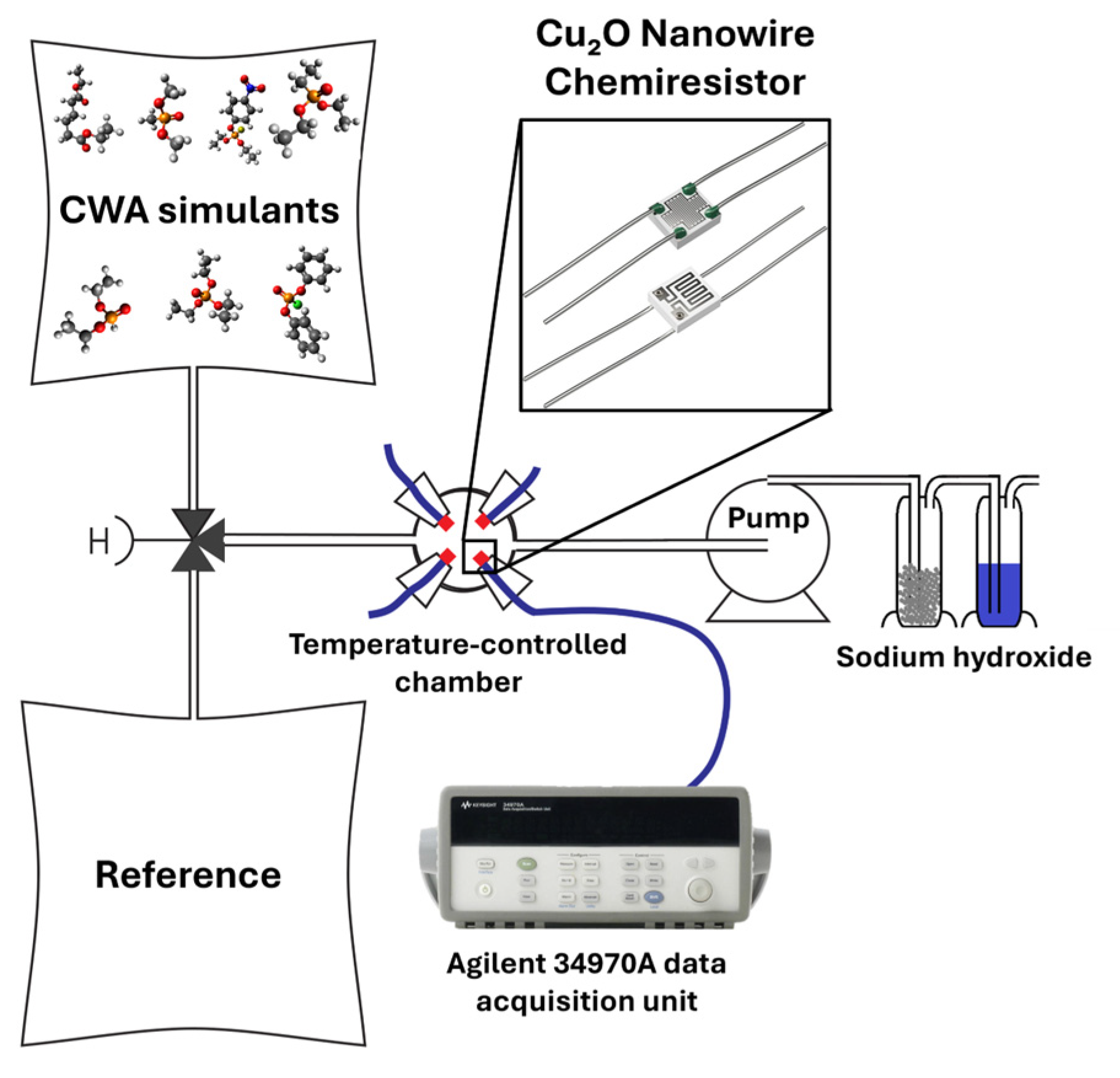
Figure 1. Schematic of the gas-response setup for the Cu2O nanowire chemiresistor. A three-way valve switches between reference (synthetic air) and CWA-simulant streams taken from Tedlar™ gas sampling bags; the total flow is 300 mL·min−1 drawn by a downstream pump. The sensor platform KBS4 (inset, Pt IDEs with a backside Pt microheater) is mounted in a temperature-controlled glass chamber. Sensor resistance (i.e., output signal) and working temperature are recorded with an Agilent 34970A. The exhaust is passed through NaOH traps.
Electronics 14 03478 g001
Figure 2. SEM image of Cu2O nanowires: (a) pristine, (b) after exposure to DEP, and (c) after exposure to DPPCl. Main panels: 10,000×, scale bar 5 µm; insets: 50,000×, scale bar 2 µm.
Electronics 14 03478 g002
Figure 3. Dynamic response curvesvariation of resistance as a function of timeof Cu2O nanowire chemiresistors at 320 °C (baseline in synthetic air; total flow 300 mL·min−1): (a,b) response to 93 ppm of DEPtwo different sensors (irreversible modification prevents their reuse); (c) response to 112 ppm of DMMPmodest positive response; and (d) response to 105 ppm of DEEPmodest positive response.
Electronics 14 03478 g003
Figure 4. Dynamic response curvesvariation of resistance as a function of timeof Cu2O nanowire chemiresistor to selected chemical warfare agent simulants (baseline in synthetic air; total flow 300 mL·min−1): (a) 120 ppm of ClCNreversible negative response, (b) 60 ppm of DEAreversible positive response, (c) 8.9 ppb of parathionnegligible positive response with drift, and (d) 49 ppb of DPPClstrong negative response with cumulative baseline shift.
Electronics 14 03478 g004
Figure 5. Dynamic response curvesvariation of resistance as a function of timeof Cu2O nanowire chemiresistor to selected chemical warfare agent simulants (baseline in synthetic air; total flow 300 mL·min−1) after the exposure to DPPCl: (a) 100 ppm of TEP, (b) 105 ppm of DEEP, and (c) 112 ppm of DMMP. Each analyte shows modest positive response with slow drift; magnitudes may be suppressed by partial poisoning from the preceding DPPCl exposure.
Electronics 14 03478 g005
Table 1. Physicochemical properties and tested gas-phase concentrations of selected chemical warfare agent (CWA) simulants.
SimulantSaturated Vapor Pressure [Pa]Prepared Concentration [ppm]DistributorPurity [%]
ClCN1,987,000120Draslovka a.s., Kolín, Czech Republic≤100.0
DEA7.7060Sigma-Aldrich®, St. Louis, MI, USA≥98.0
DEEP41.2105Sigma-Aldrich®, St. Louis, MI, USA≥98.0
DEP149093Sigma-Aldrich®, St. Louis, MI, USA≥98.0
DMMP128112Sigma-Aldrich®, St. Louis, MI, USA≥98.0
DPPCl0.00490.049Sigma-Aldrich®, St. Louis, MI, USA≥99.0
Parathion0.000890.0089Supelco® Bellefonte, PA, USA≥98.0
TEP51.8100Sigma-Aldrich®, St. Louis, MI, USA≥99.8
Table 2. Structural and physicochemical similarity of selected simulants to CWAs, expressed using ED and TC values. Simulants are compared to nerve agents (sarin, soman, tabun, VX) and a blister agent (distilled mustard). Lower ED and higher TC values indicate greater molecular similarity [32].
SimulantCAS No.ED
(Sarin)
ED
(Soman)
ED
(Tabun)
ED
(VX)
ED
(HD)
TC
(Sarin)
TC
(Soman)
TC
(Tabun)
TC
(VX)
TC
(HD)
DMMP756-79-60.1930.3030.2650.6670.6400.432
DEEP78-38-60.2420.2390.2450.4290.4170.537
TEP78-40-00.3080.3020.2860.4550.4410.525
DEP762-04-90.2300.2750.2380.3820.3710.463
DPPCl2524-64-30.5410.5410.5110.5000.4850.245
Parathion56-38-20.3950.509
DEA141-28-60.5600.310
ClCN506-77-4
Table 3. Elemental composition (in atomic %) of Cu2O nanowire surfaces after exposure to selected analytes, as determined by EDS.
AnalyteC (at. %)O (at. %)P (at. %)Cl (at. %)Cu (at. %)
Before33.48 ± 1.2820.48 ± 0.9746.04 ± 2.18
DPPCl18.65 ± 0.802.40 ± 0.1740.79 ± 1.0737.32 ± 1.64
DEP7.49 ± 0.8053.86 ± 3.2612.31 ± 0.4226.34 ± 1.46
Table 4. Sensor response and kinetic parameters of the Cu2O nanowire chemiresistor for chemical warfare agent (CWA) simulants at 320 °C in 300 mL·min−1 synthetic air.
SimulantClCNDEADEPDEEPDMMPTEP *DEEP *DMMP *DPPClParathion
RR (%)−9.469.94203.754.583.390.204.235.76−94.671.82
Responseτ(s)484510-10631120-1095113994196
τ10%(s)5154-112118-11512010196
τ90%(s)11151175-2447118-252126231021
Recoveryτ(s)1944157-780606-1838151033721984
τ10%(s)20517-8264-194159355209
τ90%(s)4477362-17951396-4233347677634569
* Responses measured after DPPCl exposure.
Disclaimer/Publisher’s Note: The statements, opinions and data contained in all publications are solely those of the individual author(s) and contributor(s) and not of MDPI and/or the editor(s). MDPI and/or the editor(s) disclaim responsibility for any injury to people or property resulting from any ideas, methods, instructions or products referred to in the content.