Article

Trial of Brain–Computer Interface for Continuous Motion Using Electroencephalography and Electromyography

by
Norihiko Saga
1,*,
Yukina Okawa
1,
Takuma Saga
1,2,
Toshiyuki Satoh
3 and
Naoki Saito
4
1
Department of Engineering, Kwansei Gakuin University, 1 Gakuen-Uegahara, Sanda 669-1330, Japan
2
Department of Mechanical Systems Engineering, The University of Shiga Prefecture, 2500 Hassaka-cho, Hikone 522-0057, Japan
3
Department of Mechanical Science and Engineering, Hirosaki University, 3 Bunkyou-cho, Hirosaki 036-8561, Japan
4
Department of Intelligent Mechatronics, Akita Prefectural University, 84-4 Aza-Ebinokuchi, Yurihonjo 015-0055, Japan
*
Author to whom correspondence should be addressed.
Electronics 2024, 13(14), 2770; https://doi.org/10.3390/electronics13142770
Submission received: 5 May 2024 / Revised: 4 July 2024 / Accepted: 8 July 2024 / Published: 15 July 2024
(This article belongs to the Special Issue Brain Computer Interface: Theory, Method, and Application)

Abstract

:
Most BCI systems used in neurorehabilitation detect EEG features indicating motor intent based on machine learning, focusing on repetitive movements, such as limb flexion and extension. These machine learning methods require large datasets and are time consuming, making them unsuitable for same-day rehabilitation training following EEG measurements. Therefore, we propose a BMI system based on fuzzy inference that bypasses the need for specific EEG features, introducing an algorithm that allows patients to progress from measurement to training within a few hours. Additionally, we explored the integration of electromyography (EMG) with conventional EEG-based motor intention estimation to capture continuous movements, which is essential for advanced motor function training, such as skill improvement. In this study, we developed an algorithm that detects the initial movement via EEG and switches to EMG for subsequent movements. This approach ensures real-time responsiveness and effective handling of continuous movements. Herein, we report the results of this study.

1. Introduction

As the fields of brain science and neuroscience have developed, we have learnt that a reorganization of brain functions (brain plasticity) can occur through intensive rehabilitation after brain damage due to stroke. If brain plasticity can be efficiently induced, it is possible to recover the motor function of paralyzed limbs. In recent years, various brain–computer interfaces (BCIs) have been developed as a method to link the brain and motor function [1,2,3]. A BCI reads signals induced in the brain through external stimuli or mental tasks, analyzes them with a computer, and then operates equipment. Closed-loop rehabilitation systems that use robots have recently been developed, as using BCIs to close the loop between motor intention and actual movement through can facilitate the motor relearning process [4,5,6].
Non-invasive BCIs, which have been studied extensively for rehabilitation applications and are considered to be less stressful to the human body, include magnetoencephalography (MEG) [7,8], functional magnetic resonance imaging (fMRI) [9,10], and near-infrared spectroscopy (NIRS) [11,12]. However, we chose EEGs to deliver brain information in a BCI to restore motor function in stroke hemiplegic patients. EEGs were selected as they only require measuring changes in scalp potentials, are subject-friendly systems, have a high temporal resolution, provide real-time feedback, and can be composed of relatively inexpensive measurement equipment [13,14].
The main EEGs used in BCIs are event-related potentials. Event-related potentials include potentials generated during specific cognitive activities; visually evoked potentials, which involve flashing visual stimuli; and motor recall potentials, which involve event-related synchronization (ERS) [15] and event-related desynchronization (ERD) [16] generated by motor recall. Studies utilizing event-related potentials (ERPs) have investigated wheelchair control and the development of communication tools for patients with paralyzed motor functions [17,18,19,20]. One well-known ERP is the P300 waveform [21,22], which appears 300 ms after stimulus presentation [23]. To analyze ERPs, the target stimulus’ time of occurrence is measured as a trigger, and additive averaging is performed approximately 100 times offline. In addition, there has been research on BCIs that generate a motion such as grasping based on SSVEPs that are generated by staring at a blinking LED, wherein the motion depends on the frequency of the blinking [24,25,26]. However, starting with flashing lights induces stress and fatigue, and it is essential to devise practical measures to reduce this.
Consequently, we developed an EEG-based BCI that synchronizes motor imagery with rehabilitation equipment and can be controlled in real time without external stimulation. BCI-based rehabilitation systems generally utilize motor recall imagery through machine learning and other methods, detecting event-related desynchronization (ERD) and event-related synchronization (ERS) in the low-frequency regions of alpha and beta waves generated by motor recall potentials [27,28,29]. This method has been combined with voluntary exercise in rehabilitation to attempt the recovery of motor function through the restructuring and strengthening of cranial nerve circuits [30], and the recovery of motor and brain functions has been reported [31]. However, the limitations of this technology need to be resolved prior to its wider use in general hospitals.
First, EEG characteristics vary because the motor imagery used to recall motor in-tensions fluctuates greatly from individual to individual [32]. Second, electroencephalograms (EEGs) are easily influenced by the external environment, change according to participants’ concentration levels, etc.; thus, individual differences are large. Therefore, even if rehabilitation training is conducted several weeks later, a brainwave feature algorithm learned on the day of brainwave measurement can only be applied for some participants. Third, formulating an algorithm by using machine learning to detect evoked EEG characteristics takes time for cognitive tasks where a specific frequency band or measurement site is fixed in advance [33,34].
We propose a heuristic BCI method based on fuzzy reasoning [35,36] that can achieve results within around one hour from the start of EEG measurement to feature extraction, improving on conventional machine learning methods. This method simply creates a template of the fuzzy rule, delineating whether an EEG is high or low for each measurement position, and efficiently generates a detection algorithm in the short time of about an hour because it only learns the characteristic EEG pattern, discriminating the motor image state from the rest state. We confirm that utilizing this method in neurorehabilitation systems is beneficial not only for healthy individuals but also for patients.
The next problem to overcome is estimating the motor intention of continuous movements, such as reaching out, grasping, swinging an arm, and releasing, instead of single movements, such as holding a hand, bending an arm, and lifting a leg, as observed in rehabilitation training that uses a pegboard. In contrast, some methods use conventional machine learning with EEGs only [37,38,39], and other methods combine motion estimation techniques with a parallel EMG to further improve accuracy [40,41,42]. However, since these methods ultimately use machine learning to learn motor intention from EEG signals, additional measurement data are added to the conventional methods, which increases the computational time required to create detection algorithms, perpetuating this fundamental problem. Therefore, these systems do not enable rehabilitation treatment immediately after EEG measurement.
The aim of this research was to capture the motion intent of continuous motion by utilizing a heuristic BCI based on a previously developed fuzzy inference. Heuristic BCI algorithms that use EEGs often capture non-targeted motion intentions during successive movements, making it difficult to identify the start of the next movement. In addition, incorporating EMG and EEG signals into a heuristic BCI algorithm in parallel is complicated because of the weighting and optimization problems of the two different signals, meaning its features cannot be fully utilized. Therefore, as a closed-loop rehabilitation system connecting brain and movement, we investigated whether the rapid motion-intention-estimation feature of this system can be adapted to continuous motion by detecting the first motion with an EEG and then switching to an EMG alone from the next motion onward to detect motion intention. The estimation of motor intention using muscle potentials has already been demonstrated with the exoskeleton-type walking aid “HAL” [43].
The novelty and contributions of this study are summarized as follows:
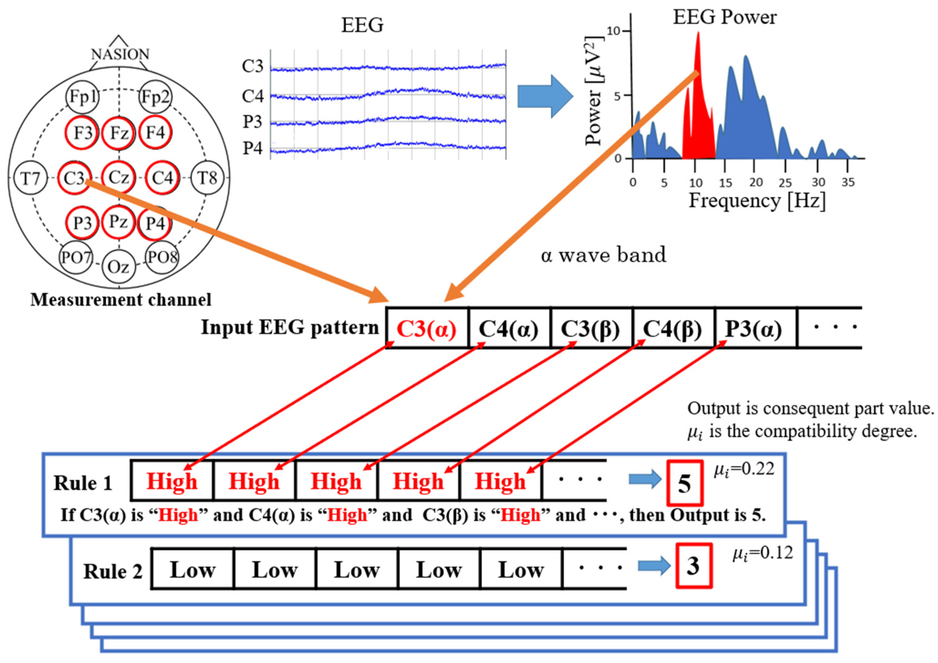
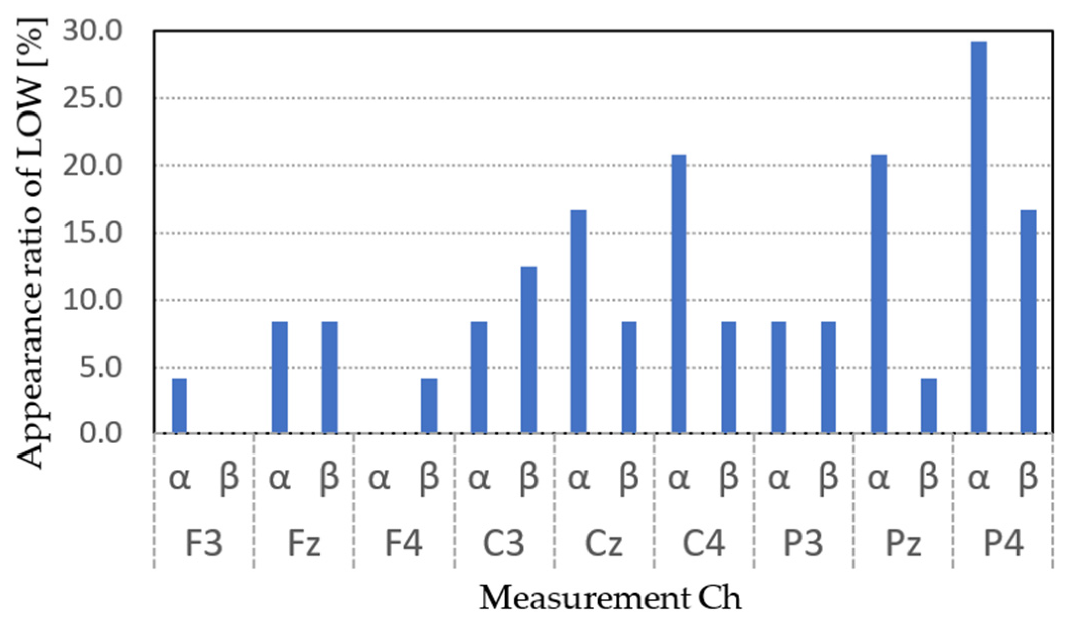
  • Heuristic BCIs can generate a motor intention detection algorithm in a short time by binarizing the measured CH EEG power to LOW or HIGH and generating patterns without seeking the optimal conditions for detection, such as machine learning.
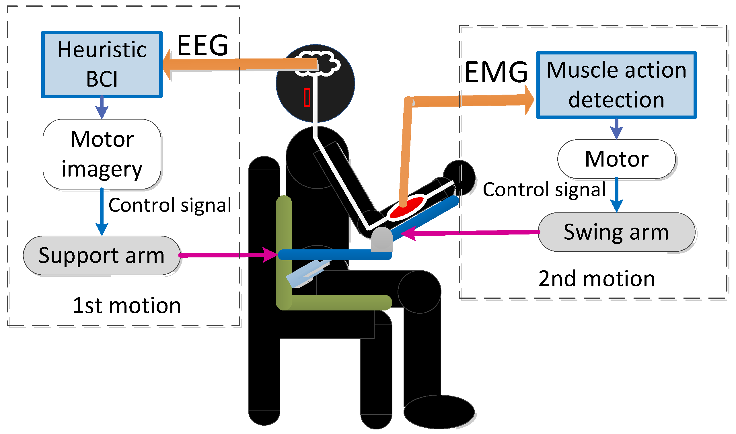
  • The algorithm is based on the assumption that the target movement is skill training and is realized by using EEG and EMG potentials to handle continuous movements rather than monotonous movements of flexion and extension.
  • The proposed system has the potential to be widely used because it simplifies the settings necessary for the system to work as much as possible, enabling users to perform advanced medical treatment at home by themselves, while reducing costs.

2. Materials and Methods

Hybrid BCIs consist of a heuristic BCI [35,36] and a muscle activity detection unit. As shown in Figure 1, after the motor intention of the first shoulder joint flexion (arm raising) using an EEG (orange arrow on the left side) with a heuristic BCI, and then it switches to the detection of the motor intention of the elbow joint flexion (elbow bending), the next motor movement, with the muscle activity detection unit (orange arrow on the right side). After detecting the motor intention with the heuristic BCI, the system switches to the muscle activity detection unit to identify the motor intention of the elbow flexion to support consecutive movements (pink arrow).
The input to the heuristic BCI is an EEG, and the output is the presence or absence of motor intention. One output value is obtained for each second of a user’s EEG, and the presence or absence of exercise intention is determined by comparing the output value with a threshold value. The input to the muscle activity detection unit is myopotential, and the output is the presence or absence of exercise intention. The threshold for electromyography was arbitrarily determined after removing noise with a low-pass filter from the values measured during forearm flexion and extension movements, by calculating the RMS (root mean square) values and basing it on the maximum value, in order to judge the presence or absence of motion intention.

2.1. Shoulder-Joint Motion Intention Detection Using a Heuristic BCI

A heuristic BCI defines task and non-task states and uses pre-defined templates (rules) to represent the proximity of brain activity during measurement to brain activity in each state by output values, which are then binarized by a threshold value. A template is created using the user’s EEG immediately before using the device. The system uses the task state as a shoulder flexion movement image and the non-task state as the resting state.
Data from 120 s in each of the task and non-task states were used to create the templates. The EEG electrodes used nine channels, and the frequency bands were an alpha wave band (8–12 Hz) and a beta wave band (13–30 Hz). The input values were the sums of the EEG power in the specified frequency bands obtained by a fast Fourier transform of the EEG data. Each rule was composed of a precondition, which was a binary label representing the EEG power of each electrode and frequency band as high or low according to its height, and a postcondition, which represented the proximity to the task or non-task state as a single real value. The concept of creating fuzzy templates (rules) for heuristic BCIs is shown in Figure 2. Initially, 262,114(=218) rules were created based on the combination composed of all electrodes, frequency bands, and the high and low binarization (the total number of rules was determined by calculating a b × c , where a = 2 based on the high and low binarization, b is the number of electrodes, and c is the number of frequency bands of the target). A membership function existed for all labels, which determined the membership value (degree of dependence) between the input value and the label. The membership function was created using the maximum and minimum values of the input values for each label, with high being an ascending function and low being a descending function. If the rule label was high and the input values were in the relatively high range, the degree of dependence was high, and if the input values were low, the degree of dependence was low. Conversely, when the label was low, the dependency was low for the relatively large input values and high for the small values.
The membership values were represented by values between 0 and 1. The membership value of each label, G j (where j is the EEG channel number), was multiplied by the compatibility degree between the input brain activity state and the brain activity state represented by each rule, μ i (where i is the i-th rule). The compatibility degree μ i was defined by Equation (1):
μ i = j = 0 n G j .
The output value Z was obtained from Equation (2) by using the compatibility degree μ i between the input EEG and the consequent value, Z i , of all the existing rules, weighting them by the compatibility degree, and weighting the consequent value Z i to obtain a weighted average, as follows:
Z = i = 1 n μ i × Z i i = 1 n μ i .
The consequent values, Z i , of each rule were adjusted by learning so that they were effective in discriminating states, and the output values, Z, could be binarized by a threshold value. The output value Z was close to 5 when the input brain activity was close to the task state, and it was close to 0 when the input brain activity was close to the non-task state. The consequent values ( Z i ) of each rule were adjusted to be highly compatible with either the task state or the non-task state so that the output values (Z) were appropriately divided into either state. However, there were cases where the compatibility degree μ i was high for both states, and the rule had a negative impact on the output values (Z). Therefore, in pruning, the sums of the compatibility degrees (Ot and On) for the task and non-task states of each rule were calculated as shown in Equation (3), respectively, and the difference ( O s ) was calculated as shown in Equation (4). The difference, normalized by the maximum values of both O b s, was compared with the threshold value, and if it was greater than or equal to the threshold value t h , as shown in Equation (5), then the rule was appropriately distributed for the task and non-task states, and the rule was retained. If it was below the threshold, the rule was deleted. Pruning was used to remove appropriate rules from the initially created rules.
O b = arg max { O t i , O n i }
O s = O t i , O n i
p l u n i n g i = r e t a i n ,   i f O s O b t h d e l e t e ,   i f O s O b < t h
The above process created a template for identifying the motor intention for the measured EEG when the device was used. The process up to this point was the preparatory process before neurorehabilitation, which was performed in real time. In actual rehabilitation, the template initially created was first used to obtain the EEG output values recorded during device use. By setting the threshold value arbitrarily, the output value of the task state with motor intention and the output value of the non-task state at rest were discriminated, and the motion signal to the support device was output only when the threshold value was exceeded.

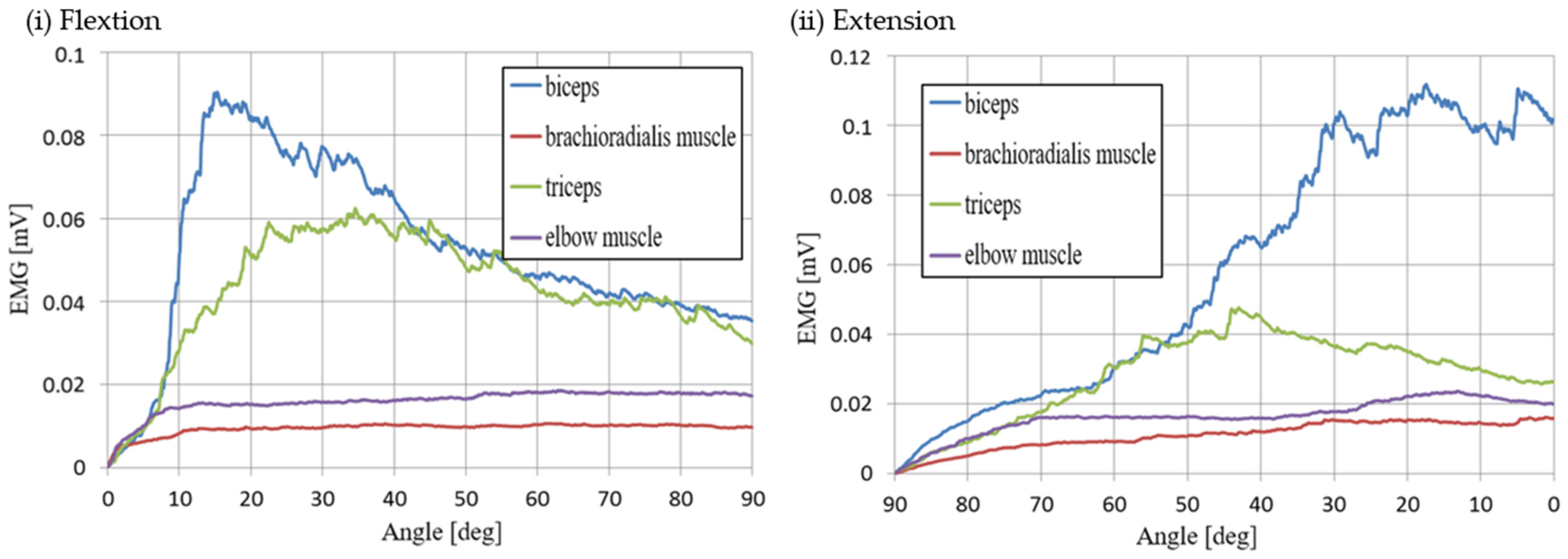
2.2. Elbow-Joint Motion Intention Detection by Muscle Activity Detection Section

The muscle activity detection part used muscle potentials as inputs and determined the presence or absence of muscle activity by comparing the root mean square (RMS) value of the muscle potentials to a threshold value as muscle activity. The first movement was a shoulder flexion movement to raise the elbow position, and the second was an elbow flexion movement to raise the forearm to the mouth. The second movement was targeted for the estimation of motor intention using electromyograms.
In the pre-measurements, muscle strength was measured by performing elbow flexion extension movements of the biceps and brachialis muscles, which are considered highly relevant to elbow flexion, and the triceps and elbow muscles, which are considered highly relevant to elbow extension, to select muscles strongly associated with the movements and determine the thresholds of the muscle potentials. This was based on the synergy hypothesis [44], which states that motor control is carried out by the coordinated action of multiple muscles and joint groups, and suggests that, for example, human upper limb movements can be explained by four or five muscle synergies [45]. In addition, Cyberdyne [46], for example, has been realized as a walking aid using muscle activity-based motion prediction.

3. Experiments

3.1. Participants

The volunteers were three healthy participants (graduate students, 23 ± 0.5 years old, male) who were seated in chairs, and they were as relaxed as possible. The studies involving human participants were reviewed and approved by the Kwansei Gakuin University Institutional Review Board for the Protection of Human Subjects of Medical Research (No.KG-IRB-20-01). The participants provided their written informed consent to participate in this study.

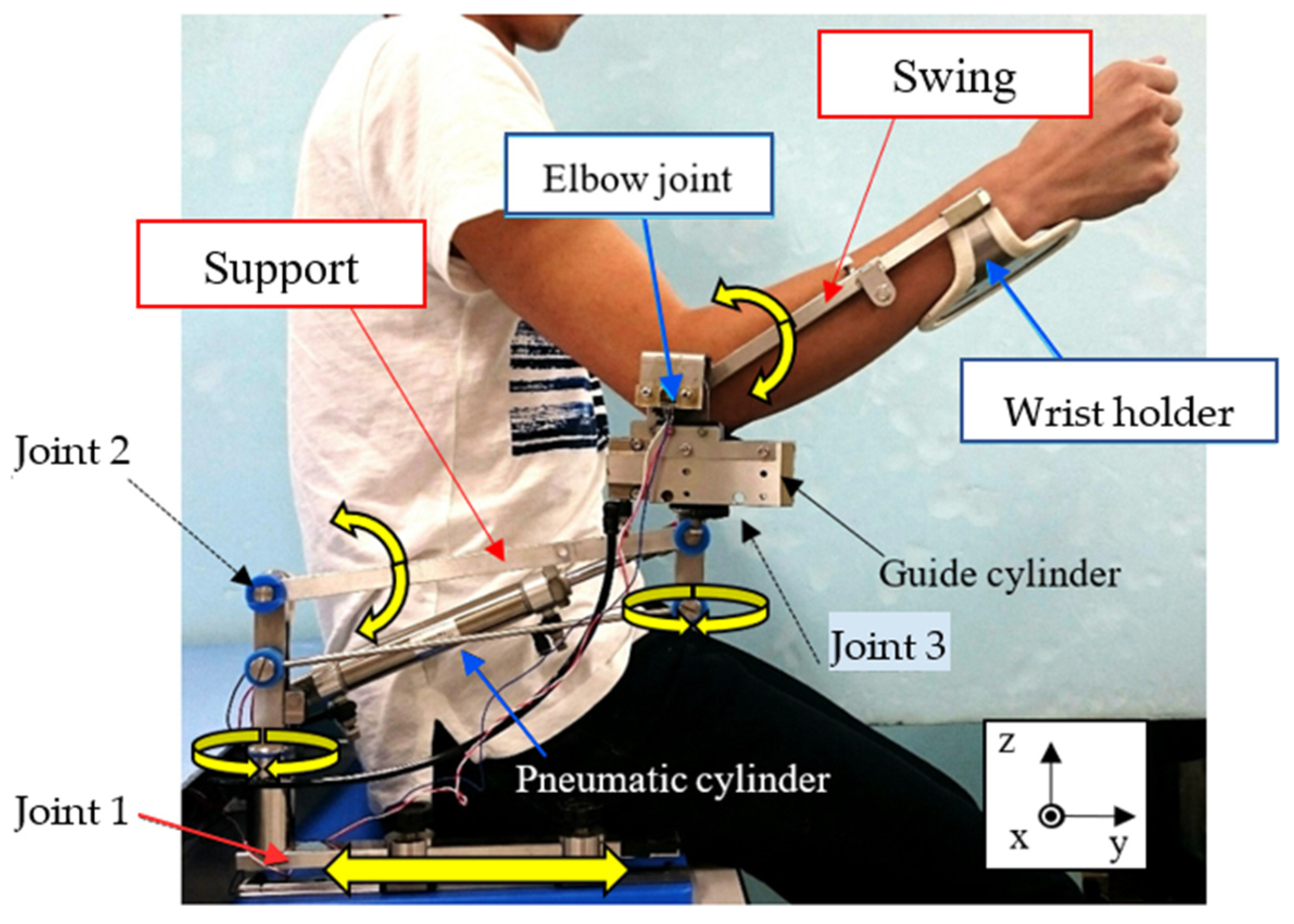
3.2. Overview of the Upper Limb Support Devices

The upper limb assistive device [47] used in the experiment was a link mechanism consisting of joint 1, joint 2, and joint 3, as shown in Figure 3. Here, the x-axis was the coronal axis, the y-axis was the sagittal axis, and the z-axis was the axial axis. Joint 1 assisted of a reaching motion by reciprocating motion in the y-axis direction. Joint 2 assisted upper arm motion (shoulder joint flexion and extension) by rotating the arm around the x-axis through the expansion and contraction of an air cylinder. Joint 3 provided forearm motion support (elbow joint flexion/extension support) by rotation of the forearm arm around the x-axis through extension/retraction of the guided air cylinder. Joints 2 and 3 could rotate around the z-axis by means of a rotary joint. The rotation of joint 2 around the x-axis and the rotation of joint 3 around the x-axis were actively supported by air cylinders, while the other joint movements were performed by the user’s own power.
In this study, the height support of the elbow position (upper arm motion support), which was the reference for the first motion—the eating motion—was performed by controlling the position of the pneumatic cylinder using the heuristic BCI, and then the forearm motion support for the eating motion was performed using electromyotics.

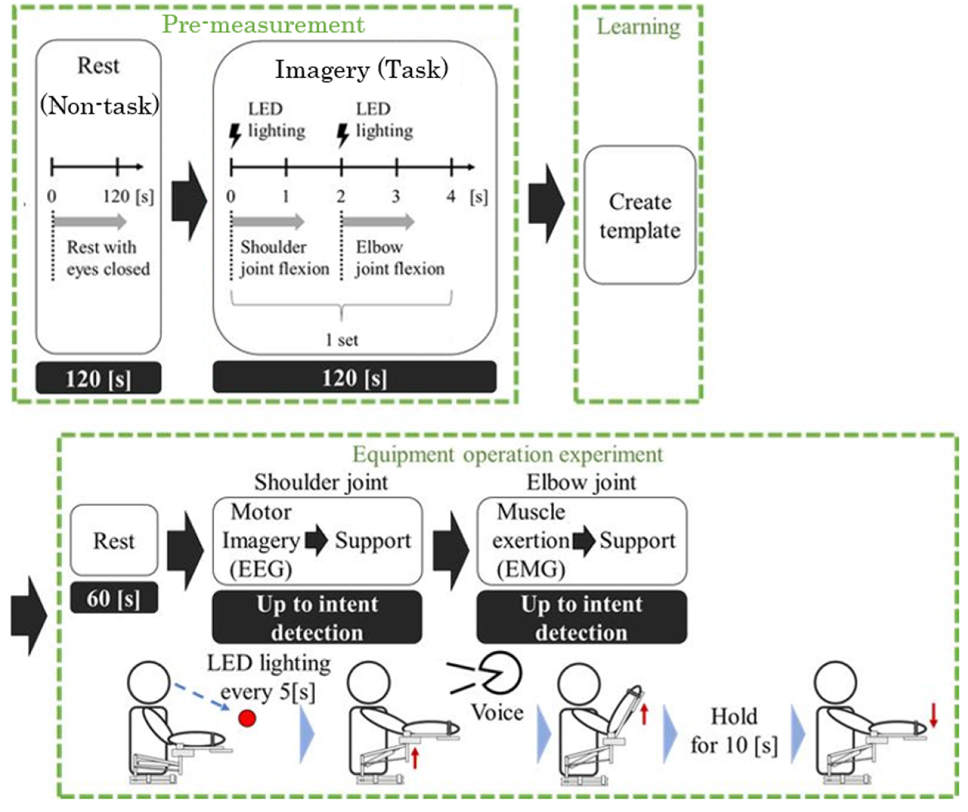
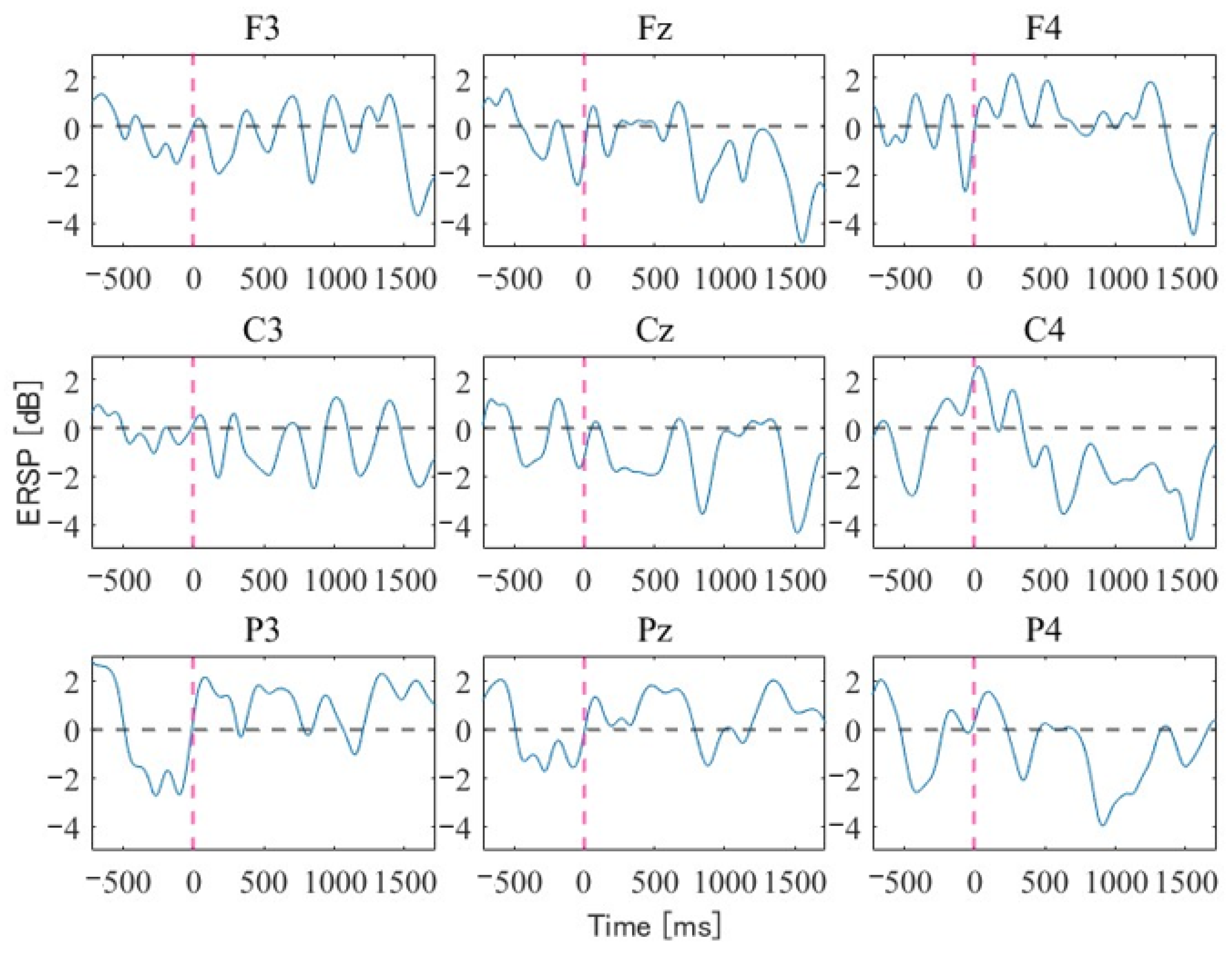
3.3. Setup for Motor Intent Detection by the EEG

As a preliminary experiment for the heuristic BCI, a g.Nautilus (g.tec medical engineering GmbH, Melbourne, Austria) was used for EEG measurements. We used dry electrodes as the measurement electrodes, with nine channels (F3, Fz, F4, C3, Cz, C4, P3, Pz, and P4) in the international 10–20 system [48]. Each electrode was positioned by wearing the head cap. The reference electrodes were placed on both earlobes.
First, the experimental participants were asked to perform a movement like drinking a glass of water, and after confirming the position of the elbow at that time and the movement of the forearm up towards the mouth with the elbow position fixed, the EEG measurement experiment was started. As shown in Figure 4, after 2 min of closed-eye rest, the experimental participants opened their eyes and performed a voluntary movement of bending the shoulder joint at the first LED light emission and extending the elbow joint at the next LED light emission two seconds later; they performed 30 sets of this single series of movements. The measured data were recorded in MATLAB Simulink (MATLAB2020a, MathWorks, Inc., Natick, MA, USA) and analyzed. The measured EEG was used as the fuzzy label placement pattern for the front part of the fuzzy rule, and a template was created by simply binarizing the EEG power at the measurement site for each alpha and beta wave band into low and high values. The real values of the posterior condition were set so that they would be close to the teacher’s signal by learning. Next, a heuristic BCI system was created by learning to discriminate between the resting state and the motor imagery state. For pre-processing, the EEG signal was notch-filtered in order to exclude 60 Hz of electrical noise, which was in turn band-pass filtered with frequencies between 0.04 and 200 Hz (FIR filtering). The frequency was chosen to include the full spectrum of the EEG signal, considering the correlation between EEG and EMG and excluding high frequencies from the EMG. Then, we conducted an independent component analysis (ICA) of whole EEG signals in order to segregate motor-related signals from the artifact components of Electrooculography (EOG) such as eye blink. In addition, frequency analysis was performed to extract time features at each measurement location. We also performed normalization of processed data with the aim of eliminating individual differences in electroencephalography and myogenic potentials. The sampling frequency of the EEG measurement was set to 500 Hz. Upon examining For heuristic BCI, we decided to apply it only to the first movement, because it is difficult to detect the second and subsequent consecutive movements with EEG alone. This is explained in Chapter 5 based on the experimental results.

3.4. Setup for Motor Intent Detection by the EMG

The second movement, flexion and extension of the elbow joint of the forearm, was assumed to be eating. The biceps brachii and brachioradialis muscles, which are considered highly relevant to elbow flexion, and the triceps brachii and elbow muscles, which are considered highly relevant to extension, were targeted, and target electrodes were placed on the right arm after palpation [49]. EMG measurements were performed using MQ16 (Kissei Comtec Co., Ltd., Matsumoto, Japan) and recorded with Vital Recorder 2 (Kissei Comtec Co., Ltd., Matsumoto, Japan) at a sampling frequency of 500 Hz to match the EEG. Although muscle potentials are typically sampled at 1000 Hz, we set the sampling frequency to 500 Hz. This decision followed an evaluation of elbow joint flexion and extension, which confirmed that the frequency response of the biceps and triceps during contraction was below 50 Hz, presenting no issues. Disposable ECG electrodes (LecTrode NP, ADVANCE Co., Ltd., Tokyo, Japan) were used. For the myopotential measurement, two electrodes were placed on the skin surface aligned with the muscle fibers, utilizing the bipolar induction method. The electrodes were spaced 15 mm apart, with the reference electrode positioned at the ulnar styloid process. The recorded waveforms underwent RMS (root mean square) processing with a 1 Hz low-pass filter and a 20 Hz high-pass filter to eliminate motion artifacts. In the measurements, the arm was flexed and extended 10 times to the 10 s period of the metronome, and the average of the five movements between them was used as the value for each participant in the experiment. The flexion movement was performed from 0 degrees to 90 degrees in the direction of the hand tip relative to the desk surface with the elbow as the fulcrum, and the extension movement was performed from 90 degrees to 0 degrees in the direction of the hand tip relative to the desk surface.
Figure 5 presents the RMS values of the EMG measured at the onset of movement for one of the experimental participants, setting the EMG at 0 × 10 3 V as the baseline. During the flexion and extension of the elbow joint, the electromyographic readings of the bicep brachii muscle were high for all three experimental participants. The electromyography for the second movement, which acts as the trigger signal for this participant, was based on the EMG recorded during flexion of the elbow joint and set at an RMS value of 0.013 × 10 3 V. The threshold for the second movement for each experimental participant was similarly determined by measuring the muscle’s EMG during the flexion and extension movements. The EMG signals were processed using the root mean square (RMS) method, followed by a moving average calculation. The actual RMS calculation was conducted with an integration interval of 0.06 s, set to 30 points (2T), as follows:
E M G   ( R M S t ) = 1 2 T T T { E M G t + τ } 2 d τ
Here, t represents the data point, and 2T is the integration interval (30 points).

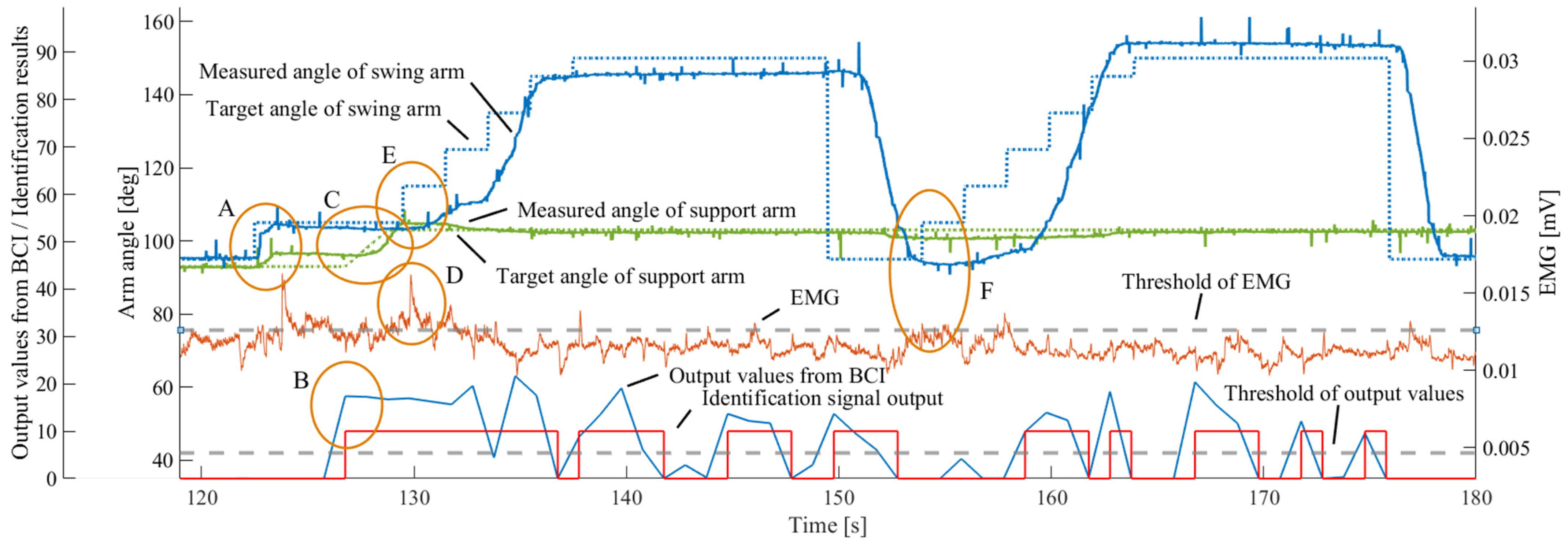
3.5. Experimental Overview of the Hybrid BCI System

The target motion of the hybrid BCI system is a simple, continuous flexion motion of the shoulder and elbow joints, as shown in Figure 6. The users were asked to imagine the movement of their upper arms, as shown in Figure 6, using the LED light as a cue, and once the motion intention was detected from the EEG, the elbow position was initially controlled by a pneumatic cylinder to the elbow position height for the eating motion. In this movement support, the height of the elbow position was fixed when it reached the horizontal position, and the angle of the upper arm increased from the initial position of 93 degrees to 103 degrees. When the upper arm angle reached 103 degrees, the forearm arm’s angle was 95 degrees (horizontal to the seat). When the biceps muscle exceeded the threshold, the guide cylinder was position-controlled to rise in a staircase-like manner to support the forearm arm. The forearm arm was set to maintain its position when the threshold was not exceeded because eating was assumed to be rehabilitation training for muscle strength recovery. In addition, after detecting the intention to exercise from the EEG, support for the next movement by electromyography was performed by switching to myoelectric potentials, and the evaluation was focused on whether the switching was smooth. The forearms were lowered slowly after a holding time of 3 s after ascending to avoid muscle fatigue.

4. Experimental Results

The experimental results for one participant are shown in Figure 7. Part A was set up so that the arm was attached to the upper limb support device and rose to its initial settings of approximately 93 degrees (green line) and 105 degrees (blue line) in the upper arm and forearm, respectively, 2.5 s after the experiment started. The rehabilitation start position was set to 105 degrees for the support arm because the dead time was minimized by preloading the pneumatic cylinder. In Part B, the heuristic BCI captured the motion intention (light blue line) in the EEG, and the motion command signal (red line) was instantly input into the device, indicating that the motion was performed smoothly. From Part C, we can see that the elbow position (green line) was increased by the support arm from the initial position of about 95 degrees to about 105 degrees 2.02 s after the start of the experiment. After the upper arm reached the target angle, the signal input to the device switched from an EEG to an EMG signal, and the support motion of the forearm was initiated once the EMG signal in Part D (vermilion line) exceeded the threshold value. As shown in Part E, the target movement for the forearm was set to a gentle stepwise increase, with an increment of 10 degrees every 2 s, and then to an increment of only 5 degrees per second just before reaching the target angle of 150 degrees. The aim was to reach the target angle in five steps over 10 s using this control, and the settling time was 10.77 s during the initial swing movement based on the EEG, as shown in Figure 5. On average, the delay remained within 1 s for the three participants. As the arm was lowered by its own weight after the forearm was maintained at a pre-set angle for 12 s, the EMG signals exceeded the threshold, as shown in Part F, and the forearm was observed to begin rising again. The duration of the hold was set based on the Barré and Mingazzini tests [50]. An undershoot due to the arm’s weight was observed, but it was confirmed that the arm was raised smoothly. The slope during arm raising and the undershoot during lowering can be improved by incorporating gravity compensation into the control system design. The offsets observed during the initial action and the second assistive movement are due to cumulative errors from the resolution of the encoders installed on the two arm rotation axes, and they can be addressed by revising the design of the control system.
These results indicate that the switch from an EEG to EMG signal was smoothly assisted without causing any discomfort to the person. In the heuristic BCI, the three participants were able to discriminate between the non-task in the resting state and during the task in the motor imagery state with high accuracy, with an average error rate of 9.4% (±7.3%), as shown in Figure 8. In addition, the three participants were able to sufficiently discriminate continuous movements in approximately 0.6 to 0.8 s from the motor intention’s detection by the EEG to the assistive device’s operation and in 1.8 to 3.1 s from the myoelectricity to the assistive device’s operation. The threshold values for the EEG and the EMG, the former of which was the output signal to the device, were set based on the measurement results for each individual. Table 1 shows the top eight rules with the highest posterior values for each participant. Similarly, Figure 9 illustrates the occurrence rate of ‘LOW’ for each channel (ch) across the three participants. Additionally, Figure 10 focuses on the alpha wave band, displaying the averaged time analysis results of event-related potentials (ERPs) for one of the participants. The ‘LOW’ indicated by the fuzzy rules in Table 1 signifies a decrease in the EEG power spectrum. From Figure 9, the channels at electrode positions Cz, C4, Pz, and P4 had the highest incidence of high posterior values among the top eight for the three participants. Further observation of the channels in Figure 9, compared to other electrodes, reveals that although the heuristic BCI system with fuzzy template matching detects motor intention without identifying specific EEG features, Table 1 and Figure 8 show a significant decrease after an event, suggesting the occurrence of ERD. This indicates that the proposed method, which uses EEG patterns represented by just two channels (a simple LOW and HIGH channel), is comparable to BMI systems that use machine learning and other methods to detect EEG features.

5. Discussion

We have proposed and verified the effectiveness of a new brain–computer interface (BCI) system capable of accommodating continuous movements based on motor imagery and electromyography, suitable for patients with post-stroke hemiplegia. To estimate movement intentions, we applied our developed heuristic BCI system based on fuzzy inference without using EEG features such as event-related desynchronization (ERD) or P300. The system reads motor imagery brain waves measured from nine electrode channels, binarizing brain wave power into low and high to construct fuzzy rules. The output values are determined by comparing rest and motor imagery states. As shown in Table 1, Figure 8 and Figure 9, it was confirmed that the proposed method, which does not identify EEG features, still includes EEG features due to event-related potentials. This indicates that the algorithm, which simply distinguishes between resting and motion states without targeting specific EEG features, is effective for the BMI system. This approach has the potential to develop algorithms capable of detecting motor intentions with even higher accuracy. Furthermore, we believe that varying the number of measurement channels, adjusting the measurement positions, narrowing the measurement bandwidth, and extending it to include the γ-wave bandwidth to study the effect of fuzzy template matching on output values will lead to the discovery of new EEG features and behavioral intentions.
Regarding continuous movements, the core of this research connects brain waves with movement through a closed loop interfaced with rehabilitation equipment via neurofeedback. Upon examining Figure 7, which presents the experimental results, the initiation of movement was successfully captured by the identification signal output, represented by the red solid line. However, for the second movement, this signal occurred frequently before and after, indicating that it was not captured effectively. Consequently, it was decided to apply the heuristic BCI solely to the initial movement. While EEGs are vital for the initial movement, the variability in EEG waveforms in response to various actions is supplanted in subsequent movements by utilizing the biceps brachii muscle activity based on forearm flexion movements alone. The results for three healthy participants confirm that the transition from brain waves to muscle activity occurs smoothly.
The target movements in this study were two continuous motions, flexion and extension of the shoulder and elbow joints. To increase the number of target movements, it is important to investigate the relationship between muscle activity and these movements. To achieve this, we believe that it is effective to enhance extensibility for continuous movement by selecting muscles with synergistic effects based on the muscle synergy hypothesis. Moreover, we consider our heuristic BCI method, which does not pin down specific EEG features, to be applicable in rehabilitation systems for stroke patients in continuous movement scenarios. The system’s utility in patients has only been demonstrated for isolated movements, and we plan to continue validating it with more trials in actual patients. Additionally, we hope to reveal new EEG features in new movements by retrospectively considering the type of EEG features our BCI system captures.

6. Conclusions

This study shows that the heuristic BCI can smoothly provide support for continuous upper limb movements based on a user’s intention. By switching from an EEG in the initial movement to an EMG in the next movement, the system provides rehabilitation on the same day that the EEG is measured.
In the future, we will further increase the number of participants to confirm motions and realize motion support for complex upper-limb movements, such as a series of multiple movements, by utilizing multiple muscle activity patterns based on muscle synergy, acceleration, position sensors, etc.

Author Contributions

Conceptualization, writing—original draft preparation, and methodology, N.S. (Norihiko Saga); algorithm construction, data curation, and experimentation, Y.O.; methodology, validation, and editing, T.S. (Takuma Saga); validation and editing, T.S. (Toshiyuki Satoh) and N.S. (Naoki Saito) All authors have read and agreed to the published version of the manuscript.

Funding

This work was partially supported by the Japan Keirin Autorace foundation (JKA) (2020M-204).

Institutional Review Board Statement

The study was conducted in accordance with the Declaration of Helsinki and approved by the Kwansei Gakuin University Medical Research Ethics Committee (No. KG-IRB-20-01) (24 September 2020).

Informed Consent Statement

Informed consent was obtained from all participants involved in the study.

Data Availability Statement

The data that support the findings of this study are available upon request from the corresponding author, N.S. The data are not publicly available due to the restriction that they contain information that requires the consent of the study participants.

Conflicts of Interest

The authors declare no conflicts of interest.

References

  1. Pfurtscheller, G.; Neuper, C. Future prospects of ERD/ERS in the context of brain–computer interface (BCI) developments. Prog. Brain Res. 2006, 159, 433–437. [Google Scholar] [CrossRef]
  2. Jackson, A.; Zimmermann, J.B. Neural interfaces for the brain and spinal cord—Restoring motor function. Nat. Rev. Neurosci. 2012, 8, 690–699. [Google Scholar] [CrossRef] [PubMed]
  3. Ethier, C.; Gallego, J.A.; Miller, L.E. Brain-controlled neuromuscular stimulation to drive neural plasticity and functional recovery. Curr. Opin. Neurobiol. 2015, 33, 95–102. [Google Scholar] [CrossRef]
  4. Chaudhary, U.; Birbaumer, N.; Murguialday, A.R. Brain–computer interfaces for communication and rehabilitation. Nat. Rev. Neurosci. 2016, 12, 513–525. [Google Scholar] [CrossRef] [PubMed]
  5. Caria, A.; Weber, C.; Brötz, D.; Ramos, A.; Ticini, L.F.; Gharabaghi, A.; Braun, C.; Birbaumer, N. Chronic stroke recovery after combined BCI training and physiotherapy: A case report. Psychophysiology 2011, 48, 578–582. [Google Scholar] [CrossRef]
  6. Sitaram, R.; Ros, T.; Stoeckel, L.; Haller, S.; Scharnowski, F.; Peacock, J.L.; Weiskopf, N.; Blefari, M.L.; Rana, M.; Oblak, E.; et al. Closed-loop brain training: The science of neurofeedback. Nat. Rev. Neurosci. 2017, 18, 86–100. [Google Scholar] [CrossRef]
  7. Halme, H.L.; Parkkonen, L. Comparing Features for Classification of MEG Responses to Motor Imagery. PLoS ONE 2016, 11, e0168766. [Google Scholar] [CrossRef]
  8. Kim, Y.J.; Park, S.W.; Yeom, H.G.; Bang, M.S.; Kim, J.S.; Chung, C.K.; Kim, S. A study on a robot arm driven by three-dimensional trajectories predicted from non-invasive neural signals. Biomed. Eng. Online 2015, 14, 81. [Google Scholar] [CrossRef] [PubMed]
  9. Lee, J.; Mukae, N.; Arata, J.; Iwata, H.; Iramina, K.; Iihara, K.; Hashizume, M. A multichannel-near-infrared-spectroscopy-triggered robotic hand rehabilitation system for stroke patients. IEEE Int. Conf. Rehabil. Robot. ICORR 2017, 2017, 158–163. [Google Scholar] [CrossRef]
  10. Logothetis, N.K. What we can do and what we cannot do with fMRI. Nature 2008, 453, 869–878. [Google Scholar] [CrossRef]
  11. Boas, D.A.; Elwell, C.E.; Ferrari, M.; Taga, G. Twenty years of functional near-infrared spectroscopy: Introduction for the special issue. Neuroimage 2014, 85, 1–5. [Google Scholar] [CrossRef]
  12. Ferrari, M.; Quaresima, V. A brief review on the history of human functional near-infrared spectroscopy (fNIRS) development and fields of application. Neuroimage 2012, 63, 921–935. [Google Scholar] [CrossRef] [PubMed]
  13. Nunez, P.L.; Srinivasan, R. Electric Fields of the Brain: The Neurophysics of EEG, 2nd ed.; Oxford University Press: New York, NY, USA, 2006. [Google Scholar] [CrossRef]
  14. Wang, Y.; Nakanishi, M.; Zhang, D. EEG-Based Brain-Computer Interfaces. Adv. Exp. Med. Biol. 2019, 1101, 41–65. [Google Scholar] [CrossRef] [PubMed]
  15. Pfurtscheller, G. Event-related synchronization (ERS): An electrophysiological correlate of cortical areas at rest. Electroencephalogr. Clin. Neurophysiol. 1992, 83, 62–69. [Google Scholar] [CrossRef] [PubMed]
  16. Pfurtscheller, G.; Aranibar, A. Event-related cortical desynchronization detected by power measurements of scalp EEG. Electroencephalogr. Clin. Neurophysiol. 1977, 42, 817–826. [Google Scholar] [CrossRef] [PubMed]
  17. Farwell, L.A.; Donchin, E. Talking off the top of your head: Toward a mental prosthesis utilizing event-related brain potentials. Electroencephalogr. Clin. Neurophysiol. 1988, 70, 510–523. [Google Scholar] [CrossRef]
  18. Kaufmann, T.; Herweg, A.; Kübler, A. Toward brain-computer interface based wheelchair control utilizing tactually-evoked event-related potentials. J. Neuroeng. Rehabil. 2014, 11, 7. [Google Scholar] [CrossRef]
  19. Herweg, A.; Gutzeit, J.; Kleih, S.; Kübler, A. Wheelchair control by elderly participants in a virtual environment with a brain-computer interface (BCI) and tactile stimulation. Biol. Psychol. 2016, 121, 117–124. [Google Scholar] [CrossRef] [PubMed]
  20. Eidel, M.; Kübler, A. Wheelchair Control in a Virtual Environment by Healthy Participants Using a P300-BCI Based on Tactile Stimulation: Training Effects and Usability. Front. Hum. Neurosci. 2020, 14, 265. [Google Scholar] [CrossRef]
  21. Pritchard, W.S. Psychophysiology of P300. Psychol. Bull. 1981, 89, 506–540. [Google Scholar] [CrossRef]
  22. Hillyard, S.A.; Kutas, M. Electrophysiology of cognitive processing. Annu. Rev. Psychol. 1983, 34, 33–61. [Google Scholar] [CrossRef] [PubMed]
  23. Rebsamen, B.; Burdet, E.; Guan, C.; Zhang, H.; Teo, C.L.; Zeng, Q.; Laugier, C.; Ang, M.H. Controlling a Wheelchair Indoors Using Thought. IEEE Intell. Syst. 2007, 22, 18–24. [Google Scholar] [CrossRef]
  24. Cheng, M.; Gao, X.; Gao, S.; Xu, D. Design and implementation of a brain-computer interface with high transfer rates. IEEE. Trans. Biomed. Eng. 2002, 49, 1181–1186. [Google Scholar] [CrossRef] [PubMed]
  25. Chi, X.; Wan, C.; Wang, C.; Zhang, Y.; Chen, X.; Cui, X. A Novel Hybrid Brain-Computer Interface Combining Motor Imagery and Intermodulation Steady-State Visual Evoked Potential. IEEE Trans. Neural Syst. Rehabil. Eng. 2022, 30, 1525–1535. [Google Scholar] [CrossRef] [PubMed]
  26. Chen, X.; Liu, B.; Wang, Y.; Cui, H.; Dong, J.; Ma, R.; Li, N.; Gao, X. Optimizing Stimulus Frequency Ranges for Building a High-Rate High Frequency SSVEP-BCI. IEEE Trans. Neural Syst. Rehabil. Eng. 2023, 31, 1277–1286. [Google Scholar] [CrossRef] [PubMed]
  27. Lotte, F.; Bougrain, L.; Cichocki, A.; Clerc, M.; Congedo, M.; Rakotomamonjy, A.; Yger, F. A review of classification algorithms for EEG-based brain-computer interfaces: A 10 year update. J. Neural Eng. 2018, 15, 31005. [Google Scholar] [CrossRef] [PubMed]
  28. Sheng, J.; Xu, J.; Li, H.; Liu, Z.; Zhou, H.; You, Y.; Song, T.; Zuo, G. A Multi-Scale Temporal Convolutional Network with Attention Mechanism for Force Level Classification during Motor Imagery of Unilateral Upper-Limb Movements. Entropy 2023, 25, 464. [Google Scholar] [CrossRef] [PubMed]
  29. Pfurtscheller, G. EEG event-related desynchronization (ERD) and synchronization (ERS). Electroencephalogr. Clin. Neurophysiol. 1997, 103, 26. [Google Scholar] [CrossRef]
  30. Benzy, V.K.; Vinod, A.P.; Subasree, R.; Alladi, S.; Raghavendra, K. Motor Imagery Hand Movement Direction Decoding Using Brain Computer Interface to Aid Stroke Recovery and Rehabilitation. IEEE Trans. Neural Syst. Rehabil. Eng. 2020, 28, 3051–3062. [Google Scholar] [CrossRef]
  31. Prasad, G.; Herman, P.; Coyle, D.; McDonough, S.; Crosbie, J. Applying a brain-computer interface to support motor imagery practice in people with stroke for upper limb recovery: A feasibility study. J. Neuroeng. Rehabil. 2010, 7, 60. [Google Scholar] [CrossRef]
  32. Blankertz, B.; Dornhege, G.; Krauledat, M.; Müller, K.R.; Curio, G. The non-invasive Berlin Brain-Computer Interface: Fast acquisition of effective performance in untrained subjects. NeuroImage 2007, 37, 539–550. [Google Scholar] [CrossRef]
  33. Krauledat, M.; Schröder, M.; Blankertz, B.; Müller, K.R. Reducing Calibration Time for Brain-Computer Interfaces: A Clustering Approach. In Advances in Neural Information Processing Systems 19: Proceedings of the 2006 Conference, Vancouver, Canada, 4–7 December 2006; MIT Press: Cambridge, MA, USA, 2007; pp. 753–760. [Google Scholar]
  34. Van Erp, J.B.F.; Lotte, F.; Tangermann, M. Brain-Computer Interfaces: Beyond Medical Applications. Comput. J. 2012, 45, 26–34. [Google Scholar] [CrossRef]
  35. Saga, N.; Tanaka, Y.; Doi, A.; Oda, T.; Kudoh, S.N.; Fujie, H. Prototype of an Ankle Neurorehabilitation System with Heuristic BCI Using Simplified Fuzzy Reasoning. Appl. Sci. 2019, 9, 2429. [Google Scholar] [CrossRef]
  36. Saga, N.; Doi, A.; Oda, T.; Kudoh, S.N. Elucidation of EEG Characteristics of Fuzzy Reasoning-Based Heuristic BCI and Its Application to Patient with Brain Infarction. Front. Neurorobot. 2021, 14, 607706. [Google Scholar] [CrossRef]
  37. Bansal, A.K.; Truccolo, W.; Irwin, C.E.V.; Donoghue, J.P. Decoding 3D reach and grasp from hybrid signals in motor and premotor cortices: Spikes, multiunit activity, and local field potentials. J. Neurophysiol. 2012, 107, 1337–1355. [Google Scholar] [CrossRef]
  38. Jeong, J.H.; Shim, K.H.; Kim, D.J.; Lee, S.W. Trajectory Decoding of Arm Reaching Movement Imageries for Brain-Controlled Robot Arm System. In Proceedings of the 2019 41st Annual International Conference of the IEEE Engineering in Medicine and Biology Society (EMBC), Berlin, Germany, 23–27 July 2019; pp. 5544–5547. [Google Scholar] [CrossRef]
  39. Kim, J.H.; Bießmann, F.; Lee, S.W. Decoding Three-Dimensional Trajectory of Executed and Imagined Arm Movements from Electroencephalogram Signals. IEEE Trans. Neural Syst. Rehabil. Eng. 2015, 23, 867–876. [Google Scholar] [CrossRef]
  40. Malesevic, N.; Markovic, D.; Kanitz, G.; Controzzi, M.; Cipriani, C.; Antfolk, C. Decoding of individual finger movements from surface EMG signals using vector autoregressive hierarchical hidden Markov models (VARHHMM). IEEE Int. Conf. Rehabil. Robot. (ICORR) 2017, 2017, 1518–1523. [Google Scholar]
  41. Zhang, Q.; Liu, R.; Chen, W.; Xiong, C. Simultaneous and Continuous Estimation of Shoulder and Elbow Kinematics from Surface EMG Signals. Front. Neurosci. 2017, 11, 280. [Google Scholar] [CrossRef]
  42. Pan, L.; Crouch, D.L.; Huang, H. Comparing EMG-Based Human-Machine Interfaces for Estimating Continuous, Coordinated Movements. IEEE Trans. Neural Syst. Rehabil. Eng. 2019, 27, 2145–2154. [Google Scholar] [CrossRef]
  43. Tsukahara, A.; Hasegawa, Y.; Eguchi, K.; Sankai, Y. Restoration of gait for spinal cord injury patients using HAL with intention estimator for preferable swing speed. IEEE Trans. Neural Syst. Rehabil. Eng. 2015, 23, 308–318. [Google Scholar] [CrossRef]
  44. Bernshteĭn, N.A. The Coordination and Regulation of Movements; Pergamon Press: Oxford, UK, 1967. [Google Scholar]
  45. d’Avella, A.; Portone, A.; Fernandez, L.; Lacquaniti, F. Control of fast-reaching movements by muscle synergy combinations. J. Neurosci. 2006, 26, 7791–7810. [Google Scholar] [CrossRef] [PubMed]
  46. Namikawa, Y.; Kawamoto, H.; Sankai, Y. Gait Evaluation with Bioelectrical Signal Patterns during Cybernic Treatment. In Proceedings of the 2021 43rd Annual International Conference of the IEEE Engineering in Medicine & Biology Society (EMBC), Virtual, 1–5 November 2021; pp. 6728–6733. [Google Scholar] [CrossRef]
  47. Saga, N.; Umeki, R.; Saito, N.; Nagase, J.; Satoh, T. Development of a Meal Support Device for Functional Recovery Using EMG Signals. IEEE Access 2020, 8, 79586–79593. [Google Scholar] [CrossRef]
  48. Towle, V.L.; Bolaños, J.; Suarez, D.; Tan, K.; Grzeszczuk, R.; Levin, D.N.; Cakmur, R.; Frank, S.A.; Spire, J.P. The spatial location of EEG electrodes: Locating the bestfitting sphere relative to cortical anatomy. Electroencephalogr. Clin. Neurophysiol. 1993, 86, 1–6. [Google Scholar] [CrossRef] [PubMed]
  49. Cael, C. Functional Anatomy: Musculoskeletal Anatomy, Kinesiology, and Palpation for Manual Therapists, 2nd ed.; Jones & Bartlett Learning: Burlington, MA, USA, 2022. [Google Scholar]
  50. Koehler, P.J. The Barrés test and Mingazzini test. In Neurological Eponyms; Oxford Academic-Oxford University Press: Oxford, UK, 2000; pp. 119–126. [Google Scholar] [CrossRef]
Figure 1. Outline of the hybrid BCI. The orange arrows are biological signals, and the pink arrows indicate the command signals to the device.
Electronics 13 02770 g001
Figure 2. Creation of fuzzy templates (rules) for a heuristic BCI. The “one-way arrows” refer to each channel of the fuzzy template. For example, C3(α) indicates the alpha waveband of the channel C3. The “two-way arrow” means that a “LOW” or “HIGH” decision was made based on the EEG power of each measured channel.
Electronics 13 02770 g002
Figure 3. Upper limb support devices.
Electronics 13 02770 g003
Figure 4. Preliminary experimental setup.
Electronics 13 02770 g004
Figure 5. RMS values of four muscle activities during flexion–extension of the elbow joint.
Electronics 13 02770 g005
Figure 6. Target motion of the hybrid BCI system.
Electronics 13 02770 g006
Figure 7. Experimental results of the hybrid BCI system. The solid blue line is the measured value of the swing arm, the dashed blue line is the target value of the swing arm, the solid green line is the measured value of the support arm, the dashed green line is the target value of the support arm, the solid orange line is the EMG, the dashed gray line is the EMG threshold value, the solid light blue line is the output value from the BCI, the solid red line is the identification signal output value of motion intention, and the dashed gray line at the bottom is the output value threshold value. Part A: Setting the support arm in its initial position, Part B: Heuristic BCI captures motion intention based on EEG, Part C: Ascending the support arm, Part D: Capturing motor intention based on EMG, Part E: Ascending the swing arm ascends gradually, Part F: Capturing motor intention based on EEG with Heuristic BCI for the second set.
Electronics 13 02770 g007
Figure 8. Output values at rest and during the motor imagery state. Eller bars denote standard errors, ** denotes significant differences (**: p < 0.01, two-tailed t-test).
Electronics 13 02770 g008
Figure 9. Appearance ratio of LOW in the measured Ch for each frequency band of the three experimental participants.
Electronics 13 02770 g009
Figure 10. Time analysis results of the event-related potential (ERP) for one experimental participant. The pink dashed line indicates the trigger signal, and the dark blue dashed line indicates the baseline.
Electronics 13 02770 g010
Table 1. Top 8 fuzzy rules with high consequent values for one experimental participant. The “LOW” markers are marked in light blue, and the “HIGH” markers are marked in red.
TopF3FzF4C3CzC4P3PzP4Consequent Value
α βαβαβαβαβαβαβαβαβ
1HighHighHighHighHighHighHighHighHighHighLowHighHighHighHighHighHighHigh3.289
2HighHighHighHighHighHighLowHighHighHighHighHighHighHighHighHighHighHigh3.099
3HighHighHighHighHighHighHighHighHighLowHighLowHighHighHighHighLowHigh2.080
4HighHighHighHighHighHighLowHighLowHighLowHighLowLowLowHighHighHigh1.794
5HighHighHighHighHighHighHighLowHighHighHighLowHighHighHighHighHighHigh1.619
6HighHighHighHighHighLowHighHighHighHighHighHighHighHighHighHighHighHigh1.465
7HighHighHighHighHighHighHighHighHighHighHighHighHighHighHighHighLowLow1.388
8HighHighHighHighHighHighHighLowHighLowHighHighHighHighHighHighHighHigh1.384
Disclaimer/Publisher’s Note: The statements, opinions and data contained in all publications are solely those of the individual author(s) and contributor(s) and not of MDPI and/or the editor(s). MDPI and/or the editor(s) disclaim responsibility for any injury to people or property resulting from any ideas, methods, instructions or products referred to in the content.

