Article

Studies on the Control of Dermanyssus gallinae via High-Voltage Impulse

1
Department of Electrical and Electronic Eng., National Institute of Technology, Oita College, Oita 870-0152, Japan
2
General Education, National Institute of Technology, Oita College, Oita 870-0152, Japan
3
Department of Parasitology and Tropical Medicine, Kitasato University School of Medicine, Sagamihara 252-0374, Japan
4
Department of Health Science, Kitasato University School of Allied Health Sciences, Sagamihara 252-0374, Japan
*
Author to whom correspondence should be addressed.
Electronics 2023, 12(4), 1038; https://doi.org/10.3390/electronics12041038
Submission received: 31 January 2023 / Revised: 13 February 2023 / Accepted: 16 February 2023 / Published: 19 February 2023

Abstract

:
Dermanyssus gallinae, a parasitic mite that subsists on the avian blood of chickens, poses a considerable threat to the poultry industry. D. gallinae infestation can result in a plethora of detrimental effects for the host birds, including decreased egg production and anemia. Pyrethroid pesticides have been the primary means of combating this issue and have demonstrated high levels of efficacy. However, in recent years, D. gallinae has exhibited resistance to these chemicals, resulting in a marked decrease in their mortality; thus, an integrated control strategy in addition to the chemical use should be required for the sustainable control of this mite. This study confirms that D. gallinae can be effectively controlled through the utilization of high-voltage impulse discharges and that various electrical parameters possess optimal values that are required for mite control. The alterations in the body surface of the mite caused by high-voltage impulses were akin to those caused by heat, but no alteration in the elemental composition of the body surface was observed, suggesting a change in organization caused by currents flowing inside the exoskeleton. Comparatively, the mite control efficacy of high-voltage impulse was found to be substantially superior to that of ultraviolet light or ozone, with up to 95% more mites being killed in as little as 30 seconds.

1. Introduction

The origins of bioelectronics date back to the 18th century, when scientist Luigi Galvani first applied voltage to the legs of a frog [1]. Research into the application of electrical engineering in biology and medicine subsequently flourished. Galvani’s theory that “animal electricity is a vital force which gave life to organic matter” was later disproven by experiments conducted by Alessandro Volta [2], though it is still regarded as the origin of electrophysiology today. In the 19th century, physiological research using electricity, led by Emile du Bois-Rémond and Helmholtz, established that nerve activity is electrical in nature [3,4,5]. In 1947, the use of electric shocks on the human body was first utilized, leading to the development of modern automated external defibrillators (AED) [6]. Today, electroconvulsive therapy is also used as a treatment for certain psychiatric disorders [7,8]. These conventional electric shocks are based on nerve stimulation and do not cause irreversible changes in the body, with instantaneous power reaching only a few kilowatts at most.
On the other hand, pulsed power, which has a duration of microseconds or less and an instantaneous power of several hundred kilowatts or more, has been employed since the 1960s to forcibly alter the structure of cell membranes, such as in non-thermal field sterilization [9] and electroporation [10]. Advances in bioanalytical techniques and the increasing performance of pulsed power generators in the 21st century have led to new biological applications of pulsed power. Pulsed power acts as a powerful electromagnetic force on the living organism, comprising dielectric materials, which leads to changes in the structure and function of cells. Additionally, by adjusting the conditions under which pulsed power is applied, it is possible to elicit a variety of biological responses in non-cellular organisms. These biological responses, induced by physical stress, are being explored for medical applications such as cancer therapy [11,12] and wound care [13], as well as cosmetic applications. Pulsed power is not limited to animal cells but is also used as a physical stimulus for plant germination promotion, growth control [14,15,16], and breeding [17].
In this study, pulsed power was applied to control one type of arachnid, the mite Dermanyssus gallinae. D. gallinae that suck blood from chickens have a significant impact on the poultry industry. This mite is a mite belonging to the class Arachnida, order Mesoptera, and family Dermanyssidae, and its infestations have been confirmed in various regions in Japan. Adult mites are about 1.0 mm long and suck blood mainly at night, forming clusters during the day in shady places such as joints in metal fittings and egg receptacles [18,19]. Once the mites invade a poultry farm, they rapidly spread by attaching themselves to humans, vehicles, carrying baskets, and containers, making complete elimination highly challenging. It causes egg contamination, reduced laying rates, and anemia in poultry [20]. Furthermore, it can cause discomfort and allergic reactions in workers, leading to job turnover.
A questionnaire survey of poultry farms around the world found most were infested with D. gallinae [21,22,23]. Pesticides based on pyrethroid chemicals were the primary method of control and were highly effective. However, in recent years, D. gallinae has developed resistance to these chemicals, leading to a significant reduction in pesticidal efficacy [24,25]. The continued spread of drug-resistant D. gallinae and the increasing damage caused by the mite necessitate the development of alternative control methods belonging to integrated pest management (IPM).
With this need in mind, we sought to investigate the efficacy of using pulsed power as a means of eliminating D. gallinae. In this experiment, optimal control efficiency was determined by manipulating the voltage, frequency, and pulse width of high-voltage impulse discharges. Despite extensive research on the cellular and medical applications of pulsed power, few studies have targeted insects [26,27,28]. Although there have been studies on the use of commercially available electric insect traps for the elimination of houseflies [29,30,31], there has been no prior research targeting D. gallinae parasitizing poultry, and no studies were found on the determination of electrical parameters necessary for mite control activity. In this study, we established the electrical parameters necessary for the control of D. gallinae and confirmed the superiority of electric mite control technology as compared to other mite control methods.

2. Materials and Methods

2.1. Voltage Applied Target

D. gallinae individuals were randomly selected, regardless of their developmental stage (immature, mature, nymphs), and the experiment was conducted under controlled conditions of 25 °C room temperature and 60% relative humidity, with ambient room lighting. The behavior of the mites was observed by utilizing a video microscope (magnification 1500), and their condition was evaluated immediately after the application of an high-voltage impulse discharge, as well as 6 and 12 h post-treatment. Mites were considered ‘‘dead’’ if no movement was visible even after a gentle touch with a brush.

2.2. Experimental Procedure

An electrode was inserted within an acrylic container measuring 100 mm on each side, and approximately 100 D. gallinae were placed within the container. A high-voltage impulse was applied for a duration of 30 s. The number of mites that had been killed, as well as the number that had been sealed within the container, was subsequently assessed. The mortality was calculated as the ratio of killed mites to the total number of mites present.

2.3. Experimental Apparatus

The entire experimental apparatus is depicted in Figure 1. Electrodes with alternating anodes and cathodes were connected via resistors to an impulse power supply set to an operating frequency of 100 Hz. The electrodes were housed within an acrylic container measuring 100 mm on each side. As is shown in Figure 1, the electrodes were square-shaped, measured 90 mm on each side, and had copper anodes and cathodes. The structure comprised alternating anodes and cathodes with widths of 2 mm.

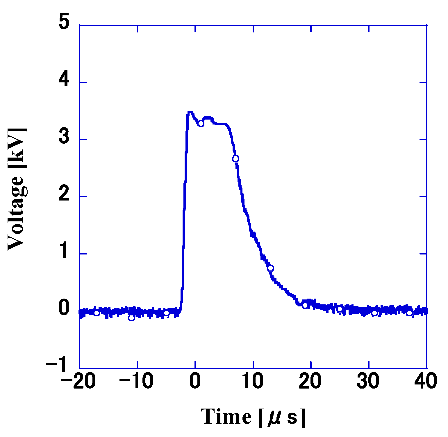
2.4. Variation in Discharge Voltage Due to Different Electrode Spacing and Insulation Materials

The electrodes developed in this experiment were designed to kill mites by electrically charging mites on one electrode and discharging through the mites just before they touched the other electrode. An high-voltage impulse discharge was applied to the electrodes, and the voltage immediately before the discharge was measured as the discharge voltage. The voltage was a pulsed waveform as shown in Figure 2, with variable wave height. The electrode spacing varied from 0.5 mm to 3 mm in 0.5 mm increments, and the discharge voltage was measured using glass epoxy and polyimide insulators as the bases of the electrodes, respectively. In addition, magnified photographs of the electrodes were taken to determine the cause of the voltage difference caused by the insulation materials.

2.5. Variation of Mortality by Electrical Parameters

Based on the above voltage levels, mortality was measured by varying the maximum voltage level, frequency, current level, and voltage pulse width to derive the electrical parameters necessary to kill mites. For the measurement of mortality through voltage variation, 1 kΩ resistors were connected to 0.5 mm spaced electrodes with glass epoxy insulators. The voltage varied from 0.5 kV to 4 kV. The mortality was also measured under similar conditions when the frequency varied from 0.1 Hz to 100 kHz.
For the measurement of mortality through current, the current was derived from the resistance connected in series with the electrodes. The resistance was varied from 1 kΩ to 1 MΩ to adjust the current flowing between the electrodes. For the measurement of mortality caused by voltage pulse width, the pulse width was adjusted from 15 ns to 10 ms by changing the capacitor connected in parallel to the electrodes. The application time was 30 s, and the frequency was kept constant at 100 Hz for all experiments except for the frequency variation experiment. The movement of the mites on the electrodes was recorded using a video camera.

2.6. Variation of Mortality Due to Ultraviolet Light and Ozone

In addition to these electrical mite control techniques, the effectiveness of ultraviolet radiation and ozone at eradicating D. gallinae was also confirmed. A Petri dish was irradiated with ultraviolet light (15W UV-C, 253.7 nm) for 60 min, and the mortality of mites was checked at each time point. To ensure that mites enclosed in the Petri dish were sufficiently irradiated by the UV light emitted from the UV-C light, a quartz glass lid with a thickness of 1.0 mm (UV transmittance = 99.9%) was used.
In the mite control experiment using ozone, a small ozone generator and a Petri dish containing D. gallinae were placed inside the container; the ozone concentration was adjusted to 10 ppm, and mites were released from the Petri dish into the container to check the mortality over time.

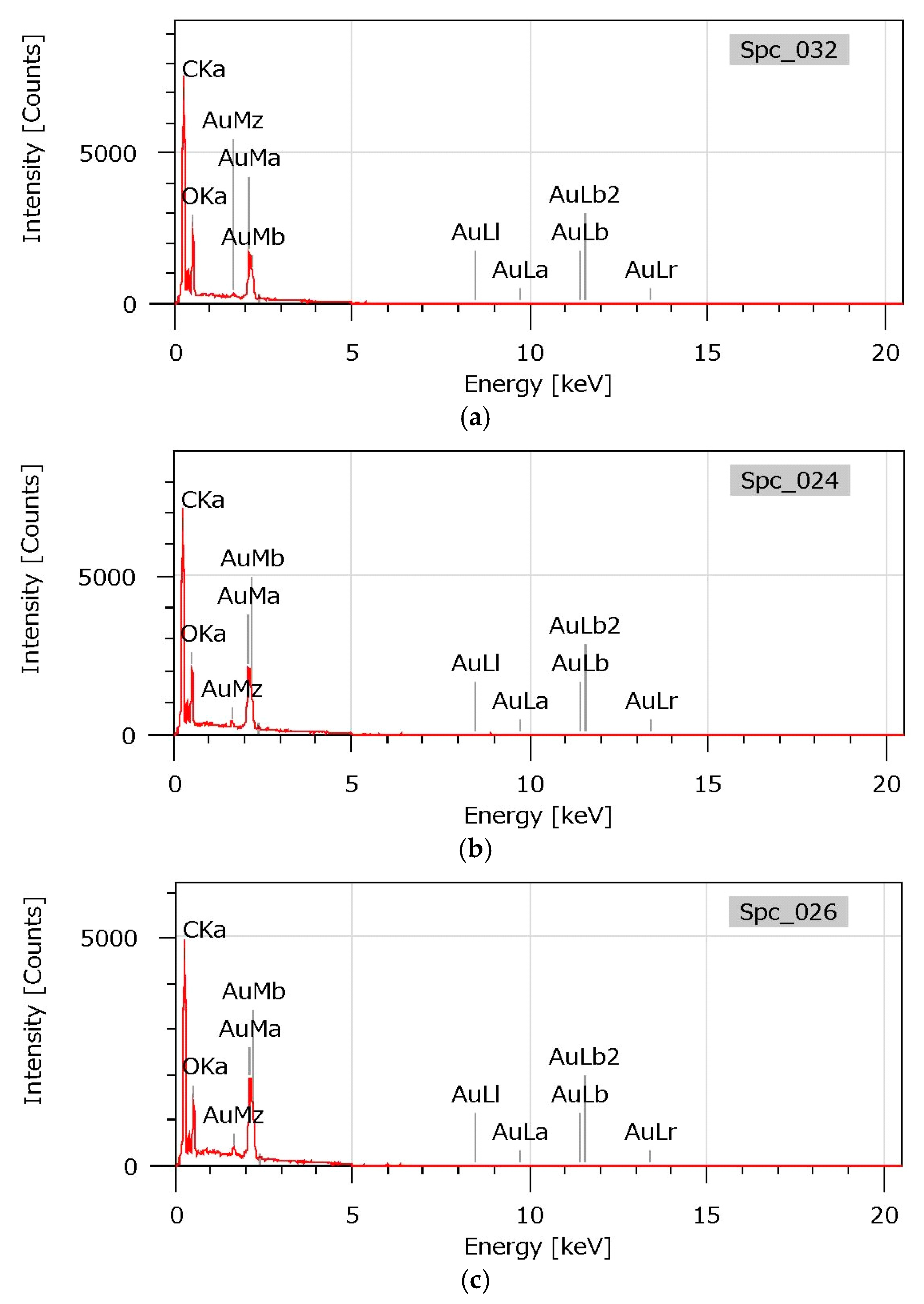
2.7. Observation of D. gallinae Killed by High-Voltage Impulse Discharges using EDS

To elucidate the control mechanism of D. gallinae using high-voltage impulses, mites under various conditions were observed under a microscope using a 12-megapixel video microscope to track changes in the body surface and appearance of the mite after the impulses were applied. Mites that had been exposed to warm air at 80 °C for 3 min and mites that had been starved for more than one week and had not moved were also observed for comparison.
Killed D. gallinae were coated with Au using an ion-sputtering system, and the EDS spectra were analyzed using a bench top SEM (JCM-7000, JEOL Ltd., Tokyo, Japan) for elemental analysis.

2.8. Statistical Analysis

Statistical analysis was conducted using Excel 2021 (Microsoft Corporation, Washington, DC, USA) to investigate the correlation between mortality and four electrical parameters: voltage, current, frequency, and pulse width. Multiple regression analysis was utilized, treating mortality as the response variable and the aforementioned electrical parameters as explanatory variables. p-values were calculated at a confidence level of 95%.

3. Results

3.1. Measurement of Discharge Voltage via Electrode Gap and Polarity

Figure 3a illustrates the discharge voltages when the insulating materials of the electrodes were glass epoxy and polyimide. As the distance between the electrodes increased, the discharge voltage correspondingly increased. At the same distance between electrodes, the negative voltage was slightly higher than the positive voltage. When the electrode spacing was 3.0 mm, the discharge voltages were 9.5 kV and −11 kV, respectively.
When the voltage amplitude exceeded approximately 10 kV, numerous instances of peeling of the copper on the substrate were observed during the discharge. When polyimide was employed as the insulating material for the electrodes, the discharge voltage tended to increase with the distance between the electrodes as in the case of glass epoxy, but the voltage values were lower. At all electrode spacings, the voltage of polyimide was lower than that of glass epoxy, with a difference of more than 2.0 kV at an electrode spacing of 3 mm. Figure 3b,c shows a magnified view of the electrode for each electrode material. Protrusions were observed on the electrode surface of the polyimide material, which were not present on the epoxy material.

3.2. Variation in Mortality of D. gallinae Using Electrical Parameters

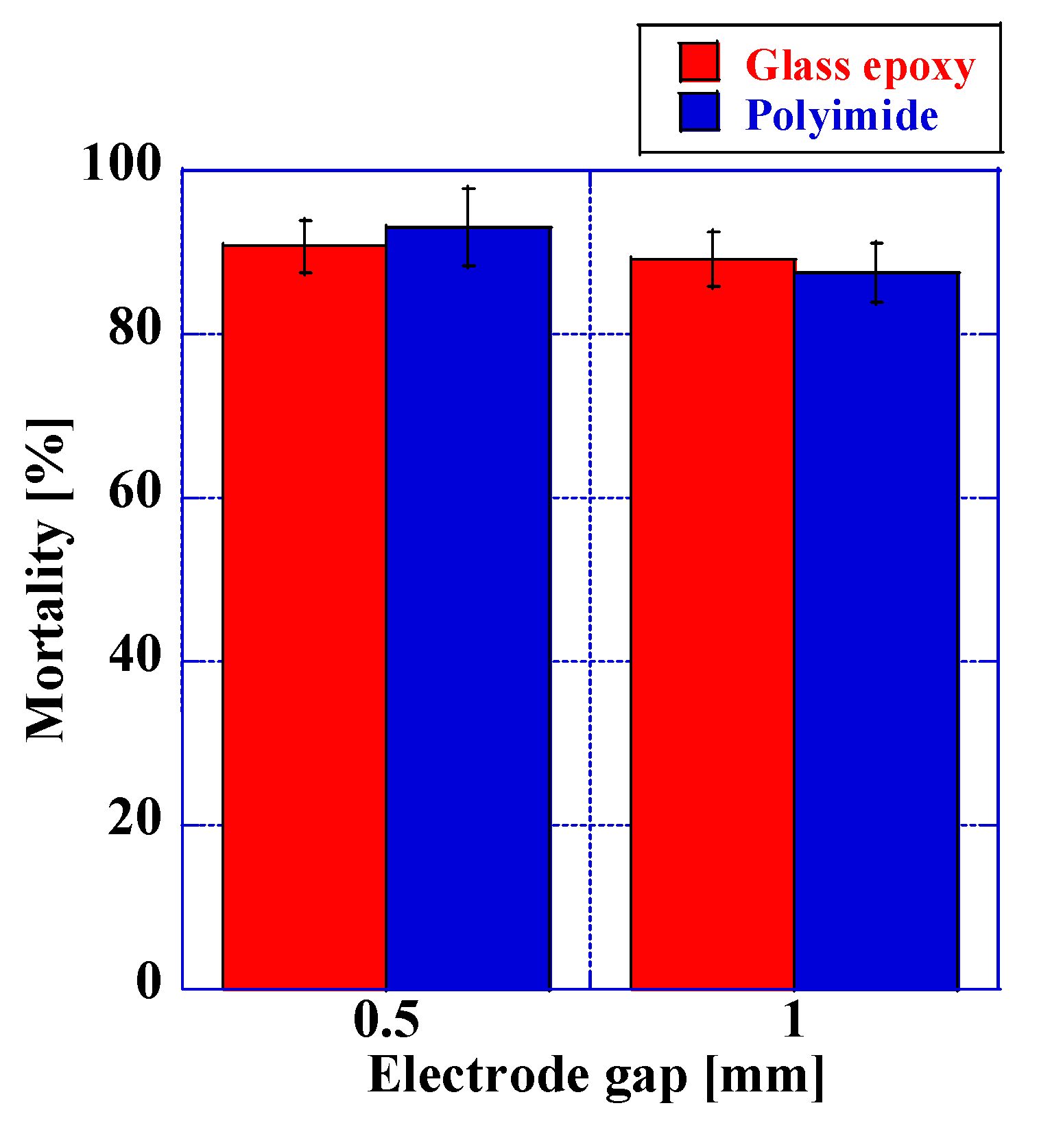
3.2.1. Variation in Mortality Depending on the Type of Insulator

Mortality when the electrode base materials were glass epoxy and polyimide is illustrated in Figure 4. At positive voltages, the mortality ranged from 92% to 94% at an electrode spacing of 0.5 mm and from 89% to 90% at an electrode spacing of 1.0 mm. There was no significant difference in the mortality for any of the electrodes, and although the negative voltage was used to check the mortality, there was no significant difference between this and the positive voltage.

3.2.2. Variation of Mortality as a Function of Discharge Voltage

Figure 5a shows the correlation between discharge voltage and mortality. Prior to 2.6 kV, no discharge was recorded between the electrodes; thus, the D. gallinae were not effectively eliminated. As the voltage was elevated, the mortality increased rapidly. At a voltage of 3.5 kV, the mortality reached 90.4%. However, as the voltage was further increased, mites could no longer be eliminated as a result of continuous discharge between the electrodes.

3.2.3. Mortality as a Function of Frequency

Under the same conditions as described in Section 3.2.2, Figure 5b shows the relationship between frequency and mortality. At frequencies below 10 Hz, no D. gallinae were eliminated on the electrodes. As the frequency exceeded 100 Hz, the mortality rapidly increased, reaching over 90%. However, at 100 kHz, the rate drastically decreased as discharge occurred at the same points on the electrodes.

3.2.4. Variation of Mortality with Current

The correlation between mortality and current variation using 0.5 mm electrodes is shown in Figure 5c. A rapid increase in the mortality percentage is observed above 10 mA, culminating in 54.4% mortality at 42 mA and 82.0% at 85 mA. As the current is further increased, the mortality tends towards saturation, reaching 94.2% at 427 mA and 92.5% at 1.28 A.

3.2.5. Variation of Mortality with Pulse Width

Figure 5d shows the relationship between mortality and pulse width. Pulse widths range from 15 ns to 10 μs. At a pulse width of 15 ns, the rate is as low as 8.8%. As the pulse width increases, the mortality also increases, reaching 93.3% at 5.6 μs. The mortality does not exceed 30% for pulses shorter than 1 μs but exceeds 85% for pulses longer than 1 μs. However, when the pulse width is extremely long (10 ms), discharges occur at the same points on the electrodes, resulting in a significant reduction in the mortality to less than 1%.

3.3. Variation in Mortality with Ultraviolet Light

The mortality caused by UV-C irradiation for different irradiation durations is shown in Figure 6a. The mortality rate increased with the duration of UV irradiation, with less than 10% of the mites being killed within the first 10 minutes. However, the mortality rate increased between 10 and 30 min, culminating in 88% of the mites being killed after 60 min. These results suggest that mite control methods using ultraviolet light require much longer exposure times compared to those using pulsed high-voltage discharges.

3.4. Variation in Mite Control Efficacy with Ozone

The mortality as a function of exposure time to ozone (10 ppm concentration) is shown in Figure 6b. In contrast to the results obtained with high-voltage impulse and ultraviolet mite control methods, only 2.7% of mites were killed after 3 to 10 min of exposure, and even after 60 min of ozone exposure, the mortality remained low at less than 5%. These results confirm that mites are relatively resistant to the ozone mite control method.

3.5. Comparison of D. gallinae Body Surfaces after Voltage Application

In order to elucidate the mechanism of the mite control effect of high-voltage impulse on D. gallinae, the condition of the exoskeleton was observed under the microscope. Figure 7 shows the exoskeletons of mites killed through high-voltage impulse discharges (b), heat (c), and starving (a). The mites killed through high-voltage impulse discharges and heat showed a change in body surface color and all legs from the first to the fourth were folded. No changes in the legs were observed in the control group, which is a significant difference from those killed through high-voltage impulse discharges and heat. EDS analysis of the body surface composition revealed that carbon (C) and oxygen (O) accounted for approximately 58% and 42% of the mass percentages, respectively, with no discernible differences between the three (Figure 8, Table 1).

4. Discussion

In the experiments measuring discharge voltage, the discharge voltage increased as the distance between the electrodes increased, regardless of the insulating material of the electrodes. The higher the electric field strength required to initiate a discharge, the more energy is required to accelerate the charged particles. This trend is the same as that seen in gaseous discharges. The relationship between discharge voltage and electrode spacing is governed by Paschen’s Law, which states that the breakdown voltage of a gas is a function of the product of gas pressure and the distance between the electrodes. In other words, if the gas pressure remains constant, the discharge voltage will be proportional to the electrode spacing. The higher the electric field strength required when initiating a discharge, the more energy is required to accelerate the charged particles. For the same distance between electrodes, the discharge voltage for the negative polarity was slightly higher than that for the positive polarity. Generally, the discharge initiation voltage of the negative polarity is higher than that of the positive polarity in the creepage discharge [32,33]. This phenomenon is attributed to the strength of the spatial electric field formed by the ionized electrons and positive ions and the polarity of the applied voltage [34,35,36]. Specifically, when the applied voltage is negative, the strength of the electric field around it is weakened by the charged particles, resulting in a higher discharge voltage compared to when a positive voltage is applied. Furthermore, a slight variation in discharge voltage was observed depending on the insulating material of the electrode. The discharge voltage of glass epoxy tended to be higher than that of polyimide. A magnified view of the electrode shows that the polyimide electrode had protrusions (Figure 3). It is hypothesized that the electric field concentration caused by these protrusions lowered the discharge voltage.
The insulation voltage of polyimide (280 kV/mm) is comparable to that of glass epoxy (290 kV/mm) [37,38,39,40]. However, the methods used to create the electrodes in this experiment differed from each other. A drill-based cutting method was utilized for the glass epoxy, whereas an etching method was employed for the polyimide. Etching is a technique employed for corroding and shaping copper substrates via a chemical reaction. Masking films are utilized in regions where copper corrosion is to be avoided, and in this experiment, minute irregularities were observed in this masking film. It is hypothesized that the unevenness of the masking film may have contributed to the protrusion of the polyimide electrode.
The mortality was approximately 90% for both substrates when utilizing glass epoxy and polyimide as the insulating material of the electrodes. The discharge voltage of the polyimide was slightly lower than that of the glass epoxy, but the mite control voltages could be considered roughly equivalent. A subsequent experiment to confirm the mite control efficacy using voltage revealed that mites could not be eradicated at voltages lower than 2.6 kV. At this voltage, no discharge occurred at all, and even when D. gallinae passed through the electrodes, the discharge phenomenon necessary for killing the mites was not observed. When the voltage was increased, the mortality increased rapidly, but when the voltage was further increased, the rate decreased drastically. This was due to the fact that the discharges occurred at the same points on the electrodes and did not spread to the entire electrode. It was found that if the applied voltage was too high, discharges occurred only at one point, and it was not possible to impart uniform discharges to the D. gallinae scattered on the electrodes.
The same trend in mortality was observed when the frequency was varied as the voltage. At low frequencies, the mortality was less than 15%, but at frequencies above 100 Hz, the rate increased to more than 90%. However, when the frequency was further increased, the discharge was formed in the same place as when the voltage was increased, and the mortality decreased drastically. D. gallinae has negative motility and prefers shadows and darkness, and the movement speed was high under the illumination in this experimental environment. Their speed of movement was measured, and the average value was 0.5 mm/s. It is postulated that at lower frequencies, the mortality decreased as the D. gallinae traversed the electrodes without any voltage being applied during the voltage off time (cycle minus pulse width). A similar trend was observed when the current and pulse width were manipulated: when these values were low, the mortality decreased, and when they were excessively high, the discharge formed at a single point on the electrode and effective mite control action could not be attained. Elevated electrical parameters such as voltage, current, frequency, and pulse width can diminish the insulating properties of solid surfaces. Unlike gases and liquids, the insulating properties of solids are difficult to restore once disrupted, and a breakdown can result in recurrent discharges at the site of the initial breakdown [32,33].
Table 2 displays the outcomes of the multiple regression analysis. The results indicate a robust correlation (p < 0.05) between voltage and current concerning the mortality. Although the pulse width also exhibited a correlation with mortality, the correlation was relatively weaker than that of voltage (p < 0.05). In contrast, frequency demonstrated no significant correlation (p > 0.05) with mortality.
In summary, D. gallinae can be killed electrically if the voltage at which a discharge can be formed on the electrodes is set as the threshold and the electrical parameters are such that discharges are not formed at the same point and continuously.
Next, a comparison of the body surfaces of D. gallinae killed through each method is discussed. D. gallinae killed uisng heat and high-voltage impulse were similar in that they displayed closed legs and exhibited changes in coloration on their body surfaces. No change in composition was detected through the EDS analysis. The exoskeleton of D. gallinae is composed of chitin, a type of glycosaminoglycan [41]. The chemical formula of chitin is (C8H13NO5)n [42], and its mass percentages are 47.3%, 6.45%, 6.90%, and 39.4% for C, H, N, and O, respectively. Hydrogen is not observable in the EDS analysis, and nitrogen is a trace element that is difficult to detect. Given these facts, the analysis result of approximately 57% C and 43% O by mass is generally consistent with the theoretical value. The fact that the visage of the exoskeleton is altered by the high-voltage impulse killing yet the composition remains unaltered suggests that the current flows through the body and alters the internal tissues via Joule heating, which may have resulted in the demise of mites.
UV-C requires a prolonged period of time (more than 60 min) to kill D. gallinae, making it challenging to the achieve the highly efficient killing of D. gallinae, which moves at a speed of only a few millimeters per second. The spot irradiation of D. gallinae is also difficult due to their preference for darkness (negative phototaxis). Similar to the results of the UV-irradiation experiments on house dust mites and spider mites, the mortality rate was also very low [43,44]. The DNA molecule displays a peak absorption spectrum ranging from 260 to 265 nm, in close proximity to the ultraviolet (UV) region used in this study [45]. UV radiation stimulates the production of reactive oxygen species (ROS), leading to indirect damage to DNA, lipids, and proteins [46]. Insects, such as Helicoverpa armigera, have been shown to experience an increase in oxidative stress following exposure to UV radiation [47]. Furthermore, insects have been found to elicit molecular responses to stress and damage caused by irradiation [48,49].
Even at a concentration of 10 ppm, which is 200 times higher than the upper limit of 0.05 ppm established by the National Institute for Occupational Safety and Health (NIOSH), the suppression of D. gallinae through ozone was notably low. There are few studies on the use of ozone for mite control, but in an experiment on mite control in food storage, it was reported that a CT (cumulative exposure time product) of 192,000 resulted in 99% mortality and a CT of 10,800 suppressed mite growth [50,51]. The CT in this trial was 600, which appears to be too low to control D. gallinae.
The acaricides that are the first choice for killing D. gallinae are organophosphate and carbamate, which inhibit acetylcholine and cholinesterase in the nervous system, and pyrethroids, which inhibit the nerve membrane (sodium channel), thereby killing D. gallinae [52]. However, their acaricidal efficacy has declined over time due to the length of time required to completely eradicate mites in poultry houses and the emergence of drug resistance in D. gallinae [53,54,55,56].
On the other hand, the proposed electroshock pesticidal method can achieve a mortality of up to 95% with the application of a high-voltage impulse for less than 30 s. The electrical parameters required for the killing are in the order of milliamps of current and microseconds of pulses; thus, the risk of electrocution to the human body is extremely low, and the pesticidal effectiveness is extremely high [57].
In recent years, a variety of alternative techniques have been proposed for controlling D. gallinae. These include biological control agents, physical methods, and natural compounds. Despite their potential benefits, these technologies have not yet been widely adopted in practical use due to various drawbacks. Biological control agents, such as predatory mites [58,59,60] and nematodes [61], are often expensive and have limited effectiveness in large-scale applications. Physical methods [62,63,64], such as high-temperature treatment and vacuum cleaning, are time-consuming and labor-intensive. Natural compounds, such as essential oils [65] and plant extracts [66], may have variable efficacy and require further research to optimize their use.
In addition, various alternative technologies, such as insect growth regulators, necessitate frequent spraying inside the poultry house. On the other hand, the proposed technology can be controlled externally through electrical signals, allowing for continuous D.gallinae eradication. Following installation, the system can be employed to eliminate D.gallinae in an automated manner.
D. gallinae is capable of entering poultry facilities and infecting the poultry via the poultry gauge. During actual installation, it is anticipated that electrodes will be placed on existing poultry gauges to control D.gallinae. The proposed polyimide substrate can be contorted and molded without damaging the electrodes, allowing it to be installed based on the shape of the gauge. However, once the D. gallinae parasite has infested the poultry, it is difficult to control it using this technique. Therefore, it is advisable to install the electrodes in a poultry house that has been completely cleared of D. gallinae during a periodic “all-out” cleaning process.

5. Conclusions

In this study, we proposed the utilization of high-voltage impulse discharges as an alternative D. gallinae control method of the IPMs that will provide us with the chance to be independent from the heavy use of chemicals. We conducted investigations into the discharge characteristics of electrodes and the mite control efficacy in relation to variations in voltage and frequency. Through experimentation, it was determined that D. gallinae can be effectively controlled through the application of high-voltage impulse discharges and that there exist optimal values for various electrical parameters crucial for mite control action. The morphological changes on the body surface of D. gallinae resulting from high-voltage impulse discharge were found to be analogous to those resulting from thermal mite control methods, yet there was no alteration in the elemental composition of the body surface, suggesting that the changes were brought about through Joule heating within the exoskeleton.
In comparison to methods involving ultraviolet light or ozone, the high-voltage impulse mite control technique has been demonstrated to achieve a mortality rate of up to 95% or greater for D. gallinae within as little as 30 s, and it exhibits exceptional mite control intensity and temporal efficiency. The results indicate that this electric shock mite control technique has potential as a novel mite control method as D. gallinae does not develop resistance to it and can be swiftly eliminated. In the future, we aim to further understand the mechanism of mite control action by examining the internal tissues of D. gallinae through staining techniques and to improve the dustproofing of the high-voltage impulse power supply and the electrode structure to facilitate highly efficient mite control action. Additionally, we intend to investigate other practical applications of this technology.

Author Contributions

Conceptualization, T.U. and T.H.; data curation, T.U., Y.M. and J.N.; formal analysis, T.F.; funding acquisition, T.U.; investigation, Y.M.; methodology, T.U.; resources, Y.M., J.N. and T.I.; supervision, T.H.; validation, T.H.; writing—original draft, T.U., T.F. and T.H.; writing—review and editing, T.F. and T.H. All authors have read and agreed to the published version of the manuscript.

Funding

This research was funded by KAKENHI (21K04013) from The Japan Society for the Promotion of Science, and from the Kieikai Research Foundation on FY2020.

Data Availability Statement

Not applicable.

Acknowledgments

The authors thank Koichiro Ogata of NIT, Oita College for his valuable suggestions and discussions.

Conflicts of Interest

The authors declare no conflict of interest.

References

  1. Galvani, L. Aloysii Galvani De Viribus Electricitatis in Motu Musculari Commentarius; Ex Typographia Instituti Scientiarium: Bononiae, Italy, 1791. [Google Scholar] [CrossRef] [Green Version]
  2. Houston, E.J. Electricity in Every-Day Life; P. F. Collier & Son: New York, NY, USA, 1905. [Google Scholar]
  3. Finkelstein, G. Emil du Bois-Reymond vs Ludimar Hermann. Comptes Rendus Biol. 2006, 329, 340–347. [Google Scholar] [CrossRef] [PubMed] [Green Version]
  4. Clarke, E.; Jacyna, L.S. Nineteenth-Century Origins of Neuroscientific Concepts; University of California Press: Berkeley, CA, USA, 1987. [Google Scholar]
  5. Wells, D.A. The Science of Common Things: A Familiar Explanation of the First Principles of Physical Science. For Schools, Families, and Young Students. Illustrated with Numerous Engravings; Ivison, Phinney & Company: New York, NY, USA, 1860. [Google Scholar]
  6. Theruvath, T.P.; Ikonomidis, J.S. Historical perspectives of The American Association for Thoracic Surgery: Claude S. Beck (1894–1971). J. Thorac. Cardiovasc. Surg. 2015, 149, 655–660. [Google Scholar] [CrossRef] [PubMed] [Green Version]
  7. Beloucif, S. Informed consent for special procedures: Electroconvulsive therapy and psychosurgery. Curr. Opin. Anaesthesiol. 2013, 26, 182–185. [Google Scholar] [CrossRef] [PubMed]
  8. Montgomery, J.H.; Vasu, D. The use of electroconvulsive therapy in atypical psychotic presentations: A case review. Psychiatry 2007, 4, 30–39. [Google Scholar]
  9. Sale, A.J.H.; Hamilton, W.A. Effects of high electric fields on micro-organisms: III. Lysis of erythrocytes and protoplasts. Biochim. Et Biophys. Acta (BBA) Biomembr. 1968, 163, 37–43. [Google Scholar] [CrossRef]
  10. Neumann, E.; Rosenheck, K. Permeability changes induced by electric impulses in vesicular membranes. J. Membr. Biol. 1972, 10, 279–290. [Google Scholar] [CrossRef] [Green Version]
  11. Nuccitelli, R.; Pliquett, U.; Chen, X.; Ford, W.; James Swanson, R.; Beebe, S.J.; Kolb, J.F.; Schoenbach, K.H. Nanosecond pulsed electric fields cause melanomas to self-destruct. Biochem. Biophys. Res. Commun. 2006, 343, 351–360. [Google Scholar] [CrossRef] [Green Version]
  12. Dev, S.B.; Rabussay, D.P.; Widera, G.; Hofmann, G.A. Medical applications of electroporation. IEEE Trans. Plasma Sci. 2000, 28, 206–223. [Google Scholar] [CrossRef]
  13. Beebe, S.J.; Blackmore, P.F.; White, J.; Joshi, R.P.; Schoenbach, K.H. Nanosecond pulsed electric fields modulate cell function through intracellular signal transduction mechanisms. Physiol. Meas. 2004, 25, 1077–1093. [Google Scholar] [CrossRef]
  14. Tsukamoto, S.; Maeda, T.; Ikeda, M.; Akiyama, H. Application of pulsed power to mushroom culturing. In Proceedings of the Digest of Technical Papers, PPC-2003, 14th IEEE International Pulsed Power Conference (IEEE Cat. No.03CH37472), Dallas, TX, USA, 15–18 June 2003; Volume 1112, pp. 1116–1119. [Google Scholar]
  15. Shimizu, H.; Hiraguri, T.; Kimoto, M.; Ota, K.; Shindo, T.; Hoshino, Y.; Takaki, K. Stimulatory growth effect of lightning strikes applied in the vicinity of shiitake mushroom bed logs. J. Phys. D Appl. Phys. 2020, 53, 204002. [Google Scholar] [CrossRef]
  16. Eing, C.J.; Bonnet, S.; Pacher, M.; Puchta, H.; Frey, W. Effects of nanosecond pulsed electric field exposure on Arabidopsis thaliana. IEEE Trans. Dielectr. Electr. Insul. 2009, 16, 1322–1328. [Google Scholar] [CrossRef] [Green Version]
  17. Wang, D.; Lin, X.; Hirayama, K.; Li, Z.; Ohno, T.; Zhang, W.; Namihira, T.; Katsuki, S.; Takano, H.; Takio, S.; et al. A New Application of Underwater Pulsed Streamerlike Discharge to Transcriptional Activation of Retrotransposon of Porphyra yezoensis. IEEE Trans. Plasma Sci. 2010, 38, 39–46. [Google Scholar] [CrossRef]
  18. Wood, H.P. The Chicken Mite: Its Life History and Habits; U.S. Dept. of Agriculture: Washington, DC, USA, 1917; Volume 553. [Google Scholar]
  19. Collins, D. Mites in the poultry house. J. Agric. North. Irel. 1976, 51, 24–26. [Google Scholar]
  20. Kilpinen, O.; Roepstorff, A.; Permin, A.; Nørgaard-Nielsen, G.; Lawson, L.G.; Simonsen, H.B. Influence of Dermanyssus gallinae and Ascaridia galli infections on behaviour and health of laying hens (Gallus gallus domesticus). Br. Poult. Sci. 2005, 46, 26–34. [Google Scholar] [CrossRef]
  21. Sparagano, O.; Pavlićević, A.; Murano, T.; Camarda, A.; Sahibi, H.; Kilpinen, O.; Mul, M.; van Emous, R.; le Bouquin, S.; Hoel, K.; et al. Prevalence and key figures for the poultry red mite Dermanyssus gallinae infections in poultry farm systems. Exp. Appl. Acarol. 2009, 48, 3–10. [Google Scholar] [CrossRef] [PubMed]
  22. Cencek, T. Prevalence of dermanyssus gallinae in poultry farms in silesia region in poland. J. Bull. Vet. Inst. Pulawy 2003, 47, 465–470. [Google Scholar]
  23. Guy, J.H.; Khajavi, M.; Hlalel, M.M.; Sparagano, O. Red mite (Dermanyssus gallinae) prevalence in laying units in Northern England. Br. Poult. Sci. 2004, 45 (Suppl. S1), S15–S16. [Google Scholar] [CrossRef]
  24. Thind, B.B.; Ford, H.L. Assessment of susceptibility of the poultry red mite Dermanyssus gallinae (Acari: Dermanyssidae) to some acaricides using an adapted filter paper based bioassay. Vet. Parasitol. 2007, 144, 344–348. [Google Scholar] [CrossRef]
  25. Zeman, P.; Železný, J. The susceptibility of the poultry red mite, Dermanyssus gallinae (De Geer, 1778), to some acaricides under laboratory conditions. Exp. Appl. Acarol. 1985, 1, 17–22. [Google Scholar] [CrossRef]
  26. Frings, H. Factors determining the effects of radio-frequency electromagnetic fields on insects and materials they infest. J. Econ. Entomol. 1952, 45, 396–408. [Google Scholar] [CrossRef]
  27. Wang, S.; Tang, J.; Johnson, J.A.; Mitcham, E.; Hansen, J.D.; Hallman, G.; Drake, S.R.; Wang, Y. Dielectric Properties of Fruits and Insect Pests as related to Radio Frequency and Microwave Treatments. Biosyst. Eng. 2003, 85, 201–212. [Google Scholar] [CrossRef]
  28. Wang, S.; Tiwari, G.; Jiao, S.; Johnson, J.A.; Tang, J. Developing postharvest disinfestation treatments for legumes using radio frequency energy. Biosyst. Eng. 2010, 105, 341–349. [Google Scholar] [CrossRef]
  29. Pickens, L.G.; Mills, G.D., Jr. Solar-powered electrocuting trap for controlling house flies and stable flies (Diptera: Muscidae). J. Med. Entomol. 1993, 30, 872–877. [Google Scholar] [CrossRef]
  30. Urban, J.E.; Broce, A. Killing of flies in electrocuting insect traps releases bacteria and viruses. Curr. Microbiol. 2000, 41, 267–270. [Google Scholar] [CrossRef] [PubMed]
  31. Pickens, L.G. Battery-powered, electrocuting trap for stable flies (Diptera: Muscidae). J. Med. Entomol. 1991, 28, 822–830. [Google Scholar] [CrossRef]
  32. O’Dwyer, J.J. Theory of Dielectric Breakdown in Solids. J. Electrochem. Soc. 1969, 116, 239. [Google Scholar] [CrossRef]
  33. Verweij, J.F.; Klootwijk, J.H. Dielectric breakdown I: A review of oxide breakdown. Microelectron. J. 1996, 27, 611–622. [Google Scholar] [CrossRef] [Green Version]
  34. Beroual, A.; Kebbabi, L. Analysis of cumulative number and polarity of creeping discharges initiated at solid/liquid interfaces subjected to AC voltage. In Proceedings of the 2009 IEEE Conference on Electrical Insulation and Dielectric Phenomena, Virginia Beach, VA, USA, 18–21 October 2009; pp. 380–383. [Google Scholar]
  35. Li, C.; Cao, B.; Li, X.; Liu, X.; Cheng, J.; Liu, Z.; Zhao, Z.; Yang, Z. Surface discharge characteristics of silicone gel and DBC under positive repetitive square voltage. Power Electron. Devices Compon. 2022, 3, 100021. [Google Scholar] [CrossRef]
  36. Donkó, Z.; Zajičková, L.; Sugimoto, S.; Harumningtyas, A.A.; Hamaguchi, S. Modeling characterisation of a bipolar pulsed discharge. Plasma Sources Sci. Technol. 2020, 29, 104001. [Google Scholar] [CrossRef]
  37. Fukuma, M.; Nagao, M.; Kosaki, M. Numerical Analysis of Electrical Breakdown Characteristics in Polypropylene Film based on Thermal and Electronic Composite Breakdown Model. IEEJ Trans. Fundam. Mater. 1994, 114, 230–235. [Google Scholar] [CrossRef] [Green Version]
  38. Nagao, M.; Yamaguchi, F.; Tokumaru, K.; Sugide, I.; Kozaki, M.; Ieda, M. DC dielectric breakdown of polypropylene films in the high temperature range. IEEJ Trans. Electr. Electron. Eng. 1985, 105, 177–182. [Google Scholar] [CrossRef] [Green Version]
  39. Yamadera, T. Environment-Friendry Printed Wiring Boards (PWB) and the Technologies for their Inflammabilities. J. Jpn. Inst. Electron. Packag. 2014, 17, 96–99. [Google Scholar] [CrossRef] [Green Version]
  40. Matsuo, M. Glass Epoxy (FR-4). Circuit Technol. 1993, 8, 402–411. [Google Scholar] [CrossRef]
  41. Crowe, J.H. Studies on acarine cuticles. III. Cuticular ridges in the citrus red mite. Trans. Am. Microsc. Soc. 1975, 94, 98–108. [Google Scholar] [CrossRef]
  42. Alberti, G.; Storch, V.; Renner, H. Über den feinstrukturellen Aufbau der Milbencuticula (Acari, Arachnida). Zoologische Jahrbücher fair Anatomie; G. Fischer: Jena, Germany, 1981; Volume 105, pp. 183–236. [Google Scholar]
  43. Lah, E.F.C.; Musa, R.N.A.R.; Ming, H.T. Effect of germicidal UV-C light (254 nm) on eggs and adult of house dustmites, Dermatophagoides pteronyssinus and Dermatophagoides farinae (Astigmata: Pyroglyhidae). Asian Pac. J. Trop. Biomed. 2012, 2, 679–683. [Google Scholar] [CrossRef] [PubMed] [Green Version]
  44. Suzuki, T.; Watanabe, M.; Takeda, M. UV tolerance in the two-spotted spider mite, Tetranychus urticae. J. Insect Physiol. 2009, 55, 649–654. [Google Scholar] [CrossRef]
  45. Beggs, C.B. A quantitative method for evaluating the photoreactivation of ultraviolet damaged microorganisms. Photochem. Photobiol. Sci. 2002, 1, 431–437. [Google Scholar] [CrossRef]
  46. Chamberlain, J.; Moss, S.H. Lipid peroxidation and other membrane damage produced in Escherichia coli K1060 by near-UV radiation and deuterium oxide. Photochem. Photobiol. 1987, 45, 625–630. [Google Scholar] [CrossRef]
  47. Meng, J.-Y.; Zhang, C.-Y.; Zhu, F.; Wang, X.-P.; Lei, C.-L. Ultraviolet light-induced oxidative stress: Effects on antioxidant response of Helicoverpa armigera adults. J. Insect Physiol. 2009, 55, 588–592. [Google Scholar] [CrossRef]
  48. Sang, W.; Ma, W.-H.; Qiu, L.; Zhu, Z.-H.; Lei, C.-L. The involvement of heat shock protein and cytochrome P450 genes in response to UV-A exposure in the beetle Tribolium castaneum. J. Insect Physiol. 2012, 58, 830–836. [Google Scholar] [CrossRef]
  49. Zhou, L.-J.; Zhu, Z.-H.; Liu, Z.-X.; Ma, W.-H.; Desneux, N.; Lei, C.-L. Identification and transcriptional profiling of differentially expressed genes associated with response to UVA radiation in Drosophila melanogaster (Diptera: Drosophilidae). Environ. Entomol. 2013, 42, 1110–1117. [Google Scholar] [CrossRef]
  50. Grasso, C.; Eramo, V.; Lembo, M.; Forniti, R.; Carboni, C.; Botondi, R. Effects of gaseous ozone treatment on the mite pest control and qualitative properties during ripening storage of pecorino cheese. J. Sci. Food Agric. 2023, 103, 2124–2133. [Google Scholar] [CrossRef]
  51. Mahmoud, R.; Abdel-Khalik, A.R. Comparison between Two Physical Methods to Control the Stored Dates Fruit Mites, Tyrophagus putrescentiae (Schrank) and Rhizoglyphus robini Claparede (Astigmata: Acaridae). Egypt. Acad. J. Biol. Sci. B. Zool. 2022, 14, 149–158. [Google Scholar] [CrossRef]
  52. Pritchard, J.; Kuster, T.; Sparagano, O.; Tomley, F. Understanding the biology and control of the poultry red mite Dermanyssus gallinae: A review. Avian Pathol. 2015, 44, 143–153. [Google Scholar] [CrossRef] [PubMed] [Green Version]
  53. Marangi, M.; Cafiero, M.A.; Capelli, G.; Camarda, A.; Sparagano, O.A.; Giangaspero, A. Evaluation of the poultry red mite, Dermanyssus gallinae (Acari: Dermanyssidae) susceptibility to some acaricides in field populations from Italy. Exp. Appl. Acarol. 2009, 48, 11–18. [Google Scholar] [CrossRef]
  54. Beugnet, F.; Chauve, C.; Gauthey, M.; Beert, L. Resistance of the red poultry mite to pyrethroids in France. Vet. Rec. 1997, 140, 577–579. [Google Scholar] [CrossRef] [PubMed]
  55. Nordenfors, H.; Höglund, J.; Tauson, R.; Chirico, J. Effect of permethrin impregnated plastic strips on Dermanyssus gallinae in loose-housing systems for laying hens. Vet. Parasitol. 2001, 102, 121–131. [Google Scholar] [CrossRef] [PubMed]
  56. Murano, T. Red mite, Dermanyssus gallinae; Current problem and trials for control in Japan. J. Jpn. Soc. Poult. Dis. 2007, 43, 23–30. [Google Scholar]
  57. IEC 60479-1:2018; Effects of Current on Human Beings and Livestock—Part 1: General Aspects. IEC: London, UK, 2018.
  58. Coleman, D.C.; Callaham, M.A.; Crossley, D.A. Fundamentals of Soil Ecology, 3rd ed.; Coleman, D.C., Callaham, M.A., Crossley, D., Eds.; Elsevier: Amsterdam, The Netherlands, 2018. [Google Scholar]
  59. Mohammadi, K.; Fathi, S.A.A.; Razmjou, J.; Naseri, B. Evaluation of the effect of strip intercropping green bean/garlic on the control of Tetranychus urticae in the field. Exp. Appl. Acarol. 2021, 83, 183–195. [Google Scholar] [CrossRef]
  60. Castilho, R.C.; Venancio, R.; Narita, J.P.Z. Mesostigmata as biological control agents, with emphasis on Rhodacaroidea and Parasitoidea. Prospect. Biol. Control Plant Feed. Mites Other Harmful Org. 2015, 19, 1–31. [Google Scholar]
  61. Rueda-Ramírez, D.; Palevsky, E.; Ruess, L. Soil Nematodes as a Means of Conservation of Soil Predatory Mites for Biocontrol. Agronomy 2023, 13, 32. [Google Scholar] [CrossRef]
  62. MAFF. Statistics on the Agricultural Labor Force: Population of Agricultural Workers and Number of Core Agricultural Workers. 2020. Available online: https://www.maff.go.jp/j/tokei/sihyo/data/08.html (accessed on 14 February 2023).
  63. MAFF. Food Price Trends Survey (Meat & Eggs). Available online: https://www.maff.go.jp/j/zyukyu/anpo/kouri/k_gyuniku/index.html (accessed on 14 February 2023).
  64. Mul, M.F.; Koenraadt, C.J. Preventing introduction and spread of Dermanyssus gallinae in poultry facilities using the HACCP method. Exp. Appl. Acarol. 2009, 48, 167–181. [Google Scholar] [CrossRef] [PubMed]
  65. George, D.R.; Olatunji, G.; Guy, J.H.; Sparagano, O.A.E. Effect of plant essential oils as acaricides against the poultry red mite, Dermanyssus gallinae, with special focus on exposure time. Vet. Parasitol. 2010, 169, 222–225. [Google Scholar] [CrossRef] [PubMed]
  66. Abdel-Ghaffar, F.; Sobhy, H.M.; Al-Quraishy, S.; Semmler, M. Field study on the efficacy of an extract of neem seed (Mite-Stop) against the red mite Dermanyssus gallinae naturally infecting poultry in Egypt. Parasitol. Res. 2008, 103, 481–485. [Google Scholar] [CrossRef] [PubMed]
Figure 1. Electrode structure and photograph. The anode and cathode are arranged alternately in a comb-shaped electrode. The D. gallinae pass between these electrodes and are killed. (a) Photo of electrodes (glass epoxy) (b) Enlarged view of substrate (0.5 mm between electrodes).
Electronics 12 01038 g001
Figure 2. Voltage waveform. Voltage wave height was 3.5 kV; pulse width (FWHM) was 10 µs.
Electronics 12 01038 g002
Figure 3. Discharge voltage variation and enlarged view of the electrodes for each material. (a) The discharge voltage of electrodes using glass epoxy and polyimide as insulators was observed. It was found that the discharge voltage increased in proportion to the distance between the electrodes for both insulating materials. It was also found that the negative discharge voltage was greater than the positive discharge voltage, with the voltage being higher when glass epoxy was used as the insulating material. The error bars in the data indicate the standard deviations. The upper figure shows a (b) glass epoxy material (c) polyimide material. The glass epoxy had smooth electrodes, while protrusions were observed on the electrode surfaces of the polyimide substrates.
Electronics 12 01038 g003
Figure 4. Variation in mortality as a function of substrate insulator. No significant differences in mite control efficacy were observed as a result of variations in the insulating material of the electrodes or the polarity of the discharge voltage. The electrodes were made of glass epoxy or polyimide with a distance between electrodes of 0.5 mm and 1.0 mm and a frequency of 100 Hz. The error bars in the data show the standard deviation.
Electronics 12 01038 g004
Figure 5. Variation in mortality with electrical parameters. (a) Variation in mortality with discharge voltage. Killing of D. gallinae was found to be highly unlikely. As the voltage was increased, the mortality increased; however, this rate decreased rapidly at higher voltage levels. At an electrode spacing of 0.5 mm, the voltage was 3.5 kV and the maximum mortality was 90.4%. The electrodes were made of glass epoxy, the electrode spacing was 0.5 mm, and the frequency was 100 Hz. The error bars in the data indicate the standard deviation. (b) Variation in mortality with frequency. The mortality was found to be significantly low at frequencies below 10 Hz, while frequencies above 100 Hz resulted in a high mortality of over 90%. However, at frequencies above 100 kHz, discharges were observed to occur at consistent locations on the electrodes, resulting in a significant reduction in mortality. The electrodes were made of glass epoxy and operated at a voltage of 3.5 kV with a spacing of 0.5 mm. Error bars in the data indicate the standard deviation. (c) Variation in mortality with current. The mortality showed a marked increase when the current exceeded 10 mA, culminating in a maximum mortality of 94.2% at a current of 427 mA. Error bars in the data indicate the standard deviation. The electrodes were made of glass epoxy and operated at a voltage of 3.5 kV, with a distance of 0.5 mm between electrodes, and a frequency of 100 Hz. (d) Variation in mortality with pulse width. When the pulse width is in the order of nanoseconds, the mortality is low, with values of less than 40%. However, when the pulse width is in the order of microseconds, the mortality exceeds 80%. In addition, increasing the pulse width to the order of milliseconds did not result in a high mortality. The electrodes were made of glass epoxy and operated at a voltage of 3.5 kV, with a distance of 0.5 mm between electrodes, and at a frequency of 100 Hz. Error bars in the data indicate the standard deviation.
Electronics 12 01038 g005
Figure 6. Variation in mortality due to ozone and UVC irradiation. (a) Change in mortality as a function of UV exposure time. The mortality showed low efficacy during the first 10 min of irradiation but then increased, culminating in a mortality of 88% after 60 min of exposure to UV radiation at a wavelength of 253.7 nm. Error bars indicate standard deviation. (b) Change in mortality as a function of ozone exposure time. The mortality did not increase with longer exposure to ozone. The concentration was 10 ppm. Error bars indicate standard deviation.
Electronics 12 01038 g006
Figure 7. Comparison of D. gallinae body surfaces. (a) Control. (b) High-voltage impulse. (c) Heat. The color of the body surface is altered compared to the mites killed through starvation. Heat and high-voltage mite-control methods caused very similar changes in the coloration of the body surface.
Electronics 12 01038 g007
Figure 8. EDS spectrum of the body surface of D. gallinae killed using pulsed high voltage. (a) Control. (b) High-voltage impulse. (c) Heat. An EDS spectrum of the surface of the mite killed through high-voltage impulse revealed the presence of 58% carbon and 42% oxygen, with no discernible changes before and after the application of voltage. The images were taken at an incident voltage of 5.0 kV, a working distance of 16.2 mm, a magnification of ×1500 and in high vacuum mode. The Au components are shown in grey.
Electronics 12 01038 g008
Table 1. Body surface composition by EDS analysis. (a) Control. (b) High-voltage impulse. (c) Heat. All values are expressed as a percentage by mass.
(a) Control(b) High-Voltage Impulse(c) Heat
Carbon (%)57.8 ± 0.1557.9 ± 0.1358.4 ± 0.13
Oxygen (%)42.2 ± 0.1542.1 ± 0.1241.6 ± 0.13
Table 2. Multiple regression analysis between mortality and electrical parameters.
CoefficientsT Statp-Value
Intercept−2.27 × 10⁻¹−1.82 × 1006.98 × 10−2
Voltage2.04 × 10−15.23 × 1004.44 × 10−7
Frequency−1.42 × 10⁻⁶−1.01 × 1003.14 × 10−1
Current4.48 × 10−43.87 × 1001.49 × 10−4
Pulse width−4.43 × 10⁻⁸−2.85 × 1004.89 × 10−3
Disclaimer/Publisher’s Note: The statements, opinions and data contained in all publications are solely those of the individual author(s) and contributor(s) and not of MDPI and/or the editor(s). MDPI and/or the editor(s) disclaim responsibility for any injury to people or property resulting from any ideas, methods, instructions or products referred to in the content.

