Article

Age-Associated Changes on Gait Smoothness in the Third and the Fourth Age

1
Department of Mechanical, Chemical and Materials Engineering, Piazza d’Armi, 09123 Cagliari, Italy
2
Department of Clinical Sciences and Community Health (DISCCO), University of Milan, 20122 Milano, Italy
3
Exercise Medicine Unit, IRCCS Istituto Auxologico Italiano, Via Pier Lombardo 22, 20135 Milano, Italy
4
Center for Cognitive Disorders and Dementia, ASL Cagliari District, 09121 Cagliari, Italy
5
Geriatric Unit, SS. Trinità Hospital, 09121 Cagliari, Italy
6
Department of Electronics, Information and Bioengineering, Politecnico di Milano, Piazza Leonardo da Vinci 32, 20133 Milano, Italy
7
Istituto Auxologico Italiano, IRCCS, S. Giuseppe Hospital, Piancavallo, 28824 Oggebbio, Italy
*
Author to whom correspondence should be addressed.
Electronics 2023, 12(3), 637; https://doi.org/10.3390/electronics12030637
Submission received: 22 December 2022 / Revised: 18 January 2023 / Accepted: 22 January 2023 / Published: 27 January 2023

Abstract

:
Although gait disorders represent a highly prevalent condition in older adults, the alterations associated with physiologic aging are often not easily differentiable from those originated by concurrent neurologic or orthopedic conditions. Thus, the detailed quantitative assessment of gait patterns represents a crucial issue. In this context, the study of trunk accelerations may represent an effective proxy of locomotion skills in terms of symmetry. This can be carried out by calculating the Harmonic Ratio (HR), a parameter obtained through the processing of trunk accelerations in the frequency domain. In this study, trunk accelerations during level walking of 449 healthy older adults (of age > 65) who were stratified into three groups (Group 1: 65–74 years, n = 175; Group 2: 75–85 years, n = 227; Group 3: >85 years, n = 47) were acquired by means of a miniaturized Inertial Measurement Unit located in the low back and processed to obtain spatio-temporal parameters of gait and HR, in antero-posterior (AP), medio-lateral (ML) and vertical (V) directions. The results show that Group 3 exhibited a 16% reduction in gait speed and a 10% reduction in stride length when compared with Group 1 (p < 0.001 in both cases). Regarding the cadence, Group 3 was characterized by a 5% reduction with respect to Groups 1 and 2 (p < 0.001 in both cases). The analysis of HR revealed a general trend of linear decrease with age in the three groups. In particular, Group 3 was characterized by HR values significantly lower (−17%) than those of Group 1 in all three directions and significantly lower than Group 2 in ML and V directions (−10%). Taken together, such results suggest that HR may represent a valid measure to quantitatively characterize the progressive deterioration of locomotor abilities associated with aging, which seems to occur until the late stages of life.

1. Introduction

Gait is a fundamental physical activity of daily life and represents an important factor for independent living. However, gait efficiency undergoes significant changes with age [1,2,3]. In fact, the physiologic decline of the musculoskeletal system and cognitive performance associated with aging [4] leads to reduced movement smoothness and cognitive reserve, thus impairing several aspects of mobility associated with daily life tasks. This primarily affects walking, which results in altered automaticity and skill [5,6] but also affects other movements, such as turning and sitting to standing and vice versa [7]. Moreover, it should be recalled that, in older adults, most falls occur while walking, which emphasizes the importance of having a stable gait as a preventive countermeasure against such hazardous events [8].
In this context, the detailed analysis of the gait characteristics appears crucial to define the current status of the individual and, where necessary, plan specific interventions able to ensure that a sufficient degree of mobility is preserved during the late stages of life. Gait is usually investigated in both spatial and temporal domains [9], and its main features can be classified into relatively independent domains, with pace, rhythm, variability, symmetry, and postural control being probably the most important ones [10]. The parameters belonging to each of these domains can be assessed using several kinds of systems, such as motion-capture systems, electronic walkways, and, more recently, wearable Inertial Measurement Units (IMUs). IMUs employed for human movement analysis are basically stand-alone microelectromechanical systems that integrate multiaxial inertial sensors. A typical configuration, which includes a 3-axis accelerometer, a 3-axis gyroscope, and a 3-axis magnetometer, allow measuring acceleration, angular speed, and magnetic vector field of a moving object in a three-dimensional space, providing up to six degrees of freedom [11,12]. Since modern IMUs are designed to be small, lightweight, economic, and unobtrusive, their use quickly gained popularity among researchers involved in human movement analysis. To date, they are considered a reliable and affordable solution to assess gait in a variety of environments, as they do not require dedicated spaces or complex laboratory settings [13,14]. In particular, contrary to the equipment present in the traditional movement analysis laboratories, they allow individuals to be tested while wearing their usual clothes and shoes, thus ensuring good ecological validity [15]. IMUs can provide a new dimension of granularity for gait analysis and are increasingly used in research studies [16,17]. Although the number and placement of sensors can be variable, the simple setup which makes use of a single sensor (usually located in the low back) is widely employed as it ensures a minimum encumbrance for tested individuals, thus allowing gait to be performed freely under habitual conditions and type of terrains [18]. Several metrics derived from trunk accelerations during gait have been associated with specific features such as pattern regularity (through Recurrence Quantification Analysis [19]), motor complexity (through Multiscale Entropy Analysis [20]), gait stability (using short Lyapunov exponents [20]), and step-to-step symmetry or rhythmicity/smoothness (through calculation of the Harmonic Ratio, HR, [21]).
Particularly the HR, which is obtained by processing trunk accelerations in the frequency domain for antero-posterior (AP), vertical (V), and medio-lateral (ML) directions, has been demonstrated as a valid and robust metric useful to quantify step-to-step symmetry and to describe the overall smoothness/rhythmicity of gait. As higher values of HRs are associated with greater smoothness/symmetry, this parameter can be considered a good indicator of whole-body balance during gait [22,23], and, to date, some evidence supports the pivotal role of HRs in discriminating gait variations consequent to neurologic [24,25,26] and orthopaedic [27] conditions. Moreover, HRs are sensitive to subtle changes in gait smoothness which may occur even in the presence of normal spatio-temporal parameters [26,28].
Among other applications, HR has been employed to characterize age-associated changes in the smoothness of gait as its value has been found to increase when passing from childhood to adolescence and maturity (where a maximum is reached), while it tends to decrease during aging [23,29,30]. In this context, such parameters would potentially be useful to discriminate physiologic gait alterations from those associated with specific pathologic conditions, including cognitive deficits, in older adults. However, it is noteworthy that there are few applications of this approach to investigate the role of aging in terms of smoothness modifications [21,23,29,31,32,33,34]. Brach et al. [29] aimed to validate the discriminative power of HR by testing groups of young and old participants across different walking conditions (i.e., straight and curved path, dual task). They found that older adults had lower HR in the AP direction, indicating a less smooth strategy in the direction of motion. Lowry et al. [23] examined age-related differences in HRs across a range of self-selected overground walking speeds, finding that young and older adults exhibited similar HRs in all directions of motion across speeds, while old-old adults exhibited lower HR in AP and V directions. However, no differences were observed in HRs calculated for natural and faster speeds, with the exception of reduced HR in the V direction in the very fast condition for the older groups. The HR in the ML direction was not different between groups and varied less across speeds. Lowry et al. [31] investigated age-related differences in locomotor strategies during an adaptive walking task (i.e., walking with narrow and wide step widths). They demonstrated that, compared to young adults, older adults generally had greater reductions in the variables used to describe forward progression (HR in AP direction) in both narrow and wide step width. In contrast, the pattern of results for ML control was similar between young and older adults. In the study by Misu et al. [32], HR was employed to assess possible changes associated with nutritional status in a group of community-dwelling older adults. They found significantly reduced HR in the ML direction in those characterized by a poor nutritional status and hypothesized that this aspect could affect lateral trunk control. Asai et al. [33] used HR to assess whether fall history and the fear of falling contribute to the smoothness of lower trunk oscillation during walking in older adults living in the local community. Row Lazzarini et al. [34] examined the effects of speed and treadmill walking (TW) on the smoothness and rhythmicity of 40 men and women aged 70–96 years. They concluded that the use of treadmills for gait smoothness and rhythmicity studies in older adults is problematic as some participants were not able to achieve overground speed during TW; walking at the overground speed on a treadmill improves rhythmicity and ML smoothness, and walking at the slower preferred treadmill walking speed worsens vertical and AP gait smoothness. At last, Pau et al. [35] reported that, in older adults, the existence of a cognitive deficit is associated with a significant reduction of HR in AP and V directions with respect to cognitively intact individuals and that HR values in all the three directions resulted moderately correlated with the cognitive performance assessed using either Mini Mental State Examination (MMSE) or Addenbrooke’s Cognitive Examination Revised (ACE-R).
The existing literature seems to support the hypothesis that HR may represent a suitable measure to describe the changes in gait smoothness associated with aging. However, studies on this topic are quite limited and often carried out in small groups and/or approximately around the age of 70–75 years. Moreover, only one study [23] included the presence of a small sample (13 participants) of the oldest-old adults (i.e., those aged 85 and over). As the effects of aging on gait become significantly stronger approximately around the age of 80 years [36], it could be interesting to specifically investigate the reductions of gait smoothness in such individuals.
Based on the aforementioned considerations, in this study, we aim to provide reference values of HR during gait useful to characterize the changes occurring during aging in a large cohort of healthy individuals aged 65 and over, including the oldest-old participants. Our hypothesis is that aging is associated, other than with changes in spatio-temporal parameters previously recognized [1,37], also by modifications of gait smoothness that may indicate a progressive deterioration of locomotor abilities.

2. Materials and Methods

2.1. Participants

During the period November 2019 to June 2022, 863 older adults were screened for eligibility at the Center for Cognitive Disorders and Dementia (in collaboration with the Geriatric Unit of “SS. Trinità” General Hospital, Cagliari, Italy) and the University of Milan (Milan, Italy). Eligibility criteria included: (1) age over 65 years; (2) ability to walk independently (i.e., without an assistive device or the assistance of another person); (3) being free from either neuromuscular disorders impairing movement (including but not limited to Parkinson’s disease, stroke, and multiple sclerosis) or spinal disorder affecting accelerometer placement; (4) being cognitively intact (i.e., MMSE score > 26); and (5) being free from depressive symptoms (i.e., score on 30-item Geriatric Depression Scale > 10).
Four hundred forty-nine individuals matched the inclusion criteria and were enrolled in the study and stratified into three groups as follows:
  • Group 1 (age 65–74 years, n = 175);
  • Group 2 (age 75–85 years, n = 227);
  • Group 3 (age >85 years, n = 47).
The selection process is shown in Figure 1.
The anthropometric features of the participants are reported in Table 1.
The study, which was conducted in accordance with the Declaration of Helsinki of 1964 and its latest amendments, was approved by the ethical committees of the University of Milan (authorization number 12_2019) and ATS Sardegna, Italy (authorization number 300/2021/CE). Written informed consent was obtained from all participants.

2.2. Data Acquisition

A small, lightweight inertial sensor (G-Sensor®, BTS Bioengineering, Italy), previously validated for the assessment of gait spatio-temporal parameters in healthy individuals [38] and previously used to assess gait in older adults [33,39,40], was attached to participants’ trunk (at the L4-L5 vertebrae level) using a dedicated semi-elastic belt (see Figure 2). After a short familiarization period, participants were required to walk, at a self-selected speed and in the most natural manner, along a 30 m hallway following a straight trajectory. The device acquired the linear accelerations in the three directions (AP, ML, and V) at 100 Hz frequency, then transmitted in real-time via Bluetooth to a personal computer to be stored as ASCII files. Subsequently, data were processed by means of a custom Matlab® routine to calculate the gait parameters of interest. In the first 5 s of the acquisition, the participant is required to stand without moving; this period was employed to confirm the sensor orientation and to adjust the acceleration vector data during the data collection. The most relevant spatio-temporal parameters (gait speed, cadence, stride length, stance, and double support phase duration) were computed starting from the raw acceleration data, according to the peak-detection algorithm formulated by Zijlstra et al. [41].
Instead, HRs were calculated using the approach proposed by Menz, Lord, and Fitzpatrick in 2003 [19]. In short, the accelerations of the trunk collected by the IMU in the three orthogonal directions are handled in the frequency domain via a finite Fourier series. Then, the HRs for the AP and V directions are calculated using Eq. 1 as the ratio between the sum of the amplitudes (A) of the first ten even harmonics (associated with the in-phase components of the signal) and the sum of the amplitudes of the first ten odd harmonics (which contain the out-of-phase components), the latter being minimized as gait smoothness improves. In the case of ML direction, the calculation is slightly different (see Eq. (2)). In fact, since the acceleration pattern is characterized by one peak per stride (thus resulting in the dominance of the first harmonic and subsequent odd harmonics), in this case, HR ML is obtained by dividing the sum of the amplitudes of the odd harmonics divided by the sum of the amplitudes of the even harmonics.
H R A P V = A e v e n   h a r m o n i c s A o d d   h a r m o n i c s
H R M L = A o d d   h a r m o n i c s A e v e n   h a r m o n i c s
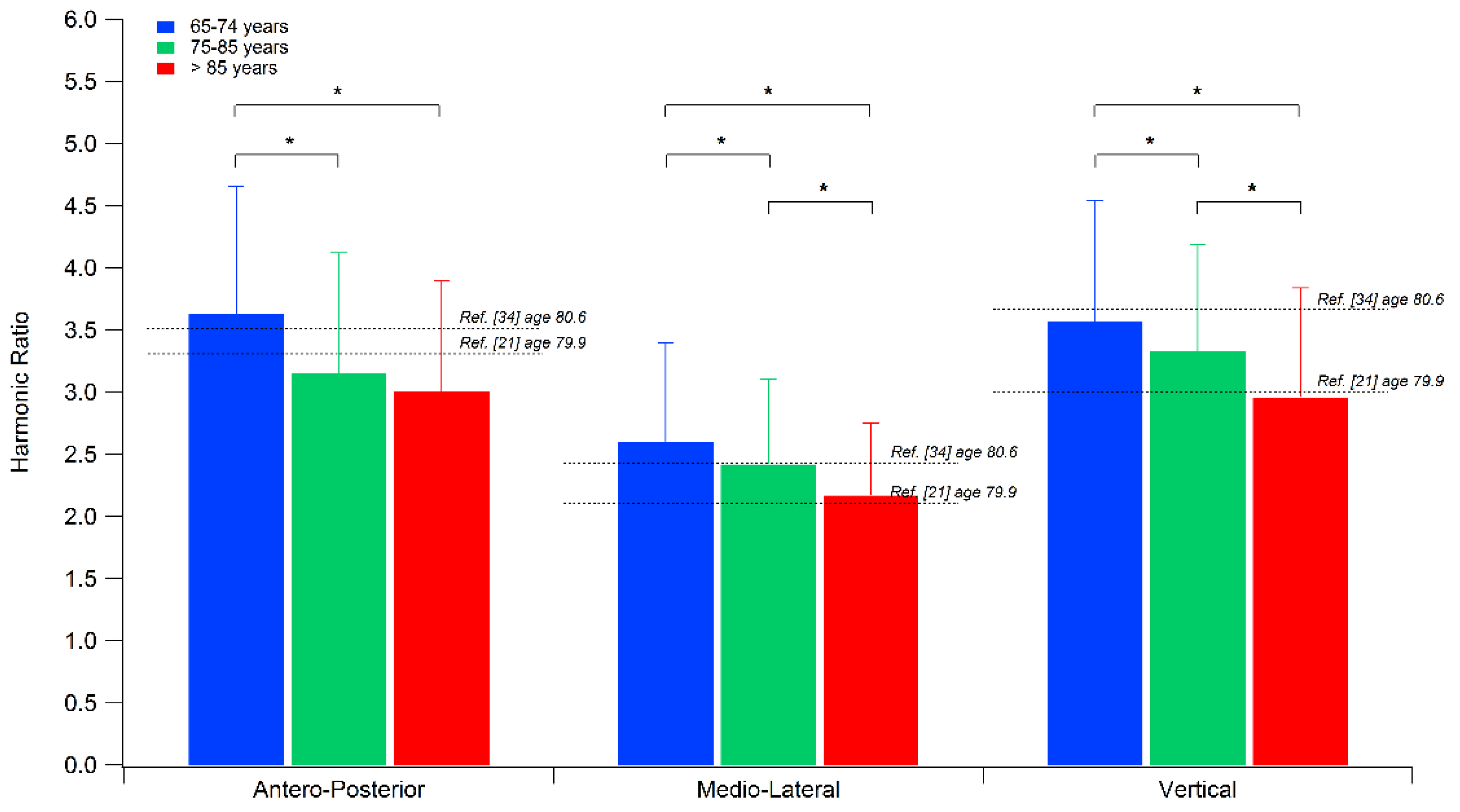
HR values are quite simple to interpret, being lower values indicative of a less smooth/symmetrical gait. Previous studies reported that healthy older adults are characterized by values of HR approximately from 3–4 in the AP and V directions and from 2.1–2.6 in the ML direction [21,23,25,34].

2.3. Statistical Analysis

A two-way multivariate analysis of variance (MANOVA) was used to verify the presence of differences among the three groups in terms of spatio-temporal parameters and HRs. In particular, regarding HRs, previous studies indicated its sensitivity to gait speed (i.e., higher speed originates higher HR values [23]), and its value is expected to differ across different age groups. Thus, it appears necessary to include it in the analysis as a covariate.
The independent variables were the participant’s age group, while the dependent variables were, in one case, the six spatio-temporal parameters and, in the other, the three HRs. The statistical significance level was set at p < 0.05, and the effect sizes were evaluated via the eta-squared (η2) coefficient. Univariate analysis of variance (ANOVA) was used as a post hoc test through a reduction of the significance level according to the Bonferroni correction for multiple comparisons (p = 0.008, 0.05/6) for spatio-temporal parameters and p = 0.016, 0.05/3) for HRs). Data were analyzed using the IBM SPSS Statistics v.23 software (IBM, Armonk, NY, USA).

3. Results

Table 2 reports the experimental test results regarding the spatio-temporal parameters of gait and HRs for each age group. A significant main effect of the group [ F(12, 878) = 4.38, p < 0.001, Wilks λ = 0.89, η2 = 0.06 ] was found by the MANOVA on spatio-temporal parameters of gait. In particular, the oldest participants (Group 3) were characterized by a 16% reduction in gait speed and by a 10% reduction in stride length (p < 0.001) when compared with the performance of the Group 1 (p < 0.001 in both cases), resulting after the post hoc analysis. Slightly smaller (yet statistically significant) reductions in speed and stride length (approximately 9% in both cases) were also observed between individuals of Group 1 and Group 2. Regarding the cadence, the statistical analysis revealed that Group 3 was characterized by a 5% reduction with respect to Groups 1 and 2 (p < 0.001 in both cases). In contrast, no significant differences were observed for the duration of the stance, swing, and double support phases.
The trend of HR for AP, ML, and V directions across the analysis groups is shown in Figure 3. Including gait speed as the covariate, MANCOVA detected a significant main effect of age on HR values [ F(6,882) = 3.10, p = 0.005, Wilks λ = 0.96, η2 = 0.02]. The post hoc analysis showed that older participants (i.e., Group 3) exhibited a quite uniform reduction of HR with respect to those of Group 1 for all three directions of approximately 17% (p < 0.001), while differences vs. Group 2 involved only the ML and V directions and were smaller (−10%). Finally, significant differences were also found between Group 2 and Group 1 for HR in all three directions, as those aged 75–85 exhibited reduced HR values (approximately between 6 and 13%).

4. Discussion

In this study, we aimed to quantitatively investigate the existence of age-related alterations of gait patterns in a large cohort of healthy older adults aged 65 and over, including the oldest-old participants, assessed in a clinical environment via a wearable inertial sensor. The hypothesis was explored by analyzing HRs computed from trunk acceleration and the most common spatio-temporal parameters. In particular, HRs, representative of gait smoothness, can be considered an effective indicator of whole-body balance during gait, and it was already demonstrated to be a measure suitable to describe the changes in ambulation associated with aging. In this regard, although there is previous evidence of age-related reductions of HRs during walking [29,33,42], little data is available as regards the oldest-old adults. We attempted here to overcome such a limitation by testing a large cohort of healthy individuals, which also included a group of 47 participants aged 85–90.
Our data confirm that aging is associated with significant changes in spatio-temporal parameters. In particular, as for gait speed and stride length, the youngest participants (Group 1) exhibited higher values when compared with the other two groups, while as regards the cadence, significantly lower values were found in Group 3 compared to Group 1 and Group 2. In contrast, no significant differences among groups were found in terms of stance, swing, and double support phase duration, although a consistent trend of variation with age (i.e., stance and double support phases increase, swing phase duration decreases) was observed. It is noteworthy that the observed speed changes are consistent with those reported in previous studies, which showed a continuous reduction in gait speed in older adults, especially from the seventh decade [43,44]. These data align with Hollman et al., 2011 [36], which presents the normative spatio-temporal gait parameters in older adults, and a more recent study [45] which reports the reference values for usual gait speed in community-dwelling older adults living in Western Europe. Moreover, since such a reduction is accompanied by a correspondent step/stride shortening (particularly when passing from 65 to 75 years), our data confirm that older adults adopt a cautious strategy in order to achieve better stability in locomotion and, consequently, decrease the risk of falling [46,47,48,49].
As regards HRs, the values calculated in the present study are quite consistent with those previously reported for individuals of similar age ranges [21,23,33,34,35]. For example, in Lowry et al. [21], the adults aged 80–86 years exhibited lower HR in the AP and V directions compared to those aged 60–69 years, and no differences in the ML direction were detected between the Groups. In Brach et al. (2011) [29], older adults (mean age: 77.5 years) had lower HR in the AP direction than young adults (mean age = 24.4 years).
This indicates that, despite the possible differences involving equipment, measurement protocol, and data processing, the HR parameter could represent a sensitive approach. The main finding which emerges from the trunk acceleration analysis is that aging is associated with a substantially linear decrease of HRs in all directions, with the oldest-old participants characterized by the lowest values in all three directions. As previously mentioned, it is known that older adults are usually characterized by lower HRs compared to young adults [23,29], and among them, the co-existence of cognitive decline enhances this phenomenon [33]. In this regard, our results demonstrate that smoothness tends to further worsen in those aged 85 and over (Group 3), thus suggesting that changes in motor control abilities continue to occur until the late stages of life. However, it is difficult to perform a comparison as the age ranges are not similar among studies, and also equipment and measurement protocols are not uniform [23,36]. Alterations in limb dynamics and a different distribution of joint torques and powers on lower limb joints towards proximal segments (i.e., older adults tend to perform more work at the hip and less at the ankle) have been identified as possible factors able to increase irregularity of trunk accelerations and thus consequently able originate reduced smoothness [23]. However, it should also be recalled that the control of ML motion during the stance phase of gait represents a main risk factor for falls in older adults [50,51]. It is, thus, possible that reduced HRs (in particular those of ML directions) are one of the expressions of the cautious strategy adopted to keep the center of mass safely between their feet and thus preserve balance. In fact, an optimal balance during walking requires continuous integrative control, particularly in a lateral direction, due to inherent instability associated with single limb support [52]. There is now considerable evidence for the effects of age on ML motion [53,54,55,56], some of which have been associated with increased fall risk [57].
As HR is a parameter that is very sensitive to even subtle changes in gait smoothness, it could be used as an outcome measure of rehabilitation/training programs aimed at improving gait in older people in combination with the conventional spatio-temporal parameters. In particular, the literature suggests that structured exercise programs, prescribed and designed according to individual clinical conditions, age, and goal/s to reach, can now be considered a strategy to maintain and improve physical function in older people [58,59,60]. In particular, it was demonstrated that exercise programs reduce the rate of falls and the number of people experiencing falls in older people living in the community [61] and that physical exercises, including functional mobility training, especially walking, have better results than physical programs with only static, resistance, and flexibility training, especially in those with cognitive deficit [62]. It is also important to stimulate them to regularly perform physical activity to improve general well-being and cognition, also considering home-based exercise programs.
Some limitations of the study should be acknowledged. Firstly, the proposed stratification resulted in a Group (i.e., those aged 85 and over) that was markedly smaller than the others. This reflects, to some extent, the need to include participants free from significant mobility restrictions and cognitively intact, which is not easy to achieve considering that such issues are quite common during the late stage of life. Secondly, since this study focused solely on spatio-temporal parameters and HRs, we could only speculate about the mechanisms underlying the pattern of results. Future developments of the study should aim to combine kinematic and kinetic features of gait with HRs and other trunk acceleration-derived measures, to have a detailed and exhaustive picture of the control of body motion during walking and even to understand which measures are most sensitive to age-related changes in gait.

5. Conclusions

In the present study, the possible changes in spatio-temporal parameters and smoothness of gait associated with aging were explored in a cohort of youngest-, middle-, and oldest-old, using parameters derived from trunk acceleration collected in a clinical context using a simple setup composed by a single miniaturized IMU. Our data showed the presence of significant alterations in gait according to aging, reporting a reduced speed and stride length and a reduction of HR in the three directions. The latter changes were similar in magnitude across the three groups and suggested that smoothness similarly worsens in all directions until the late stage of life. Considering the sensitivity of HR to the presence of physical and cognitive conditions which interfere with mobility, the analysis of smoothness of gait may be considered a useful and valid tool for the early detection of subtle changes in gait in older adults, which that the spatio-temporal parameters alone could fail to highlight. This parameter could also be used in clinical practice by a physician and a physical therapist.

Author Contributions

Conceptualization, M.P. (Massimiliano Pau); G.B.; M.G. and V.C.; methodology, M.P. (Massimiliano Pau) and V.C.; software, B.L.; M.P. (Micaela Porta) and S.C.; formal analysis, M.P. (Massimiliano Pau); Clinical Assessment of the Participants, G.B.; V.P.; D.V.; D.R. and G.A.; data curation, M.P. (Massimiliano Pau) and V.C.; writing—original draft preparation, M.P. (Massimiliano Pau); B.L.; M.P. (Micaela Porta); S.C. and V.C.; writing—review and editing, M.P. (Massimiliano Pau), G.B. and V.C.; supervision, M.P. (Massimiliano Pau) and V.C. All authors have read and agreed to the published version of the manuscript.

Funding

This research received no external funding.

Institutional Review Board Statement

The study was conducted in accordance with the Declaration of Helsinki, and approved by the ethical committees of the University of Milan (authorization number 12_2019) and ATS Sardegna, Italy (authorization number 300/2021/CE).

Informed Consent Statement

Informed consent was obtained from all subjects involved in the study.

Data Availability Statement

Data available on request due to restrictions, e.g., privacy or ethical.

Conflicts of Interest

The authors declare no conflict of interest.

References

  1. DeVita, P.; Hortobagyi, T. Age Causes a Redistribution of Joint Torques and Powers during Gait. J. Appl. Physiol. 2000, 88, 1804–1811. [Google Scholar] [CrossRef] [PubMed] [Green Version]
  2. McGibbon, C.A.; Krebs, D.E. Age-Related Changes in Lower Trunk Coordination and Energy Transfer during Gait. J. Neurophysiol. 2001, 85, 1923–1931. [Google Scholar] [CrossRef] [PubMed] [Green Version]
  3. Ko, S.; Stenholm, S.; Metter, E.J.; Ferrucci, L. Age-Associated Gait Patterns and the Role of Lower Extremity Strength - Results from the Baltimore Longitudinal Study of Aging. Arch. Gerontol. Geriatr. 2012, 55, 474–479. [Google Scholar] [CrossRef] [Green Version]
  4. Harada, C.N.; Natelson Love, M.C.; Triebel, K.L. Normal Cognitive Aging. Clin. Geriatr. Med. 2013, 29, 737–752. [Google Scholar] [CrossRef] [Green Version]
  5. Lajoie, Y.; Teasdale, N.; Bard, C.; Fleury, M. Upright Standing and Gait: Are There Changes in Attentional Requirements Related to Normal Aging? Exp. Aging Res. 1996, 22, 185–198. [Google Scholar] [CrossRef]
  6. Woollacott, M.; Shumway-Cook, A. Attention and the Control of Posture and Gait: A Review of an Emerging Area of Research. Gait Posture 2002, 16, 1–14. [Google Scholar] [CrossRef] [PubMed]
  7. Sunderaraman, P.; Maidan, I.; Kozlovski, T.; Apa, Z.; Mirelman, A.; Hausdorff, J.M.; Stern, Y. Differential Associations between Distinct Components of Cognitive Function and Mobility: Implications for Understanding Aging, Turning and Dual-Task Walking. Front. Aging Neurosci. 2019, 11, 1–13. [Google Scholar] [CrossRef] [PubMed] [Green Version]
  8. Boyé, N.D.A.; Mattace-Raso, F.U.S.; Van der Velde, N.; Van Lieshout, E.M.M.; De Vries, O.J.; Hartholt, K.A.; Kerver, A.J.H.; Bruijninckx, M.M.M.; Van der Cammen, T.J.M.; Patka, P.; et al. Circumstances Leading to Injurious Falls in Older Men and Women in the Netherlands. Injury 2014, 45, 1224–1230. [Google Scholar] [CrossRef] [Green Version]
  9. Zijlstra, W.; Hof, A.L. Assessment of Spatio-Temporal Gait Parameters from Trunk Accelerations during Human Walking. Gait Posture 2003, 18, 1–10. [Google Scholar] [CrossRef] [Green Version]
  10. Lord, S.; Galna, B.; Verghese, J.; Coleman, S.; Burn, D.; Rochester, L. Independent Domains of Gait in Older Adults and Associated Motor and Nonmotor Attributes: Validation of a Factor Analysis Approach. J. Gerontol Ser. A Biol. Sci. Med. Sci. 2013, 68, 820–827. [Google Scholar] [CrossRef]
  11. Roetenberg, D.; Luinge, H.; Slycke, P. Xsens MVN: Full 6DOF Human Motion Tracking Using Miniature Inertial Sensors. Xsens Motion Technol. BV 2009, 1–7. [Google Scholar]
  12. Seel, T.; Raisch, J.; Schauer, T. IMU-Based Joint Angle Measurement for Gait Analysis. Sensors 2014, 14, 6891–6909. [Google Scholar] [CrossRef] [PubMed] [Green Version]
  13. Shrivastava, S.; Trung, T.Q.; Lee, N.E. Recent Progress, Challenges, and Prospects of Fully Integrated Mobile and Wearable Point-of-Care Testing Systems for Self-Testing. Chem. Soc. Rev. 2020, 49, 1812–1866. [Google Scholar] [CrossRef] [PubMed]
  14. Ji, X.; Rao, Z.; Zhang, W.; Liu, C.; Wang, Z.; Zhang, S.; Zhang, B.; Hu, M.; Servati, P.; Xiao, X. Airline Point-of-Care System on Seat Belt for Hybrid Physiological Signal Monitoring. Micromachines 2022, 13, 1880. [Google Scholar] [CrossRef]
  15. Cho, Y.-S.; Jang, S.-H.; Cho, J.-S.; Kim, M.-J.; Lee, H.D.; Lee, S.Y.; Moon, S.-B. Evaluation of Validity and Reliability of Inertial Measurement Unit-Based Gait Analysis Systems. Ann. Rehabil. Med. 2018, 42, 872–883. [Google Scholar] [CrossRef] [PubMed] [Green Version]
  16. Horak, F.; King, L.; Mancini, M. Role of Body-Worn Movement Monitor Technology for Balance and Gait Rehabilitation. Phys. Ther. 2015, 95, 461–470. [Google Scholar] [CrossRef] [Green Version]
  17. Maetzler, W.; Rochester, L. Body-Worn Sensors--the Brave New World of Clinical Measurement? Mov. Disord. Off. J. Mov. Disord. Soc. 2015, 30, 1203–1205. [Google Scholar] [CrossRef]
  18. Del Din, S.; Godfrey, A.; Galna, B.; Lord, S.; Rochester, L. Free-Living Gait Characteristics in Ageing and Parkinson’s Disease: Impact of Environment and Ambulatory Bout Length. J. Neuroeng. Rehabil. 2016, 13, 1–12. [Google Scholar] [CrossRef] [Green Version]
  19. Labini, F.S.; Meli, A.; Ivanenko, Y.P.; Tufarelli, D. Recurrence Quantification Analysis of Gait in Normal and Hypovestibular Subjects. Gait Posture 2012, 35, 48–55. [Google Scholar] [CrossRef]
  20. Bisi, M.C.; Riva, F.; Stagni, R. Measures of Gait Stability: Performance on Adults and Toddlers at the Beginning of Independent Walking. J. Neuroeng. Rehabil. 2014, 11, 131. [Google Scholar] [CrossRef] [Green Version]
  21. Menz, H.B.; Lord, S.R.; Fitzpatrick, R.C. Acceleration Patterns of the Head and Pelvis When Walking Are Associated with Risk of Falling in Community-Dwelling Older People. J. Gerontol. Ser. A Biol. Sci. Med. Sci. 2003, 58, M446–M452. [Google Scholar] [CrossRef]
  22. Bellanca, J.L.; Lowry, K.A.; VanSwearingen, J.M.; Brach, J.S.; Redfern, M.S. Harmonic Ratios: A Quantification of Step to Step Symmetry. J. Biomech. 2013, 46, 828–831. [Google Scholar] [CrossRef] [PubMed] [Green Version]
  23. Lowry, K.A.; Lokenvitz, N.; Smiley-Oyen, A.L. Age- and Speed-Related Differences in Harmonic Ratios during Walking. Gait Posture 2012, 35, 272–276. [Google Scholar] [CrossRef] [PubMed]
  24. Menz, H.B.; Lord, S.R.; St George, R.; Fitzpatrick, R.C. Walking Stability and Sensorimotor Function in Older People with Diabetic Peripheral Neuropathy. Arch. Phys. Med. Rehabil. 2004, 85, 245–252. [Google Scholar] [CrossRef]
  25. Lowry, K.A.; Smiley-Oyen, A.L.; Carrel, A.J.; Kerr, J.P. Walking Stability Using Harmonic Ratios in Parkinson’s Disease. Mov. Disord. Off. J. Mov. Disord. Soc. 2009, 24, 261–267. [Google Scholar] [CrossRef]
  26. Pau, M.; Mandaresu, S.; Pilloni, G.; Porta, M.; Coghe, G.; Marrosu, M.G.; Cocco, E. Smoothness of Gait Detects Early Alterations of Walking in Persons with Multiple Sclerosis without Disability. Gait Posture 2017, 58, 307–309. [Google Scholar] [CrossRef] [PubMed]
  27. Iosa, M.; Paradisi, F.; Brunelli, S.; Delussu, A.S.; Pellegrini, R.; Zenardi, D.; Paolucci, S.; Traballesi, M. Assessment of Gait Stability, Harmony, and Symmetry in Subjects with Lower-Limb Amputation Evaluated by Trunk Accelerations. J. Rehabil. Res. Dev. 2014, 51, 623–634. [Google Scholar] [CrossRef]
  28. Cimolin, V.; Cau, N.; Sartorio, A.; Capodaglio, P.; Galli, M.; Tringali, G.; Leban, B.; Porta, M.; Pau, M. Symmetry of Gait in Underweight, Normal and Overweight Children and Adolescents. Sensors 2019, 19, 2054. [Google Scholar] [CrossRef]
  29. Brach, J.S.; McGurl, D.; Wert, D.; Vanswearingen, J.M.; Perera, S.; Cham, R.; Studenski, S. Validation of a Measure of Smoothness of Walking. J. Gerontol. Ser. A Biol. Sci. Med. Sci. 2011, 66, 136–141. [Google Scholar] [CrossRef]
  30. Leban, B.; Cimolin, V.; Porta, M.; Arippa, F.; Pilloni, G.; Galli, M.; Pau, M. Age-Related Changes in Smoothness of Gait of Healthy Children and Early Adolescents. J. Mot. Behav. 2020, 52, 694–702. [Google Scholar] [CrossRef] [PubMed]
  31. Lowry, K.A.; Sebastian, K.; Perera, S.; Van Swearingen, J.; Smiley-Oyen, A.L. Age-Related Differences in Locomotor Strategies During Adaptive Walking. J. Mot. Behav. 2017, 49, 435–440. [Google Scholar] [CrossRef] [PubMed]
  32. Misu, S.; Asai, T.; Doi, T.; Sawa, R.; Ueda, Y.; Saito, T.; Nakamura, R.; Murata, S.; Sugimoto, T.; Yamada, M.; et al. Association between Gait Abnormality and Malnutrition in a Community-Dwelling Elderly Population. Geriatr. Gerontol. Int. 2017, 17, 1155–1160. [Google Scholar] [CrossRef]
  33. Asai, T.; Misu, S.; Sawa, R.; Doi, T.; Yamada, M. The Association between Fear of Falling and Smoothness of Lower Trunk Oscillation in Gait Varies According to Gait Speed in Community-Dwelling Older Adults. J. Neuroeng. Rehabil. 2017, 14, 1–9. [Google Scholar] [CrossRef] [Green Version]
  34. Row Lazzarini, B.S.; Kataras, T.J. Treadmill Walking Is Not Equivalent to Overground Walking for the Study of Walking Smoothness and Rhythmicity in Older Adults. Gait Posture 2016, 46, 42–46. [Google Scholar] [CrossRef] [PubMed]
  35. Pau, M.; Mulas, I.; Putzu, V.; Asoni, G.; Viale, D.; Mameli, I.; Leban, B.; Allali, G. Smoothness of Gait in Healthy and Cognitively Impaired Individuals: A Study on Italian Elderly Using Wearable Inertial Sensor. Sensors 2020, 20, 3577. [Google Scholar] [CrossRef]
  36. Hollman, J.H.; McDade, E.M.; Petersen, R.C. Normative Spatiotemporal Gait Parameters in Older Adults. Gait Posture 2011, 34, 111–118. [Google Scholar] [CrossRef] [PubMed] [Green Version]
  37. Brach, J.S.; Perera, S.; Studenski, S.; Katz, M.; Hall, C.; Verghese, J. Meaningful Change in Measures of Gait Variability in Older Adults. Gait Posture 2010, 31, 175–179. [Google Scholar] [CrossRef] [Green Version]
  38. Bugané, F.; Benedetti, M.G.; Casadio, G.; Attala, S.; Biagi, F.; Manca, M.; Leardini, A. Estimation of Spatial-Temporal Gait Parameters in Level Walking Based on a Single Accelerometer: Validation on Normal Subjects by Standard Gait Analysis. Comput. Methods Programs Biomed. 2012, 108, 129–137. [Google Scholar] [CrossRef] [PubMed]
  39. Ilaria Viscione; Francesca D’Elia; Rodolfo Vastola; Maurizio Sibilio the Correlation Between Technologies and Rating Scales in Gait Analysis. J. Sport. Sci. 2016, 4, 119–123. [CrossRef]
  40. Pau, M.; Porta, M.; Pilloni, G.; Corona, F.; Fastame, M.C.; Hitchcott, P.K.; Penna, M.P. Texting While Walking Induces Gait Pattern Alterations in Healthy Older Adults. Proc. Hum. Factors Ergon. Soc. Annu. Meet. 2018, 62, 1908–1912. [Google Scholar] [CrossRef] [Green Version]
  41. Zijlstra, W. Assessment of Spatio-Temporal Parameters during Unconstrained Walking. Eur. J. Appl. Physiol. 2004, 92, 39–44. [Google Scholar] [CrossRef] [PubMed]
  42. Mazzà, C.; Iosa, M.; Pecoraro, F.; Cappozzo, A. Control of the Upper Body Accelerations in Young and Elderly Women during Level Walking. J. Neuroeng. Rehabil. 2008, 5, 1–10. [Google Scholar] [CrossRef] [PubMed] [Green Version]
  43. Abellan van Kan, G.; Rolland, Y.; Andrieu, S.; Bauer, J.; Beauchet, O.; Bonnefoy, M.; Cesari, M.; Donini, L.M.; Gillette Guyonnet, S.; Inzitari, M.; et al. Gait Speed at Usual Pace as a Predictor of Adverse Outcomes in Community-Dwelling Older People an International Academy on Nutrition and Aging (IANA) Task Force. J. Nutr. Health Aging 2009, 13, 881–889. [Google Scholar] [CrossRef]
  44. Studenski, S.; Perera, S.; Patel, K.; Rosano, C.; Faulkner, K.; Inzitari, M.; Brach, J.; Chandler, J.; Cawthon, P.; Connor, E.B.; et al. Gait Speed and Survival in Older Adults. JAMA 2011, 305, 50–58. [Google Scholar] [CrossRef] [PubMed] [Green Version]
  45. Dommershuijsen, L.J.; Ragunathan, J.; Ruiter, T.R.; Groothof, D.; Mattace-Raso, F.U.S.; Ikram, M.A.; Polinder-Bos, H.A. Gait Speed Reference Values in Community-Dwelling Older Adults – Cross-Sectional Analysis from the Rotterdam Study. Exp. Gerontol. 2022, 158, 111646. [Google Scholar] [CrossRef] [PubMed]
  46. Aboutorabi, A.; Arazpour, M.; Bahramizadeh, M.; Hutchins, S.W.; Fadayevatan, R. The Effect of Aging on Gait Parameters in Able-Bodied Older Subjects: A Literature Review. Aging Clin. Exp. Res. 2016, 28, 393–405. [Google Scholar] [CrossRef] [PubMed]
  47. Brach, J.S.; Studenski, S.A.; Perera, S.; VanSwearingen, J.M.; Newman, A.B. Gait Variability and the Risk of Incident Mobility Disability in Community-Dwelling Older Adults. J. Gerontol. Ser. A 2007, 62, 983–988. [Google Scholar] [CrossRef]
  48. Brach, J.S.; Studenski, S.; Perera, S.; VanSwearingen, J.M.; Newman, A.B. Stance Time and Step Width Variability Have Unique Contributing Impairments in Older Persons. Gait Posture 2008, 27, 431–439. [Google Scholar] [CrossRef] [Green Version]
  49. Jones, L.M.; Waters, D.L.; Legge, M. Walking Speed at Self-Selected Exercise Pace Is Lower but Energy Cost Higher in Older versus Younger Women. J. Phys. Act. Health 2009, 6, 327–332. [Google Scholar] [CrossRef] [Green Version]
  50. Kavanagh, J.J.; Barrett, R.S.; Morrison, S. Age-Related Differences in Head and Trunk Coordination during Walking. Hum. Mov. Sci. 2005, 24, 574–587. [Google Scholar] [CrossRef]
  51. Mesure, S.; Azulay, J.P.; Pouget, J.; Amblard, B. Strategies of Segmental Stabilization during Gait in Parkinson’s Disease. Exp. Brain Res. 1999, 129, 573–581. [Google Scholar] [CrossRef] [PubMed]
  52. O’Connor, S.M.; Kuo, A.D. Direction-Dependent Control of Balance during Walking and Standing. J. Neurophysiol. 2009, 102, 1411–1419. [Google Scholar] [CrossRef] [Green Version]
  53. Brodie, M.A.D.; Menz, H.B.; Lord, S.R. Age-Associated Changes in Head Jerk While Walking Reveal Altered Dynamic Stability in Older People. Exp. Brain Res. 2014, 232, 51–60. [Google Scholar] [CrossRef] [PubMed]
  54. Chou, L.-S.; Kaufman, K.R.; Hahn, M.E.; Brey, R.H. Medio-Lateral Motion of the Center of Mass during Obstacle Crossing Distinguishes Elderly Individuals with Imbalance. Gait Posture 2003, 18, 125–133. [Google Scholar] [CrossRef] [PubMed]
  55. Cofré Lizama, L.E.; Pijnappels, M.; Reeves, N.P.; Verschueren, S.M.; van Dieën, J.H. Centre of Pressure or Centre of Mass Feedback in Mediolateral Balance Assessment. J. Biomech. 2015, 48, 539–543. [Google Scholar] [CrossRef] [Green Version]
  56. Singer, J.C.; McIlroy, W.E.; Prentice, S.D. Kinetic Measures of Restabilisation during Volitional Stepping Reveal Age-Related Alterations in the Control of Mediolateral Dynamic Stability. J. Biomech. 2014, 47, 3539–3545. [Google Scholar] [CrossRef]
  57. Maki, B.E. Gait Changes in Older Adults: Predictors of Falls or Indicators of Fear. J. Am. Geriatr. Soc. 1997, 45, 313–320. [Google Scholar] [CrossRef]
  58. Casas-Herrero, A.; Anton-Rodrigo, I.; Zambom-Ferraresi, F.; Sáez de Asteasu, M.L.; Martinez-Velilla, N.; Elexpuru-Estomba, J.; Marin-Epelde, I.; Ramon-Espinoza, F.; Petidier-Torregrosa, R.; Sanchez-Sanchez, J.L.; et al. Effect of a Multicomponent Exercise Programme (VIVIFRAIL) on Functional Capacity in Frail Community Elders with Cognitive Decline: Study Protocol for a Randomized Multicentre Control Trial. Trials 2019, 20, 362. [Google Scholar] [CrossRef]
  59. Amorese, A.J.; Ryan, A.S. Home-Based Tele-Exercise in Musculoskeletal Conditions and Chronic Disease: A Literature Review. Front. Rehabil. Sci. 2022, 3. [Google Scholar] [CrossRef]
  60. Baez, M.; Khaghani Far, I.; Ibarra, F.; Ferron, M.; Didino, D.; Casati, F. Effects of Online Group Exercises for Older Adults on Physical, Psychological and Social Wellbeing: A Randomized Pilot Trial. PeerJ 2017, 5, e3150. [Google Scholar] [CrossRef] [Green Version]
  61. Sherrington, C.; Fairhall, N.J.; Wallbank, G.K.; Tiedemann, A.; Michaleff, Z.A.; Howard, K.; Clemson, L.; Hopewell, S.; Lamb, S.E. Exercise for Preventing Falls in Older People Living in the Community. Cochrane Database Syst. Rev. 2019, 1, CD012424. [Google Scholar] [CrossRef] [PubMed]
  62. Zhang, W.; Low, L.-F.; Gwynn, J.D.; Clemson, L. Interventions to Improve Gait in Older Adults with Cognitive Impairment: A Systematic Review. J. Am. Geriatr. Soc. 2019, 67, 381–391. [Google Scholar] [CrossRef] [PubMed]
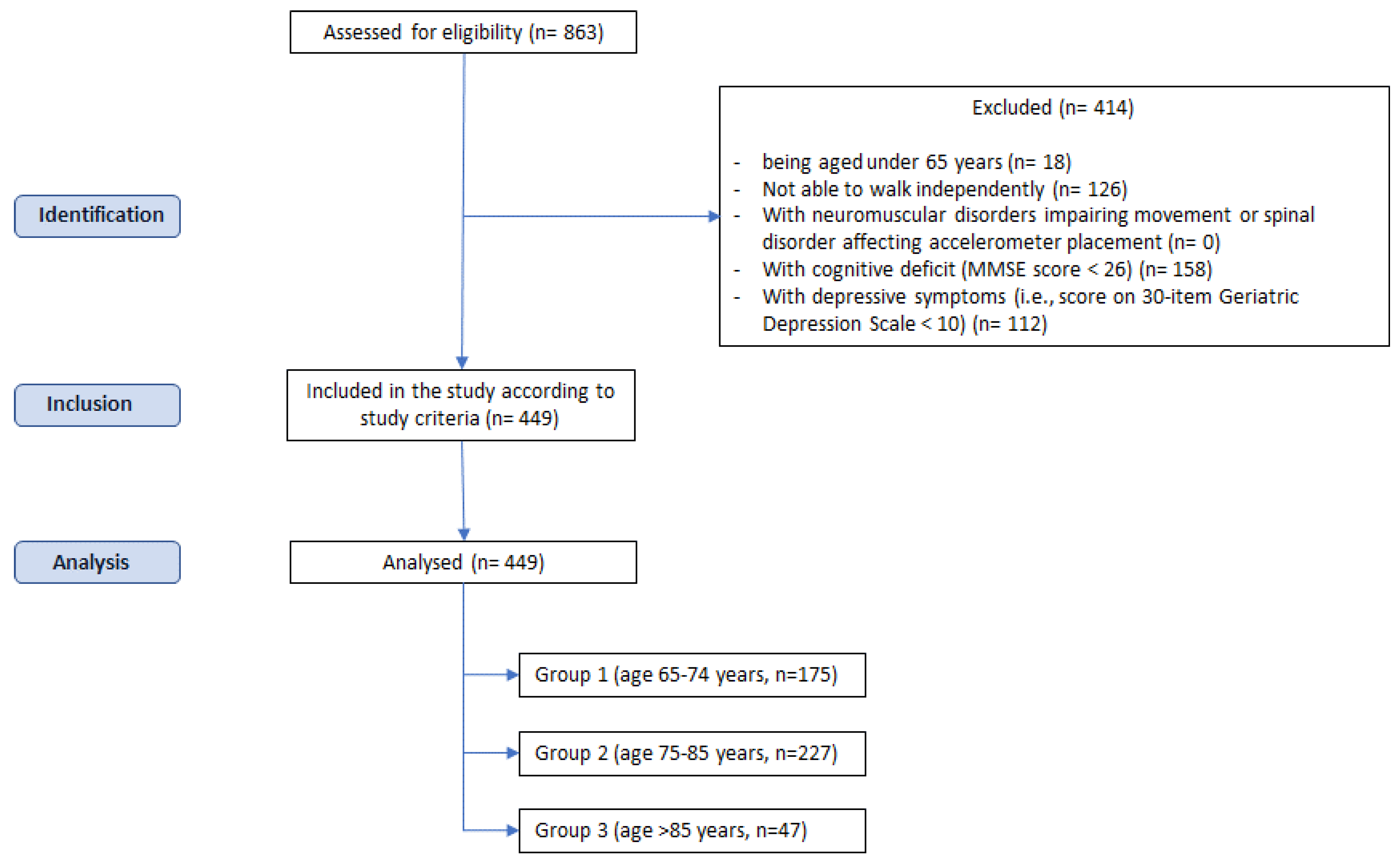
Figure 1. Process of participants’ selection.
Electronics 12 00637 g001
Figure 2. From left to right: (a) participant equipped with the IMU for the gait trials; (b) detail of the IMU positioning inside the semi-elastic belt; (c) the BTS G-Sensor IMU used for the experimental tests.
Electronics 12 00637 g002
Figure 3. Harmonic Ratio values in AP, ML, and V directions for the three groups of tested older adults. Data from the present study are compared with those previously reported for similar age ranges (Refs. [21,34]).
Electronics 12 00637 g003
Table 1. Participant’s characteristics. Values are expressed as mean ± SD.
Group 1
(65–74 Years)
Group 2
(75–85 Years)
Group 3
(>85 Years)
Participants # (F, M)175 (103 F, 72 M)228 (128 F, 100 M)47 (28 F, 19 M)
Participants percentage (F, M)F 59%, M 41%F 56%, M 44%F 60%, M 40%
Age (years)70.4 ± 2.579.1 ± 2.8 a86.5 ± 1.7 a,b
Body Mass (kg)66.8 ± 12.465.6 ± 11.461.3 ± 13.6 a,b
Height (cm)162.0 ± 8.4160.0 ± 8.7 a158.6 ± 8.5 a
The symbol a indicates a significant difference for Group 1; the symbol b indicates a significant difference for Group 2.
Table 2. Mean and standard deviation values of the spatio-temporal and HR (Harmonic Ratio) parameters in the three considered groups.
Group 1
(65–74 Years)
Group 2
(75–85 Years)
Group 3
(>85 Years)
Spatial-temporal parameters of gaitGait speed (m s−1)1.08 ± 0.240.98 ± 0.24 a0.91 ± 0.26 a
Stride length (m)1.16 ± 0.211.05 ± 0.22 a1.04 ± 0.27 a
Cadence (steps min−1)111.20 ± 9.46111.61 ± 10.75105.20 ± 13.00 a,b
Stance phase (% of the GC)60.46 ± 2.5560.82 ± 1.9761.28 ± 2.74
Swing phase (% of the GC)39.42 ± 3.0039.06 ± 2.7339.04 ± 3.32
Double support phase (% of the GC)10.56 ± 2.0810.80 ± 1.9811.21 ± 2.71
Harmonic Ratio *AP direction *3.63 ± 1.033.15 ± 0.97 a3.01 ± 0.89 a
ML direction *2.60 ± 0.80 2.42 ± 0.69 a2.17 ± 0.58 a,b
V direction *3.57 ± 0.973.33 ±0.86 a2.96 ± 0.88 a,b
The symbol a indicates a significant difference for Group 1 after Bonferroni correction (p = 0.016), the symbol b indicates a significant difference for Group 2 after Bonferroni correction (p = 0.016); * controlled for gait speed; GC: Gait Cycle.
Disclaimer/Publisher’s Note: The statements, opinions and data contained in all publications are solely those of the individual author(s) and contributor(s) and not of MDPI and/or the editor(s). MDPI and/or the editor(s) disclaim responsibility for any injury to people or property resulting from any ideas, methods, instructions or products referred to in the content.