Article

Experimental Analysis to Detect Corona COVID-19 Virus Symptoms in Male Patients through Breath Pattern Using Machine Learning Algorithms

by
Abdulilah Mohammad Mayet
1,
Neeraj Kumar Shukla
1,
M. Ramkumar Raja
1,
Ijaz Ahmad
2,*,
Ramy Mohammed Aiesh Qaisi
3,
Ali Awadh Al-Qahtani
1,
Anita Taparwal
4,
Vineet Tirth
5 and
Reem AL-Dossary
6
1
Electrical Engineering Department, College of Engineering, King Khalid University, Abha 61411, Saudi Arabia
2
Shenzhen College of Advanced Technology, University of Chinese Academy of Sciences (UCAS), Shenzhen 518055, China
3
Department Electrical and of Electronic Engineering, College of Engineering, University of Jeddah, Jeddah 21589, Saudi Arabia
4
Gurgaon ESIC Hospital, Delhi 122001, India
5
Mechanical Engineering Department, College of Engineering, King Khalid University, Abha 61421, Saudi Arabia
6
Nursing Education Department, Nursing College, Imam Abdulrahman Bin Faisal University, Dammam 34221, Saudi Arabia
*
Author to whom correspondence should be addressed.
Electronics 2023, 12(1), 10; https://doi.org/10.3390/electronics12010010
Submission received: 7 November 2022 / Revised: 4 December 2022 / Accepted: 9 December 2022 / Published: 20 December 2022
(This article belongs to the Special Issue Application of Artificial Neural Network in Non-destructive Testing)

Abstract

:
In the fourth quarter of the year 2019, the planet became overwhelmed by the pandemic caused by the coronavirus disease (COVID-19). This virus imperiled human life and have affected a considerable percentage of the world population much before its early stage detection mechanisms were discovered and made available at the grassroots level. As there is no specific drug available to treat this infection, the vaccine was intended to serve as the ultimate weapon in the war against this species of coronavirus, but like other viruses, being an RNA virus, this virus also mutates continuously while it passes from one human to the other, making the development of highly potent vaccines even more challenging. This work is being sketched at the juncture when a huge percentage of the human population is already affected by this virus globally. In this work, we are proposing an idea to develop an app to detect coronavirus (COVID-19) symptoms at an early stage by self-diagnosis at home or at the clinical level. An experimental study has been performed on a dummy dataset with 11000 entries of various breadth patterns based on the spirometry analysis, lung volume analysis, and lung capacity analysis of normal male subjects and detailed breath patterns of infected male patients. A logistic regression model is trained after using SMOTE oversampling to balance the data and the predictive accuracy levels of 80%, 78%, and 90%. The results accomplished through this study and experiments may not only aid the clinicians in their medical practice but may also bestow a blue chip to the masterminds engaged in the biomedical research for inventing more evolved, sophisticated, user-friendly, miniaturized, portable, and economical medical app/devices in the future.

1. Introduction

The 21st century has withstood many deadly and highly virulent viruses and their subsequent morbidities and mortalities, e.g., Middle East respiratory syndrome coronavirus (MERS-CoV) [1]; Ebola virus disease; swine flu pandemic caused by H1N1; Zika virus; human immunodeficiency virus (HIV) epidemic, which is continuing globally; the highly pathogenic and continually mutating avian (bird) influenza (flu) type-A virus subtype H5N1 (bird flu) [2], which is causing a grave global concern as a potential pandemic threat; severe acute respiratory syndrome coronavirus epidemic (SARS CoV1); and the ongoing novel SARS-Cov2 (Corona COVID-19) crisis [3,4]. The entire world has been put in a lockdown situation [5] and the economies have gone into drastic recessions, as most nations are staying the course of this unprecedented calamity. On the one hand, health professionals are putting their unswerving and painstaking endeavors on extended and exhaustive working hours under extreme conditions to combat the disease, and on the other hand, scientists are ceaselessly researching to formulate the antidote or vaccine and discover newer testing modalities. The governing bodies are grappling to deal with the unremittingly emerging dearth of resources as the stocks and funds are being exhausted. Hot-spot areas (HSAs) have been identified and containment zones (CZs) have been created in different areas of afflicted countries depending on the scale of the prevalence of the disease. Foundations are being laid to augment the testing facilities, open new laboratories, and create make-shift hospitals at a breakneck pace, while the research is in full swing so that we human beings can combat the COVID-19 disease caused by this deadly tiny virus (approx. diameter 120–140 nm). The COVID-19 disease has been a massive setback for the entire world as the death toll due to this virus has crossed 5.31 lakhs, while more than 1.13 crores have been affected across 216 countries when this article is being written, according to the Data Source WHO [6]. The existing testing facilities are time-consuming, involve exorbitant costs, and the number of test kits available is also very limited. Depending on the vulnerability of the target population and the probability of contracting the virus, various governments and regulatory bodies have set their standards and guidelines about the indications for laboratory testing, precautions to be followed to limit the spread of the disease and precautions to be taken by the public, health professionals, and paramedical professionals, viz. use of masks, caps, face shields, gloves, personal protective measures, maintaining adequate social distancing, quarantining the contacts and isolating the cases, etc. Performing the battery cell testing of laboratory tests to confirm the disease and arriving at an accurate diagnosis comes with several challenges in terms of time, expenditure, the modality of testing, and ascertaining the probability of false-negative and false-positive results.
Scientists and researchers propose different testing procedures and techniques to diagnose whether the patient has COVID-19-related symptoms. The detection of the symptoms at an early stage shall help in curing the patient before the onset of any complications, and this may help to stop the spread of the virus in the community. As per the reports, COVID-19 symptoms usually appear around five days after the person contracts the virus in most cases, but this incubation may vary from just two days to four weeks. During this incubation period, reports show that a coronavirus-infected person (coronavirus carrier) has unwittingly already infected many people who came into their contact and thus serve as an asymptomatic carrier [7] of the disease. Therefore, the detection of the virus at an early stage is not only mandatory but also very crucial to minimize complications and mortality. This timely detection of the disease is of pivotal significance, especially when extensive research is underway to develop antiviral drugs and produce a potent vaccine with high sensitivity and specificity for therapeutic and prophylactic purposes.
Despite the centrality of the research on the study of the virus and the incessant multidimensional efforts to find its cure, much more remains to be done to battle the deadly virus. Based on the reviews of the available literature and the depiction of the views from various scientists and clinicians, it may be proposed that the coronavirus disease (COVID-19) may be detected by the study and careful evaluation of the patterns of human (male, female, and child) breath, i.e., how a person breathes (inhale/exhale), sneezes, coughs, etc. [8]. This proposed virus detection model, through the recording and analysis of the human breathing pattern (to be sensed by breath sensor [9] and electronic nose [10]) shall use algorithms of signal processing [11,12], filtration, machine learning, and artificial intelligence to predict whether the patient is COVID-19-positive. This study and analysis shall be conducted on a predefined set of the target population (vulnerable people with multiple comorbidities, infected and symptomatic, infected but asymptomatic, and the non-infected subjects, which serve as the control group) with different breath patterns to diagnose whether an infection is due to the corona (COVID-19) virus [13].
The exhaled air analysis and the breath sound patterns provide vital information about the anatomical, physiological, and psychological state of the person. Any variation in any of these parameters is reflected in the patient’s breathing pattern, e.g., rate of breathing, the quantity of air exhaled, quality of breath sounds, duration of the inspiratory and expiratory rate, and breath-holding time. In this study and analysis, these breathing parameters shall be used to predict the target variable in various datasets related to spirometry analysis, lung volume analysis, and lung capacity analysis parameters. The respiratory sounds, when heard subjectively or through the auscultation of the chest with a stethoscope, can bring forth multiple clues in the evaluation of every patient because the breath sounds are the noises produced by the structures of the respiratory tract during breathing. It is a demonstrable fact that the human breath pattern may show alterations depending on the emotional state of the individual, i.e., a state of happiness, sadness, anger, fear, tiredness, and laughter may give different results. However, it appears to be challenging research and it is a tough assignment to correlate these psychological changes with the state of physical health. Therefore, it is proposed that this experimental analysis may help to detect the virus by examining the breathing patterns, breath sounds, and rate of breathing most effortlessly, at almost nil cost, and without any medical science expertise.

2. Background

In our study, we are convinced that the human breath dataset, data filtration, breath datasets, and ML models (e.g., logistic regression and SMOTE analysis, etc.) can be used in the detection process of the existing and future viral diseases. In [14], the technique for recording the breath sounds [15] and analysis of the effects of sound recordings from the human respiratory passages are explained. The successful outcome of this study may open a gateway for further research on human breath patterns and AI methodology to foster modifications that can eventually help in the screening and diagnosis of other pathological-related respiratory symptoms, besides COVID-19. A report on an artificially intelligent nano-array based on gold nanoparticles modified at a molecular level and a random network of single-walled nanotubes of carbon, which could be utilized in diagnosing respiratory tract pathologies through breath analysis with commendable accuracy levels during the blind experiments, are presented [16]. The artificial neural network (ANN) algorithm has been used in [17] for breath pattern recognition and classification with Xi as an extracted breathing features from the breathing dataset. In [18], the authors proposed that the ANN algorithms are inspired by the biological nervous system of the human brain. It is made up of several highly interconnected neurons (e.g., multi-layer perceptron) that work together to analyze and solve an ANN problem.
The available resources on the different artificially intelligent nano-arrays have revealed that there is a distinct breath-print of every pathology, which may reflect the functioning of the respiratory tract of humans and that the presence of one disease would not preclude other coexisting pathologies [19]. Clinical and environmental factors did not make a significant difference in the results achieved by the performance of nano-arrays. The results accomplished through in this study and experiments may not only aid the clinicians in their medical practice but would also bestow a blue chip to the masterminds engaged in the biomedical research for inventing more evolved, sophisticated, user-friendly, miniaturized, portable, and economical medical app/devices in the future. This experimental analysis can be used as a reference for a novel app/device design for self-screening and long-term follow-up of a gamut of respiratory diseases. It shall supplement the clinical examination to clarify the suspicion of coronavirus (COVID-19) symptoms and signs at the outset. If the app indicates a higher probability of being COVID-19-positive for the patient, the person can approach the nearby COVID-19 medical testing facility center for a confirmatory test and a definitive diagnosis without any delay and the patient can start taking precautions and symptomatic treatment without further ado, such as self/institutional quarantine [20], regular oxygen saturation monitoring, etc., as per the World Health Organization (WHO) and State Government guidelines. This prompt action (as an SSR—Self–Social Responsibility) taken by the patient in a timely manner may certainly address the biggest challenge of the spread of the virus and may thus cut back on its community transmission [21,22]. Moreover, this will help to reduce a significant amount of efforts, expenditure, investments, resources, manpower, and time involved at the health industry levels and society per se.

3. Human Breathe Pattern and Virus Detection

The objective of this research analysis is to ideate a novel “AI App” (let us name it as CVBA-COVID Virus Breadth Analyzer) based on spirometry principles, lung volume analysis, and lung capacity analysis and ML algorithms which can predict the COVID-19 symptoms at an admissible precision level. The predictive accuracy depends on the exactitude and training of the dataset based on human breath patterns and breadth sounds collected during the dataset preparation of the patient. The infected person manifests several changes in certain of his/her body parameters, viz. temperature, fatigue, nausea, blood pressure, heartbeat, breath, etc. These parameters affect the patient’s physiology of respiration, i.e., the amount of oxygen inhaled, the amount of carbon dioxide exhaled, the oxygen saturation levels in the blood, etc., and these variations can be reflected in his/her breath signals, which can be recorded in the form of a .csv (or a .xlsx) data file to be used as an ML dataset for further experimental study and analysis. In Figure 1, the human respiration mechanism and detection system are depicted.
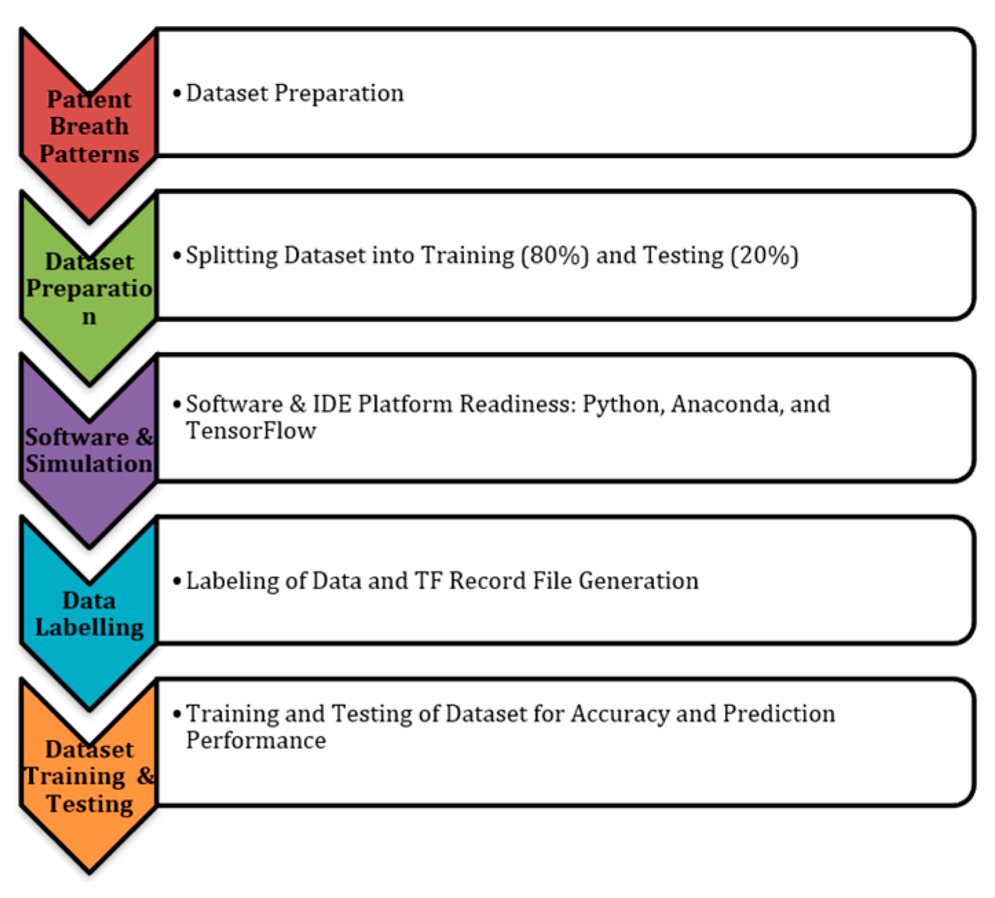
This work proposed a predictive supervised ML model to predict whether a male subject is infected with the virus. This process involves the proper selection of suspected patients, the selection of control subjects, obtaining proper consent for the study, data collection, data preparation, data exploration, model building, model evaluation, data interpretation, and lastly the prediction. In conformity with the acceptable standards of machine learning, approximately 80% of the dataset shall be used as the training dataset and the remaining 20% dataset [21] shall be used as a testing dataset to make a robust model for data prediction. Recently, numerical methods have been used in different fields [23,24,25,26,27,28,29,30,31]. It is worth mentioning that in these methods, AI has been widely used as a potential technique for various applications in recent years [30,31,32,33,34,35,36,37,38,39]. The proposed Flowchart of the Breath Detection Model with various stages of AI framework, e.g., breadth patterns recording, dataset preparation, simulation, data labelling, data training and testing are presented in Figure 2.

Corona COVID-19 Virus Dataset Challenge

In order to capture the finer details and variations of the human breath by the nano-arrays, the medical data shall include the following human (adult male/female, and child) breath components (proposed), e.g., breath-holding duration, breath sound intensity, and quality of air during normal breathing and forced inspiration as well as during forced expiration. The COVID-19 patient data shall also include the patient’s gender (male/female), age (adult/child) with relevant clinical history, viz. hypertension, heart disease, blood pressure, thyroid disorder, diabetes, TB, and recent history of travel or contact with COVID-19-infected persons, etc. The data (for dataset) are proposed to be collected from various sources such as hospitals or medical data records centers (as per stated medical and state norms). Thereafter, the data shall be prepared for exploratory data analysis (EDA) and then the data will be used in the experimental study of the initial diagnosis using ML algorithms. The foremost challenge in this work is about optimum data collection and data preparation from healthy and infected people of different age groups of both genders (males/females) in equal numbers with due consent from the participant after obtaining concurrence from various medical bodies, viz. state healthcare organizations, including hospitals and pathology services.

4. Breath Parameter and Dataset Preparation—The Implementation Strategy

In the implementation strategy, the study and analysis were carried out on a random and hypothetical dummy dataset with more than one thousand parameter samples and their important limits, provided in Table 1, Table 2 and Table 3; the spirometry, lung volumes, and lung capacities parameters, respectively. Similar studies can be carried out on these parameters in the state-of-the-art clinical patients’ dataset to identify the symptoms at an early phase of infection.

4.1. Electronic Spirometry (E-Spirometry) Analysis (Data Is Collected through the Mobile App)

A common office test is used to assess how well your lungs work by measuring how much air you inhale, how much you exhale, and how rapidly you exhale. The following spirometry parameters are being considered in the dataset with dummy values for predictive analysis. FVC—forced vital capacity: the total volume of air that can be exhaled during a maximally forced expiration effort (normal range FVC ≥ 80%).
  • FEV1—forced expiratory volume in one second: the volume of air exhaled in the first second under force after a maximal inhalation (normal range FEV1 ≥ 80%).
  • FEV1/FVC ratio: the percentage of the FVC expired in one second (normal range FEV1/FVC ≥ 70%).
  • FEV6—forced expiratory volume in six seconds: the amount of air exhaled after full inhalation within the first six seconds under force (normal range FEV6 ≥ 80%). FEF25–75%—forced expiratory flow: the FEP over the middle one half of the FVC; the average flow from the point at which 25% of the FVC has been exhaled to the point at which 75% of the FVC has been exhaled.
  • MVV—maximal voluntary ventilation: the volume of air expired in a specified period during repetitive maximal effort (normal range is 15–20 times and the resting minute volume average values for males and females are 140–180 and 80–120 L per minute, respectively).
  • PEF—peak expiratory flow: the highest forced expiratory flow is measured with a peak flow meter (normal range is 400–700 L/min).
Given the e-spirometry breath parameters (Table 1), an experimental analysis was performed using ML algorithms on the dummy dataset of 11,000 entries with about 1000 of them being COVID-19-positive data points, of which the majority has breathing parameters outside of the normal and acceptable range.
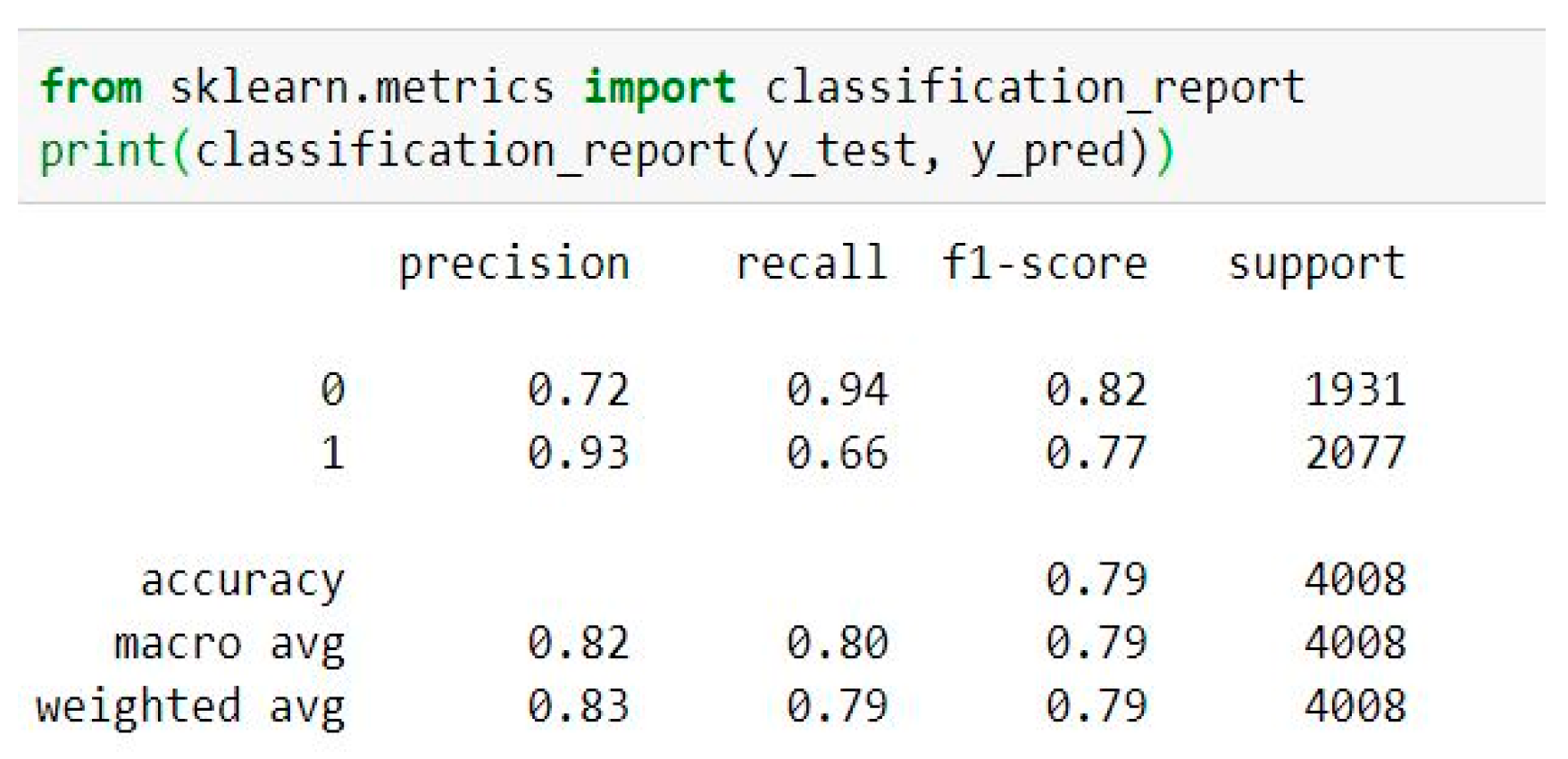
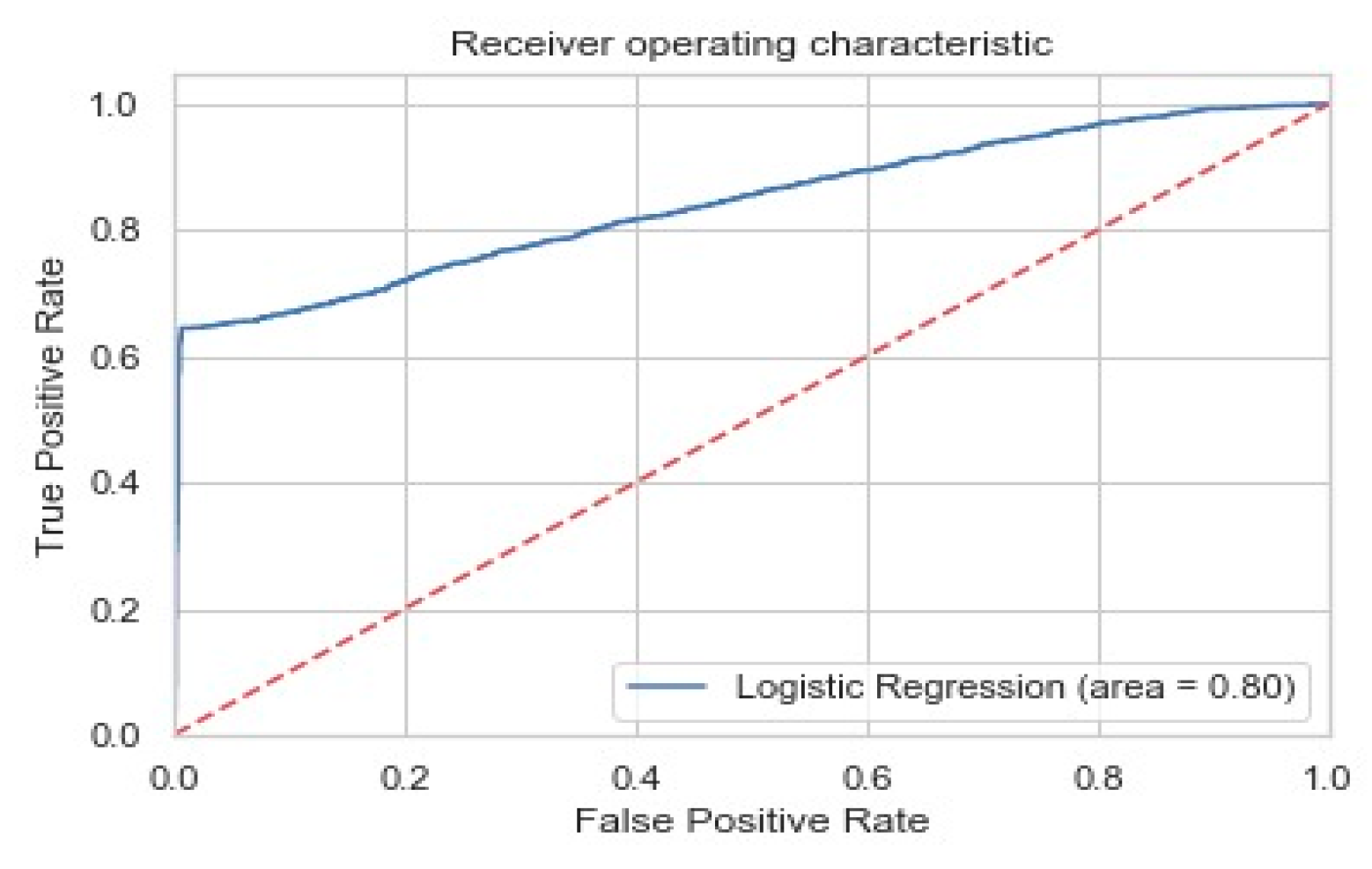
These results are presented in Figure 3 (confusion matrix), 4 (classification metrics report of the model), and 5 (ROCs curve of the LR model), which are obtained using a dummy dataset of 11,000 entries with about 1000 of them being COVID-19-positive data points. The imbalanced data are then converted into balanced data using SMOTE oversampling and a logistic regression model is trained. On plotting the confusion matrix (Figure 3) and calculating the classification metrics (Figure 4), namely precision, recall, and F1 score, a high value of precision (0.93) for COVID-19-positive data points is observed, which corresponds to a high value of true positives correctly predicted on this test data. This can also be shown by plotting the model’s ROCs (receiver operating characteristics) curve (Figure 5) and measuring the AUC (area under the curve), which appears to be 0.8, which means that our model’s overall accuracy is 80%. This accuracy can be further improved by hyper-parameter tuning when dealing with a real-life dataset.

4.2. Lung Volumes Analysis

In the dataset with dummy values for probabilistic predictive analysis, the following parameters are considered for lung volume parameters:
  • ERV—expiratory reserve volume: at the end of each exhalation, the maximum amount of air exhaled (normal range = 1.1 L (males) and 0.7 L (females));
  • IRV—inspiratory reserve volume: the maximum amount of air inhaled as a result of end-inspiration [40] (normal range = 3.3 L (males) and 1.9 L (females));
  • RV—residual volume: after a maximal exhale, the amount of air left in the lungs [40] (normal range = 1.2 L (males) and 1.1 L (females));
  • TV—tidal volume: during each respiratory cycle, the amount of air inhaled or exhaled (normal range = 0.5 L).
In Table 2, the lung volume parameters are considered as ERV, IRV, RV, and TV for male/female genders with their typical range values. To predict whether a patient (gender: male) is corona COVID-19-positive, a dummy dataset of 11,000 entries is considered, of which 1000 data points are corona COVID-19-positive, where a majority of the data points are outside of the normal acceptable range.
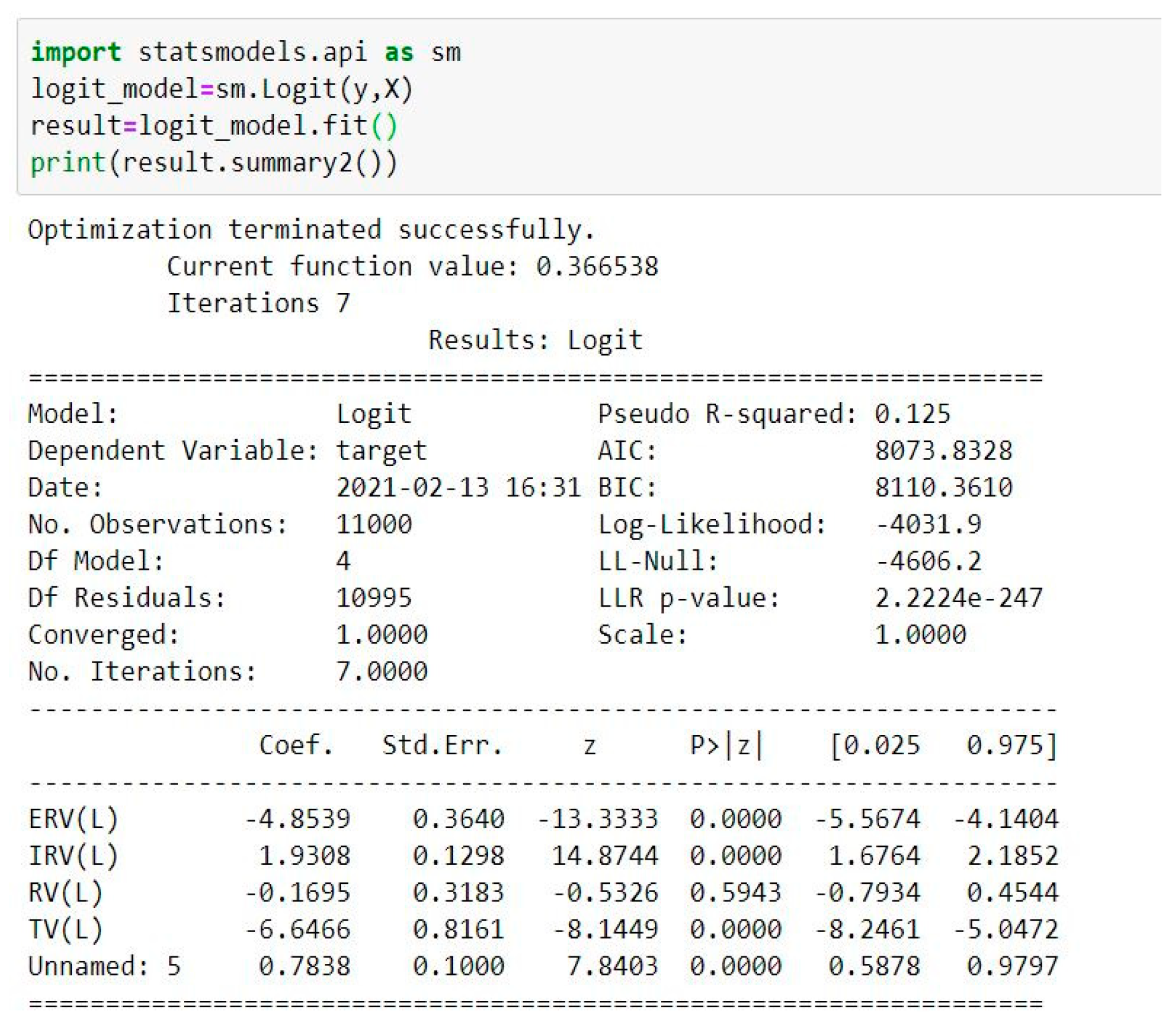
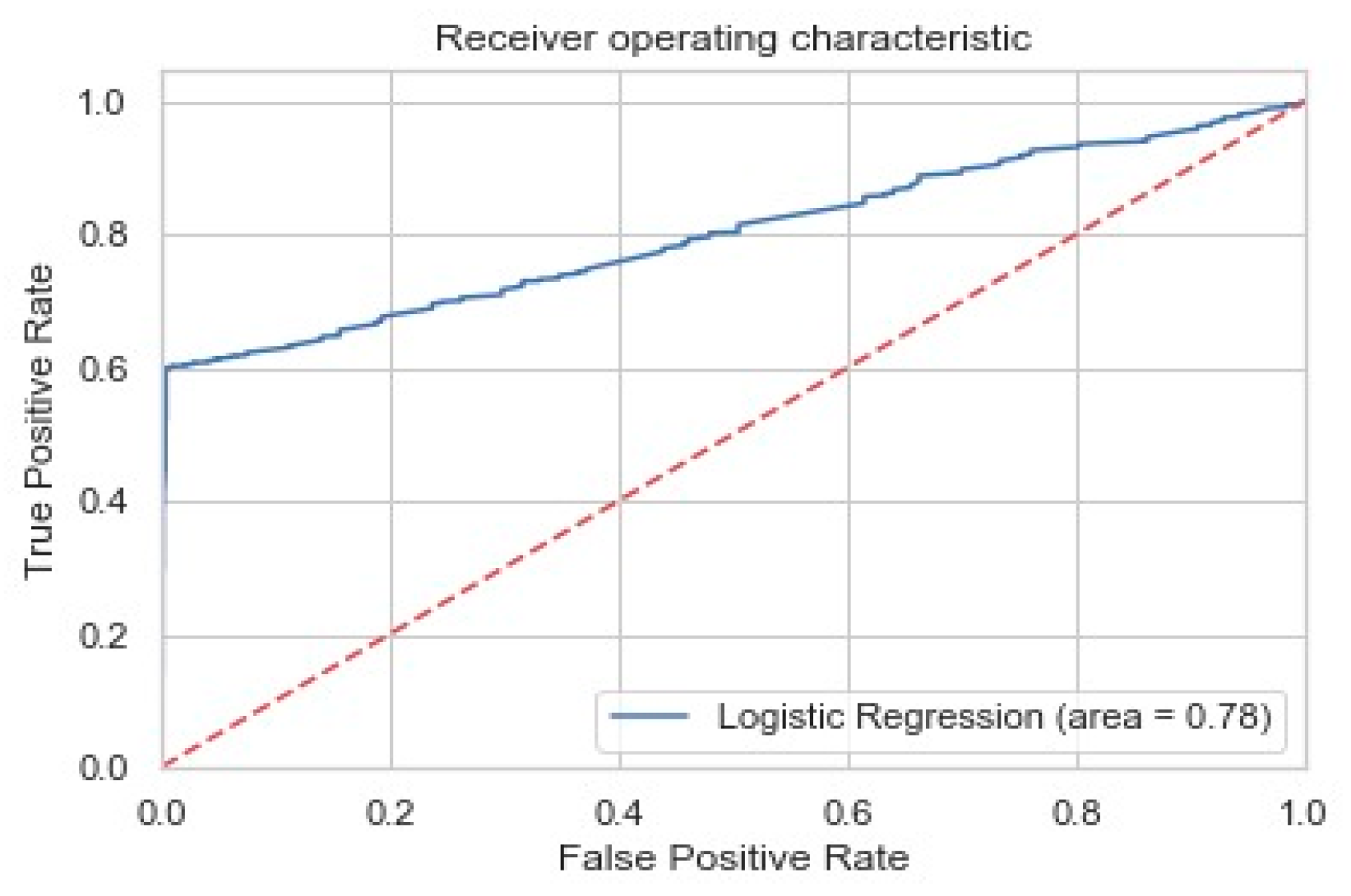
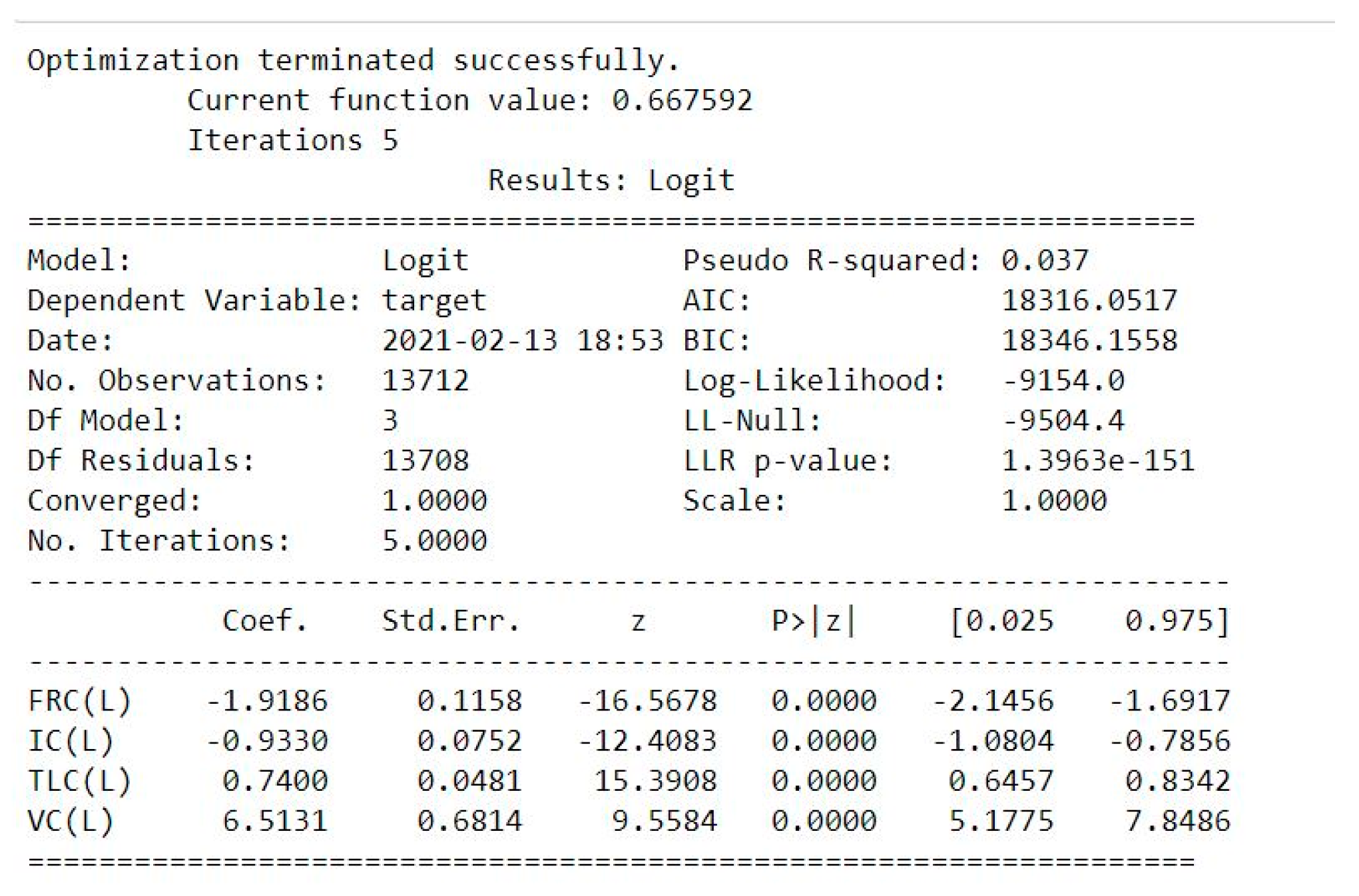
On the dataset with 11,000 entries, a logistic regression model is trained by selecting the parameters with a p-value of less than 0.05 from the model summary (Figure 6) to ensure max. AIC and BIC scores leading to the max. overall accuracy of the model. The confusion matrix (Figure 7) and other classification metrics (Figure 8) also indicate a high value of recall (0.95) value for COVID-19-negative patients, which indicates 95% of COVID-19-negative patients were identified correctly. The AUC for ROCs curve (Figure 9) for this model indicated an overall accuracy of 78%, which can be further improved by hyper-parameter tuning using the grid search cv pipeline for logistic regression in real-time patient data.

4.3. Lung Capacities Analysis

  • FRC—functional residual capacity: at resting end-expiration, the volume of air in the lungs [41] (normal range = 2.4 L (males) and 1.8 L (females));
  • IC—inspiratory capacity: from the resting expiratory level, the maximum volume of air that can be inhaled [41] (normal range = 3.8 L (males) and 2.4 L (females));
  • TLC—total lung capacity: the amount of air in the lungs when fully inflated (normal range = 6.0 L (males) and 4.2 L (females));
  • VC—vital capacity: after full inspiration, the highest volume is measured on complete exhale [40] (normal range = 4.8 L (males) and 3.1 L (females)).
The lung capacities parameters, e.g., FRC, IC, TLC, and VC, are taken into consideration (for male gender) with 11,000 entries in the dummy dataset and with 1000 of them as corona COVID-19-positive data points, with a majority of these are outside the normal range of allowable limits.
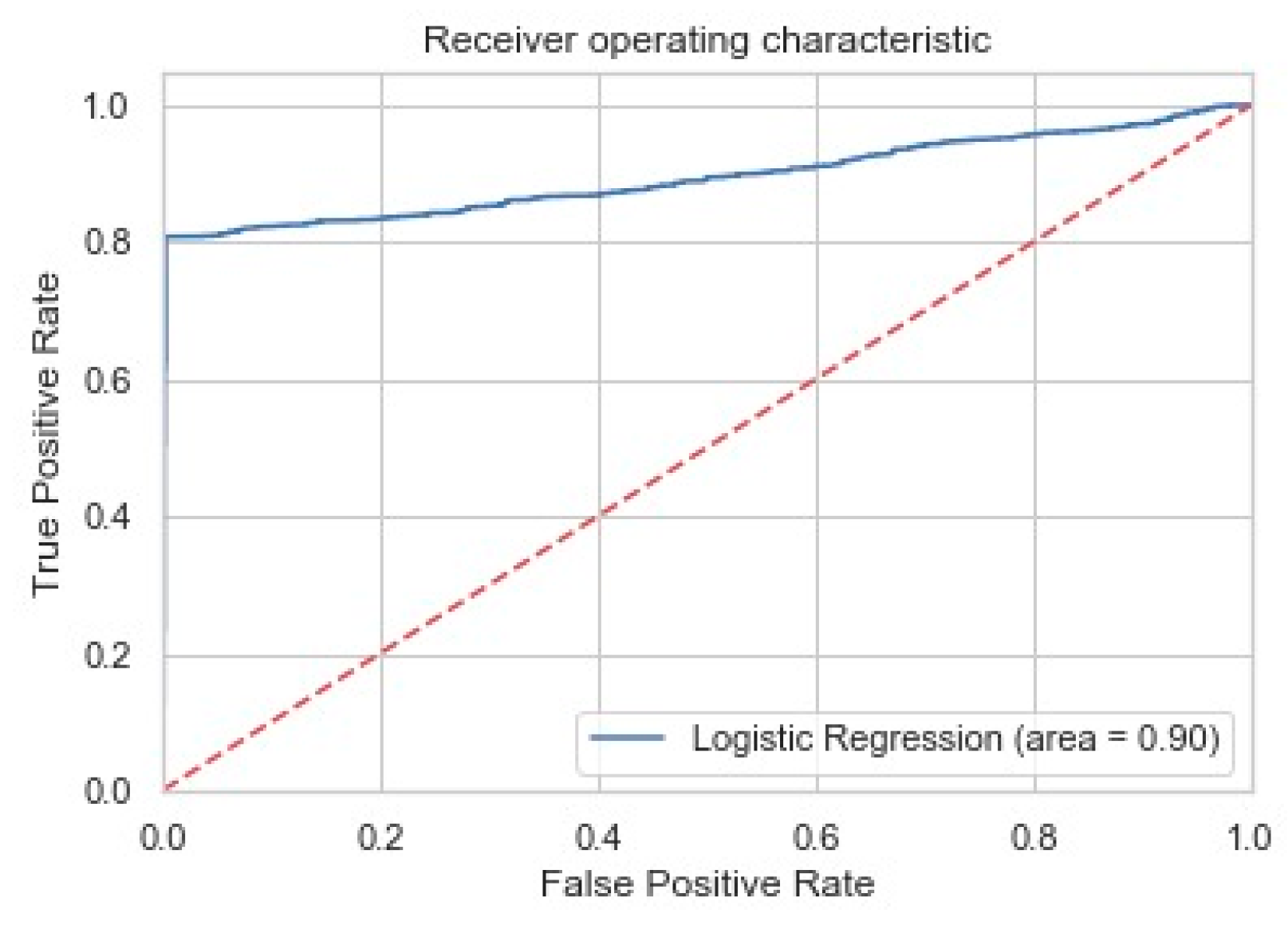
We now predict whether the patient is COVID-19-positive using the lung capacity parameters (FRC, IC, VC, and TLC) for male gender using a similar dummy dataset of 11,000 entries with about 1000 of them being COVID-19-positive data points, the majority of which have lung capacities outside of the normal range. Here, a logistic regression model is also trained after oversampling to convert the data into the balanced form and then using the parameters from the model summary which have a characteristic p-value of less than 0.05 (Figure 10). It is worth mentioning that the p-value is a number describing how likely it is that data would have occurred by random chance (i.e., that the null hypothesis is true). A p-value of less than 0.05 (typically ≤ 0.05) is statistically significant. It indicates strong evidence against the null hypothesis, as there is less than a 5% probability that the null is correct (and the results are random). On plotting the confusion matrix (Figure 11) and calculating the classification metrics, we observe that the model performs the best on this dataset, delivering very high values for both precision and recall (up to 98%) (Figure 12). The ROCs plot of the model indicates the AUC (overall accuracy of the model) coming out to be 90% (Figure 13). The results of the three different logistic regression curves explained above can further be the ensemble to enhance the overall accuracy of our resulting model.

5. Conclusions

This research work aims to develop an app to detect infections caused by the coronavirus COVID-19 using the patient’s available dataset, data preparation, algorithms of signal processing, and machine learning. The successful implementation of the breath testing and its analysis may proffer an early stage detection mechanism for not only a wide spectrum of human diseases affecting the respiratory tract, such as asthma, chronic obstructive pulmonary disease, and other underlying pathologies of heart, thyroid, etc., but also the status of the lungs, the strength of respiratory muscles, exercise tolerance, etc. For this research work, a logistic regression model is trained after using SMOTE oversampling to balance the data, and the accuracy levels achieved are 80%, 78%, and 90%. This also opens up the opportunity of using multi-layered ANN frameworks which can be useful in capturing more complex patterns when dealing with a real-life dataset. The human breath can also be used to monitor, record, and diagnose the psychological and emotional effects of the pandemic due to the lockdown, illness or death of kith or kin, and loss of jobs, the crash of businesses, or income. In the times ahead, the use of this study and invention could be extended to the regular patient diagnosis of many other ailments, such as tuberculosis, emphysema, pneumonia, etc., after approval from the competent bodies.
The results accomplished through in this study and experiments may not only aid the clinicians in their medical practice but may also bestow a blue chip to the masterminds engaged in the biomedical research for inventing more evolved, sophisticated, user-friendly, miniaturized, portable, and economical medical app/devices in the future.

Author Contributions

Methodology, A.M.M., N.K.S., M.R.R., I.A., R.M.A.Q., A.A.A.-Q., A.T., V.T. and R.A.-D.; Conceptualization, A.M.M., N.K.S., M.R.R., R.M.A.Q., A.A.A.-Q., A.T., V.T. and R.A.-D. All authors have read and agreed to the published version of the manuscript.

Funding

This research received no external funding.

Institutional Review Board Statement

Not applicable.

Informed Consent Statement

Not applicable.

Data Availability Statement

Not applicable.

Conflicts of Interest

The authors declare no conflict of interest.

References

  1. World Health Organization. Middle East Respiratory Syndrome Coronavirus (MERS-CoV); WHO: Geneva, Switzerland, 2019; Available online: http://www.who.int/emergencies/mers-cov/en/ (accessed on 16 September 2022).
  2. Crawford, D.H. Viruses: A Very Short Introduction, 2nd ed.; Oxford University Press Print: Oxford, UK, 2018; ISBN 13 9780198811718. [Google Scholar] [CrossRef]
  3. European Centre for Disease Control and Prevention Rapid Risk Assessment. Outbreak of Severe Acute Respiratory Syndrome Coronavirus 2 (SARS-CoV-2): Increased Transmission beyond China—Fourth Update; ECDC: Stockholm, Sweden, 2020. [Google Scholar]
  4. Zaki, A.M.; van Boheemen, S.; Bestebroer, T.M.; Osterhaus, A.D.M.E.; Fouchier, R.A.M. Isolation of a novel coronavirus from a man with pneumonia in Saudi Arabia. N. Engl. J. Med. 2012, 367, 1814–1820. [Google Scholar] [CrossRef] [PubMed]
  5. Bhargava, A.; Shewade, H.D. The potential impact of the COVID-19 response-related lockdown on TB incidence and mortality in India. Indian J. Tuberc. 2020, 67, S139–S146. [Google Scholar] [CrossRef] [PubMed]
  6. Coronavirus disease (COVID-19) Pandemic. World Health Organization (WHO). 7 July 2020. Available online: https://www.who.int/emergencies/diseases/novel-coronavirus-2019 (accessed on 16 September 2022).
  7. Diamond, F. Asymptomatic Carriers May Play a Huge Part in COVID-19 Surge. 12 January 2021. Available online: https://www.infectioncontroltoday.com/view/asymptomatic-carriers-may-play-a-huge-part-in-covid-19-surge (accessed on 16 September 2022).
  8. Edwards, R.; Davidson, E.; Jamieson, L.; Weller, S. Theory and the breadth-and-depth method of analyzing large amounts of qualitative data: A research note. Qual. Quant. 2021, 55, 1275–1280. [Google Scholar] [CrossRef]
  9. Adiguzel, Y.; Kulah, H. Breath sensors for lung cancer diagnosis. Biosens. Bioelectron. 2015, 65, 121–138. [Google Scholar] [CrossRef]
  10. Kononov, A.; Korotetsky, B.; Jahatspanian, I.; Gubal, A.; Vasiliev, A.; Arsenjev, A.; Nefedov, A.; Barchuk, A.; Gorbunov, I.; Kozyrev, K.; et al. Online breath analysis using metal oxide semiconductor sensors (electronic nose) for diagnosis of lung cancer. J. Breath Res. 2019, 14, 016004. [Google Scholar] [CrossRef]
  11. Rehman, A.U.; Jiang, A.; Rehman, A.; Paul, A.; Din, S.; Sadiq, M.T. Identification and role of opinion leaders in information diffusion for online discussion network. J. Ambient. Intell. Humaniz. Comput. 2020, 1–13. [Google Scholar] [CrossRef]
  12. Rehman, A.U.; Tariq, R.; Rehman, A.; Paul, A. Collapse of Online Social Networks: Structural Evaluation, Open Challenges, and Proposed Solutions. In Proceedings of the 2020 IEEE Globecom Workshops (GC Wkshps), Taipei, Taiwan, 7–11 December 2020; pp. 1–6. [Google Scholar] [CrossRef]
  13. Davidson, E.; Edwards, R.; Jamieson, L.; Weller, S. Big data, qualitative style: A breadth-and-depth method for working with large amounts of secondary qualitative data. Qual. Quant. 2018, 53, 363–376. [Google Scholar] [CrossRef] [Green Version]
  14. Priftis, K.N.; Hadjileontiadis, L.J.; Everard, M.L. Breath Sounds. In From Basic Science to Clinical Practice, 1st ed.; Springer: Cham, Switzerland, 2018. [Google Scholar] [CrossRef]
  15. Valenti, M.; Tonelli, D.; Vesperini, F.; Principi, E.; Squartini, S. A Neural Network Approach for Sound Event Detection in Real Life Audio. In Proceedings of the 2017 25th European Signal Processing Conference (EUSIPCO), Kos, Greece, 28 August–2 September 2017. [Google Scholar] [CrossRef] [Green Version]
  16. Nakhleh, M.K.; Amal, H.; Jeries, R.; Broza, Y.Y.; Aboud, M.; Gharra, A.; Ivgi, H.; Khatib, S.; Badarneh, S.; Har-Shai, L.; et al. Diagnosis and classification of 17 diseases from 1404 subjects via pattern analysis of exhaled molecules. ACS Nano 2017, 11, 112–125. [Google Scholar] [CrossRef] [Green Version]
  17. Lee, S.J.; Motai, Y.; Weiss, E.; Sun, S.S. Irregular Breathing Classification From Multiple Patient Datasets Using Neural Networks. IEEE Trans. Inf. Technol. Biomed. 2012, 16, 1253–1264. [Google Scholar]
  18. Ardabili, S.; Mosavi, A.; Ghamisi, P.; Ferdinand, F.; Varkonyi-Koczy, A.; Reuter, U.; Rabczuk, T.; Atkinson, P. COVID-19 Outbreak Prediction with Machine Learning. Algorithms 2020, 13, 249. [Google Scholar] [CrossRef]
  19. Mok, K. Scientists’ Artificially Intelligent Nanoarray Can Diagnose Disease Using Your Breath. 8 January 2017. Available online: https://thenewstack.io/scientists-artificially-intelligent-nanoarray-can-diagnose-disease-using-breath/ (accessed on 16 September 2022).
  20. UNICEF for Every Child. What Is the Difference between Home Self-Isolation and Quarantine? Ministry of Health, Romania. Available online: https://www.unicef.org/romania/documents/what-difference-between-home-self-isolation-and-quarantine (accessed on 16 September 2022).
  21. Ahmed, M.A.; Colebunders, R.; Fodjo, J.N.S. Evidence for significant COVID-19 community transmission in Somalia using a clinical case definition. Int. J. Infect. Dis. 2020, 98, 206–207. [Google Scholar] [CrossRef] [PubMed]
  22. Pitol, A.K.; Julian, T.R. Community Transmission of SARS-CoV-2 by Surfaces: Risks and Risk Reduction Strategies. Environ. Sci. Technol. Lett. 2021, 8, 263–269. [Google Scholar] [CrossRef]
  23. Nguyen, G.; Dlugolinsky, S.; Bobák, M.; Tran, V.; García, L.; Heredia, I.; Malík, P.; Hluchý, L. Machine Learning and Deep Learning frameworks and libraries for large-scale data mining: A survey. Artif. Intell. Rev. 2019, 52, 77–124. [Google Scholar] [CrossRef] [Green Version]
  24. Nazemi, E.; Roshani, G.H.; Feghhi, S.A.H.; Setayeshi, S.; Zadeh, E.E.; Fatehi, A. Optimization of a method for identifying the flow regime and measuring void fraction in a broad beam gamma-ray attenuation technique. Int. J. Hydrogen Energy 2016, 41, 7438–7444. [Google Scholar] [CrossRef]
  25. Shukla, N.K.; Mayet, A.M.; Vats, A.; Aggarwal, M.; Raja, R.K.; Verma, R.; Muqeet, M.A. High speed integrated RF–VLC data communication system: Performance constraints and capacity considerations. Phys. Commun. 2021, 50, 101492. [Google Scholar] [CrossRef]
  26. Roshani, G.H.; Nazemi, E.; Feghhi, S.A.; Setayeshi, S. Flow regime identification and void fraction prediction in two-phase flows based on gamma ray attenuation. Measurment 2015, 62, 25–32. [Google Scholar] [CrossRef]
  27. Mayet, A.; Hussain, M. Amorphous WNx Metal for Accelerometers and Gyroscope. In Proceedings of the MRS Fall Meeting, Boston, MA, USA, 30 November–5 December 2014. [Google Scholar]
  28. Karami, A.; Roshani, G.H.; Khazaei, A.; Nazemi, E.; Fallahi, M. Investigation of different sources in order to optimize the nuclear metering system of gas–oil–water annular flows. Neural Comput. Appl. 2018, 32, 3619–3631. [Google Scholar] [CrossRef]
  29. Mayet, A.; Smith, C.; Hussain, M.M. Amorphous metal based nanoelectromechanical switch. In Proceedings of the 2013 Saudi International Electronics, Communications and Photonics Conference (SIECPC), Riyadh, Saudi Arabia, 27–30 April 2013; pp. 1–5. [Google Scholar]
  30. Nazemi, E.; Feghhi, S.A.H.; Roshani, G.H.; Peyvandi, R.G.; Setayeshi, S. Precise Void Fraction Measurement in Two-phase Flows Independent of the Flow Regime Using Gamma-ray Attenuation. Nucl. Eng. Technol. 2016, 48, 64–71. [Google Scholar] [CrossRef] [Green Version]
  31. Mayet, A.M.; Alizadeh, S.M.; Kakarash, Z.A.; Al-Qahtani, A.A.; Alanazi, A.K.; Alhashimi, H.H.; Eftekhari-Zadeh, E.; Nazemi, E. Introducing a Precise System for Determining Volume Percentages Independent of Scale Thickness and Type of Flow Regime. Mathematics 2022, 10, 1770. [Google Scholar] [CrossRef]
  32. Alanazi, A.K.; Alizadeh, S.M.; Nurgalieva, K.S.; Nesic, S.; Guerrero, J.W.G.; Abo-Dief, H.M.; Eftekhari-Zadeh, E.; Nazemi, E.; Narozhnyy, I.M. Application of Neural Network and Time-Domain Feature Extraction Techniques for Determining Volumetric Percentages and the Type of Two Phase Flow Regimes Independent of Scale Layer Thickness. Appl. Sci. 2022, 12, 1336. [Google Scholar] [CrossRef]
  33. Mayet, A.M.; Alizadeh, S.M.; Nurgalieva, K.S.; Hanus, R.; Nazemi, E.; Narozhnyy, I.M. Extraction of Time-Domain Characteristics and Selection of Effective Features Using Correlation Analysis to Increase the Accuracy of Petroleum Fluid Monitoring Systems. Energies 2022, 15, 1986. [Google Scholar] [CrossRef]
  34. Hosseini, S.; Taylan, O.; Abusurrah, M.; Akilan, T.; Nazemi, E.; Eftekhari-Zadeh, E.; Bano, F.; Roshani, G.H. Application of Wavelet Feature Extraction and Artificial Neural Networks for Improving the Performance of Gas–Liquid Two-Phase Flow Meters Used in Oil and Petrochemical Industries. Polymers 2021, 13, 3647. [Google Scholar] [CrossRef]
  35. Mayet, A.; Smith, C.E.; Hussain, M.M. Energy reversible switching from amorphous metal based nanoelectromechanical switch. In Proceedings of the 2013 13th IEEE Conference on Nanotechnology (IEEE-NANO), Beijing, China, 5–8 August 2013; pp. 366–369. [Google Scholar]
  36. Roshani, G.; Hanus, R.; Khazaei, A.; Zych, M.; Nazemi, E.; Mosorov, V. Density and velocity determination for single-phase flow based on radiotracer technique and neural networks. Flow Meas. Instrum. 2018, 61, 9–14. [Google Scholar] [CrossRef]
  37. Mayet, A.M.; Salama, A.S.; Alizadeh, S.M.; Nesic, S.; Guerrero, J.W.G.; Eftekhari-Zadeh, E.; Nazemi, E.; Iliyasu, A.M. Applying Data Mining and Artificial Intelligence Techniques for High Precision Measuring of the Two-Phase Flow’s Characteristics Independent of the Pipe’s Scale Layer. Electronics 2022, 11, 459. [Google Scholar] [CrossRef]
  38. Iliyasu, A.M.; Alizadeh, S.M.; Salama, A.S.; Nazemi, E.; Hirota, K. Application of artificial intelligence to calculate the volume percentages of a stratified regime three-phase flow regardless of the pipe’s scale thickness. Appl. Sci. 2022, 10, 1996. [Google Scholar] [CrossRef]
  39. Mayet, A.; Hussain, A.; Hussain, M. Three-terminal nanoelectromechanical switch based on tungsten nitride, an amorphous metallic material. Nanotechnology 2016, 27. [Google Scholar] [CrossRef] [PubMed]
  40. Thafasal Ijyas, V.P.; Mayet, A.M. Electronic circuit implementation of the compartmental models for population dynamics of COVID-19 like epidemics. Sādhanā 2022, 47, 1–6. [Google Scholar] [CrossRef]
  41. Barreiro, T.J.; Perillo, I. An approach to interpreting spirometry. Am. Fam. Physician 2004, 69, 1107–1114. [Google Scholar] [PubMed]
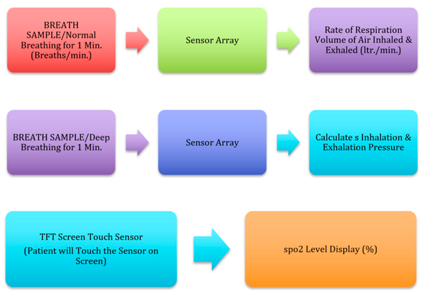
Figure 1. Human respiration mechanism and detection system.
Electronics 12 00010 g001
Figure 2. Flowchart of breath detection model.
Electronics 12 00010 g002
Figure 3. Confusion matrix of spirometry breath parameters dataset [21].
Electronics 12 00010 g003
Figure 4. Classification metrics report of the model of e-spirometry breath parameters dataset.
Electronics 12 00010 g004
Figure 5. ROCs curve of the LR model of e-spirometry breath parameters dataset.
Electronics 12 00010 g005
Figure 6. Logistic regression model summary for lung volume analysis.
Electronics 12 00010 g006
Figure 7. Confusion matrix for lung volume analysis.
Electronics 12 00010 g007
Figure 8. Classification metrics report for lung volume analysis.
Electronics 12 00010 g008
Figure 9. ROCs curve of the LR model for lung volume analysis.
Electronics 12 00010 g009
Figure 10. Logistic regression model summary of lung capacities parameters.
Electronics 12 00010 g010
Figure 11. Confusion matrix of lung capacities parameters.
Electronics 12 00010 g011
Figure 12. Classification metrics report of lung capacities parameters.
Electronics 12 00010 g012
Figure 13. ROCs curve of the LR model of lung capacities parameters.
Electronics 12 00010 g013
Table 1. E-Spirometry breadth parameters.
ParameterFVCFEV1RATIO FEV1/FVCFEV6FEFMVVPEF
Normal80% or more80% or more/second70% or more80%/6 s or more25–75%140–180 per liter per minute (Male)80–120 per liter per minute (Female)400–700 L/min
Table 2. Lung volumes parameters.
ParameterERVIRVRVTV
Normal1.2 L (Male)
0.7 L (Female)
3.3 L (Male)
1.9 L (Female)
1.2 L (Male)
1.1 L (Female)
0.5 L
Table 3. Lung Capacities Parameters.
ParameterFRCICTLCVC
Normal2.4 L (Male)
1.8 L (Female)
3.8 L (Male)
2.4 L (Female)
6.0 L (Male)
4.2 L (Female)
4.8 L (Male)
3.1 L (Female)
Disclaimer/Publisher’s Note: The statements, opinions and data contained in all publications are solely those of the individual author(s) and contributor(s) and not of MDPI and/or the editor(s). MDPI and/or the editor(s) disclaim responsibility for any injury to people or property resulting from any ideas, methods, instructions or products referred to in the content.