Review

Overview of Machine Learning and Deep Learning Approaches for Detecting Shockable Rhythms in AED in the Absence or Presence of CPR

Department of Electrical and Computer Engineering, The University of Memphis, Memphis, TN 38152, USA
*
Author to whom correspondence should be addressed.
Electronics 2022, 11(21), 3593; https://doi.org/10.3390/electronics11213593
Submission received: 6 October 2022 / Revised: 28 October 2022 / Accepted: 1 November 2022 / Published: 3 November 2022
(This article belongs to the Section Artificial Intelligence)

Abstract

:
Sudden Cardiac Arrest (SCA) is one of the leading causes of death worldwide. Therefore, timely and accurate detection of such arrests and immediate defibrillation support for the victim is critical. An automated external defibrillator (AED) is a medical device that diagnoses the rhythms and provides electric shocks to SCA patients to restore normal heart rhythms. Machine learning and deep learning-based approaches are popular in AEDs for detecting shockable rhythms and automating defibrillation. There are some works in the literature for reviewing various machine learning (ML) and deep learning (DL) algorithms for shockable ECG signals in AED. Starting in 2017 and beyond, different DL algorithms were proposed for the AED. This paper provides an overview of AED, including its circuit diagram and application to SCA patients. It also presents the most up-to-date ML and DL approaches for detecting shockable rhythms in AEDs without cardiopulmonary resuscitation (CPR) or during CPR. It also provides a performance comparison of these approaches and discusses other researchers’ results that lay the foundation for researchers to delve in-depth. Furthermore, the research gaps and recommendations for future research provided in this review paper will be helpful to the researchers, scientists, and engineers in conducting further research in this critical field.

1. Introduction

An automated external defibrillator (AED) is a medical device that provides defibrillation to treat persons suffering from SCA [1,2]. It delivers electric shocks to the heart through the chest to restore the normal heart rhythm of SCA victims [3]. SCA is a significant public health problem that causes the patient’s heart to stop beating suddenly and unexpectedly, causing patients to lose consciousness, become unresponsive, and perhaps die if medical help is not given within a few minutes. There is a 90–95% chance of patients dying if the treatment is not provided within 10 min [4]. Hence, more treatment delay for SCA victims means more chance of patient death. When defibrillation is delayed for more than 10 min, the recovery rate of victims drops to 5–10% [5]. Because response time is critical for SCA victims, we can shorten it by utilizing nearby AED immediately. Furthermore, AED is portable, easy to use, and readily available in public areas, such as universities, shopping malls, cinemas, train/bus stations, and office buildings, leading to a reliable and affordable way to treat a person having a cardiac arrest. Furthermore, only those SCA sufferers with shockable cardiac rhythms can be treated with an AED. As a result, the heart rhythms of SCA victims are initially analyzed by AED to determine whether the heart rhythms are shockable or non-shockable. Shockable rhythms are those rhythms than can be treated with AED. If the cardiac rhythm becomes shockable, the AED administers an electrical shock to bring it back to normal [6]. However, non-shockable rhythms have a minimal chance of defibrillation. Furthermore, suppose the patient has a non-shockable heart rhythm such as Asystole or pulseless electrical activity (PEA). In that case, the patient will require chest compression, ventilator, and medication; thus, getting to the hospital is critical [7].
AED’s main components are a battery, a capacitor, electrodes, and an electrical circuit designed to analyze the rhythm and send an electric shock when needed [8]. An operator is needed to connect the AED device to the SCA victims and run the device. The housing of the AED device includes an ON/OFF button to turn the device on or off, a charging button to charge the device, and a discharge button to provide an electric shock to the patient. In addition, it has a monitor to display instructions, a voice prompt to assist the operator easily, and two electronic pads to collect information about the patient’s heart rhythms. Currently, all AEDs have biphasic defibrillation waveform shock technology. The biphasic AEDs use a bidirectional current flow and offer defibrillation successfully at lower energy levels comparable with monophasic defibrillators [9]. In addition, most AEDs contain memory cards that can be used to record the use of each AED, which can be reviewed in full after the event.
Even though AEDs have contributed to better survival of out-of-hospital cardiac arrest victims, there have been incidents of their malfunctioning [10]. AED is a life-saving device for critical patients with SCA; its failure could result in death. SCA victims’ unexpected deaths due to device failure and late response can be reduced by reducing AED equipment errors and time delays in diagnosing the rhythms. As a result, when an AED is in use, it must function appropriately while being used. Because AED faults are rare, the total number of AED malfunctions is insignificant compared with the total number of lives saved [11]. However, if an AED fails, a person’s chance of unexpected dying increases.
Furthermore, the time required to determine whether the patient’s heart rhythm is shockable should be minimal so that the AED can select whether to deliver an electric shock immediately. Different machine-learning techniques can detect shockable rhythms in AED [12]. In 2017 and after, deep learning algorithms became more popular because of their better performance. However, we must develop a model with no error to detect the shockable or non-shockable rhythm in a short time (less than seconds) and provide defibrillation to the patient as quickly as feasible when necessary. In addition, we need to develop a reliable approach for regularly locating and repairing possibly defective AED devices [11].
Since chest compressions produce artifacts in the ECG, cardiopulmonary resuscitation (CPR) must be stopped for a reliable automated rhythm analysis [13]. However, interrupting CPR has a detrimental effect on survival. Indeed, interruption of chest compressions decreases the chances of adequate resuscitation by up to 50%. According to AHA, immediate CPR can double or triple a SCA patient’s chance of survival [14]. Current AEDs necessitate pausing CPR as ECG data is processed, depriving the brain of essential oxygen [15]. One of the most important goals for growing the out-of-hospital cardiac arrest survival rate is to detect shockable rhythms early and accurately without interrupting CPR.
This paper reviews the different types of machine learning and deep learning techniques for detecting shockable rhythms in AED during the absence or presence of CPR and compares these models in terms of specificity and sensitivity. The specificity and sensitivity must be greater than 95% and 90%, respectively, to meet the American Health Association’s (AHA)’s criteria for AED. Overall, this paper analyzes different ML and DL algorithms’ performance, research gaps, and future directions in AED, leading to further development of this field.
The remainder of the paper is organized as follows. Section 2 gives rhythm annotations. Section 3 explains ECG databases, and Section 4 discusses the calculation of sensitivity and specificity. Section 5 and Section 6 discuss the DL/ML methods that have been applied while CPR is interrupted and during CPR. Section 7 provides a discussion, and Section 8 contains limitations of surveyed works and recommendations for future research. Finally, the conclusion is stated in Section 9.

2. Rhythms Annotation External Defibrillators

The American Heart Association rhythm classification scheme defines the following basic shockable and non-shockable rhythms below [16,17].

2.1. Shockable Rhythms

Shockable rhythms are those heart rhythms that are faster than normal heart rhythms and can be treated with defibrillation shocks. The primary purpose of AED is to provide defibrillation to the SCA patient only after detecting shockable rhythms.
There are two types of shockable rhythms, which are discussed below:

2.1.1. Ventricular Fibrillation (VF)

In this type of rhythm, the heart rate is too high and goes up to 500 bpm. It is a dangerous cardiac disturbance; if no shock is delivered within a few minutes, this may result in hypoxic brain damage and death.

2.1.2. Ventricular Tachycardia (VT)

In this type of rhythm, the heart rate is higher than normal, in the range of 150–250 bpm. Defibrillation can reduce such heart rate to normal.

2.2. Non-Shockable Rhythms

Non-shockable rhythms are those rhythms that cannot be treated with defibrillation shocks. These types of rhythms vary from normal to very dangerous. The AED does not provide defibrillation if it detects non-shockable rhythms.
There are three types of non-shockable rhythms:

2.2.1. Asystole (ASYS)

This is a flatline rhythm meaning there is no heartbeat or electrical activity. The heart is not functioning, and defibrillation does not work; hence, less than 2% of people with Asystole survive.

2.2.2. Pulseless Electrical Activity (PEA)

This is a life-threatening and unshockable cardiac rhythm. Despite the presence of coordinated cardiac electrical activity in this rhythm, there is no perceptible pulse.

2.2.3. Other Non-Shockable Rhythms (ONR)

These rhythms are not associated with cardiac arrest. Examples include normal sinus rhythm (NSR), supraventricular tachycardias, sinus bradycardia, atrial fibrillation and flutter, heart blocks, idioventricular rhythms, premature atrial or ventricular contractions, atrioventricular nodal reentrant tachycardia (AVNRT), bigeminy, etc.
Figure 1 [16] shows different types of heart rhythms. ASYS is the most dangerous; there is a minimal chance of defibrillation and a higher chance of a patient’s death if immediate treatment is not provided. VT and VF rhythms are faster than NSR, whereas ASYS is slower than NSR.

3. ECG Databases

The widely used ECG databases are public Holter ECG databases, which continuously monitor patients with ventricular arrhythmias, and OHCA databases, recorded by AEDs from cardiac arrest patients. The sample rate of both databases is 250 Hz.

3.1. Public Holter Databases

The following three public databases are used primarily in VF detection in AED.
  • AHA fibrillation database (AHADB) [18]: includes 30 min ECG recordings from 10 patients.
  • Massachusetts Institute of Technology-Beth Israel Hospital (MIT-BIH) malignant ventricular ectopy database (VFDB) [19]: includes 22 half-hour ECG recordings of patients who experienced ventricular tachycardia, ventricular flutter, and ventricular fibrillation.
  • Creighton University (CU) ventricular tachyarrhythmia database (CUDB) [20]: includes 35 eight-minute ECG recordings of people who have undergone sustained ventricular tachycardia, ventricular flutter, and ventricular fibrillation episodes.
  • MIT-BIH Normal Sinus Rhythm Database ((NSRDB) [21]: includes 18 long-term ECG recordings of 18 subjects.
  • MIT-BIH AF Database (AFDB) [22]: includes 25 long-term ECG recordings (of human subjects with AF.
  • Sudden Cardiac Death Holter Database (SDBB) [23]: includes 18 patients with underlying sinus rhythm.

3.2. Out-of-Hospital Cardiac Arrests (OHAC) Databases

The OHCA databases include ventricular fibrillation, ventricular tachycardia, Asystole, and coordinated pulse rhythms (Pulsed Rhythms, PR) and, without pulses, rhythms collected by AEDs from SCA patients during treatment [24].

4. Performances Metrics

AED’s effectiveness depends on its ability to detect shockable (Sh) rhythms and how correctly the operator can use them [25]. An AED device’s accuracy is measured by quantifying the performance of the machine learning/deep learning technique to classify Sh or NSh rhythms. Specificity and sensitivity are the most widely used metrics among various performance metrics. For AED, sensitivity is the percentage of the total number of shock advisories for patients with Sh rhythm, and specificity is the percentage of the total number of no-shock advisories for patients with NSh rhythm [25]. “The American Health Association recommends a sensitivity (Se) higher than 90% for Sh rhythms, and a specificity higher than 95% for NSh rhythms, and above 99% in the case of normal sinus rhythms” [12].
Table 1 represents the confusion matrix that generally reflects how efficiently a machine learning algorithm classifies the ECG data, where TP, FP, FN, and TN are performance parameters and are described below [26]:
  • True positive (TP): shock is correctly advised for a shockable rhythm
  • False positive (FP): shock incorrectly advised for a non-shockable rhythm
  • False negative (FN): no shock is advised for a shockable rhythm
  • True negative (TN): no shock is advised for a non-shockable rhythm
Furthermore, the sensitivity and specificity equations are given below [27]:
Sensitivity   Se = TP TP + FN
Specificity   Sp = TN TN + FP

5. Deep Learning and Machine Learning Techniques for Detecting Shockable Rhythms in AED While CPR Is Not Being Applied

The conventional ML approaches to identifying shockable rhythms include preprocessing, extracting features, selecting features, and classification carried out by independent algorithms [28]. However, DL methods automatically perform feature extraction. The DL approaches have been used widely since 2017 for AED to detect shockable rhythms. The DL approaches outperform the classical feature extraction classification algorithms [12]. In addition, currently, DL (for feature extraction and selection) and ML (for classification) are also used to detect and analyze AED rhythms. Some of these approaches are discussed below:

5.1. Support Vector Machine (SVM)

An SVM is a binary supervised classifier used for classification, regression, and outlier detection [28]. It classifies both linear and non-linear data by utilizing a hyperplane by considering a more considerable margin between the two classes. However, it uses a kernel trick for the classification of non-linear data. Kernel trick means mapping the input data from input space to higher dimensional spaces (called feature space). SVM is a robust classification algorithm because of its simple structure and fewer feature requirements [29].
Li et al. [30] proposed a machine-learning approach to classify VF and rapid VT using an SVM. They used three public domain ECG databases: AHADB as training, the CUDB as a test, and VFDB as a validation dataset. A genetic algorithm (GA) was applied to select optimal features. Furthermore, the best combination of features was selected for the training and testing data for VF classification using the SVM classifier in the development phase. Then, in the validation phase, the fivefold CV was applied to the validation dataset, and then SVM was used for classification. They reported that they obtained accuracy (Acc) of 98.1%, Se of 98.4%, and Sp of 98.0% for training data with 5 s window size, whereas Acc of 96.3%, Se of 96.2%, and Sp of 96.2% were obtained by fivefold cross-validation for validation data.
Nam et al. [31] proposed a detection system using SVM for classifying shockable and non-shockable rhythms based on a single feature. They used multiple databases, e.g., MITDB, CUDB, and AHADB, which were sampled at 360, 250, and 250 Hz, respectively. They first down-sampled MITDB to 250 Hz and then employed it. Before applying the classifier, they extracted a single feature from raw ECG signals using a discrete wavelet transform (DWT). For the transform, Harr’s wavelet was chosen, as it has a high processing speed, and then various features were extracted at various window lengths (WLs) of 3–5 s. The extracted features were fed into the SVM classifier per WL. The kernel function was decided upon to be the radial basis function. The allowed range of classification error C and the width of the kernel were established empirically. The fivefold cross-validation was performed on various WLs and calculated sensitivities and specificities to evaluate the performance of the applied model. They achieved Se of 97.8%, 97.7%, and 98.8% for WL 3 s, 4 s, and 5 s, respectively. Similarly, Sp of 98.0%, 98.3%, and 98.4% were achieved for WL 3 s, 4 s, and 5 s, respectively.

5.2. Random Forest (RF)

RF is a classification system and regression based on the aggregation of many decision trees [32]. Specifically, an ensemble of trees constructed from a training data set and internally validated to yield a prediction of the answer provided the predictors for future observations.
Tripathy et al. [33] proposed a novel method for detecting and classifying shockable and non-shockable rhythms. To decompose the input ECG signals into the number of modes, they used variational mode decomposition (VMD). They evaluated the first three modes: energy, real entropy, and permutation entropy. Then, these modes were used for feature extraction. Furthermore, the extracted features were evaluated using the RF classifier. The proposed model achieved Ac of 97.23%, Se of 96.54%, and Sp of 97.97%.

5.3. Boosting and Logistic Regression (B-LR)

A boosting classifier is one ensemble classifier that selects optimal features to reduce the classifier’s error rate [34]. To increase the classifier’s performance, it converts the weak classifier into a strong classifier by selecting those features that the classifier can classify. Logistic regression is a supervised classifier to build a binary classification model [35]. It uses a weighted sum of some predictor variables to differentiate two classes.
Figuera et al. [12] proposed a model for detecting VF applied to defibrillators using machine learning algorithms. They used the boosting algorithm and logistic regression to detect and extract optimal features and compared their performance on two different databases: public and OHCA data. They claimed the public data performance was significantly better than OCHA data from testing datasets. Indeed, they obtained a mean Se of 96.6%, Sp of 98.8%, and Acc of 97.8% for public data, whereas a mean Se of 94.7%, Sp of 96.5%, and Acc of 95.6% were obtained for OHCA data. With the selection of optical features, they obtained identical results for Se and Sp for public and OHCA data for ECG segments of 8 s and 4 s duration.

5.4. Convolution Neural Network (CNN)

A convolution neural network (CNN) is an artificial neural network that automatically and adaptively learns a hierarchical collection of features [36]. It is an improvised neural network version composed of numerous layers coupled in a feedforward way. In CNN, the main three layers used for extracting features are the convolution, normalization, and pooling layers, whereas the fully-connected layer is used for the classification [37]. As the big data age progresses, CNN, with more hidden layers, has a more complex network structure, and more efficient feature learning and feature speech abilities than conventional ML approaches [38].
Acharya et al. [39] proposed a new CNN model for automatically classifying 2 s segmented ECG signals into Sh and NSh ventricular arrhythmias. They de-noised the input signals initially before feeding into the 11-layer CNN model for classification. They used the proposed CNN model for feature extraction, selection, and classification. They employed a total of 54,096 ECG segments (6001(Sh) + 48,095(NSh) from three datasets (MITDB, VFDB, and CUDH). The proposed system was 10-fold cross-validated. They reported that their proposed model detects shockable rhythms with high sensitivity and specificity, with a maximum accuracy of 93.18%, a sensitivity of 95.32%, and a specificity of 91.04%.

5.5. Convolution Neural Network and Boosting Algorithm (CNN-BS)

The CNN-BS represents the combined use of CNN and BS, where CNN is used for feature extraction, and BS is used for classification.
Nguyen et al. [40] proposed a novel algorithm for detecting SCA on electrocardiogram (ECG) signals applied to AED. They used a hybrid model combining a convolution neural network as a feature extractor (CNNE) and a boosting (BS) algorithm as a classifier. The CNNE combines CNN with an RF classifier to extract in-depth features, which are then fed to the boosting classifier to validate its performance using 5-fold cross-validation (CV). They used the Creighton University Ventricular Tachyarrhythmia Database (CUDB) and the MIT-BIH Malignant Ventricular Arrhythmia Database (VFDB) as training and validation datasets. Furthermore, they applied the 5-fold CV in the training and validation phases. They implemented a modified variational mode decomposition (MVMD) technique to reconstruct the ECG 8 s signals into the NSh and Sh signals. Hence, three input signals (preprocessed ECG, Sh, and NSh signals) were applied to the CNNE, and the extracted features were fed to the BS classifier for further classification of Sh or NSh rhythms. For 8 s segments of training data, their proposed model achieved 96.26% accuracy, 97.07% sensitivity, and 99.44% specificity. They claimed that CNNE is less time-consuming and complex than traditional feature extraction approaches.

5.6. Convolution Neural Network and Support Vector Machine (CNN-SVM)

The CNN-SVM approach includes the DL method, with CNN as a feature extractor, and the ML approach, with SVM as a classifier algorithm.
Nguyen et al. [41] proposed a novel feature extraction scheme and shock advice algorithm (SAA) to detect SCA on ECG signals. They implemented CNN as a feature extractor and SVM as a classifier on VFDB and CUDB databases. They used the MVDM technique to reconstruct the Sh and NSh signals from ECG signals of the user databases to produce the three input channels (ECGs segments, Sh signal, and NSh signal) implemented to CNN to extract the features. Then, they applied the 5-fold CV procedure to the extracted features and fed it to the SVM classifier to identify a 5 s ECG segment as shockable or non-shockable rhythms. They achieved a relatively high Acc of 99.02%, Se of 95.21%, and Sp of 99.31%.

5.7. CNN and Long Short-Term Memory (CNN-LSTM)

The CNN-LSTM approach includes the DL methods, with CNN as a feature extractor and LSTM as a classifier algorithm.
LSTM is a recurrent neural network (RNN) variation developed to solve the RNN’s vanishing gradient problem and is substantially more sophisticated than ordinary neural units [42,43]. For dealing with long-term dependencies, the LSTM is a preferable structure [44].
Picon et al. [24] proposed a hybrid model that combined CNN and LSTM networks for detecting ventricular fibrillation (VF) in the AED shock-decision algorithm. They used both 4 s ECG segmented public Holter and OCHA databases for testing their proposed mode. They achieved an Acc of 99.3%, Se of 99.7%, and Sp of 98.9% for the public Holter data and Acc of 98.0%, Se of 99.2%, and Sp of 96.7% for the OHCA data. They claimed that their proposed model (CNN-LSTM) performed better than standalone CNN and SVM architectures. Their model learned 20 features that provided higher sensitivity and specificity values by adding the LSTM network.

5.8. Optimized CNN

Optimized CNN refers to CNN with optimized hyperparameters [45]. These hyperparameters are the number of sequential CNN blocks (N), number of filters (Fi), kernel size (Ki), max-pooling size, dropout rate, etc.
To detect shockable or non-shockable rhythms in AED, Krasteva et al. [26] proposed an optimized CNN with 1 to 7 CNN layers and 5 to 23 hidden layers. They optimized the hyperparameters of CNN and validated the best hyperparameter setting for short and long (2–10 s) ECG segments. They observed Acc = 99.5%, Se = 99.6%, and Sp = 99.4% for a 5 s analysis on OCHA data. They achieved Acc of 98.2%, Se of 97.6%, and Sp of 98.7%, with a tolerable reduction in performance using a 2 s analysis.

5.9. CNN and Recurrent Neural Network (CNN-RNN)

Andersen et al. [46] proposed a deep learning approach for automatically detecting AF using an end-to-end combination of CNN and RNN. The ECG signals from three public databases—AFDB, MITDB, and NSRDB—were first converted to RRI sequences, and each sequence was segmented into smaller signals of length L beats (L = 31). These segmented signals were fed to CNN to extract temporal features. The extracted features were fed to the RNN model (i.e., LSTM) for classification. Using 5-fold cross-validation, they achieved Se and Sp of 98.98% and 96.95%, respectively, on AFDB. On MITDB and NSRDB, they evaluated the robustness of the AF detection for new recordings and achieved Se of 98.96% and 86.04%, respectively, using MITDB and Sp of 95.01% using NSRDB. According to their findings, the proposed model was computationally effective and could analyze 24 h worth of ECG records within 1 s.

6. Deep Learning and Machine Learning Techniques for Detecting Shockable Rhythms in AED during Chest Compression

CPR must be stopped for accurate shock advice analysis in automatic external defibrillators [47]. By removing the CPR artifacts, we can improve the defibrillation success rate in the case of AED application during chest compression. Because of the variety of ECG arrhythmias, the variability of ECG waveforms, and, most importantly, variations in CPR artifacts due to different CPR delivery among performers, distinguishing between shockable and non-shockable arrhythmia during CPR is challenging [15]. The following methods were used to differentiate between shockable and non-shockable rhythms without interrupting CPR in AED.

6.1. SVM

Ayala et al. [48] developed a model for analyzing CPR rhythms that incorporates two strategies: an adaptive LMS filter to suppress CPR artifacts and a shock advice algorithm (SAA) that classifies the filtered signal optimally. The SAA uses SVM as a shock/no-shock decision algorithm. The proposed model used the OHCA patient dataset to observe Se of 91.0% and Sp of 96.6% for rhythm analysis during CPR. They reported that the proposed method significantly improves specificity compared with previous research without losing sensitivity.

6.2. CNN

Isasi et al. [49] proposed a deep learning algorithm for accurately detecting shockable rhythms for the defibrillator during chest compressions provided by a load distributing band (LDB) device. LBD is a mechanical chest compression device used to treat OCHA patients. The proposed method comprises an adaptive recursive least squares (RLS) filter to remove chest compression artifacts from the ECG and a CNN-based algorithm to classify filtered ECG into shockable or non-shockable rhythms. The proposed model observed the Se of 92.2% and Sp of 96.65% using the OHCA dataset, which consisted of 2644 non-shockable rhythms (Asystole, sinus natural, and other standard rhythms) and 780 shockable rhythms (ventricular fibrillation and rapid ventricular tachycardia).

6.3. CNN with Bidirectional LSTM and Residual Networks

In the case of bidirectional LSTM, there are two separate LSTMs; each sequence is presented with forward and backward LSTMs, allowing complete information to be accessed before and after each stage of each sequence [50]. The reverse path of LSTM smooths the data even further and reduces noise effects. A residual network is a form of NN that allows for very deep networks by only using short paths during training [51]. Paths across residual networks differ in length, unlike conventional models.
Hajeb-M Shirin et al. [15] proposed a deep learning algorithm that uses convolutional layers, residual networks, and a bidirectional LSTM approach to distinguish between shockable and non-shockable rhythms in the presence and absence of CPR artifacts. They claimed their proposed trained model would make shock vs. non-shock decisions in AED, regardless of CPR status. They observed Se of 92.71%, Sp of 97.6%, and Acc of 96.33% for ECGs with CPR artifact in the case of leave-one-subject-out validation, whereas Se of 99.04%, and Sp of 95.2% were observed for ECGs without CPR artifact. They used various arrhythmias from CUDB, MIT-BIH, VFDB, and the sudden cardiac death Holter database (SDDB).

6.4. Backpropagation Neural Network (BP-NN)

BP-NN is a popular method for training multilayer feedforward artificial neural networks. [52]. However, this method’s effective application is limited, as selecting the learning and inertial factors affects the BP-NN convergence.
Ming et al. [53] proposed a robust model using BP-NN construction to distinguish between various ECG signals, even in extreme CPR artifacts. They first used the feature selection technique to select 13 metrics out of 21, and the selected features were passed through the BP neural network to evaluate the proposed model. The proposed model observed Se of 99% and Sp of 95%, even during chest compression.

7. Discussion

This section provides the results from different papers on detecting shockable rhythms during and in the absence of CPR. We have selected 12 papers.
Table 2 shows the value of Sp, Se, and Acc for different ECG segments and databases for different ML and DL algorithms during the absence of CPR. It shows that the optimized CNN algorithm performs better with Se = 97.6% and Sp = 98.7 for most short analysis duration (2 s) for OCHA data. For CNN-LSTM for 2 s segments, Sp = 93.7% is less than 95%, meaning it does not meet the AHA target. However, CNN-LSTM architecture met the AHA requirements (95% Sp and 90% Se) in both public and OHCA datasets for segment lengths as short as 3 s. Deep learning performs better than SVM for short ECG analysis segments (<4 s). However, SVM is less time-consuming and more complex for training the data. For feature extraction, deep learning algorithms are better than conventional ML algorithms. In addition, CNN with LSTM is better in feature extraction than CNN alone. Overall, according to the literature review, the most accurate VF detection algorithm in a very short time is optimized CNN architecture, especially on OHCA data.
Table 3 shows the accuracy of DL and ML algorithms for AED connected to the patient during CPR. In the presence of CPR, the accuracy of DL is more significant than ML. The best algorithm for higher rhythms classification performance is BP neural network with Se of 99.0% and Sp of 95.0%. CPR interruption is required to improve the success rate of the defibrillator [54]. However, stopping CPR also decreases critical SCA victims’ chances of survival, and hence, CPR is needed to increase out-of-hospital cardiac arrest survival [55]. Usually, before connecting with the AED device, chest compression is provided to the SCA victims, and then the CPR must be stopped to administer defibrillation. Currently, different filters are used to remove CPR artifacts to provide defibrillation during CPR.
The algorithms mentioned in Table 2 and Table 3 require high-quality, labeled, and well-balanced data for training to achieve higher performance. Furthermore, these algorithms require a sufficient sample of data to learn, and even the sample for each class inside the dataset should be balanced for better model performance for each class. Otherwise, insufficient data samples for different classes can hamper these models’ learning. The literature shows the issues with the class imbalance problem in the ECG dataset. Most researchers used sampling methods to overcome it, whereas some focused on extracting and selecting better features to improve the model’s performance. As the available ECG datasets are unbalanced, sample sizes for shockable rhythms are much less than non-shockable rhythms; we need to handle imbalanced data problems by producing more realistic samples for low-sample-size data. In addition, less data-dependent models can be implemented instead of supervised and unsupervised models to solve this research problem. Some future works are explained in Section 8.

8. Limitations of Surveyed Works and Recommendations for Future Research

8.1. Limitations of Surveyed Works

Most of the researchers mentioned in this review paper used public Holter databases. These databases were collected from a smaller number of subjects/patients. For example, the AHADB includes 10 patients, MIT-BIH includes 22 patients, and CUDB includes 35 patient ECG recordings. Researchers also combined the same type of public datasets. That increased the number of data points, but the total number of subjects was less than 100. However, ECG datasets with a higher number of patient ECG recordings are needed for better generalization of learning models. In addition, the ECG datasets found in the literature review are unbalanced, meaning the samples for shockable rhythms are much smaller than non-shockable rhythms. These low-sample-size shockable rhythms are responsible for SCA. ML/DL algorithms require a sufficient sample of data to learn, and even the sample for each class inside the dataset should be balanced for better model performance for each class. Otherwise, insufficient data samples for different classes can constrain these models’ learning. Hence, these works from the literature review suffer from the issues associated with the class imbalance problem. Some researchers used traditional oversampling methods such as synthetic minority oversampling technique (SMOTE) and adaptive synthetic sampling approach (ADASYN) to overcome it. In contrast, some works focused on extracting and selecting better features to improve the model’s performance and ignored the imbalanced data issues. The major drawback of SMOTE is overfitting since it randomly combines minority data samples while ignoring the importance of the majority class. Similarly, the major drawbacks of ADASYN are that the minority examples are distributed sparsely, and its precision may suffer due to its adaptable nature. To better perform learning algorithms, we can handle imbalanced data problems by producing more realistic samples for low-sample-size data. Recently, generative adversarial networks (GAN) have been very successful in image generation [56] and speech emotion recognition [57]. In addition, it can be used to generate synthetic tabular data instead of images [58,59]. The GAN-generated samples are more realistic and superior to oversampling techniques for generating synthetic data [56]. GAN generates unique data from existing samples while still resembling real data and can be used to supplement real ones during the training of any learning algorithms. In addition, this approach can capture the true data distribution to generate new samples for the minority class, addressing the class imbalance problem [60]. In the case of standard data augmentation, they generate unrealistic or overgeneralized samples [58]. Even though they addressed the imbalanced class problem by generating a minority class, the performance matrices using these synthetic data might be less than GAN. Furthermore, reinforcement learning can be applied to detecting shockable rhythms in AED. The implementation of reinforcement learning can be applied to reduce the dependency of the model on the data since it focuses on learning by maximizing the expected rewards.

8.2. Limitations of Surveyed Works

In the future, more work can be done in this field. From the limitations of the reviewed works of the literature, the following research gaps for AED detection systems are observed:

8.2.1. False Alarm Rate

For accurate prediction of the shockable and non-shockable rhythm, the ML/DL model implemented in AED must have a very low false alarm rate. False alarm means AED can classify the rhythm incorrectly and initiate defibrillation though there is no sign of SCA. DL and ML algorithms may suffer from this, and one can significantly exploit this gap to reduce the false alarm rate.

8.2.2. Lack of Databases with a Higher Number of Patient ECG Recordings

One major limitation of the available ECG datasets is that it has a few hundred to a thousand patient ECG recordings, which might induce biases due to data limitation. Larger sample size is needed to capture the real spatiotemporal pattern or the real distribution of the population. The variation of patients would give more generalization to any DL/ML models. There is a gap between seeing the performance of different ML/DL algorithms on the dataset with a large number of patient ECG recordings.

8.2.3. Imbalanced Datasets

ML/DL models are data-driven and need almost-balanced datasets for better generalization. Hence, even though these models with the unbalanced dataset have higher detection accuracy, they might not be guaranteed higher precision and recall for each class in the datasets.

8.2.4. Lack of Standard Datasets

It is difficult to say which algorithm performs best since different algorithms use different datasets in the literature. Because ML/DL is the data-driven approach, its performance depends on data cleaning, preprocessing, traffic distribution, etc. There is a gap in investigating the performance of different ML/DL algorithms on a standard dataset.

8.2.5. Lacks the Application of Unsupervised and Reinforcement Learning

The algorithms used for this problem are supervised algorithms, and the supervised algorithm requires the tedious labeling of data. Therefore, the literature lacks the application of unsupervised and reinforcement learning for the problem.

9. Conclusions

As time is crucial for SCA victims, reliable early detection of shockable rhythms, followed by defibrillation support, is needed to increase their chance of survival. The review paper briefly describes the software and hardware implemented in AED. It provides a detailed description of AED, including a circuit diagram and how to use it for SCA victims. It also provides the comparative performance of different ML and DL algorithms to detect SCA during CPR or when CPR is stopped. Finally, it also recommends future steps with research gaps in this field. The following conclusions can be drawn from this review paper.
  • From the literature review, the optimized CNN is the best-known algorithm with the shortest detection time and higher specificity and sensitivity when CPR is stopped than other DL and ML algorithms. Similarly, during CPR, DL gives better performance than ML algorithms.
  • DL/ML-based algorithms are data-driven approaches; therefore, data preprocessing impacts the algorithm’s performance.
  • There is a considerable research gap in reducing the false alarm rate, standardization of algorithms and datasets, balancing the datasets, collecting large datasets from many patients, and implementing less tedious learning algorithms, such as unsupervised and reinforcement learning.
Hence, this study provides a comprehensive review of shockable rhythms detection applied to defibrillators in the absence or presence of CPR with the performance analysis of different ML and Dl algorithms, datasets description, research gap, and future directions analysis, which will hopefully help future AED developments.

Author Contributions

Writing—original draft preparation, K.D.; writing—review and editing, M.H.A. All authors have read and agreed to the published version of the manuscript.

Funding

The authors are pleased to acknowledge the partial financial support from the Dept. of Electrical and Computer Engineering at the University of Memphis, USA, to complete this work.

Institutional Review Board Statement

Not applicable.

Data Availability Statement

Not applicable.

Acknowledgments

The authors are pleased to acknowledge the partial financial support from the Dept. of Electrical and Computer Engineering at the University of Memphis, USA, to complete this work.

Conflicts of Interest

The authors declare no conflict of interest.

References

  1. Ferretti, J.; di Pietro, L.; de Maria, C. Open-source automated external defibrillator. HardwareX 2017, 2, 61–70. [Google Scholar] [CrossRef]
  2. Patil Kaustubha, D.; Halperin Henry, R.; Becker Lance, B. Cardiac Arrest. Circ. Res. 2015, 116, 2041–2049. [Google Scholar] [CrossRef] [PubMed] [Green Version]
  3. Available online: https://www.heart.org/-/media/files/health-topics/answers-by-heart/pe-abh-what-is-an-automated-external-defibrillator-ucm_300340.pdf (accessed on 12 January 2021).
  4. Sudden Cardiac Arrest (SCA). Available online: https://www.rqhealth.ca/department/cardiosciences/sudden-cardiac-arrest-sca (accessed on 9 May 2022).
  5. Fabbri, A.; Marchesini, G.; Spada, M.; Iervese, T.; Dente, M.; Galvani, M.; Vandelli, A. Monitoring intervention programmes for out-of-hospital cardiac arrest in a mixed urban and rural setting. Resuscitation 2006, 71, 180–187. [Google Scholar] [CrossRef] [PubMed]
  6. Kennedy Space Center Automated External Defibrillator (AED) Program—Home Page. Available online: https://aed.ksc.nasa.gov/FAQs/AED%20FAQs (accessed on 12 January 2021).
  7. Sleightholm, K. Non-Shockable Rhythms. 2019. Available online: https://www.firstaidshow.com/non-shockable-rhythms/ (accessed on 12 January 2021).
  8. Delgado, H.; Toquero, J.; Mitroi, C.; Castro, V.; Lozano, I.F. Principles of External Defibrillators; IntechOpen: London, UK, 2013. [Google Scholar] [CrossRef] [Green Version]
  9. Cummins, R.O.; Hazinski, M.F.; Kerber, R.E.; Kudenchuk, P.; Becker, l.; Nichol, G.; Malanga, B.; Aufderheide, P.T.; Stapleton, E.M.; Kern, K.; et al. Low-Energy Biphasic Waveform Defibrillation: Evidence-Based Review Applied to Emergency Cardiovascular Care Guidelines. Circulation 1998, 97, 1654–1667. [Google Scholar] [CrossRef] [PubMed]
  10. P. 27 F. 2012|20:17 GMT, The Shocking Truth About Defibrillators—IEEE Spectrum. IEEE Spectrum: Technology, Engineering, and Science News. 2012. Available online: https://spectrum.ieee.org/biomedical/devices/the-shocking-truth-about-defibrillators (accessed on 12 January 2021).
  11. Shah, J.; Maisel, W.H. Recalls and safety alerts affecting automated external defibrillators. JAMA 2006, 296, 655–660. [Google Scholar] [CrossRef] [Green Version]
  12. Figuera, C.; Irusta, U.; Morgado, E.; Aramendi, E.; Ayala, U.; Wik, L.; Kramer-Johansen, J.; Eftestøl, T.; Alonso-Atienza, F. Machine Learning Techniques for the Detection of Shockable Rhythms in Automated External Defibrillators. PLoS ONE 2016, 11, e0159654. [Google Scholar] [CrossRef] [Green Version]
  13. De Gauna, S.R.; Irusta, U.; Ruiz, J.; Ayala, U.; Aramendi, E.; Eftestøl, T. Rhythm Analysis during Cardiopulmonary Resuscitation: Past, Present, and Future. BioMed. Res. Int. 2014, 2014, e386010. [Google Scholar] [CrossRef] [Green Version]
  14. Ibrahim, W.H. Recent advances and controversies in adult cardiopulmonary resuscitation. Postgrad. Med. J. 2007, 83, 649–654. [Google Scholar] [CrossRef] [Green Version]
  15. Shirin, H.; Alicia, C.; Matt, V.; Chon, K.H. Deep Neural Network Approach for Continuous ECG-Based Automated External Defibrillator Shock Advisory System During Cardiopulmonary Resuscitation. J. Am. Heart Assoc. 2021, 10, e019065. [Google Scholar] [CrossRef]
  16. Krasteva, V.; Ménétré, S.; Didon, J.-P.; Jekova, I. Fully Convolutional Deep Neural Networks with Optimized Hyperparameters for Detection of Shockable and Non-Shockable Rhythms. Sensors 2020, 20, 2875. [Google Scholar] [CrossRef]
  17. Jekova, I. Real time detection of ventricular fibrillation and tachycardia. Physiol. Meas. Available online: https://www.academia.edu/3331968/Real_time_detection_of_ventricular_fibrillation_and_tachycardia (accessed on 16 March 2021).
  18. Association, T.A.H. AHA Database Sample Excluded Record. Physionet.Org. 2003. Available online: https://physionet.org/content/ahadb/1.0.0/ (accessed on 23 October 2022).
  19. Greenwald, S.D. The MIT-BIH Malignant Ventricular Arrhythmia Database. Physionet.Org. 1992. Available online: https://physionet.org/content/vfdb/1.0.0/ (accessed on 23 October 2022).
  20. Nolle, F.M.; Bowser, R.W. Creighton University Ventricular Tachyarrhythmia Database. Physionet.Org. 1992. Available online: https://physionet.org/content/cudb/1.0.0/ (accessed on 23 October 2022).
  21. Moody, G.B. The Beth Israel Deaconess Medical Center, The MIT-BIH Normal Sinus Rhythm Database. Physionet.Org. 1990. Available online: https://physionet.org/content/nsrdb/1.0.0/ (accessed on 23 October 2022).
  22. Moody, G.B.; Mark, R.G. MIT-BIH Atrial Fibrillation Database. Physionet.Org . 1992. Available online: https://physionet.org/content/afdb/1.0.0/ (accessed on 23 October 2022).
  23. Greenwald, S.D. Sudden Cardiac Death Holter Database. Physionet.Org . 1984. Available online: https://physionet.org/content/sddb/1.0.0/ (accessed on 23 October 2022).
  24. Picon, A.; Irusta, U.; Álvarez-Gila, A.; Aramendi, E.; Alonso-Atienza, F.; Figuera, C.; Ayala, U.; Garrote, E.; Wik, L.; Kramer-Johansen, J.; et al. Mixed convolutional and long short-term memory network for the detection of lethal ventricular arrhythmia. PLoS ONE 2019, 14, e0216756. [Google Scholar] [CrossRef] [Green Version]
  25. Nishiyama, T.; Nishiyama, A.; Negishi, M.; Kashimura, S.; Katsumata, Y.; Kimura, T.; Nishiyama, N.; Tanimoto, Y.; Aizawa, Y.; Mitamura, H.; et al. Diagnostic Accuracy of Commercially Available Automated External Defibrillators. J. Am. Heart Assoc. 2015, 4, e002465. [Google Scholar] [CrossRef] [Green Version]
  26. Jekova, I.; Krasteva, V. Optimization of End-to-End Convolutional Neural Networks for Analysis of Out-of-Hospital Cardiac Arrest Rhythms during Cardiopulmonary Resuscitation. Sensors 2021, 21, 4105. [Google Scholar] [CrossRef]
  27. Isasi, I.; Irusta, U.; Aramendi, E.; Eftestøl, T.; Kramer-Johansen, J.; Wik, L. Rhythm Analysis during Cardiopulmonary Resuscitation Using Convolutional Neural Networks. Entropy 2020, 22, 595. [Google Scholar] [CrossRef]
  28. Amarappa, S.; Sathyanarayana, D.S.V. Data classification using Support vector Machine (SVM), a simplified approach. Int. J. Electron. Comput. Sci. Eng. 2014, 3, 435–445. [Google Scholar]
  29. Support Vector Machine—An Overview|ScienceDirect Topics. Available online: https://www.sciencedirect.com/topics/computer-science/support-vector-machine (accessed on 17 March 2021).
  30. Li, Q.; Rajagopalan, C.; Clifford, G.D. Ventricular Fibrillation and Tachycardia Classification Using a Machine Learning Approach. IEEE Trans. Biomed. Eng. 2014, 61, 1607–1613. [Google Scholar] [CrossRef]
  31. Nam, D.H.; Kang, D.W.; Myoung, H.S.; Lee, K.J. Detection method for shockable rhythm based on a single feature. Electron. Lett. 2016, 52, 686–688. [Google Scholar] [CrossRef]
  32. Boulesteix, A.-L.; Janitza, S.; Kruppa, J.; König, I.R. Overview of random forest methodology and practical guidance with emphasis on computational biology and bioinformatics. WIREs Data Min. Knowl. Discov. 2012, 2, 493–507. [Google Scholar] [CrossRef] [Green Version]
  33. Tripathy, R.K.; Sharma, L.N.; Dandapat, S. Detection of Shockable Ventricular Arrhythmia using Variational Mode Decomposition. J. Med. Syst. 2016, 40, 79. [Google Scholar] [CrossRef]
  34. Redpath, D.B.; Lebart, K. Boosting feature selection. In Proceedings of the Pattern Recognition and Data Mining: Third International Conference on Advances in Pattern Recognition, ICAPR 2005, Bath, UK, 22–25 August 2005; pp. 305–314. [Google Scholar] [CrossRef]
  35. Zhang, Y.; Qi, J.; Shu, H.; Cao, J. A Hybrid KNN-LR Classifier and its Application in Customer Churn Prediction. In Proceedings of the 2007 IEEE International Conference on Systems, Man and Cybernetics, Montreal, QC, Canada, 7–10 October 2007. [Google Scholar] [CrossRef]
  36. Yamashita, R.; Nishio, M.; Do, R.K.G.; Togashi, K. Convolutional neural networks: An overview and application in radiology. Insights Imaging 2018, 9, 611–629. [Google Scholar] [CrossRef] [Green Version]
  37. Ebrahimi, Z.; Loni, M.; Daneshtalab, M.; Gharehbaghi, A. A review on deep learning methods for ECG arrhythmia classification. Expert Syst. Appl. X 2020, 7, 100033. [Google Scholar] [CrossRef]
  38. Al-Saffar, A.A.M.; Tao, H.; Talab, M.A. Review of deep convolution neural network in image classification. In Proceedings of the 2017 International Conference on Radar, Antenna, Microwave, Electronics, and Telecommunications (ICRAMET), Jakarta, Indonesia, 22–23 October 2017; pp. 26–31. [Google Scholar] [CrossRef]
  39. Acharya, U.R.; Fujita, H.; Oh, S.L.; Raghavendra, U.; Tan, J.H.; Adam, M.; Gertych, A.; Hagiwara, Y. Automated identification of shockable and non-shockable life-threatening ventricular arrhythmias using convolutional neural network. Future Gener. Comput. Syst. 2018, 79, 952–959. [Google Scholar] [CrossRef]
  40. Nguyen, M.T.; Nguyen, B.V.; Kim, K. Deep Feature Learning for Sudden Cardiac Arrest Detection in Automated External Defibrillators. Sci. Rep. 2018, 8, 17196. [Google Scholar] [CrossRef] [PubMed] [Green Version]
  41. Nguyen, M.; Kiseon, K. Feature Learning Using Convolutional Neural Network for Cardiac Arrest Detection. In Proceedings of the 2018 International Conference on Smart Green Technology in Electrical and Information Systems (ICSGTEIS), Bali, Indonesia, 25–27 October 2018; pp. 39–42. [Google Scholar] [CrossRef]
  42. Basnet, M.; Ali, M.H. Deep Learning-Based Intrusion DETECTION system For Electric Vehicle charging station. In Proceedings of the 2020 2nd International Conference on Smart Power & Internet Energy Systems (SPIES), Bangkok, Thailand, 2–4 June 2020; 2020; pp. 408–413. [Google Scholar]
  43. Basnet, M.; Poudyal, S.; Ali, M.H.; Dasgupta, D. Ransomware Detection Using Deep Learning in the SCADA System of Electric Vehicle Charging Station. In Proceedings of the 2021 IEEE PES Innovative Smart Grid Technologies Conference—Latin America (ISGT Latin America), Lima, Peru, 15–17 September 2021; pp. 1–5. [Google Scholar] [CrossRef]
  44. Shewalkar, A.; Nyavanandi, D.; Ludwig, S. Performance Evaluation of Deep neural networks Applied to Speech Recognition: Rnn, LSTM and GRU. J. Artif. Intell. Soft Comput. Res. 2019, 9, 235–245. [Google Scholar] [CrossRef] [Green Version]
  45. Performance Aspects of Automated Rhythm Detection Capabilities for AEDs. Available online: https://www.eplabdigest.com/articles/Performance-Aspects-Automated-Rhythm-Detection-Capabilities-AEDs (accessed on 12 January 2021).
  46. Andersen, R.S.; Peimankar, A.; Puthusserypady, S. A deep learning approach for real-time detection of atrial fibrillation. Expert Syst. Appl. 2019, 115, 465–473. [Google Scholar] [CrossRef]
  47. Eilevstjønn, J.; Eftestøl, T.; Aase, S.O.; Myklebust, H.; Husøy, J.H.; Steen, P.A. Feasibility of shock advice analysis during CPR through removal of CPR artefacts from the human ECG. Resuscitation 2004, 61, 131–141. [Google Scholar] [CrossRef]
  48. Ayala, U.; Irusta, U.; Ruiz, J.; Eftestøl, T.; Kramer-Johansen, J.; Alonso-Atienza, F.; Alonso, E.; González-Otero, D. A reliable method for rhythm analysis during cardiopulmonary resuscitation. BioMed. Res. Int. 2014, 2014, 872470. [Google Scholar] [CrossRef] [Green Version]
  49. Isasi, I.; Irusta, U.; Aramendi, E.; Olsen, J.-Å.; Wik, L. Detection of Shockable Rhythms Using Convolutional Neural Networks During Chest Compressions Provided by a Load Distributing Band. Comput. Cardiol. 2020, 47, 1–4. [Google Scholar] [CrossRef]
  50. Zhang, J.; Wang, P.; Yan, R.; Gao, R.X. Long short-term memory for machine remaining life prediction. J. Manuf. Syst. 2018, 48, 78–86. [Google Scholar] [CrossRef]
  51. Veit, A.; Wilber, M.; Belongie, S. Residual Networks Behave Like Ensembles of Relatively Shallow Networks. arXiv 2016, arXiv:160506431. Available online: http://arxiv.org/abs/1605.06431 (accessed on 17 March 2021).
  52. Jin, W.; Li, Z.J.; Wei, L.S.; Zhen, H. The improvements of BP neural network learning algorithm. In Proceedings of the WCC 2000—ICSP 2000. 2000 5th International Conference on Signal Processing Proceedings. 16th World Computer Congress 2000, Beijing, China, 21–25 August 2000; pp. 1647–1649. [Google Scholar] [CrossRef]
  53. Ming, Y.; Wu, T.; Yang, P.; Lv, M.; Hou, F.; Zhang, G.; Feng, C. Detection of Shockable Rhythm during Chest Compression based on Machine Learning. In Proceedings of the 2019 IEEE 8th Joint International Information Technology and Artificial Intelligence Conference (ITAIC), Chongqing, China, 24–26 May 2019; pp. 365–370. [Google Scholar] [CrossRef]
  54. Krasteva, V.; Jekova, I.; Dotsinsky, I.; Didon, J.-P. Shock Advisory System for Heart Rhythm Analysis During Cardiopulmonary Resuscitation Using a Single ECG Input of Automated External Defibrillators. Ann. Biomed. Eng. 2010, 38, 1326–1336. [Google Scholar] [CrossRef]
  55. Association, A.H. Compression-only CPR Increases Survival of Out-Of-Hospital Cardiac Arrest. Available online: https://medicalxpress.com/news/2019-04-compression-only-cpr-survival-out-of-hospital-cardiac.html (accessed on 16 March 2021).
  56. Haque, A. EC-GAN: Low-Sample Classification using Semi-Supervised Algorithms and GANs (Student Abstract). Proc. AAAI Conf. Artif. Intell. 2021, 35, 15797–15798. [Google Scholar] [CrossRef]
  57. Chatziagapi, A.; Paraskevopoulos, G.; Sgouropoulos, D.; Pantazopoulos, G.; Nikandrou, M.; Giannakopoulos, T.; Katsamanis, A.; Potamianos, A.; Narayanan, S. Data Augmentation Using GANs for Speech Emotion Recognition. Interspeech 2019, 171–175. [Google Scholar] [CrossRef] [Green Version]
  58. Walia, M.; Tierney, B.; McKeever, S. Synthesising Tabular Data using Wasserstein Conditional GANs with Gradient Penalty. AICS 2020, 12, 325–336. [Google Scholar]
  59. Engelmann, J.; Lessmann, S. Conditional Wasserstein GAN-based oversampling of tabular data for imbalanced learning. Expert Syst. Appl. 2021, 174, 114582. [Google Scholar] [CrossRef]
  60. Zhu, B.; Pan, X.; Broucke, S.V.; Xiao, J. A GAN-based hybrid sampling method for imbalanced customer classification. Inf. Sci. 2022, 609, 1397–1411. [Google Scholar] [CrossRef]
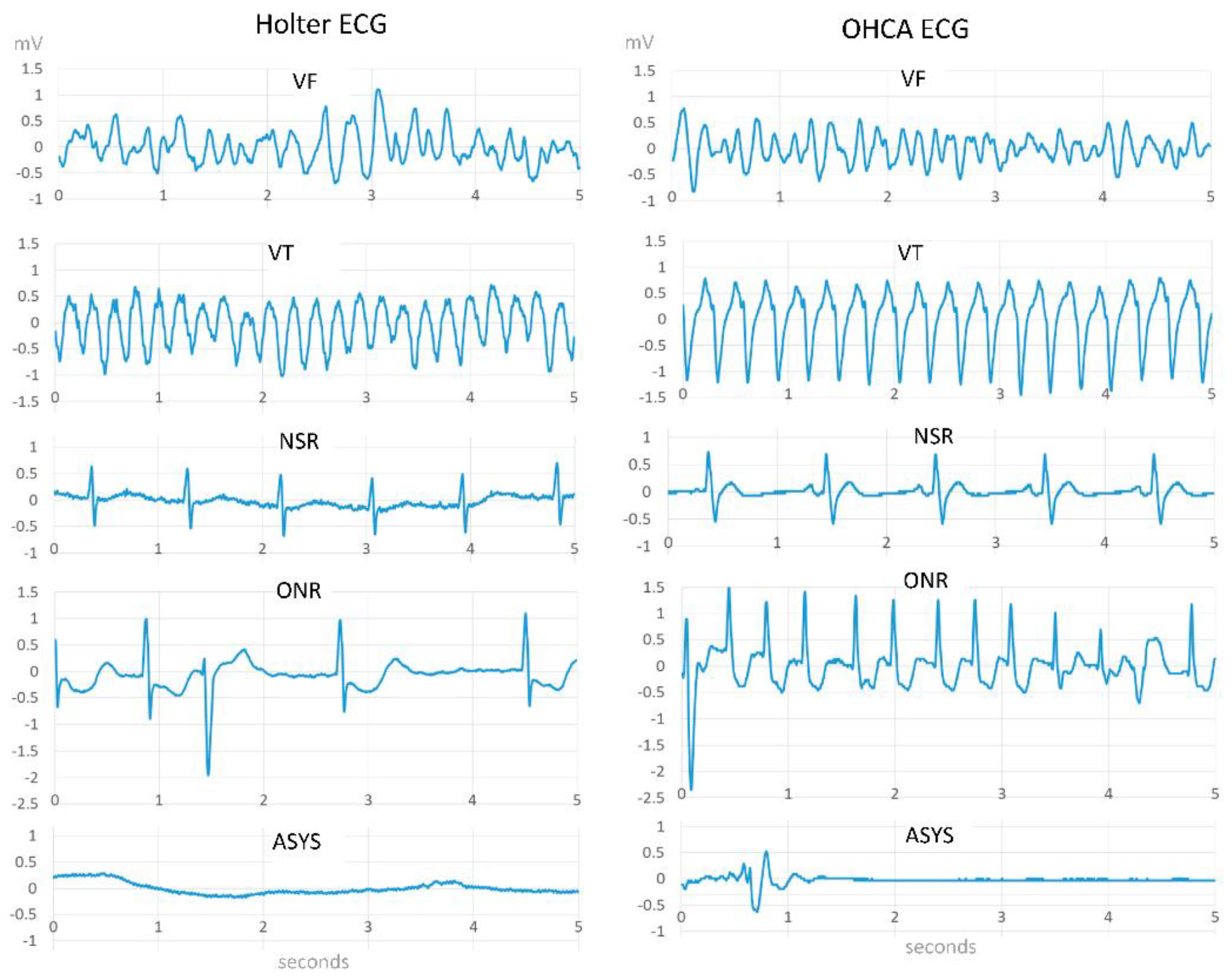
Figure 1. Examples of electrocardiogram (ECG) strips for VF, VT, NSR, ONR, and ASYS rhythms [16].
Electronics 11 03593 g001
Table 1. Confusion Matrix.
Shockable
(Sh)
Non-Shockable
(NSh)
AED algorithm decisionShockTrue Positive (TP)False Positive (FP)
No-shockFalse Negative (FN)True Negative (TN)
Table 2. Performance comparison of different ML and DL algorithms in the absence of CPR.
RefType of
Methods
ApproachSegmentSe (%)Sp (%)Databases
[30]MLSVM, Genetic algorithm5 s96.296.2AHADB, CUDB, VFDB
[31]MLSVM, DWT3 s
4 s
5 s
97.8
97.7
98.8
98.0
98.3
98.4
MITDB, AHADB, CUDB
[33]MLRF, VMDN/A95.291.04N/A
[12]MLLR, BSN/A96.6
94.7
98.8
96.5
Public
OCHA
[39]DLCNN2 s95.3291.04MITDB, VFDB, CUDB
[40]DL and
ML
CNN, BS, MVMD8 s97.099.44VFDB, CUDB
[41]DL and
ML
CNN, SVM, MVMD5 s95.299.31VFDB, CUDB
[24]DL and MLCNN, LSTM4 s
4 s
2 s
2 s
99.7
99.2
97.5
97.5
98.9
96.7
93.6
97.5
Public
OHCA
OHCA
Public
[26]DL and MLDCNN, HP optimization5 s
2 s
96.6
97.6
99.4
98.7
OCHA
OCHA
[46]DL and MLCNN, RNNN/A98.98
98.96
N/A
96.95
86.04
95.01
AFDB
MITDB
NSRDB
Table 3. Performance comparison of different ML and DL algorithms during CPR.
RefType of MethodApproachesSe (%)Sp (%)Databases
[36]MLSVM91.096.6OHCA
[49]DLCNN92.296.65OHCA
[15]DLCNN,
LSTM
92.7197.6CUDB,
SDBB
[53]DLBP-NN99.095.0N/A
Publisher’s Note: MDPI stays neutral with regard to jurisdictional claims in published maps and institutional affiliations.