
Article

Efficient Perineural Invasion Detection of Histopathological Images Using U-Net
1
School of Electronic and Electrical Engineering, Kyungpook National University, Daegu 41566, Korea
2
Neopons, Daegu 41404, Korea
3
School of Electronics Engineering, Kyungpook National University, Daegu 41566, Korea
*
Author to whom correspondence should be addressed.
These authors contributed equally to this work.
Electronics 2022, 11(10), 1649; https://doi.org/10.3390/electronics11101649

Submission received: 25 April 2022 / Revised: 18 May 2022 / Accepted: 20 May 2022 / Published: 22 May 2022
(This article belongs to the Special Issue Deep Learning in Medical Image Process)

Abstract

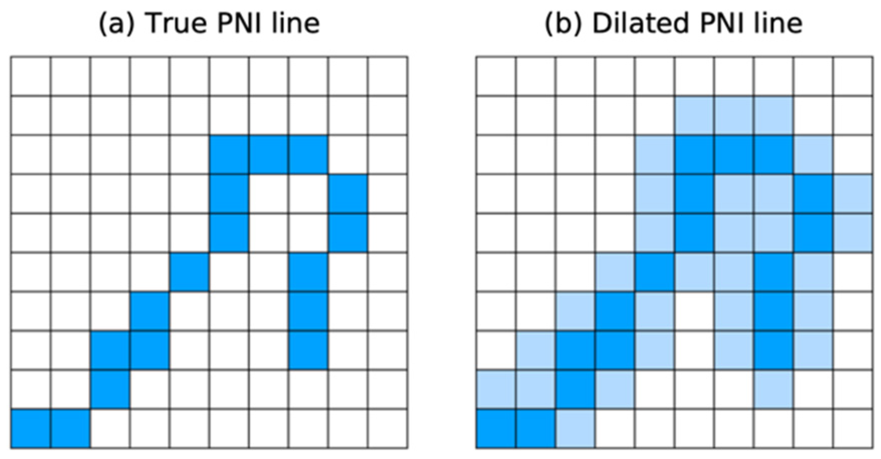
Perineural invasion (PNI), a sign of poor diagnosis and tumor metastasis, is common in a variety of malignant tumors. The infiltrating patterns and morphologies of tumors vary by organ and histological diversity, making PNI detection difficult in biopsy, which must be performed manually by pathologists. As the diameters of PNI nerves are measured on a millimeter scale, the PNI region is extremely small compared to the whole pathological image. In this study, an efficient deep learning-based method is proposed for detecting PNI regions in multiple types of cancers using only PNI annotations without detailed segmentation maps for each nerve and tumor cells obtained by pathologists. The key idea of the proposed method is to train the adopted deep learning model, U-Net, to capture the boundary regions where two features coexist. A boundary dilation method and a loss combination technique are proposed to improve the detection performance of PNI without requiring full segmentation maps. Experiments were conducted with various combinations of boundary dilation widths and loss functions. It is confirmed that the proposed method effectively improves PNI detection performance from 0.188 to 0.275. Additional experiments were also performed on normal nerve detection to validate the applicability of the proposed method to the general boundary detection tasks. The experimental results demonstrate that the proposed method is also effective for general tasks, and it improved nerve detection performance from 0.511 to 0.693.
Keywords: deep learning; U-Net; boundary detection; perineural invasion detection; histopathological image deep learning; U-Net; boundary detection; perineural invasion detection; histopathological image