
Efficient Pre-Processing and Segmentation for Lung Cancer Detection Using Fused CT Images

 , , ,
, , ,  , and
, and
Abstract
:1. Introduction
2. Background of the Theory
2.1. Sparse Representation Method
2.2. Image-Decomposition-Based Fusion Methods
2.3. Deep-Learning-Based Fusion Methods
2.4. Rolling Guidance Filtering
2.5. Dictionary Learning
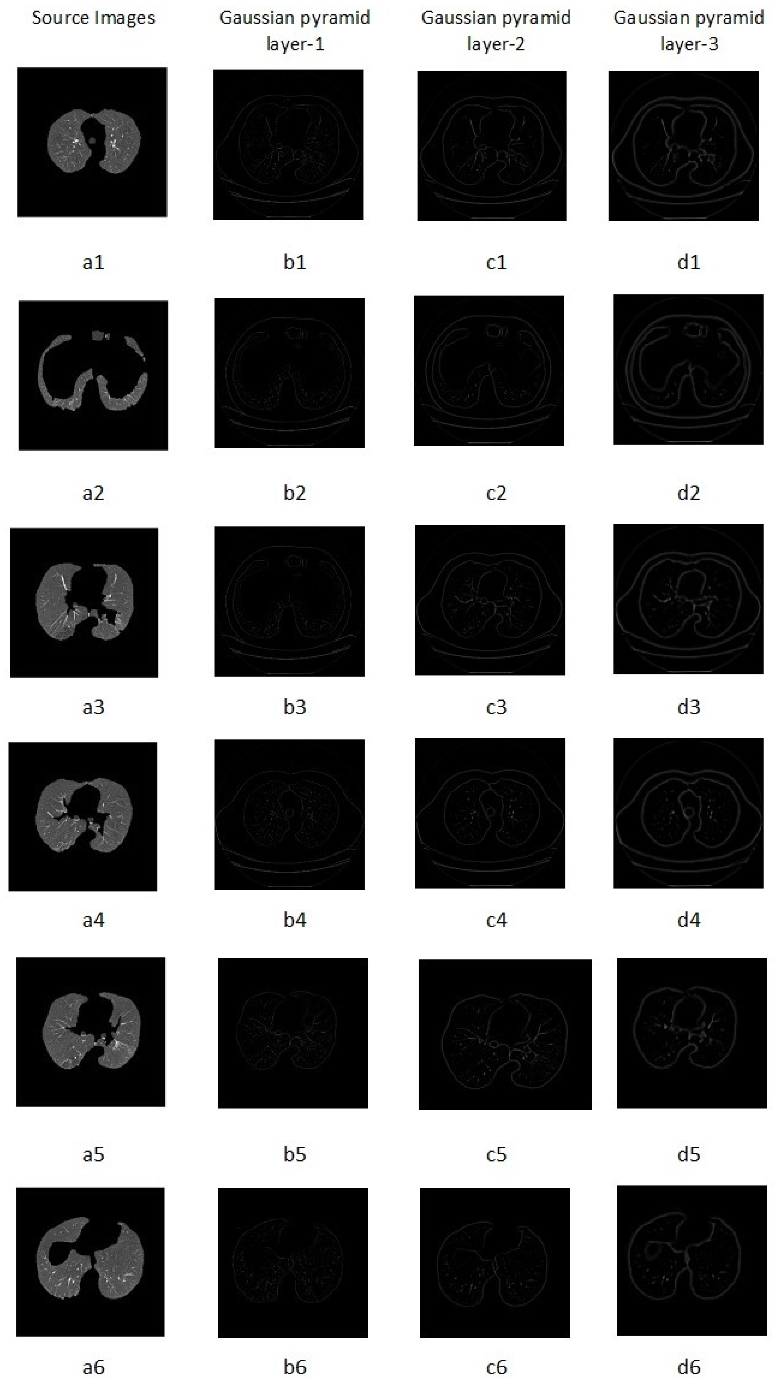
2.6. Laplacian Pyramid Method
3. Materials and Methods
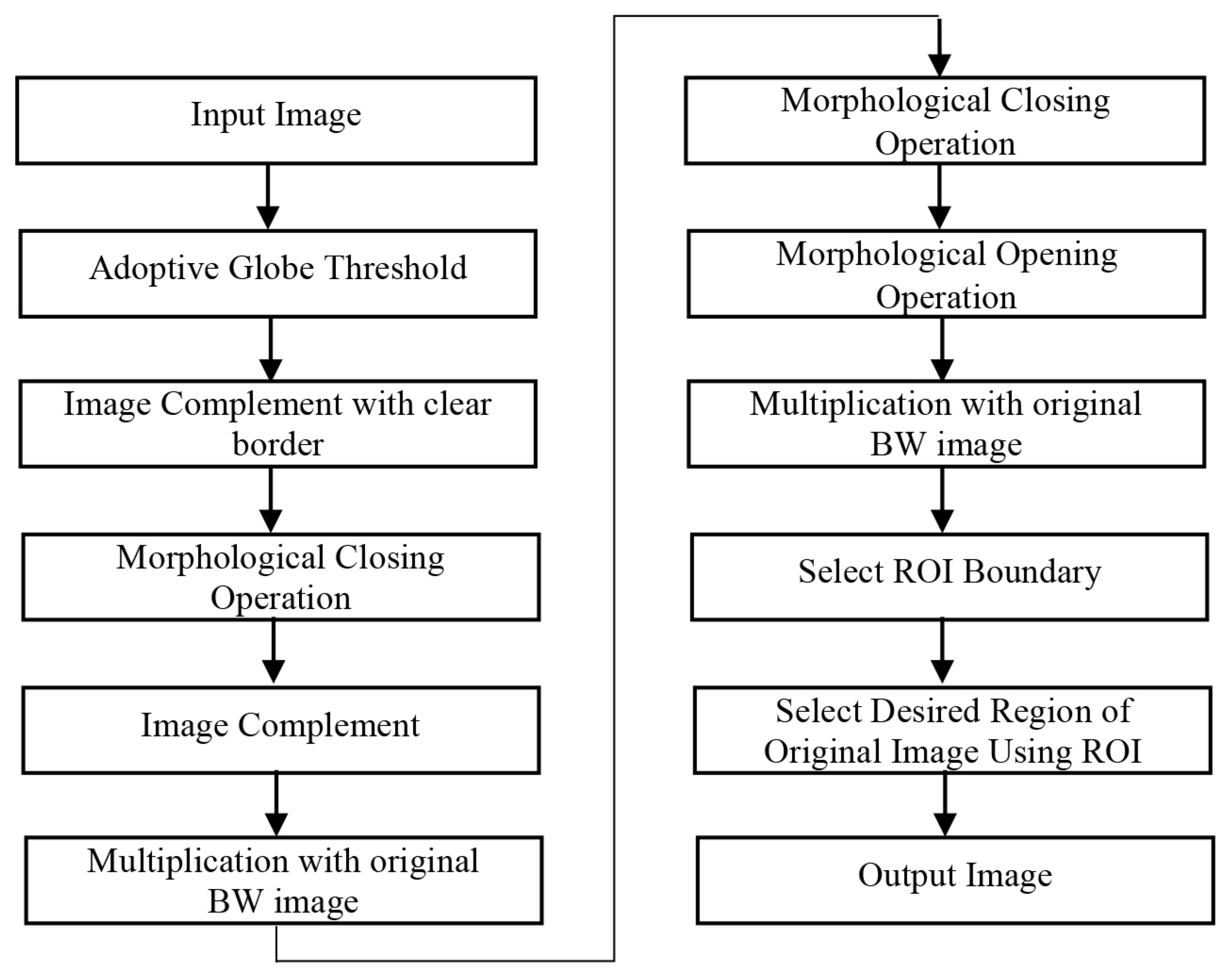
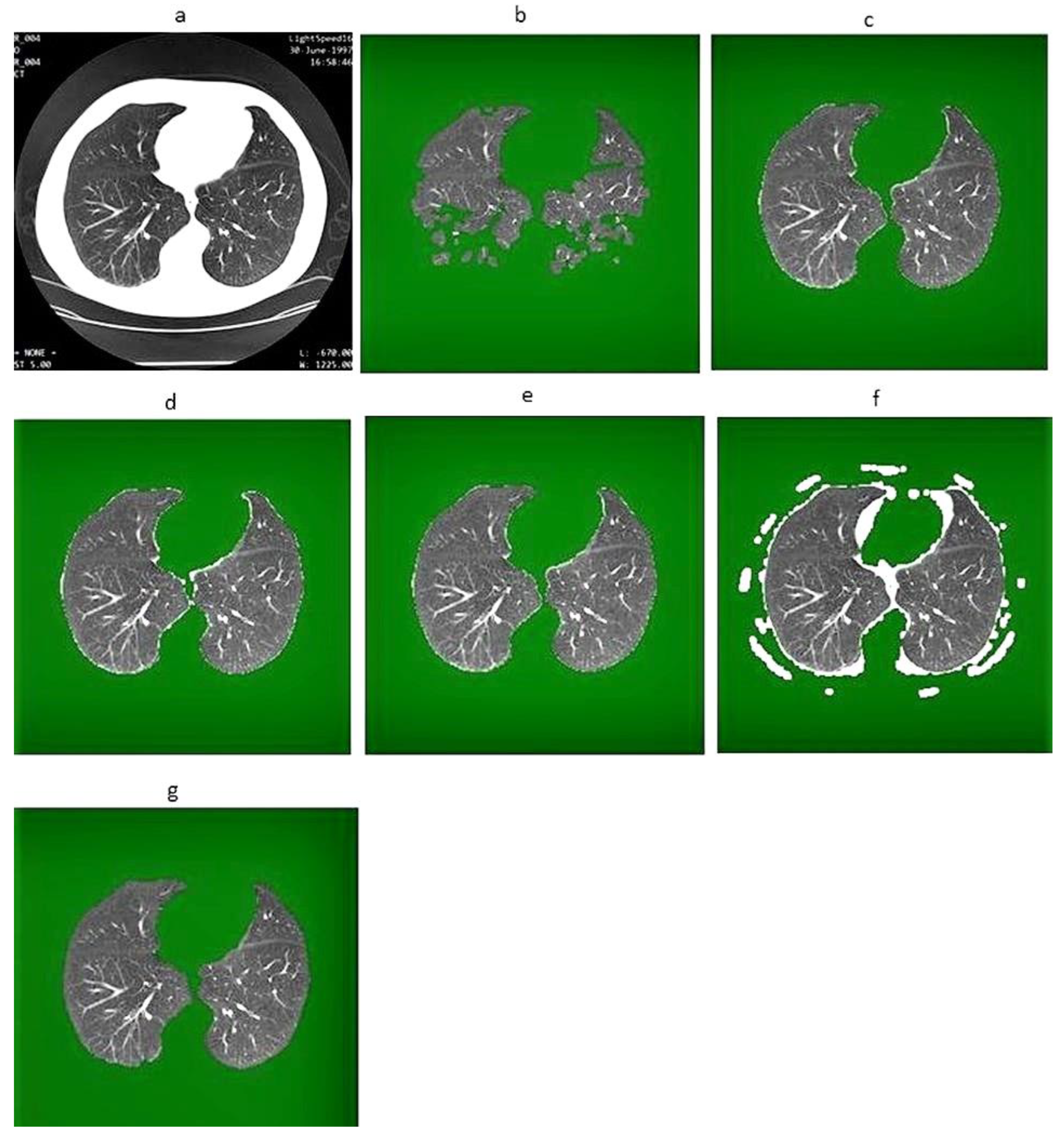
3.1. Image-Segmentation Method
| Algorithm 1 Proposed segmentation algorithm. |
Input: Output: Initialization:
|
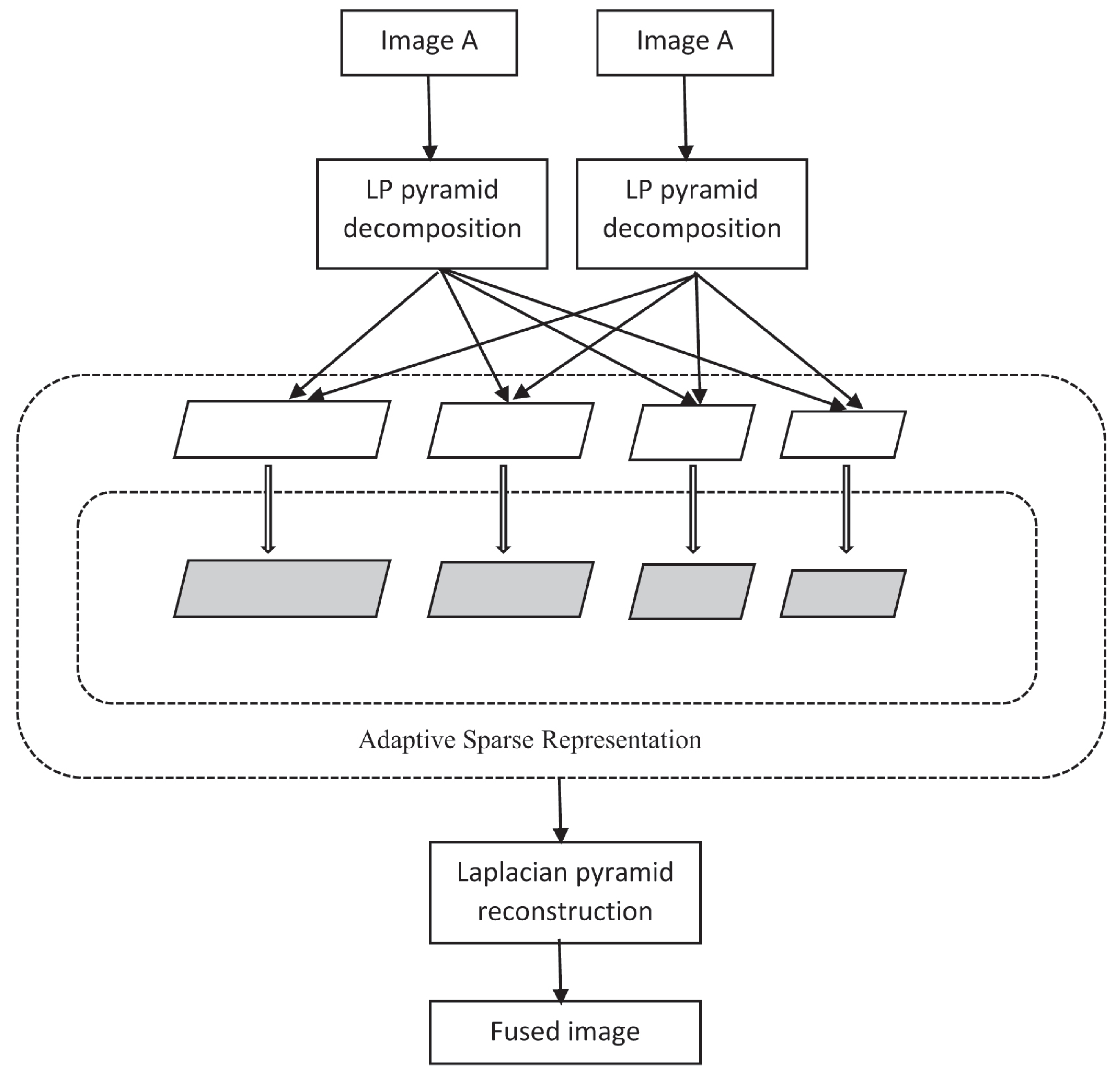
3.2. Image Fusion Method
| Algorithm 2 Proposed fusion algorithm. |
Input: Output:
|
3.2.1. Decomposition of the Segmented Source Image
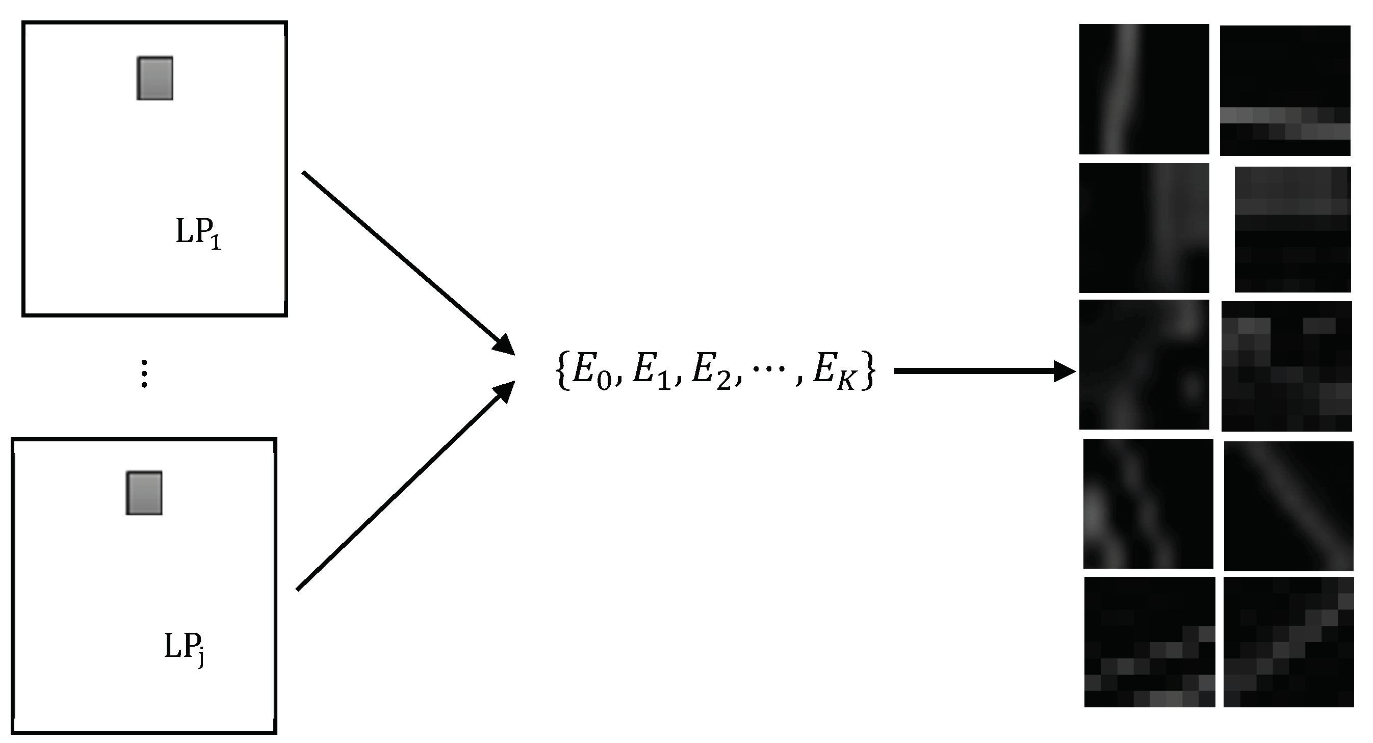
3.2.2. ASR Method
- For each input image , a sliding window with a size of was used to delete all patches with a step length of one pixel from top to bottom and left to right. It was assumed the was a set of patches in for the ith image. is the number of patches sampled from each input image;
- The column vectors were obtained by rearranging the patches , and each column vector was made to be zero mean by subtracting the the mean value from each value of the column vector.where 1 is the unit vector of ;
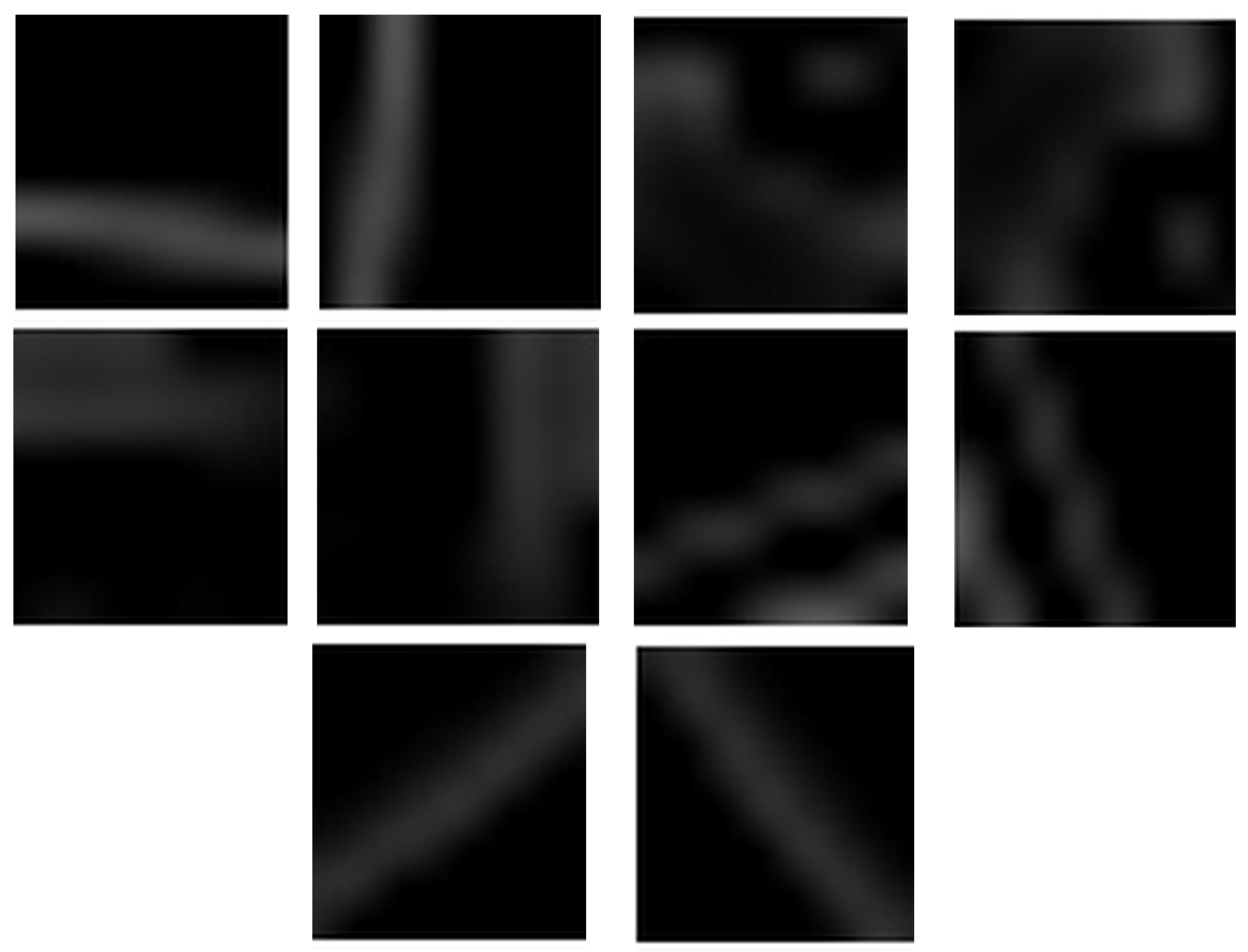
- From the set , the with the greatest variance was chosen. Then, using , a gradient orientation histogram was generated, and one sub-dictionary was chosen from , which had a total of sub-dictionaries. The gradient orientation histogram can be written as:is defined as an adaptive sub-dictionary, with being the index of into which the patch should be divided. The procedure for selecting is shown below:where is:and the index of is shown as:
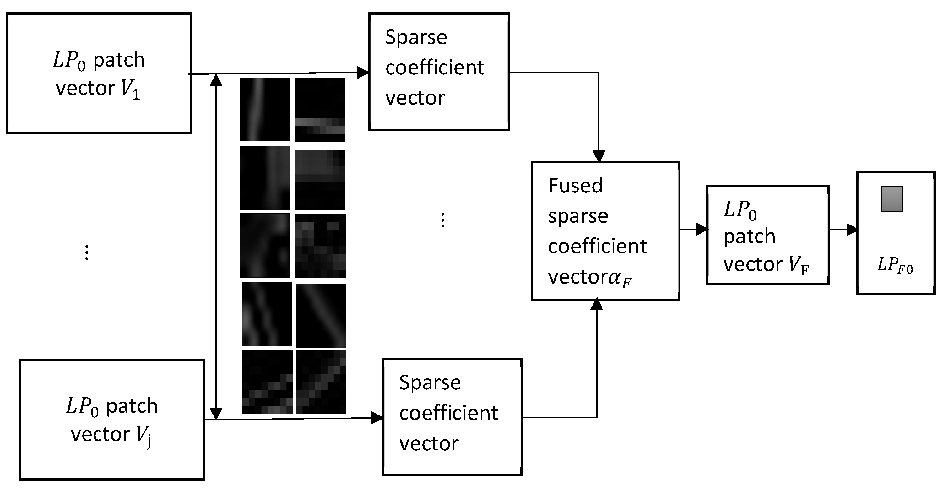
- The dictionary that was chosen for SR fusion was . The sparse vectors of were obtained after extracting vector from the of both source images.where is a constant and is the error tolerance. The steps of this method are shown in Figure 5.The Max-L1 fusion rule was used for the fusion of sparse vectors ,where:It is recommended that the merged mean value be set to:Finally, the fused results of the 1st layer of is estimated by:
- In for the source image patches, Steps 2 to 4 are repeated to obtain the fused results of . To fuse the remaining three layers of the pyramid, the step of selecting the sub-dictionary is repeated. Finally, we are able to build the fused LP image .
3.2.3. Image Reconstruction and Fusion
4. Results
4.1. Dataset
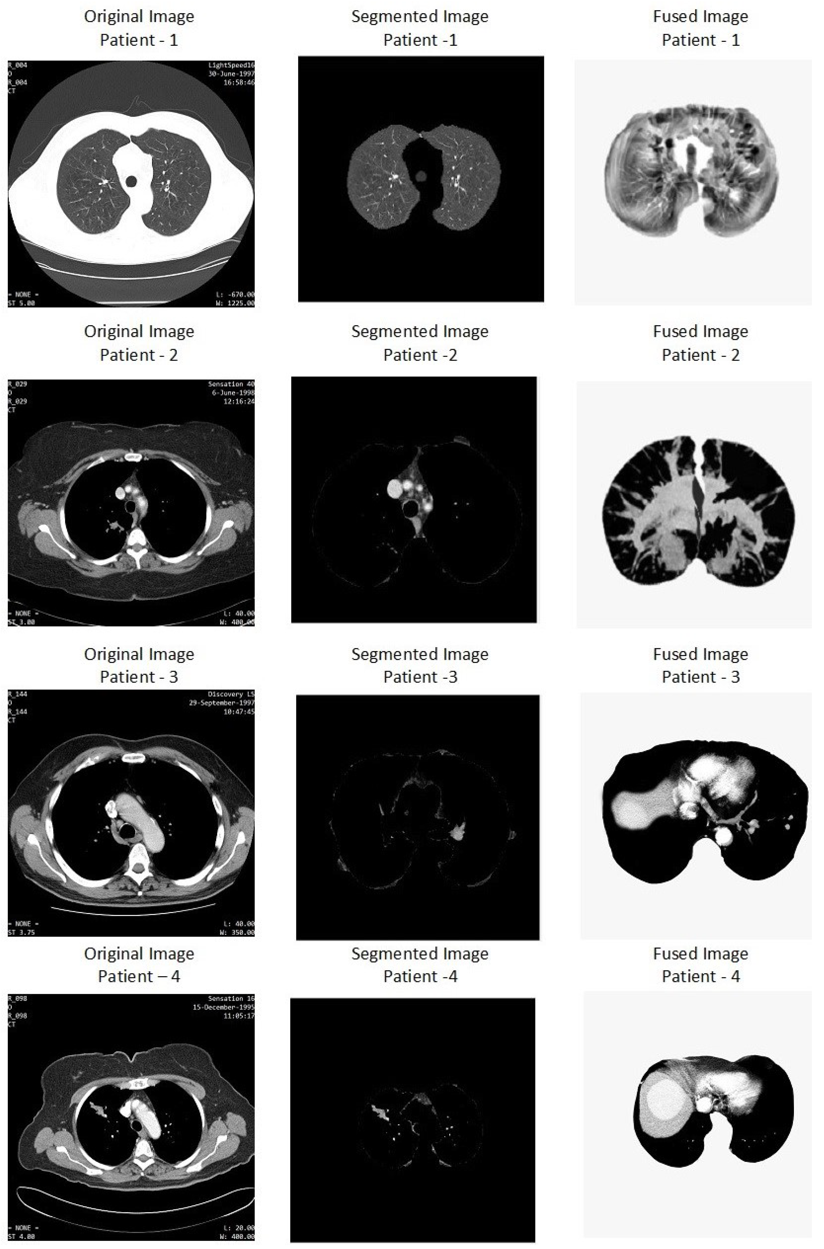
4.2. Image Segmentation
4.3. Image Fusion Results
4.4. Discussion
5. Conclusions and Future Work
Author Contributions
Funding
Acknowledgments
Conflicts of Interest
Abbreviations
| CT | Computed Tomography |
| LP | Laplacian Pyramid |
| ASR | Adaptive Sparse Representative |
| LIDC | Lung Image Database Consortium |
| PET | Positron Emission Tomography |
| HRCT | High-Resolution Computed Tomography |
| CAD | Computer-Aided Design |
| GCPSO | Guaranteed Convergence Particle Swarm Optimization |
| SD | Spatial Domain |
| TD | Transform Domain |
| PF | Pyramid Fusion |
| DWT | Discrete Wavelet Transform |
| CVT | Curvelet Transform |
| NSCT | Non-Subsampled Contour Transform |
| SR | Sparse Representation |
| SVD | Singular-Value Decomposition |
| DL-GSGR | Dictionary Learning with Group Sparsity and Graph Regularization |
| RWT | Redundant Wavelet Transform |
| R-DWT | Redundant Discrete Wavelet Transform |
| RMLP | Region Mosaicking on Laplacian Pyramids |
| NCA | Neighborhood Component Analysis |
| LDSB | Lung Data Science Bowl |
| MI | Mutual Information |
| NCC | Normalized Cross-Correlation |
| FMI | Feature Mutual Information |
| PCA | Principal Component Analysis |
| LIDC-IDRI | Lung Image Database Consortium and Image Database Resource Initiative |
| ROI | Region Of Interest |
| DICOM | Digital Imaging and Communications in Medicine |
| RGB | Red Green Blue |
| DSC | Distributed Source Coding |
| RD | Region Detection |
| LSWI | Level Set Without Initialization |
| RM | Re-initialization Methods |
| API | Average Pixel Intensity |
| SD | Standard Deviation |
| AG | Average Gradient |
| MI | Mutual Information |
| SF | Spatial Frequency |
| BiSe-Net | Bilateral Segmentation Network |
| ESP-Net | Efficient Spatial Pyramid Network |
| GDRLSE | Generalized Distance Regulated Level Set Evolution |
| RASM | Robust Active Shape Model |
| MSGC | Multi-Scale Grid Clustering |
| GMM | Gaussian Mixture Model |
References
- Vijaya, G.; Suhasini, A. An adaptive preprocessing of lung CT images with various filters for better enhancement. Acad. J. Cancer Res. 2014, 7, 179–184. [Google Scholar]
- Litjens, G.; Kooi, T.; Bejnordi, B.E.; Setio, A.A.A.; Ciompi, F.; Ghafoorian, M.; Van Der Laak, J.A.; Van Ginneken, B.; Sánchez, C.I. A survey on deep learning in medical image analysi. Med. Image Anal. 2017, 42, 60–88. [Google Scholar] [CrossRef] [Green Version]
- Pu, J.; Roos, J.; Chin, A.Y.; Napel, S.; Rubin, G.D.; Paik, D.S. Adaptive border marching algorithm: Automatic lung segmentation on chest CT images. Comput. Med. Imaging Graph. 2008, 32, 452–462. [Google Scholar] [CrossRef] [PubMed] [Green Version]
- Senthil Kumar, K.; Venkatalakshmi, K.; Karthikeyan, K. Lung cancer detection using image segmentation by means of various evolutionary algorithms. Comput. Math. Methods Med. 2019, 2019, 4909846. [Google Scholar] [CrossRef] [Green Version]
- Akter, O.; Moni, M.A.; Islam, M.M.; Quinn, J.M.; Kamal, A. Lung cancer detection using enhanced segmentation accuracy. Appl. Intell. 2020, 50, 1–14. [Google Scholar] [CrossRef]
- Du, J.; Li, W.; Lu, K.; Xiao, B. An overview of multi-modal medical image fusion. Neurocomputing 2016, 215, 3–20. [Google Scholar] [CrossRef]
- Li, S.; Yin, H.; Fang, L. Group-sparse representation with dictionary learning for medical image denoising and fusion. IEEE Trans. Biomed. Eng. 2012, 59, 3450–3459. [Google Scholar] [CrossRef]
- Liu, H.; Cao, H.; Song, E.; Ma, G.; Xu, X.; Jin, R.; Jin, Y.; Hung, C.C. A cascaded dual-pathway residual network for lung nodule segmentation in CT images. Phys. Medica 2019, 63, 112–121. [Google Scholar] [CrossRef] [Green Version]
- Aishwarya, N.; Bennila Thangammal, C. A novel multimodal medical image fusion using sparse representation and modified spatial frequency. Int. J. Imaging Syst. Technol. 2018, 28, 175–185. [Google Scholar] [CrossRef]
- Kaur, R.; Kaur, E.G. Medical image fusion using redundant wavelet based ICA co-variance analysis. Int. J. Eng. Comp. Sci. 2015, 4, 28. [Google Scholar] [CrossRef]
- Liu, X.; Mei, W.; Du, H. Detail-enhanced multimodality medical image fusion based on gradient minimization smoothing filter and shearing filter. Med. Biol. Eng. Comput. 2018, 56, 1565–1578. [Google Scholar] [CrossRef] [PubMed]
- Matsopoulos, G.; Marshall, S.; Brunt, J. Multiresolution morphological fusion of MR and CT images of the human brain. IEE Proc.-Vis. Image Signal Process. 1994, 141, 137–142. [Google Scholar] [CrossRef]
- Du, J.; Li, W.; Xiao, B.; Nawaz, Q. Union Laplacian Pyramid with multiple features for medical image fusion. Neurocomputing 2016, 194, 326–339. [Google Scholar] [CrossRef]
- Kou, L.; Zhang, L.; Zhang, K.; Sun, J.; Han, Q.; Jin, Z. A multi-focus image fusion method via region mosaicking on Laplacian Pyramids. PLoS ONE 2018, 13, e0191085. [Google Scholar] [CrossRef] [PubMed]
- Li, X.; Zhao, J. A novel multi-modal medical image fusion algorithm. J. Ambient. Intell. Humaniz. Comput. 2021, 12, 1995–2002. [Google Scholar] [CrossRef]
- Soliman, A.; Khalifa, F.; Elnakib, A.; Abou El-Ghar, M.; Dunlap, N.; Wang, B.; Gimel’farb, G.; Keynton, R.; El-Baz, A. Accurate lung segmentation on CT chest images by adaptive appearance-guided shape modeling. IEEE Trans. Med. Imaging 2016, 36, 263–276. [Google Scholar] [CrossRef] [PubMed]
- Khan, M.A.; Rubab, S.; Kashif, A.; Sharif, M.I.; Muhammad, N.; Shah, J.H.; Zhang, Y.D.; Satapathy, S.C. Lungs cancer classification from CT images: An integrated design of contrast based classical features fusion and selection. Pattern Recognit. Lett. 2020, 129, 77–85. [Google Scholar] [CrossRef]
- Azam, M.A.; Khan, K.B.; Ahmad, M.; Mazzara, M. Multimodal Medical Image Registration and Fusion for Quality Enhancement. Cmc-Comput. Mater. Contin. 2021, 68, 821–840. [Google Scholar] [CrossRef]
- Chen, T.; Ma, X.; Ying, X.; Wang, W.; Yuan, C.; Lu, W.; Chen, D.Z.; Wu, J. Multi-modal fusion learning for cervical dysplasia diagnosis. In Proceedings of the 2019 IEEE 16th International Symposium on Biomedical Imaging (ISBI 2019), Venice, Italy, 8–11 April 2019; pp. 1505–1509. [Google Scholar]
- Liu, Y.; Chen, X.; Cheng, J.; Peng, H. A medical image fusion method based on convolutional neural networks. In Proceedings of the 2017 20th International Conference on Information Fusion (Fusion), Xi’an, China, 10–13 July 2017; pp. 1–7. [Google Scholar]
- Ma, X.; Hu, S.; Liu, S.; Fang, J.; Xu, S. Multi-focus image fusion based on joint sparse representation and optimum theory. Signal Process. Image Commun. 2019, 78, 125–134. [Google Scholar] [CrossRef]
- Zhu, Z.; Chai, Y.; Yin, H.; Li, Y.; Liu, Z. A novel dictionary learning approach for multi-modality medical image fusion. Neurocomputing 2016, 214, 471–482. [Google Scholar] [CrossRef]
- Li, H.; He, X.; Tao, D.; Tang, Y.; Wang, R. Joint medical image fusion, denoising and enhancement via discriminative low-rank sparse dictionaries learning. Pattern Recognit. 2018, 79, 130–146. [Google Scholar] [CrossRef]
- Liu, Y.; Chen, X.; Ward, R.K.; Wang, Z.J. Medical image fusion via convolutional sparsity based morphological component analysis. IEEE Signal Process. Lett. 2019, 26, 485–489. [Google Scholar] [CrossRef]
- Jiang, W.; Yang, X.; Wu, W.; Liu, K.; Ahmad, A.; Sangaiah, A.K.; Jeon, G. Medical images fusion by using weighted least squares filter and sparse representation. Comput. Electr. Eng. 2018, 67, 252–266. [Google Scholar] [CrossRef]
- Xu, Z. Medical image fusion using multi-level local extrema. Inf. Fusion 2014, 19, 38–48. [Google Scholar] [CrossRef]
- Maqsood, S.; Javed, U. Multi-modal medical image fusion based on two-scale image decomposition and sparse representation. Biomed. Signal Process. Control 2020, 57, 101810. [Google Scholar] [CrossRef]
- Guo, X.; Nie, R.; Cao, J.; Zhou, D.; Qian, W. Fully convolutional network-based multifocus image fusion. Neural Comput. 2018, 30, 1775–1800. [Google Scholar] [CrossRef]
- Li, H.; Wu, X.J. DenseFuse: A fusion approach to infrared and visible images. IEEE Trans. Image Process. 2018, 28, 2614–2623. [Google Scholar] [CrossRef] [Green Version]
- Zhang, Y.; Liu, Y.; Sun, P.; Yan, H.; Zhao, X.; Zhang, L. IFCNN: A general image fusion framework based on convolutional neural network. Inf. Fusion 2020, 54, 99–118. [Google Scholar] [CrossRef]
- Ma, J.; Xu, H.; Jiang, J.; Mei, X.; Zhang, X.P. DDcGAN: A dual-discriminator conditional generative adversarial network for multi-resolution image fusion. IEEE Trans. Image Process. 2020, 29, 4980–4995. [Google Scholar] [CrossRef]
- Zhang, Q.; Shen, X.; Xu, L.; Jia, J. Rolling guidance filter. In European Conference on Computer Vision; Springer: Berlin/Heidelberg, Germany, 2014; pp. 815–830. [Google Scholar]
- Mao, R.; Fu, X.S.; Niu, P.J.; Wang, H.Q.; Pan, J.; Li, S.S.; Liu, L. Multi-directional laplacian Pyramid image fusion algorithm. In Proceedings of the 2018 3rd International Conference on Mechanical, Control and Computer Engineering (ICMCCE), Huhhot, China, 14–16 September 2018; pp. 568–572. [Google Scholar]
- Liu, Y.; Wang, Z. Simultaneous image fusion and denoising with adaptive sparse representation. IET Image Process. 2015, 9, 347–357. [Google Scholar] [CrossRef] [Green Version]
- Armato, S.G., III; McLennan, G.; Bidaut, L.; McNitt-Gray, M.F.; Meyer, C.R.; Reeves, A.P.; Zhao, B.; Aberle, D.R.; Henschke, C.I.; Hoffman, E.A.; et al. The lung image database consortium (LIDC) and image database resource initiative (IDRI): A completed reference database of lung nodules on CT scans. Med. Phys. 2011, 38, 915–931. [Google Scholar] [CrossRef] [PubMed]
- Hollaus, F.; Diem, M.; Sablatnig, R. MultiSpectral image binarization using GMMs. In Proceedings of the 2018 16th International Conference on Frontiers in Handwriting Recognition (ICFHR), Niagara Falls, NY, USA, 5–8 August 2018; pp. 570–575. [Google Scholar]
- Skourt, B.A.; El Hassani, A.; Majda, A. Lung CT image segmentation using deep neural networks. Procedia Comput. Sci. 2018, 127, 109–113. [Google Scholar] [CrossRef]
- Banu, S.F.; Sarker, M.; Kamal, M.; Abdel-Nasser, M.; Puig, D.; A Raswan, H. AWEU-Net: An Attention-Aware Weight Excitation U-Net for Lung Nodule Segmentation. Appl. Sci. 2021, 11, 10132. [Google Scholar] [CrossRef]
- Rocha, J.; Cunha, A.; Mendonça, A.M. Conventional filtering versus u-net based models for pulmonary nodule segmentation in ct images. J. Med. Syst. 2020, 44, 1–8. [Google Scholar] [CrossRef]
- Mukherjee, S.; Huang, X.; Bhagalia, R.R. Lung nodule segmentation using deep learned prior based graph cut. In Proceedings of the 2017 IEEE 14th International Symposium on Biomedical Imaging (ISBI 2017), Melbourne, VIC, Australia, 18–21 April 2017; pp. 1205–1208. [Google Scholar]
- Wang, W.; Lu, Y.; Wu, B.; Chen, T.; Chen, D.Z.; Wu, J. Deep active self-paced learning for accurate pulmonary nodule segmentation. In International Conference on Medical Image Computing and Computer-Assisted Intervention; Springer: Berlin/Heidelberg, Germany, 2018; pp. 723–731. [Google Scholar]
- Zhang, G.; Guo, M.; Gong, Z.; Bi, J.; Kim, Y.; Guo, W. Pulmonary nodules segmentation method based on auto-encoder. In Proceedings of the 10th International Conference on Digital Image Processing (ICDIP 2018), Shanghai, China, 11–14 May 2018; Volume 10806, p. 108062P. [Google Scholar]
- Feng, X.; Yang, J.; Laine, A.F.; Angelini, E.D. Discriminative localization in CNNs for weakly-supervised segmentation of pulmonary nodules. In International Conference on Medical Image Computing and Computer-Assisted Intervention; Springer: Berlin/Heidelberg, Germany, 2017; pp. 568–576. [Google Scholar]
- Tan, J.; Jing, L.; Huo, Y.; Li, L.; Akin, O.; Tian, Y. Lgan: Lung segmentation in ct scans using generative adversarial network. Comput. Med. Imaging Graph. 2021, 87, 101817. [Google Scholar] [CrossRef]
- Chen, S.; Wang, Y. Pulmonary Nodule Segmentation in Computed Tomography with an Encoder-Decoder Architecture. In Proceedings of the 2019 10th International Conference on Information Technology in Medicine and Education (ITME), Qingdao, China, 23–25 August 2019; pp. 157–162. [Google Scholar]
- Piella, G.; Heijmans, H. A new quality metric for image fusion. In Proceedings of the 2003 International Conference on Image Processing (Cat. No. 03CH37429), Barcelona, Spain, 14–17 September 2003; Volume 3, pp. III–173. [Google Scholar]
- Singh, H.; Kumar, V.; Bhooshan, S. Weighted least squares based detail enhanced exposure fusion. Int. Sch. Res. Not. 2014, 2014. [Google Scholar] [CrossRef] [Green Version]
- Wang, Z.; Cui, Z.; Zhu, Y. Multi-modal medical image fusion by Laplacian Pyramid and adaptive sparse representation. Comput. Biol. Med. 2020, 123, 103823. [Google Scholar] [CrossRef] [PubMed]
- Petrovic, V.; Xydeas, C. Objective image fusion performance characterisation. In Proceedings of the Tenth IEEE International Conference on Computer Vision (ICCV’05), Beijing, China, 17–21 October 2005; Volume 2, pp. 1866–1871. [Google Scholar]
- Sundar, K.J.A.; Jahnavi, M.; Lakshmisaritha, K. Multi-sensor image fusion based on empirical wavelet transform. In Proceedings of the 2017 International Conference on Electrical, Electronics, Communication, Computer, and Optimization Techniques (ICEECCOT), Mysuru, India, 15–16 December 2017; pp. 93–97. [Google Scholar]
- Nencini, F.; Garzelli, A.; Baronti, S.; Alparone, L. Remote sensing image fusion using the curvelet transform. Inf. Fusion 2007, 8, 143–156. [Google Scholar] [CrossRef]
- Zhang, Q.; Guo, B.L. Multifocus image fusion using the nonsubsampled contourlet transform. Signal Process. 2009, 89, 1334–1346. [Google Scholar] [CrossRef]
- Yang, B.; Li, S. Multifocus image fusion and restoration with sparse representation. IEEE Trans. Instrum. Meas. 2009, 59, 884–892. [Google Scholar] [CrossRef]


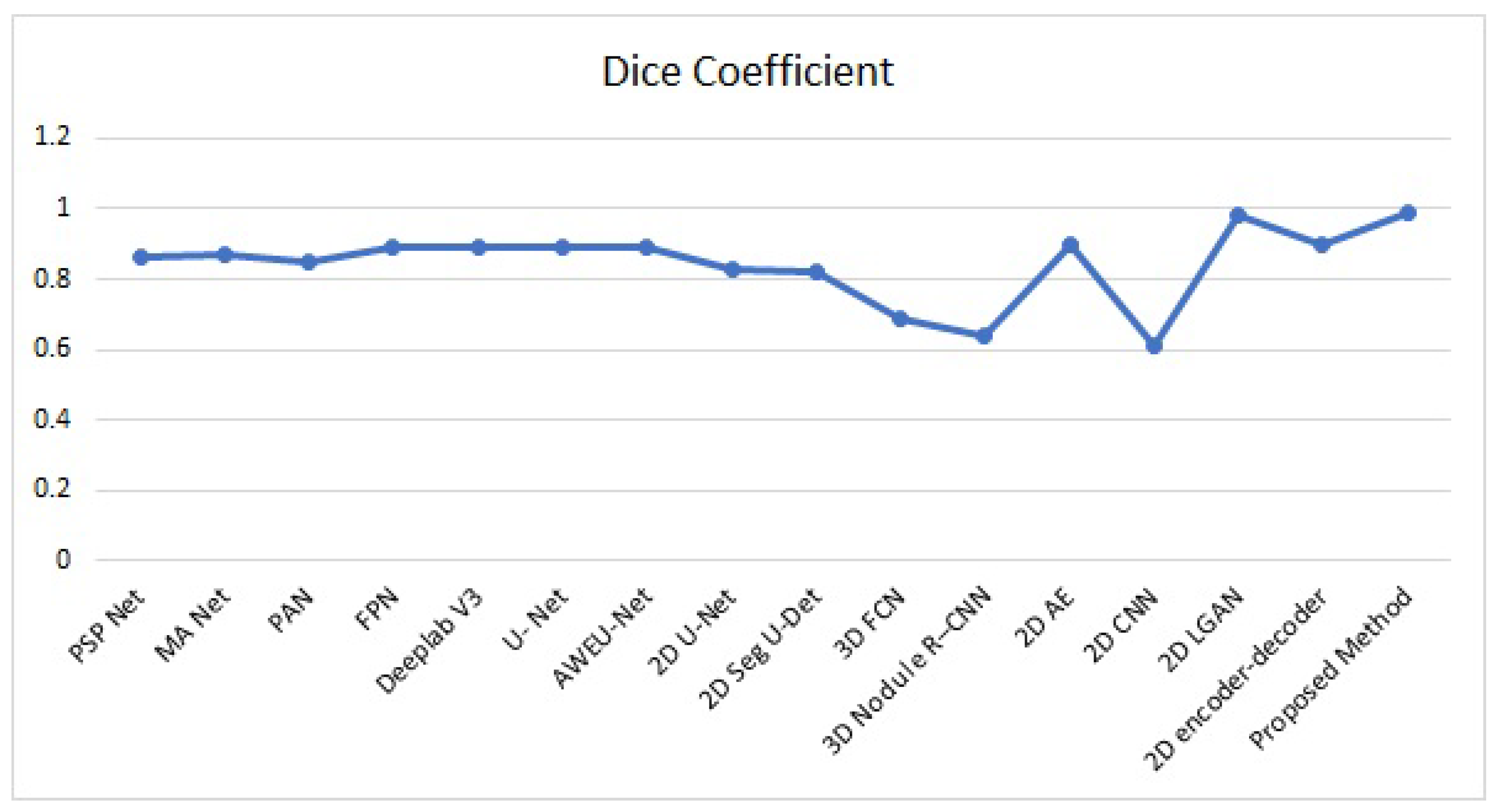
| Method | Dataset | Dice Coefficient | Running Time (s) |
|---|---|---|---|
| U-Net | LIDC-IDRI | 0.89 | - |
| AWEU-Net | LIDC-IDRI | 0.89 | - |
| 2D U-Net | LIDC-IDRI | 0.83 | - |
| 2D Seg U-Det | LIDC-IDRI | 0.82 | - |
| 3D FCN | LIDC-IDRI | 0.69 | 5.0 |
| 3D Nodule R-CNN | LIDC-IDRI | 0.64 | - |
| 2D AE | LIDC-IDRI | 0.90 | - |
| 2D CNN | LIDC-IDRI | 0.61 | - |
| 2D-LGAN | LIDC-IDRI | 0.98 | - |
| 2D Encoder–Decoder | LIDC-IDRI | 0.90 | - |
| Proposed Method | LIDC-IDRI | 0.99 | 1.2252 |
| Methodology | Dataset | Sensitivity (%) | Specificity (%) | Accuracy (%) |
|---|---|---|---|---|
| U-Net | LIDC-IDRI | 84.0 | 96.3 | 94.3 |
| AWEU-Net | LIDC-IDRI | 90.0 | 96.4 | 94.6 |
| 2D U-Net | LIDC-IDRI | 89.0 | - | - |
| 2D Seg U-Det | LIDC-IDRI | 85.0 | - | - |
| 3D FCN | LIDC-IDRI | - | - | - |
| 3D Nodule R-CNN | LIDC-IDRI | - | - | - |
| 2D AE | LIDC-IDRI | - | - | - |
| 2D CNN | LIDC-IDRI | - | - | - |
| 2D LGAN | LIDC-IDRI | - | - | - |
| 2D Encoder–Decoder | LIDC-IDRI | 90.0 | - | - |
| Proposed Method | LIDC-IDRI | 89.0 | 98.0 | 99.0 |
| Method | API | SD | AG | H | MI | SF | Q | L | N |
|---|---|---|---|---|---|---|---|---|---|
| LP [33] | 4.60 | 7.84 | 9.19 | 3.88 | 2.71 | 2.16 | 0.80 | 0.17 | 0.02 |
| DWT [50] | 5.30 | 7.07 | 8.41 | 4.10 | 2.68 | 1.89 | 0.76 | 0.22 | 0.01 |
| CVT [51] | 5.46 | 7.22 | 9.51 | 5.22 | 2.42 | 2.08 | 0.77 | 0.20 | 0.01 |
| NSCT [52] | 5.42 | 7.42 | 9.38 | 4.66 | 2.57 | 2.13 | 0.81 | 0.16 | 0.02 |
| SR [53] | 5.33 | 7.48 | 9.16 | 3.72 | 3.59 | 2.53 | 0.75 | 0.20 | 0.03 |
| ASR [34] | 5.37 | 7.27 | 9.68 | 3.99 | 2.64 | 2.17 | 0.76 | 0.22 | 0.02 |
| Proposed Method | 5.76 | 8.13 | 10.64 | 5.62 | 3.78 | 2.70 | 0.79 | 0.16 | 0.01 |
Publisher’s Note: MDPI stays neutral with regard to jurisdictional claims in published maps and institutional affiliations. |
© 2021 by the authors. Licensee MDPI, Basel, Switzerland. This article is an open access article distributed under the terms and conditions of the Creative Commons Attribution (CC BY) license (https://creativecommons.org/licenses/by/4.0/).
Share and Cite
Nazir, I.; Haq, I.U.; Khan, M.M.; Qureshi, M.B.; Ullah, H.; Butt, S. Efficient Pre-Processing and Segmentation for Lung Cancer Detection Using Fused CT Images. Electronics 2022, 11, 34. https://doi.org/10.3390/electronics11010034
Nazir I, Haq IU, Khan MM, Qureshi MB, Ullah H, Butt S. Efficient Pre-Processing and Segmentation for Lung Cancer Detection Using Fused CT Images. Electronics. 2022; 11(1):34. https://doi.org/10.3390/electronics11010034
Chicago/Turabian StyleNazir, Imran, Ihsan Ul Haq, Muhammad Mohsin Khan, Muhammad Bilal Qureshi, Hayat Ullah, and Sharjeel Butt. 2022. "Efficient Pre-Processing and Segmentation for Lung Cancer Detection Using Fused CT Images" Electronics 11, no. 1: 34. https://doi.org/10.3390/electronics11010034
APA StyleNazir, I., Haq, I. U., Khan, M. M., Qureshi, M. B., Ullah, H., & Butt, S. (2022). Efficient Pre-Processing and Segmentation for Lung Cancer Detection Using Fused CT Images. Electronics, 11(1), 34. https://doi.org/10.3390/electronics11010034